Review

Next-Generation Cancer Models for Drug Testing: Recent Advances in Immunocompetent Microphysiological Systems

Department of Urology, University Medicine Greifswald, 17475 Greifswald, Germany
*
Author to whom correspondence should be addressed.
Future Pharmacol. 2025, 5(3), 36; https://doi.org/10.3390/futurepharmacol5030036
Submission received: 23 April 2025 / Revised: 20 June 2025 / Accepted: 24 June 2025 / Published: 7 July 2025
(This article belongs to the Special Issue Feature Papers in Future Pharmacology 2025)

Abstract

The success of checkpoint inhibitors in improving cancer patient survival has demonstrated the therapeutic potential of immunotherapies. This advancement has reshaped oncology treatment and driven interest in harnessing immune modulation for a wider range of diseases. However, developing drugs that modulate immune activity presents unique challenges. A major limitation in preclinical research is the inefficiency of testing human-specific immune targets in animal models, which often fail to translate to clinical outcomes. Additionally, conventional in vitro systems lack immune reactivity due to their static and monocellular nature, limiting their predictive value. Advanced in vitro models can bridge this gap by offering increasingly relevant human physiology for testing drug efficacy and safety, along with absorption, distribution, metabolism, and excretion (ADME). In particular, immune-competent spheroids, organoids, and organs-on-a-chip (OoC) have emerged as promising tools. Although still in their infancy, these microphysiological systems (MPSs) have demonstrated the feasibility of replicating immune responses ex vivo, providing a new avenue for studying immune-targeting drugs with higher translational potential. In this review, we explore recent advances in immune-competent organoid and OoC models, highlighting their capabilities and limitations. We provide a perspective on their applications for cancer drug testing, discussing how these systems could refine preclinical immuno-oncology research and accelerate the development of next-generation immunotherapies.

1. Introduction

Cancer remains a leading cause of mortality worldwide, requiring continuous innovation in therapeutic strategies and model systems for drug development [1]. According to the World Health Organization, cancer accounted for nearly 10 million deaths globally in 2022, and its incidence continues to rise with aging populations and evolving risk factors [2]. While traditional treatments such as chemotherapy and radiation therapy have long been the foundation of oncology, the clinical focus is increasingly shifting towards immunotherapeutic approaches. These therapies harness the body’s immune system to detect and destroy tumor cells, offering more targeted and, in some cases, long-lasting responses compared to conventional therapies [3,4].
Among immunotherapies, immune checkpoint inhibitors (ICIs) have gained extensive attention since their initial approval in 2011 [5]. These agents, which block regulatory pathways such as PD-1/PD-L1 and CTLA-4, have led to significant clinical successes in several cancers, including melanoma, non-small cell lung cancer, and renal cell carcinoma [6]. As of March 2025, eleven ICIs have been approved for cancer treatment, reflecting their rapid expansion in clinical use [7,8]. Additionally, chimeric antigen receptor (CAR) T-cell therapies have shown remarkable success, particularly in treating certain hematologic cancers [9]. There is also a renewed interest in cancer vaccines and other modalities aimed at boosting immune recognition of tumors [10]. Collectively, these advances highlight the growing importance of immunotherapy in modern oncology and its expected dominance in future cancer treatment strategies.
While significant progress has been made in the development of anti-cancer therapies, researchers continue to face several challenges in preclinical testing. Alarmingly, fewer than 11% of anticancer therapies and immunotherapies that show efficacy in preclinical animal models ultimately receive approval following phase III clinical trials. This high attrition rate is mainly attributed to the limited translational relevance of existing models. Animal systems often fail to replicate the heterogeneity and complexity of human tumors, particularly in terms of immune components, due to species-specific differences and, in many cases, compromised or simplified immune functions [11,12].
A central issue in immuno-oncology is the intricate nature of the tumor microenvironment (TME). The TME comprises not only tumor cells but also stromal cells, immune infiltrates, endothelial cells, and extracellular matrix components that collectively regulate tumor growth, immune evasion, and therapeutic response. Immunotherapies often face resistance due to the immunosuppressive features of the TME, such as the presence of regulatory T cells, myeloid-derived suppressor cells, and inhibitory cytokines [13,14]. These nuances are difficult to capture using standard preclinical models.
Even within human patients, responses to immunotherapy vary significantly [15], highlighting the need for predictive, human-relevant preclinical models. Conventional two-dimensional (2D) cell cultures fail to replicate the complex architecture and cellular interactions of the tumor microenvironment (TME) [16,17]. While animal models provide a more physiological milieu, they present ethical challenges and inherent biological differences that compromise their utility, particularly in immuno-oncology research.
To overcome these limitations, researchers have increasingly turned to more physiologically relevant in vitro systems. Initial efforts focused on the development of three-dimensional (3D) tumor spheroids, which offer improved simulation of tumor morphology and biochemical gradients compared to 2D cultures [16]. These models offer enhanced insights into tumor proliferation, drug penetration, and oxygenation gradients. However, they still lack several key features necessary for studying immune-oncology applications, such as dynamic perfusion, immune–stromal–tumor interactions, and tissue-level architecture. To partially address these limitations, researchers have turned to multicellular spheroid models, which offer improved representation of the tumor microenvironment (TME) compared to traditional two-dimensional cultures. By incorporating multiple cell types, multicellular spheroid models better represent the TME and extracellular matrix (ECM), allowing for more realistic investigations of immune cell infiltration and tumor invasion [18]. These models also offer a superior platform for evaluating drug delivery and efficacy compared to 2D cultures, where complex cell–cell and cell–matrix interactions are absent [18,19]. For instance, a study using both epithelial and menenchymal-like (EMT) A549 lung adenocarcinoma cells demonstrated the formation of N-cadherin/E-cadherin adhesion complexes, highlighting the role of ECM crosstalk. This interplay with the ECM is critical for designing biomimetic models that capture tumor heterogeneity [19]. Furthermore, communication within tumor complexes often occurs via the release of soluble factors. In co-culture systems involving breast cancer cells and fibroblasts, for example, cells released vimentin, matrix metalloproteinases (MMPs), and platelet-derived growth factor (PDGF), which are factors known to influence tumor progression and the surrounding stroma [18].
Microphysiological systems (MPSs) refer to in vitro models designed to replicate complex human physiologies on a small scale. This term applies to organ-on-a-chip (OoC) technologies, as well as organoids and spheroids [20]. These platforms aim to recreate the physical, chemical, and biological conditions of human tissues, allowing the integration of vascular networks, immune cells, and stromal components, and are emerging as next-generation tools for cancer research. OoC systems allow researchers to control factors like gradient formation and cellular communication within a perfused, microfluidic environment that closely mimics in vivo physiology [21,22].
Recent studies have demonstrated the capacity of OoC models to incorporate T cells, CAR-T therapies, and cytokine signaling components, making them particularly well-suited for studying immunotherapeutic responses. OoC platforms have been used to model T cell extravasation across endothelium, CAR-T cytotoxicity against 3D tumor spheroids, and immune checkpoint blockade within vascularized microenvironments [23,24]. These systems support real-time imaging, scalability, and personalized testing while reducing reliance on animal experimentation, which is an increasingly important consideration aligned with evolving regulatory and ethical standards.
This review aims to highlight the transformative role of organ-on-a-chip (OoC) technologies in the field of cancer immunotherapy. It evaluates recent advances, examines their applications across various cancer types, and outlines how these platforms are being utilized to model immune–tumor interactions, evaluate immunotherapeutic efficacy, and improve preclinical drug testing strategies. Seven representative cancer models were examined to illustrate the most common approaches replicating complex tumor architecture and immune interactions in organ-on-a-chip and 3D culture systems. The models were selected for their methodological diversity and recent publication date, primarily within the last five years, providing complementary perspectives on cytokine signaling, vascular infiltration, and 3D tumor–immune co-culture. A comprehensive point-by-point description of the models is provided as Supplementary Information (Table S1: Organ-on-a-chip_Cancer_Immune_Research). Each model was selected for the distinct physiological features it replicates, such as immune infiltration, cytokine gradients, vascular perfusion, and multicellular organization, highlighting their relevance to preclinical immunotherapy research.

2. Organ-on-a-Chip Models for Cancer Immunotherapy

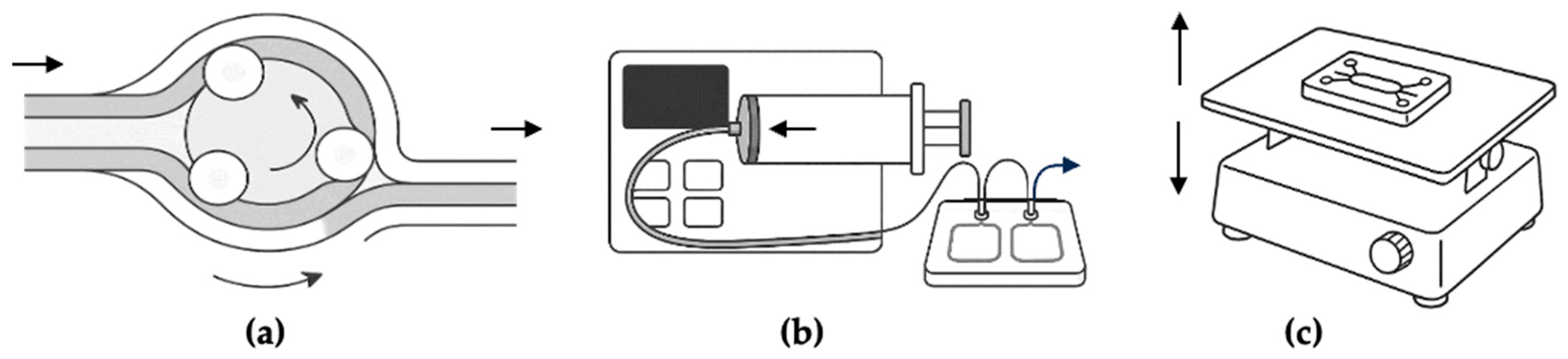
2.1. Technological Overview

OoC platforms are engineered microfluidic systems designed to replicate key aspects of human tissue architecture and function within a controlled, perfusable microenvironment. These systems enable the co-culture of different cell types, such as tumor and immune cells, under dynamic flow conditions, allowing researchers to study cellular interactions, cytokine signaling, and the effects of applied therapies in a more physiologically relevant context [24,25]. All models examined in this review simulate immune–tumor interactions and include features such as fluid flow, compartmentalization, and immune cell tracking. OoC platforms can be tailored to specific research needs, depending on the organ and cancer type being studied [25].
Most systems use polydimethylsiloxane (PDMS)-based microfluidic chips, mainly because PDMS is biocompatible, optically clear, and easy to work with [26,27]. In some platforms, polyethylene glycol diacrylate (PEGDA) hydrogels are used to provide a matrix in which cells and spheroids can grow in a 3D structure. These hydrogels have adjustable mechanical properties, helping to mimic the physical characteristics of the tumor environment [28]. Other approaches combine hybrid spheroids, consisting of multiple cell types to better replicate the tumor microenvironment (TME), with microfluidic chips to study spatially organized tumor–immune interactions [28,29,30].
One major advantage of these platforms is their ability to incorporate different immune cell types into co-culture with tumor cells. This allows researchers to study immune cell infiltration, cytotoxicity, and cytokine-influenced immune behavior of CD4+ T cells, macrophages, monocytes, and even CAR-T cells.
These models utilize perfusion to apply physiological shear stress, support nutrient exchange, and mimic immune cell migration (Figure 1). Perfusion durations range from 48 h up to eight days [22,28,29,30,31,32,33,34,35,36,37,38]. In systems not focused on drug testing, continuous perfusion has been maintained for up to one month, enabling both short-term interaction studies and long-term observation of immune–tumor dynamics [39]. Perfusion is often achieved through gravity-driven flow, using a rocker platform that tilts the chip and causes fluid to move between reservoirs. This setup offers a relatively simple and low-maintenance solution, avoiding the need for sterile connections to external pumps. While it does not fully replicate vascular flow, it still generates shear stress and supports transport processes [35,40,41]. Some systems do not use active flow at all, instead relying on passive diffusion through Matrigel membranes or across microchannels. In these cases, immune cell migration and media transport appear to be driven by chemotaxis [33,34,37]. Other platforms use syringe pumps to create unidirectional flow through continuous withdrawal at a defined rate [28,29,38,42,43]. More advanced systems incorporate closed-loop circulation with peristaltic pumps to better simulate dynamic physiological conditions [22,36,39].
Beyond flow control, channel architecture is a critical factor in determining physiological relevance. Common formats include dual-channel systems, which are separated by porous membranes that mimic vascular–tissue interfaces [31,33,37,40]; single-channel designs, which are used for tumor spheroids [30]; and multichamber systems, which allow for sequential tissue zones [34,37,42]. These configurations enable the modeling of tissue barriers, immune extravasation, and inter-tissue signaling.
Readout techniques used across these models include live-cell imaging with fluorescence and confocal microscopy to track immune and tumor cell behavior [29,30,33,35,37,40,42,43,44], as well as immunofluorescence for endpoint characterization [19,22,30,31,36,44]. Cytokine release is often quantified using ELISA or multiplex bead assays (e.g., IFN-γ, IL-6, and TNF-α), and flow cytometry (FACS) is used to evaluate immune cell activation and exhaustion markers (e.g., PD-1 and CTLA-4) as well as changes in immune subtypes [22,29,30,31,36,37,38,40,43,44]. Some platforms enable real-time monitoring of immune cell infiltration into 3D tumor spheroids, cytotoxic activity, and the dynamics of checkpoint inhibitor responses. Together, these tools offer multi-parametric insight into immune–tumor interactions under dynamic, physiologically relevant conditions.

2.2. Current Industrial Developments and Companies Commercializing MPSs in 2025

Considering their novelty, commercial OoC applications of immune-competent models are only now emerging, as reflected in a growing number of industrial platforms designed to replicate immune–tumor interactions (Table 1).

2.3. Application by Tumor Type
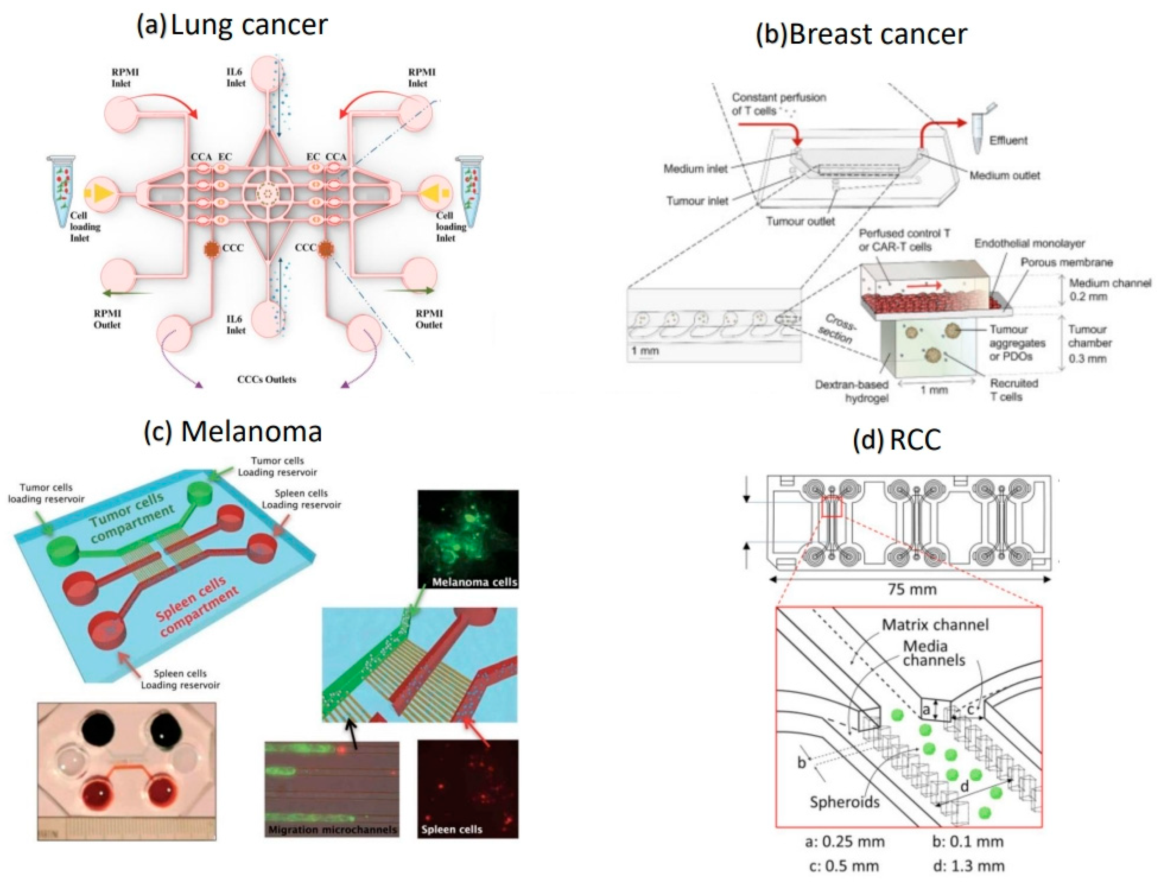
2.3.1. Lung Cancer

With around 2.2 million newly diagnosed cases and 1.8 million deaths in 2020, lung cancer ranks as the second most commonly diagnosed cancer worldwide and remains the leading cause of cancer-related death. More deaths occurred due to lung cancer than from breast, colorectal, and prostate cancers combined. The disease disproportionately affects individuals from lower socioeconomic backgrounds. Although about 80% of all lung cancers are linked to tobacco use, the disease also ranks among the top ten cancer-related causes of death in non-smokers globally [1,35,50].
The poor prognosis is largely due to late-stage diagnosis, as most cases are identified when treatment options are already limited. Over the past two decades, however, advances in tumor biology have led to the development of targeted therapies and immunotherapeutic approaches, including immune checkpoint inhibitors such as nivolumab and pembrolizumab. These therapies have significantly improved survival, especially in patients with advanced non-small cell lung carcinoma (NSCLC), and are now firmly established in clinical practice. Improvements in screening, particularly through low-dose computed tomography (LDCT), have also contributed to earlier diagnosis in high-risk populations [1,35,50].
Conventional 2D models fail to replicate the multicellular epithelium required to mimic the pulmonic architecture, and several microfluidic organ-on-a-chip (OoC) models have been developed to recreate aspects of the lung tumor microenvironment and provide more physiologically relevant tools for studying immunotherapeutic interventions [51].
One such model, described by Sardarabadi et al. [42], used a PDMS-based chip bonded to glass, with a gelatin methacryloyl (GelMA) hydrogel to embed cells in a three-dimensional matrix. The authors co-cultured A549 lung carcinoma cells with Jurkat T cells to simulate the tumor–immune microenvironment. A central IL-6 reservoir, flanked by parallel flow channels, was used to establish a chemotactic gradient. This setup allowed the researchers to observe real-time migration of T cells under flow conditions and analyze cytokine responses using ELISA. Interestingly, IL-6 was shown to have a dual role: it initially promoted T-cell activation and migration, but subsequently led to an upregulation of PD-L1 expression on tumor cells. While no drugs were tested in this particular study, the model provides a valuable foundation for future evaluations of cytokine-modulating agents and checkpoint inhibitors. The ability to monitor immune activation and exhaustion through cytokine profiling could be especially useful in preclinical screening.
Another MPS focused on NSCLC (HCC0827) employed a 3-lane chip developed by MIMETAS, incorporating a perfusable 3D endothelium. Endothelial cells (HUVECs) were cultured next to tumor cells, separated by a collagen barrier, to recreate the vascular interface. T-cells were introduced into the endothelial compartment and allowed to migrate toward the tumor under continuous perfusion. The platform enabled detailed observation of T-cell infiltration, a process strongly associated with improved clinical outcomes in immunotherapy. Although the system does not fully capture the complexity of lung tissue, it offers a reproducible and scalable method for assessing therapies that aim to enhance immune access to solid tumors [35].
Both OoC platforms demonstrate distinct strategies for modeling lung cancer immune dynamics in a setting that allows for precise manipulation of variables such as flow, cytokine gradients, and tissue architecture. One emphasized cytokine-driven migration within the tumor matrix, while the other focused on trans-endothelial infiltration. Each approach highlights specific mechanistic insights relevant for immunotherapy development in lung cancer.

2.3.2. Breast Cancer

Breast cancer is the most commonly diagnosed carcinoma among women worldwide. However, it also affects men, although at a significantly lower rate. It remains a significant global health concern. Statistically, 1 in 20 women develop breast cancer during their lifespan. Especially in countries with a low human development index, it is predicted that by 2050, 3.2 million new cases and 1.1 million deaths will occur due to this disease [52].
Despite all the developments in early diagnoses and therapy, the development of effective and safe drugs is a crucial challenge. Breast cancer is a heterogeneous disease with various molecular subtypes, including ER-positive/HER2-negative and triple-negative breast cancer (TNBC). Every subtype has its own biological properties that require different therapeutic approaches [53,54]. Triple-negative breast cancer is very aggressive and hard to treat, whereas ER-positive tumors react well to hormonal therapies. The therapy for breast cancer underwent significant development during recent years. New approaches target the hormone sensitivity of breast cancer. Treatments like CDK4&-inhibitors, SERD-Therapies (which degrade the selective estrogen receptor), and immunotherapy improved survival rates and provided longer life spans while simultaneously reducing side effects, especially for metastasized breast cancer [53,55,56].
Organ-on-chip technologies have also made it possible to replicate the complexity of breast cancer in vitro, capturing key features of the tumor microenvironment and disease progression. A remarkable example developed by Lee et al. is a dual-chip model that integrates breast carcinoma and cardiac tissue to study the cardiotoxic side effects of chemotherapy [44].
To support personalized CAR-T cell testing, Maulana et al. [43] developed a PDMS-based microfluidic platform featuring perfusable endothelialized channels next to chambers with tumor cells. These chambers contained either TNBC spheroids (MDA-MB-231) or patient-derived organoids (PDOs) embedded in a dextran-based hydrogel. CAR-T cells, engineered to recognize the Receptor Tyrosine Kinase-Like Orphan Receptor 1 (ROR1), which is overexpressed in TNBC, were continuously perfused through the endothelial channel, mimicking extravasation into the tumor site. Functional responses were evaluated using live imaging, immunofluorescence, flow cytometry, and cytokine quantification (e.g., IFN-γ, IL-2, granzyme B). The system demonstrated target-specific T-cell infiltration and tumor lysis while also capturing cytokine release syndrome (CRS)-like responses. Importantly, the use of dasatinib effectively reduced cytokine levels without compromising CAR-T cell viability. This study highlights the potential of microfluidic OoC models for evaluating both the efficacy and safety of patient-specific immunotherapies.
In another study, Berger Fridman et al. [31] established a high-throughput microfluidic platform using alginate-based hydrogels to recreate inflammatory and immunosuppressive tumor environments. By co-culturing breast tumor cells, fibroblasts, T cells, and macrophages, the platform was able to simulate dynamic shifts within the TME. Immunosuppressive shifting of macrophages to tumor-associated macrophages (TAMs) and changes in cytokine secretion were observed depending on platform configuration. Proteomic analysis revealed expression patterns that closely resemble those found in vivo, supporting the model’s relevance for studying immune cell regulation and TME progression.
Lee et al. [44] approached breast cancer modeling through a dual-organ microfluidic platform integrating breast cancer spheroids (SK-BR-3) with induced pluripotent stem cell-derived cardiac spheroids. The system allowed real-time monitoring of HER2+ tumors and cardiac responses to the drug doxorubicin using electrochemical aptasensors. While not focused on immunotherapy, the model demonstrated the different drug sensitivity in cancer versus healthy and fibrotic cardiac tissue, highlighting its value in predicting off-target cardiotoxicity of anticancer treatments in vulnerable patient populations.
A vascularized breast cancer-on-chip model developed by Boussommier-Calleja et al. [37] further investigated the relevance of immune integration by introducing primary human monocytes into a 3D microfluidic system. Tumor cells (MDA-MB-231) were seeded alongside HUVECs and fibroblasts in a fibrin-based matrix, forming perfusable microvessels. Monocytes were shown to significantly reduce tumor cell extravasation, not through direct contact, but likely via paracrine signaling involving factors such as IL-8 and CCL2. These findings emphasize the value of immune-integrated microfluidic models for investigating metastatic processes and tumor–immune interactions.
Together, these models reflect the diversity and adaptability of OoC technologies in breast cancer research. From simulating tumor–immune interactions and T-cell infiltration to evaluating patient-specific CAR-T responses or off-target toxicities, these platforms enable mechanistic insight and therapeutic evaluation under human-relevant conditions.

2.3.3. Melanoma

Melanoma is the most aggressive form of skin cancer and represents a significant health burden. Although it accounts for only 1% of all skin cancers, it causes 75% of skin cancer deaths [57,58].
The global incidence of melanoma has risen sharply in recent decades, especially in countries with populations with predominantly lighter skin tones. In 2020, 325,000 new cases were diagnosed worldwide, an increase of 41% compared to 2012. The highest rates will be seen in Australia and New Zealand, followed by North America and Europe [57].
The prognosis is highly dependent on the stage of the disease. Localized melanoma, which accounts for 78% of cases, has a 5-year survival rate of 99.6%, while the survival rate for metastatic stage IV melanoma is around 35%, despite recent advances. These improvements are due to the introduction of targeted therapies such as BRAF and MEK inhibitors and immunotherapies with checkpoint inhibitors such as anti-PD-1 and PD-L1. In addition, new approaches such as CAR T-cell therapy are being tested. Despite these advances, challenges remain, particularly in dealing with resistance and metastatic disease, such as brain metastases [58].
Preclinical models, such as animal models, are often insufficient to reproduce the complex physiology of melanoma. Consequently, emerging tools like organ-on-chip systems and 3D cultures are essential for building more accurate and translational melanoma models.
In a study by Agliari et al. [33], a microfluidic co-culture model was introduced to study the migration behavior of immune cells in response to melanoma cells. The system consisted of two chambers, one containing immune cells and the other seeded with melanoma cells. Both were connected by microchannels to mimic tissue architecture and allow real-time observation of immune cell migration. The study showed that wild-type (WT) immune cells migrated towards melanoma cells by testing spleen-derived immune cells from both wild-type and IRF-8 knockout (KO) mice. WT cells showed collective and chemotactic movement, whereas KO cells moved randomly and failed to infiltrate the tumor compartment. The work demonstrated the critical role of IRF-8 in coordinating immune cell behavior and showed how immune functionality can be measured through movement analysis (e.g., migration path, movement behavior, and directionality). The system allows quantitative analysis of immune behavior and provides a useful platform for testing therapies designed to enhance immune infiltration.
Complementing this 2D dynamic system, another study by Seo et al. [29] developed a 3D mixed spheroid model in which tumor cells were co-cultured with immune cells in a scaffold-free environment to more closely mimic in vivo tumor composition. This approach allowed the study of immune infiltration, cytotoxicity, and cytokine-driven transformation of tumor spheroids. Real-time imaging showed how immune pressure not only suppressed tumor cell viability but also altered the structure of the spheroids, reflecting the interplay between immune activation and tumor adaptation. The hybrid model is adaptable in terms of matrix composition and cell types and is suitable for testing a variety of immunomodulatory therapies, including checkpoint inhibitors and CAR-T cells.
Together, these two systems illustrate complementary strategies for modelling melanoma–immune interactions. The microfluidic chip allows real-time tracking and quantitative analysis of immune migration, while the spheroid-based system illustrates the architectural complexity of tumor–immune dynamics. Both highlight the value of immune-competent in vitro models for studying mechanisms of immune evasion and evaluating the efficacy of immunotherapeutic agents in melanoma.

2.3.4. Renal Cell Carcinoma

Renal cell carcinoma (RCC) accounts for approximately 3% of all malignant tumors and, although relatively rare, represents a clinically significant entity in oncology. In Germany alone, approximately 8350 new cases in men and 4059 in women were reported in 2019, corresponding to age-standardized incidence rates of 17.7 and 7.3 per 100,000, respectively [59]. Although incidence rates have remained relatively stable since the 1990s, mortality has decreased slightly due to advances in early detection and treatment [60].
RCC is histologically and molecularly heterogeneous, comprising several distinct subtypes with different prognoses and responses to treatment. The most common form is clear cell RCC (ccRCC), which accounts for 55–60% of cases and is often associated with VHL gene mutations and activation of hypoxia-related signaling pathways [61].
Papillary RCC (pRCC), which accounts for 9–17% of cases, is often associated with MET gene alterations, while the chromophobe subtype (chRCC), although rare (~6%), is characterized by mitochondrial-rich tumor cells. Other variants, such as sarcomatoid differentiation and collecting duct carcinoma, are associated with a particularly poor prognosis [60].
Survival outcomes vary widely depending on the stage of the disease. While localized RCC (UICC stage I) is associated with a five-year survival rate of over 95%, the prognosis drops dramatically to around 17% in cases of distant metastasis (UICC IV). Approximately 40% of RCCs are detected at an early stage, but approximately 10% are already metastatic at the time of diagnosis. The overall relative five-year survival rate is 77% for men and 79% for women [60].
Despite advances in targeted therapies—including tyrosine kinase inhibitors—and immunotherapy with checkpoint inhibitors, the treatment of metastatic RCC remains a clinical challenge. Intratumoral heterogeneity, activation of resistance pathways such as mTOR signaling, and a lack of predictive biomarkers significantly limit long-term therapeutic success [61].
Several organ-on-chip models have been developed that replicate key aspects of the RCC phenotype and are used to study its pathophysiology and drug resistance (Figure 2).
A study by Miller et al. [30] introduced a 3D RCC spheroid model cultured in a microphysiological chip. Using A498 and RCC4 cell lines embedded in a collagen matrix, the system mimicked essential features of the tumor microenvironment, including hypoxia, collective migration, and gene expression patterns consistent with in vivo RCC tissue. The platform enabled detailed drug response analysis. For example, bortezomib treatment resulted in significantly higher cytotoxicity in 3D cultures compared to 2D models, underscoring the physiological relevance of the chip. In addition, the model revealed migratory tumor behavior associated with EMT-like gene expression, enabling screening of anti-metastatic compounds such as latrunculin A and AT13148. The system also supported immune-related applications, demonstrating that ROR1-targeted CAR T cells could selectively kill RCC spheroids, although extracellular matrix penetration was a barrier. This highlights the potential of this model for testing both cytotoxic and immunomodulatory therapies in RCC.
A complementary approach was developed by Somova et al. [22,62], who designed a multi-compartment microphysiological system that co-cultured RCC cells (Caki-1) with healthy renal epithelium (RPTEC/TERT1). In their approach, tumor and healthy tissues were cultured in spatially separated but perfused chambers. Exposure to tumor-conditioned medium induced cytokine expression (IL-8, TNF-α) and metabolic reprogramming, reflecting tumor–host interactions. While immune cells were not added, the chip is compatible with future immune co-culture configurations, offering a basis for evaluating tumor–host and immunotherapy–host interactions.
Taken together, these studies highlight the growing value of MPSs in RCC research. They offer a reliable way to model the tumor microenvironment, enabling long-term perfused co-cultures of tumor and surrounding tissue. These platforms also support real-time analysis of cytotoxicity, metabolism, and cytokine signaling, making it possible to assess how drugs affect both the tumor and its microenvironment.
There are still limitations, especially when it comes to capturing the full complexity of the immune system or simulating systemic immune responses and vascular trafficking [22,30,62]. Moving forward, integrating more immune components and dynamic vascular features will be essential to increase the translational potential of these models.

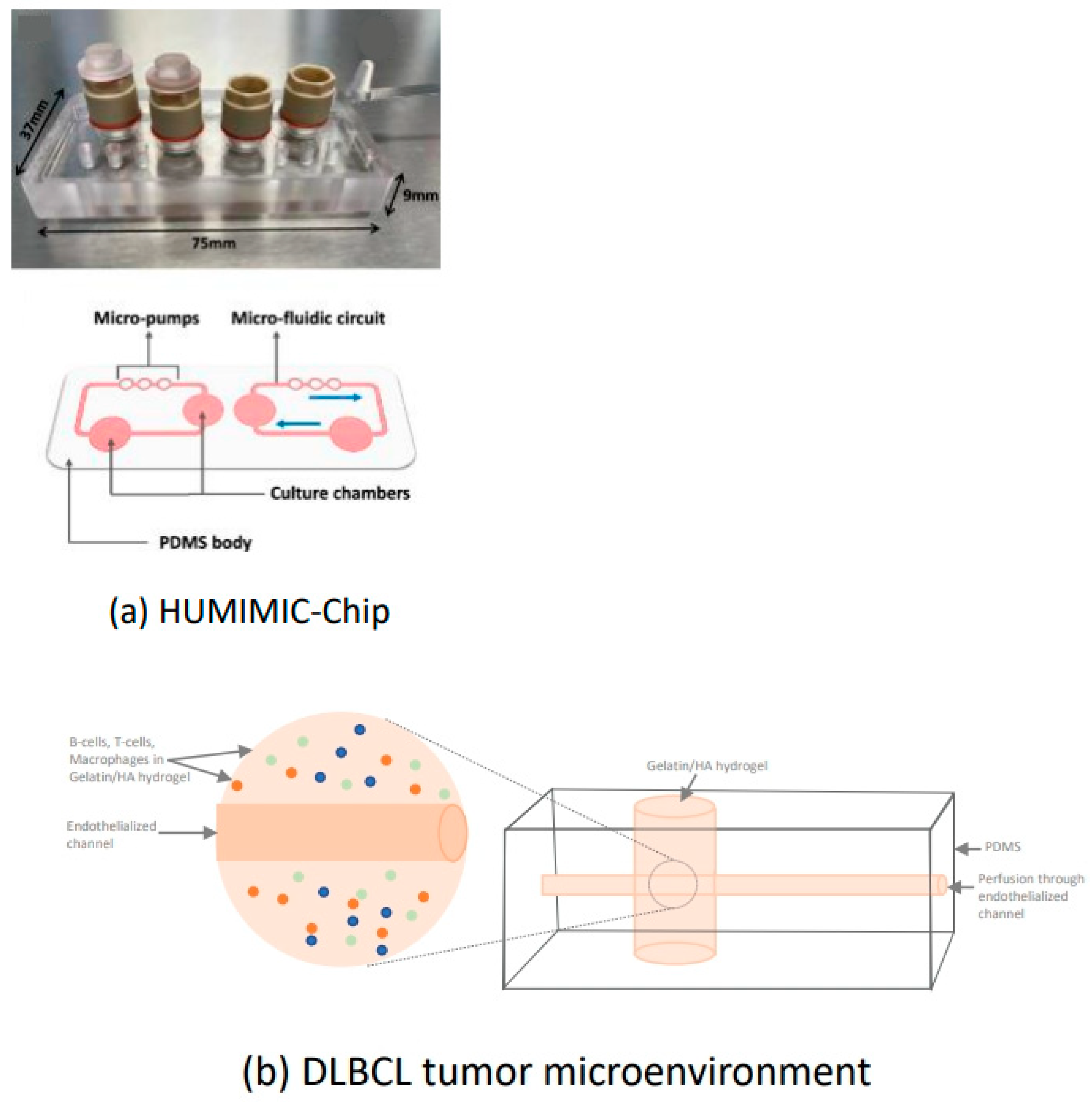
2.3.5. Diffuse Large B-Cell Lymphoma

DLBCL is the most common form of non-Hodgkin lymphoma, accounting for 30–40% of all cases, and represents an aggressive, life-threatening disease. The annual incidence is 7 per 100,000 people, with a mean age of onset of 64 years and a higher prevalence in men. Without treatment, the disease progresses rapidly, emphasizing the urgent need for effective therapeutic strategies [63].
DLBCL is a highly heterogeneous disease with multiple molecular subtypes. The most clinically relevant are the germinal center B-cell-like (GCB) and activated B-cell-like (ABC) subtypes, which differ in both genetic signature and clinical behavior. Genetic alterations such as translocations involving BCL2, BCL6, or MYC play central roles in disease pathogenesis and influence treatment response. The current standard of care is the R-CHOP regimen (rituximab, cyclophosphamide, doxorubicin, vincristine, and prednisone), which is potentially curative for many patients. However, relapse occurs in approximately 40% of cases, and the prognosis remains particularly poor in aggressive subtypes [64].
A study by Mannino et al. [38] introduced a lymphoma-on-a-chip platform designed to replicate key features of the DLBCL tumor microenvironment (Figure 3). The model uses a hyaluronic acid-based hydrogel embedded with tumor B-cells, T-cells, and macrophages, along with a perfusable, endothelialized microchannel to simulate tumor vasculature. This configuration enables the spatiotemporal analysis of drug delivery under dynamic flow conditions. The chip supports fluorescence-based diffusion tracking, flow cytometry analysis, and incorporates spatially distinct regions that mimic both tumor tissue and healthy lymphatic environments.
As a proof of concept for therapeutic testing, the system was treated with an anti-CSF-1R antibody, resulting in selective macrophage depletion within the tumor compartment. This demonstrated the model’s capability to evaluate drug efficacy in real time and under immune-competent, physiologically relevant conditions. The inclusion of physically separated compartments also allowed for the analysis of localized versus systemic immune effects, which is a feature not commonly addressed in traditional models.
While the system offers advanced spatial and functional complexity, several technical limitations were noted. For example, the removal of the steel wire used in fabrication occasionally disrupted the hydrogel structure, leading to variability in channel geometry. Additionally, despite its sophistication, the model does not yet replicate systemic immune circulation or multiorgan interactions [38].
A second study by Foxall et al. [32] focused on developing a scaffold-free 3D tumor microenvironment (TME) model for B-cell lymphoma to assess antibody-based therapies. This model was particularly suited to evaluating monoclonal antibody responses, including responses to rituximab. Compared to standard 2D cultures, it more accurately reproduced tumor morphology and endothelial barrier integrity, revealing resistance mechanisms that align with in vivo behavior. However, the absence of vasculature and dynamic perfusion limited its ability to simulate drug delivery kinetics and immune cell migration.
Together, these two approaches reflect the increasing importance of 3D and microfluidic models in lymphoma research. While one emphasizes spatial control and vascularized architecture, the other demonstrates the value of preserving tumor structure for therapeutic assessment. Both offer greater precision in modeling tumor–immune interactions than conventional systems and underscore the need for continued development toward more physiologically complete platforms.

2.3.6. Hepatocellular Carcinoma

Hepatocellular carcinoma (HCC) is the most common primary liver cancer, accounting for more than 90% of cases, with a global incidence of 500,000 to 1 million new diagnoses per year [65,66]. Its pathogenesis is closely associated with chronic liver diseases such as viral hepatitis (HBV/HCV), cirrhosis, and metabolic disorders. The tumor microenvironment is characterized by hypoxia, angiogenic signaling (e.g., VEGF, HIF-1α), and immune cell infiltration [67,68]. These complex interactions make HCC a particularly challenging target for conventional therapies, especially given that current in vivo models poorly recapitulate human liver physiology and immune context.
Treatment strategies for HCC are highly stage dependent. Early-stage disease is typically treated by resection, liver transplantation, or radiofrequency ablation for tumors less than 3 cm [69,70]. Intermediate stages are treated with transarterial chemoembolization (TACE), with a median survival of 20–26 months [69]. Emerging strategies now combine TACE with immunotherapy to improve efficacy [71]. For advanced HCC, first-line treatments include atezolizumab/bevacizumab (anti-PD-L1 + anti-VEGF) or durvalumab/tremelimumab (anti-PD-L1 + anti-CTLA-4) combinations [71]. Targeted therapies such as lenvatinib and sorafenib continue to be used, although resistance, often due to reactivation of the MAPK pathway, continues to limit long-term success [72].
In order to enhance understanding of immune cell trafficking in the HCC tumor microenvironment, Kennedy et al. [34] developed a two-channel liver-on-a-chip model that reproduces key features of hepatic sinusoidal flow and endothelial cell interactions. The study’s primary focus is on the adhesion and migration of immune cells across primary human liver sinusoidal endothelial cells (LSEC) under physiological shear stress, with implications for the testing of immunotherapies and the delivery of chimeric antigen receptor (CAR)-T cells. The model is constructed using the Emulate Liver-Chip platform, a microfluidic device with two parallel channels separated by a porous membrane. LSECs are cultured in the lower (endothelial) channel, while Huh-7 hepatoma cells are cultured in the upper (epithelial) channel. Perfusion is applied to mimic the sinusoidal shear stress of the liver (0.05 Pa) [34]. Following a 24 h period of cytokine stimulation with TNF-α and IFN-γ, the LSEC layer showed increased immune activation and morphological changes. Pre-labeled peripheral blood mononuclear cells (PBMCs) were introduced into the chip and exposed to the endothelial layer. Confocal and multiphoton imaging techniques were used to visualize immune cell adhesion and migration across the LSEC. It was observed that cytokine treatment significantly enhanced immune cell adhesion, as compared to the control group [34].
This system enables the real-time study of leukocyte recruitment and migration across the human liver endothelium. This system has the potential to test the efficacy of immunomodulatory therapies, which include checkpoint inhibitors and cell therapies such as CAR-T or CAR macrophages, in enhancing immune infiltration into HCC tissues [34].

2.3.7. Prostate Cancer

Prostate cancer (PCa) is the most common cancer in men in the USA and Europe, with a projected incidence of over 313,000 new cases and 35,000 deaths in the US for the year 2025 [73].
Therapeutic strategies for prostate carcinoma vary depending on the stage of the disease. For localized, low-risk tumors, active surveillance is often the recommended approach. In intermediate- and high-risk patients, radical prostatectomy is considered standard, often complemented by radiation therapy [74].
For advanced and metastatic prostate cancer, androgen deprivation therapy (ADT) remains the foundation of treatment. In many cases, ADT is initiated in combination with newer hormonal agents such as abiraterone or enzalutamide to improve outcomes. Chemotherapy may also be considered [75].
In castration-resistant prostate cancer (CRPC), where the disease progresses despite low testosterone levels, targeted therapies have emerged. Olaparib is used in patients with BRCA1/2 mutations, while radioligand therapy with 177Lu-PSMA-617 offers a treatment option for patients with PSMA-positive metastases [76,77].
PCa research is hampered by the limited availability of robust and representative cell lines as well as highly inefficient preclinical animal models [78].
To address these limitations, Peng et al. [28] developed a sophisticated microphysiological tumor model using PEGDA hydrogel microbeads to recreate a tunable, 3D tumor microenvironment containing prostate cancer cells and fibroblast-associated stromal components. The authors co-cultured PSCA-expressing PC3 prostate cancer cells with HT1080 fibrosarcoma cells modified to express fibroblast activation protein (FAP), a stromal marker associated with immune suppression. These composite spheroids were encapsulated in PEGDA beads with mechanical properties that mimic the tissue stiffness of common metastatic sites such as the liver and lung.
This model enabled long-term spheroid viability, and was used to evaluate UniCAR T cell therapy targeting both PSCA (tumor antigen) and FAP (stromal antigen). The study showed that dual-targeting UniCAR T cells could efficiently infiltrate the matrix, accumulate on the spheroid surface, and induce potent tumor cell killing. Cytokine analysis (e.g., IFN-γ, TNF-α) and granzyme B immunostaining confirmed T cell activation and cytotoxicity, while migration assays tracked T cell behavior in real time [28].
A study by Xie et al. [40] used a pump-free, dual-channel microfluidic chip to study interactions between natural killer (NK) cells and DU145 prostate tumor spheroids. The chip’s gravity-driven design created unidirectional fluid flow to mimic physiological perfusion without external pumps. Tumor spheroids sat in Matrigel-coated paper stacks within the chip, and NK-92 cells were introduced into adjacent channels. The flow conditions allowed NK-92 cells to migrate and infiltrate the spheroids more, and the flow influenced the cells’ localization and activation.
Static and perfused conditions revealed that dynamic culture better reflected in vivo NK cell phenotypes, particularly the transition to tumor-infiltrating NK cell (TINK) subtypes, as shown by changes in CD56/CD16 marker expression. The setup also allowed separation and phenotypic analysis of free versus infiltrated NK cells, highlighting the chip’s utility for prostate tumor immune dynamics [40].
In a complementary approach, Padmyastuti et al. [36] performed a study using the HUMIMIC Chip platform (TissUse) to investigate molecular and secretory changes in prostate cancer models under 3D and microfluidic conditions (Figure 3). LNCaP and PC3 prostate cancer cells were cultured in both 3D static and within the perfused HUMIMIC platforms. The model sustained PSA secretion in LNCaP cells for over 10 days and induced changes in microRNA expression profiles compared to 2D cultures. MicroRNAs linked to castration-resistant prostate cancer were upregulated under MPS conditions, suggesting that dynamic flow impacts molecular traits relevant to disease progression. PSA and PSMA levels were more robustly expressed in microfluidic culture, indicating that the system supports physiological cell behavior.
Together, these platforms demonstrate the diverse capabilities of prostate cancer–focused OoC systems. The PEGDA-based model emphasizes dual-targeted CAR-T cell immunotherapy, the pump-free chip reveals NK cell behavior under dynamic conditions, and the dynamic MPSs showcase phenotypic changes and biomarker expression. Each contributes valuable insights toward building predictive, immune-integrated models for prostate cancer drug development and immunotherapy testing.

3. Key Experimental Approaches Across Models

Across the different cancer-specific OoC and microphysiological systems reviewed, several recurring experimental strategies and design principles can be observed. These approaches define the flexibility and translational potential of in vitro immuno-oncology modeling.

3.1. Cell Sources and Cell Types

The majority of models include a combination of immortalized tumor cell lines (e.g., A549 for NSCLC, Huh-7 for HCC, CAKI-1 for renal cell carcinoma), endothelial cells (often HUVECs), and stromal cells such as fibroblasts. The integration of immune cells is critical to reflect the specific mechanisms of immunotherapy. These include CD4+ T-cells, Jurkat cells, CAR-T cells, monocytes, macrophages, and peripheral blood mononuclear cells (PBMCs). In certain models, tumor spheroids are constructed using PEGDA hydrogels or Matrigel to mimic the 3D tumor architecture [28].

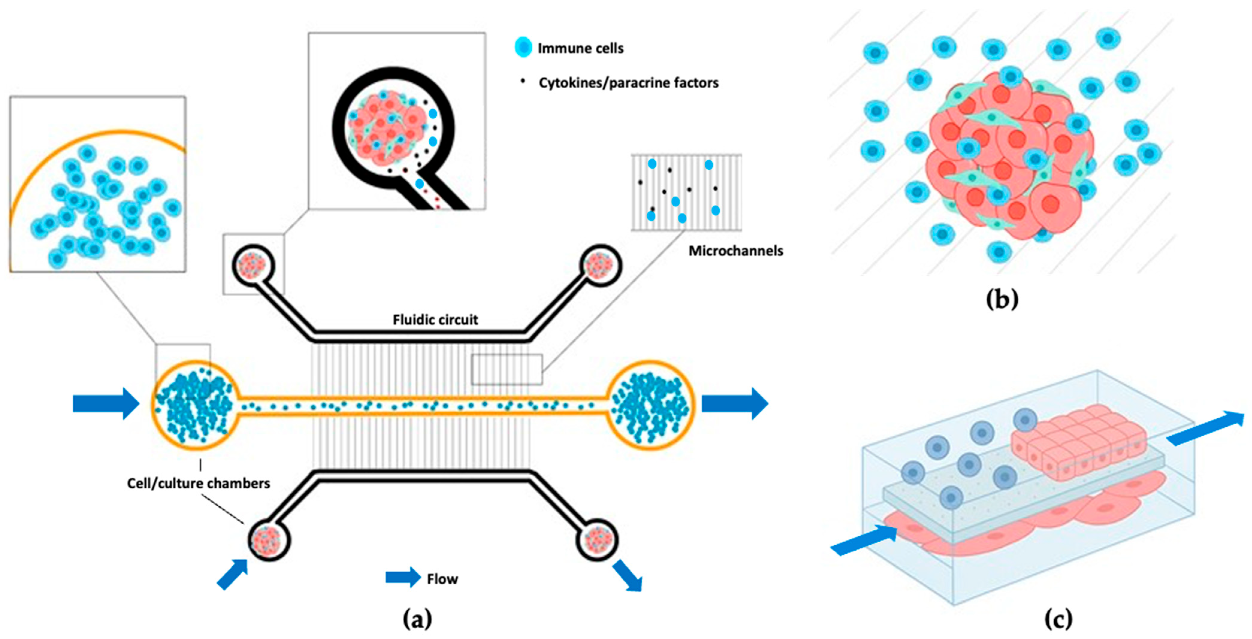
3.2. Immune Cell Integration Methods

The integration of immune cells can be achieved by direct embedding in hydrogels, perfusion through adjacent channels, or co-culture in separate but interconnected compartments (Figure 4). For instance, the addition of CAR-T cells is frequently achieved through a recirculating perfusion loop or a syringe–pump system to simulate vascular infiltration [34,43]. Monocytes and neutrophils are perfused through channels to study transmigration or immune suppression [31,35,37].

3.3. Model Viability and Stability

A variety of techniques are used for model characterization and response assessment. For imaging, confocal microscopy [33,37,39,41,42,79], live-cell fluorescence imaging, and immunofluorescence are employed to track cell localization, toxicity, and infiltration [29,31,32,38,41,42,80]. For cytokine analysis, ELISA and multiplex assays are frequently utilized, for instance, for IFN-γ, TNF-α, and IL-6. FACS and immunostaining are employed to evaluate activation markers or PD-1/PD-L1 expression. Spheroids are analyzed for viability, area, and morphological changes in 3D cultures [29,31,42,43].

3.4. Engineering Innovations

Engineering innovations have led to the development of novel platforms that enhance physiological relevance in vitro. These include PEGDA hydrogels with tunable stiffness to mimic matrix mechanics and cytokine gradient systems to simulate inflammation-driven chemotaxis [28]. The drive to better mimic the tumor microenvironment (TME) was evident in nearly all cancer types reviewed. The incorporation of diverse cell types into spheroids appears to be the next evolution in simplified 3D culture systems [28,29,31,34,42]. Dual-compartment chips offer a practical means of replicating different anatomical or physiological compartments, allowing the study of local versus systemic immune responses. Taken together, these experimental designs highlight the potential of organ-on-chip (OoC) platforms to model complex immune–tumor interactions under dynamic and modular conditions (Table 2).
OoC have introduced new levels of physiological relevance and experimental control, overcoming current limitations will be key to advancing their translational value. A major challenge remains the limited immune complexity, as most models incorporate only one or two immune cell types [88]. A potential solution lies in the integration of some form of bone marrow-on-a-chip components or immune organoids, which could provide a more diverse and functional immune repertoire within the system [39,89]. Expanding this to include a broader immune repertoire, such as a combination of dendritic cells, NK cells, and B cells, could be essential for more detailed studies of immune suppression, activation thresholds, interactions, and immune escape mechanisms. In addition, immune memory, exhaustion, and chronic activation states remain difficult to model in short-term cultures.
Similarly, the limited biological complexity of tumor models can be partially overcome by incorporating patient-derived organoids that contain stromal, vascular, and autologous immune cells, allowing for more realistic tumor–immune interactions. Combined with microfluidic perfusion and real-time analytics, this could enable ex vivo immunotherapy testing customized to individual patients, supporting personalized oncology [90,91].
On the technical side, standardization of chip fabrication methods and operating protocols will be essential to improve interlaboratory reproducibility. Initiatives such as the organ-on-a-chip standards that may be developed by the European Committee for Standardization (CEN) are an important step in enabling wider adoption of these systems in both research and regulatory contexts.
The short lifespan of immune–tumor co-cultures and the lack of systemic circulation remain persistent barriers to modeling long-term immune responses and metastatic progression. These challenges could be addressed by the development of interconnected multi-organ platforms or vascularized microfluidic circuits that would allow recirculation of immune cells and study of inter-tissue immune dynamics over extended periods of time.
Current systems are optimized for acute responses, with experiments rarely extending beyond a week. However, many immunotherapeutic outcomes depend on long-term cell–cell communication, adaptive resistance, and tumor evolution. Increasing the duration and stability of co-cultures will be critical for modeling events such as checkpoint inhibitor resistance or CAR-T cell exhaustion.
Finally, some functional limitations, such as immune cell adhesion or vascular blockage, can be mitigated by better design strategies, including the use of surface coatings, optimized channel geometries, and adaptive flow control mechanisms. Addressing these technical and biological gaps will be critical to transforming OoC systems into robust tools for immunotherapy development and precision medicine [86,87].

4. Conclusions

OoC technologies have attracted considerable interest as tools for modeling cancer–immune interactions in vitro. However, many of the models described in the current literature should be considered primarily as proof-of-concept platforms. While some have tested immunotherapies or novel agents, comprehensive drug screening remains scarce. The field has clear potential for predictive drug testing, but most systems have not been developed or validated for this purpose on a large scale.
Many second-generation OoC platforms have evolved from early 3D spheroid culture approaches to incorporate controlled perfusion, spatial compartmentalization, and increasingly complex tumor microenvironments. While these systems demonstrate considerable creativity, only a subset is likely to remain relevant or scalable over the long term. Those with simplified operation, cost-effective design, and robust, reproducible outputs are most likely to transition from academic research tools to translational platforms.
While these systems do not yet replace animal models, they successfully replicate specific immune and tissue-level functions that do not require whole-organism systems. As such, they may serve as viable alternatives to xenograft models, particularly for mechanistic studies and early-stage therapeutic development. Over time and with further refinement, they could ultimately help to reduce and in some cases replace the use of animal testing in oncology altogether. The recent roll-out of the Federal Drug Administration (FDA) 3-year roadmap to reduce animal testing (April 2025) is seen as a major boost for MPS applications. It plans for a phased reduction in animal experimentation requirements in the evaluation of monoclonal antibody therapies, and subsequent expansion to biologics and small-molecule drugs (https://www.fda.gov/media/186092/download?attachment; accessed on 30 May 2025).
The growing interest in employing OoC platforms to study not only cancer immunology but also metabolic diseases, sepsis, and vaccine responses underscores their broader potential. As these tools evolve, they will become increasingly relevant for studying individualized therapies such as CAR-T cells or personalized cancer vaccines. While the field is still in its early stages, its momentum and versatility suggest that OoC models should be taken seriously as part of the future landscape of immuno-oncology research.

Supplementary Materials

The following supporting information can be downloaded at: https://www.mdpi.com/article/10.3390/futurepharmacol5030036/s1, Table S1: Organ-on-a-chip_Cancer_Immune_Research.

Author Contributions

M.G. was responsible for the conceptualization, literature research, manuscript and figure preparation, editing, and proofreading; M.B. supported the conceptualization of the review; and P.C.P. supported the manuscript and figure preparation, editing, and proofreading. All authors have read and agreed to the published version of the manuscript.

Funding

This review received no external funding.

Acknowledgments

Special thanks to Maryna Somova for her valuable advice and support throughout the development of this review. During the preparation of this manuscript, the author used ChatGPT (OpenAI, GPT-4, April 2025) for linguistic refinement, creation of figures, and structural organization of the review. The authors have reviewed and edited the output and take responsibility for the content of this publication.

Conflicts of Interest

The authors declare no conflicts of interest.

References

  1. Sung, H.; Ferlay, J.; Siegel, R.L.; Laversanne, M.; Soerjomataram, I.; Jemal, A.; Bray, F. Global Cancer Statistics 2020: GLOBOCAN Estimates of Incidence and Mortality Worldwide for 36 Cancers in 185 Countries. CA Cancer J. Clin. 2021, 71, 209–249. [Google Scholar] [CrossRef] [PubMed]
  2. World Health Organization Cancer. Available online: https://www.who.int/news-room/fact-sheets/detail/cancer (accessed on 27 June 2025).
  3. Sharma, P.; Allison, J.P. The Future of Immune Checkpoint Therapy. Science 2015, 348, 56–61. [Google Scholar] [CrossRef]
  4. Ribas, A.; Wolchok, J.D. Cancer Immunotherapy Using Checkpoint Blockade. Science 2018, 359, 1350–1355. [Google Scholar] [CrossRef]
  5. Pardoll, D.M. The Blockade of Immune Checkpoints in Cancer Immunotherapy. Nat. Rev. Cancer 2012, 12, 252–264. [Google Scholar] [CrossRef]
  6. Topalian, S.L.; Drake, C.G.; Pardoll, D.M. Immune Checkpoint Blockade: A Common Denominator Approach to Cancer Therapy. Cancer Cell 2015, 27, 450–461. [Google Scholar] [CrossRef]
  7. U.S. Food and Drug Administration. Oncology (Cancer)/Hematologic Malignancies Approval Notifications. Available online: https://www.fda.gov/drugs/resources-information-approved-drugs/oncology-cancerhematologic-malignancies-approval-notifications (accessed on 27 June 2025).
  8. Paul, J.; Mitchell, A.P.; Kesselheim, A.S.; Rome, B.N. Overlapping and Non-Overlapping Indications for Checkpoint Inhibitors in the US. J. Clin. Oncol. 2024, 42, 11057. [Google Scholar] [CrossRef]
  9. Tao, Z.; Chyra, Z.; Kotulová, J.; Celichowski, P.; Mihályová, J.; Charvátová, S.; Hájek, R. Impact of T Cell Characteristics on CAR-T Cell Therapy in Hematological Malignancies. Blood Cancer J. 2024, 14, 213. [Google Scholar] [CrossRef]
  10. Marchal, I. Cancer Vaccines: The Experts Speak. Nat. Biotechnol. 2025, 43, 477–481. [Google Scholar] [CrossRef]
  11. Park, J.S.; Withers, S.S.; Modiano, J.F.; Kent, M.S.; Chen, M.; Luna, J.I.; Culp, W.T.N.; Sparger, E.E.; Rebhun, R.B.; Monjazeb, A.M.; et al. Canine Cancer Immunotherapy Studies: Linking Mouse and Human. J. Immunother. Cancer 2016, 4, 97. [Google Scholar] [CrossRef]
  12. Mak, I.W.; Evaniew, N.; Ghert, M. Lost in Translation: Animal Models and Clinical Trials in Cancer Treatment. Am. J. Transl. Res. 2014, 6, 114–118. [Google Scholar]
  13. Togashi, Y.; Shitara, K.; Nishikawa, H. Regulatory T Cells in Cancer Immunosuppression—Implications for Anticancer Therapy. Nat. Rev. Clin. Oncol. 2019, 16, 356–371. [Google Scholar] [CrossRef] [PubMed]
  14. Joyce, J.A.; Fearon, D.T. T Cell Exclusion, Immune Privilege, and the Tumor Microenvironment. Science 2015, 348, 74–80. [Google Scholar] [CrossRef] [PubMed]
  15. Ventola, C.L. Cancer Immunotherapy, Part 3: Challenges and Future Trends. Pharm. Ther. 2017, 42, 514–521. [Google Scholar]
  16. Mangani, S.; Kremmydas, S.; Karamanos, N.K. Mimicking the Complexity of Solid Tumors: How Spheroids Could Advance Cancer Preclinical Transformative Approaches. Cancers 2025, 17, 1161. [Google Scholar] [CrossRef]
  17. Kapałczyńska, M.; Kolenda, T.; Przybyła, W.; Zajączkowska, M.; Teresiak, A.; Filas, V.; Ibbs, M.; Bliźniak, R.; Łuczewski, Ł.; Lamperska, K. 2D and 3D Cell Cultures—A Comparison of Different Types of Cancer Cell Cultures. Arch. Med. Sci. 2018, 14, 910–919. [Google Scholar] [CrossRef]
  18. Courau, T.; Bonnereau, J.; Chicoteau, J.; Bottois, H.; Remark, R.; Assante Miranda, L.; Toubert, A.; Blery, M.; Aparicio, T.; Allez, M.; et al. Cocultures of Human Colorectal Tumor Spheroids with Immune Cells Reveal the Therapeutic Potential of MICA/B and NKG2A Targeting for Cancer Treatment. J. Immunother. Cancer 2019, 7, 74. [Google Scholar] [CrossRef]
  19. Rodrigues, D.B.; Reis, R.L.; Pirraco, R.P. Modelling the Complex Nature of the Tumor Microenvironment: 3D Tumor Spheroids as an Evolving Tool. J. Biomed. Sci. 2024, 31, 13. [Google Scholar] [CrossRef]
  20. Hartung, T.; Smirnova, L. A Path Forward Advancing Microphysiological Systems. ALTEX 2025, 42, 183–203. [Google Scholar] [CrossRef]
  21. Bhatia, S.N.; Ingber, D.E. Microfluidic Organs-on-Chips. Nat. Biotechnol. 2014, 32, 760–772. [Google Scholar] [CrossRef]
  22. Somova, M.; Simm, S.; Padmyastuti, A.; Ehrdardt, J.; Schoon, J.; Wolff, I.; Burchardt, M.; Roennau, C.; Pinto, P.C. Integrating Tumor and Healthy Epithelium in a Micro-Physiology Multi-Compartment Approach to Study Renal Cell Carcinoma Pathophysiology. Sci. Rep. 2024, 14, 9357. [Google Scholar] [CrossRef]
  23. Chernyavska, M.; Masoudnia, M.; Valerius, T.; Verdurmen, W.P.R. Organ-on-a-Chip Models for Development of Cancer Immunotherapies. Cancer Immunol. Immunother. 2023, 72, 3971–3983. [Google Scholar] [CrossRef] [PubMed]
  24. Avula, L.R.; Grodzinski, P. How Organ-on-a-Chip Is Advancing Cancer Research and Oncology—A Cancer Hallmarks’ Perspective. Front. Lab. A Chip Technol. 2024, 3, 1487377. [Google Scholar] [CrossRef]
  25. Leung, C.M.; de Haan, P.; Ronaldson-Bouchard, K.; Kim, G.-A.; Ko, J.; Rho, H.S.; Chen, Z.; Habibovic, P.; Jeon, N.L.; Takayama, S.; et al. A Guide to the Organ-on-a-Chip. Nat. Rev. Methods Primers 2022, 2, 33. [Google Scholar] [CrossRef]
  26. Khot, M.I.; Levenstein, M.A.; de Boer, G.N.; Armstrong, G.; Maisey, T.; Svavarsdottir, H.S.; Andrew, H.; Perry, S.L.; Kapur, N.; Jayne, D.G. Characterising a PDMS Based 3D Cell Culturing Microfluidic Platform for Screening Chemotherapeutic Drug Cytotoxic Activity. Sci. Rep. 2020, 10, 15915. [Google Scholar] [CrossRef]
  27. Banik, S.; Uchil, A.; Kalsang, T.; Chakrabarty, S.; Ali, M.A.; Srisungsitthisunti, P.; Mahato, K.K.; Surdo, S.; Mazumder, N. The Revolution of PDMS Microfluidics in Cellular Biology. Crit. Rev. Biotechnol. 2023, 43, 465–483. [Google Scholar] [CrossRef]
  28. Peng, X.; Janićijević, Ž.; Loureiro, L.R.; Hoffmann, L.; Lee, P.S.; Cela, I.; Kruppke, B.; Kegler, A.; Feldmann, A.; Bachmann, M.; et al. 3D Microphysiological Tumor Model for Dual-Targeting CAR T Cell Immunotherapy. BioRxiv 2024. [Google Scholar] [CrossRef]
  29. Seo, J.; Ha, G.; Lee, G.; Nasiri, R.; Lee, J. Modeling Tumor-Immune Interactions Using Hybrid Spheroids and Microfluidic Platforms for Studying Tumor-Associated Macrophage Polarization in Melanoma. Acta Biomater. 2024, 190, 233–246. [Google Scholar] [CrossRef]
  30. Miller, C.P.; Fung, M.; Jaeger-Ruckstuhl, C.A.; Xu, Y.; Warren, E.H.; Akilesh, S.; Tykodi, S.S. Therapeutic Targeting of Tumor Spheroids in a 3D Microphysiological Renal Cell Carcinoma-on-a-Chip System. Neoplasia 2023, 46, 100948. [Google Scholar] [CrossRef]
  31. Berger Fridman, I.; Kostas, J.; Gregus, M.; Ray, S.; Sullivan, M.R.; Ivanov, A.R.; Cohen, S.; Konry, T. High-Throughput Microfluidic 3D Biomimetic Model Enabling Quantitative Description of the Human Breast Tumor Microenvironment. Acta Biomater. 2021, 132, 473–488. [Google Scholar] [CrossRef]
  32. Foxall, R.; Narang, P.; Glaysher, B.; Hub, E.; Teal, E.; Coles, M.C.; Ashton-Key, M.; Beers, S.A.; Cragg, M.S. Developing a 3D B Cell Lymphoma Culture System to Model Antibody Therapy. Front. Immunol. 2021, 11, 605231. [Google Scholar] [CrossRef]
  33. Agliari, E.; Biselli, E.; De Ninno, A.; Schiavoni, G.; Gabriele, L.; Gerardino, A.; Mattei, F.; Barra, A.; Businaro, L. Cancer-Driven Dynamics of Immune Cells in a Microfluidic Environment. Sci. Rep. 2014, 4, 6639. [Google Scholar] [CrossRef] [PubMed]
  34. Kennedy, J.I.; Davies, S.P.; Hewett, P.W.; Wilkinson, A.L.; Oo, Y.H.; Lu, W.Y.; El Haj, A.J.; Shetty, S. Organ-on-a-Chip for Studying Immune Cell Adhesion to Liver Sinusoidal Endothelial Cells: The Potential for Testing Immunotherapies and Cell Therapy Trafficking. Front. Cell Dev. Biol. 2024, 12, 1359451. [Google Scholar] [CrossRef] [PubMed]
  35. Wisdom, K.M.; Suijker, J.; van den Broek, L.; Sridharan, B.; Grandhi, T.S.P.; Cheng, A.; Lamb, M.; Titus, S.A.; Gehman, A.; Poore, D.; et al. Lung Tumor Microphysiological System with 3D Endothelium to Evaluate Modulators of T-Cell Infiltration. BioRxiv 2022. [Google Scholar] [CrossRef]
  36. Padmyastuti, A.; Sarmiento, M.G.; Dib, M.; Ehrhardt, J.; Schoon, J.; Somova, M.; Burchardt, M.; Roennau, C.; Pinto, P.C. Microfluidic-Based Prostate Cancer Model for Investigating the Secretion of Prostate-Specific Antigen and MicroRNAs In Vitro. Sci. Rep. 2023, 13, 11623. [Google Scholar] [CrossRef]
  37. Boussommier-Calleja, A.; Atiyas, Y.; Haase, K.; Headley, M.; Lewis, C.; Kamm, R.D. The Effects of Monocytes on Tumor Cell Extravasation in a 3D Vascularized Microfluidic Model. Biomaterials 2019, 198, 180–193. [Google Scholar] [CrossRef]
  38. Mannino, R.G.; Santiago-Miranda, A.N.; Pradhan, P.; Qiu, Y.; Mejias, J.C.; Neelapu, S.S.; Roy, K.; Lam, W.A. 3D Microvascular Model Recapitulates the Diffuse Large B-Cell Lymphoma Tumor Microenvironment in Vitro. Lab Chip 2017, 17, 407–414. [Google Scholar] [CrossRef]
  39. Georgescu, A.; Oved, J.H.; Galarraga, J.H.; Cantrell, T.; Mehta, S.; Dulmovits, B.M.; Olson, T.S.; Fattahi, P.; Wang, A.; Candarlioglu, P.L.; et al. Self-Organization of the Hematopoietic Vascular Niche and Emergent Innate Immunity on a Chip. Cell Stem Cell 2024, 31, 1847–1864.e6. [Google Scholar] [CrossRef]
  40. Xie, Y.; Ning, K.; Sun, W.; Feng, L.; Chen, Y.; Sun, W.; Li, Y.; Yu, L. A Pump-Free Microfluidic Co-Culture System for Investigating NK Cell-Tumor Spheroid Interactions in Flow Conditions. J. Biotechnol. 2025, 397, 11–21. [Google Scholar] [CrossRef]
  41. van Os, L.; Yeoh, J.; Witz, G.; Ferrari, D.; Krebs, P.; Chandorkar, Y.; Zeinali, S.; Sengupta, A.; Guenat, O.T. Immune Cell Extravasation in an Organ-on-Chip to Model Lung Inflammation. Eur. J. Pharm. Sci. 2023, 187, 106485. [Google Scholar] [CrossRef]
  42. Sardarabadi, P.; Lee, K.Y.; Sun, W.L.; Kojabad, A.A.; Liu, C.H. Investigating T Cell Immune Dynamics and IL-6’s Duality in a Microfluidic Lung Tumor Model. ACS Appl. Mater. Interfaces 2024, 17, 4354–4367. [Google Scholar] [CrossRef]
  43. Maulana, T.I.; Teufel, C.; Cipriano, M.; Roosz, J.; Lazarevski, L.; van den Hil, F.E.; Scheller, L.; Orlova, V.; Koch, A.; Hudecek, M.; et al. Breast Cancer-on-Chip for Patient-Specific Efficacy and Safety Testing of CAR-T Cells. Cell Stem Cell 2024, 31, 989–1002.e9. [Google Scholar] [CrossRef] [PubMed]
  44. Lee, J.; Mehrotra, S.; Zare-Eelanjegh, E.; Rodrigues, R.O.; Akbarinejad, A.; Ge, D.; Amato, L.; Kiaee, K.; Fang, Y.; Rosenkranz, A.; et al. A Heart-Breast Cancer-on-a-Chip Platform for Disease Modeling and Monitoring of Cardiotoxicity Induced by Cancer Chemotherapy. Small 2021, 17, 2004258. [Google Scholar] [CrossRef] [PubMed]
  45. Morrison, A.I.; Jäger, J.; de Winde, C.M.; Roest, H.P.; van der Laan, L.J.W.; Gibbs, S.; Koning, J.J.; Mebius, R.E. Integration of Lymphatic Vasculature to a Human Lymph Node-on-Chip Enhances Physiological Immune Properties. BioRxiv 2025. [Google Scholar] [CrossRef]
  46. Ehlers, H.; Nicolas, A.; Schavemaker, F.; Heijmans, J.P.M.; Bulst, M.; Trietsch, S.J.; van den Broek, L.J. Vascular Inflammation on a Chip: A Scalable Platform for Trans-Endothelial Electrical Resistance and Immune Cell Migration. Front. Immunol. 2023, 14, 1118624. [Google Scholar] [CrossRef]
  47. Sasserath, T.; Rumsey, J.W.; McAleer, C.W.; Bridges, L.R.; Long, C.J.; Elbrecht, D.; Schuler, F.; Roth, A.; Bertinetti-LaPatki, C.; Shuler, M.L.; et al. Differential Monocyte Actuation in a Three-Organ Functional Innate Immune System-on-a-Chip. Adv. Sci. 2020, 7, 2000323. [Google Scholar] [CrossRef]
  48. Michielon, E.; Boninsegna, M.; Waaijman, T.; Fassini, D.; Spiekstra, S.W.; Cramer, J.; Gaudriault, P.; Kodolányi, J.; de Gruijl, T.D.; Homs-Corbera, A.; et al. Environmentally Controlled Microfluidic System Enabling Immune Cell Flow and Activation in an Endothelialised Skin-On-Chip. Adv. Healthc. Mater. 2024, 13, e2400750. [Google Scholar] [CrossRef]
  49. Roldan, N.; Rapet, A.; Stucki, A.O.; Raggi, G.; Fytianos, K.; Geiser, T.; Hobi, N.; Guenat, O.T. Acute Lung Injury On-Chip: Building a Disease Predictive Model That Emulates Alveolar Biomechanics. Toxicol. Lett. 2018, 295, S126–S127. [Google Scholar] [CrossRef]
  50. Siegel, R.L.; Miller, K.D.; Wagle, N.S.; Jemal, A. Cancer Statistics, 2023. CA Cancer J. Clin. 2023, 73, 17–48. [Google Scholar] [CrossRef]
  51. Zhu, L.; Zhang, J.; Guo, Q.; Kuang, J.; Li, D.; Wu, M.; Mo, Y.; Zhang, T.; Gao, X.; Tan, J. Advanced Lung Organoids and Lung-on-a-Chip for Cancer Research and Drug Evaluation: A Review. Front. Bioeng. Biotechnol. 2023, 11, 1299033. [Google Scholar] [CrossRef]
  52. International Agency for Research on Cancer (IARC). Press Release 361: Breast Cancer Cases and Deaths Are Projected to Rise Globally (PR 361_E.pdf). February 2025. Available online: https://www.iarc.who.int/wp-content/uploads/2025/02/pr361_E.pdf (accessed on 27 June 2025).
  53. Subhan, M.A.; Parveen, F.; Shah, H.; Yalamarty, S.S.K.; Ataide, J.A.; Torchilin, V.P. Recent Advances with Precision Medicine Treatment for Breast Cancer Including Triple-Negative Sub-Type. Cancers 2023, 15, 2204. [Google Scholar] [CrossRef]
  54. Sledge, G.W.; Mamounas, E.P.; Hortobagyi, G.N.; Burstein, H.J.; Goodwin, P.J.; Wolff, A.C. Past, Present, and Future Challenges in Breast Cancer Treatment. J. Clin. Oncol. 2014, 32, 1979–1986. [Google Scholar] [CrossRef] [PubMed]
  55. Liu, X.-Y.; Yu, T.-J.; Shao, Z.-M. Precision Medicine for Breast Cancer: Advances and Challenges. Transl. Breast Cancer Res. 2024, 5, 35. [Google Scholar] [CrossRef] [PubMed]
  56. Yu, J.; Mu, Q.; Fung, M.; Xu, X.; Zhu, L.; Ho, R.J.Y. Challenges and Opportunities in Metastatic Breast Cancer Treatments: Nano-Drug Combinations Delivered Preferentially to Metastatic Cells May Enhance Therapeutic Response. Pharmacol. Ther. 2022, 236, 108108. [Google Scholar] [CrossRef] [PubMed]
  57. Arnold, M.; Singh, D.; Laversanne, M.; Vignat, J.; Vaccarella, S.; Meheus, F.; Cust, A.E.; de Vries, E.; Whiteman, D.C.; Bray, F. Global Burden of Cutaneous Melanoma in 2020 and Projections to 2040. JAMA Dermatol. 2022, 158, 495. [Google Scholar] [CrossRef]
  58. Waseh, S.; Lee, J.B. Advances in Melanoma: Epidemiology, Diagnosis, and Prognosis. Front. Med. 2023, 10, 1268479. [Google Scholar] [CrossRef]
  59. Möller, L.; Stang, A.; Kajüter, H. Epidemiologie Des Nierenzellkarzinoms in Deutschland 2019. Die Onkol. 2023, 29, 565–570. [Google Scholar] [CrossRef]
  60. Fiebig, J.; Kraywinkel, K. Epidemiologie Des Nierenzellkarzinoms in Deutschland. Die Onkol. 2019, 25, 483–487. [Google Scholar] [CrossRef]
  61. Onkopedia. Nierenzellkarzinom (Hypernephrom). Available online: https://www.onkopedia.com/de/onkopedia/guidelines/nierenzellkarzinom-hypernephrom (accessed on 27 June 2025).
  62. Somova, M.; Grosse, M.; Schulze, F.; Burchardt, M.; Pinto, P.C. Microfluidic Co-Culture of Renal Healthy and Tumor Epithelium to Model Kidney Cancer Progression. J. Vis. Exp. 2025, 215, e67456. [Google Scholar] [CrossRef]
  63. Ernst, M.; Dührsen, U.; Hellwig, D.; Lenz, G.; Skoetz, N.; Borchmann, P. Diffuse Large B-Cell Lymphoma and Related Entities—Diagnosis, Treatment, and Follow-Up. Dtsch. Arztebl. Int. 2023, 120, 289. [Google Scholar] [CrossRef]
  64. Onkopedia. Diffuses Großzelliges B-Zell-Lymphom. Available online: https://www.onkopedia.com/de/onkopedia/guidelines/diffuses-grosszelliges-b-zell-lymphom/@@guideline/html/index.html (accessed on 27 June 2025).
  65. Motola-Kuba, D.; Zamora-Valdés, D.; Uribe, M.; Méndez-Sánchez, N. Hepatocellular Carcinoma. An Overview. Ann. Hepatol. 2006, 5, 16–24. [Google Scholar] [CrossRef]
  66. Waller, L.P. Hepatocellular Carcinoma: A Comprehensive Review. World J. Hepatol. 2015, 7, 2648. [Google Scholar] [CrossRef] [PubMed]
  67. Gabbia, D.; De Martin, S. Insights into Hepatocellular Carcinoma: From Pathophysiology to Novel Therapies. Int. J. Mol. Sci. 2024, 25, 4188. [Google Scholar] [CrossRef] [PubMed]
  68. Ghouri, Y.; Mian, I.; Rowe, J. Review of Hepatocellular Carcinoma: Epidemiology, Etiology, and Carcinogenesis. J. Carcinog. 2017, 16, 1. [Google Scholar] [CrossRef] [PubMed]
  69. Samant, H.; Amiri, H.S.; Zibari, G.B. Addressing the Worldwide Hepatocellular Carcinoma: Epidemiology, Prevention and Management. J. Gastrointest. Oncol. 2021, 12, S361–S373. [Google Scholar] [CrossRef]
  70. McGlynn, K.A.; Petrick, J.L.; El-Serag, H.B. Epidemiology of Hepatocellular Carcinoma. Hepatology 2021, 73, 4–13. [Google Scholar] [CrossRef]
  71. He, P.; Wan, H.; Wan, J.; Jiang, H.; Yang, Y.; Xie, K.; Wu, H. Systemic Therapies in Hepatocellular Carcinoma: Existing and Emerging Biomarkers for Treatment Response. Front. Oncol. 2022, 12, 1015527. [Google Scholar] [CrossRef]
  72. Tang, W.; Chen, Z.; Zhang, W.; Cheng, Y.; Zhang, B.; Wu, F.; Wang, Q.; Wang, S.; Rong, D.; Reiter, F.P.; et al. The Mechanisms of Sorafenib Resistance in Hepatocellular Carcinoma: Theoretical Basis and Therapeutic Aspects. Signal Transduct. Target. Ther. 2020, 5, 87. [Google Scholar] [CrossRef]
  73. Siegel, R.L.; Kratzer, T.B.; Giaquinto, A.N.; Sung, H.; Jemal, A. Cancer Statistics, 2025. CA Cancer J. Clin. 2025, 75, 10–45. [Google Scholar] [CrossRef]
  74. Wirth, M.F.M. Lokal Begrenztes Prostatakarzinom: Therapie. In Uroonkologie; Springer Medizin: Berlin/Heidelberg, Germany, 2022. [Google Scholar]
  75. Leitlinienprogramm Onkologie. S3-Leitlinie Prostatakarzinom–Langversion 7.0. Available online: https://www.leitlinienprogramm-onkologie.de/fileadmin/user_upload/Downloads/Leitlinien/Prostatatkarzinom/Version_7/LL_Prostatakarzinom_Langversion_7.0.pdf (accessed on 27 June 2025).
  76. Mateo, J.; de Bono, J.S.; Fizazi, K.; Saad, F.; Shore, N.; Sandhu, S.; Chi, K.N.; Agarwal, N.; Olmos, D.; Thiery-Vuillemin, A.; et al. Olaparib for the Treatment of Patients with Metastatic Castration-Resistant Prostate Cancer and Alterations in BRCA1 and/or BRCA2 in the PROfound Trial. J. Clin. Oncol. 2024, 42, 571–583. [Google Scholar] [CrossRef]
  77. Rahbar, K.; Ahmadzadehfar, H.; Kratochwil, C.; Haberkorn, U.; Schäfers, M.; Essler, M.; Baum, R.P.; Kulkarni, H.R.; Schmidt, M.; Drzezga, A.; et al. German Multicenter Study Investigating 177Lu-PSMA-617 Radioligand Therapy in Advanced Prostate Cancer Patients. J. Nucl. Med. 2017, 58, 85–90. [Google Scholar] [CrossRef]
  78. Namekawa, T.; Ikeda, K.; Horie-Inoue, K.; Inoue, S. Application of Prostate Cancer Models for Preclinical Study: Advantages and Limitations of Cell Lines, Patient-Derived Xenografts, and Three-Dimensional Culture of Patient-Derived Cells. Cells 2019, 8, 74. [Google Scholar] [CrossRef] [PubMed]
  79. Zhang, M.; Wang, P.; Luo, R.; Wang, Y.; Li, Z.; Guo, Y.; Yao, Y.; Li, M.; Tao, T.; Chen, W.; et al. Biomimetic Human Disease Model of SARS-CoV-2-Induced Lung Injury and Immune Responses on Organ Chip System. Adv. Sci. 2021, 8, 2002928. [Google Scholar] [CrossRef] [PubMed]
  80. Kromidas, E.; Geier, A.; Weghofer, A.; Liu, H.Y.; Weiss, M.; Loskill, P. Immunocompetent PDMS-Free Organ-on-Chip Model of Cervical Cancer Integrating Patient-Specific Cervical Fibroblasts and Neutrophils. Adv. Healthc. Mater. 2024, 13, e2302714. [Google Scholar] [CrossRef]
  81. Sung, K.E.; Beebe, D.J. Microfluidic 3D Models of Cancer. Adv. Drug Deliv. Rev. 2014, 79–80, 68–78. [Google Scholar] [CrossRef]
  82. Gaebler, D.; Hachey, S.J.; Hughes, C.C.W. Microphysiological Systems as Models for Immunologically ‘Cold’ Tumors. Front. Cell Dev. Biol. 2024, 12, 1389012. [Google Scholar] [CrossRef]
  83. Gil, J.F.; Moura, C.S.; Silverio, V.; Gonçalves, G.; Santos, H.A. Cancer Models on Chip: Paving the Way to Large-Scale Trial Applications. Adv. Mater. 2023, 35, e2300692. [Google Scholar] [CrossRef]
  84. Ayuso, J.M.; Rehman, S.; Virumbrales-Munoz, M.; McMinn, P.H.; Geiger, P.; Fitzgerald, C.; Heaster, T.; Skala, M.C.; Beebe, D.J. Microfluidic Tumor-on-a-Chip Model to Evaluate the Role of Tumor Environmental Stress on NK Cell Exhaustion. Sci. Adv. 2021, 7, eabc2331. [Google Scholar] [CrossRef]
  85. Liu, X.; Fang, J.; Huang, S.; Wu, X.; Xie, X.; Wang, J.; Liu, F.; Zhang, M.; Peng, Z.; Hu, N. Tumor-on-a-Chip: From Bioinspired Design to Biomedical Application. Microsyst. Nanoeng. 2021, 7, 50. [Google Scholar] [CrossRef]
  86. Boussommier-Calleja, A.; Li, R.; Chen, M.B.; Wong, S.C.; Kamm, R.D. Microfluidics: A New Tool for Modeling Cancer–Immune Interactions. Trends Cancer 2016, 2, 6–19. [Google Scholar] [CrossRef]
  87. Ajikumar, A.; Lei, K.F. Microfluidic Technologies in Advancing Cancer Research. Micromachines 2024, 15, 1444. [Google Scholar] [CrossRef]
  88. Morrison, A.I.; Sjoerds, M.J.; Vonk, L.A.; Gibbs, S.; Koning, J.J. In Vitro Immunity: An Overview of Immunocompetent Organ-on-Chip Models. Front. Immunol. 2024, 15, 1373186. [Google Scholar] [CrossRef] [PubMed]
  89. Khan, A.O.; Rodriguez-Romera, A.; Reyat, J.S.; Olijnik, A.-A.; Colombo, M.; Wang, G.; Wen, W.X.; Sousos, N.; Murphy, L.C.; Grygielska, B.; et al. Human Bone Marrow Organoids for Disease Modeling, Discovery, and Validation of Therapeutic Targets in Hematologic Malignancies. Cancer Discov. 2023, 13, 364–385. [Google Scholar] [CrossRef] [PubMed]
  90. Qu, S.; Xu, R.; Yi, G.; Li, Z.; Zhang, H.; Qi, S.; Huang, G. Patient-Derived Organoids in Human Cancer: A Platform for Fundamental Research and Precision Medicine. Mol. Biomed. 2024, 5, 6. [Google Scholar] [CrossRef] [PubMed]
  91. Magré, L.; Verstegen, M.M.A.; Buschow, S.; van der Laan, L.J.W.; Peppelenbosch, M.; Desai, J. Emerging Organoid-Immune Co-Culture Models for Cancer Research: From Oncoimmunology to Personalized Immunotherapies. J. Immunother. Cancer 2023, 11, e006290. [Google Scholar] [CrossRef]
Figure 1. Examples of liquid delivery methods used in organ-on-chip applications, including active and passive perfusion approaches. (a) Schematic representation of a peristaltic pump mechanism. The system consists of a flexible tubing compressed by rotating rollers, which move fluid from the inlet to the outlet in a unidirectional, pulseless flow. As the rollers rotate, they sequentially compress and release the tubing, propelling the fluid forward without direct contact between the fluid and mechanical components. (b) Schematic illustration of a syringe pump. A motor drives the plunger of a mounted syringe, which forces fluid from the cylinder through connected tubing toward the outlet. This system allows for very precise dosing. (c) Rocker-based perfusion system. A culture platform or reservoir is placed on a tilting base that periodically rocks back and forth. This motion creates a gravity-driven bi-directional flow of fluid within the connected channels or wells. As the platform tilts, fluid moves from one side to the other, promoting passive mixing and nutrient exchange without the need for external pumps or tubing.
Futurepharmacol 05 00036 g001
Figure 2. Representative organ-on-a-chip (OoC) designs used in immune-competent cancer models. Schematic and experimental illustrations of four microphysiological systems simulating tumor–immune interactions across different cancer types: (a) Microfluidic chip with central IL-6 reservoirs and co-culture of endothelial cells and carcinoma cells, used to study cytokine-driven t-cell migration under flow. Adapted with permission from [42], published by American Chemical Society, 2024. (b) PDMS-based dual-channel chip perfused with CAR-T cells through endothelialized channel next to tumor organoids or patient-derived organoids embedded in hydrogel. Adapted with permission from [43], published by Elsevier, 2024. (c) Co-culture platform with separate reservoirs for melanoma cells and spleen-derived immune cells, connected by migration microchannels. Adapted with permission from [33], published by Springer Nature, 2014. (d) RCC spheroids embedded in a collagen matrix within a chip featuring distinct matrix and media channels. Adapted with permission from [30], published by Wiley, 2023.
Futurepharmacol 05 00036 g002
Figure 3. Organ-on-a-chip systems modeling lymphatic and hematologic tumor microenvironments. (a) HUMIMIC Chip platform by TissUse, a multi-organ microfluidic device with integrated micro-pumps and multiple culture chambers embedded in a PDMS body. This system supports dynamic recirculation and is designed for modular assembly of interconnected organ models under perfused conditions. Adapted from [36], published by Springer Nature, 2023. (b) Diffuse Large B-Cell Lymphoma (DLBCL) tumor microenvironment-on-a-chip. Schematic of a lymphoma chip architecture showing a perfusable, endothelialized microchannel embedded in a gelatin/hyaluronic acid (HA) hydrogel matrix containing B-cells, T-cells, and macrophages. The setup enables spatial separation of tumor and vasculature compartments while supporting immune infiltration, cytokine signaling, and drug delivery studies under controlled flow conditions.
Futurepharmacol 05 00036 g003
Figure 4. Immune cell integration methods. (a) Representative model to represent co-culture of immune and tumor cells. Multiple reservoirs contain different cell types, and immune cells migrate through microchannels. (b) Schematic of a hybrid spheroid model incorporating stromal, immune, and tumor cells to recapitulate key features of the TME. The construct is suitable for hydrogel embedding. (c) Schematic generalization of a microfluidic chip that recapitulates in vivo vascular or tissue-specific channels. The chip consists of two microchannels separated by a porous membrane. Endothelial cells are cultured in one channel and epithelial cells in the other. Perfusion through both compartments allows dynamic replication of physiological flow conditions and tissue–tissue interactions.
Futurepharmacol 05 00036 g004
Table 1. Immune-competent models based on commercial OoC platforms.
CompanyPlatformModel DescriptionRef.
TissUseHUMIMIC Chip2Human lymph node-on-chip model with integrated lymphatic vasculature and recirculating human T- and dendritic cells, enabling the study of lymphatic–endothelial interactions, immune cell trafficking, and antigen-specific immune responses.[45]
MimetasOrganoplatePlatform using 64 perfused HUVEC tubules to model endothelial inflammation under exposure to cytokines and immune cells, capturing barrier disruption and morphological change for studying vascular inflammation.[46]
Hesperos3-Organ systemHuman multi-organ immune system-on-a-chip with recirculating monocytic cells simulating both targeted and systemic immune responses, enabling assessment of tissue-specific infiltration, cytokine profiles, and immune-mediated toxicity. [47]
Cherry BiotechCubixMulti-well microfluidic adaptor integrated with reconstructed human skin and flowing monocyte-like immune cells, enabling dynamic, skin-on-chip culture with control over gas and flow conditions, supporting immune activation studies.[48]
AlveolixAXLung-on-Chip SystemLung-on-chip model combining human pulmonary endothelial cells, circulating peripheral blood mononuclear cells, and mechanical breathing motion reveals that immune–endothelial interactions under lipopolysaccharide challenge significantly exacerbate inflammation and barrier disruption.[49]
AIM BiotechidenTxRCC-on-a-chip platform recreating tumor spheroids in a collagen extracellular matrix, enabling assessment of drug responses and tumor cell migration, incorporating engineered human cytotoxic T lymphocytes to study antigen-specific immune-mediated tumor killing.[30]
Table 2. Strengths and limitations of current OoC models and future directions.
StrengthsCurrent Limitations
Physiologically relevant design: Enables simulation of tissue-specific architecture, flow conditions, and mechanical stress, better reflecting the tumor microenvironment [81,82,83].Restricted immune complexity: Most models only include one or two immune cell types, lacking full immune repertoire [82].
Immune–tumor co-culture compatibility: Supports direct interaction between tumor cells and immune components (e.g., T cells, CAR-T, macrophages) [82].Short experimental lifespan: Co-cultures are typically viable for less than 10 days, limiting long-term studies [84].
Real-time monitoring: Optical clarity and microfluidic control allow for live-cell imaging, migration tracking, and real-time immune response evaluation [83,85].Absence of systemic dynamics: Models lack full-body immune circulation or multiorgan crosstalk relevant in metastatic or immunomodulatory settings [82,86].
Ethically favorable: Helps reduce reliance on animal testing, aligning with regulatory efforts toward human-relevant in vitro alternatives [81,83].Inconsistency in chip fabrication: Manual handling and variable gel properties can lead to differences in structure and flow patterns [85].
Customizable and modular: Devices can be tailored for specific cancer types, immune interactions, or treatment scenarios [82,83].Standardization hurdles: Lack of harmonized protocols complicates cross-platform comparison and clinical translation [85,87].
Useful for immunotherapy evaluation: Some platforms allow testing of immune checkpoint inhibitors, cytokine blockers, or engineered cell therapies [82].Immune cell adhesion and blockage: Immune cells can adhere to microchannel surfaces or become blocked in MPSs [86,87].
Disclaimer/Publisher’s Note: The statements, opinions and data contained in all publications are solely those of the individual author(s) and contributor(s) and not of MDPI and/or the editor(s). MDPI and/or the editor(s) disclaim responsibility for any injury to people or property resulting from any ideas, methods, instructions or products referred to in the content.

