Article

Erythrocyte Folyl Polyglutamate Synthetase Activity Profiling as a Potential Tool for the Prediction of Methotrexate Efficacy and Toxicity in Rheumatoid Arthritis

1
Center for Computational Biology, University of Kansas, Kansas City, KS 66047, USA
2
Department of Pharmacy Practice, University of Kansas, Kansas City, KS 66047-1620, USA
3
Division of Allergy, Clinical Immunology, and Rheumatology, University of Kansas Medical Center, Kansas City, KS 66160, USA
*
Author to whom correspondence should be addressed.
Future Pharmacol. 2023, 3(4), 819-833; https://doi.org/10.3390/futurepharmacol3040049
Submission received: 5 September 2023 / Revised: 25 October 2023 / Accepted: 1 November 2023 / Published: 6 November 2023

Abstract

:
Methotrexate (MTX) is the cornerstone of therapy in the treatment of rheumatoid arthritis (RA). However, its efficacy and toxicity are variable and remain unpredictable. Interindividual variation in the metabolism of MTX by the enzyme folyl polyglutamate synthetase (FPGS) has been associated with response variability in RA. In this work, we propose the development of a FPGS phenotyping assay that can be evaluated as a tool for the prediction of efficacy and toxicity in patients with RA prior to initiating MTX therapy. FPGS activity was measured in erythrocyte lysate by monitoring methotrexate polyglutamate (MTX + Glun) formation using ultra-performance liquid chromatography tandem–mass spectrometry (UPLC/MS/MS). Erythrocyte FPGS activity was measured in newly diagnosed RA (n = 35) and osteoarthritis (n = 7) patients. The enzymatic assay was optimized for measuring FPGS activity in 25 µL of packed erythrocytes over two hours. The coefficient of variation for intra- and inter-day analysis was found to be 5% and 12%, respectively. The method was used to measure FPGS enzyme kinetics, resulting in a mean (SD) Km of 30.3 (4.8) µM and a Vmax of 612 (193) pmol MTX + Glu2/h/mL of packed erythrocytes. Mean (SD) erythrocyte FPGS activity in patients with RA was found to be 445.93 (344.50) pmol MTX + Glu2/h/mL and with a 26-fold difference in the range (range: 83–2179 pmol MTX + Glu2/h/mL) whereas for patients with OA, it was found to be 409.80 (157.66) pmol MTX + Glu2/h/mL with a 3.5-fold difference in the range (range: 200.95–683.93 pmol MTX + Glu2/h/mL). Monitoring erythrocyte FPGS activity may be a feasible strategy of phenotyping for methotrexate efficacy and toxicity in patients with RA.

1. Introduction

Methotrexate (MTX) (4-amino-N10-methylpteroyl glutamic acid) is a synthetic organic compound which belongs to the antifolate therapeutic class [1,2,3,4]. MTX is commonly regarded as the first-line disease-modifying therapy for the treatment of rheumatoid arthritis (RA) and other forms of inflammatory arthritis because of its favorable risk–benefit profile [5,6,7]. It is a disease-modifying anti-rheumatic drug (DMARD) that decreases pain and swelling while slowing joint degradation and preventing long-term disability [3,8,9].
Despite being the first-choice disease-modifying antirheumatic drug (DMARD) used for rheumatoid arthritis (RA), a significant proportion of RA patients (approximately 30–40%) do not experience a satisfactory response to methotrexate (MTX) and need supplementary therapies [10]. Several studies have indicated that around two-thirds of RA patients do not achieve a sufficient response when treated with MTX [11,12,13]. Currently, no reliable laboratory or clinical predictors of MTX efficacy or toxicity are available to guide MTX therapy in RA. Prior studies have found erythrocyte concentrations of polyglutamated metabolites of MTX (i.e., MTX + Glun) to be associated with MTX efficacy in RA [14,15]. These findings have suggested the monitoring of erythrocyte MTX + Glun as a biomarker strategy to monitor MTX adherence and efficacy [16,17,18,19,20]. However, the slow accumulation of these metabolites in erythrocytes, requiring several months of therapy before reaching a steady state, precludes their use as pre-treatment or early treatment markers of MTX response in RA [20,21,22,23,24,25]. Recognizing that the enzyme responsible for the intracellular formation of MTX + Glun is folyl polyglutamate synthetase (FPGS), this work seeks to develop an assay to measure erythrocyte FPGS activity in patients as a novel approach to predicting MTX response in RA.
Upon entering the cell through either the reduced folate carrier-1 (RFC-1) [26] or the proton-coupled folate transporter, MTX is reversibly metabolized through the addition of glutamic acid residues by FPGS. FPGS catalyzes the serial addition of up to six additional glutamic acid residues to the gamma-carboxyl group of MTX forming polyglutamyl products, MTX + Glun (n = # of glutamic acid residues). Polyglutamate products have been demonstrated to be increasingly potent inhibitors of several folate-dependent enzymes including thymidylate synthase (TYMS), 5-aminoimidazole 4-carboxamide ribonucleotide transformylase (ATIC), and phosphoribosylglycinamide formyltransformylase (GART) [27,28]. The addition of glutamic acid residues by FPGS also increases the cellular retention of MTX by increasing the stearic bulk and anionic charge of the molecule, effectively resulting in intracellular drug trapping. As a result, FPGS activity is a major regulator of the cellular disposition of MTX [29,30], with increased FPGS activity resulting in enhanced the tissue accumulation of MTX [31,32].
In a genome-wide association study conducted on patients with RA who received MTX treatment, FPGS was identified as one of the genes with polymorphisms associated with a poor response to MTX [24]. In the context of the MTX response, previous studies have also monitored intracellular FPGS mRNA levels to assess the expression and activity of FPGS involved in drug metabolism or drug targets. The measurements of FPGS mRNA levels in isolated monocytes from RA patients receiving MTX revealed a link between higher mRNA levels and poor MTX responses [33]. A follow-up investigation identified an increased production of dysfunctional pre-mRNA splice variants associated with decreased responsiveness to MTX [34]. The development of a UHPLC-MS/MS method to measure FPGS activity in isolated peripheral blood mononuclear cells has demonstrated significant variation in enzyme activity and supports the potential role of enzyme activity profiling to predict MTX metabolism and response in patients with RA [35].
Despite previous reports suggesting minimal FPGS activity in erythrocytes, these cells are the primary site for monitoring MTX + Glun levels and offer practical advantages such as abundance, ease of isolation, and preparation compared to PBMCs. Therefore, in this study, the previously established UHPLC-MS/MS methodology is modified and validated to measure FPGS activity and its variability in isolated erythrocytes from a group of 42 MTX-naïve patients with arthritis.

2. Materials and Methods

2.1. Chemicals

The list of chemicals that were obtained from ThermoFisher Scientific, Waltham, MA, USA includes Tris (catalog number: 17926), potassium chloride (KCl) (white crystals; catalog number: BP366–500), magnesium chloride (MgCl2) (anhydrous; 99%, catalog number: AA12315A1), DTT (dithiothreitol; catalog number: R0862), and Adenosine-5’-triphosphate disodium salt hydrate (ATP), 98% (catalog number: J61125.06). Methotrexate (MTX) was obtained from Toronto Research Chemicals (catalog number: M260675) and L-glutamic acid (catalog number: G8415) was obtained from Sigma Aldrich, St. Louis, MO, USA.

2.2. Patient Information

This research was conducted at University of Kansas Medical Center, Kansas City, Kansas, and received ethical approval from the University of Kansas Medical Center’s institutional review board. Patient samples were collected between 29 August 2019 and 17 March 2022. Informed consent was obtained from all participating patients prior to the collection and utilization of their samples for this investigation. To evaluate the viability of our proof-of-concept approach, we enrolled 35 patients from the rheumatoid arthritis clinic that were newly diagnosed with rheumatoid arthritis (male = 5; female = 30) and 7 osteoarthritis patients (male = 1; female = 4; gender information not available = 2).

2.3. Clinical Samples

In the initial evaluation and validation of the method, we used erythrocytes from a single otherwise healthy donor, while erythrocyte samples from RA and osteoarthritis patients (n = 42) were obtained from our in-lab RA biorepository. To adapt the method described in by Muller et al., we made specific modifications to the sampling and lysing process to accommodate the use of packed erythrocytes for our specific method’s development.

2.4. FPGS Assay

FPGS activity was measured in erythrocyte lysate based on the enzymatic addition of glutamic acid to MTX (i.e., MTX + Glu2). The reaction was conducted in tris buffer (or reaction buffer) supplemented with 0.25 mM MTX, 4 mM L-glutamic acid, 10 mM ATP, 20 mM MgCl2, 20 mM KCl, and 10 mM DTT at pH 8.85. The buffers were prepared in RO water. The assay was optimized for erythrocyte sample volume, incubation time, and concentrations of MTX and L-glutamic acid.

2.5. UHPLC-MS/MS

Erythrocyte sample analysis was carried out using an LC-MS/MS system, which included a Waters Acquity UPLC equipped with a 20 μL sample loop. Additionally, a Quattro Premier XE triple quadrupole mass spectrometer from Waters Corporation (Etten-Leur, The Netherlands), was used with an electrospray ionization source (ESI) operating in the positive mode. The instrument settings and chromatographic conditions were adopted from the analytical set-up established by den Boer et al. [36]. Graphical representations of the data were generated using GraphPad Prism 8.0.1 and Microsoft Excel 365. The assay method used in this study was based on the protocol developed by Muller et al. with some modifications in sample processing, which will be discussed briefly in the following sections.

2.5.1. Sample Preparation

RBC samples were combined with an extraction buffer (50 mM Tris-HCl, 20 mM KCl, 10 mM MgCl2, and 5 mM DTT, adjusted to pH 7.5 with HCl) and reaction buffer. Samples were sonicated for 5 s and vortex-mixed for 5 s, and then this was repeated. The samples were incubated for 2 h at 37 °C in a water bath. At the end of the incubation, samples were put on ice and a MTX + Glu2 internal standard was added along with ice-cold 16% perchloric acid to stop the reaction. The sample was again mixed via repeat sonication and vortex mixing. The sample tube was then incubated on ice for 30 min and then the sample was subjected to centrifugation at 2700× g and 4 °C for 15 min. Finally, the supernatant was transferred to a new fresh labeled tube for use in sample analysis in UPLC-MS/MS.

2.5.2. Assay Conditions Tested for Optimization

For the initial optimization of the enzyme assay, the MTX + Glun calibration curve was tested for concentrations ranging from 5.0 to 100 nM. Then, we established the linearity between polyglutamate products and erythrocyte volume. Various sample volumes were tested in an arithmetic progression, starting from 0 and increasing in increments of 12.5 and 50, up to 100 μL. Subsequently, to assess the linearity between polyglutamate products and time, we monitored product formation at five time points, starting at T = 0 and progressing in arithmetic intervals of 0.5, 1, 2, and 4 h. Each time point was evaluated in triplicate, except for the 4 h time point, which was duplicated due to sample contamination. To determine FPGS enzyme kinetics concerning MTX concentration, we measured peak intensities for both the product and the internal standard. This was carried out across a range of MTX concentrations and its blank (2.5 μM, 5.0 μM, 12.5 μM, 75 μM, 125 μM, and 250 μM), with a reference standard concentration of 50 nM. The experiment was replicated in triplicate on three separate occasions to assess both intra- and inter-day variability.

2.5.3. MTX + Glu2 Detection and FPGS Activity Calculations

The assay underwent optimization concerning factors such as erythrocyte sample volume, incubation time, and concentrations of MTX. The subsequent calculation of FPGS activity relied on specific parameters extracted from the UHPLC-MS/MS analysis, encompassing observed peak areas for MTX + Glu2 and corresponding internal standard (IS) peaks for MTX + Glu2.
We first obtained the peak values for MTX + Glu2 at different concentrations ranging from 2.5 nM to 250 nM with a fixed concentration of MTX + Glu2-IS of 50 nM. For each of these concentration measurements, blank sample peaks were also recorded. Next, to perform relative quantification, we calculated the ratio of the compound to its internal standard as follows:
R a t i o b l a n k   o r   s a m p l e = M T X + G l u 2 M T X + G l u 2 I S
To ensure accurate quantification and validate our analytical method, we further considered obtaining the reference standard peaks for both MTX + Glu2 and MTX + Glu2-IS and obtained their relative quantification ratios as well, as shown in Equation (1).
Furthermore, all the relative quantification ratio values obtained from different concentrations were multiplied by the reaction volume (µL) to determine the amount of MTX + Glu2 in the sample, as shown below:
R a t i o b l a n k   o r   s a m p l e = R e a c t i o n   V o l u m e ( µ L ) M T X + G l u 2 M T X + G l u 2 I S
Minor amounts of MTX + Glu2 were observed in blank samples (i.e., contamination from the MTX-containing reaction buffer) and required a blank subtraction (Equation (3a)). FPGS enzyme activity was expressed as the amount of MTX + Glu2 formed normalized to both time and the RBC extraction volume (Equation (3b)).
A d j u s t e d   M T X + G l u 2   s a m p l e ( p m o l ) = M T X + G l u 2   s a m p l e M T X + G l u 2   b l a n k
F P G S   a c t i v i t y = A d j u s t e d   M T X + G l u 2 ( s a m p l e ) / t i m e ( h r s ) / e x t r a c t e d   v o l u m e ( m L )
where the units for the FPGS activity of MTX + Glu2 formation are expressed in pmol/h/mL of erythrocytes (RBC).

2.5.4. Enzyme Kinetics

In this assay, we used the Michaelis–Menten model for enzymatic reactions to calculate the enzyme kinetic parameters (Vmax and Km) of MTX.
V = V m a x . [ S ] K m + [ S ]
where V = rate of reaction, Vmax = maximum rate of reaction, [S] = concentration of the substrate, and Km = concentration of the substrate at half Vmax.
Instead of using Equation (4), we will use its reciprocal to obtain the double reciprocal curve.
1 V = K m + [ S ] V m a x [ S ]
Upon simplification,
1 V = K m V m a x . 1 S + 1 V m a x
Equation (6) now corresponds to the equation of a line, i.e., y = mx + c, where 1/V = y-axis, Km/Vmax = slope (m), 1/[S] = x-axis, and 1/Vmax = y intercept (c).

2.5.5. Statistical Analysis

We obtained descriptive statistics to determine the median and interquartile range (IQR) for both genders (male and female, N = 40) and for osteoarthritis (OA) versus RA groups (disease-based, N = 42) based on their enzyme activity values. Additionally, we conducted non-parametric two-tailed Mann–Whitney U tests to assess enzyme activity differences between males and females, as well as between OA and RA, with a significance level set at 0.05.

3. Results

In this UHPLC-MS/MS-based assay, packed human erythrocyte samples were used to validate and confirm FPGS enzyme activity as a function of the synthesis of the polyglutamate products using MTX as the enzyme substrate (Figure 1).

3.1. Assay Development/Optimization/Validation

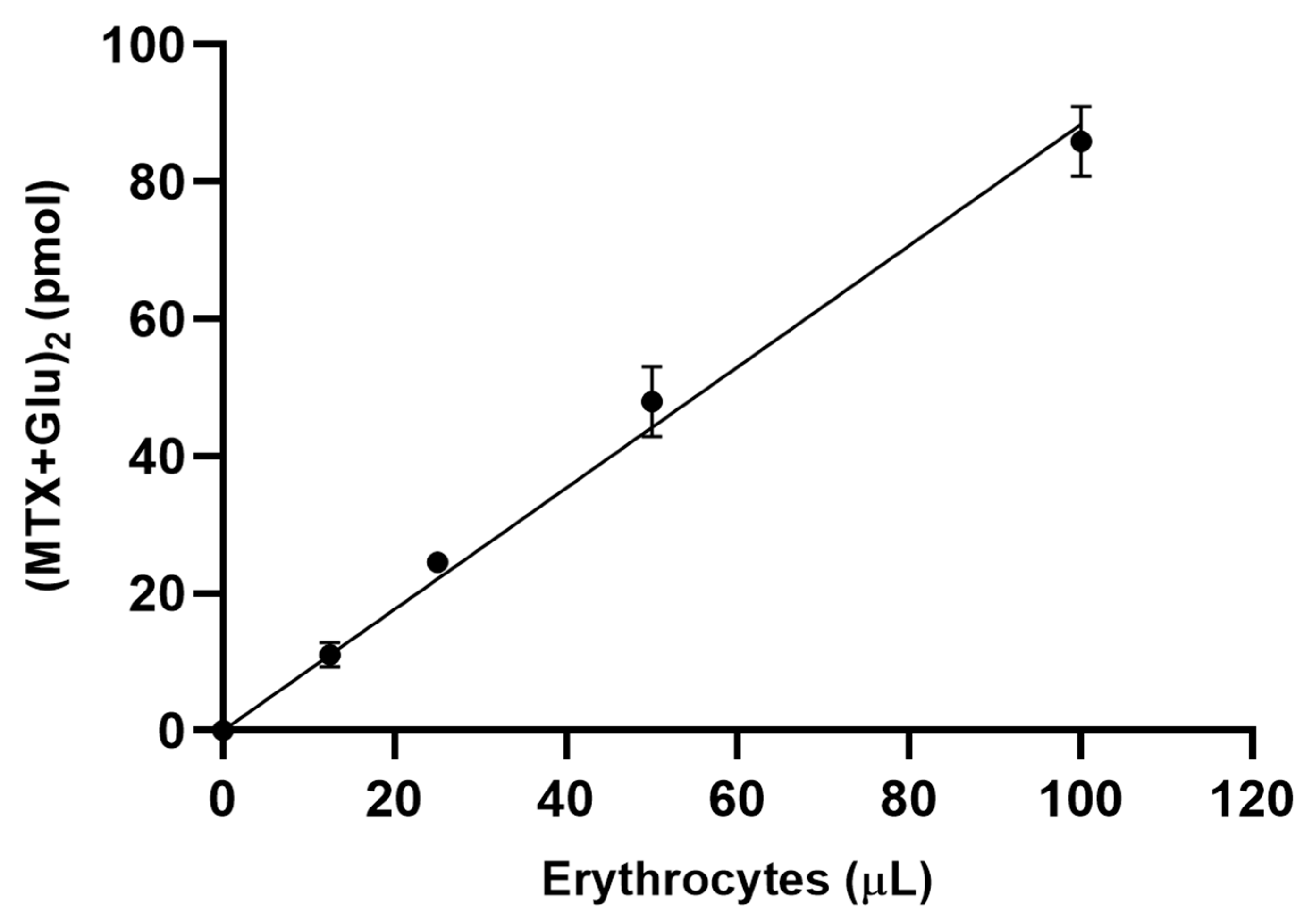
Assay conditions were optimized by incubating extracted erythrocyte samples in conditions that included an incubation period of up to four hours and a range of erythrocyte volumes from 0 to 100 µL. Regression curves for erythrocyte volume as a function of picomoles of MTX + Glu2 formation demonstrated linearity and reproducibility over the volumes tested (Figure 2). A sample volume of 25 µL was chosen for subsequent method validation based on the need to minimize the sample volume.
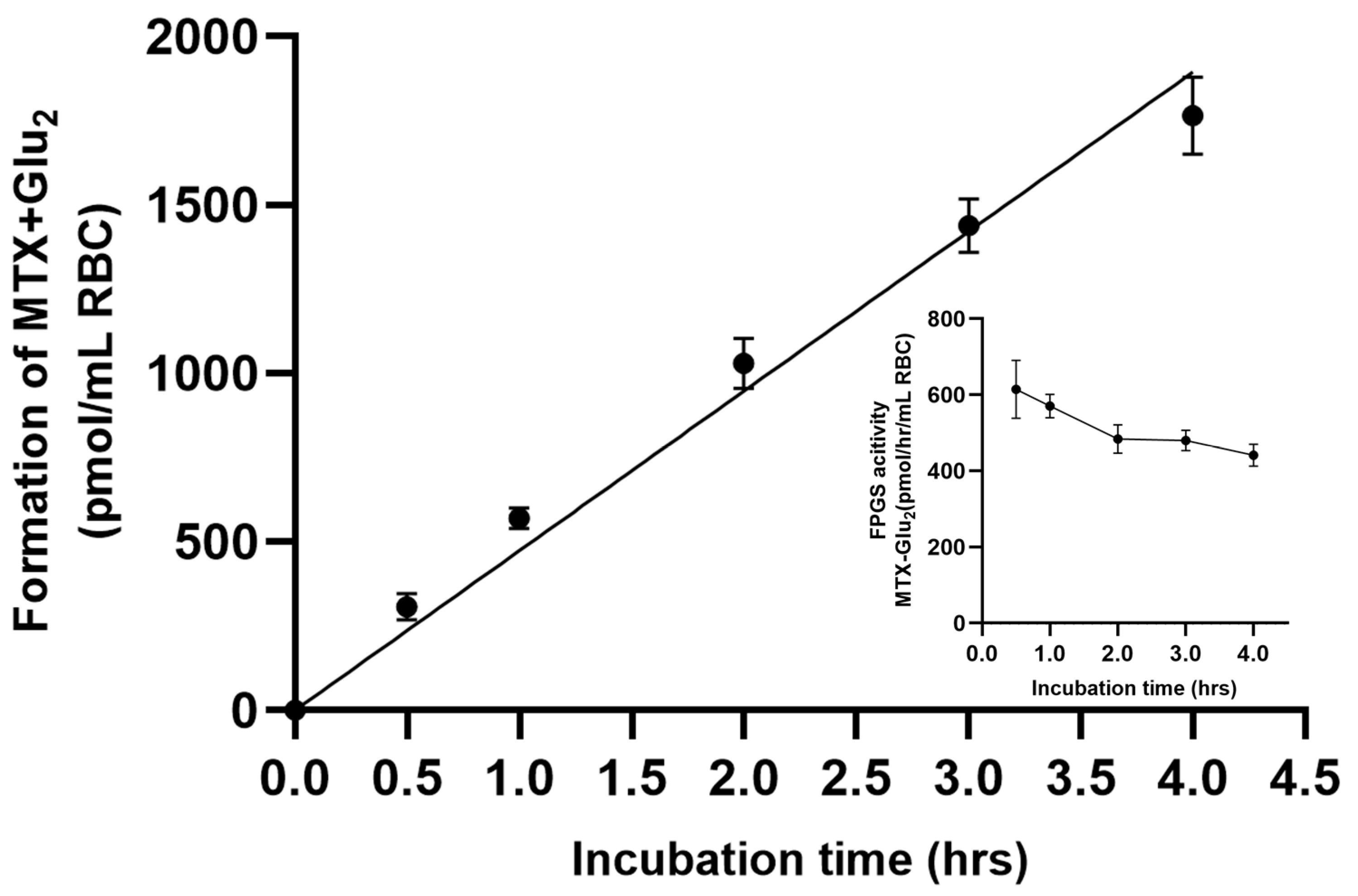
Similarly, a linear relationship was observed between product (i.e., MTX + Glu2) formation and incubation time. However, there was a slight drop-off at 4 h, as shown in Figure 3, while the regression analysis demonstrated linearity between product formation over the incubation time from 0 to 4 h via the line of best fit (R2 = 0.991). Additionally, another linear trend was observed between the rate of FPGS enzyme activity and incubation time, ranging from 30 min to 4 h. Initially, a slight fluctuation was observed in the assay due to the time needed for the enzyme to reach its optimal state. As the experiment progressed, enzyme activity gradually stabilized, indicating a more consistent trend over time. This can be observed in the inset graph of Figure 3. To ensure that the measurements remained within the linear range of the assay, a sample incubation time of 2 h was selected for further experiments.

3.2. Enzyme Kinetics Analysis

Further, FPGS enzyme kinetics were evaluated as a function of substrate concentration (MTX) using the Michaelis–Menten equation to derive kinetic constants and to compare them to previously published data. We observed that for initial MTX concentrations, the Michaelis–Menten curve follows a linear trend and slowly approaches a near-saturation (or Vmax per the Michaelis–Menten equation) level asymptotically at 250 µM (Figure 4a).
The absolute values for both Vmax and Km were estimated by fitting the data assuming Michaelis–Menton kinetics through the double-reciprocal plot or the Lineweaver–Burk plot (Figure 4b). The Lineweaver–Burk plot generated from the Michaelis–Menten curves (equation IV), for MTX, gives a mean (SD) Km of 30.3(4.8) (µM)−1 and a mean (SD) Vmax of 612(193) (pmol/h/mL)−1 is presented in Table 1. The observed Km value in our assay is significantly lower than that in the previously reported data [35,37,38,39], indicating a high affinity of the enzyme for MTX as a substrate.
Within-run and between-run precision was assessed using healthy human erythrocyte samples by measuring replicates (n = 3) over the course of the next three days. This allowed us to examine both intra-day and inter-day variability, ensuring the validation and reproducibility of the enzymatic assay. The difference in measured FPGS activity between intra-day and inter-day measurements is presented in Table 2, showing a relatively low coefficient of variation that is in the acceptable range for a bioanalytical assay [35].

3.3. FPGS Activity in Arthritis Patients

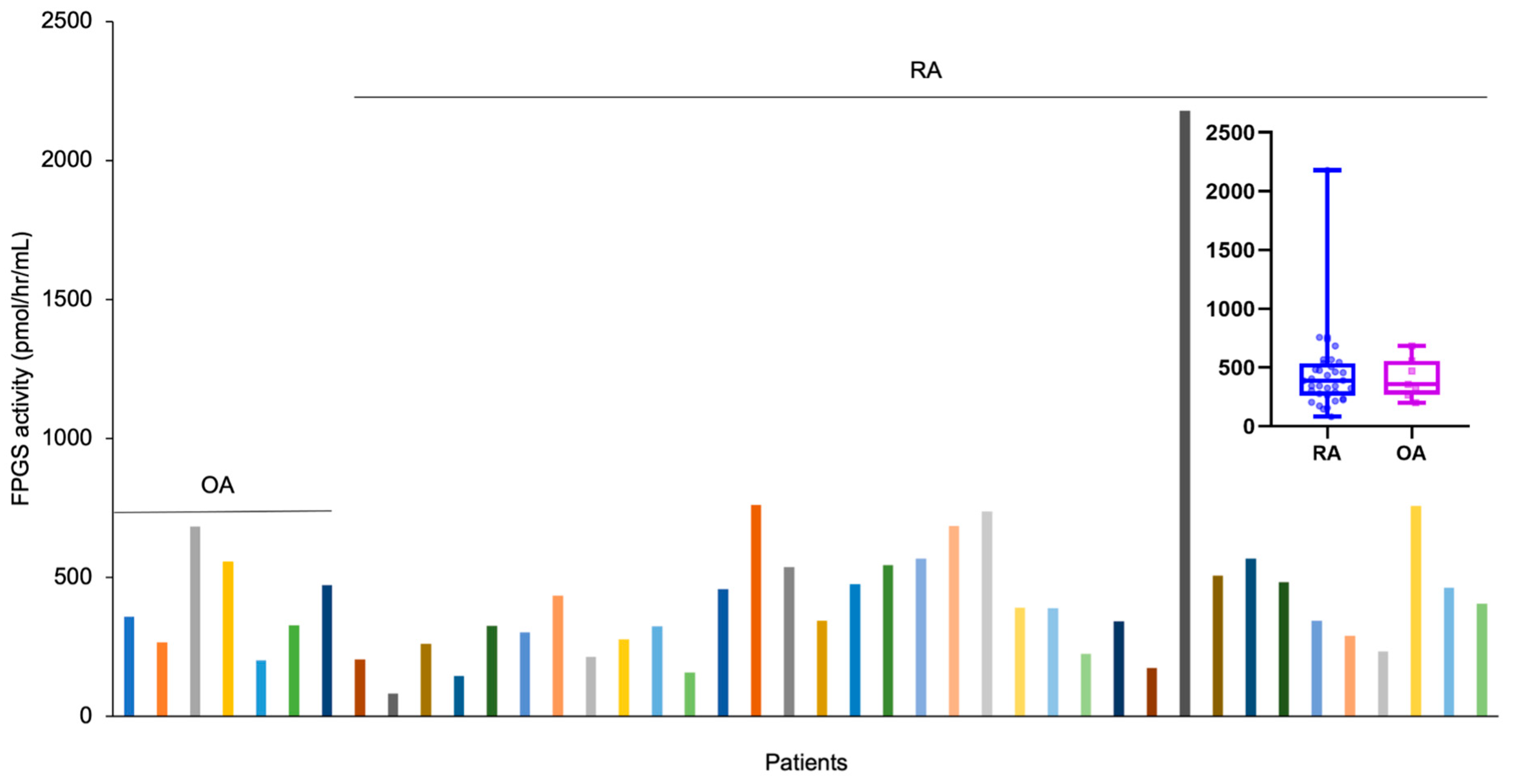
Using the optimized method, we measured FPGS activity in 42 patient erythrocyte samples from our RA biorepository, operating under the Vmax conditions with a 250 µM MTX substrate concentration and a 2 h incubation at 37 °C (Figure 5).
We determined the median [interquartile range] (M[IQR]) along with the non-parametric test values for FPGS activity values for both gender-based analysis and type of arthritis. For gender-based analysis, the M[IQR] for males was (456.15[293.14]) while that for females was (351.64[260.08]). The non-parametric test led to the finding that there were no significant (N = 40, p = 0.46) differences in enzyme activity between males and females. Similarly, for the type of arthritis, the M[IQR] for the OA was 358.08[217.07]) and that for RA was (388.87[252.18]). The non-parametric test results suggested that there were no significant (N = 42, p = 0.94) differences in enzyme activity between the two types of arthritis.

4. Discussion

In this study, we reported the development and optimization of an enzyme-based method using UHPLC-MS/MS to measure FPGS activity as a function of MTX + Glu2 formation in packed erythrocytes. The method was adapted from a FPGS-based study on human PBMCs by Muller et al. [35], hence providing the same advantages to our study in terms of being less labor-intensive, being highly sensitive, and having no requirements for any radioactive labeled FPGS substrate [35]. The instrumentation parameters, such as ionization mode, charge/mass ratio for MTX + Glu2 and MTX + Glu2-IS, ion spray volume, collision energy, declustering potential, and collision exit potential for MS/MS were consistent with the settings described by den Boer et al. [36]. The mobile phase solvent, including its concentration and pH, was also kept same as that their protocol. Subsequently, quantification was performed using a similar integration method to determine the area under the curve in counts per second. The assay’s linearity, sensitivity, and reproducibility were validated using authentic reference standards, as reported in Figure 2, Figure 3 and Figure 4.
Enzyme activity assays have several advantages over genetic assays and protein quantitative assays. Firstly, they directly measure functional activity, providing valuable insights into enzymatic function and its impact on cellular processes [40]. Additionally, enzyme activity assays offer a more direct reflection of the phenotype as they measure actual enzymatic function and activity [41,42]. Unlike genetic assays and protein quantitative assays, they can also detect post-translation modifications and other factors that influence enzyme function [43,44]. Moreover, enzyme activity assays enable the evaluation of enzyme activity and its response to environmental and physiological changes. In addition to that, using an enzyme-based cell-type assay is advantageous for the current study as enzymes can be easily identified based on the specific reactions they catalyze, unlike nucleic acids and functional proteins [45].
Unlike Muller et al.’s method, where they preferred peripheral blood mononuclear cells (PBMC’s) for studying FPGS activity, we implemented some modifications in sample processing that included a cellular lysis step in the extraction buffer since we used packed erythrocytes in our method’s development. Interestingly, there are conflicting opinions among studies regarding the most suitable cell line for quantifying FPGS activity. Some studies have chosen peripheral blood mononuclear cells (PBMCs) due to their relevance in inflammation [35], while Stoforidis et al. have suggested erythrocytes as appropriate cells due to the difficulty in collecting a sufficient quantity of peripheral blood lymphocytes [46]. However, Meeberg et al. and Barredo et al. argue that erythrocytes have substantially lower or negligible FPGS activity, making them unsuitable for studying FPGS activity [47,48].
Nevertheless, several studies have demonstrated a correlation between erythrocyte polyglutamate concentration and MTX efficacy in RA [20,46,49,50,51], acute lymphoblastic leukemia, and psoriasis [52,53]. This suggests that investigating FPGS activity in erythrocytes may remain a valid approach for the development of biomarkers. Owing to these previously reported successful studies on erythrocytes, we studied kinetic parameters (time of incubation, volume of substrate, etc.) and were successfully able to detect the formation of polyglutamation products (i.e., (MTX + Glu2))_ in erythrocytes, proving the method to be sensitive in detecting low-concentration products with acceptable precision.
The idea of this study stemmed from the successful phenotyping of thiopurine S-methyltransferase (TPMT) in patients that receive azathioprine (AZA) therapy. By phenotyping TPMT expression/activity and thus characterizing the degree of AZA metabolism, this method has been shown to improve outcomes and avoid life-threatening toxicities in patients that may receive AZA therapy [54]. Similarly, the principle of this study is to measure FPGS enzyme activity prior to initiating MTX to predict the degree of drug metabolism via polyglutamation.
Predicting FPGS enzyme activity before starting MTX treatment is important in its potential use as an a priori marker or early marker of MTX efficacy or toxicity, due to the time required to reach steady-state MTX + Glun concentrations [34,55,56]. Consequently, performing FPGS phenotyping in advance could be valuable tool for predicting MTX metabolism and treatment responses.
The efficacy of MTX is dependent on FPGS, which is responsible for the addition of glutamate residues on the drug [25,57,58,59,60]. Previous studies suggest that a longer polyglutamate product is related to a more rapid anti-inflammatory response [5,24,25,61], but can also adversely affect the immune system, whereas the absence of polyglutamation can result in decreased drug effectiveness, potentially leading to inadequate treatment outcomes [18,59,62]. This will help clinicians to both adjust patients’ MTX dosing regimens and select an alternative therapy for non-responders, effectively minimizing joint degradation due to a lack of therapeutic benefit from the insufficient polyglutamation of MTX.
In our method, we selected MTX as the substrate for several reasons, including its stability and affordability. It is worth noting that, similar to what was carried out in the study conducted by Muller et al., we ensured that the concentration of the turnover product of the FPGS reaction (MTX + Glu2) remained significantly below its Km. This was carried out to prevent it from serving as a substrate for subsequent FPGS reaction, where additional glutamic acid residues could have been added to form MTX + Glu3. Therefore, our kinetic studies focused only on the FPGS-catalyzed reaction that resulted in the addition of one glutamic acid moiety, forming the MTX + Glu2 product.
Ideally, for an analytical assay, it is not recommended that relative activity values alone be calculated; rather, a standard curve is preferred and is both an essential and logical way to extrapolate enzyme activity. Therefore, a standard curve was constructed by measuring the assay signal with standard solutions of the reaction product over a suitable range (5.0–100 nM) of MTX + Glu2 concentrations with a correlation coefficient of R2 > 0.99 and an acceptable coefficient of variation of 6%. Since most assays (such as the enzyme activity assay, kinetic assays, immunoassays, etc.) use a 25–50 µL sample volume [63,64,65,66,67], which ideally requires 1–2 h of sample incubation. However, the specific volume requirements can vary depending on the particular enzyme assay and the experimental protocol being used. We opted for the minimum recommended sample volume of 25 µL for our assay.
In our method, we also tested the time-dependent formation of the product, which is common in all enzyme-catalyzed reactions. We observed that FPGS activity was linear over an incubation time range of 0–4 h. Since it was difficult to determine sensitivity and specificity in most of the enzyme assays, we tested certain time points for our method and observed 4 h to be the optimal time before the curve levels off and becomes saturated. It is also important to note that longer incubation times in enzyme assays might lead to the consumption of the substrate, and that a change in co-factor condition, such as pH, can affect the efficiency of enzyme activity. Additionally, we know that increasing substrate concentrations increases the rate of reaction; however, at a certain concentration, any increase has no effect because the enzyme FPGS has effectively become saturated and acts at its maximum feasible rate [68,69,70]. Therefore, in our proposed condition, we observed a good turnover from substrate to product after 2 h of incubation.
After assay optimization, we tested our method on 42 clinical samples from our repository and observed the interpatient variability of FPGS activity among the patient samples. Interpatient variability has been consistently reported in previous works [25,56,57,58,71,72,73,74,75]. The variation in FPGS enzyme activity observed among patients not only reflects differences in individual affinities for the formation of polyglutamate product, but also indicates genuine biological heterogeneity. Based on our results, we did not observe any statistically significant differences in enzyme activity with respect to gender (p = 0.46) or disease subtypes (p = 0.94). However, it is important to note that the limited number of patients in each group may have impacted our ability to detect meaningful differences. While the small sample size prevents us from drawing definitive conclusions regarding the lack of significance, it does suggest that factors beyond gender and disease subtypes, such as genetics, diet, or overall health status, may play a more influential role in influencing enzyme activity during comparative analyses. An exploration of the factors underlying this variability in enzyme activity and biological heterogeneity can be conducted through demographic studies. Previous demographic studies on joint diseases such as osteoarthritis (OA) and RA have identified demographic factors that contribute to differences in disease etiology. Factors such as age, sex, ethnicity, body mass index, and smoking habits have been shown to impact diagnosis and the variability in drug responses among patients with OA and RA [76]. Although FPGS enzyme activity can be a potential biomarker for the variability in MTX metabolism, there are other factors that can also contribute to this variability, including age, MTX dosage, renal function, smoking or tobacco usage, erythrocyte folate levels, and MTX + Glun concentrations [59,77]. In addition, Inoue et al. suggest that MTX’s pharmacokinetics and pharmacodynamics may be affected by drug–drug interactions through transporters and their genetic polymorphisms, resulting in interpatient variability in efficacy [78].
In summary, we were successful in validating the linearity of ranges for volume, time, and enzyme kinetics (in Figure 2, Figure 3 and Figure 4) using the chromatographic approach. The initial evaluation and validation were further validated for FPGS activity with the arthritis samples. Despite the limited number of patients in our study, we adopted a conservative approach when interpreting the results to address potential limitations in applicability. Furthermore, in our study, we found that FPGS activity between males and females was not significant. The relatively small number of male participants (N = 6) in comparison to females (N = 34) in our study, as previously mentioned, could have been a contributing factor to the lack of statistically significant gender-related differences in enzyme activity. Therefore, larger male sample sizes in future studies may strengthen the reliability of these findings. It is noteworthy that RA exhibits gender-based variations, with a higher prevalence in women. This gender disparity can result in differences in how the disease manifests, progresses, and responds to treatments [79,80]. Considering these gender-specific characteristics is crucial for personalized and effective healthcare in RA research.
Overall, our method marks a notable step forward in the development of an enzyme assay with predictive capabilities for phenotypic assessment. Unlike the time-consuming measurement of drug metabolite levels, this FPGS assay offers the potential to predict the range of MTX metabolism in arthritis patients, encompassing low, normal, and high levels. In summary, we conducted a post hoc analysis to explore a previously non-proposed biomarker, FPGS, and its potential relationship with treatment responses in patients with arthritis.

5. Conclusions

Our current study proposes a UHPLC-MS/MS-based method for measuring FPGS activity in erythrocytes. It reveals variations among RA patients, showing potential in drug monitoring and phenotyping for RA treatment. Future investigations could explore population variations and correlations with MTX + Glun levels in RA, alongside FPGS activity differences among responsive and non-responsive RA patients.

Author Contributions

Conceptualization, R.F. and P.B.; methodology, R.F.; validation, R.F.; formal analysis, R.F. and A.K.; data curation, R.F. and P.B..; writing—original draft preparation, A.K. and M.I.A.; writing—review and editing, R.F. and P.B.; visualization, A.K.; supervision, R.F.; funding acquisition, R.F. All authors have read and agreed to the published version of the manuscript.

Funding

This research was supported by the University of Kansas, via a CTSA grant from NCATS awarded to the University of Kansas for Frontiers: University of Kansas Clinical and Translational Science Institute, KL2TR002367 (R.S.F.), and via a Kansas Institute of Precision Medicine Centers of Biomedical Research Excellence grant from NIGMS awarded to the University of Kansas Medical Center, P20GM130423 (R.S.F.).

Institutional Review Board Statement

The study was conducted in accordance with the guidelines of the Declaration of Helsinki and approved by the Institutional Review Board of The University of Kansas Medical Center.

Informed Consent Statement

Informed consent was obtained from all subjects involved in the study.

Data Availability Statement

The data presented in this study are available on request from the corresponding author.

Conflicts of Interest

The authors declare no conflict of interest.

Correction Statement

This article has been republished with a minor correction to the position of Figures. This change does not affect the scientific content of the article.

References

  1. Visentin, M.; Zhao, R.; Goldman, I.D. The Antifolates. Hematol. Oncol. Clin. N. Am. 2012, 26, 629–648. [Google Scholar] [CrossRef] [PubMed]
  2. Kremer, J.M. Methotrexate and Leflunomide: Biochemical Basis for Combination Therapy in the Treatment of Rheumatoid Arthritis. Semin. Arthritis Rheum. 1999, 29, 14–26. [Google Scholar] [CrossRef] [PubMed]
  3. Benjamin, O.; Goyal, A.; Lappin, S.L. Disease-Modifying Antirheumatic Drugs (DMARD). In StatPearls; StatPearls Publishing: Treasure Island, FL, USA, 2023. [Google Scholar]
  4. Swierkot, J.; Szechiński, J. Methotrexate in Rheumatoid Arthritis. Pharmacol. Rep. PR 2006, 58, 473–492. [Google Scholar] [PubMed]
  5. Taylor, P.C.; Balsa Criado, A.; Mongey, A.-B.; Avouac, J.; Marotte, H.; Mueller, R.B. How to Get the Most from Methotrexate (MTX) Treatment for Your Rheumatoid Arthritis Patient?-MTX in the Treat-to-Target Strategy. J. Clin. Med. 2019, 8, 515. [Google Scholar] [CrossRef]
  6. Lopez-Olivo, M.A.; Siddhanamatha, H.R.; Shea, B.; Tugwell, P.; Wells, G.A.; Suarez-Almazor, M.E. Methotrexate for Treating Rheumatoid Arthritis. Cochrane Database Syst. Rev. 2014, 2014, CD000957. [Google Scholar] [CrossRef]
  7. Dogra, S.; Singh, N.; Kumar, S.; Narang, T.; Handa, S. Comparison of Overall Efficacy and Safety of Oral versus Subcutaneous Methotrexate in Severe Psoriasis. Dermatol. Ther. 2022, 35, e15656. [Google Scholar] [CrossRef] [PubMed]
  8. Raza, K.; Buckley, C.E.; Salmon, M.; Buckley, C.D. Treating Very Early Rheumatoid Arthritis. Best Pract. Res. Clin. Rheumatol. 2006, 20, 849–863. [Google Scholar] [CrossRef] [PubMed]
  9. Bedoui, Y.; Guillot, X.; Sélambarom, J.; Guiraud, P.; Giry, C.; Jaffar-Bandjee, M.C.; Ralandison, S.; Gasque, P. Methotrexate an Old Drug with New Tricks. Int. J. Mol. Sci. 2019, 20, 5023. [Google Scholar] [CrossRef]
  10. Plant, D.; Maciejewski, M.; Smith, S.; Nair, N.; Maximising Therapeutic Utility in Rheumatoid Arthritis Consortium, the RAMS Study Group; Hyrich, K.; Ziemek, D.; Barton, A.; Verstappen, S. Profiling of Gene Expression Biomarkers as a Classifier of Methotrexate Nonresponse in Patients With Rheumatoid Arthritis. Arthritis Rheumatol. Hoboken NJ 2019, 71, 678–684. [Google Scholar] [CrossRef]
  11. Emery, P.; Pope, J.E.; Kruger, K.; Lippe, R.; DeMasi, R.; Lula, S.; Kola, B. Efficacy of Monotherapy with Biologics and JAK Inhibitors for the Treatment of Rheumatoid Arthritis: A Systematic Review. Adv. Ther. 2018, 35, 1535–1563. [Google Scholar] [CrossRef]
  12. Weinblatt, M.E. Methotrexate in Rheumatoid Arthritis: A Quarter Century of Development. Trans. Am. Clin. Climatol. Assoc. 2013, 124, 16–25. [Google Scholar]
  13. Fleischmann, R.; Schiff, M.; van der Heijde, D.; Ramos-Remus, C.; Spindler, A.; Stanislav, M.; Zerbini, C.A.F.; Gurbuz, S.; Dickson, C.; de Bono, S.; et al. Baricitinib, Methotrexate, or Combination in Patients With Rheumatoid Arthritis and No or Limited Prior Disease-Modifying Antirheumatic Drug Treatment. Arthritis Rheumatol. Hoboken NJ 2017, 69, 506–517. [Google Scholar] [CrossRef]
  14. de Rotte, M.C.F.J.; den Boer, E.; de Jong, P.H.P.; Pluijm, S.M.F.; Ćalasan, M.B.; Weel, A.E.; Huisman, A.M.; Gerards, A.H.; van Schaeybroeck, B.; Wulffraat, N.M.; et al. Methotrexate Polyglutamates in Erythrocytes Are Associated with Lower Disease Activity in Patients with Rheumatoid Arthritis. Ann. Rheum. Dis. 2015, 74, 408–414. [Google Scholar] [CrossRef]
  15. Pan, S.; Stamp, L.K.; Duffull, S.B.; Barclay, M.L.; Dalrymple, J.M.; Drake, J.; Zhang, M.; Korell, J. Assessment of the Relationship between Methotrexate Polyglutamates in Red Blood Cells and Clinical Response in Patients Commencing Methotrexate for Rheumatoid Arthritis. Clin. Pharmacokinet. 2014, 53, 1161–1170. [Google Scholar] [CrossRef]
  16. Daraghmeh, D.N.; Moghaddami, M.; Bobrovskaya, L.; Proudman, S.M.; Wiese, M.D. Quantitation of Methotrexate Polyglutamates in Human Whole Blood, Erythrocytes and Leukocytes Collected via Venepuncture and Volumetric Absorptive Micro-Sampling: A Green LC-MS/MS-Based Method. Anal. Bioanal. Chem. 2022, 414, 6029–6046. [Google Scholar] [CrossRef]
  17. Becker, M.L.; van Haandel, L.; Gaedigk, R.; Lasky, A.; Hoeltzel, M.; Stobaugh, J.; Leeder, J.S. Analysis of Intracellular Methotrexate Polyglutamates in Patients with Juvenile Idiopathic Arthritis: Effect of Route of Administration on Variability in Intracellular Methotrexate Polyglutamate Concentrations. Arthritis Rheum. 2010, 62, 1803–1812. [Google Scholar] [CrossRef]
  18. Hawwa, A.F.; AlBawab, A.; Rooney, M.; Wedderburn, L.R.; Beresford, M.W.; McElnay, J.C. Methotrexate Polyglutamates as a Potential Marker of Adherence to Long-Term Therapy in Children with Juvenile Idiopathic Arthritis and Juvenile Dermatomyositis: An Observational, Cross-Sectional Study. Arthritis Res. Ther. 2015, 17, 295. [Google Scholar] [CrossRef]
  19. van de Meeberg, M.M.; Hebing, R.C.F.; Nurmohamed, M.T.; Fidder, H.H.; Heymans, M.W.; Bouma, G.; de Bruin-Weller, M.S.; Tekstra, J.; van den Bemt, B.; de Jonge, R.; et al. A Meta-Analysis of Methotrexate Polyglutamates in Relation to Efficacy and Toxicity of Methotrexate in Inflammatory Arthritis, Colitis and Dermatitis. Br. J. Clin. Pharmacol. 2023, 89, 61–79. [Google Scholar] [CrossRef]
  20. Dervieux, T.; Zablocki, R.; Kremer, J. Red Blood Cell Methotrexate Polyglutamates Emerge as a Function of Dosage Intensity and Route of Administration during Pulse Methotrexate Therapy in Rheumatoid Arthritis. Rheumatol. Oxf. Engl. 2010, 49, 2337–2345. [Google Scholar] [CrossRef]
  21. Halilova, K.I.; Brown, E.E.; Morgan, S.L.; Bridges, S.L.; Hwang, M.-H.; Arnett, D.K.; Danila, M.I. Markers of Treatment Response to Methotrexate in Rheumatoid Arthritis: Where Do We Stand? Int. J. Rheumatol. 2012, 2012, 978396. [Google Scholar] [CrossRef]
  22. Yan, H.; Su, R.; Xue, H.; Gao, C.; Li, X.; Wang, C. Pharmacomicrobiology of Methotrexate in Rheumatoid Arthritis: Gut Microbiome as Predictor of Therapeutic Response. Front. Immunol. 2021, 12, 789334. [Google Scholar] [CrossRef]
  23. Takahashi, C.; Kaneko, Y.; Okano, Y.; Taguchi, H.; Oshima, H.; Izumi, K.; Yamaoka, K.; Takeuchi, T. Association of Erythrocyte Methotrexate-Polyglutamate Levels with the Efficacy and Hepatotoxicity of Methotrexate in Patients with Rheumatoid Arthritis: A 76-Week Prospective Study. RMD Open 2017, 3, e000363. [Google Scholar] [CrossRef]
  24. Wei, K.; Jiang, P.; Zhao, J.; Jin, Y.; Zhang, R.; Chang, C.; Xu, L.; Xu, L.; Shi, Y.; Guo, S.; et al. Biomarkers to Predict DMARDs Efficacy and Adverse Effect in Rheumatoid Arthritis. Front. Immunol. 2022, 13, 865267. [Google Scholar] [CrossRef]
  25. Becker, M.L. Using Methotrexate Metabolites to Make Clinical Decisions in JIA. Curr. Treat. Options Rheumatol. 2015, 1, 381–395. [Google Scholar] [CrossRef]
  26. Castaldo, P.; Magi, S.; Assunta Nasti, A.; Arcangeli, S.; Lariccia, V.; Alesi, N.; Tocchini, M.; Amoroso, S. Clinical Pharmacogenetics of Methotrexate. Curr. Drug Metab. 2011, 12, 278–286. [Google Scholar] [CrossRef]
  27. Brown, P.M.; Pratt, A.G.; Isaacs, J.D. Mechanism of Action of Methotrexate in Rheumatoid Arthritis, and the Search for Biomarkers. Nat. Rev. Rheumatol. 2016, 12, 731–742. [Google Scholar] [CrossRef]
  28. Stamp, L.K.; Roberts, R.L. Effect of Genetic Polymorphisms in the Folate Pathway on Methotrexate Therapy in Rheumatic Diseases. Pharmacogenomics 2011, 12, 1449–1463. [Google Scholar] [CrossRef] [PubMed]
  29. Zheng, Y.; Cantley, L.C. Toward a Better Understanding of Folate Metabolism in Health and Disease. J. Exp. Med. 2019, 216, 253–266. [Google Scholar] [CrossRef]
  30. Raz, S.; Stark, M.; Assaraf, Y.G. Folylpoly-γ-Glutamate Synthetase: A Key Determinant of Folate Homeostasis and Antifolate Resistance in Cancer. Drug Resist. Updat. 2016, 28, 43–64. [Google Scholar] [CrossRef]
  31. Shane, B. Folate chemistry and metabolism*. Clin. Res. Regul. Aff. 2001, 18, 137–159. [Google Scholar] [CrossRef]
  32. Moran, R.G. Roles of Folylpoly-Gamma-Glutamate Synthetase in Therapeutics with Tetrahydrofolate Antimetabolites: An Overview. Semin. Oncol. 1999, 26 (Suppl. S6), 24–32. [Google Scholar] [PubMed]
  33. Stranzl, T.; Wolf, J.; Leeb, B.F.; Smolen, J.S.; Pirker, R.; Filipits, M. Expression of Folylpolyglutamyl Synthetase Predicts Poor Response to Methotrexate Therapy in Patients with Rheumatoid Arthritis. Clin. Exp. Rheumatol. 2003, 21, 27–32. [Google Scholar] [PubMed]
  34. Muller, I.B.; Lin, M.; Lems, W.F.; Ter Wee, M.M.; Wojtuszkiewicz, A.; Nurmohamed, M.T.; Cloos, J.; Assaraf, Y.G.; Jansen, G.; de Jonge, R. Association of Altered Folylpolyglutamate Synthetase Pre-mRNA Splicing with Methotrexate Unresponsiveness in Early Rheumatoid Arthritis. Rheumatol. Oxf. Engl. 2021, 60, 1273–1281. [Google Scholar] [CrossRef] [PubMed]
  35. Muller, I.B.; Lin, M.; Struys, E.A.; Heydari, P.; Hebing, R.C.F.; Nurmohamed, M.T.; Van Der Laken, C.; Lems, W.F.; Cloos, J.; Jansen, G.; et al. Development and Validation of a Sensitive UHPLC-MS/MS–Based Method for the Analysis of Folylpolyglutamate Synthetase Enzymatic Activity in Peripheral Blood Mononuclear Cells: Application in Rheumatoid Arthritis and Leukemia Patients. Ther. Drug Monit. 2019, 41, 598–606. [Google Scholar] [CrossRef]
  36. den Boer, E.; Meesters, R.J.W.; Van Zelst, B.D.; Luider, T.M.; Hazes, J.M.W.; Heil, S.G.; de Jonge, R. Measuring Methotrexate Polyglutamates in Red Blood Cells: A New LC-MS/MS-Based Method. Anal. Bioanal. Chem. 2012, 405, 1673–1681. [Google Scholar] [CrossRef]
  37. Liani, E.; Rothem, L.; Bunni, M.A.; Smith, C.A.; Jansen, G.; Assaraf, Y.G. Loss of Folylpoly-?-Glutamate Synthetase Activity Is a Dominant Mechanism of Resistance to Polyglutamylation-Dependent Novel Antifolates in Multiple Human Leukemia Sublines. Int. J. Cancer 2003, 103, 587–599. [Google Scholar] [CrossRef]
  38. Leil, T.A.; Endo, C.; Adjei, A.A.; Dy, G.K.; Salavaggione, O.E.; Reid, J.R.; Ames, M.M.; Adjei, A.A. Identification and Characterization of Genetic Variation in the Folylpolyglutamate Synthase Gene. Cancer Res. 2007, 67, 8772–8782. [Google Scholar] [CrossRef]
  39. Chen, L.; Qi, H.; Korenberg, J.; Garrow, T.A.; Choi, Y.-J.; Shane, B. Purification and Properties of Human Cytosolic Folylpoly-γ-Glutamate Synthetase and Organization, Localization, and Differential Splicing of Its Gene. J. Biol. Chem. 1996, 271, 13077–13087. [Google Scholar] [CrossRef]
  40. Kovarik, M.L.; Allbritton, N.L. Measuring Enzyme Activity in Single Cells. Trends Biotechnol. 2011, 29, 222–230. [Google Scholar] [CrossRef] [PubMed]
  41. Lee, O.J.; Kim, S.-J.; Sohn, Y.B.; Park, H.-D.; Lee, S.-Y.; Kim, C.-H.; Ko, A.-R.; Yook, Y.-J.; Lee, S.-J.; Park, S.W.; et al. A Study of the Relationship between Clinical Phenotypes and Plasma Iduronate-2-Sulfatase Enzyme Activities in Hunter Syndrome Patients. Korean J. Pediatr. 2012, 55, 88. [Google Scholar] [CrossRef]
  42. Huen, K.; Richter, R.; Furlong, C.; Eskenazi, B.; Holland, N. Validation of PON1 Enzyme Activity Assays for Longitudinal Studies. Clin. Chim. Acta 2009, 402, 67–74. [Google Scholar] [CrossRef]
  43. Wang, S.; Ramamurthy, D.; Tan, J.; Liu, J.; Yip, J.; Chua, A.; Yu, Z.; Lim, T.K.; Lin, Q.; Pines, O.; et al. Post-Translational Modifications of Fumarase Regulate Its Enzyme Activity and Function in Respiration and the DNA Damage Response. J. Mol. Biol. 2020, 432, 6108–6126. [Google Scholar] [CrossRef]
  44. Härmä, H.; Tong-Ochoa, N.; Van Adrichem, A.J.; Jelesarov, I.; Wennerberg, K.; Kopra, K. Toward Universal Protein Post-Translational Modification Detection in High Throughput Format. Chem. Commun. 2018, 54, 2910–2913. [Google Scholar] [CrossRef] [PubMed]
  45. Bisswanger, H. Enzyme Assays. Perspect. Sci. 2014, 1, 41–55. [Google Scholar] [CrossRef]
  46. Angelis-Stoforidis, P.; Vajda, F.J.; Christophidis, N. Methotrexate Polyglutamate Levels in Circulating Erythrocytes and Polymorphs Correlate with Clinical Efficacy in Rheumatoid Arthritis. Clin. Exp. Rheumatol. 1999, 17, 313–320. [Google Scholar] [PubMed]
  47. Van De Meeberg, M.M.; Seinen, M.L.; Fidder, H.H.; Lin, M.; Oldenburg, B.; De Boer, N.K.; Bouma, G.; De Jonge, R.; Bulatović Ćalasan, M., on behalf of the Dutch Initiative on Crohn and Colitis (ICC). Subcutaneous Administration, Higher Age and Lower Renal Function Are Associated with Erythrocyte Methotrexate Accumulation in Crohn’s Disease: A Cross-Sectional Study. BMC Gastroenterol. 2022, 22, 365. [Google Scholar] [CrossRef]
  48. Barredo, J.; Moran, R.G. Determinants of Antifolate Cytotoxicity: Folylpolyglutamate Synthetase Activity during Cellular Proliferation and Development. Mol. Pharmacol. 1992, 42, 687–694. [Google Scholar] [PubMed]
  49. Dervieux, T.; Furst, D.; Lein, D.O.; Capps, R.; Smith, K.; Walsh, M.; Kremer, J. Polyglutamation of Methotrexate with Common Polymorphisms in Reduced Folate Carrier, Aminoimidazole Carboxamide Ribonucleotide Transformylase, and Thymidylate Synthase Are Associated with Methotrexate Effects in Rheumatoid Arthritis. Arthritis Rheum. 2004, 50, 2766–2774. [Google Scholar] [CrossRef] [PubMed]
  50. Dervieux, T. Pharmacogenetic and Metabolite Measurements Are Associated with Clinical Status in Patients with Rheumatoid Arthritis Treated with Methotrexate: Results of a Multicentred Cross Sectional Observational Study. Ann. Rheum. Dis. 2005, 64, 1180–1185. [Google Scholar] [CrossRef]
  51. Hornung, N.; Ellingsen, T.; Attermann, J.; Stengaard-Pedersen, K.; Poulsen, J.H. Patients with Rheumatoid Arthritis Treated with Methotrexate (MTX): Concentrations of Steady-State Erythrocyte MTX Correlate to Plasma Concentrations and Clinical Efficacy. J. Rheumatol. 2008, 35, 1709–1715. [Google Scholar] [PubMed]
  52. Chládek, J.; Simková, M.; Vanecková, J.; Hroch, M.; Chládkova, J.; Martínková, J.; Vávrová, J.; Beránek, M. The Effect of Folic Acid Supplementation on the Pharmacokinetics and Pharmacodynamics of Oral Methotrexate during the Remission-Induction Period of Treatment for Moderate-to-Severe Plaque Psoriasis. Eur. J. Clin. Pharmacol. 2008, 64, 347–355. [Google Scholar] [CrossRef]
  53. Schmiegelow, K.; Schrøder, H.; Gustafsson, G.; Kristinsson, J.; Glomstein, A.; Salmi, T.; Wranne, L. Risk of Relapse in Childhood Acute Lymphoblastic Leukemia Is Related to RBC Methotrexate and Mercaptopurine Metabolites during Maintenance Chemotherapy. Nordic Society for Pediatric Hematology and Oncology. J. Clin. Oncol. 1995, 13, 345–351. [Google Scholar] [CrossRef] [PubMed]
  54. TPMT Testing before Azathioprine Therapy? Drug Ther. Bull. 2009, 47, 9–12. [CrossRef]
  55. Danila, M.I.; Hughes, L.B.; Brown, E.E.; Morgan, S.L.; Baggott, J.E.; Arnett, D.K.; Bridges, S.L. Measurement of Erythrocyte Methotrexate Polyglutamate Levels: Ready for Clinical Use in Rheumatoid Arthritis? Curr. Rheumatol. Rep. 2010, 12, 342–347. [Google Scholar] [CrossRef]
  56. Muller, I.B.; Hebing, R.F.; Jansen, G.; Nurmohamed, M.T.; Lems, W.F.; Peters, G.J.; De Jonge, R. Personalized Medicine in Rheumatoid Arthritis: Methotrexate Polyglutamylation Revisited. Expert Rev. Precis. Med. Drug Dev. 2018, 3, 331–334. [Google Scholar] [CrossRef]
  57. den Boer, E. Therapeutic Drug-Monitoring of Methotrexate-Polyglutamates in Rheumatoid Arthritis; Erasmus University Rotterdam: Rotterdam, The Netherlands, 2014. [Google Scholar]
  58. Dalrymple, J.M.; Stamp, L.K.; O’Donnell, J.L.; Chapman, P.T.; Zhang, M.; Barclay, M.L. Pharmacokinetics of Oral Methotrexate in Patients with Rheumatoid Arthritis. Arthritis Rheum. 2008, 58, 3299–3308. [Google Scholar] [CrossRef]
  59. Yamamoto, T.; Shikano, K.; Nanki, T.; Kawai, S. Folylpolyglutamate Synthase Is a Major Determinant of Intracellular Methotrexate Polyglutamates in Patients with Rheumatoid Arthritis. Sci. Rep. 2016, 6, 35615. [Google Scholar] [CrossRef]
  60. Owen, S.A.; Hider, S.L.; Martin, P.; Bruce, I.N.; Barton, A.; Thomson, W. Genetic Polymorphisms in Key Methotrexate Pathway Genes Are Associated with Response to Treatment in Rheumatoid Arthritis Patients. Pharmacogenom. J. 2013, 13, 227–234. [Google Scholar] [CrossRef]
  61. Pasnoor, M.; Heim, A.J.; Herbelin, L.; Statland, J.; Dimachkie, M.M.; Becker, M.; Barohn, R.J.; Methotrexate in MG Investigators of the Muscle Group Study. Methotrexate Polyglutamation in a Myasthenia Gravis Clinical Trial. Kans. J. Med. 2020, 13 (Suppl. S2), 10–13. [Google Scholar] [CrossRef]
  62. Zarou, M.M.; Vazquez, A.; Vignir Helgason, G. Folate Metabolism: A Re-Emerging Therapeutic Target in Haematological Cancers. Leukemia 2021, 35, 1539–1551. [Google Scholar] [CrossRef]
  63. Foldesi, B. Guide to Enzyme Unit Definitions and Assay Design. Biomol GmbH—Life Science Shop. Available online: https://www.biomol.com/resources/biomol-blog/guide-to-enzyme-unit-definitions-and-assay-design (accessed on 13 June 2023).
  64. Restriction Enzyme Resource Guide. Promega.com. Available online: https://www.promega.com/resources/guides/nucleic-acid-analysis/restriction-enzyme-resource/ (accessed on 13 June 2023).
  65. New England Biolabs. Optimizing Restriction Endonuclease Reactions|NEB. Neb.com. Available online: https://www.neb.com/tools-and-resources/usage-guidelines/optimizing-restriction-endonuclease-reactions (accessed on 13 June 2023).
  66. Cox, K.L.; Devanarayan, V.; Kriauciunas, A.; Manetta, J.; Montrose, C. Immunoassay Methods. Nih.gov. Available online: https://www.ncbi.nlm.nih.gov/books/NBK92434/ (accessed on 13 June 2023).
  67. Cox, K.L.; Devanarayan, V.; Kriauciunas, A.; Manetta, J.; Montrose, C.; Sittampalam, S. Immunoassay Methods. In Assay Guidance Manual; Markossian, S., Grossman, A., Brimacombe, K., Arkin, M., Auld, D., Austin, C., Baell, J., Chung, T.D.Y., Coussens, N.P., Dahlin, J.L., et al., Eds.; Eli Lilly & Company and the National Center for Advancing Translational Sciences: Bethesda, MD, USA, 2004. [Google Scholar]
  68. Voet, D.; Voet, J.G.; Pratt, C.W. Fundamentals of Biochemistry: Life at the Molecular Level, 5th ed.; Wiley: Hoboken, NJ, USA, 2016. [Google Scholar]
  69. Boyer, R.F. Concepts in Biochemistry, 2nd ed.; John Wiley & Sons, Inc.: New York, NY, USA, 2002. [Google Scholar]
  70. Punekar, N.S. Enzymes: Catalysis, Kinetics and Mechanisms; Springer: Singapore, 2018. [Google Scholar]
  71. CTG Labs—NCBI. clinicaltrials.gov. Available online: https://clinicaltrials.gov/ct2/show/NCT00695188 (accessed on 2 October 2023).
  72. Becker, M.L. Role of Methotrexate in Juvenile Idiopathic Arthritis: Where We Have Been and Where We Are Going. Int. J. Clin. Rheumatol. 2013, 8, 123–135. [Google Scholar] [CrossRef]
  73. Den Boer, E.; De Rotte, M.C.J.F.; Pluijm, S.M.F.; Heil, S.G.; Hazes, J.M.; De Jonge, R. Determinants of Erythrocyte Methotrexate Polyglutamate Levels in Rheumatoid Arthritis. J. Rheumatol. 2014, 41, 2167–2178. [Google Scholar] [CrossRef]
  74. Jansen, G. Folate Supplementations for Methotrexate Therapies in Cancer and Arthritis: Rationales Revisited. J. Mol. Clin. Med. 2022, 5, 1. [Google Scholar] [CrossRef]
  75. Qiu, Q.; Huang, J.; Shu, X.; Fan, H.; Zhou, Y.; Xiao, C. Polymorphisms and Pharmacogenomics for the Clinical Efficacy of Methotrexate in Patients with Rheumatoid Arthritis: A Systematic Review and Meta-Analysis. Sci. Rep. 2017, 7, 44015. [Google Scholar] [CrossRef] [PubMed]
  76. Mohammed, A.; Alshamarri, T.; Adeyeye, T.; Lazariu, V.; McNutt, L.-A.; Carpenter, D.O. A Comparison of Risk Factors for Osteo- and Rheumatoid Arthritis Using NHANES Data. Prev. Med. Rep. 2020, 20, 101242. [Google Scholar] [CrossRef]
  77. Stamp, L.K.; O’Donnell, J.L.; Chapman, P.T.; Zhang, M.; Frampton, C.; James, J.; Barclay, M.L. Determinants of Red Blood Cell Methotrexate Polyglutamate Concentrations in Rheumatoid Arthritis Patients Receiving Long-Term Methotrexate Treatment. Arthritis Rheum. 2009, 60, 2248–2256. [Google Scholar] [CrossRef] [PubMed]
  78. Inoue, K.; Yuasa, H. Molecular Basis for Pharmacokinetics and Pharmacodynamics of Methotrexate in Rheumatoid Arthritis Therapy. Drug Metab. Pharmacokinet. 2014, 29, 12–19. [Google Scholar] [CrossRef]
  79. Intriago, M.; Maldonado, G.; Cárdenas, J.; Ríos, C. Clinical Characteristics in Patients with Rheumatoid Arthritis: Differences between Genders. Sci. World J. 2019, 2019, 8103812. [Google Scholar] [CrossRef]
  80. van Vollenhoven, R.F. Sex Differences in Rheumatoid Arthritis: More than Meets the Eye. BMC Med. 2009, 7, 12. [Google Scholar] [CrossRef]
Figure 1. A reaction scheme representing the FPGS-mediated conversion of methotrexate into methotrexate polyglutamate. Here, “n” represents the number of glutamyl residues that can be sequential added. It can range from one to six residues with n = 1 representing the parent form of MTX.
Futurepharmacol 03 00049 g001
Figure 2. Validation of the formation of MTX + Glu2 in picomoles as a function of erythrocyte volume (µL). Triplicate samples were evaluated for each sample volume, and MTX + Glu2 formation is represented as the mean and standard deviation.
Futurepharmacol 03 00049 g002
Figure 3. The formation rate of MTX + Glu2 (picomole/mL of RBC) in erythrocytes, as measured via UHPLC-MS/MS, validated by observing the increase in incubation time (hours). Each data point corresponds to a set of triplicate time points of sample incubation. The error bars indicate the standard deviation of the incubation data sets from the mean for that set. Inset: graph illustrating FPGS activity (pmol/h/mL of RBC) as a function of incubation time (0.5–4 h).
Futurepharmacol 03 00049 g003
Figure 4. Graphs representing the enzyme kinetics analysis, where the Michaelis–Menten curve indicates the saturation point of FPGS and its maximum efficiency at a specific MTX concentration. Each curve point represents the mean of three substrate concentration data points from three independent experimental evaluations, enabling slope calculation for the initial reaction rate at different substrate concentrations. The error bars in the curve indicate deviations from the mean for each substrate concentration set. The linear transformation of the Michaelis–Menten curve is represented by the Lineweaver–Burk plot. (a) FPGS activity (pmol of MTX + Glu2/h/mL RBC) as a function of substrate concentration, e.g., of methotrexate (µM), represented through a Michaelis–Menten plot; (b) Lineweaver–Burk plot depicting 1/FPGS activity (1/V) as a function of 1/substrate (1/MTX concentration).
Futurepharmacol 03 00049 g004
Figure 5. Histogram showing the distribution of the FPGS activity analyses of erythrocytes from 42 MTX-naïve RA (N = 35) and osteoarthritis (N = 7) patients. Inset: a boxplot displaying the distribution of FPGS activity.
Futurepharmacol 03 00049 g005
Table 1. FPGS enzyme kinetics as measured by varying MTX concentrations in triplicate over three independent experimental evaluations.
Kinetic ParametersMeanStandard Deviation
Vmax (pmol/h/mL)−1611.95193.36
Km (µM)−130.294.81
Table 2. Between-run imprecision experiments for samples (n = 3) measured by comparing replicates independently on three separate days.
Intra-DayInter-Day
Day-1Day-2Day-3Combined
Average (pmol/h/mL)754.9599.6642.2665.6
Standard deviation (%)3.037.903.1012.05
Disclaimer/Publisher’s Note: The statements, opinions and data contained in all publications are solely those of the individual author(s) and contributor(s) and not of MDPI and/or the editor(s). MDPI and/or the editor(s) disclaim responsibility for any injury to people or property resulting from any ideas, methods, instructions or products referred to in the content.