Review

Therapeutic Potential of Mitotic Kinases’ Inhibitors in Cancers of the Gastrointestinal System

by
Aadil Javed
1,†,
Gianluca Malagraba
2,†,
Mahdieh Yarmohammadi
3,†,
Catalina M. Perelló-Reus
2,
Carles Barceló
2 and
Teresa Rubio-Tomás
4,*
1
Department of Bioengineering, Faculty of Engineering, Cancer Biology Laboratory, Ege University, Izmir 35040, Turkey
2
Translational Pancreatic Cancer Oncogenesis Group, Health Research Institute of the Balearic Islands (IdISBA), 07120 Palma de Mallorca, Spain
3
Department of Biology, Faculty of Sciences, Central Tehran Branch, Islamic Azad University, Tehran 33817-74895, Iran
4
School of Medicine, University of Crete, 70013 Herakleion, Crete, Greece
*
Author to whom correspondence should be addressed.
These authors contributed equally to this work.
Future Pharmacol. 2022, 2(3), 214-237; https://doi.org/10.3390/futurepharmacol2030015
Submission received: 9 May 2022 / Revised: 21 June 2022 / Accepted: 24 June 2022 / Published: 30 June 2022
(This article belongs to the Special Issue Feature Papers in Future Pharmacology)

Abstract

:
Mitosis entails mechanistic changes required for maintaining the genomic integrity in all dividing cells. The process is intricate and temporally and spatially regulated by the ordered series of activation and de-activation of protein kinases. The mitotic kinases ensure the stepwise progression of entry into mitosis after the G2 phase of the cell cycle, followed by prophase, pro-metaphase, metaphase, anaphase, telophase, and subsequently cytokinesis and birth of two daughter cells with equal segregation and distribution of the genome. The major mitotic kinases include cyclin-dependent kinase 1 (CDK1), Aurora A and B Kinases, and Polo-Like-Kinase 1 (PLK1), among others. Overexpression of some of these kinases has been reported in many cancers as the mitotic fidelity and genome integrity are interlinked and dependent on these regulators, the native irregularities in these factors can be targeted as therapeutic strategies for various cancers. Here, we report and summarize the recent updates on the literature describing the various mitotic inhibitors targeting kinases, which can be used as potential therapeutic interventions for gastrointestinal cancers including gastric cancer, liver cancer, pancreatic cancer and colorectal cancer.

1. Introduction

The division of living cells into identical daughter cells requires mitosis and cytokinesis which together bring out an elegant dance entailing exquisite mechanisms involving many cellular machines including protein complexes and other factors and enzymes called kinases [1]. The process of mitosis has been studied in detail and reviewed [2,3]. Briefly, the life cycle of a cell consists of interphase (which is considered the time period between two successive mitoses) and mitosis. During mitosis, the cells go through a round of division. Between cell division rounds, cells duplicate their DNA during the interphase and double the organelles and cellular materials. Specific molecular events take place when cells enter into mitosis, where equal segregation of DNA content in the form of chromosomes to two daughter nuclei occurs, followed by cytokinesis causing cell division and generation of daughter cells [4]. Therefore, the cells have tightly regulated mechanisms aiming to maintain genome integrity. Failure of these mechanisms leads to chromosomal aberrations, aneuploidy, and DNA damage that are passed to the progeny. Moreover, the deregulated cell cycle along with the genome instability characterizes cancers or malignant phenotypes of cancers [5].
The effective strategies to combat cancer, therefore, involve inducing aberrant mitosis to trigger apoptosis in dividing cells. These strategies are considered relatively selective as cancer cell populations rapidly divide and become susceptible to therapy if mitosis is targeted specifically [6]. However, there are some complications with such strategies. For instance, strategies targeting the microtubules can inhibit and trigger the death of healthy non-dividing cells such as neurons [7]. Therefore, a new generation of anti-mitotic agents that can target the protein kinases involved in the regulation of mitosis has been developed [8]. For cell cycle progression, there are many kinases such as cyclin-dependent kinases (CDKs) functioning in intra-cellular environments. The master regulator kinase is CDK1, which is a mitotic kinase that interacts with cyclin B to become active and phosphorylates its substrates in mitotic progression [9,10]. Furthermore, there are other pivotal kinases that coordinate their activities to maintain the genomic integrity in normal cell division, which can be targeted to provide therapeutic efficacy against cancers. These kinases include two families of kinases; Polo-like kinases and Aurora kinases [11].
Aurora A overexpression in many cancers has been reported including cervical cancer, neuroblastoma, prostate, ovarian, colon, and breast cancers. The overexpression of Aurora A has been associated with multinucleation and cytokinesis failure induced by centrosome amplification [12]. Aurora A is considered to be regulated by p53. Indeed, cells lacking p53 exhibited chromosome instability, higher centrosome numbers, and higher aneuploidy due to failed rounds of cellular division [13,14]. Moreover, in cells possessing p53, Aurora A overexpression resulted in tetraploidy along apoptosis and growth arrest, consistent with activation of the post-mitotic checkpoint. The mechanism through which p53 inhibits Aurora A is by inducing the localization of a known inhibitor of Aurora A called GADD45a at centrosomes in response to DNA damage [15].
Similar to Aurora A, Aurora B overexpression has also been reported in cancers and is attributed as either effect or cause of tumorigenesis [16]. The higher Aurora-B expression is linked to its mislocalization within cells that cause the promotion of growth and tetraploidy, aneuploidy, near-diploids and chromosomal instability. The extent of overexpression of Aurora-B has a direct relationship with cancer progression. The tight regulation of Aurora-B in mitosis provides a balance for the cells to go through normal cell division and any perturbation in Aurora B can lead to cells being susceptible to chromosomal aberrations [17].
Polo Like Kinase 1 (PLK1) is another mitotic kinase that is one of the major regulators of the progression of the cell cycle, and its levels and kinase activity are intricately regulated for ensuring genome integrity [18]. A variety of human cancers exhibit overexpression of PLK1, which contributes to tumorigenesis. The overexpression of PLK1 is reported to favor proliferation by inducing the transcription of FoxM1 and its downstream gene targets [19]. Hyperactivity of PLK1 leads to downregulation of the G2 checkpoint signaling pathways and the entry into mitosis without stringent DNA damage response and resultant acute DNA damage [20]. Elevated levels of PLK1 give rise to cytokinesis failure resulting from multinucleation and centrosome amplification and subsequently causing cellular transformation and deleterious aneuploidy. Invasion of extracellular matrix upon PLK1 overexpression is one of the mechanisms that cells utilize to transition towards malignancy [21].
Here we report the advances made in the field of targeting of the mitotic kinases as potential therapeutic agents in the last five years. The review is focused on summarizing the role of mitotic kinases and their involvement in the progression of gastrointestinal cancers along with various inhibitors that have been utilized in preclinical and clinical settings to treat these cancers.

2. Gastric Cancer

Gastric Cancer (GC) is one of the most prevalent cancers, with a poor prognosis and a high incidence of disease-related deaths. Scientific advances in the treatment of GC have global implications, even in low-incidence countries [22]. The biological differences in tumors between Eastern and Western countries define standard treatment regimens based on international trials. Systemic chemotherapy, radiotherapy, surgery, immunotherapy, combined treatments, and targeted therapy have proven efficacy in GC [23].

2.1. Standard-of-Care Treatment of Gastric Cancer

Surgery is the gold standard for GC treatment without distant metastases. The optimization of endoscopic resection and minimally invasive surgery has significantly impacted treatment strategies for GC over the last few decades [24]. Surgery should be performed in the time window when the tumor is responsive to chemotherapy. Preoperative and postoperative chemotherapy is commonly used to reduce tumor volume. Ychou et al. showed that the postoperative adjuvant chemotherapy based on the fluorouracil regimen significantly reduced mortality in GC patients compared to surgery alone [25]. In addition, chemotherapy increased overall 5-year survival from 49.6% to 55.3%, and oral administration of fluoropyrimidines may also be effective in advanced GC [26]. Furthermore, radiation therapy uses high-energy rays or particles to eliminate cancer cells and is combined with chemotherapy (chemoradiation) to improve survival outcomes for patients with GC [27].
The main treatment options based on the molecular properties of gastric tumors are ramucirumab and trastuzumab, which target vascular endothelial growth factor receptor 2 (VEGFR2) and Erb-B2 receptor tyrosine kinase 2 (HER2), respectively. De Vita et al. compared trastuzumab plus chemotherapy (i.e., cisplatin, capecitabine) with chemotherapy alone in patients with advanced HER2-positive stomach cancer in a phase II trial. They demonstrated that the ramucirumab and paclitaxel combination was the most appropriate treatment for patients who received first-line therapy with trastuzumab [28]. Hence, there is an urgent need to investigate new treatment strategies, including the identification of novel biomarkers for patient stratification, but also specific pharmacological drugs for GC.

2.2. General Overview of Mitotic Kinases in Gastric Cancer

2.2.1. Cyclin-Dependent Kinases (CDKs)

Due to the critical role of mitotic kinases in GC progress, they may serve as an attractive target for molecular targeting cancer therapy. CDKs are cell cycle-dependent kinases that regulate cell cycle progression and response to DNA damage and are over-expressed in many cancers as mentioned previously.
Tang et al. have reported that CDK2 modulates aerobic glycolysis in GC cell lines since CDK2 knockdown reduced glycolytic mRNA levels. Sirtuin 5 (SIRT5) is a mitochondria-localized tumor suppressor that plays a negative role in metabolic reprogramming. CDK2 knockdown increased SIRT5 mRNA expression levels in SGC-7901 and MGC-803 cells. In summary, the authors uncovered new roles for CDK2 and SIRT5 in GC, that could potentially be targeted to improve the overall survival of GC patients [29].

2.2.2. Aurora Kinases

Aurora kinases are a family of serine/threonine kinases that regulate G2/M phase transitions through several key factors. Compelling data indicate that the G2/M transition is promoted by Aurora kinase [30]. Ding and colleagues reported that Aurora B and cyclin B1 are co-expressed during the G2/M phase in GC cells. In addition, they demonstrated that, in the presence of high levels of cyclin B1, Aurora B was upregulated during the G2/M phase. Taken together, their results show that Aurora B interacts with Cell-cycle Related and Expression-elevated Protein in Tumor (CREPT) to modulate the expression of Cyclin B1 during G2 phase [31].

2.2.3. Polo-Like Kinase 1

Polo-like kinase (PLK) family plays a critical role in regulating cell cycle, DNA synthesis and p53 transactivation. Their overexpression is involved in the pathogenesis of multiple human cancers [32]. Dang et al. discovered that PLK1 was elevated upon transfection of GC cell lines with anti-miR-505. Furthermore, they found that miR-505 directly targets Polo-like kinase 1 (PLK1) to regulate its expression levels in GC cell lines [33].
Most importantly, PLK1 has been identified as having a great effect on cell division and maintaining genomic stability in mitosis. Indeed, the expression of PLK1 is increased in GC tissues and cells. Furthermore, PLK1 inhibition resulted in cell cycle G2-phase arrest and inhibited the proliferation, migration, and apoptosis of GC cells, whereas its overexpression had the opposite effect. Moreover, inhibition of PLK1 reduced the activation of Mitogen-activated protein kinase (MEK) and Extracellular Signal-Regulated Kinases (ERK) pathway [34].
Similarly, Cai et al. suggested that PLK1 is an essential positive regulator of GC cell migration, invasion and epithelial-mesenchymal transition (EMT). They reported that most GC cell lines have higher PLK1 mRNA and protein levels compared to human gastric mucosal epithelial cell line (GES1). Silencing intracellular PLK1 expression by siRNA demonstrated the involvement of PLK1 in migration of both SGC7901 and MKN28 cells. Hence, overexpression of PLK1 promotes the EMT program [35].

2.2.4. Wee1-Like Protein Kinase (WEE1)

WEE1 kinase is a member of the Serine/Threonine protein kinase family and regulates the DNA damage checkpoint (G2/M cell-cycle checkpoint) that allows DNA repair before mitotic entry. Notably, the expression of WEE1 is significantly high in cancer cells [36].
Kim et al. have investigated the tumorigenic role of WEE1 in GC, and they identified high WEE1 expression and WEE1 protein secretion in GC cell lines. Furthermore, they transfected GC lines with siRNA against WEEI to demonstrate the effect of WEE1 silencing on cell viability, concluding that cell viability was decreased in WEE1 siRNA-transfected cells [37].

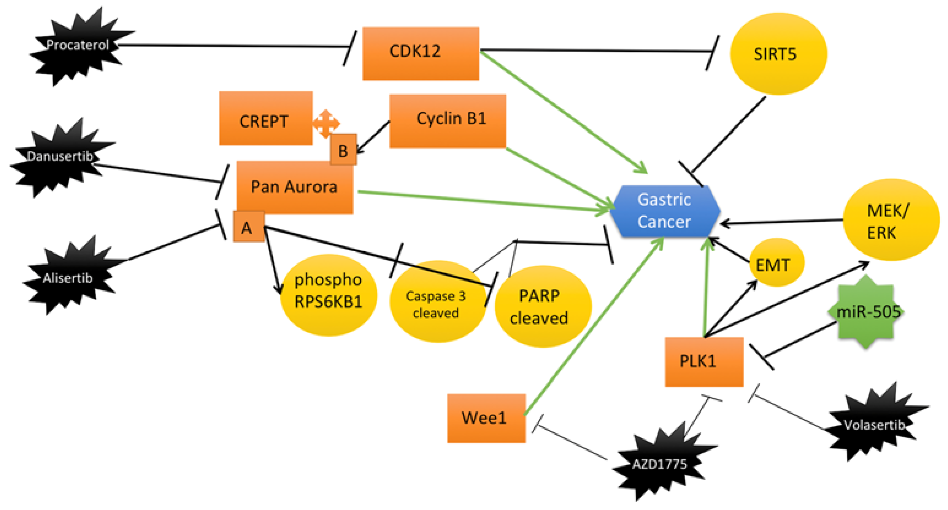
2.3. Recent Mitotic Kinase Inhibitors as Therapeutic Interventions for Gastric Cancer

2.3.1. Aurora Kinases

Danusertib, formerly known as PHA739358, is a potent pan-Aurora kinase inhibitor. Yuan et al. reported that danusertib reduces the survival rate of human GC cell lines. Indeed, danusertib had a potent inhibitory effect on the growth of GC cells. In addition, treatment with danusertib decreased the proportion of cancer cells in the G1 phase, as well as expression of cyclin-dependent kinase 2 (CDK2) [38].
Aurora A knockdown or inhibition with alisertib decreased the levels of phosphorylated ribosomal protein S6 kinase B1 (RPS6KB1) and increased the levels of pro-apoptotic proteins, including poly (ADP-ribose) polymerase cleaved (PARP) and cleaved caspase 3. Administration of alisertib to mice with xenograft tumors significantly reduced tumor volumes. Furthermore, Aurora A was upregulated in gastric tumor tissue compared to non-tumor tissue [39].

2.3.2. Polo-Like Kinase

Nokihara et al. described volasertib as a selective Polo-like kinase inhibitor that induces mitotic arrest and apoptosis in patient with GC. The results of phase I of their study have proofed the inhibitory effects of the volasertib (300 mg of volasertib administered on day 1 of a 3-week cycle) in Japanese patients with advanced GC, as well as its safety for clinical use [40].
Lin et al. have shown the inhibitory effect of AZD1775 on PLK1 in GC cells. AZD1775 targeted WEE1/PLK1 and reduced the expression of PLK1 in a dose-dependent manner in AGS and MKN45 cell lines. AZD1775 exerted its antitumor activity by inducing apoptosis in GC cells, since the cleavage of caspase 3 and the expression of Bax significantly increased in AZD1775-treated AGS and MKN45 cell lines. In addition, the combination of AZD1775 with Olaparib led to enhanced inhibition of gastric tumor growth compared to monotherapy. To sum up, they suggested that targeting WEE1 with AZD1775-Olaparib is an alternative promising method against GC [36].

2.3.3. Wee1-Like Protein Kinase (WEE1)

Wee1-like protein kinase (WEE1) belongs to the serine/threonine-protein kinase family and is a key regulator of cell cycle progression. WEE1 is highly expressed in various cancers and is known to promote carcinogenesis. Kim et al. detected overexpression of WEE1 in male GC patients with lymph node metastasis. Moreover, WEE1 ablation reduced GC cell viability, invasion and migration, whereas treatment with the WEE1 inhibitor AZD1775 decreased viability of GC cell lines and induced cell cycle arrest. Remarkably, treatment with AZD1775 reduced tumor size and weight in animal models of cancer. In addition, combination therapy enhanced suppression of GC growth in a mouse model, compared to monotherapy [37].

2.3.4. Cyclin-Dependent Kinases (CDKs)

Liu et al. proposed procaterol treatment as a new treatment strategy for human GC. They found that procaterol, a clinically used drug as β2-receptor agonist against bronchitis, suppresses cell viability and colony formation of GC cell lines and attenuates tumor growth of patient-derived gastric tumor xenografts. Additionally, they reported that procaterol bounds and inhibits the kinase activity of cyclin-dependent kinase 12 (CDK12), which is highly expressed in GC. In conclusion, they found that procaterol inhibits CDK12 in vitro and in vivo [41].
The main recent findings concerning mitotic kinase inhibitors (along with their chemical structures) in gastric cancer are summarised in Table 1 and Figure 1.

3. Liver Cancer

Liver cancers are a great health challenge with cases increasing every year with the expected number of cases reaching over a million in 2025 according to estimates by epidemiologists [42,43]. Usually, chronic liver diseases along with cirrhosis caused by infections from hepatitis B and C are associated with hepatocellular carcinomas (HCC) that drive Liver cancers [42]. Long-term liver diseases include the major risk factors for HCC, which is considered the primary liver cancer [44] along with the accumulation of fat in the liver and higher alcohol consumption [45]. The liver function blood tests, imaging tests, and liver biopsies are the standard procedures for the diagnosis of liver cancer [46].

3.1. Standard-of-Care Treatment of Liver Cancer

HCC and other cancers of the liver can be treated by immunotherapy, targeted drug delivery approaches, radiation therapy, chemotherapy, surgical resection, ablation therapy, and transplant surgery depending on the condition of the patients ranging from early dysplasia to metastatic stages of the disease [47,48,49].

3.2. General Overview of Mitotic Kinases in Liver Cancer

3.2.1. CDK1

The cyclin-dependent kinase 1 (CDK1) is responsible for upregulating the cellular proliferation, G2-M transition, and cell cycle progression in HCC, and its overexpression is associated with poor clinicopathological outcomes along with the recurrence of the disease [50]. CDK1 activity is also higher in tumor tissues as compared to non-tumorous tissues in HCC [51]. The CDK1 mRNA and protein levels are consistently higher HCC tissues. The shRNA-mediated knockdown of CDK1 resulted in a decrease in induction of apoptosis due to the regulation of apoptin subcellular localization, which is known to trigger apoptosis in cancer cells selectively [52].
Both in vivo and in vitro inhibition of CDK1 caused a blockage of cell cycle progression at G2-M [53]. Furthermore, CDK1 protein has been introduced as one of the hub proteins that is selectively enriched in protein–protein interaction networks associated with HCC [54]. In HCC cell lines, a pan-CDK inhibitor, which significantly inhibits CDK1 activity, known as Flavopiridol is reported to induce doxorubicin-associated cytotoxicity. Furthermore, CDK1 inhibitors such as P276-00, roscovitine, and flavopiridol increase apoptosis and decrease the migration and proliferation of HCC cells [55]. Flavopiridol and irinotecan treatments in phase I clinical trial showed that patients with advanced HCC exhibit stabilization of the disease for a period of more than 14 months [56]. It has also been demonstrated that HCC cells isolated from pleural fluids and ascites develop resistance to chemotherapeutic drugs; however, flavopiridol treatment show effectiveness against this resistance [57].
CDK1, along with CCNB2 and CCNB1, which are cyclin B genes, are considered prognostic biomarkers for HCC and are associated with immune cell infiltration. Therefore, CDK1 can be utilized for the prediction of immunotherapy for HCC as well. It has been hypothesized that combinatorial inhibition of these aforementioned genes can improve the overall survival rate for HCC patients [58]. Transcriptomic analysis of human HCC tissue concluded that CDK1 overexpression was associated with other mitotic factors such as MCM2, BUB1B, BUB1, the substrate of CDK1 named CDC20, and CCNB1. Remarkably, overexpression of these genes termed the poor survival in HCC and can be targeted as a therapeutic strategy for HCC [59].
Apart from higher expression of CDK1 and other core genes in HCC, knockdown of CDK1 led to suppression of SGOL2, ANLN, and PLK1, showing a pathway termed as CDK1-PLK1/SGOL2/ANLN in regulating the cell division for the development of HCC [60]. A circular RNA circ-ADD3 is known to influence the CDK1 and EZH2 interaction positively, leading to higher ubiquitination of EZH2 and its degradation and induction of anti-metastatic genes along with reduced H3K27me3 on the promoter region of target genes, and ultimately reducing metastasis of HCC. Therefore, in normal liver cells, CDK1 function is balanced by circ-ADD3, which is abrogated in HCC [61]. There are other forms of RNA such as long non-coding RNA that are implicated in regulating CDK1 in HCC. LINC00346 promotes CDK1 and Cyclin B1 pathways by inhibiting miR-1991-3p, p21, and p53. The regulation of the cell cycle in HCC cells by LINC00346 includes the adsorption of miR-1991-3p, which directly affects the signaling pathway of p53; therefore, CDK1 regulation is critical for HCC development [62].

3.2.2. Aurora Kinases

Aurora A is a serine/threonine kinase encoded by the AURKA gene that specifically phosphorylates its substrate at either serine or threonine residues and is known to be involved in the processes of cytokinesis, chromosome alignment in metaphase, spindle assembly, and centrosome duplication and separation in dividing cells. Aurora A expression is in the G2-M phases of the cell cycle and is often localized to mitotic spindles and centrosomes [63]. Aurora A is overexpressed in HCC and is considered a potent oncogene with higher expression associated with metastasis of HCC. Its overexpression is also associated with an increased EMT along with the CSC behavior of HCC cells mediated by the PI3K/Akt/mTOR pathway [64].
Aurora A expression is post-transcriptionally regulated by miR-26a-5p to control the growth of cells in HCC. The overexpression of miR-26a-5p results in a direct reduction of growth and Aurora A levels [65]. Genetic alterations in the Aurora A gene have been associated with the development of HCC and can be termed as potential genetic biomarkers for the early prediction of HCC [66]. Another upstream factor of Aurora A is Inhibitor of Differentiation 1 (ID-1), which competitively binds to CDH1 and serves as a co-factor of anaphase-promoting complex/cyclosome (APC/C) and leads to the accumulation of Aurora A, which is one of the substrates of APC/C. Increased Aurora A expression and activity then subsequently increases Myc and leads to an oncogenic ID1/Aurora A/Myc axis and progression of HCC [67]. One of the major mechanisms through which Aurora A functions in promoting HCC is an Myc/Aurora A feedback loop involving Aurora A and Myc regulating the expression of each other transcriptionally [68].
Aurora B is another critical cancer-related Aurora kinase that is overexpressed in HCC and its upregulation has been associated with hyperpolyploidization for promoting HCC. During cytokinesis, Aurora B is localized at the midbody, and its overexpression results in failure of abscission leading to hyperpolyploidy [69]. There is a variant of AURKB known as AURKB-Sv2, which is significantly expressed and linked with HCC and can be used as a marker for the poor prognosis of HCC. The multiple malignant formations of the tumor along with the recurrence of HCC are also associated with the Aurora B variant [70].

3.2.3. PLK1

PLK1 overexpression has been associated with poor outcomes of HCC and can be regarded as a biomarker of the disease [71]. PLK1 suppression leads to inhibition of growth in HCC cell lines [72]. PLK1 inhibition leads to the induction of apoptosis by stabilized pro-apoptotic proteins [73]. PLK1 is a cell cycle-related protein, which functions in mitosis by regulating the spindle assembly checkpoint. PLK1 is repressed by retinoblastoma protein (RB), which in turn is activated by SLAMF3 in HCC [74].
The kinase STK39 acts as an upstream factor for activation of PLK1 apart from the aforementioned CDK1-PLK1 axis. STK39 stabilizes the ERK signaling by activating PLK1 in HCC cells and therefore PLK1 and STK39 both are targets for therapy in HCC [75]. FBXO45 is another protein involved in the PLK1 mediated progression of HCC. It ubiquitinates IGF2BP1 and activates PLK1 by stabilizing it [76]. In HCC cells, a microRNA let-7b regulates PLK1 for induction of apoptosis by reducing the surviving phosphorylations [77]. PLK1 is also known to phosphorylate and induce the proteasomal degradation of ZBF198 and SUZ12, which subsequently increases the HOTAIR expression in virus-induced hepatocytes showing an epigenetic reprogramming related mechanism of PLK1 in Liver cancer [78].

3.3. Recent Mitotic Kinase Inhibitors as Therapeutic Interventions for Liver Cancer
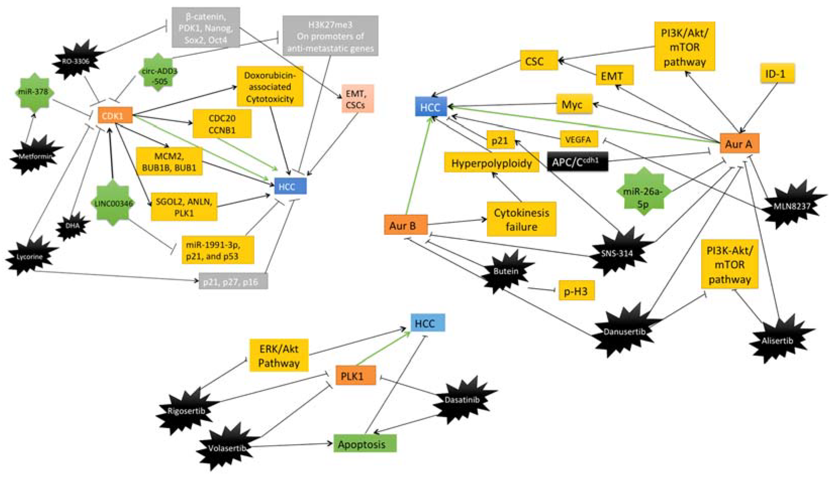
3.3.1. CDK1

It has been established that CDK1 is frequently higher in HCC tissues and is directly correlated with the poor outcome of the disease. Hence, CDK1 inhibition by specific kinase inhibitor RO-3306 is usually combined with chemotherapeutic agents such as sorafenib. Therefore, the effect on tumor growth of this combinatorial method was investigated, and it was observed that CDK1 inhibition after RO-3306 treatments in PDX models, downregulated β-catenin and PDK1 along with Nanog, Sox2, and Oct4 that are pluripotency-inducing proteins. Furthermore, CDK1 inhibition leads to a decreased epithelial-mesenchymal-transition (EMT), S-phase populations, and cancer stem cell (CSC) growth [79].
Metformin is an antihyperglycemic agent, which exhibits antineoplastic effects. It has been demonstrated that metformin treatment induces microRNA miR-378, which inhibits CDK1 and leads to anti-proliferative effects in HCC cells [80]. Lycorine, a natural product of Lyroris radiate, also inhibits CDK1 expression and induces senescence in HCC cells. Lycorine treatment resulted in the upregulation of p21, p27, and p16 proteins which halts the progression of HCC cells. Therefore, Lycorine is a potent direct inhibitor of CDK1, which can be utilized for the treatment of HCC [81]. Dihydroartemisinin (DHA) derived from Artemisia annua Linn is another naturally occurring inhibitor of CDK1, which has been shown to inhibit the cyclin B1-CDK1 pathway and HCC cell proliferation [82].

3.3.2. Aurora Kinase

Sorafenib is currently a standard treatment option against HCC. There is ongoing research aiming to boost the effects of sorafenib with an additional inhibitor and MLN8237 is an Aurora A kinase inhibitor that has been explored together with sorafenib in vivo and in vitro models of HCC. The synergistic effect of MLN8237 with sorafenib decreased angiogenesis, invasion, and cell growth and increased the apoptosis and inhibition of HCC. The mechanism of action included the inhibition of VEGFA, cyclin D1, CDK4, phosphor-p38 MAPK, and phospho-Akt. Therefore, it has been demonstrated that sorafenib-MLN8237 is a novel therapy that can be used for the inhibition of HCC progression [83]. Moreover, a pan-Aurora kinase inhibitor SNS-314 is known to induce apoptosis and polyploidy in HCC by accumulating p21 and is considered a valid therapeutic option. SNS-314 reduces the Yes-associated protein (YAP) and directly regulates p21 for induction of inhibitory effect in HCC cells [84].
Butein is a chalcone, which is usually isolated from Rhus cerniciflua and is known to directly link with Aurora B to inhibit its kinase activity. It decreases the histone 3 phosphorylations and Aurora B to induce the G2-M arrest and causes apoptosis in HCC cells [85]. Danusertib, a pan-Aurora inhibitor arrests HCC cells in G2-M transition and induces mitochondria-dependent apoptosis. The induction of autophagy and apoptosis resulting from danusertib treatment is the result of a perturbed PI3K-Akt/mTOR pathway [86]. Alisertib is a specific Aurora A Kinase inhibitor, which causes G2-M arrest in HepG2 cells and leads to aneuploidy and autophagy. The mechanism of action of alisertib involves the regulation of the PI3K/Akt/mTOR pathway. Moreover, alisertib can increase the cisplatin and doxorubicin sensitivity in HepG2 cells [87].

3.3.3. PLK1

Volasertib and GSK461364 are PLK1 inhibitors that have been used in Huh7 and HepG2 cells to demonstrate that these inhibitors selectively target the p53 mutated cells and can be used in synergy with BIRC5 (survivin) inhibitor to increase the efficacy of the treatment in xenograft models [88]. Rigosertib is a dual inhibitor, which inhibits PLK1 and RAS, and reduces cell proliferation, and causes a cell cycle arrest in HCC cells by inhibiting the activation of Akt and ERK showing disrupted RAS signaling [89]. Dasatinib is an inhibitor of PLK1 protein synthesis and its synergistic effect with irinotecan leads to a significantly higher apoptosis rate in HCC cells in vitro and in vivo [90].
The main recent findings concerning mitotic kinase inhibitors (along with their chemical structures) in liver cancer are summarised in Table 2 and Figure 2.

4. Pancreatic Cancer

Pancreatic cancer is one of the most lethal cancers worldwide [91,92]. The World Health Organization (WHO) estimates that in 2020 there were almost half a million new pancreatic cancer cases and almost half a million deaths due to pancreatic cancer. Therefore, despite not being the most common cancer type, pancreatic cancer presents high mortality [93]. In fact, pancreatic ductal adenocarcinoma (PDAC), the most common form of pancreatic cancer, has an overall survival in five years that does not exceed the 10%. PDAC incidence and mortality are increasing year by year making researchers believe that PDAC will become the second leading cause of cancer-related mortality by 2030 [91,92,94,95].

4.1. Standard-of-Care Treatment of Pancreatic Cancer

Pancreatic cancer’s high mortality is due to delayed detection and limited effects exerted by available treatments. Currently, the only curative treatment for pancreatic cancer is surgical resection of the tumor, but not all pancreatic tumors are treatable this way. Pancreatic tumors that extend beyond the pancreas or involve major vessels like the superior mesenteric artery or celiac axis are not considered safe for resection. Less than 20% of PDAC patients are eligible for surgery, and more than 75% of surgical treatments fail to eliminate all microscopically visible cancerous cells [91,92,96].
When resection of the tumor is not an option, chemotherapy remains the standard treatment with agents like gemcitabine plus nab-paclitaxel or FOLFIRINOX (5-fluorouracil, folinic acid, irinotecan plus oxaliplatin). Despite these treatments being useful, they present limited effects due to short-term response and toxicity, so there is still need for developing better treatments [91,92,94,96,97].

4.2. General Overview of Mitotic Kinases in Pancreatic Cancer

Cell cycle regulation has been one of the fields studied to develop cancer treatment options as it is known that one of cancer’s hallmarks is sustaining proliferative signaling [98]. Amongst the possible therapeutic targets of cell cycle associated proteins, mitotic kinases are prominent [99,100].

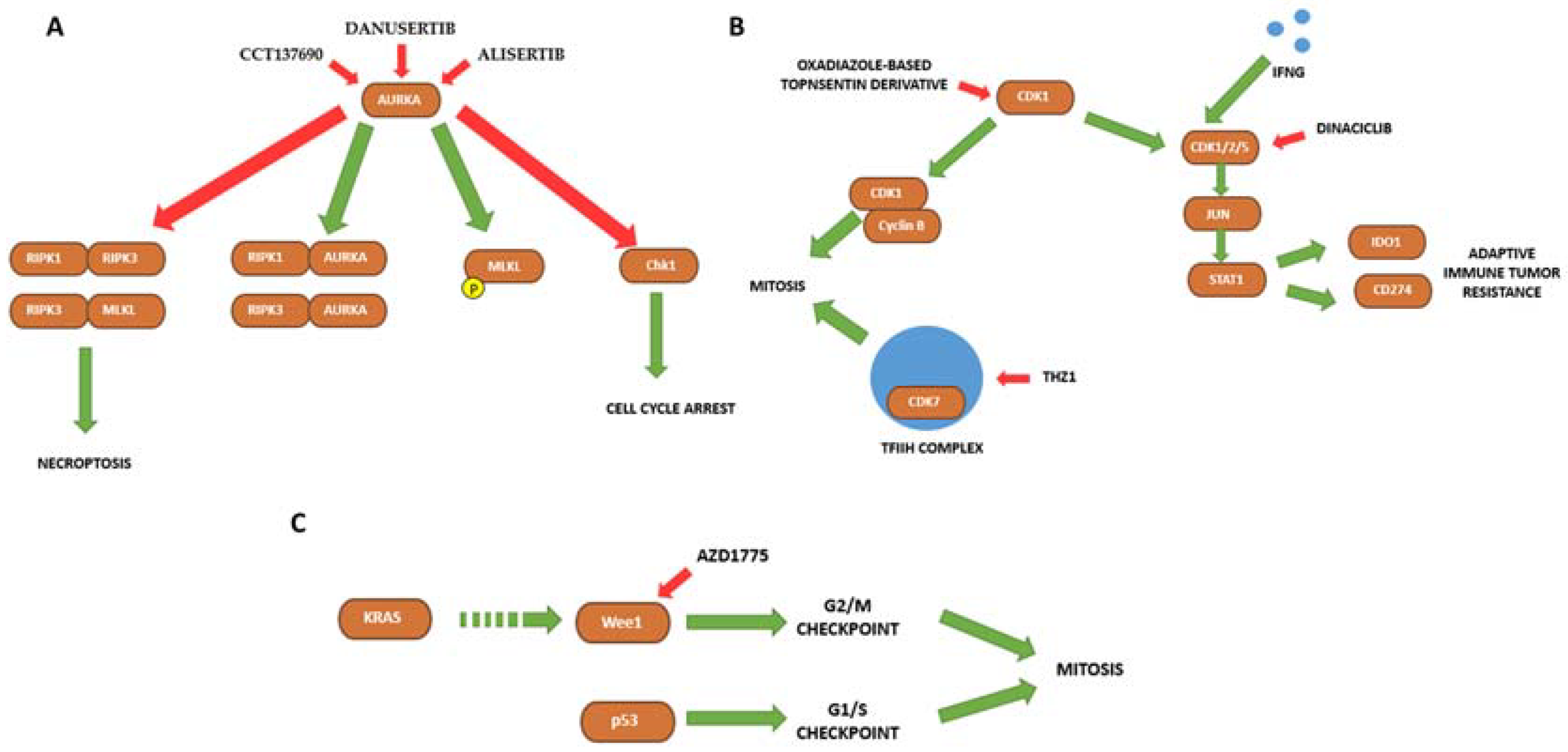
4.2.1. Aurora Kinases

Aurora kinases are a family of kinases with a well-known role in cell cycle and mitosis. Multiple studies show that the inhibition of Aurora Kinase A (AURKA) may be a therapeutic option for pancreatic cancer [101]. Researchers have described an increment of AURKA expression in PDAC patient samples in comparison to their matched non-tumor pancreatic tissue [102]. A higher AURKA expression is associated with a shorter patient survival and the presence of oncogenic KRAS mutations [101].

4.2.2. Cyclin-Dependent Kinases (CDKs)

CDKs play an important role in cell cycle control. It is not uncanny to find cell cycle dysregulation via altered CDKs function in various tumors, including pancreatic cancer [103]. For instance, CDK1 is overexpressed in pancreatic cancer cells, and it correlates with a higher tumor size and histological grade. Thus, CDK1 higher expression is associated with a poorer prognosis. These findings lead researchers to believe that CDK1 may play a role in pancreatic cancer progression and could be either a prognostic biomarker or a therapeutic target [104,105,106].

4.2.3. Wee1

Wee1 is a mitotic kinase that plays a key role in cell cycle regulation, particularly the entry into mitosis. This kinase is mutated in several cancer types. Similar to other aforementioned mitotic kinases, WEE1 also presents a higher expression in pancreatic cancer cells, and likely plays an important role in PDAC [105,107]. Moreover, it has been described that miRNA-15a (the miRNA that suppresses WEE1 among other genes) is reduced in pancreatic cancer cells leading to WEE1 overexpression. This mechanism allows cell proliferation and evasion of cell cycle arrest [108].

4.3. Recent Mitotic Kinase Inhibitors as Therapeutic Interventions for Pancreatic Cancer

4.3.1. Aurora A

Aurora kinases are a family of kinases with a well-known role in cell cycle and mitosis. Multiple studies show that Aurora A inhibition may be a therapeutic option for pancreatic cancer [101].
Xie et al., using a library of kinase inhibitors, found CCT137690, an Aurora A inhibitor which induces necrosis-like death and hinders cell growth in pancreatic cancer cell lines (PANC1, PANC2.02, BxPC3, CFPAC1, and MiaPaCa-2). Moreover, xenografts models (xenografts with PANC1 in immunodeficient nu/nu mice) and KDC PDACs mice models show that CCT137690 reduces tumor growth and increases IFNγ producing tumor infiltrating CD8+ cells, suggesting that Aurora A inhibition induces immunogenic cell death in vivo. Considering that necroptosis is an apoptosis-independent process, these results may serve as a solution to one of the major issues with gemcitabine-based therapy, resistance to apoptosis [109]. Another Aurora A inhibitor, danusertib, has shown interesting results in pancreatic cancer, as it exerts cytotoxic effects, induces apoptosis, and accumulates cells at S and G2/M phases in CFPAC-1 cell line [110].
Not only are Aurora A inhibitors showing interesting results as single agent treatments, but also as part of combination therapies. For example, the Mathison et al. study the use of Aurora A and H3K9 methyltransferase (H3K9me-HMT) inhibitors. Combination of alisertib (MLN8237), an Aurora A inhibitor, and chaetocin, a H3K9me-HMT inhibitor is more effective in reducing pancreatic cancer cell viability both in vitro (using several pancreatic cancer cell lines, spheroids, and organoids), and in vivo (orthotopic xenografts injecting Pan02 cells into the pancreas of C57BL/6 mice). The combination of both drugs presents cytotoxic effects as they induce mitotic catastrophe characterized by aberrant mitotic checkpoint signaling and decreased centromeric H3K9 methylation. This treatment forbids a normal mitotic progression; therefore, cells enter cell death pathways [111].
Beyond preclinical studies, there are phase I pancreatic cancer clinical trials based on combination therapies with alisertib and gemcitabine (NCT01924260) or alisertib and nab-paclitaxel (NCT01677559), where researchers established the safety of these combinations [112,113].

4.3.2. Cyclin-Dependent Kinases (CDKs)

CDKs have been a target to develop novel therapies for a long time. Currently, a range of diverse CDKs inhibitors are being developed. It has already been established that CDK1 is overexpressed in pancreatic cancer and may serve as a therapeutic target [106]. For example, some new oxadiazole-based topsentin derivative exert antiproliferative activity and induce apoptosis in PDAC cell lines [114]. Moreover, a CDK1/2/5 inhibitor, dinaciclib, has been identified as an immune checkpoint inhibitor that triggers caspase-dependent apoptosis and histone-dependent immunogenic cell death. Combination of dinaciclib and IFNG induces immunogenic cell death and exerts anticancer activity in pancreatic cancer both in vitro (pancreatic cancer cell lines) and in vivo (using subcutaneous, orthotopic and transgenic mouse models) through Jun and the STAT1 pathway [115]. A clinical trial with dinaciclib in combination with AKT inhibitors for pancreatic cancer treatment (NCT01783171) established that, despite this combination being safe, it does not display clinical benefits [116].
CDK7, a CDK and a subunit of the multi-protein basal transcription factor TFIIH is also an interesting therapeutic target for pancreatic cancer. THZ1, a CDK7 inhibitor, has cytotoxic effects and induces apoptosis both in vitro (PDAC cell lines and pancreatic patient-derived cancer cells) and in vivo (PDX models of PDAC and murine models), where models did not present toxicity signs. Nevertheless, not all cell lines were susceptible to THZ1 treatment as THZ1 susceptibility in PDAC cells is associated with MYC expression [117].

4.3.3. WEE1

Inhibition of cell cycle checkpoints has the potential to sensitize cancer cells to chemotherapy, as they lose the ability to stop division and repair DNA damage. For example, WEE1 inhibitor AZD1775 in combination with other treatment agents show promising results. AZD1775 as a single agent treatment does not present effective results, but in combination with capecitabine or irinotecan it exerts significant antiproliferative effects in p53 mutated cells both in vitro (using several pancreatic cancer cell lines) and in vivo (using PDX models). P53 mutated cells present an increased sensitivity to WEE1 targeted drugs as these cells have functionally inactive G1/S checkpoints. As 60% of PDAC patients present mutations in p53, WEE1 inhibition in combination therapies has the potential to be a novel PDAC therapy, although more studies are needed [118]. Furthermore, 95% of PDACs patients present KRAS activating mutations which has been associated with WEE1 pathway. Data suggest that WEE1 contributes to KRAS-mutant PDAC proliferation, meaning that direct inhibition of WEE1 may suppress KRAS mediated cell proliferation. In different KRAS mutated pancreatic cancer cell lines, AZD1775 inhibits proliferation, increases apoptosis, and leads to an accumulation of cells in S and G2/M phases. Combined inhibition of WEE1 and ERK, both associated with KRAS, causes synergistic growth arrest and apoptosis in vitro (pancreatic cancer cell lines and organoids) [119].
Currently, there are some ongoing clinical trials for pancreatic cancer regarding AZD1775 in combination with gemcitabine (NCT05212025) or in combination with gemcitabine and paclitaxel (NCT02194829). Another study assessing the use of AZD1775 with gemcitabine and radiation for unresectable pancreatic adenocarcinomas (NCT02037230) showed in 2019 that this combination therapy was well tolerated and increases overall survival [120].

4.3.4. TTK

TTK is a protein kinase required for spindle assembly checkpoint and is overexpressed in PDAC. TTK is required to prevent aneuploidy (mitotic fidelity) and apoptosis; therefore, its inhibition with AZ3146 decreased cell proliferation in PDAC cell lines [121].
The main recent findings concerning mitotic kinase inhibitors (along with their chemical structures) in pancreatic cancer are summarized in Table 3 and Figure 3.

5. Colorectal Cancer

Colorectal cancer (CRC) ranks as a leading cause of cancer-related death and the third most prevalent malignant tumor worldwide [122,123].

5.1. Standard-of-Care Treatment of Colorectal Cancer

Treatment of CRC aims at complete resection of the tumor and metastases, by surgery, chemotherapy and immunotherapy. Treatment depends on the stage of the disease according to the tumor, nodes, and metastases (TNM) classification, the patients’ health status, and the goals of treatment or palliative care [124].
Immune escape, i.e., cancer cells evading the host’s immune recognition and response, is a common feature of various types of cancer [125]. Several anti-PD1 (nivolumab, pembrolizumab) and anti-PDL1 (MPDL3280A, Medi4736) monoclonal antibodies have been studied for the treatment of gastrointestinal cancer. Checkpoint inhibition therapy has shown promising results in metastatic CRC with high microsatellite instability (MSIH). Therefore, its use under the Critical Path Initiative (CPI) in these patients has been approved by the Food and Drug Administration (FDA) [126].
Treatment of CRC with chemotherapy includes a single component therapy primarily based on fluoropyrimidines (5FU) and one or more drugs, including oxaliplatin (OX), irinotecan (IRI) and capecitabine (CAP or XELODA or XEL) [127]. Targeted therapies specifically inhibit cancerous cells’ proliferation, differentiation, and/or migration [128]. Small molecules are a group of molecules with a molecular weight below 900 Da that acts primarily inside the cell to inactivate selected enzymes, preventing tumor cell growth and triggering apoptosis. Cycle-independent kinases, proteasomes and poly(A) polymerases account for the majority of molecular targets for CRC [129].

5.2. General Overview of Mitotic Kinases Inhibitors in Colorectal Cancer

Identification of novel molecular targets permits the development of specific pharmacological treatment for CRC. Mitotic kinases have emerged as targets for CRC treatment. They play an essential role in the elongation stage of the global transcription process. The list of mitotic kinases includes but is not limited to CDK, Aurora and Polo-like kinase (Plk) families, budding uninhibited by benzimidazoles 1 (Bub1), histone H3 associated protein kinase (Haspin) and mucopolysaccharidosis I (Mps1) [130,131,132].

5.2.1. CDKs

CDKs are classified into two groups. CDK-1, CDK-4, and CDK-5 fall within the cell-cycle-related subfamilies, while CDK-7, CDK-8, CDK-9, CDK-11, and CDK-20 are classified as transcriptional subfamilies. CDK-1 is one of the most important CDKs for cell-cycle regulation since it promotes the G2–M transition. Fadaka and Sibuyi reported that mRNA expression of CDK-1 was overexpressed in CRC types compared to normal colorectal tissues. Furthermore, the authors assessed the expression of CDKs in OMICS data from CRC subtypes, concluding that CDK expression was higher in males and females with CRC compared to normal counterparts [133].

5.2.2. Aurora A

Aurora A is a member of the serine/threonine kinase family involved in the initiation of mitosis, the formation of bipolar spindles, the regulation of centrosome maturation, and the separation during mitosis. Overexpression and amplification of the Aurora A protein are commonly noticed in CRC. Koh et al. revealed that Aurora A protein expression level correlates with colorectal cancer. Moreover, Aurora A protein was upregulated in 45% of patients with colorectal adenocarcinoma using immunohistochemistry. Expression of Aurora A, along with other clinicopathological parameters, was associated with the patient’s gender and tumor site [134].

5.2.3. Mps1

Zhang et al. identified high mitotic checkpoint kinase (Mps1) protein expression in CRC and, remarkably, Mps1 expression was even higher in poorly differentiated patients’ CRC tissues, indicating that Mps1 may be associated to tumor differentiation. The authors proposed that Mps1 promotes the growth of colon cancer cells through activation of PKCα/ERK1/2 and inhibits differentiation through downregulation of the PI3K/Akt pathway [135].

5.3. Recent Mitotic Kinase Inhibitors as Therapeutic Interventions for Colorectal Cancer

5.3.1. PLK1

Phospho Polo Like Kinase (Plk) family proteins are key regulators of multiple stages of mitotic progression [136]. The upregulation of Plk1 in CRC is associated to poor prognosis [137]. Klauck et al. reported that TAK-960 is a mitotic kinase inhibitor limiting colony formation colorectal cancer cell lines in a dose-dependent manner. Most importantly, they performed Immunoblotting to elucidate the mechanism of action of TAK-960 in CRC cell lines treated with increasing concentration of TAK-960. Interestingly, P-Plk1 was reduced upon exposure to TAK-960 [138].

5.3.2. Aurora Kinases

ENMD-2076 is a recently-discovered orally active small molecule multi-kinase inhibitor targeting the mitotic kinases Aurora A and B in human CRC cell lines. Capasso and colleagues reported G2/M arrest and apoptosis induction in CRC cell lines treated with ENMD-2076 [139].

5.3.3. CDKs

Robb et al. demonstrated that CP668863, a 3-aminopyrazole analog previously indetified by Pfizer, inhibits CDKs and could potentially serve as a therapy for CRC. CP668863 inhibited CDK2 and CDK5 in a dose-dependent manner. Data from colorectal cancer xenograft model using GEO cells indicated that CP668863-treated tumors displayed a decrease in the phospho focal adhesion kinase (pFAK) levels, a phosphorylation site-specific to CDK5, thereby indication that CP668863 inhibits CDK5 in vivo [140].

5.3.4. Mps1

Faisal et al. reported that CCT271850 suppressed Mitotic kinase Monopolar Spindle1(Mps1 kinase) phosphorylation and activity in human colon cancer HCT116 cells. They identified that histone H3 phosphorylation was inhibited in CCT271850-treated HCT116 cells and apoptotic cell death was induced upon CCT271850 treatment in a time-dependent manner as determined by the levels of cleaved PARP. Additionally, in vivo studies using HCT116 tumor xenografts revealed that 100 mg/kg of CCT271850 is sufficient to inhibit MPS1 and promote cell cycle arrest [141].
The main recent findings concerning mitotic kinase inhibitors (along with their chemical structures) in colorectal cancer are summarized in Table 4 and Figure 4.

6. Conclusions

In summary, mitotic kinase inhibitors are promising therapies for the treatment of gastrointestinal cancers including gastric cancer, liver cancer, pancreatic cancer and colorectal cancer. Nevertheless, further studies are needed to understand their 3D structure, as well as their binding to their substrates and the effects on cell cultures, animal models and, ultimately, in human patients, to allow safety and efficacy in their translation to the clinics.

Author Contributions

A.J., G.M., M.Y. and T.R.-T. conceived, wrote and reviewed the manuscript. C.M.P.-R. and C.B. critically reviewed the manuscript. All authors have read and agreed to the published version of the manuscript.

Funding

This research received no external funding.

Conflicts of Interest

The authors declare no conflict of interest.

References

  1. Kapoor, T.M.; Compton, D.A. Searching for the middle ground: Mechanisms of chromosome alignment during mitosis. J. Cell Biol. 2002, 157, 551–556. [Google Scholar] [CrossRef]
  2. Pines, J. Mitosis: A matter of getting rid of the right protein at the right time. Trends Cell Biol. 2006, 16, 55–63. [Google Scholar] [CrossRef]
  3. Walczak, C.E.; Cai, S.; Khodjakov, A. Mechanisms of chromosome behaviour during mitosis. Nat. Rev. Mol. Cell Biol. 2010, 11, 91–102. [Google Scholar] [CrossRef]
  4. Vermeulen, K.; Van Bockstaele, D.R.; Berneman, Z.N. The cell cycle: A review of regulation, deregulation and therapeutic targets in cancer. Cell Prolif. 2003, 36, 131–149. [Google Scholar] [CrossRef]
  5. Ovejero, S.; Bueno, A.; Sacristán, M.P. Working on Genomic Stability: From the S-Phase to Mitosis. Genes 2020, 11, 225. [Google Scholar] [CrossRef]
  6. Manchado, E.; Guillamot, M.; Malumbres, M. Killing cells by targeting mitosis. Cell Death Differ. 2012, 19, 369–377. [Google Scholar] [CrossRef]
  7. Chan, K.-S.; Koh, C.-G.; Li, H.-Y. Mitosis-targeted anti-cancer therapies: Where they stand. Cell Death Dis. 2012, 3, e411. [Google Scholar] [CrossRef] [PubMed]
  8. De Wolf, B.; Kops, G.J.P.L.; Kops, G.J.P.L. Cell Division Machinery and Disease. In Advances in Experimental Medicine and Biology; Springer: Berlin, Germany, 2017. [Google Scholar]
  9. Goga, A.; Yang, D.; Tward, A.D.; Morgan, D.O.; Bishop, J.M. Inhibition of CDK1 as a potential therapy for tumors over-expressing MYC. Nat. Med. 2007, 13, 820–827. [Google Scholar] [CrossRef] [PubMed]
  10. Ying, X.; Che, X.; Wang, J.; Zou, G.; Yu, Q.; Zhang, X. CDK1 serves as a novel therapeutic target for endometrioid endometrial cancer. J. Cancer 2021, 12, 2206–2215. [Google Scholar] [CrossRef] [PubMed]
  11. Wang, G.; Jiang, Q.; Zhang, C. The role of mitotic kinases in coupling the centrosome cycle with the assembly of the mitotic spindle. J. Cell Sci. 2014, 127, 4111–4122. [Google Scholar] [CrossRef]
  12. D’Assoro, A.B.; Ehaddad, T.; Egalanis, E. Aurora-A Kinase as a Promising Therapeutic Target in Cancer. Front. Oncol. 2016, 5, 295. [Google Scholar] [CrossRef] [PubMed]
  13. Cicenas, J. The Aurora kinase inhibitors in cancer research and therapy. J. Cancer Res. Clin. Oncol. 2016, 142, 1995–2012. [Google Scholar] [CrossRef] [PubMed]
  14. Lens, S.M.A.; Medema, R.H. Cytokinesis defects and cancer. Nat. Cancer 2018, 19, 32–45. [Google Scholar] [CrossRef]
  15. Ma, H.T.; Poon, R.Y. Aurora kinases and DNA damage response. Mutat. Res. Mol. Mech. Mutagen. 2020, 821, 111716. [Google Scholar] [CrossRef]
  16. Passaro, C.; Chieffi, P. Aurora B: A New Prognostic Marker and Therapeutic Target in Cancer. Curr. Med. Chem. 2011, 18, 482–496. [Google Scholar] [CrossRef]
  17. Honma, K.; Nakanishi, R.; Nakanoko, T.; Ando, K.; Saeki, H.; Oki, E.; Iimori, M.; Kitao, H.; Kakeji, Y.; Maehara, Y. Contribution of Aurora-A and -B expression to DNA aneuploidy in gastric cancers. Surg. Today 2013, 44, 454–461. [Google Scholar] [CrossRef] [PubMed]
  18. Liu, Z.; Sun, Q.; Wang, X. PLK1, A Potential Target for Cancer Therapy. Transl. Oncol. 2016, 10, 22–32. [Google Scholar] [CrossRef]
  19. Zhang, Z.; Zhang, G.; Kong, C. FOXM1 participates in PLK1-regulated cell cycle progression in renal cell cancer cells. Oncol. Lett. 2016, 11, 2685–2691. [Google Scholar] [CrossRef]
  20. Jung, Y.; Kraikivski, P.; Shafiekhani, S.; Terhune, S.S.; Dash, R.K. Crosstalk between Plk1, p53, cell cycle, and G2/M DNA damage checkpoint regulation in cancer: Computational modeling and analysis. NPJ Syst. Biol. Appl. 2021, 7, 46. [Google Scholar] [CrossRef] [PubMed]
  21. Jang, H.-R.; Shin, S.-B.; Kim, C.-H.; Won, J.-Y.; Xu, R.; Kim, D.-E.; Yim, H. PLK1/vimentin signaling facilitates immune escape by recruiting Smad2/3 to PD-L1 promoter in metastatic lung adenocarcinoma. Cell Death Differ. 2021, 28, 2745–2764. [Google Scholar] [CrossRef] [PubMed]
  22. Pei, G.; Luo, M.; Ni, X.; Wu, J.; Wang, S.; Ma, Y.; Yu, J. Autophagy Facilitates Metadherin-Induced Chemotherapy Resistance Through the AMPK/ATG5 Pathway in Gastric Cancer. Cell. Physiol. Biochem. 2018, 46, 847–859. [Google Scholar] [CrossRef] [PubMed]
  23. Fuchs, C.S.; Özgüroğlu, M.; Bang, Y.-J.; Di Bartolomeo, M.; Mandalà, M.; Ryu, M.-H.; Fornaro, L.; Olesinski, T.; Caglevic, C.; Chung, H.C.; et al. Pembrolizumab versus paclitaxel for previously treated patients with PD-L1–positive advanced gastric or gastroesophageal junction cancer (GC): Update from the phase III KEYNOTE-061 trial. J. Clin. Oncol. 2020, 38 (Suppl. 15), 4503. [Google Scholar] [CrossRef]
  24. Parisi, A.; Reim, D.; Borghi, F.; Nguyen, N.T.; Qi, F.; Coratti, A.; Cianchi, F.; Cesari, M. Minimally invasive surgery for gastric cancer: A comparison between robotic, laparoscopic and open surgery. World J. Gastroenterol. 2017, 23, 2376–2384. [Google Scholar] [CrossRef]
  25. Ychou, M.; Boige, V.; Pignon, J.-P.; Conroy, T.; Bouché, O.; Lebreton, G.; Ducourtieux, M.; Bedenne, L.; Fabre, J.-M.; Saint-Aubert, B.; et al. Perioperative Chemotherapy Compared with Surgery Alone for Resectable Gastroesophageal Adenocarcinoma: An FNCLCC and FFCD Multicenter Phase III Trial. J. Clin. Oncol. 2011, 29, 1715–1721. [Google Scholar] [CrossRef] [PubMed]
  26. Ende, T.V.D.; ter Veer, E.; Machiels, M.; Mali, R.M.A.; Nijenhuis, F.A.A.; de Waal, L.; Laarman, M.; Gisbertz, S.S.; Hulshof, M.C.C.M.; van Oijen, M.G.H.; et al. The Efficacy and Safety of (Neo)Adjuvant Therapy for Gastric Cancer: A Network Meta-analysis. Cancers 2019, 11, 80. [Google Scholar] [CrossRef]
  27. Tsai, C.; Mueller, A.; Maubach, J.; Warschkow, R.; Nussbaum, D.P.; Schmied, B.M.; Blazer, D.; Gloor, B.; Worni, M. No Difference in Survival between Neo-Adjuvant Chemotherapy and Neo-Adjuvant Chemoradiation Therapy in Gastric Cardia Cancer Patients: A Contemporary View from the National Cancer Database. Dig. Surg. 2019, 37, 249–257. [Google Scholar] [CrossRef]
  28. De Vita, F.; Borg, C.; Farina, G.; Geva, R.; Carton, I.; Cuku, H.; Wei, R.; Muro, K. Ramucirumab and paclitaxel in patients with gastric cancer and prior trastuzumab: Subgroup analysis from RAINBOW study. Future Oncol. 2019, 15, 2723–2731. [Google Scholar] [CrossRef]
  29. Tang, Z.; Li, L.; Tang, Y.; Xie, D.; Wu, K.; Wei, W.; Xiao, Q. CDK 2 positively regulates aerobic glycolysis by suppressing SIRT 5 in gastric cancer. Cancer Sci. 2018, 109, 2590–2598. [Google Scholar] [CrossRef]
  30. Kamran, M.; Long, Z.-J.; Xu, D.; Lv, S.-S.; Liu, B.; Wang, C.-L.; Xu, J.; Lam, E.; Liu, Q. Aurora kinase A regulates Survivin stability through targeting FBXL7 in gastric cancer drug resistance and prognosis. Oncogenesis 2017, 6, e298. [Google Scholar] [CrossRef]
  31. Ding, L.; Yang, L.; He, Y.; Zhu, B.; Ren, F.; Fan, X.; Wang, Y.; Li, M.; Li, J.; Kuang, Y.; et al. CREPT/RPRD1B associates with Aurora B to regulate Cyclin B1 expression for accelerating the G2/M transition in gastric cancer. Cell Death Dis. 2018, 9, 1172. [Google Scholar] [CrossRef] [PubMed]
  32. Wakida, T.; Ikura, M.; Kuriya, K.; Ito, S.; Shiroiwa, Y.; Habu, T.; Kawamoto, T.; Okumura, K.; Ikura, T.; Furuya, K. The CDK-PLK1 axis targets the DNA damage checkpoint sensor protein RAD9 to promote cell proliferation and tolerance to genotoxic stress. eLife 2017, 6, e29953. [Google Scholar] [CrossRef]
  33. Dang, S.-C.; Wang, F.; Qian, X.-B.; Abdul, M.; Naseer, Q.-A.; Jin, W.; Hu, R.; Gu, Q.; Gu, M. MicroRNA-505 suppresses gastric cancer cell proliferation and invasion by directly targeting Polo-like kinase-1. OncoTargets Ther. 2019, 12, 795–803. [Google Scholar] [CrossRef]
  34. Dang, S.-C.; Fan, Y.-Y.; Cui, L.; Chen, J.-X.; Qu, J.-G.; Gu, M. PLK1 as a potential prognostic marker of gastric cancer through MEK-ERK pathway on PDTX models. OncoTargets Ther. 2018, 11, 6239–6247. [Google Scholar] [CrossRef]
  35. Cai, X.P.; Chen, L.D.; Bin Song, H.; Zhang, C.X.; Yuan, Z.W.; Xiang, Z.X. PLK1 promotes epithelial-mesenchymal transition and metastasis of gastric carcinoma cells. Am. J. Transl. Res. 2016, 8, 4172–4183. [Google Scholar]
  36. Lin, X.; Chen, D.; Zhang, C.; Zhang, X.; Li, Z.; Dong, B.; Gao, J.; Shen, L. Augmented antitumor activity by olaparib plus AZD1775 in gastric cancer through disrupting DNA damage repair pathways and DNA damage checkpoint. J. Exp. Clin. Cancer Res. 2018, 37, 129. [Google Scholar] [CrossRef]
  37. Kim, H.-Y.; Cho, Y.; Kang, H.; Yim, Y.-S.; Kim, S.-J.; Song, J.; Chun, K.-H. Targeting the WEE1 kinase as a molecular targeted therapy for gastric cancer. Oncotarget 2016, 7, 49902–49916. [Google Scholar] [CrossRef]
  38. Yuan, C.X.; Zhou, Z.W.; Yang, Y.X.; He, Z.X.; Zhang, X.; Wang, D.; Yang, T.; Pan, S.Y.; Chen, X.W.; Zhou, S.F. Danusertib, a potent pan-Aurora kinase and ABL kinase inhibitor, induces cell cycle arrest and programmed cell death and inhibits epithelial to mesenchymal transition involving the PI3K/Akt/mTOR-mediated signaling pathway in human gastric cancer AGS and NCI-N78 cells. Drug Des. Dev. Ther. 2015, 9, 1293–1318. [Google Scholar] [CrossRef]
  39. Wang-Bishop, L.; Chen, Z.; Gomaa, A.; Lockhart, A.C.; Salaria, S.; Wang, J.; Lewis, K.B.; Ecsedy, J.; Washington, K.; Beauchamp, R.D.; et al. Inhibition of AURKA Reduces Proliferation and Survival of Gastrointestinal Cancer Cells with Activated KRAS by Preventing Activation of RPS6KB1. Gastroenterology 2019, 156, 662–675.e7. [Google Scholar] [CrossRef]
  40. Nokihara, H.; Yamada, Y.; Fujiwara, Y.; Yamamoto, N.; Wakui, H.; Nakamichi, S.; Kitazono, S.; Inoue, K.; Harada, A.; Taube, T.; et al. Phase I trial of volasertib, a Polo-like kinase inhibitor, in Japanese patients with advanced solid tumors. Investig. New Drugs 2015, 34, 66–74. [Google Scholar] [CrossRef]
  41. Liu, H.; Shin, S.H.; Chen, H.; Liu, T.; Li, Z.; Hu, Y.; Liu, F.; Zhang, C.; Kim, D.J.; Liu, K.; et al. CDK12 and PAK2 as novel therapeutic targets for human gastric cancer. Theranostics 2020, 10, 6201–6215. [Google Scholar] [CrossRef] [PubMed]
  42. Llovet, J.M.; Zucman-Rossi, J.; Pikarsky, E.; Sangro, B.; Schwartz, M.; Sherman, M.; Gores, G. Hepatocellular carcinoma. Nat. Rev. Dis. Prim. 2016, 2, 16018. [Google Scholar] [CrossRef]
  43. Caines, A.; Selim, R.; Salgia, R. The Changing Global Epidemiology of Hepatocellular Carcinoma. Clin. Liver Dis. 2020, 24, 535–547. [Google Scholar] [CrossRef]
  44. Marcon, P.D.S.; Tovo, C.V.; Kliemann, D.A.; Fisch, P.; De Mattos, A.A. Incidence of hepatocellular carcinoma in patients with chronic liver disease due to hepatitis B or C and coinfected with the human immunodeficiency virus: A retrospective cohort study. World J. Gastroenterol. 2018, 24, 613–622. [Google Scholar] [CrossRef] [PubMed]
  45. Ochiai, Y.; Kawamura, Y.; Kobayashi, M.; Shindoh, J.; Kobayashi, Y.; Okubo, S.; Muraishi, N.; Kajiwara, A.; Iritani, S.; Fujiyama, S.; et al. Effects of alcohol consumption on multiple hepatocarcinogenesis in patients with fatty liver disease. Hepatol. Res. 2020, 51, 62–68. [Google Scholar] [CrossRef]
  46. Ayuso, C.; Rimola, J.; Vilana, R.; Burrel, M.; Darnell, A.; García-Criado, Á.; Bianchi, L.; Belmonte, E.; Caparroz, C.; Barrufet, M.; et al. Diagnosis and staging of hepatocellular carcinoma (HCC): Current guidelines. Eur. J. Radiol. 2018, 101, 72–81. [Google Scholar] [CrossRef] [PubMed]
  47. Vogel, A.; Cervantes, A.; Chau, I.; Daniele, B.; Llovet, J.M.; Meyer, T.; Nault, J.-C.; Neumann, U.; Ricke, J.; Sangro, B.; et al. Hepatocellular carcinoma: ESMO Clinical Practice Guidelines for diagnosis, treatment and follow-up. Ann. Oncol. 2018, 29, iv238–iv255. [Google Scholar] [CrossRef]
  48. Bruix, J.; Chan, S.L.; Galle, P.R.; Rimassa, L.; Sangro, B. Systemic treatment of hepatocellular carcinoma: An EASL position paper. J. Hepatol. 2021, 75, 960–974. [Google Scholar] [CrossRef]
  49. Lee, G.C.; Ferrone, C.R.; Vagefi, P.A.; Uppot, R.N.; Tanabe, K.K.; Lillemoe, K.D.; Blaszkowsky, L.S.; Qadan, M. Surgical resection versus ablation for early-stage hepatocellular carcinoma: A retrospective cohort analysis. Am. J. Surg. 2019, 218, 157–163. [Google Scholar] [CrossRef] [PubMed]
  50. Shen, S.; Dean, D.C.; Yu, Z.; Duan, Z. Role of cyclin-dependent kinases (CDKs) in hepatocellular carcinoma: Therapeutic potential of targeting the CDK signaling pathway. Hepatol. Res. 2019, 49, 1097–1108. [Google Scholar] [CrossRef] [PubMed]
  51. Li, K.K.W.; Ng, I.O.-L.; Fan, S.T.; Albrecht, J.H.; Yamashita, K.; Poon, R.Y.C. Activation of cyclin-dependent kinases CDC2 and CDK2 in hepatocellular carcinoma. Liver Int. 2002, 22, 259–268. [Google Scholar] [CrossRef]
  52. Zhao, J.; Han, S.-X.; Ma, J.-L.; Ying, X.; Liu, P.; Li, J.; Wang, L.; Zhang, Y.; Ma, J.; Zhang, L.; et al. The role of CDK1 in apoptin-induced apoptosis in hepatocellular carcinoma cells. Oncol. Rep. 2013, 30, 253–259. [Google Scholar] [CrossRef] [PubMed]
  53. Nguyen, C.B.; Kotturi, H.; Waris, G.; Mohammed, A.; Chandrakesan, P.; May, R.; Sureban, S.; Weygant, N.; Qu, D.; Rao, C.V.; et al. (Z)-3,5,4′-Trimethoxystilbene Limits Hepatitis C and Cancer Pathophysiology by Blocking Microtubule Dynamics and Cell-Cycle Progression. Cancer Res. 2016, 76, 4887–4896. [Google Scholar] [CrossRef]
  54. He, B.; Yin, J.; Gong, S.; Gu, J.; Xiao, J.; Shi, W.; Ding, W.; He, Y. Bioinformatics analysis of key genes and pathways for hepatocellular carcinoma transformed from cirrhosis. Medicine 2017, 96, e6938. [Google Scholar] [CrossRef]
  55. Jung, E.U.; Yoon, J.-H.; Lee, Y.-J.; Lee, J.-H.; Kim, B.H.; Yu, S.J.; Myung, S.J.; Kim, Y.J.; Lee, H.-S. Hypoxia and retinoic acid-inducible NDRG1 expression is responsible for doxorubicin and retinoic acid resistance in hepatocellular carcinoma cells. Cancer Lett. 2010, 298, 9–15. [Google Scholar] [CrossRef]
  56. Shah, M.A.; Kortmansky, J.; Motwani, M.; Drobnjak, M.; Gonen, M.; Yi, S.; Weyerbacher, A.; Cordon-Cardo, C.; Lefkowitz, R.; Brenner, B.; et al. A Phase I Clinical Trial of the Sequential Combination of Irinotecan Followed by Flavopiridol. Clin. Cancer Res. 2005, 11, 3836–3845. [Google Scholar] [CrossRef] [PubMed]
  57. Richard, C.; Matthews, D.; Duivenvoorden, W.; Yau, J.; Wright, P.S.; Th’Ng, J.P. Flavopiridol Sensitivity of Cancer Cells Isolated from Ascites and Pleural Fluids. Clin. Cancer Res. 2005, 11, 3523–3529. [Google Scholar] [CrossRef]
  58. Zou, Y.; Ruan, S.; Jin, L.; Chen, Z.; Han, H.; Zhang, Y.; Jian, Z.; Lin, Y.; Shi, N.; Jin, H. CDK1, CCNB1, and CCNB2 are Prognostic Biomarkers and Correlated with Immune Infiltration in Hepatocellular Carcinoma. Med. Sci. Monit. 2020, 26, e925289-1–e925289-14. [Google Scholar] [CrossRef] [PubMed]
  59. Yang, W.-X.; Pan, Y.-Y.; You, C.-G. CDK1, CCNB1, CDC20, BUB1, MAD2L1, MCM3, BUB1B, MCM2, and RFC4 May Be Potential Therapeutic Targets for Hepatocellular Carcinoma Using Integrated Bioinformatic Analysis. Biomed Res. Int. 2019, 2019, 1245072. [Google Scholar] [CrossRef]
  60. Li, L.; Huang, K.; Zhao, H.; Chen, B.; Ye, Q.; Yue, J. CDK1-PLK1/SGOL2/ANLN pathway mediating abnormal cell division in cell cycle may be a critical process in hepatocellular carcinoma. Cell Cycle 2020, 19, 1236–1252. [Google Scholar] [CrossRef]
  61. Sun, S.; Wang, W.; Luo, X.; Li, Y.; Liu, B.; Li, X.; Zhang, B.; Han, S.; Li, X. Circular RNA circ-ADD3 inhibits hepatocellular carcinoma metastasis through facilitating EZH2 degradation via CDK1-mediated ubiquitination. Am. J. Cancer Res. 2019, 9, 1695–1707. Available online: http://www.ncbi.nlm.nih.gov/pubmed/31497351%0Ahttp://www.pubmedcentral.nih.gov/articlerender.fcgi?artid=PMC6726993 (accessed on 20 March 2022).
  62. Jin, J.; Xu, H.; Li, W.; Xu, X.; Liu, H.; Wei, F. LINC00346 Acts as a Competing Endogenous RNA Regulating Development of Hepatocellular Carcinoma via Modulating CDK1/CCNB1 Axis. Front. Bioeng. Biotechnol. 2020, 8, 54. [Google Scholar] [CrossRef]
  63. Nikonova, A.S.; Astsaturov, I.; Serebriiskii, I.G.; Dunbrack, R.L., Jr.; Golemis, E.A. Aurora A kinase (AURKA) in normal and pathological cell division. Cell. Mol. Life Sci. 2013, 70, 661–687. [Google Scholar] [CrossRef]
  64. Chen, C.; Song, G.; Xiang, J.; Zhang, H.; Zhao, S.; Zhan, Y. AURKA promotes cancer metastasis by regulating epithelial-mesenchymal transition and cancer stem cell properties in hepatocellular carcinoma. Biochem. Biophys. Res. Commun. 2017, 486, 514–520. [Google Scholar] [CrossRef] [PubMed]
  65. Yuan, Y.L.; Yu, H.; Mu, S.-M.; Dong, Y.D.; Li, D.Y. MiR-26a-5p Inhibits Cell Proliferation and Enhances Doxorubicin Sensitivity in HCC Cells via Targeting AURKA. Technol. Cancer Res. Treat. 2019, 18, 1533033819851833. [Google Scholar] [CrossRef] [PubMed]
  66. Wang, B.; Hsu, C.-J.; Chou, C.-H.; Lee, H.-L.; Chiang, W.-L.; Su, C.-M.; Tsai, H.-C.; Yang, S.-F.; Tang, C.-H. Variations in the AURKA Gene: Biomarkers for the Development and Progression of Hepatocellular Carcinoma. Int. J. Med. Sci. 2018, 15, 170–175. [Google Scholar] [CrossRef]
  67. Wu, M.; Zhou, Y.; Fei, C.; Chen, T.; Yin, X.; Zhang, L.; Ren, Z. ID1 overexpression promotes HCC progression by amplifying the AURKA/Myc signaling pathway. Int. J. Oncol. 2020, 57, 845–857. [Google Scholar] [CrossRef]
  68. Lu, L.; Han, H.; Tian, Y.; Li, W.; Zhang, J.; Feng, M.; Li, Y. Aurora kinase A mediates c-Myc’s oncogenic effects in hepatocellular carcinoma. Mol. Carcinog. 2014, 54, 1467–1479. [Google Scholar] [CrossRef]
  69. Lin, H.; Huang, Y.-S.; Fustin, J.-M.; Doi, M.; Chen, H.; Lai, H.-H.; Lin, S.-H.; Lee, Y.-L.; King, P.-C.; Hou, H.-S.; et al. Hyperpolyploidization of hepatocyte initiates preneoplastic lesion formation in the liver. Nat. Commun. 2021, 12, 645. [Google Scholar] [CrossRef]
  70. Yasen, M.; Mizushima, H.; Mogushi, K.; Obulhasim, G.; Miyaguchi, K.; Inoue, K.; Nakahara, I.; Ohta, T.; Aihara, A.; Tanaka, S.; et al. Expression of Aurora B and alternative variant forms in hepatocellular carcinoma and adjacent tissue. Cancer Sci. 2009, 100, 472–480. [Google Scholar] [CrossRef]
  71. He, Z.-L.; Zheng, H.; Lin, H.; Miao, X.-Y.; Zhong, D.-W. Overexpression of polo-like kinase1 predicts a poor prognosis in hepatocellular carcinoma patients. World J. Gastroenterol. 2009, 15, 4177–4182. [Google Scholar] [CrossRef]
  72. Pellegrino, R.; Calvisi, D.F.; Ladu, S.; Ehemann, V.; Staniscia, T.; Evert, M.; Dombrowski, F.; Schirmacher, P.; Longerich, T. Oncogenic and tumor suppressive roles of polo-like kinases in human hepatocellular carcinoma. Hepatology 2009, 51, 857–868. [Google Scholar] [CrossRef]
  73. Sun, W.; Su, Q.; Cao, X.; Shang, B.; Chen, A.; Yin, H.; Liu, B. High Expression of Polo-Like Kinase 1 Is Associated with Early Development of Hepatocellular Carcinoma. J. Genom. 2014, 2014, 312130. [Google Scholar] [CrossRef]
  74. Bouhlal, H.; Ouled-Haddou, H.; DeBuysscher, V.; Singh, A.R.; Ossart, C.; Reignier, A.; Hocini, H.; Fouquet, G.; Al Baghami, M.; Eugenio, M.S.; et al. RB/PLK1-dependent induced pathway by SLAMF3 expression inhibits mitosis and control hepatocarcinoma cell proliferation. Oncotarget 2016, 7, 9832–9843. [Google Scholar] [CrossRef]
  75. Zhang, C.; Wang, X.; Fang, D.; Xu, P.; Mo, X.; Hu, C.; Abdelatty, A.; Wang, M.; Xu, H.; Sun, Q.; et al. STK39 is a novel kinase contributing to the progression of hepatocellular carcinoma by the PLK1/ERK signaling pathway. Theranostics 2021, 11, 2108–2122. [Google Scholar] [CrossRef] [PubMed]
  76. Lin, X.-T.; Yu, H.-Q.; Fang, L.; Tan, Y.; Liu, Z.-Y.; Wu, D.; Zhang, J.; Xiong, H.-J.; Xie, C.-M. Elevated FBXO45 promotes liver tumorigenesis through enhancing IGF2BP1 ubiquitination and subsequent PLK1 upregulation. eLife 2021, 10, e70715. [Google Scholar] [CrossRef] [PubMed]
  77. He, Z.; Deng, W.; Jiang, B.; Liu, S.; Tang, M.; Liu, Y.; Zhang, J. Hsa-let-7b inhibits cell proliferation by targeting PLK1 in HCC. Gene 2018, 673, 46–55. [Google Scholar] [CrossRef]
  78. Zhang, H.; Diab, A.; Fan, H.; Mani, S.K.; Hullinger, R.; Merle, P.; Andrisani, O. PLK1 and HOTAIR Accelerate Proteasomal Degradation of SUZ12 and ZNF198 during Hepatitis B Virus–Induced Liver Carcinogenesis. Cancer Res. 2015, 75, 2363–2374. [Google Scholar] [CrossRef]
  79. Wu, C.X.; Wang, X.Q.; Chok, S.H.; Man, K.; Tsang, S.H.Y.; Chan, A.C.Y.; Ma, K.W.; Xia, W.; Cheung, T.T. Blocking CDK1/PDK1/β-Catenin signaling by CDK1 inhibitor RO3306 increased the efficacy of sorafenib treatment by targeting cancer stem cells in a preclinical model of hepatocellular carcinoma. Theranostics 2018, 8, 3737–3750. [Google Scholar] [CrossRef]
  80. Zhou, J.; Han, S.; Qian, W.; Gu, Y.; Li, X.; Yang, K. Metformin induces miR-378 to downregulate the CDK1, leading to suppression of cell proliferation in hepatocellular carcinoma. OncoTargets Ther. 2018, ume 11, 4451–4459. [Google Scholar] [CrossRef]
  81. Yin, S.; Yang, S.; Luo, Y.; Lu, J.; Hu, G.; Wang, K.; Shao, Y.; Zhou, S.; Koo, S.; Qiu, Y.; et al. Cyclin-dependent kinase 1 as a potential target for lycorine against hepatocellular carcinoma. Biochem. Pharmacol. 2021, 193, 114806. [Google Scholar] [CrossRef] [PubMed]
  82. Hao, L.; Li, S.; Peng, Q.; Guo, Y.; Ji, J.; Zhang, Z.; Xue, Y.; Liu, Y.; Shi, X. Anti-malarial drug dihydroartemisinin downregulates the expression levels of CDK1 and CCNB1 in liver cancer. Oncol. Lett. 2021, 22, 653. [Google Scholar] [CrossRef]
  83. Zhang, K.; Wang, T.; Zhou, H.; Feng, B.; Chen, Y.; Zhi, Y.; Wang, R. A Novel Aurora-A Inhibitor (MLN8237) Synergistically Enhances the Antitumor Activity of Sorafenib in Hepatocellular Carcinoma. Mol. Ther.-Nucleic Acids 2018, 13, 176–188. [Google Scholar] [CrossRef]
  84. Liu, F.; Wang, G.; Wang, X.; Che, Z.; Dong, W.; Guo, X.; Wang, Z.; Chen, P.; Hou, D.; Zhang, Q.; et al. Targeting high Aurora kinases expression as an innovative therapy for hepatocellular carcinoma. Oncotarget 2017, 8, 27953–27965. [Google Scholar] [CrossRef] [PubMed]
  85. Zhou, Y.; Li, M.; Yu, X.; Liu, T.; Li, T.; Zhou, L.; Liu, W.; Li, W.; Gao, F. Butein suppresses hepatocellular carcinoma growth via modulating Aurora B kinase activity. Int. J. Biol. Sci. 2018, 14, 1521–1534. [Google Scholar] [CrossRef]
  86. Zhu, Q.; Yu, X.; Zhou, Z.-W.; Luo, M.; Zhou, C.; He, Z.-X.; Chen, Y.; Zhou, S.-F. A quantitative proteomic response of hepatocellular carcinoma Hep3B cells to danusertib, a pan-Aurora kinase inhibitor. J. Cancer 2018, 9, 2061–2071. [Google Scholar] [CrossRef] [PubMed]
  87. Zhu, Q.; Yu, X.; Zhou, Z.-W.; Zhou, C.; Chen, X.-W.; Zhou, S.-F. Inhibition of Aurora A Kinase by Alisertib Induces Autophagy and Cell Cycle Arrest and Increases Chemosensitivity in Human Hepatocellular Carcinoma HepG2 Cells. Curr. Cancer Drug Targets 2017, 17, 386–401. [Google Scholar] [CrossRef] [PubMed]
  88. Li, Y.; Zhao, Z.-G.; Luo, Y.; Cui, H.; Wang, H.-Y.; Jia, Y.-F.; Gao, Y.-T. Dual targeting of Polo-like kinase 1 and baculoviral inhibitor of apoptosis repeat-containing 5 in TP53-mutated hepatocellular carcinoma. World J. Gastroenterol. 2020, 26, 4786–4801. [Google Scholar] [CrossRef]
  89. Dietrich, P.; Freese, K.; Mahli, A.; Thasler, W.E.; Hellerbrand, C.; Bosserhoff, A.K. Combined effects of PLK1 and RAS in hepatocellular carcinoma reveal rigosertib as promising novel therapeutic “dual-hit” option. Oncotarget 2017, 9, 3605–3618. [Google Scholar] [CrossRef]
  90. Xu, L.; Zhu, Y.; Shao, J.; Chen, M.; Yan, H.; Li, G.; Zhu, Y.; Xu, Z.; Yang, B.; Luo, P.; et al. Dasatinib synergises with irinotecan to suppress hepatocellular carcinoma via inhibiting the protein synthesis of PLK1. Br. J. Cancer 2017, 116, 1027–1036. [Google Scholar] [CrossRef]
  91. Fawzy, F.; Canada, A.; Fawzy, N. Cancer Treatment. Encycl. Stress 2007, 384–387. [Google Scholar] [CrossRef]
  92. Milella, M.; Bassi, C.; Boggi, U.; Brunetti, O.; Cavaliere, A.; Crippa, S.; De Vita, F.; Falconi, M.; Frassineti, G.L.; Giommoni, E.; et al. Evolving pancreatic cancer treatment: From diagnosis to healthcare management. Crit. Rev. Oncol. 2021, 169, 103571. [Google Scholar] [CrossRef] [PubMed]
  93. Cancer Today. (n.d.). Available online: https://gco.iarc.fr/today/online-analysis-pie?v=2020&mode=cancer&mode_population=continents&population=900&populations=900&key=total&sex=0&cancer=39&type=1&statistic=5&prevalence=0&population_group=0&ages_group%5B%5D=0&ages_group%5B%5D=17&nb_items=7&group_cancer=1&include_nmsc=1&include_nmsc_other=1&half_pie=0&donut=0 (accessed on 31 March 2022).
  94. Chandana, S.; Babiker, H.M.; Mahadevan, D. Therapeutic trends in pancreatic ductal adenocarcinoma (PDAC). Expert Opin. Investig. Drugs 2018, 28, 161–177. [Google Scholar] [CrossRef] [PubMed]
  95. Park, W.; Chawla, A.; O’Reilly, E.M. Pancreatic Cancer. JAMA J. Am. Med. Assoc. 2021, 326, 851–862. [Google Scholar] [CrossRef] [PubMed]
  96. PDQ Adult Treatment Editorial Board. Pancreatic Cancer Treatment (Adult) (PDQ®). PDQ Cancer Information Summaries. 2022. Available online: https://www-ncbi-nlm-nih-gov.sire.ub.edu/books/NBK65957/ (accessed on 31 March 2022).
  97. Gugenheim, J.; Crovetto, A.; Petrucciani, N. Neoadjuvant therapy for pancreatic cancer. Update Surg. 2021, 74, 35–42. [Google Scholar] [CrossRef] [PubMed]
  98. Hanahan, D. Hallmarks of Cancer: New Dimensions. Cancer Discov. 2022, 12, 31–46. [Google Scholar] [CrossRef] [PubMed]
  99. Tischer, J.; Gergely, F. Anti-mitotic therapies in cancer. J. Cell Biol. 2018, 218, 10–11. [Google Scholar] [CrossRef]
  100. Bayraktar, S.; Lima, C.M.R. Emerging cell-cycle inhibitors for pancreatic cancer therapy. Expert Opin. Emerg. Drugs 2012, 17, 571–582. [Google Scholar] [CrossRef] [PubMed]
  101. Gomes-Filho, S.M.; dos Santos, E.O.; Bertoldi, E.R.M.; Scalabrini, L.C.; Heidrich, V.; Dazzani, B.; Levantini, E.; Reis, E.M.; Bassères, D.S. Aurora A kinase and its activator TPX2 are potential therapeutic targets in KRAS-induced pancreatic cancer. Cell. Oncol. 2020, 43, 445–460. [Google Scholar] [CrossRef]
  102. Bavetsias, V.; Linardopoulos, S. Aurora Kinase Inhibitors: Current Status and Outlook. Front. Oncol. 2015, 5, 278. [Google Scholar] [CrossRef]
  103. García-Reyes, B.; Kretz, A.-L.; Ruff, J.-P.; von Karstedt, S.; Hillenbrand, A.; Knippschild, U.; Henne-Bruns, D.; Lemke, J. The Emerging Role of Cyclin-Dependent Kinases (CDKs) in Pancreatic Ductal Adenocarcinoma. Int. J. Mol. Sci. 2018, 19, 3219. [Google Scholar] [CrossRef]
  104. Piao, J.; Zhu, L.; Sun, J.; Li, N.; Dong, B.; Yang, Y.; Chen, L. High expression of CDK1 and BUB1 predicts poor prognosis of pancreatic ductal adenocarcinoma. Gene 2019, 701, 15–22. [Google Scholar] [CrossRef]
  105. Dong, S.; Huang, F.; Zhang, H.; Chen, Q. Overexpression of BUB1B, CCNA2, CDC20, and CDK1 in tumor tissues predicts poor survival in pancreatic ductal adenocarcinoma. Biosci. Rep. 2019, 39, BSR20182306. [Google Scholar] [CrossRef] [PubMed]
  106. Wijnen, R.; Pecoraro, C.; Carbone, D.; Fiuji, H.; Avan, A.; Peters, G.J.; Giovannetti, E.; Diana, P. Cyclin Dependent Kinase-1 (CDK-1) Inhibition as a Novel Therapeutic Strategy against Pancreatic Ductal Adenocarcinoma (PDAC). Cancers 2021, 13, 4389. [Google Scholar] [CrossRef]
  107. di Rorà, A.G.L.; Cerchione, C.; Martinelli, G.; Simonetti, G. A WEE1 family business: Regulation of mitosis, cancer progression, and therapeutic target. J. Hematol. Oncol. 2020, 13, 126. [Google Scholar] [CrossRef]
  108. Guo, S.; Fesler, A.; Huang, W.; Wang, Y.; Yang, J.; Wang, X.; Zheng, Y.; Hwang, G.-R.; Ju, J. Functional Significance and Therapeutic Potential of miR-15a Mimic in Pancreatic Ductal Adenocarcinoma. Mol. Ther.-Nucleic Acids 2019, 19, 228–239. [Google Scholar] [CrossRef] [PubMed]
  109. Xie, Y.; Zhu, S.; Zhong, M.; Yang, M.; Sun, X.; Liu, J.; Kroemer, G.; Lotze, M.; Zeh, H.J.; Kang, R.; et al. Inhibition of Aurora Kinase A Induces Necroptosis in Pancreatic Carcinoma. Gastroenterology 2017, 153, 1429–1443.e5. [Google Scholar] [CrossRef]
  110. Ozcimen, A.; Kirbiyik, I. The effect of danusertib, an Aurora kinase inhibitor, onto the cytotoxicity, cell cycle and apoptosis in pancreatic ductal adenocarcinoma cells. J. Cancer Res. Ther. 2021, 17, 1419. [Google Scholar] [CrossRef] [PubMed]
  111. Mathison, A.; Salmonson, A.; Missfeldt, M.; Bintz, J.; Williams, M.; Kossak, S.; Nair, A.; de Assuncao, T.M.; Christensen, T.; Buttar, N.; et al. Combined AURKA and H3K9 Methyltransferase Targeting Inhibits Cell Growth by Inducing Mitotic Catastrophe. Mol. Cancer Res. 2017, 15, 984–997. [Google Scholar] [CrossRef]
  112. Kim, E.J.-H.; Semrad, T.J.; Gandara, D.R.; Riess, J.; Li, T.; Yu, A.; Matsukuma, K.; Mack, P.C.; Kelly, K. Phase I study of the combination of alisertib (MLN8237) and gemcitabine in advanced solid tumors. J. Clin. Oncol. 2015, 33, 2526. [Google Scholar] [CrossRef]
  113. Uboha, N.V.; Milhem, M.M.; Kovacs, C.; Amin, A.; Magley, A.; Das Purkayastha, D.; Piha-Paul, S.A. Phase II study of spartalizumab (PDR001) and LAG525 in advanced solid tumors and hematologic malignancies. J. Clin. Oncol. 2019, 37, 2553. [Google Scholar] [CrossRef]
  114. Pecoraro, C.; Parrino, B.; Cascioferro, S.; Puerta, A.; Avan, A.; Peters, G.J.; Diana, P.; Giovannetti, E.; Carbone, D. A New Oxadiazole-Based Topsentin Derivative Modulates Cyclin-Dependent Kinase 1 Expression and Exerts Cytotoxic Effects on Pancreatic Cancer Cells. Molecules 2021, 27, 19. [Google Scholar] [CrossRef] [PubMed]
  115. Huang, J.; Chen, P.; Liu, K.; Liu, J.; Zhou, B.; Wu, R.; Peng, Q.; Liu, Z.-X.; Li, C.; Kroemer, G.; et al. CDK1/2/5 inhibition overcomes IFNG-mediated adaptive immune resistance in pancreatic cancer. Gut 2020, 70, 890–899. [Google Scholar] [CrossRef] [PubMed]
  116. Murphy, A.G.; Zahurak, M.; Shah, M.; Weekes, C.D.; Hansen, A.; Siu, L.L.; Spreafico, A.; LoConte, N.; Anders, N.M.; Miles, T.; et al. A Phase I Study of Dinaciclib in Combination with MK-2206 in Patients with Advanced Pancreatic Cancer. Clin. Transl. Sci. 2020, 13, 1178–1188. [Google Scholar] [CrossRef] [PubMed]
  117. Lu, P.; Geng, J.; Zhang, L.; Wang, Y.; Niu, N.; Fang, Y.; Liu, F.; Shi, J.; Zhang, Z.-G.; Sun, Y.-W.; et al. THZ1 reveals CDK7-dependent transcriptional addictions in pancreatic cancer. Oncogene 2019, 38, 3932–3945. [Google Scholar] [CrossRef]
  118. Hartman, S.J.; Bagby, S.M.; Yacob, B.W.; Simmons, D.M.; MacBeth, M.; Lieu, C.H.; Davis, S.L.; Leal, A.D.; Tentler, J.J.; Diamond, J.R.; et al. WEE1 Inhibition in Combination with Targeted Agents and Standard Chemotherapy in Preclinical Models of Pancreatic Ductal Adenocarcinoma. Front. Oncol. 2021, 11, 642328. [Google Scholar] [CrossRef]
  119. Diehl, J.N.; Klomp, J.E.; Snare, K.R.; Hibshman, P.S.; Blake, D.R.; Kaiser, Z.D.; Gilbert, T.S.; Baldelli, E.; Pierobon, M.; Papke, B.; et al. The KRAS-regulated kinome identifies WEE1 and ERK coinhibition as a potential therapeutic strategy in KRAS-mutant pancreatic cancer. J. Biol. Chem. 2021, 297, 101335. [Google Scholar] [CrossRef] [PubMed]
  120. Cuneo, K.C.; Morgan, M.A.; Sahai, V.; Schipper, M.J.; Parsels, L.A.; Parsels, J.D.; Devasia, T.; Al-Hawaray, M.; Cho, C.S.; Nathan, H.; et al. Dose Escalation Trial of the Wee1 Inhibitor Adavosertib (AZD1775) in Combination with Gemcitabine and Radiation for Patients with Locally Advanced Pancreatic Cancer. J. Clin. Oncol. 2019, 37, 2643–2650. [Google Scholar] [CrossRef] [PubMed]
  121. Stratford, J.K.; Yan, F.; Hill, R.A.; Major, M.; Graves, L.M.; Der, C.; Yeh, J.J. Genetic and pharmacological inhibition of TTK impairs pancreatic cancer cell line growth by inducing lethal chromosomal instability. PLoS ONE 2017, 12, e0174863. [Google Scholar] [CrossRef]
  122. Ferlay, J.; Colombet, M.; Soerjomataram, I.; Dyba, T.; Randi, G.; Bettio, M.; Gavin, A.; Visser, O.; Bray, F. Cancer incidence and mortality patterns in Europe: Estimates for 40 countries and 25 major cancers in 2018. Eur. J. Cancer 2018, 103, 356–387. [Google Scholar] [CrossRef]
  123. Siegel, R.L.; Miller, K.D.; Jemal, A. Cancer statistics, 2019. CA Cancer J. Clin. 2019, 69, 7–34. [Google Scholar] [CrossRef]
  124. Bender, U.; Rho, Y.; Barrera, I.; Aghajanyan, S.; Acoba, J.; Kavan, P. Adjuvant Therapy for Stages II and III Colon Cancer: Risk Stratification, Treatment Duration, and Future Directions. Curr. Oncol. 2019, 26, 43–52. [Google Scholar] [CrossRef]
  125. Mielcarska, S.; Kula, A.; Dawidowicz, M.; Kiczmer, P.; Chrabańska, M.; Rynkiewicz, M.; Wziątek-Kuczmik, D.; Świętochowska, E.; Waniczek, D. Assessment of the RANTES Level Correlation and Selected Inflammatory and Pro-Angiogenic Molecules Evaluation of Their Influence on CRC Clinical Features: A Preliminary Observational Study. Medicina 2022, 58, 203. [Google Scholar] [CrossRef] [PubMed]
  126. Bin Wang, H.; Yao, H.; Li, C.S.; Liang, L.X.; Zhang, Y.; Chen, Y.X.; Fang, J.-Y.; Xu, J. Rise of PD-L1 expression during metastasis of colorectal cancer: Implications for immunotherapy. J. Dig. Dis. 2017, 18, 574–581. [Google Scholar] [CrossRef]
  127. Venook, A.P.; Niedzwiecki, D.; Lenz, H.-J.; Innocenti, F.; Fruth, B.; Meyerhardt, J.A.; Schrag, D.; Greene, C.; O’Neil, B.H.; Atkins, J.N.; et al. Effect of First-Line Chemotherapy Combined with Cetuximab or Bevacizumab on Overall Survival in Patients with KRAS Wild-Type Advanced or Metastatic Colorectal Cancer: A randomized clinical trial. JAMA 2017, 317, 2392–2401. [Google Scholar] [CrossRef]
  128. Zhao, Q.; Ouyang, X.; Wan, X.; Gajiwala, K.S.; Kath, J.C.; Jones, L.H.; Burlingame, A.L.; Taunton, J. Broad-Spectrum Kinase Profiling in Live Cells with Lysine-Targeted Sulfonyl Fluoride Probes. J. Am. Chem. Soc. 2017, 139, 680–685. [Google Scholar] [CrossRef] [PubMed]
  129. Baur, F.; Nietzer, S.L.; Kunz, M.; Saal, F.; Jeromin, J.; Matschos, S.; Linnebacher, M.; Walles, H.; Dandekar, T.; Dandekar, G. Connecting Cancer Pathways to Tumor Engines: A Stratification Tool for Colorectal Cancer Combining Human in Vitro Tissue Models with Boolean in Silico Models. Cancers 2019, 12, 28. [Google Scholar] [CrossRef] [PubMed]
  130. Rahaman, M.H.; Lam, F.; Zhong, L.; Teo, T.; Adams, J.; Yu, M.; Milne, R.W.; Pepper, C.; Lokman, N.A.; Ricciardelli, C.; et al. Targeting CDK9 for treatment of colorectal cancer. Mol. Oncol. 2019, 13, 2178–2193. [Google Scholar] [CrossRef]
  131. Tanaka, H.; Wada, M.; Park, J. HASPIN kinase inhibitor CHR-6494 suppresses intestinal polyp development, cachexia, and hypogonadism in Apc min/+ mice. Eur. J. Cancer Prev. 2019, 29, 481–485. [Google Scholar] [CrossRef]
  132. Schmit, T.L.; Ahmad, N. Regulation of mitosis via mitotic kinases: New opportunities for cancer management. Mol. Cancer Ther. 2007, 6, 1920–1931. [Google Scholar] [CrossRef]
  133. Fadaka, A.O.; Sibuyi, N.R.S.; Bakare, O.O.; Klein, A.; Madiehe, A.M.; Meyer, M. Expression of cyclin-dependent kinases and their clinical significance with immune infiltrates could predict prognosis in colorectal cancer. Biotechnol. Rep. 2021, 29, e00602. [Google Scholar] [CrossRef]
  134. Koh, H.M.; Jang, B.G.; Hyun, C.L.; Kim, Y.S.; Hyun, J.W.; Chang, W.Y.; Maeng, Y.H. Aurora Kinase A Is a Prognostic Marker in Colorectal Adenocarcinoma. J. Pathol. Transl. Med. 2017, 51, 32–39. [Google Scholar] [CrossRef]
  135. Zhang, L.; Jiang, B.; Zhu, N.; Tao, M.; Jun, Y.; Chen, X.; Wang, Q.; Luo, C. Mitotic checkpoint kinase Mps1/TTK predicts prognosis of colon cancer patients and regulates tumor proliferation and differentiation via PKCα/ERK1/2 and PI3K/Akt pathway. Med. Oncol. 2019, 37, 5. [Google Scholar] [CrossRef]
  136. Welburn, J.P.I.; Jeyaprakash, A.A. Mechanisms of Mitotic Kinase Regulation: A Structural Perspective. Front. Cell Dev. Biol. 2018, 6, 6. [Google Scholar] [CrossRef] [PubMed]
  137. Wang, C.; Lu, T.; Wang, L.; Wu, C.; Chen, M.; Kuo, C.; Shyu, R.; Tsai, F. Tazarotene-induced gene 1 interacts with Polo-like kinase 2 and inhibits cell proliferation in HCT116 colorectal cancer cells. Cell Biol. Int. 2021, 45, 2347–2356. [Google Scholar] [CrossRef]
  138. Klauck, P.J.; Bagby, S.M.; Capasso, A.; Bradshaw-Pierce, E.L.; Selby, H.M.; Spreafico, A.; Tentler, J.J.; Tan, A.C.; Kim, J.; Arcaroli, J.J.; et al. Antitumor activity of the polo-like kinase inhibitor, TAK-960, against preclinical models of colorectal cancer. BMC Cancer 2018, 18, 136. [Google Scholar] [CrossRef] [PubMed]
  139. Capasso, A.; Pitts, T.M.; Klauck, P.J.; Bagby, S.M.; Westbrook, L.; Kaplan, J.; Soleimani, M.; Spreafico, A.; Tentler, J.J.; Diamond, J.R.; et al. Dual compartmental targeting of cell cycle and angiogenic kinases in colorectal cancer models. Anti-Cancer Drugs 2018, 29, 827–838. [Google Scholar] [CrossRef]
  140. Robb, C.M.; Kour, S.; Contreras, J.I.; Agarwal, E.; Barger, C.J.; Rana, S.; Sonawane, Y.; Neilsen, B.K.; Taylor, M.; Kizhake, S.; et al. Characterization of CDK(5) inhibitor, 20-223 (aka CP668863) for colorectal cancer therapy. Oncotarget 2017, 9, 5216–5232. [Google Scholar] [CrossRef]
  141. Faisal, A.; Mak, G.W.Y.; Gurden, M.D.; Xavier, C.P.R.; Anderhub, S.J.; Innocenti, P.; Westwood, I.M.; Naud, S.; Hayes, A.; Box, G.; et al. Characterisation of CCT271850, a selective, oral and potent MPS1 inhibitor, used to directly measure in vivo MPS1 inhibition vs therapeutic efficacy. Br. J. Cancer 2017, 116, 1166–1176. [Google Scholar] [CrossRef]
Figure 1. A schematic representation of the main molecules involved in gastric cancer and the drugs that target them.
Futurepharmacol 02 00015 g001
Figure 2. A schematic representation of the main molecules involved in liver cancer and the drugs that target them.
Futurepharmacol 02 00015 g002
Figure 3. (A) A schematic representation of AURKA implication in mitosis and its inhibitors. AURKA inhibition reduces pancreatic cancer growth and induces necroptosis. (B) A schematic representation of CDKs implication in mitosis and its inhibitors. Data shows that CDKs inhibition reduces pancreatic cancer growth rate and may also serve as a way to sort out IFNG mediated immune resistance. (C) A schematic representation of Wee1 role in mitosis and its inhibitors. Wee1 is associated with KRAS, and both enhance cell growth in pancreatic cancer, thus Wee1 and KRAS inhibition combined is effective in reducing cell growth. Moreover, cells with p53 mutations may benefit from Wee1 inhibition because this way neither G1/S and G2/M checkpoints are active and cells become more sensitive to DNA damaging therapies.
Futurepharmacol 02 00015 g003
Figure 4. A schematic representation of the main molecules involved in colorectar cancer and the drugs that target them.
Futurepharmacol 02 00015 g004
Table 1. Recent studies on mitotic kinase inhibitors as therapeutic interventions for gastric cancer.
TargetInhibitorModelMain FindingsReference
Aurora and CDK2Danusertib
Futurepharmacol 02 00015 i001
NCI-N78 and AGS human GC cell line.Danusertib inhibits Aurora and CDK2 expression in GC cell lines. The proportion of GC cells in the G1 phase is reduced.[38]
Aurora AAlisertib
Futurepharmacol 02 00015 i002
Mice with xenograft tumors tissues.Alisertib reduces tumor volume. Downregulation of Aurora A by alisertib reduces the level of phosphorylated RPS6KB1. [39]
Wee1AZD1775
Futurepharmacol 02 00015 i003
The GC cell lines (AGS, YCC-2, MKN28, KATO III, SNU-216, SNU-601, SNU-638, SNU-668, and SNU-719. Orthotopic mouse model for GC. AZD1775 induces apoptosis and cell cycle arrest. Tumor size and weight were reduced in mice treated with AZD1775 compared to control mice.[37]
CDK12Procaterol
Futurepharmacol 02 00015 i004
Cell xenograft NU/NU mouse models (CDXs) and patient-derived xenograft NOD/SCID mouse models (PDXs) were conducted to study.Procaterol bounds to and inhibits CDK12 kinase activity.[41]
PLK1Volasertib
Futurepharmacol 02 00015 i005
Japanese patients with advanced solid tumors.
Methods In this phase I, open-label, dose-escalation trial, sequential patient cohorts.
Volasertib has been investigated as a selective Polo-like kinase inhibitor that resulted in mitotic arrest and apoptosis in Japanese patients with gastric cancer.[40]
WEE1/(PLK1)AZD1775
Futurepharmacol 02 00015 i006
Human GC cell lines MKN45 and AGS.
female NOD/SCID mice.
Administration of AZD1775-olaparib amed at WEE1/PLK1 and the expression of PLK1 reduced in AGS and MKN45 cell lines, which exerted stronger antigrowth efficacy than olaparib alone.
The combination of AZD1775 and olaparib has led to higher inhibition in gastric tumor growth than their monotherapy in mice.
[36]
Abbreviations: GC = gastric cancer, Aurora = Aurora kinase family, CDK2 = Cyclin-dependent kinase 2, CDK12 = Cyclin-dependent kinase 12, NCI-N78 = gastric carcinoma cell line, AGS = human gastric adenocarcinoma cell, MKN28 = human gastric adenocarcinoma, RPS6KB1 = Ribosomal Protein S6 Kinase B1, PLKs = Polo-like kinases, PLK1 = Polo-like kinase 1, Wee1 = Wee1-like protein kinase, YCC-2 = human gastric cancer cell line, KATO III = human gastric carcinoma cell line, SNU-216, SNU-601, SNU-638, SNU-668, and SNU-719 = Gastric Cell Lines, AZD1775 = Adavosertib, CDXs = cell line-based xenografts, PDXs = patient-derived xenografts, NOD/SCID = Nonobese diabetic/severe combined immunodeficiency.
Table 2. Recent studies on mitotic kinase inhibitors as therapeutic interventions for liver cancer.
TargetInhibitorModelMain FindingsReference
CDK1RO-3306
Futurepharmacol 02 00015 i007
HCC patient-derived xenograft (PDX) tumor models treated with sorafenib alone or with RO-3306Downregulation of PK1-beta-Catenin pathway, reduced CSC growth, decrease in EMT profile[79]
CDK1Metformin
Futurepharmacol 02 00015 i008
Human HCC cell lines HepG2 and Hep3B, and nude xenograft mice modelsInduction of miR-378 which inhibits CDK1 mRNA and causes reduction in HCC cell proliferation[80]
CDK1Lycorine
Futurepharmacol 02 00015 i009
HCC cellsInhibition of CDK1 expression directly and induces expression of p21, p16 and p21[81]
CDK1Dihydroartemisinin (DHA)
Futurepharmacol 02 00015 i010
HCC cellsInhibition of the cyclin B1-CDK1 pathway and reduces the HCC cell proliferation[82]
Aurora AMLN8237
Futurepharmacol 02 00015 i011
HepG2 and SMMC-7721 cell lines, HepG2 xenograft mice modelsInhibition of p-Akt, p-MAPK, CDK4, cyclin D1, and ultimately progression of HCC.[83]
Pan-Aurora kinasesSNS-314
Futurepharmacol 02 00015 i012
HepG2, SMMC-7721 and HCC-LM6 Liver cancer cell linesReduction of YAP and accumulates p21 to induce polyploidy and apoptosis in HCC cells.[84]
Aurora BButein
Futurepharmacol 02 00015 i013
HepG2 and Hep3B cells, xenograft modelsInduction of apoptosis by decreasing p-H3 and Aurora B, marked by reduced Ki67[85]
Pan-Aurora kinasesDanusertib
Futurepharmacol 02 00015 i014
Hep3B cell lineInduction of autophagy and mitochondria-dependent apoptosis by changing the PI3K/Akt/mTOR signaling pathway[86]
Aurora AAlisertib
Futurepharmacol 02 00015 i015
HepG2 cell lineRegulation of PI3K/Akt/mTOR pathway, induces autophagy, and increase sensitivity to doxorubicin and cisplatin[87]
PLK1Volasertib
Futurepharmacol 02 00015 i016
P53 mutated HCC cells, Xenograft modelInduction of apoptosis selectively in p53 mutated HCC cells[88]
PLK1Rigosertib
Futurepharmacol 02 00015 i017
HCC cell linesInhibition of ERK and Akt signaling, reduces proliferation and causes cell cycle arrest[89]
PLK1Dasatinib
Futurepharmacol 02 00015 i018
HCC cell lines and xenograft modelsInhibition of protein synthesis of PLK1 and increases the apoptosis rate of HCC cells[90]
Abbreviations: Aurora A = Aurora kinase family A, Aurora B = Aurora kinase family B, CDK1 = Cyclin-dependent kinase 1, PLK1 = Polo Like Kinase 1, RO-3306 = ATP-competitive cyclin-dependent kinase 1, DHA = Dihydroartemisinin, MLN8237 = Aurora A inhibitor, SNS-314 = Inhibitor of Aurora Kinases A, B and C, HCC = Hepatocellular carcinoma, PDX = Patient-Derived Xenografts, HepG2 = Human Liver Cancer Cell Line, Hep3B = human hepatoma Hep3B cell line, SMMC-7721 = Human Hepatoma Cell Line, HCC-LM6 = Hepatocellular Carcinoma LM6, P53 = Tumor Protein, CSC = Cancer Stem Cell, EMT = Emergency Medical Technicians, miR-378 = MicroRNA-378, mRNA = Messenger Ribonucleic Acid, p21 and P16 = Tumor Suppressor Protein = Cyclin-Dependent Kinase Inhibitor, Cyclin B1-CDK1 = Cyclin B–Cdk1 Kinase, p-Akt = Phospho-Protein kinase B, MAPK = Phospho-Mitogen-Activated Protein Kinase, CDK4 = Cyclin Dependent Kinase 4, YAP = Yes-associated protein, p-H3 = Phospho-Histone H3, pKi67 = Nuclear Protein, PI3K = Phosphoinositide 3-Kinases, AKT = Protein Kinase B, mTOR = Mammalian Target of Rapamycin, ERKs = Extracellular Signal-Regulated Kinases.
Table 3. Recent studies on mitotic kinase inhibitors as therapeutic interventions for pancreatic cancer.
TargetInhibitorModelMain FindingsReference
Aurora Kinase ACCT137690
Futurepharmacol 02 00015 i019
Human pancreatic cancer cell lines (PANC1, PANC2.03, CFPAC1, MiaPaCa2, BxPc-3 and PANC02) and mouse PDAC cell line KPC. Murine subcutaneous tumors (PANC1, PANC02 and KPC injected in athymic nude or B6 mice). Orthotopic tumors (KPC cells in B6 mice).CCT137690 serves as an Aurora Kinase A inhibitor that induces necrosis-like death in pancreatic cancer cells in vitro and in vivo. [109]
Aurora Kinase ADanusertib
Futurepharmacol 02 00015 i020
Human pancreatic cancer cell line CFPAC-1.Danusertib exerts cytotoxic effects, induces apoptosis, and accumulates cells at S and G2/M phases in CFPAC-1 cell line. [110]
Aurora Kinase AAlisertib
Futurepharmacol 02 00015 i021
Human pancreatic cancer cell lines (BxPC-3, Capan-2, L3.6, MiaPaCa-2, PANC-1) and mouse PDAC cell line Pan02.
Organoids from Ela-Kran mice. Orthotopic xenografts injecting Pan02 cells in C57BL/6 mice.
Alisertib in combination with H3K9 methyltransferase inhibitors is more effective in reducing pancreatic cancer cell viability in vitro and in vivo via inducing mitotic catastrophe. [111]
CDK1/2/5Dinaciclib
Futurepharmacol 02 00015 i022
Human pancreatic cancer cell line CFPAC-1, and mouse cell lines KPC and CT26. Human clinical samples from pancreatic cancer patients who underwent surgery. Animal models: vaccination murine model, subcutaneous xenografts, orthotopic xenografts, Pdx-1-Cre and KRASG12D/+ transgenic mice.Dinaciclib treatment triggers caspase-dependent apoptosis and histone-dependent immunogenic cell death in pancreatic cancer cells. Dinaciclib in combination of IGNF exerts anticancer effects both in vitro and in vivo. [115]
CDK7THZ1
Futurepharmacol 02 00015 i023
Human pancreatic cancer cell lines (BxPC-3, MiaPaCa-2, SW1990, PANC-1, ASPC-1) and human pancreatic epithelial cell line HPDE6-C7. Pancreatic patient-derived tumor cells. Patient-derived xenografts in BALB/c mice. KC and KPC mice.CDK7 inhibition has cytotoxic effects and induces apoptosis in pancreatic cancer cells both in vitro and in vivo. Susceptibility to THZ1 treatment in pancreatic cancer cells is associated with MYC expression. [117]
Wee1AZD1775
Futurepharmacol 02 00015 i024
Human pancreatic cancer cell lines (BxPC-3, MiaPaCa-2, PANC-1, and L3.3). Patient-Derived Xenografts.AZD1775 in combination with irinotecan or capecitabine shows anti-tumor effects in PDAC both in vitro and in vivo. [118]
Wee1AZD1775
Futurepharmacol 02 00015 i025
Human pancreatic cancer cell lines (AsPC-1, Panc10.05, SW-1990, MiaPaCa-2, PANC-1, HPAC, HPAF-II, Pa01C, Pa02C, Pa14C, and Pa16C) and organoids.AZD1775 treatment exerts cytotoxic effects in KRAS mutated PDAC. AZD1775 and ERK inhibition causes synergistic growth arrest and apoptosis in vitro.[119]
TTKAZ3146
Futurepharmacol 02 00015 i026
Human pancreatic cancer cell lines (HPDE, HPNE, BxPC-3, Panc02.03, MiaPaCa-2, HPAC, PANC-1, Panc 10.05, Capan2, T3M4, AsPC-1, HPAF-II, SW1990, HuPT3, Capan1, CFPAC).AZ3146 treatment decreases cell proliferation in PDAC cell lies.[121]
Abbreviations: CDK1 = Cyclin-dependent kinase 1, CDK2 = Cyclin-dependent kinase 2, CDK2 = Cyclin-dependent kinase 5, Wee1 = Wee1-like protein kinase.
Table 4. Recent studies on mitotic kinase inhibitors as therapeutic interventions for colorectal cancer.
TargetInhibitorModelMain FindingsReference
P-Plk1TAK-960
Futurepharmacol 02 00015 i027
CRC cell lines (HCT116, WIDR, DLD1 and COLO678)Colonization of CRC cell lines treated with TAK960 was reduced in a dose-dependent manner. PPlk1 decreased when CRC cell lines were treated with TAK960.[138]
Aurora A/BENMD-2076
Futurepharmacol 02 00015 i028
Forty-seven CRC cell lines had been tested.
patient-derived xenograft (PDX) models.
Exposure to ENMD2076 resulted in G2/M cell cycle arrest, increased aneuploidy, and cell death in responsive cell lines.[139]
CDKsCP668863
Futurepharmacol 02 00015 i029
CRC cell lines included GEO, HCT116 and HT29
their colorectal cancer xenograft model used GEO cells.
CP668863to target CDK2 and CDK5 in CRC cell lines. CP668863-treated tumors had low phospho focal adhesion kinase (pFAK) levels.[140]
Mps1 CCT271850
Futurepharmacol 02 00015 i030
Human colon cancer HCT116 cells.
Xenograft mice models using DLD1-GFP-MPS1 cells.
CCT271850 strongly suppressed MPS1 kinase activity in biochemical and cellular assays and in vivo models. Moderate efficacy of CCT271850 as a single agent in a xenograft model of human colorectal cancer.[141]
Abbreviations: CRC = colorectal cancer, Aurora A/B = Aurora kinase A and B, CDKs = Cyclin-dependent kinase families, P-Plk1 = phospho Polo Like Kinase 1, ENMD-2076 = Aurora + Angiogenic Kinase Inhibitor, TAK-960 = ATP-competitive Plk1 inhibitor, HT29 = human colon cancer cell line, HCT116 = Human Colorectal Carcinoma Cell Line, WIDR = Colon Adenocarcinoma Cell Line, DLD1 = Colon Adenocarcinoma Cell Line, COLO678 = Human colon carcinoma cell line, PDXs = patient-derived xenografts, GEO = Human Colon Cancer Cell Line, CP668863 = cyclin-dependent kinase 5 inhibitor, Mps1 = Mitotic kinase Monopolar spindle1, DLD1 = colorectal adenocarcinoma cell line, GFP = Green Fluorescent protein, CCT271850 = MPS1 kinase inhibitor.
Publisher’s Note: MDPI stays neutral with regard to jurisdictional claims in published maps and institutional affiliations.

