Review

Snake Venom and 3D Microenvironment Cell Culture: From Production to Drug Development

1
Laboratory of Pathophysiology, Butantan Institute, São Paulo 05503-900, Brazil
2
ESIB, Escola Superior do Instituto Butantan, São Paulo 05508-210, Brazil
3
Laboratory of Biochemistry, Butantan Institute, São Paulo 05503-900, Brazil
4
Department of Pharmacology, Institute of Biomedical Sciences, University of São Paulo, São Paulo 05508-000, Brazil
*
Author to whom correspondence should be addressed.
Future Pharmacol. 2022, 2(2), 117-125; https://doi.org/10.3390/futurepharmacol2020009
Submission received: 28 March 2022 / Revised: 22 April 2022 / Accepted: 23 April 2022 / Published: 25 April 2022
(This article belongs to the Special Issue Three-Dimensional In Vitro Cell Culture Models in Drug Discovery)

Abstract

:
Snake venoms are a natural biological source of bioactive compounds, mainly composed of proteins and peptides with specific pathophysiological functions. The diversity of protein families found in snake venoms is reflected by the range of targets and toxicological effects observed, and consequently, a wide variety of potential pharmacological activities. In this context, in vitro biomimetic models such as spheroid and organoid systems, which are three-dimensional (3D) cell culture models, enable extensive screening and identification of substances with pharmacological potential and the determination of the mechanisms underlying their activities. In this review we summarize the main findings of 3D microenvironment cell culture as a promising model for snake venom research, from producing snake toxins on venom gland organoids to screening pharmacological active compounds on spheroids for drug development.

1. Introduction

Three-dimensional (3D) in vitro models have wide applications in several fields of biology, including cancer research, stem cell research, drug discovery, pharmacological research, etc. The 3D models provide an in-vivo-like microenvironment that better reflects physiological properties, such as cell–cell and cell–matrix interactions, tissue-specific architecture, cell differentiation, cell metabolism, signal transduction, gene and protein expressions, etc., as opposed to traditional two-dimensional (2D) monolayer cell models [1,2,3,4,5]. The 2D cell models are currently the standard technology applied in life science research as these serve as useful tools for the selection and evaluation of a drug candidate in terms of its efficiency and toxicity toward several targets and pathways [6,7]. However, due to the lack of tridimensionality, 2D cell models do not effectively imitate the cell–cell or cell–matrix interactions and the spatial architecture and microenvironments observed in vivo [7]. Therefore, 3D cell culture is emerging as an attractive tool to recapitulate the in vivo architecture and microenvironment of healthy tissue and organs, as well as those of solid tumors, to evaluate the in vivo-like response to drug candidates and obtain better insights into the molecular mechanisms becoming an interesting preclinical model [8,9,10]. Several 3D cell cultures have become available currently, including scaffold-free models such as spheroids and organoids, as well as scaffold-based technologies such as hydrogels and bioprinting [1,6].
Natural products obtained from animal venoms, especially snake venoms, have demonstrated significant therapeutic potential in this regard. Therefore, due to varied classes of molecules exhibiting a wide range of pharmacological activities, snake venom compounds were used as a design for novel therapeutic agents [11]. A classic example of drug development is an anti-hypertensive agent, Captopril, the first approved drug that was designed based on the structure of a bradykinin-potentiating peptide isolated from Bothrops jararaca venom [12,13,14].
The traditional 2D in vitro models have been applied extensively for characterizing the biological functions of the constituents from animal venoms, including their anti-inflammatory, immunomodulation, anti-viral, anti-microbial, and anti-cancer activities. However, in recent studies, 3D models have been gaining preference for understanding the functional role of these venoms in biomimetic microenvironments, thereby emerging as important tools for the development of novel drugs. This article summarizes the findings of relevant studies conducted on animal venoms, with a focus on snake venom toxins as potential drug candidates and 3D microenvironment models as an effective platform for such evaluations.

2. Snake Venom Toxins

Snake venoms are complex mixtures of biologically active compounds, mainly comprising proteins and peptides, secreted from specialized venom glands [15,16]. Venomous snake species have relied for millions of years on their venom for hunting to obtain food, evolving in these years to render their venom toxic to a wide range of animals [17,18]. The constituents of these venoms are diverse at all taxonomic and biological levels, from family to populations, and even between gender and age, which could result in unexpected variation in the toxicity and mechanism of action of the compounds obtained from different sources, even if the source species are closely related [16,19]. Snake toxins are capable of selectively recognizing different biological targets and interfering with one or more physiological processes, causing different manifestations, including interruption of nervous system function, blood clotting, hemolysis, local and systemic hemorrhages, tissue necrosis, etc. [11,20,21]. The constituents of snake venoms may be classified into enzyme components and non-enzyme components. The most common enzymatic snake toxins include phospholipase A2 (PLA2), snake venom metalloproteinases (SVMP), snake venom serine protease (SVSP), and L-amino acid oxidases (LAAO). The non-enzymatic snake venom components include disintegrins (DIS), three-finger toxins (3FTx), Kunitz-type protease inhibitors (KUNs), cysteine-rich secretory proteins (CRISP), C-type lectins (CTL) and bradykinin-potentiating peptides (BPP) [22,23,24,25]. However, despite their toxicity, isolated snake toxins may exhibit a wide range of therapeutic effects owing to their diverse and distinct pharmacological activity, high target affinity, and receptor selectivity (Table 1) [22,23].

3. Spheroid Technology

Spheroids are cellular aggregates that self-assemble in a scaffold-free manner, thereby preserving cell–cell interactions and the tissue-specific phenotype [1,6,41,42]. Spheroids may be obtained using different strategies, such as liquid overlay, microfluidic-based assembly, magnetic levitation, spinner flasks, the hanging drop method, etc. [1,4,8]. The principle underlying these techniques is to induce spontaneous cellular adherence and assembly by minimizing cellular interaction with the substrate via physical forces, such as gravitational or centrifugal forces, thereby allowing the formation of a compact, well-defined structure [43]. The hanging drop method is the most commonly used technique for preparing spheroids owing to its simplicity and low cost [8]. Spheroids recapitulate the physiological characteristics of tissues and tumors, as this model reproduces the cell–cell and cell–matrix interactions, cellular heterogeneity, the nutrient, metabolite, and oxygen gradients, gene expression, and drug resistance observed in the in vivo conditions [4]. Moreover, the extracellular matrix found in spheroids is synthesized by the cells of the model, without interference from an external hydrogel or scaffold allowing for natural cell–matrix interactions [42,44]. Tumor spheroids resemble the initial avascular aggregates of malignant cells and/or micrometastatic regions in vivo and are, therefore, very useful in cancer research and drug screening [45]. Importantly, when using tumor spheroids, the tumor size is correlated to its function, drug penetration, and transport [41]. While larger spheroids (>400 µm) allow for imitating the oxygen, nutrient, and catabolite gradients and hypoxic regions in the poorly vascularized tumors, smaller spheroids (<200 µm) may be used for drug evaluations [41].

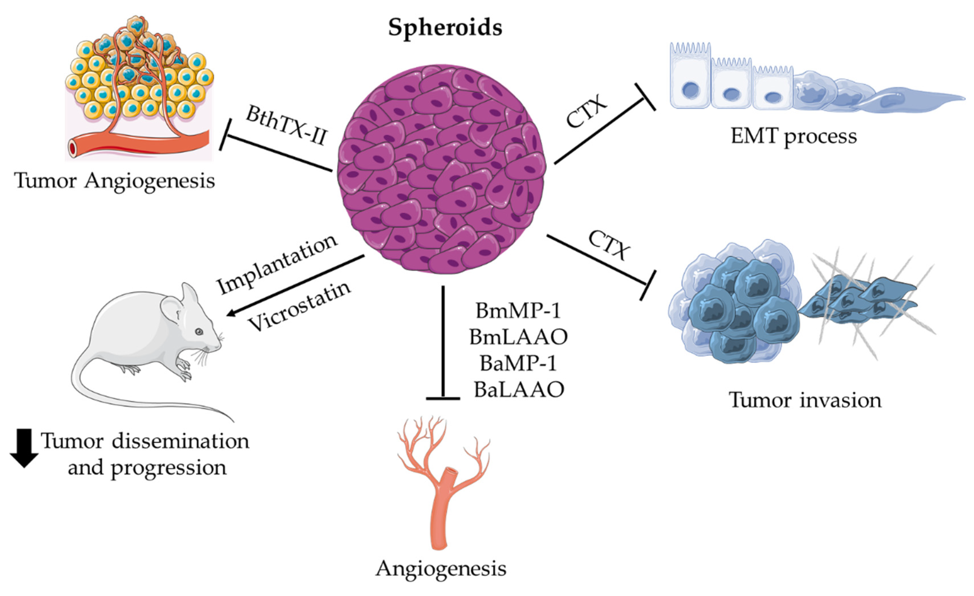
Applications of Spheroids for Evaluating Snake Venom Components

The compound PLA2 derived from different snake venoms has demonstrated the potential for antitumor, antimetastatic, and antiangiogenic effects in vitro in 2D monolayer culture [46,47,48,49]. Further, Azevedo and colleagues demonstrated the antitumor and antimetastatic effects of BthTX-II, an Asp49-PLA2 isolated from the venom of Bothrops jararacussu, on MDA-MB-231 human triple-negative breast cancer cells using the 3D culture technique [50]. The authors monitored the development of spheroids in Matrigel for 7 days and reported that the presence of different concentrations of BthTX-II (1, 10, and 50 µg/mL) impaired spheroid formation and tumor growth compared to the non-tumorigenic MCF10A cell line [50]. In their recent study, these authors demonstrated the antiangiogenic effect of the BthTX-II molecule by co-culturing MDA-MB-231 spheroids with an HUVEC vessel network on Matrigel. The authors reported that during the interaction between tumor spheroids and endothelial cells, BthTX-II promoted the disruption of the HUVEC vessel network by inhibiting endothelial cell aggregation, exhibiting a complete inhibition of cell co-culture migration and proliferation compared to the control cells cultured in Matrigel [51]. A different study reported the modulatory effect of Crotoxin (CTX), a heterodimeric PLA2 present in the Crotalus durissus terrificus venom, during the epithelial–mesenchymal transition (EMT) process. In this study, spheroids composed of non-small-cell lung cancer cells (NSCLC, A549, and Calu-3 cell lines) and normal lung fibroblasts cells (MCR-5 cell line) were utilized to imitate the early tumor–stroma interactions, i.e., to mimic the avascular tumor initiation step [52]. However, unlike BthTX-II, CTX did not interfere with spheroid formation. The use of 12.5 nM of CTX promoted the reduction in the invasion area of the cells that migrated out from the spheroids toward the 3D collagen matrix. Moreover, this effect was observed to be correlated to the decreased protein expression levels of EMT markers, such as N-cadherin, α-SMA, and integrin αv, and was accompanied by decreased secretory levels of MMP-9 and MMP-13 as well as the cytokines and growth factors associated with the EMT process, particularly from the CXCL5/CXCR2 and IL-8/CXCR1/CXCR2 axes [52]. Both of the above-stated studies demonstrated that the antimetastatic and antiangiogenic effects of the snake venom constituent PLA2 were not related to catalytic activity but due to interaction with integrin-mediated signaling pathways [51,52]. Another component from C. d. terrificus, a myo-neurotoxin named crotamine (Crot), a cell-penetrating peptide (CPP), was synthesized as an analog (sCrot) [53]. It was demonstrated that sCrot could selectively penetrate a spheroid model composed of melanoma cells as well as other tumor cells in suspension. As opposed to CTX and BthTX-II, which exhibit antitumor effects, sCrot could serve as a model for tumor microenvironment investigations for studying cancer and stromal cell interactions and also as a carrier of antitumor drugs for evaluating novel therapeutics in drug development [53].
Another class of snake venom components is disintegrins, a family of small, non-enzymatic substances containing the arginine-glycine-aspartic acid (RGD) peptide sequence. Disintegrins have been detected in the venoms derived from the Viperidae, Crotalidae, Atractaspididae, Elapidae, and Colubridae snake families. Disintegrins bind specifically to certain integrins expressed by tumor cells and the endothelial cells in the tumor microenvironment, such as αvβ3, αvβ5, α5β1, and αIIbβ3 [23,54,55]. Swenson and colleagues studied the effect of vicrostatin, a disintegrin synthesized from the natural snake venom disintegrin named contortrostatin, on a spheroid model composed of SKOV3 ovarian cancer cells that was implanted in a mouse model to simulate a condition of solid tumor resistance to chemotherapy [56]. Vicrostatin was observed to promote the highly effective inhibition of ovarian cancer dissemination and progression. In addition, bioluminescence imaging results revealed ~95–98% inhibition of tumor growth [56].
Bhat and colleagues reported the application of P-I metalloproteinases and l-amino acid oxidases (LAAO) derived from the genus Bothrops in the spheroids composed of endothelial cells [57]. P-I metalloproteinases belong to the group of SVMPs, which are large multidomain proteins containing a pro-enzyme domain and a conserved zinc-protease domain. SVMPs are classified into three types, P-I to P-III, among which the P-I metalloproteinases contain only one metalloproteinase domain [58]. LAAO is a flavoenzyme that converts the stereospecific l-amino acid into the corresponding alpha-keto acid along with hydrogen peroxide and ammonia as byproducts [23]. In the above-stated study, the P-I metalloproteinases and LAAO toxins were isolated from Bothrops moojeni (BmMP-1 and BmLAAO, respectively) and Bothrops atrox (BaMP-1 and BaLAAO, respectively), and subsequently evaluated using HUVEC spheroids within a collagen matrix. The P-I metalloproteinases and LAAO from both the Bothrops species promoted the modulation of angiogenesis by reducing sprout outgrowth in the 3D spheroid model. The authors, therefore, recommended using these toxins for designing an antiangiogenic strategy for pathological conditions in which angiogenesis is exacerbated, such as tumor growth, metastasis, diabetic retinopathy, and inflammatory diseases [57]. Figure 1 summarizes the main effects of different snake venom toxins applied in the spheroid model.

4. Organoid Technology

The term organoid was initially used to refer to a specialized and individual organ-like structure constructed in vitro within 3D gels from small tissue fragments derived from the patients’ tissue separated from stroma [1]. Currently, however, the organoid technology involves a variety of tissue culture techniques for preparing self-organizing and self-renewing 3D cultures from embryonic stem cells (ESC) or organ-restricted adult stem cells (ASC) [59]. Both ESC and ASC approaches exploit the seemingly infinite expansion potential of normal stem cells in culture to recapitulate the cell functionality and morphology similar to that exhibited by the native tissue [42]. In order to induce a self-organizing structure, cells are embedded in a hydrogel rich in matrix extracellular proteins, such as Matrigel or Basement Membrane Extract (BME), to simulate the appropriate physiological microenvironment [42,60].
In the context of drug development, organoids serve as a reliable 3D model in a physiologically relevant manner for investigating the pharmacokinetics and toxicity of drug candidates [9]. Since the phenotype, genotype, and metabolic profiles of organoids are highly similar to those of the native tissues, such as cancer tissues, tumor organoids may be utilized as a valuable model for drug candidate screening and understanding the pharmacodynamics of a potential pharmacological drug candidate [1].

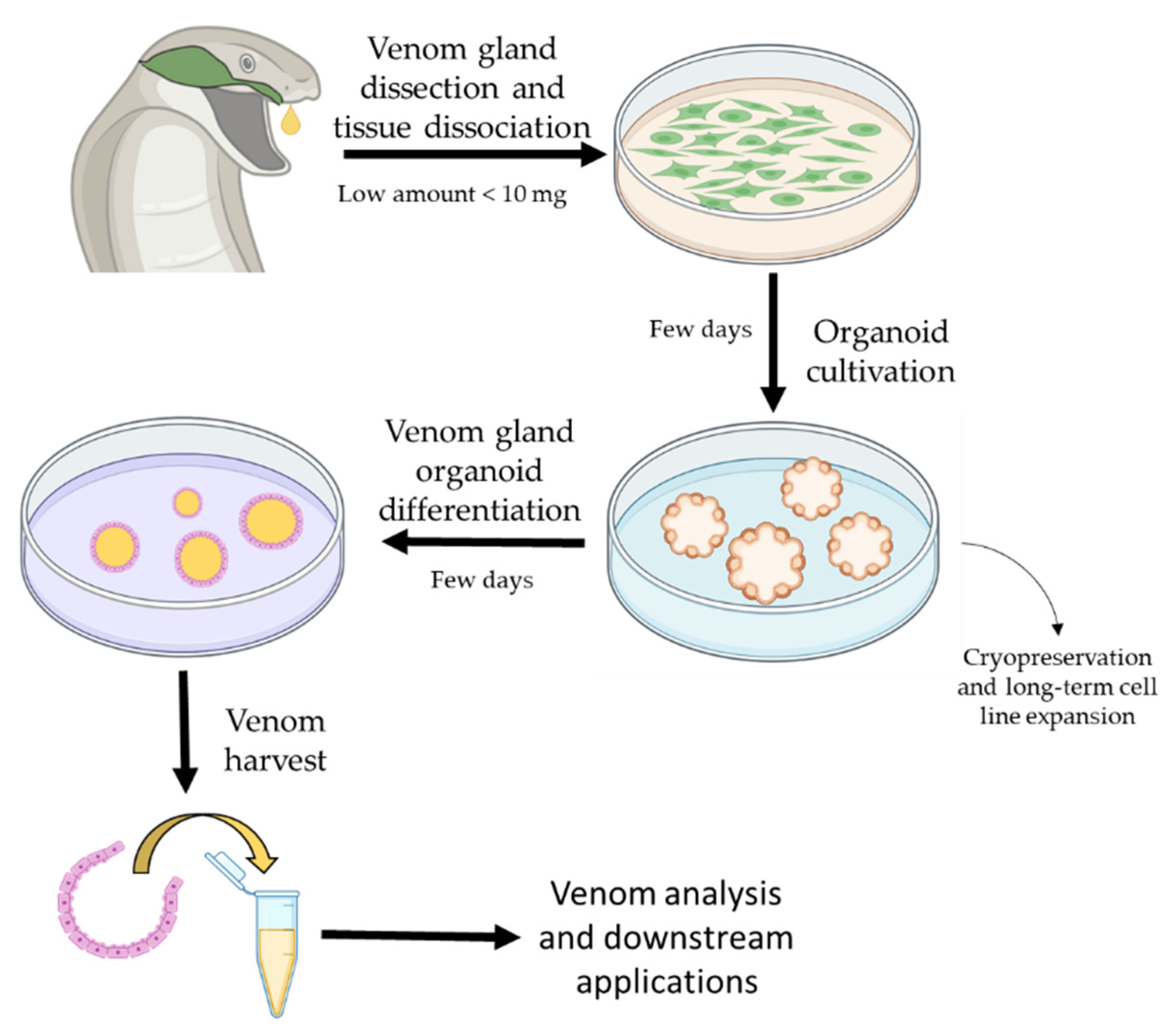
Application of Organoids in Snake Venom Research

Recently, an organoid platform with the potential for in vitro snake venom production has been established. Yorick and collaborators reported developing functional snake venom glands from nine different species belonging to two major families of venomous snakes—the Elapidae (Naja pallida, Naja annulifera, Naja nivea, Naja atra, and Aspidelaps lubricus cowlesi) and the Viperidae (Echis ocellatus, Deinagkistrodon acutus, Crotalus atrox, and Bitis arietans) [61]. The authors dissected both late-stage embryo and adult specimens to obtain the venom gland tissue cells, which were then cultured into mini-organs in long-term culture [61]. First, the venom gland tissue was dissociated to release cells, which were homogenized and then embedded into a 3D environment BME for generating organoids that were allowed to expand (Figure 2) [61,62,63]. The technology produced organoids that were phenotypically similar to natural venom glands and also exhibited similar functions as these secreted, functionally active venom components. Interestingly, the venom gland organoids exhibited cellular heterogeneity as different cell types produced different compounds of the venom [61,62]. The authors also used another 3D cell culture technology known as the organ-on-chip technology to assess the biological activity of the organoid-secreted venom peptides by exposing the murine muscle cells to the organoid supernatant. The results revealed that the firing of the muscle cells was terminated, which simulated the paralysis condition observed upon snakebite envenoming [61]. Studies have also reported the in vitro cell suspension culture of secretory cells from Bothrops jararaca venom glands for up to 21 days [64,65,66], with the synthesized toxin exhibiting hemorrhagic activity similar to that of the toxin from the venom glands harvested directly from B. jararaca specimens [66,67]. However, due to the lack of representation of tissue complexity, as demonstrated by the organoids, the in vitro cell suspension culture of secretory cells had a short lifespan, as opposed to the organoids model that could be cultured for over 2 years [62]. The above study contributed immensely to the establishment of a platform for venom-based drug development and also to the research on antivenoms.

5. Conclusions

The current challenge is not just the identification of natural substances with potential pharmacological and therapeutic properties but also the establishment of cellular and biomimetic microenvironment platforms, considering the cellular, extracellular matrix, and tissue elements for the expansion of mechanistic studies that require low concentrations of snake venoms, toxins, and other isolated compounds. In this context, 3D cell culture models, such as spheroids and organoids, have demonstrated great potential. The mechanistic studies that were conducted to reveal the effect of snake venom components on normal as well as tumor cells and the interactions of these substances with other elements in the cellular microenvironment, such as stromal cells and the extracellular matrix, have reported promising outcomes. The main advances in the determination of the antitumor activity of venoms and toxins include the unraveling of the mechanisms of action of these substances in relation to each element of the cellular microenvironment as well as the role of autocrine and paracrine mediation in this process. Therefore, the use of a combination of in vitro 3D culture technology and natural substances, such as snake venom components, represents a promising preclinical strategy for the fields of drug development, cancer target therapy, antivenom research, and tissue replacement.

Author Contributions

Conceptualization, E.E.K.; writing—original draft preparation, E.E.K.; writing—review and editing, E.E.K., V.L.V., S.C.S.; visualization, V.L.V.; S.C.S.; supervision, E.E.K. All authors have read and agreed to the published version of the manuscript.

Funding

This research received no external funding.

Institutional Review Board Statement

Not applicable.

Informed Consent Statement

Not applicable.

Data Availability Statement

Not applicable.

Conflicts of Interest

The authors declare no conflict of interest.

References

  1. Langhans, S.A. Three-dimensional in vitro cell culture models in drug discovery and drug repositioning. Front. Pharmacol. 2018, 9, 6. [Google Scholar] [CrossRef] [PubMed]
  2. Ravi, M.; Paramesh, V.; Kaviya, S.R.; Anuradha, E.; Paul Solomon, F.D. 3D cell culture systems: Advantages and applications. J. Cell. Physiol. 2015, 230, 16–26. [Google Scholar] [CrossRef]
  3. Jensen, C.; Teng, Y. Is It Time to Start Transitioning From 2D to 3D Cell Culture? Front. Mol. Biosci. 2020, 7, 33. [Google Scholar] [CrossRef] [Green Version]
  4. Costa, E.C.; Moreira, A.F.; de Melo-Diogo, D.; Gaspar, V.M.; Carvalho, M.P.; Correia, I.J. 3D tumor spheroids: An overview on the tools and techniques used for their analysis. Biotechnol. Adv. 2016, 34, 1427–1441. [Google Scholar] [CrossRef]
  5. Ravi, M.; Ramesh, A.; Pattabhi, A. Contributions of 3D Cell Cultures for Cancer Research. J. Cell. Physiol. 2017, 232, 2679–2697. [Google Scholar] [CrossRef]
  6. Fang, Y.; Eglen, R.M. Three-Dimensional Cell Cultures in Drug Discovery and Development. SLAS Discov. 2017, 22, 456–472. [Google Scholar] [CrossRef] [PubMed] [Green Version]
  7. Lv, D.; Hu, Z.; Lu, L.; Lu, H.; Xu, X. Three-dimensional cell culture: A powerful tool in tumor research and drug discovery. Oncol. Lett. 2017, 14, 6999–7010. [Google Scholar] [CrossRef] [PubMed] [Green Version]
  8. Decarli, M.C.; do Amaral, R.L.; Dos Santos, D.P.; Tofani, L.B.; Katayama, E.; Rezende, R.A.; da Silva, J.V.; Swiech, K.; Suazo, C.A.; Mota, C.; et al. Cell spheroids as a versatile research platform: Formation mechanisms, high throughput production, characterization and applications. Biofabrication 2021, 13, 032002. [Google Scholar] [CrossRef]
  9. Zhou, Z.; Zhu, J.; Jiang, M.; Sang, L.; Hao, K.; He, H. The Combination of Cell Cultured Technology and In Silico Model to Inform the Drug Development. Pharmaceutics 2021, 13, 704. [Google Scholar] [CrossRef]
  10. Ong, C.S.; Zhou, X.; Han, J.; Huang, C.Y.; Nashed, A.; Khatri, S.; Mattson, G.; Fukunishi, T.; Zhang, H.; Hibino, N. In vivo therapeutic applications of cell spheroids. Biotechnol. Adv. 2018, 36, 494–505. [Google Scholar] [CrossRef]
  11. Bordon, K.C.F.; Cologna, C.T.; Fornari-Baldo, E.C.; Pinheiro-Júnior, E.L.; Cerni, F.A.; Amorim, F.G.; Anjolette, F.A.P.; Cordeiro, F.A.; Wiezel, G.A.; Cardoso, I.A.; et al. From Animal Poisons and Venoms to Medicines: Achievements, Challenges and Perspectives in Drug Discovery. Front. Pharmacol. 2020, 11, 1132. [Google Scholar] [CrossRef]
  12. Waheed, H.; Moin, S.F.; Choudhary, M.I. Snake Venom: From Deadly Toxins to Life-saving Therapeutics. Curr. Med. Chem. 2017, 24, 1874–1891. [Google Scholar] [CrossRef]
  13. Camargo, A.C.M.; Ianzer, D.; Guerreiro, J.R.; Serrano, S.M.T. Bradykinin-potentiating peptides: Beyond captopril. Toxicon 2012, 59, 516–523. [Google Scholar] [CrossRef] [PubMed]
  14. Ferreira, S.H.; Rocha e Silva, M. Potentiation of bradykinin and eledoisin by BPF (bradykinin potentiating factor) fromBothrops jararaca venom. Experientia 1965, 21, 347–349. [Google Scholar] [CrossRef]
  15. Kochva, E. The origin of snakes and evolution of the venom apparatus. Toxicon 1987, 25, 65–106. [Google Scholar] [CrossRef]
  16. Casewell, N.R.; Wüster, W.; Vonk, F.J.; Harrison, R.A.; Fry, B.G. Complex cocktails: The evolutionary novelty of venoms. Trends Ecol. Evol. 2013, 28, 219–229. [Google Scholar] [CrossRef]
  17. Fry, B.G.; Vidal, N.; Norman, J.A.; Vonk, F.J.; Scheib, H.; Ramjan, S.F.R.; Kuruppu, S.; Fung, K.; Hedges, S.B.; Richardson, M.K.; et al. Early evolution of the venom system in lizards and snakes. Nature 2005, 439, 584–588. [Google Scholar] [CrossRef]
  18. Underwood, G. An overview of venomous snake evolution. In Venomous Snakes: Ecology, Evolution and Snakebite; Clarendon Press: Oxford, UK, 1997; pp. 1–13. [Google Scholar]
  19. Chippaux, J.P.; Williams, V.; White, J. Snake venom variability: Methods of study, results and interpretation. Toxicon 1991, 29, 1279–1303. [Google Scholar] [CrossRef]
  20. Gutiérrez, J.M.; Calvete, J.J.; Habib, A.G.; Harrison, R.A.; Williams, D.J.; Warrell, D.A. Snakebite envenoming. Nat. Rev. Dis. Prim. 2017, 3, 1–21. [Google Scholar] [CrossRef]
  21. Fox, J.W.; Serrano, S.M.T. Timeline of key events in snake venom metalloproteinase research. J. Proteom. 2009, 72, 200–209. [Google Scholar] [CrossRef] [PubMed]
  22. Li, L.; Huang, J.; Lin, Y. Snake Venoms in Cancer Therapy: Past, Present and Future. Toxins 2018, 10, 346. [Google Scholar] [CrossRef] [PubMed] [Green Version]
  23. Calderon, L.A.; Sobrinho, J.C.; Zaqueo, K.D.; de Moura, A.A.; Grabner, A.N.; Mazzi, M.V.; Marcussi, S.; Nomizo, A.; Fernandes, C.F.C.; Zuliani, J.P.; et al. Antitumoral Activity of Snake Venom Proteins: New Trends in Cancer Therapy. Biomed Res. Int. 2014, 2014, 203639. [Google Scholar] [CrossRef] [PubMed] [Green Version]
  24. Koh, D.C.I.; Armugam, A.; Jeyaseelan, K. Snake venom components and their applications in biomedicine. Cell. Mol. Life Sci. 2006, 63, 3030–3041. [Google Scholar] [CrossRef]
  25. Sanhajariya, S.; Duffull, S.B.; Isbister, G.K. Pharmacokinetics of snake venom. Toxins 2018, 10, 73. [Google Scholar] [CrossRef] [PubMed] [Green Version]
  26. Xiao, H.; Pan, H.; Liao, K.; Yang, M.; Huang, C. Snake Venom PLA2, a Promising Target for Broad-Spectrum Antivenom Drug Development. Biomed Res. Int. 2017, 2017, 6592820. [Google Scholar] [CrossRef] [PubMed] [Green Version]
  27. Gutiérrez, J.M.; Lomonte, B. Phospholipases A2: Unveiling the secrets of a functionally versatile group of snake venom toxins. Toxicon 2013, 62, 27–39. [Google Scholar] [CrossRef] [PubMed]
  28. Ferraz, C.R.; Arrahman, A.; Xie, C.; Casewell, N.R.; Lewis, R.J.; Kool, J.; Cardoso, F.C. Multifunctional toxins in snake venoms and therapeutic implications: From pain to hemorrhage and necrosis. Front. Ecol. Evol. 2019, 7, 218. [Google Scholar] [CrossRef] [Green Version]
  29. El-Aziz, T.M.A.; Soares, A.G.; Stockand, J.D. Snake Venoms in Drug Discovery: Valuable Therapeutic Tools for Life Saving. Toxins 2019, 11, 564. [Google Scholar] [CrossRef] [Green Version]
  30. Mackessy, S.P. Thrombin-Like Enzymes in Snake Venoms. In Toxins and Hemostasis; Springer: Heidelberg, Germany, 2010; pp. 519–557. [Google Scholar] [CrossRef]
  31. Slagboom, J.; Kool, J.; Harrison, R.A.; Casewell, N.R. Haemotoxic snake venoms: Their functional activity, impact on snakebite victims and pharmaceutical promise. Br. J. Haematol. 2017, 177, 947–959. [Google Scholar] [CrossRef] [Green Version]
  32. Izidoro, L.F.M.; Sobrinho, J.C.; Mendes, M.M.; Costa, T.R.; Grabner, A.N.; Rodrigues, V.M.; Da Silva, S.L.; Zanchi, F.B.; Zuliani, J.P.; Fernandes, C.F.C.; et al. Snake venom L-amino acid oxidases: Trends in pharmacology and biochemistry. Biomed Res. Int. 2014, 2014, 196754. [Google Scholar] [CrossRef] [PubMed] [Green Version]
  33. Bickler, P.E. Amplification of Snake Venom Toxicity by Endogenous Signaling Pathways. Toxins 2020, 12, 68. [Google Scholar] [CrossRef] [PubMed] [Green Version]
  34. Rivas-Mercado, E.A.; Garza-Ocañas, L. Disintegrins obtained from snake venom and their pharmacological potential. Med. Univ. 2017, 19, 32–37. [Google Scholar] [CrossRef]
  35. Arlinghaus, F.T.; Eble, J.A. C-type lectin-like proteins from snake venoms. Toxicon 2012, 60, 512–519. [Google Scholar] [CrossRef] [PubMed]
  36. Eble, J.A. Structurally Robust and Functionally Highly Versatile—C-Type Lectin (-Related) Proteins in Snake Venoms. Toxins 2019, 11, 136. [Google Scholar] [CrossRef] [Green Version]
  37. Adade, C.M.; Carvalho, A.L.O.; Tomaz, M.A.; Costa, T.F.R.; Godinho, J.L.; Melo, P.A.; Lima, A.P.C.A.; Rodrigues, J.C.F.; Zingali, R.B.; Souto-Padrón, T. Crovirin, a Snake Venom Cysteine-Rich Secretory Protein (CRISP) with Promising Activity against Trypanosomes and Leishmania. PLoS Negl. Trop. Dis. 2014, 8, e3252. [Google Scholar] [CrossRef] [PubMed]
  38. Yamazaki, Y.; Morita, T. Structure and function of snake venom cysteine-rich secretory proteins. Toxicon 2004, 44, 227–231. [Google Scholar] [CrossRef]
  39. Péterfi, O.; Boda, F.; Szabó, Z.; Ferencz, E.; Bába, L. Hypotensive Snake Venom Components—A Mini-Review. Molecules 2019, 24, 2778. [Google Scholar] [CrossRef] [Green Version]
  40. Murayama, N.; Hayashi, M.A.F.; Ohi, H.; Ferreira, L.A.F.; Hermann, V.V.; Saito, H.; Fujita, Y.; Higuchi, S.; Fernandes, B.L.; Yamane, T.; et al. Cloning and sequence analysis of a Bothrops jararaca cDNA encoding a precursor of seven bradykinin-potentiating peptides and a C-type natriuretic peptide. Proc. Natl. Acad. Sci. USA 1997, 94, 1189–1193. [Google Scholar] [CrossRef] [PubMed] [Green Version]
  41. Katt, M.E.; Placone, A.L.; Wong, A.D.; Xu, Z.S.; Searson, P.C. In Vitro Tumor Models: Advantages, Disadvantages, Variables, and Selecting the Right Platform. Front. Bioeng. Biotechnol. 2016, 4, 12. [Google Scholar] [CrossRef]
  42. Kronemberger, G.S.; Carneiro, F.A.; Rezende, D.F.; Baptista, L.S. Spheroids and organoids as humanized 3D scaffold-free engineered tissues for SARS-CoV-2 viral infection and drug screening. Artif. Organs 2021, 45, 548–558. [Google Scholar] [CrossRef] [PubMed]
  43. Kim, S.; Kim, E.M.; Yamamoto, M.; Park, H.; Shin, H. Engineering Multi-Cellular Spheroids for Tissue Engineering and Regenerative Medicine. Adv. Healthc. Mater. 2020, 9, 2000608. [Google Scholar] [CrossRef]
  44. Nunes, A.S.; Barros, A.S.; Costa, E.C.; Moreira, A.F.; Correia, I.J. 3D tumor spheroids as in vitro models to mimic in vivo human solid tumors resistance to therapeutic drugs. Biotechnol. Bioeng. 2018, 116, 206–226. [Google Scholar] [CrossRef] [Green Version]
  45. Kim, J. Bin Three-dimensional tissue culture models in cancer biology. Semin. Cancer Biol. 2005, 15, 365–377. [Google Scholar] [CrossRef]
  46. Bazaa, A.; Luis, J.; Srairi-Abid, N.; Kallech-Ziri, O.; Kessentini-Zouari, R.; Defilles, C.; Lissitzky, J.-C.; El Ayeb, M.; Marrakchi, N. MVL-PLA2, a phospholipase A2 from Macrovipera lebetina transmediterranea venom, inhibits tumor cells adhesion and migration. Matrix Biol. 2009, 28, 188–193. [Google Scholar] [CrossRef]
  47. Bazaa, A.; Pasquier, E.; Defilles, C.; Limam, I.; Kessentini-Zouari, R.; Kallech-Ziri, O.; El Battari, A.; Braguer, D.; El Ayeb, M.; Marrakchi, N.; et al. MVL-PLA2, a snake venom phospholipase A2, inhibits angiogenesis through an increase in microtubule dynamics and disorganization of focal adhesions. PLoS ONE 2010, 5, e10124. [Google Scholar] [CrossRef] [Green Version]
  48. Zouari-Kessentini, R.; Srairi-Abid, N.; Bazaa, A.; El Ayeb, M.; Luis, J.; Marrakchi, N. Antitumoral potential of Tunisian snake venoms secreted phospholipases A2. Biomed Res. Int. 2013, 2013, 391389. [Google Scholar] [CrossRef] [Green Version]
  49. Kato, E.E.; Pimenta, L.A.; de Almeida, M.E.S.; Zambelli, V.O.; Santos, M.F.; Sampaio, S.C. Crotoxin Inhibits Endothelial Cell Functions in Two- and Three-dimensional Tumor Microenvironment. Front. Pharmacol. 2021, 12, 1. [Google Scholar] [CrossRef]
  50. De Vasconcelos Azevedo, F.V.P.; Zóia, M.A.P.; Lopes, D.S.; Gimenes, S.N.; Vecchi, L.; Alves, P.T.; Rodrigues, R.S.; Silva, A.C.A.; Yoneyama, K.A.G.; Goulart, L.R.; et al. Antitumor and antimetastatic effects of PLA2-BthTX-II from Bothrops jararacussu venom on human breast cancer cells. Int. J. Biol. Macromol. 2019, 135, 261–273. [Google Scholar] [CrossRef]
  51. Van Petten de Vasconcelos Azevedo, F.; Lopes, D.S.; Zóia, M.A.P.; Correia, L.I.V.; Saito, N.; Fonseca, B.B.; Polloni, L.; Teixeira, S.C.; Goulart, L.R.; de Melo Rodrigues Ávila, V. A New Approach to Inhibiting Triple-Negative Breast Cancer: In Vitro, Ex Vivo and In Vivo Antiangiogenic Effect of BthTx-II, a PLA2-Asp-49 from Bothrops jararacussu Venom. Biomolecules 2022, 12, 258. [Google Scholar] [CrossRef]
  52. Kato, E.E.; Sampaio, S.C. Crotoxin Modulates Events Involved in Epithelial–Mesenchymal Transition in 3D Spheroid Model. Toxins 2021, 13, 830. [Google Scholar] [CrossRef] [PubMed]
  53. Mambelli-Lisboa, N.C.; Sciani, J.M.; da Silva, A.R.B.P.; Kerkis, I. Co-Localization of Crotamine with Internal Membranes and Accentuated Accumulation in Tumor Cells. Molecules 2018, 23, 968. [Google Scholar] [CrossRef] [Green Version]
  54. Arruda Macêdo, J.K.; Fox, J.W.; de Souza Castro, M. Disintegrins from snake venoms and their applications in cancer research and therapy. Curr. Protein Pept. Sci. 2015, 16, 532–548. [Google Scholar] [CrossRef] [Green Version]
  55. Calvete, J.J.; Marcinkiewicz, C.; Monleón, D.; Esteve, V.; Celda, B.; Juárez, P.; Sanz, L. Snake venom disintegrins: Evolution of structure and function. Toxicon 2005, 45, 1063–1074. [Google Scholar] [CrossRef]
  56. Swenson, S.D.; Markland, F.S.; Minea, R. A Novel, Non-Cytotoxic, Anti-Invasive Therapeutic Agent for Ovarian Cancer. In Advances in Biological Sciences Research; Atlantis Press: Paris, France, 2016; pp. 159–165. [Google Scholar] [CrossRef] [Green Version]
  57. Bhat, S.K.; Joshi, M.B.; Vasishta, S.; Jagadale, R.N.; Biligiri, S.G.; Coronado, M.A.; Arni, R.K.; Satyamoorthy, K. P-I metalloproteinases and L-amino acid oxidases from Bothrops species inhibit angiogenesis. J. Venom. Anim. Toxins Incl. Trop. Dis. 2021, 27, 1–15. [Google Scholar] [CrossRef]
  58. Takeda, S.; Takeya, H.; Iwanaga, S. Snake venom metalloproteinases: Structure, function and relevance to the mammalian ADAM/ADAMTS family proteins. Biochim. Biophys. Acta-Proteins Proteom. 2012, 1824, 164–176. [Google Scholar] [CrossRef]
  59. Drost, J.; Clevers, H. Organoids in cancer research. Nat. Rev. Cancer 2018, 18, 407–418. [Google Scholar] [CrossRef]
  60. Kretzschmar, K. Cancer research using organoid technology. J. Mol. Med. 2021, 99, 501–515. [Google Scholar] [CrossRef] [PubMed]
  61. Post, Y.; Puschhof, J.; Beumer, J.; Kerkkamp, H.M.; de Bakker, M.A.G.; Slagboom, J.; de Barbanson, B.; Wevers, N.R.; Spijkers, X.M.; Olivier, T.; et al. Snake Venom Gland Organoids. Cell 2020, 180, 233–247.e21. [Google Scholar] [CrossRef]
  62. Puschhof, J.; Post, Y.; Beumer, J.; Kerkkamp, H.M.; Bittenbinder, M.; Vonk, F.J.; Casewell, N.R.; Richardson, M.K.; Clevers, H. Derivation of snake venom gland organoids for in vitro venom production. Nat. Protoc. 2021, 16, 1494–1510. [Google Scholar] [CrossRef]
  63. Vogt, N. Venomous organoids. Nat. Methods 2020, 17, 360. [Google Scholar] [CrossRef]
  64. Carneiro, S.M.; Zablith, M.B.; Kerchove, C.M.; Moura-Da-Silva, A.M.; Quissell, D.O.; Markus, R.P.; Yamanouye, N. Venom production in long-term primary culture of secretory cells of the Bothrops jararaca venom gland. Toxicon 2006, 47, 87–94. [Google Scholar] [CrossRef]
  65. Yamanouye, N.; Kerchove, C.M.; Moura-da-Silva, A.M.; Carneiro, S.M.; Markus, R.P. Long-term primary culture of secretory cells of Bothrops jararaca venom gland for venom production in vitro. Nat. Protoc. 2007, 1, 2763–2766. [Google Scholar] [CrossRef]
  66. Viana, L.G.; Valente, R.H.; Heluany, C.S.; Souza-Imberg, A.; Luna, M.S.; Perales, J.; Yamanouye, N. Bothrops jararaca venom gland secretory cells in culture: Effects of noradrenaline on toxin production and secretion. Toxicon 2017, 133, 1–9. [Google Scholar] [CrossRef] [PubMed]
  67. Luna, M.S.; Valente, R.H.; Perales, J.; Vieira, M.L.; Yamanouye, N. Activation of Bothrops jararaca snake venom gland and venom production: A proteomic approach. J. Proteom. 2013, 94, 460–472. [Google Scholar] [CrossRef] [PubMed]
Figure 1. Spheroid applications for the study of snake toxins. This figure was created using Smart-Servier Medical Art (https://smart.servier.com, Accessed on 13 April 2022).
Futurepharmacol 02 00009 g001
Figure 2. Schematic illustration of the venom gland’s organoid generation. Created with BioRender.com.
Futurepharmacol 02 00009 g002
Table 1. Snake venom compounds and their known toxicological effects illustrate their wide range of potential pharmacological applications.
Snake Venom Toxin FamilySnake FamilyToxicological
Effects
Pharmacological
Effects
Reviewed by
PLA2Viperidae, Elapidae, Colubridae.Neurotoxicity, Myotoxicity, Cytotoxicity, Cardiotoxicity, Hemolysis, Edema, HyperalgesiaAntiinflammatory Analgesic, Antitumoral, Antiangiogenic, Antibacterial, Antiviral[26,27]
SVMPViperidae, Elapidae, Atractaspididae, Colubridae.Hemorrhage, Myonecrosis,
Coagulopathy, Tissue Damage
-[28,29]
SVSPViperidae, Elapidae, Colubridae.Hemotoxic, Rupture Capillary Vessels, Pro-coagulant or Anti-coagulant, Fibrinolysis, Platelet AggregationThrombolytic[30,31]
LAAOViperidae, Elapidae.Apoptosis, Hemorrhage, Cytotoxicity, EdemaAntibacterial, Antitumoral, Antiprotozoan, Antiviral[32]
3FTxElapidae, Colubridae.Neurotoxicity, ParalysisAnalgesic[28]
DISViperidae, Atractaspididae, Elapidae, Colubridae.Inhibit Cell-ECM, Loosen
Anchoring Tissue
Antitumor, Anti-platelet[33,34]
CTLViperidae, Elapidae.Induction or inhibition of platelet
aggregation
Antimetastatic, Antiangiogenic[35,36]
CRISPViperidae, Elapidae, Colubridae.Myotoxicity, Inhibition of smooth muscle contractionAntiparasitary[37,38]
BPPViperidae.HypotensionAnti-hypertensive[39,40]
PLA2: Phospholipase A2; SVMP: Snake venom metalloproteinase; SVSP: Snake venom serine protease; LAAO: l-amino acid oxidase; 3FTx: three-finger toxin; DIS: Disintegrin; CTL: C-type lectin; CRISP: Cysteine-rich secretory protein; BPP: Bradykinin-potentiating peptide.
Publisher’s Note: MDPI stays neutral with regard to jurisdictional claims in published maps and institutional affiliations.

