Article

Real-Time Electroencephalography-Guided Binaural Beat Audio Enhances Relaxation and Cognitive Performance: A Randomized, Double-Blind, Sham-Controlled Repeated-Measures Crossover Trial

by
Chanaka N. Kahathuduwa
1,2,
Jessica Blume
2,
Chinnadurai Mani
3 and
Chathurika S. Dhanasekara
2,4,5,*
1
Department of Neurology, Texas Tech University Health Sciences Center, Lubbock, TX 79430, USA
2
MelodiaSync LLC, Lubbock, TX 79415, USA
3
Department of Medical Education, Texas Tech University Health Sciences Center, Lubbock, TX 79430, USA
4
Department of Surgery, Texas Tech University Health Sciences Center, Lubbock, TX 79430, USA
5
Department of Pharmacology and Neuroscience, Texas Tech University Health Sciences Center, Lubbock, TX 79430, USA
*
Author to whom correspondence should be addressed.
Physiologia 2025, 5(4), 44; https://doi.org/10.3390/physiologia5040044 (registering DOI)
Submission received: 11 August 2025 / Revised: 20 October 2025 / Accepted: 22 October 2025 / Published: 24 October 2025

Abstract

Background/Objectives: Binaural beat audio has gained popularity as a non-invasive tool to promote relaxation and enhance cognitive performance, though empirical support has been inconsistent. We developed a novel algorithm integrating real-time electroencephalography (EEG) feedback to dynamically tailor binaural beats to induce relaxed brain states. This study aimed to examine the efficacy and feasibility of this algorithm in a clinical trial. Methods: In a randomized, double-blinded, sham-controlled crossover trial, 25 healthy adults completed two 30 min sessions (EEG-guided intervention versus sham). EEG (Fp1) was recorded using a consumer-grade single-electrode headset, with auditory stimulation adjusted in real time based on EEG data. Outcomes included EEG frequency profiles, stop signal reaction time (SSRT), and novelty encoding task performance. Results: The intervention rapidly reduced dominant EEG frequency in all participants, with 100% achieving <8 Hz and 96% achieving <4 Hz within median 7.4 and 9.0 min, respectively. Compared to the sham, the intervention was associated with an faster novelty encoding reaction time (p = 0.039, dz = −0.225) and trends towards improved SSRT (p = 0.098, dz = −0.209), increased boundary separation in stop trials (p = 0.065, dz = 0.350), and improved inhibitory drift rate (p = 0.067, dz = 0.452) within the limits of the exploratory nature of these findings. Twenty-four (96%) participants reached a target level of <4 Hz with the intervention, while none reached this level with the sham. Conclusions: Real-time EEG-guided binaural beats may rapidly induce low-frequency brain states while potentially preserving or enhancing aspects of executive function. These findings support the feasibility of personalized, closed-loop auditory entrainment for promoting “relaxed alertness.” The results are preliminary and hypothesis-generating, warranting larger, multi-channel EEG studies in ecologically valid contexts.

1. Introduction

Audio-assisted relaxation techniques have gained widespread use for modulating brain states to achieve enhanced relaxation, focus, and cognitive performance. Among these, binaural beats, a form of auditory stimulation that delivers two tones of slightly different frequencies to each ear, have gained significant attention for their potential to entrain brain activity [1,2]. Auditory signals travel from the cochlea through the brainstem auditory nuclei, including the superior olivary complex, where binaural integration occurs, to the inferior colliculus and medial geniculate nucleus before reaching the primary auditory cortex. The auditory cortex maintains direct connections with the prefrontal cortex through the dorsal and ventral auditory streams, as well as the hippocampus and limbic structures, potentially allowing auditory stimulation to influence executive functions such as working memory, inhibitory control, and cognitive flexibility. The superior olivary nucleus, specifically the medial superior olive, detects frequency differences between the tones received by each ear, thus generating perceptual beat frequencies in response to binaural beats [3]. This phenomenon is thought to induce a frequency-following response, wherein the brain’s dominant oscillatory activity, as measured using electroencephalography (EEG), aligns with the frequency of the binaural beats, theoretically promoting shifts towards desired mental states [4,5,6]. The entrainment effect is thought to occur through phase-locking of neural populations, particularly in the auditory cortex and connected regions, potentially spreading to other brain areas through thalamocortical loops and cortico-cortical connections [7].
Despite their popularity, empirical support for the efficacy of binaural beats remains mixed [8,9]. Recent systematic reviews and meta-analyses have provided increasingly nuanced perspectives on auditory entrainment efficacy. Garcia-Argibay et al. [10] reported an overall medium effect size (g = 0.45) for binaural beats on cognition, anxiety, and pain across 22 studies, with effectiveness modulated by exposure duration and frequency. However, Ingendoh et al. [11] found inconsistent evidence for the brainwave entrainment hypothesis, with only 36% of studies supporting entrainment effects and significant methodological heterogeneity limiting comparability. Despite these inconsistencies, meta-analyses have documented robust clinical outcomes: Wang et al. [12] demonstrated that acoustic stimulation significantly improved insomnia severity in eight RCTs. Recent experimental work has demonstrated that daily theta-frequency exposure (6 Hz) over four weeks enhances cognitive function and modulates event-related potentials [13]. Mechanistic investigations have revealed that auditory slow-wave stimulation enhances brain oscillatory activity with immediate cardiovascular changes, including increased parasympathetic activity [14,15]. While collective evidence supports therapeutic potential across cognitive enhancement, anxiety reduction, and sleep improvement, significant methodological heterogeneity underscores the need for standardized protocols [11].
A critical examination of studies included in systematic reviews and meta-analyses that report null or inconsistent results regarding the efficacy of binaural beats on brain entrainment [11,16] reveals several methodological factors that may explain such inconsistencies. First, many studies employed fixed-frequency protocols without considering individual differences in baseline brain activity. For instance, Goodin et al. [17] used a standardized 6 Hz binaural beat protocol but found no anxiety reduction, possibly because participants’ baseline alpha frequencies varied considerably (8–12 Hz range). Second, exposure duration appears critical, with studies using shorter exposures (<10 min) more likely to report null effects compared to longer protocols (>20 min). Third, individual differences in auditory processing and neural plasticity may influence susceptibility to entrainment. Vernon et al. [6] found that only 60% of participants showed measurable entrainment effects, suggesting that one-size-fits-all approaches may be inherently limited. Fourth, the choice of control condition significantly impacts results, with studies using active controls (e.g., different frequencies) showing smaller effect sizes than those using passive controls (i.e., silence). Finally, such approaches risk delivering frequencies that are incongruent with endogenous neural rhythms, which may attenuate efficacy or even result in adverse effects such as dizziness and headaches [18,19]. These findings collectively suggest that personalized, adaptive approaches accounting for individual neural characteristics and providing real-time feedback may be necessary to achieve consistent entrainment effects.
To address these limitations, we developed a novel electroencephalography (EEG)-guided binaural beat intervention that dynamically adapts stimulation frequency based on real-time EEG feedback. We selected a single-electrode, consumer-grade configuration to evaluate feasibility for scalable, wearable use, while targeting prefrontal activity, given its role in executive control and stress regulation [20,21]. As the predominant EEG frequency approaches the target state, the stimulation is maintained or fine-tuned to reinforce the desired state, offering a closed-loop, individualized neuromodulation experience. This EEG-guided approach has the potential to overcome the shortcomings of static stimulation by aligning external auditory inputs with intrinsic neural activity, maximizing the probability of achieving and sustaining a physiologically coherent brain state. While not sufficiently supported empirically, such a strategy may hold promise not only for enhancing subjective relaxation and well-being but also for improving objective markers of cognitive performance, including attention, inhibitory control, and memory encoding [22,23,24]. Given the increasing interest in non-pharmacologic, scalable tools for mental optimization and stress modulation, this personalized intervention represents a timely and evidence-informed innovation.
To evaluate the efficacy of this novel EEG-guided binaural beat intervention, we conducted a randomized, double-blinded, sham-controlled, repeated-measures crossover trial among healthy adults. We specifically aimed to compare the effects of exposure to a 30 min EEG-guided binaural-beat audio intervention on cognitive measures including sustained attention and inhibitory control (stop signal reaction task—SSRT), novel memory encoding and retrieval (novelty encoding task—NET), objective neurophysiological measures of relaxation (predominant EEG frequency bands), as well as subjective self-reported relaxation and cognitive performance among healthy adults. Considering the primary outcomes, we hypothesized that we would observe faster stop signal reaction times in SSRT and novel information retrieval reaction times in NET, along with significantly lower mean predominant EEG frequency bands measured at 5 min intervals with the intervention compared to the sham control condition. Considering secondary outcomes, we hypothesized that exposure to the intervention would result in improvements in subjective indicators of relaxation and cognitive performance (i.e., visual analogue scales), while improving hit rates and false alarm rates of NET. Observations related to deeper low-frequency states were treated as exploratory, as the study’s primary intent was to evaluate cognition and relaxation under closed-loop guidance. Furthermore, additional mechanistic derivatives of SSRT (e.g., drift diffusion parameters) and NET (e.g., d-prime) were considered as exploratory outcomes. We hypothesized that exposure to the intervention would result in improvements in the above measures towards enhancing both subjective and objective indicators of relaxation and improving subjective and objective markers of cognitive performance.

2. Results

2.1. Participant Characteristics

Twenty-eight participants were screened. Three participants were excluded during screening for the following reasons: history of childhood seizures, taking anti-depressants, and lack of availability. All recruited participants (13 males and 12 females) completed the study. The mean age ± SD of participants was 32.2 ± 12.9 years (range: 18–64 years), with similar age distributions between males (32.9 ± 13.3 years) and females (31.5 ± 12.4 years).

2.2. Primary Relaxation EEG Indices

At baseline, predominant EEG frequency did not differ between conditions (mean ± SD of intervention 18.44 ± 2.60 Hz; sham 17.92 ± 2.74 Hz; p = 0.520; Table 1). During exposure, the intervention elicited a rapid downshift in predominant frequency relative to sham (Figure 1), with highly significant between-condition differences at every intermediate time point (5–20 min and pre-awakening; all p ≤ 0.005; Table 1).
Frequency band distributions corroborated these effects (Table 2). In the intervention condition, 100% of participants reached <8 Hz (“relaxed”) with a median time of 441.6 s (7.4 min), and all who reached <8 Hz sustained it for ≥3 min. By 10 min, 80% were in delta (0–4 Hz), increasing to 96% by 20 min. In contrast, the sham condition remained predominantly beta-dominant throughout (88–96%), with only 12% reaching <8 Hz (median 723.8 s; 12.1 min), and just one participant sustaining <8 Hz for ≥3 min.

2.3. Exploratory EEG Indices

Exploratory analyses examined sustained deep low-frequency dominance. In the intervention condition, 24/25 (96%) participants reached <4 Hz (median 541.6 s; 9.0 min), and 22/25 sustained <4 Hz for ≥3 min (Table 2). In the sham condition, 1/25 (4%) reached <4 Hz (at 1080 s) and none sustained it for ≥3 min. These indices are reported as exploratory and are not intended to imply sleep staging.

2.4. Cognitive Outcomes

2.4.1. Stop Signal Task

Results of the stop signal reaction task are summarized in Table 3 and Table S1. A statistical trend was noted for the intervention x time interaction for SSRT (F1, 68.8 = 2.82, p = 0.098, dz = −0.209), driven by a mean improvement of (i.e., decreased) SSRT, while worsening (i.e., increased) of the mean SSRT associated with the control condition. This finding suggested a directional trend of improvement in inhibitory control following exposure to the intervention. Analysis of drift diffusion parameters was consistent with the above finding. Specifically, a mean increase in boundary separation while considering stop trials was seen with the intervention, while a decrease in mean boundary separation was observed with exposure to the control condition, resulting in a statistical trend in the interaction (F1, 68.0 = 3.52, p = 0.065, dz = 0.350). This suggested that exposure to the intervention was associated with potentially enhanced cognitive caution during stop trials, allowing more evidence to be accumulated before making decisions, which could translate to fewer impulsive actions. Exposure to the intervention was also associated with a mean decrease in inhibitory drift rate, while the control condition was associated with an increase in the inhibitory drift rate, resulting in a significant main effect of time (F1, 69.5 = 4.80, p = 0.032) and a statistical trend of the interaction (F1, 69.8 = 3.40, p = 0.070, dz = 0.452), indicating directional improvement of efficiency in processing stop signals with the intervention.
Comprehensive reliability analyses were conducted to evaluate the stability of SSRT estimates obtained from 90-trial sessions [25]. Split-half reliability (i.e., odd vs. even visits) yielded a Spearman–Brown corrected coefficient of 0.71, indicating acceptable internal consistency. Test–retest reliability across four visits showed an intra-class correlation coefficient (2,1) of 0.478 [0.258, 0.701], suggesting fair temporal stability [26,27], with pairwise visit correlations ranging from r = 0.282 to r = 0.698. Between-subject (SD = 0.0387) and within-subject (mean SD = 0.036) variability were comparable (variance ratio = 1.077), with a mean within-subject coefficient of variation of 0.379 (median = 0.360). Cross-correlation analysis across intervention conditions and time points also revealed moderate to strong correlations (r = 0.271 to r = 0.685). These reliability indices demonstrate that drift diffusion model-based SSRT estimates obtained through maximum likelihood estimation exhibited acceptable psychometric properties despite the limited trial count.

2.4.2. Novelty Encoding Task

Results of the novelty encoding task are summarized in Table 3 and Table S2. The intervention was associated with improved reaction time performance compared to the control condition, with a significant interaction effect (F1, 54.4 = 4.50, p = 0.039, dz = −0.225). Specifically, mean reaction time decreased from 1.23 s to 1.10 s in the intervention group, while slightly increasing from 1.13 s to 1.16 s in the control group. This finding may suggest enhanced information processing speed following the binaural beat intervention. However, this finding should be interpreted cautiously, given the exploratory nature of the analysis and the potential for Type I error. Both groups showed improvements in false alarm rates (and consequently, specificity) across time, with a significant main effect of time (F1, 57.2 = 4.18, p = 0.046) but no significant interaction. Similarly, response bias showed a trend towards more conservative decision-making in both groups over time (F1, 56.2 = 3.26, p = 0.077). Other cognitive performance measures, including hit rate, d-prime, precision, and F1 score, showed numerical improvements in the intervention group, but these changes did not reach statistical significance compared to the control condition. Collectively, these results indicate that the EEG-guided binaural beat intervention primarily enhanced information retrieval speed while maintaining accuracy metrics.

2.5. Subjective Ratings

Results of the subjective ratings are summarized in Table 3 and Table S3. Subjective measures of relaxation and sleepiness increased significantly over time in both conditions (main effect of time: F1,72 = 15.02, p < 0.001 and F1,72 = 33.73, p < 0.001, respectively), while self-reported focus decreased in both conditions (main effect of time: F1,72 = 6.78, p = 0.011) without significant condition × time interactions. Perceived cognitive performance showed a time effect (F1,72 = 4.99, p = 0.029) and a marginal interaction (F1,72 = 2.79, p = 0.099, dz = 0.449), reflecting a decline with sham from 69.3 mm to 62.2 mm and relative maintenance with intervention at 66.9 mm to 67.3 mm. Well-being did not change significantly.

2.6. Sensitivity Analyses

Exploratory sensitivity analyses performed with the addition of the order of assignment and carryover effects (i.e., number of days between the two sessions) did not reveal any significant effects for these covariates.

3. Discussion

In this randomized, double-blinded, sham-controlled crossover study, real-time EEG-guided binaural beats were associated with rapid, reproducible downshifts in dominant EEG frequency relative to sham and were accompanied by potential behavioral benefits, including faster reaction times on the novelty-encoding task, reflecting a small effect size of improvement. Statistical trends also favored directional improvements in inhibitory control on the stop signal task (decreased SSRT showing a small effect size improvement, while increased a2 and v2 trajectories showed small–moderate effect size improvements). These cognitive benefits were also reflected in the trend of small–moderate effect size improvement of subjective sense of cognitive performance. Observations of sustained <4 Hz activity occurred frequently during the intervention; however, as measures of sleep were not pre-specified outcomes and no polysomnography was collected, we treat them as exploratory indices of deep low-frequency dominance rather than sleep staging. Taken together, the data suggest that closed-loop stimulation may induce a state of “relaxed alertness” without compromising, and potentially supporting, aspects of executive functions. However, it should be emphasized that these findings remain preliminary, given the pilot and exploratory nature of our study and the multiplicity of outcomes.
The EEG findings of this study provide supportive evidence for the primary feasibility but also efficacy of personalized real-time binaural beat delivery in modulating brainwave activity. These findings sharply contrast with prior reports where binaural beat exposure failed to elicit consistent frequency-following responses (FFR) or showed minimal EEG entrainment effects, particularly when delivered at static frequencies without consideration of endogenous brain state [4,9]. Previous studies, including those by Gao et al. [4] as well as López-Caballero et al. [9], have highlighted the variability and limited robustness of brainwave entrainment with conventional binaural beats. Furthermore, the median time of 7.4 min to achieve the theta state in our study reflects a considerably faster induction compared to previous static-frequency paradigms [12]. Thus, our preliminary results challenge the status quo by demonstrating that a personalized, closed-loop approach can align stimulation with ongoing neural dynamics and yield rapid, robust low-frequency dominance using a single prefrontal electrode. By adapting the beat frequency minute-by-minute to the immediately preceding EEG, the protocol addresses a key limitation of prior open-loop methods that ignore moment-to-moment brain state. This is consistent with prior intracranial EEG studies suggesting that effective entrainment may depend not only on external stimulation parameters but also on real-time alignment with neural phase dynamics [6]. We do not claim evidence that EEG guidance reduces the adverse events previously reported with prior static-frequency interventions; rather, the contribution here is methodological novelty and feasibility with consumer-grade hardware intended for scalable, wearable deployment. Given that both conditions involved identical background audio and seated rest, and participants could not confidently identify the condition, expectancy, and simple time-on-task effects are unlikely to explain the differential EEG and cognitive patterns observed. Taken together, evidence from our study demonstrates the potential importance of considering the current brain state, at least using minimally invasive, low-fidelity EEG activity, in the effective delivery of binaural beat interventions aimed at changing the brain state.
We interpret this pattern as a form of “relaxed alertness” in which low-frequency dominance reduces background cortical noise while preserving, and possibly facilitating, top-down control processes. The observed trends of cognitive benefits following EEG-guided binaural beat exposure, particularly in inhibitory control and decision-making precision, align with mechanistic expectations derived from drift diffusion modeling. In the stop signal task, participants exhibited trends towards decreased SSRT, increased boundary separation in stop trials (a2), and improved inhibitory drift rate (v2). Within the drift diffusion framework, increased boundary separation reflects greater response caution, while a reduced drift rate towards the stop boundary is indicative of stronger inhibitory signaling, both of which are desirable in high-stakes or distraction-prone environments [28,29]. These findings mirror results reported in fMRI-based SSRT studies showing improved inhibition following prefrontal modulation [23] and are consistent with theoretical models of controlled processing under low-arousal yet attentive brain states. The integration of personalized EEG feedback likely enhanced cognitive performance by promoting a state of “relaxed alertness,” in which cognitive resources are selectively allocated to top-down control while minimizing reactive, impulsive responses. This state is particularly advantageous in tasks requiring real-time conflict monitoring and response suppression, such as the SSRT task. Prior research using drift diffusion models in populations with attentional or executive control deficits, such as ADHD, has shown that impairments in these parameters, specifically lower boundary separation and altered drift rates, are associated with compromised inhibitory control [30]. Taken together, this ‘relaxed alert” state may represent the neurophysiological processes involved in a hypnogogic state or a theta-dominant meditative state while maintaining heightened cognitive control as well as alertness. However, given the preliminary and exploratory nature of our data, the results should only be considered as hypothesis-generating rather than definitive evidence of cognitive enhancement.
In addition to improvements in inhibitory control, the EEG-guided binaural beat intervention was associated with significantly faster reaction times on the novelty encoding task, without compromising recognition accuracy or increasing false alarms. This finding suggests a selective enhancement of information processing speed within the context of incidental memory encoding. Reaction time improvements in the absence of accuracy trade-offs are consistent with a shift towards more efficient sensory and attentional processing, potentially reflecting heightened signal-to-noise ratio under conditions of low-arousal cognitive readiness. Prior studies on novelty encoding have demonstrated that relaxed, low-stress mental states facilitate deeper encoding of novel stimuli by freeing executive resources that would otherwise be allocated to environmental threat monitoring or task-related anxiety [31]. The improvements observed here may also reflect enhanced filtering of irrelevant stimuli, possibly mediated by increased coherence within attentional and salience networks during low-frequency oscillatory states. In this context, reduced reaction times can be interpreted not as a speed–accuracy trade-off but as a marker of more streamlined neural computation and faster decision thresholds. This is particularly relevant in tasks involving complex or unfamiliar visual stimuli, where the cognitive system benefits from reduced interference and heightened perceptual receptivity. Yet, it should be emphasized that our study only provides evidence to demonstrate feasibility; as such, studies conducted in better-controlled settings are required to deduce definitive conclusions regarding hypothesized benefits.
This study offers several important strengths that enhance its scientific and translational value. The use of a randomized, double-blinded, sham-controlled, repeated-measures crossover design provides a rigorous framework for isolating the specific effects of the EEG-guided binaural beat intervention. By ensuring that each participant served as their own control, we minimized interindividual variability and enhanced statistical power, particularly important given the relatively modest sample size. The integration of continuous EEG monitoring with dynamic binaural beat adjustment represents a novel, closed-loop approach to brainwave entrainment, moving beyond the traditional static-frequency models used in previous studies. Additionally, the inclusion of both neurophysiological (EEG) and behavioral (stop signal and novelty encoding tasks) outcomes, along with subjective self-report measures, allowed for a multidimensional assessment of relaxation and cognitive performance. The application of drift diffusion modeling to behavioral data further strengthened the mechanistic interpretation of the cognitive findings, offering insights into underlying changes in decision processes and inhibitory control.
Nonetheless, several limitations should be acknowledged. First, trial registration occurred after data collection, creating potential selective reporting bias. To mitigate this concern, we have clearly distinguished between pre-specified primary outcomes (i.e., stop signal reaction times in SSRT, novel information retrieval reaction times in NET, and mean predominant EEG frequency bands measured at 5 min intervals), secondary outcomes (i.e., subjective self-reported measures, hit rates and false alarm rates of NET) and exploratory analyses (drift diffusion parameters and detailed frequency band distributions) throughout the manuscript. Second, the small sample size (n = 25) limits generalizability and statistical power, particularly for cognitive outcomes. Moreover, the computed effect sizes were in the small–medium range, thus requiring much larger sample sizes to observe statistical significance. Furthermore, the study included multiple outcomes, yet the results were unadjusted, considering the exploratory nature. Third, the single-channel EEG recording at Fp1 is susceptible to ocular and muscle artifacts, which may have influenced our frequency measurements despite implemented artifact rejection procedures and utilization of the Fourier transformed output. Fourth, while the selected cognitive tasks were theoretically grounded and sensitive to executive function changes, additional measures such as functional neuroimaging, multi-channel EEG, physiological stress biomarkers, or ecological decision-making assessments could provide complementary insights in future studies. Fifth, while our sham control used a similar setting, audio, and background frequency, and all possible measures were taken to implement a double-blind design, we cannot fully rule out expectancy effects or placebo responses. Furthermore, while we attempted to formally evaluate blinding success based on self-reported ability to differentiate between intervention versus control conditions, due to the nature of the open-ended question used and most participants not responding to the question, formal statistical analyses could not be performed to assess the robustness of blinding. Finally, considering our sample comprising healthy adults in brief laboratory sessions and the short-term nature of the intervention, definitive conclusions regarding sustained effects or clinical applicability, as well as generalizability to other populations and real-world demands, warrant support from future studies. Therefore, we emphasize the need for confirmatory, preregistered trials with larger samples and explicit multiplicity control; multichannel EEG to map spatial dynamics; and concurrent physiological arousal indices (e.g., HRV, skin conductance, pupillometry, and cortisol).

4. Materials and Methods

4.1. Study Design and Participants

We conducted a randomized, double-blinded, sham-controlled, repeated-measures crossover trial in healthy adults (Figure 2). Twenty-five participants recruited from the local community completed two experimental sessions (intervention and sham), scheduled at least one day and no more than one week apart to minimize carryover. Exclusion criteria included significant visual, auditory, motor, or cognitive impairments; history of epilepsy or seizures; known intracranial lesions; daily alcohol or substance use; current use of medications that could affect EEG activity or lower the seizure threshold; and self-reported sensitivity to auditory stimuli. The protocol was approved by the institutional Human Research Protection Program (IRB-FY2024-286), and all participants provided written informed consent. The study was registered in ClinicalTrials.gov (NCT07165899, 9 September 2025) after data collection was completed. The protocol registration had to be intentionally delayed due to a patent application being prepared at the time of data collection.

4.2. Procedure

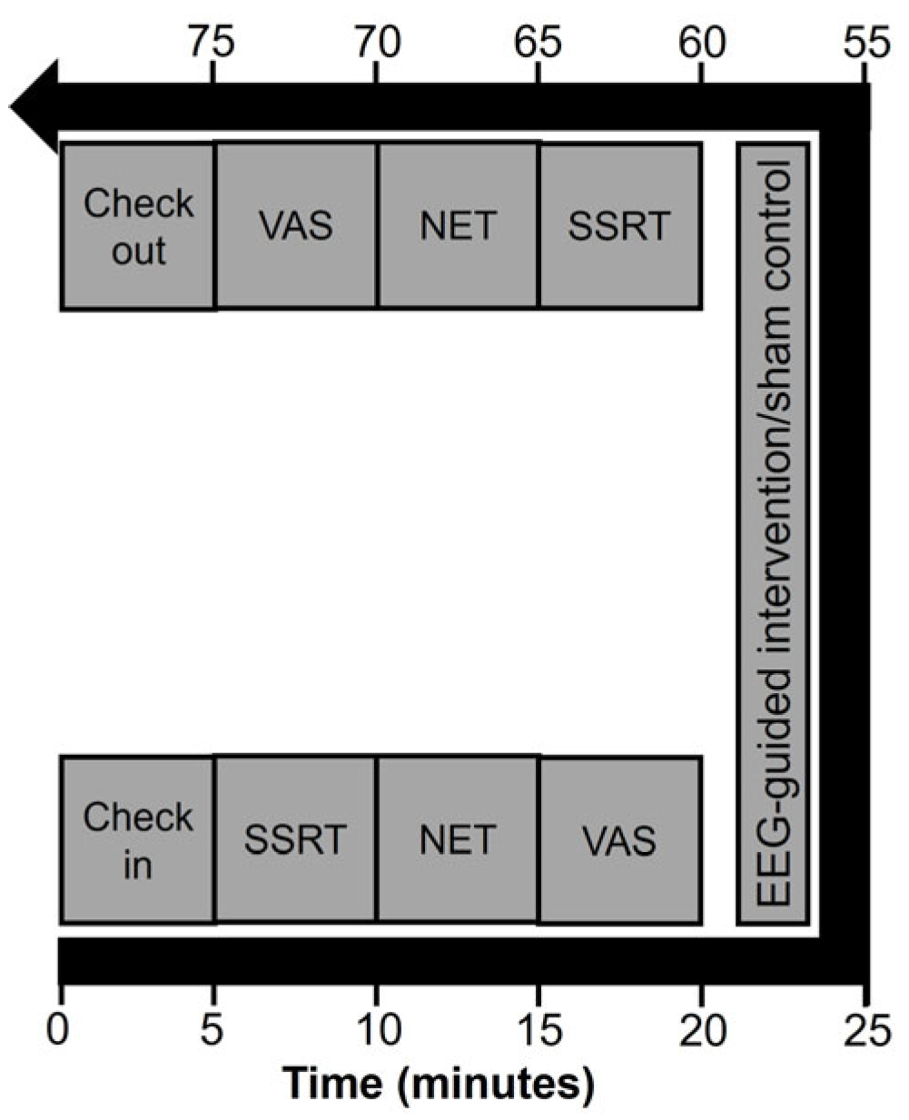
Participants attended three visits (Figure 3). Visit 1 included eligibility screening, informed consent, and training on the cognitive tasks. Visits 2 and 3 followed an identical structure: pre-session assessments (visual analogue scales [VAS] and cognitive tasks), a 30 min session (EEG-guided binaural beats or sham), and post-session assessments (VAS and cognitive tasks). Randomization was performed using a computer-generated sequence written into the Python code that was delivering the intervention and sham conditions. The code was concealed from the study team involved in data collection and analysis until the interpretation stage. The study team was only expected to enter a participant and a session number (i.e., indicating the first versus second session), and the Python script was coded to use this information and randomly allocate participants to receive the intervention versus sham-control treatment during the first session, a Bernoulli distribution of 0.5 probability, and the remaining treatment during the second session. Audio stimuli were delivered through MDR-15EXAP (Sony Corp., New York, NY, USA) in-ear headphones. While SPL calibration was not used, the devices and audio settings were held constant for all subjects and across sessions to ensure identical delivery of intervention and sham-control conditions. Both conditions included identical background ambient sounds to maintain auditory masking. Participants were asked at the end of each session whether they could distinguish any differences between sessions, and blinding success was assessed qualitatively; self-reports did not indicate confident discrimination between intervention and sham.

4.3. EEG Acquisition and Preprocessing

While traditional multi-channel EEG systems provide comprehensive spatial coverage, they are often impractical for real-world applications due to high costs, extended setup times, and complexity [32]. Consumer-grade EEG devices offer a practical alternative by prioritizing accessibility and ease of use, with validation studies demonstrating that single-electrode systems can provide adequate signal quality for specific applications despite inherent trade-offs in spatial resolution and artifact susceptibility [33,34]. The use of consumer-grade single-channel EEG devices has been validated across 916 studies [35].
Considering future translation for more feasible usage, NeuroSky MindWave Mobile 2 (NeuroSky Inc., San Jose, CA, USA) was used for data collection. MindWave Mobile 2 is a portable, single-channel EEG headset designed for noninvasive acquisition of frontal brain activity in naturalistic environments. The system uses a dry active electrode positioned at the left frontal pole (Fp1, based on the international 10–20 system) with reference and ground electrodes integrated into a stainless-steel ear-clip placed on the left earlobe (A1). The Fp1 electrode location has been validated for effectiveness in single-channel applications, achieving 70.0–85.7% detection accuracy in brain-computer interface paradigms [36]. The device records 12-bit EEG at a sampling rate of 512 Hz and transmits data wirelessly via Bluetooth using the proprietary ThinkGear ASIC Module. Several studies have demonstrated that, despite its simplicity, the MindWave platform yields reliable EEG signals suitable for spectral analyses and cognitive-state monitoring. Comparative validation with medical-grade systems has shown strong correlations of spectral power (r ≈ 0.89–0.90) and consistent detection of alpha-band differences between eyes-open and eyes-closed conditions [33,37]. Further work reported good test–retest reliability for eyes-closed EEG power (intraclass correlations = 0.76–0.85) across sessions up to one month apart [38]. A head-to-head evaluation with an ambulatory research-grade EEG demonstrated that while low-frequency (<3 Hz) sensitivity is reduced due to an in-built high-pass filter, MindWave recordings preserve signal quality above 3 Hz [33]. Moreover, recent scoping reviews examining the published evidence conclude that consumer-grade systems such as the MindWave achieve adequate validity and reliability for assessing broad spectral trends and engagement states when used with proper artifact handling and within their technical limits [35]. Although the choice of a single channel and its frontal placement is susceptible to ocular and muscle artifacts [39,40], the device represents an acceptable compromise between measurement feasibility and signal fidelity for developing a portable neurofeedback system. Collectively, these findings support the feasibility and ecological validity of single-channel dry-sensor EEG for monitoring of cognitive and affective states in applied settings.
EEG data processing and analysis were performed using custom Python scripts utilizing the MNE-Python library (version 0.24.1) and NumPy/SciPy packages. Continuous EEG from Fp1 was segmented into 2-s epochs for preprocessing, following established standards for regular epoching without event markers [41]. Signals were band-pass filtered from 1 to 32 Hz 4th-order Butterworth filter, followed by a 60 Hz notch filter to remove power line interference, adhering to best practices for EEG filtering [42] and implementing multi-taper regression for line noise removal without affecting nearby frequencies [43]. Artifact suppression was performed primarily via discrete wavelet decomposition (Daubechies-4, level 5) with adaptive soft-thresholding of detail coefficients [44]. The adaptive soft-thresholding approach employs time-scale adaptive denoising using Stein’s Unbiased Risk Estimate (SURE), providing gradient-based optimization of wavelet coefficients [45,46]. Given the single-channel recording, we applied a one-component FastICA (n = 1) as an affine, robust normalization (i.e., whitening with a non-Gaussian scalar projection) to standardize the signal for downstream analyses, rather than for source separation or artifact removal [47,48]. In our experience, for single-channel applications, the combination of the above preprocessing techniques enables effective denoising robust to artifacts [49]. Sample entropy was computed for each epoch to quantify signal irregularity; epochs exceeding the 75th percentile of entropy values were classified as high-noise and excluded from subsequent analyses [50,51]. Remaining epochs were z-scored [52] and power spectral densities were computed via Fast Fourier Transform (FFT) with 1 Hz frequency resolution, and were further summarized into standard bands (i.e., delta 0.5–4 Hz, theta 4–8 Hz, low-alpha 8–10 Hz, high-alpha 10–12 Hz, low-beta 12–18 Hz, high-beta 18–32 Hz) as well as 1 Hz frequency bands. The dominant frequency band of each 2 s epoch was determined and used to guide the binaural beat intervention.

4.4. EEG-Guided Binaural Beat Intervention

The first 2 min of each session established the participant’s predominant baseline EEG frequency [53]. During the intervention, a 144 Hz tone was presented to the left ear and 144 Hz plus the participant’s predominant baseline frequency to the right ear (subliminal intensity), yielding an initial binaural beat equal to the baseline frequency. For instance, if the baseline frequency was 20 Hz, in the intervention condition, 144 Hz was played to the left ear and 164 Hz was played to the right ear, and the stimulating frequency to the right ear was adjusted until the target state was reached. In the sham control condition, 144 Hz was played to both ears. The frequency of 144 Hz was chosen arbitrarily; we specifically avoided frequencies (e.g., 111 Hz, 174 Hz, 285 Hz, 396 Hz, 417 Hz, 432 Hz, 528 Hz, 639 Hz, 741 Hz, 852 Hz, and 963 Hz) that are commonly purported to have beneficial effects, yet none of such frequencies, to our knowledge, had sufficient empirical support. Research indicates carrier frequencies between 180 and 250 Hz are effective for theta and alpha entrainment [7], with the 144 Hz frequency falling within ranges demonstrating frequency-following responses and auditory steady-state responses [11]. Stimulation parameters were updated once per minute based on the immediately preceding minute of EEG. Decisions about parameter updates were made at fixed 1 min intervals using the prior minute’s data, thereby avoiding any impact of processing latency on closed-loop control. The binaural beat frequency was decreased from the current predominant frequency in the power spectrum towards a 2 Hz target frequency using a proprietary algorithm. If the predominant EEG frequency was observed to decrease towards the target, the stimulating frequency was decreased. The stimulating frequency was held constant if the predominant frequency was observed to be stable or increasing. The binaural beat frequency was decreased to accommodate rapid decreases in observed predominant frequency (e.g., theta states associated with drowsiness). When the predominant EEG frequency reached 2 Hz, the binaural beat was maintained at 2 Hz for the remainder of the session, with brief adjustments as needed to re-center the predominant frequency on target. In the sham condition, the background audio and overall presentation were identical except that both ears received a 144 Hz tone (i.e., no interaural frequency difference), thus maintaining identical acoustic energy per established protocols for binaural beats research [11]; EEG was recorded continuously in both conditions.

4.5. Outcome Measures

4.5.1. EEG Outcomes

The EEG data derived and saved from the real-time preprocessing pipeline were used to compute the EEG outcomes. Primary EEG outcomes indexed relaxation via dominant frequency trajectories and band distributions over time. We quantified (a) time to <8 Hz and <4 Hz, (b) the proportion of participants who reached and sustained <8 Hz for ≥3 min, (c) the proportion who reached and sustained <4 Hz for ≥3 min, and (d) distributions of predominant frequencies at prespecified time points (baseline, 5, 10, 15, 20 min, final 5 min, and end of session). Indices consistent with “sleep-like” low-frequency states (e.g., <4 Hz for ≥3 min) were analyzed as exploratory outcomes.

4.5.2. Subjective Measures

Five 100 mm VAS were administered immediately before and after each session to assess perceived relaxation, sleepiness, focus, cognitive performance, and overall well-being (anchors 0–100 mm).

4.5.3. Stop Signal Task

The stop signal task combined a primary go task and a secondary stop task [23,29]. Each trial began with a fixation cross (2500 ms ± 1500 ms), followed by either the letter X or O (500 ms duration), to which participants were instructed to press designated response buttons. In one-third of trials, the background turned red 250 ms after letter onset (stop signal delay; SSD), indicating participants should withhold their response. In the remaining trials, the background remained white (go trials). The SSD was dynamically adjusted, increasing by 50 ms following successful inhibition and decreasing by 50 ms following failed inhibition. The task comprised 90 trials (60 go trials, 30 stop trials). We derived stop signal reaction time (SSRT) and drift diffusion parameters [30]: boundary separation (a1, go; a2, stop), drift rate (v1, go; v2, stop), and starting point (z1, go; z2, stop). Specifically, a1 and a2 represent the response caution or decision threshold in go and stop trials, respectively, with larger values indicating more cautious decision-making. Parameters v1 and v2 reflect the rate of information accumulation in go and stop trials, respectively, with higher absolute values indicating more efficient information processing. Variables z1 and z2 were surrogates of starting points in decision making in go and stop trials, respectively, with values closer to 0.5 representing unbiased processing, with lower values denoting a bias towards stop trials and higher values depicting a bias towards go trials.

4.5.4. Novelty Encoding Task

This task utilized complex visual stimuli created by placing combinations of shapes (circles, triangles, and squares) and colors (red, green, and blue) within a 3 × 3 matrix [31]. The task consisted of three phases: familiarization phase in which five basic images were presented five times each; incidental learning phase in which the participants viewed the original basic images along with novel variations (color changes, shape changes, or both) derived from the basic images; and a recognition phase in which the participants viewed previously shown novel images and entirely new novel images derived from unseen basic images. Each image was shown for 3000 ms with jittered interstimulus intervals (1000 ms ± 500 ms). Participants were instructed to press a key if they saw an image that was previously presented that day. Novel images were used for each administration of the task to minimize biases associated with familiarization with the images of prior administrations. Several performance metrics were computed to characterize different aspects of memory encoding and retrieval [54]: hit rate, false alarm rate, d′, response bias, beta, precision, specificity, F1 score, and reaction time.

4.6. Statistical Analysis

With a sample size of 25 subjects, the study was at least 80% powered to detect a between-group effect size of Cohen’s d ≥ 0.75 after a conservative Bonferroni correction at a significance level of 0.05. Cognitive data were analyzed using linear mixed-effects ANOVA models computed with the lmerTest package in R statistical software (version 4.4.2). The models used for analysis included fixed effects for Intervention (control vs. intervention), Time (pre vs. post), and their interaction. First, a maximal random effects structure accounting for by-subject variability in intercepts and slopes: SSRT ~ Intervention × Time + (1 + Intervention + Time | Subject) was attempted, yet failed to converge, likely due to the limited sample size. As such, in the final analyses, only random intercepts for subjects were to account for repeated measurements: SSRT ~ Intervention × Time + (1 | Subject). This approach is appropriate for the fully within-subjects (repeated measures) design, where each participant contributed data under all combinations of Intervention and Time conditions. Type III analysis of variance with Satterthwaite’s method for degrees of freedom approximation was used to test the significance of fixed effects. Post hoc analyses were adjusted for multiple comparisons for each outcome using Holm–Bonferroni correction. Model assumptions (i.e., linearity, homoscedasticity, and normality of residuals) were evaluated with residual diagnostics. Given that participants were randomized to condition and a washout interval, as well as pre-specified diagnostics were used, session order was not included as a model term in the primary analyses. For the EEG data, we calculated the proportion of participants reaching target frequency states (<8 Hz and <4 Hz) and the median time (with interquartile range) to reach each brain state. Frequency distributions across different time points were analyzed to track the progression of brain states throughout the session. Comparisons between intervention and control conditions were made using appropriate non-parametric tests for proportional data. For behavioral and subjective measures, effect sizes were expressed as standardized differences in change (i.e., Cohen’s d) derived from the model-estimated marginal means, and F-statistics with degrees of freedom as well as p-values were reported. Outliers were defined as values exceeding 3 standard deviations from the group mean and were eliminated, considering likely biases that could be introduced by winsorizing cognitive data. Resulting missingness was handled using maximum likelihood estimation inherent in mixed-effects models. Results were considered statistically significant at p < 0.05, with trends noted at p < 0.10. Considering the pilot and largely exploratory nature of the study, we did not implement a correction for FDR at the outcomes level.

5. Conclusions

In summary, this study demonstrates the feasibility of real-time EEG-guided binaural beat interventions for rapidly inducing low-frequency brain states. Preliminary evidence suggests potential preservation or enhancement of certain cognitive functions during these states, though these findings require replication in larger, more rigorously controlled studies. While the results may support the potential for personalized, closed-loop auditory entrainment approaches, the findings are preliminary, given the pilot nature of the study and notable limitations; thus, they should be considered hypothesis-generating rather than definitive. Our findings motivate adequately powered, preregistered, mechanistically rich, rigorously controlled trials to confirm efficacy, delineate boundary conditions, and test real-world efficacy.

6. Patents

Texas Tech University System submitted a patent application (PCT/US25/22377) for the software algorithm used in the proposed study on 31 March 2025 with USPTO for auditory binaural beats guided by electroencephalography feedback.

Supplementary Materials

The following supporting information can be downloaded at https://www.mdpi.com/article/10.3390/physiologia5040044/s1. Table S1: Comparison of stop signal reaction task outcomes between the intervention and sham-control conditions; Table S2: Comparison of novelty encoding task outcomes between the intervention and sham-control conditions; Table S3: Comparison of subjective visual analogue scale outcomes between the intervention and sham-control conditions.

Author Contributions

Conceptualization, C.N.K. and C.S.D.; methodology, C.N.K., C.M. and C.S.D.; software, C.N.K.; validation, C.S.D. and C.M.; formal analysis, C.N.K. and C.S.D.; investigation, C.S.D. and C.M.; resources, C.S.D.; data curation, C.N.K. and C.S.D.; writing—original draft preparation, C.N.K. and C.S.D.; writing—review and editing, C.S.D., J.B. and C.M.; visualization, C.S.D.; supervision, C.S.D.; project administration, C.S.D.; funding acquisition, C.S.D. All authors have read and agreed to the published version of the manuscript.

Funding

This study was partially funded by the Department of Surgery, TTUHSC (internal seed grant), and through funding received from the National Science Foundation Regional (South West) I-Corps program conducted by the Innovation Hub of the Texas Tech University System.

Institutional Review Board Statement

This study was conducted in accordance with the Declaration of Helsinki and approved by the Institutional Review Board of Texas Tech University Health Sciences Center (protocol code IRB-FY2024-286, approved on 16 August 2024).

Informed Consent Statement

Informed written consent was obtained from all subjects involved in the study.

Data Availability Statement

The data presented in this study are available from the corresponding author upon request. The data are not publicly available due to privacy restrictions.

Acknowledgments

The authors wish to acknowledge the Office of Research Commercialization of Texas Tech University System for providing a conditional license to use the technology described in this manuscript for conducting the clinical trial and the Clinical Research Institute at Texas Tech University Health Sciences Center for their administrative assistance in getting the institutional IRB approval.

Conflicts of Interest

Author Jessica Blume is employed by MelodiaSync LLC. Authors Chanaka N. Kahathuduwa and Chathurika S. Dhanasekara are co-founders of MelodiaSync LLC. Texas Tech University System is the owner of a patent application for the software algorithm used in the proposed study (PCT application PCT/US25/22377 filed on 31 March 2025 with USPTO for auditory binaural beats guided by electroencephalography feedback). The authors are also the inventors of this intellectual property. Furthermore, the right to use the software algorithm has been granted back to the study team by the TTU System under a Limited License Agreement dated 2 April 2024. This fact was explicitly disclosed to the IRB and all participants of the study.

References

  1. Garcia-Argibay, M.; Santed, M.A.; Reales, J.M. Binaural auditory beats affect long-term memory. Psychol. Res. 2019, 83, 1124–1136. [Google Scholar] [CrossRef]
  2. Oster, G. Auditory beats in the brain. Sci. Am. 1973, 229, 94–103. [Google Scholar] [CrossRef]
  3. Marsh, J.T.; Worden, F.G. Sound evoked frequency-following responses in the central auditory pathway. Laryngoscope 1968, 78, 1149–1163. [Google Scholar] [CrossRef]
  4. Becher, A.K.; Höhne, M.; Axmacher, N.; Chaieb, L.; Elger, C.E.; Fell, J. Intracranial electroencephalography power and phase synchronization changes during monaural and binaural beat stimulation. Eur. J. Neurosci. 2015, 41, 254–263. [Google Scholar] [CrossRef] [PubMed]
  5. Jirakittayakorn, N.; Wongsawat, Y. Brain responses to a 6-Hz binaural beat: Effects on general theta rhythm and frontal midline theta activity. Front. Neurosci. 2017, 11, 365. [Google Scholar] [CrossRef]
  6. Vernon, D.; Peryer, G.; Louch, J.; Shaw, M. Tracking EEG changes in response to alpha and beta binaural beats. Int. J. Psychophysiol. 2014, 93, 134–139. [Google Scholar] [CrossRef] [PubMed]
  7. Perez, H.D.O.; Dumas, G.; Lehmann, A. Binaural beats through the auditory pathway: From brainstem to connectivity patterns. eNeuro 2020, 7, ENEURO.0232-19.2020. [Google Scholar] [CrossRef] [PubMed]
  8. Engelbregt, H.; Barmentlo, M.; Keeser, D.; Pogarell, O.; Deijen, J.B. Effects of binaural and monaural beat stimulation on attention and EEG. Exp. Brain Res. 2021, 239, 2781–2791. [Google Scholar] [CrossRef]
  9. López-Caballero, F.; Escera, C. Binaural beat: A failure to enhance EEG power and emotional arousal. Front. Hum. Neurosci. 2017, 11, 557. [Google Scholar] [CrossRef]
  10. Garcia-Argibay, M.; Santed, M.A.; Reales, J.M. Efficacy of binaural auditory beats in cognition, anxiety, and pain perception: A meta-analysis. Psychol. Res. 2019, 83, 357–372. [Google Scholar] [CrossRef]
  11. Ingendoh, R.M.; Posny, E.S.; Heine, A. Binaural beats to entrain the brain? A systematic review of the effects of binaural beat stimulation on brain oscillatory activity, and the implications for psychological research and intervention. PLoS ONE 2023, 18, e0286023. [Google Scholar] [CrossRef]
  12. Wang, Y.; Zhang, L.; Chen, X.; Liu, H. Efficacy of acoustic stimulation for insomnia: A meta-analysis of randomized controlled trials. Front. Neurosci. 2025, 19, 1345678. [Google Scholar] [CrossRef]
  13. Chockboondee, M.; Jatupornpoonsub, T.; Lertsukprasert, K.; Wongsawat, Y. Effects of daily listening to 6 Hz binaural beats over one month: An event-related potentials study. Sci. Rep. 2024, 14, 18059. [Google Scholar] [CrossRef] [PubMed]
  14. Huwiler, S.; Ferster, M.L.; Brogli, L.; Huber, R.; Karlen, W.; Lustenberger, C. Sleep and cardiac autonomic modulation in older adults: Insights from an at-home study with auditory deep sleep stimulation. J Sleep Res. 2025, 34, e14328. [Google Scholar] [CrossRef]
  15. Dos Anjos, T.; Altimari, L.R.; Bortolotti, H.; Braz, T.V.; Melo, E.S.; Coimbra, D.R.; Fontes, E.B. Brain wave modulation and EEG power changes during auditory beats stimulation. Neuroscience 2024, 545, 86–96. [Google Scholar] [CrossRef] [PubMed]
  16. Chaieb, L.; Wilpert, E.C.; Reber, T.P.; Fell, J. Auditory beat stimulation and its effects on cognition and mood states. Front. Psychiatry 2015, 6, 70. [Google Scholar] [CrossRef] [PubMed]
  17. Goodin, P.; Ciorciari, J.; Baker, K.; Carrey, A.M.; Harper, M.; Kaufman, J. A high-density EEG investigation into steady state binaural beat stimulation. PLoS ONE 2012, 7, e34789. [Google Scholar] [CrossRef]
  18. Gao, X.; Cao, H.; Ming, D.; Qi, H.; Wang, X.; Chen, R.; Zhou, P. Analysis of EEG activity in response to binaural beats with different frequencies. Int. J. Psychophysiol. 2014, 94, 399–406. [Google Scholar] [CrossRef]
  19. Noor, W.M.F.W.M.; Zaini, N.; Norhazman, H.; Latip, M.F.A. Dynamic encoding of binaural beats for brainwave entrainment. In Proceedings of the 2013 IEEE International Conference on Control System, Computing and Engineering, Penang, Malaysia, 29 November–1 December 2013. [Google Scholar] [CrossRef]
  20. Crowley, K.; Sliney, A.; Pitt, I.; Murphy, D. Evaluating a brain-computer interface to categorise human emotional response. In Proceedings of the 2010 10th IEEE International Conference on Advanced Learning Technologies, Sousse, Tunisia, 5–7 July 2010. [Google Scholar] [CrossRef]
  21. Kane, N.; Acharya, J.; Benickzy, S.; Caboclo, L.; Finnigan, S.; Kaplan, P.W.; Shibasaki, H.; Pressler, R.; van Putten, M. A revised glossary of terms most commonly used by clinical electroencephalographers and updated proposal for the report format of the EEG findings. Revision 2017. Clin. Neurophysiol. Pract. 2017, 2, 170–185. [Google Scholar] [CrossRef]
  22. Abeln, V.; Kleinert, J.; Strüder, H.K.; Schneider, S. Brainwave entrainment for better sleep and post-sleep state of young elite soccer players—A pilot study. Eur. J. Sport Sci. 2014, 14, 393–402. [Google Scholar] [CrossRef]
  23. Chevrier, A.D.; Noseworthy, M.D.; Schachar, R. Dissociation of response inhibition and performance monitoring in the stop signal task using event-related fMRI. Hum. Brain Mapp. 2007, 28, 1347–1358. [Google Scholar] [CrossRef] [PubMed]
  24. Stone, J.L.; Hughes, J.R. Early history of electroencephalography and establishment of the American Clinical Neurophysiology Society. J. Clin. Neurophysiol. 2013, 30, 28–44. [Google Scholar] [CrossRef] [PubMed]
  25. Enkavi, A.Z.; Eisenberg, I.W.; Bissett, P.G.; Mazza, G.L.; MacKinnon, D.P.; Marsch, L.A.; Poldrack, R.A. Large-scale analysis of test-retest reliabilities of self-regulation measures. Proc. Natl. Acad. Sci. USA 2019, 116, 5472–5477. [Google Scholar] [CrossRef] [PubMed]
  26. Fleiss, J.L. Design and Analysis of Clinical Experiments; John Wiley & Sons: Hoboken, NJ, USA, 2011. [Google Scholar] [CrossRef]
  27. Koo, T.K.; Li, M.Y. A guideline of selecting and reporting intraclass correlation coefficients for reliability research. J. Chiropr. Med. 2016, 15, 155–163. [Google Scholar] [CrossRef]
  28. Band, G.P.; Van Der Molen, M.W.; Logan, G.D. Horse-race model simulations of the stop-signal procedure. Acta Psychol. 2003, 112, 105–142. [Google Scholar] [CrossRef]
  29. Ratcliff, R. A theory of memory retrieval. Psychol. Rev. 1978, 85, 59. [Google Scholar] [CrossRef]
  30. Huang-Pollock, C.; Ratcliff, R.; McKoon, G.; Shapiro, Z.; Weigard, A.; Galloway-Long, H. Using the diffusion model to explain cognitive deficits in attention deficit hyperactivity disorder. J. Abnorm. Child Psychol. 2017, 45, 57–68. [Google Scholar] [CrossRef]
  31. Reichardt, R.; Polner, B.; Simor, P. The graded novelty encoding task: Novelty gradually improves recognition of visual stimuli under incidental learning conditions. Behav. Res. Methods 2023, 55, 1587–1600. [Google Scholar] [CrossRef]
  32. LaRocco, J.; Le, M.D.; Paeng, D.G. A systemic review of available low-cost EEG headsets used for drowsiness detection. Front. Neuroinform. 2020, 14, 553352. [Google Scholar] [CrossRef]
  33. Rieiro, H.; Diaz-Piedra, C.; Morales, J.M.; Catena, A.; Romero, S.; Roca-Gonzalez, J.; Fuentes, L.J.; Di Stasi, L.L. Validation of electroencephalographic recordings obtained with a consumer-grade, single dry electrode, low-cost device: A comparative study. Sensors 2019, 19, 2808. [Google Scholar] [CrossRef]
  34. Sawangjai, P.; Hompoonsup, S.; Leelaarporn, P.; Kongwudhikunakorn, S.; Wilaiprasitporn, T. Consumer grade EEG measuring sensors as research tools: A review. IEEE Sens. J. 2019, 20, 3996–4024. [Google Scholar] [CrossRef]
  35. Sabio, J.; Williams, N.S.; McArthur, G.M.; Badcock, N.A. A scoping review on the use of consumer-grade EEG devices for research. PLoS ONE 2024, 19, e0291186. [Google Scholar] [CrossRef]
  36. Ogino, M.; Kanoga, S.; Muto, M.; Mitsukura, Y. Analysis of prefrontal single-channel EEG data for portable auditory ERP-based brain–computer interfaces. Front. Hum. Neurosci. 2019, 13, 250. [Google Scholar] [CrossRef]
  37. Johnstone, S.J.; Blackman, R.; Bruggemann, J.M. EEG from a single-channel dry-sensor recording device. Clin. EEG Neurosci. 2012, 43, 112–120. [Google Scholar] [CrossRef] [PubMed]
  38. Rogers, J.M.; Johnstone, S.J.; Aminov, A. Test–retest reliability of a single-channel, wireless EEG system. Int. J. Psychophysiol. 2016, 106, 87–96. [Google Scholar] [CrossRef] [PubMed]
  39. Chang, W.D.; Cha, H.S.; Kim, K.; Im, C.H. Detection of eye blink artifacts from single prefrontal channel electroencephalogram. Comput. Methods Programs Biomed. 2016, 124, 19–30. [Google Scholar] [CrossRef]
  40. Plöchl, M.; Ossandón, J.P.; König, P. Combining EEG and eye tracking: Identification, characterization, and correction of eye movement artifacts in electroencephalographic data. Front. Hum. Neurosci. 2012, 6, 278. [Google Scholar] [CrossRef]
  41. Delorme, A.; Makeig, S. EEGLAB: An open source toolbox for analysis of single-trial EEG dynamics including independent component analysis. J. Neurosci. Methods 2004, 134, 9–21. [Google Scholar] [CrossRef]
  42. Widmann, A.; Schröger, E.; Maess, B. Digital filter design for electrophysiological data—A practical approach. J. Neurosci. Methods 2015, 250, 34–46. [Google Scholar] [CrossRef]
  43. Mullen, T.; Kothe, C.; Chi, Y.M.; Ojeda, A.; Kerth, T.; Makeig, S.; Cauwenberghs, G.; Jung, T.P. Real-time modeling and 3D visualization of source dynamics and connectivity using wearable EEG. In Proceedings of the 2013 35th Annual International Conference of the IEEE Engineering in Medicine and Biology Society (EMBC), Osaka, Japan, 3–7 July 2013; pp. 2184–2187. [Google Scholar] [CrossRef]
  44. Roy, V.; Shukla, S. Automatic removal of artifacts from EEG signal based on spatially constrained ICA using Daubechies wavelet. Int. J. Mod. Educ. Comput. Sci. 2014, 6, 31–39. [Google Scholar] [CrossRef]
  45. Krishnaveni, V.; Jayaraman, S.; Anitha, L.; Ramadoss, K. Removal of ocular artifacts from EEG using adaptive thresholding of wavelet coefficients. J. Neural Eng. 2006, 3, 338–346. [Google Scholar] [CrossRef] [PubMed]
  46. Mammone, N.; La Foresta, F.; Morabito, F.C. Automatic artifact rejection from multichannel scalp EEG by wavelet ICA. IEEE Sens. J. 2012, 12, 533–542. [Google Scholar] [CrossRef]
  47. Davies, M.E.; James, C.J. Source separation using single channel ICA. Signal Process. 2007, 87, 1819–1832. [Google Scholar] [CrossRef]
  48. Dharmaprani, D.; Nguyen, H.K.; Lewis, T.W.; DeLosAngeles, D.; Willoughby, J.O.; Pope, K.J. A comparison of independent component analysis algorithms and measures to discriminate between EEG and artifact components. Clin. Neurophysiol. 2017, 128, 768–776. [Google Scholar] [CrossRef]
  49. Noorbasha, S.K.; Sudha, G.F. Removal of EOG artifacts and separation of different cerebral activity components from single channel EEG—An efficient approach combining SSA–ICA with wavelet thresholding for BCI applications. Biomed. Signal Process. Control 2020, 63, 102201. [Google Scholar] [CrossRef]
  50. Richman, J.S.; Moorman, J.R. Physiological time-series analysis using approximate entropy and sample entropy. Am. J. Physiol. Heart Circ. Physiol. 2000, 278, H2039–H2049. [Google Scholar] [CrossRef]
  51. Mariani, S.; Borges, A.F.; Henriques, T.; Goldberger, A.L.; Costa, M.D. Use of multiscale entropy to facilitate artifact detection in electroencephalographic signals. In Proceedings of the 2015 37th Annual International Conference of the IEEE Engineering in Medicine and Biology Society (EMBC), Milan, Italy, 25–29 August 2015; pp. 7869–7872. [Google Scholar] [CrossRef]
  52. Lee, T.H.; Kim, M.; Hwang, W.J.; Kim, T.; Kwak, Y.B.; Kwon, J.S. Relationship between resting-state theta phase-gamma amplitude coupling and neurocognitive functioning in patients with first-episode psychosis. Schizophr. Res. 2022, 239, 134–145. [Google Scholar] [CrossRef]
  53. Enriquez-Geppert, S.; Huster, R.J.; Herrmann, C.S. EEG-neurofeedback as a tool to modulate cognition and behavior: A review tutorial. Front. Hum. Neurosci. 2017, 11, 51. [Google Scholar] [CrossRef]
  54. Tanner, W.P., Jr.; Swets, J.A. A decision-making theory of visual detection. Psychol. Rev. 1954, 61, 401. [Google Scholar] [CrossRef]
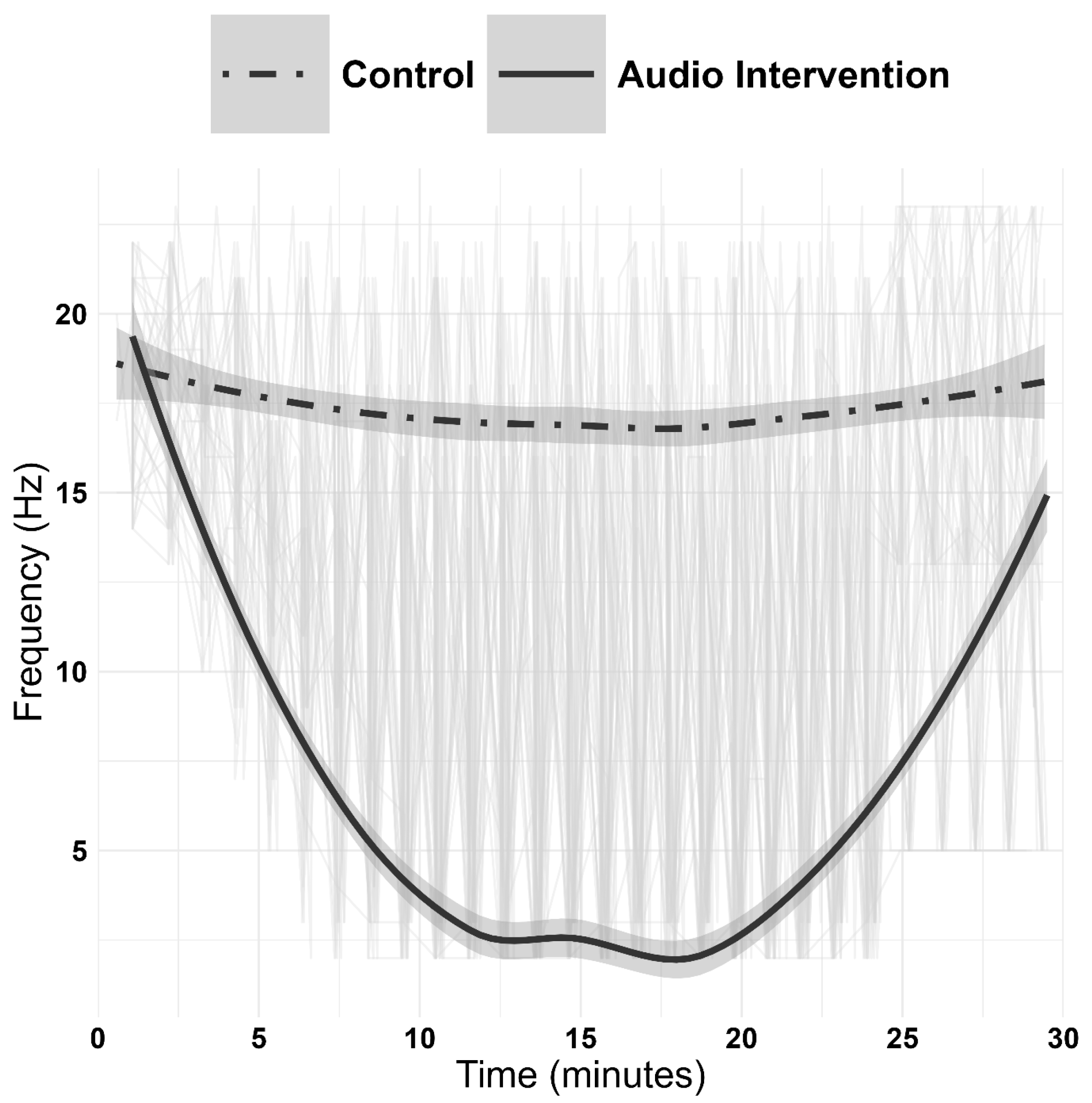
Figure 1. Smoothed mean predominant frequency curves over time for the intervention and sham-control conditions. Each gray line represents an individual participant’s frequency trajectory throughout the 30 min session, illustrating the variability in individual responses. The bold solid line shows the smoothed group average for the intervention condition, while the bold dashed line shows the smoothed group average for the sham-control condition. The shaded areas represent 95% confidence intervals around the group means, calculated using locally weighted scatterplot smoothing (LOESS). Time is shown in minutes from the start of the audio session. The intervention condition shows a rapid and sustained decrease in predominant frequency, while the sham condition remains relatively stable throughout the session.
Physiologia 05 00044 g001
Figure 2. Study flowchart showing the randomized crossover design. Following screening and enrollment, participants completed two experimental visits separated by a 7-day washout period. In each visit, participants completed pre-intervention cognitive assessments (Stop Signal Task and Novelty Encoding Task), followed by the 30 min intervention (either EEG-guided binaural beats or sham control), and then post-intervention cognitive assessments. The order of intervention and sham conditions was randomized and counterbalanced across participants. EEG was recorded continuously throughout each session.
Physiologia 05 00044 g002
Figure 3. Timeline of cognitive assessments and EEG-guided intervention/sham control during study visits 2 and 3. VAS, visual analogue scales; NET, novelty encoding task; SSRT, stop signal task.
Physiologia 05 00044 g003
Table 1. Comparison of mean predominant frequencies (Hz) between the intervention and sham-control conditions.
Sham-Control
( x ¯ ± SD)
Intervention
( x ¯ ± SD)
t-Statisticp-Value
Baseline17.92 ± 2.7418.44 ± 2.60−0.6530.520
5 min17.63 ± 2.7510.00 ± 2.86−8.495<0.001
10 min17.50 ± 4.103.08 ± 1.75−15.358<0.001
15 min17.79 ± 4.232.84 ± 0.80−16.749<0.001
20 min17.71 ± 4.082.52 ± 0.71−17.52<0.001
Pre-awakening17.52 ± 3.402.72 ± 0.84−21.288<0.001
Awakening16.92 ± 3.0312.16 ± 7.05−3.2970.003
Table 2. EEG frequency band distribution between the intervention and sham control conditions.
Time PointSham-ControlIntervention
δΘLow
α
High
α
βδθLow
α
High
α
β
Baseline000025000025
(0)(0)(0)(0)(100)(0)(0)(0)(0)(100)
5 min00012407567
(0)(0)(0)(4)(96)(0)(28)(20)(24)(28)
10 min010123204010
(0)(4)(0)(4)(92)(80)(16)(0)(4)(0)
15 min010123232000
(0)(4)(0)(4)(92)(92)(8)(0)(0)(0)
20 min010223241000
(0)(4)(0)(8)(88)(96)(4)(0)(0)(0)
Pre-
awakening
001024232000
(0)(0)(4)(0)(96)(92)(8)(0)(0)(0
Awakening001024092113
(0)(0)(4)(0)(96)(0)(36)(8)(4)(52)
Table 3. Comparison of selected stop signal reaction task, novelty encoding task, and subjective visual analogue scale outcomes between the intervention and sham-control conditions.
VariableControlInterventionMain EffectsInteraction
F(df1, df2) p
Pre
x ¯ ± SD
Post
x ¯ ± SD
Pre
x ¯ ± SD
Post
x ¯ ± SD
Condition
F(df1, df2) p
Time
F(df1, df2) p
SSRT
(s)
0.089
± 0.040
0.102
± 0.059
0.109
± 0.060
0.093
± 0.045
2.82(1,68.2)
0.097
1.11(1,68.5)
0.295
2.82(1,68.8)
0.098
a21.91
± 0.119
1.85
± 0.219
1.86
± 0.161
1.92
± 0.122
0.97(1,67.0)
0.328
2.19(1,67.6)
0.143
3.52(1,68.0)
0.065
v2−4.75
± 1.21
−5.43
± 0.916
−5.11
± 1.04
−4.99
± 1.08
1.41(1,68.7)
0.240
4.80(1,69.5)
0.032 *
3.40(1,69.8)
0.070
NET-RT
(s)
1.13
± 0.209
1.16
± 0.229
1.23
± 0.259
1.10
± 0.217
2.45(1,54.6)
0.124
0.00(1,54.6)
0.974
4.50(1,54.4)
0.039 *
Cognitive
Performance
69.3
± 13.8
62.2
± 20.1
66.9
± 16.3
67.3
± 19.6
0.57(1,72)
0.454
4.99(1,72)
0.029 *
2.79(1,72)
0.099
Values are presented as the mean ± standard deviation; SSRT, Stop Signal Reaction Time; a2, boundary separation during stop trials in stop signal task; v2, drift rate during stop trials in stop signal task; NET-RT, reaction time during novel information retrieval in novelty encoding task; *, p < 0.05; , 0.05 < p < 0.10.
Disclaimer/Publisher’s Note: The statements, opinions and data contained in all publications are solely those of the individual author(s) and contributor(s) and not of MDPI and/or the editor(s). MDPI and/or the editor(s) disclaim responsibility for any injury to people or property resulting from any ideas, methods, instructions or products referred to in the content.