Article

Emotional Salience of Evolutionary and Modern Disgust-Relevant Threats Measured Through Electrodermal Activity

1
Department of Zoology, Faculty of Science, Charles University, Viničná 7, Prague 2, 128 43 Prague, Czech Republic
2
National Institute of Mental Health, Topolová 748, 250 67 Klecany, Czech Republic
*
Author to whom correspondence should be addressed.
Physiologia 2025, 5(4), 41; https://doi.org/10.3390/physiologia5040041 (registering DOI)
Submission received: 29 August 2025 / Revised: 24 September 2025 / Accepted: 9 October 2025 / Published: 11 October 2025

Abstract

Background: The study of psychophysiological responses to disgust-evoking stimuli has long been neglected in favour of other emotional stimuli, especially those evoking fear. While the basic cascade of responses to a frightening stimulus is relatively well-understood, psychophysiological responses to disgust-related threats, such as parasites or rotten food, are scarcely studied. Methods: Here, we aimed to assess skin resistance (SR) change as a measure of electrodermal response to visual cues that signal the presence of disgust-relevant threats. To this aim, we recruited 123 participants and presented them with one of the following varieties of disgust-relevant threats: disgust-evoking animals (e.g., parasites, worms), spoiled food, threat of pandemic, or pollution and toxicity. The latter two represented modern threats to test whether also these modern stimuli can initiate immediate automatic reaction. Results: We found significant differences between the categories: Participants responded with the highest probability to disgust-evoking animals (38%) and sneezing (52%), suggesting that only ancestral cues of pathogen disgust trigger automatic physiological response. Moreover, we found significant inter-sexual differences: women exhibited more SR change responses than men, and the amplitude of these responses was overall larger. Finally, we report a weak effect of subjectively perceived disgust intensity on reactivity to threat stimuli. Conclusions: We discuss heterogeneity of disgust-relevant threats, their adequate behavioural responses, and subsequent heterogeneity of respective SR responses. We conclude that large interindividual variability might eclipse systematic differences between participants with high or low sensitivity to disgust, and that subjectively perceived intensity of disgust is only a weak predictor of electrodermal response to its elicitor.

1. Introduction

In everyday life, people are confronted with a variety of situations. Some situations may evoke different kinds of emotions that assist the cognitive categorization and facilitate the appropriate behavioural response [1]. Moreover, these emotions are often part of a more complex, specific pattern of physiological reactions controlled by the autonomic nervous system (ANS), which also affects such parameters as pupil dilatation, heart rate and blood pressure, or activity of sweat glands [2,3]. Such integrated response of many bodily systems allows for coordinated behavioural reaction, potentially maximizing well-being and survival. The ANS is a part of the peripheral nervous system responsible for regulating physiological processes, operating independently of conscious control. It is generally divided into two functionally distinct branches: the sympathetic and the parasympathetic nervous systems. The sympathetic division is primarily associated with the response to stress or perceived threats, mediating the so-called “fight-or-flight” response, which includes increased heart rate, dilated pupils, and increased electrodermal activity (EDA), among other effects [4,5]. In contrast, the parasympathetic division predominates during restful states, resulting in reduced cardiac output or decreased EDA [6]. Together, these components enable the ANS to coordinate complex, adaptive responses to both internal and external stimuli, ensuring physiological stability across varying conditions.
Most of the studies investigating the connection of emotions and the physiological responses use as physiological parameters heart rate (HR) and electrodermal activity (EDA), sometimes accompanied by facial electromyography (EMG) [3,7,8,9,10,11,12]. The connection between emotional and automatic physiological response is reasonably well-understood: Besides thermoregulation, sweating also plays a role in metabolism and has proved to be excreted during emotional states [13]. It is regulated by both the central nervous system and ANS. The thermoregulation centre is the hypothalamus, which is also influenced by emotional stimuli. The emotional sweating arises independently of ambient temperature and is mediated by cholinergic fibres of sympathetic system [14]. However, recent studies have also found evidence of an association with the adrenergic system, suggesting that our understanding of this pathway is not yet complete (reviewed in [15,16]).
While HR may be useful for measuring valence of the emotion during long-term physiological states, EDA might be better to detect rapid, subtle, and stimulus-specific emotional responses, since the delay of skin conductance response (SCR) is quite fast, often within 1–3 s [17]. Another advantage of EDA measurement is that it seems slightly less affected by small body movements compared to heart rate, which can fluctuate due to posture changes, breathing, or minor exertion [18]. This makes EDA more stable in controlled lab settings where subtle emotional shifts are being monitored. Although EMG enables differentiation of specific negative emotions by recording activity of individual facial muscles [19,20], it is relatively intrusive, as electrodes must be placed directly on the face, and improper placement can introduce substantial artefacts. In contrast, EDA is easier to administer, less intrusive, and generally less vulnerable to movement artefacts. In this study, we were interested in immediate automatic reactions to emotional visual stimuli; therefore, we chose the measurements of skin resistance (SR) as the main physiological method. Skin resistance is an EDA measurement, and it refers to how much the skin impedes the passage of electric charge due to activity of sweat glands activated by ANS, which is widely considered a biomarker of arousal [3,17]. It can be easily inversed into skin conductance, which is a predominantly used measurement of EDA in psychophysiological research studying emotions, stress, or psychopathologies such as phobias (e.g., [21,22,23,24]). Additionally, different parameters of the SCR, such as latency or magnitude, can be distinguished. For example, Grus and Hromadko [25] measured SCR to pictures with different level of pathogen saliency, and even though they found no significant effect on SCR magnitude, the latency period for high pathogen-salient pictures was significantly shorter compared to low-salient pictures. This suggests that, in addition to measuring reaction magnitude, SCR can also provide insight into the temporal dynamics of emotional processing.

1.1. Psychophysiology of Disgust

Traditionally, the emotion of disgust has been linked to increased parasympathetic activity, particularly in the cardiac system [2,26,27]. Nevertheless, results of studies focusing on responses of autonomic nervous system to disgust-evoking stimulation are not always consistent. For example, Ekman et al. [2] reported decreased HR and SC (resp. increase SR); Lang et al. [3] found decreased HR but increased SC compared to other emotions, while Levenson et al. [28] reported increases in both HR and SC. Although different methods of disgust elicitation were used in some of those mentioned studies, taken together, these results support a more complex activation of the autonomic nervous system [11].
Bradley et al. [7] proposed that an initial orientation of attention toward a potential threat causes co-activation of the sympathetic and parasympathetic systems, which then switches to only sympathetic activation after the threat is recognized as relevant. The suggestion that sympathetic activity is indeed part of the response to disgust-eliciting threat is also supported by increased skin conductance during the viewing of disgusting pictures [3]. In several other studies [12,29,30,31], authors also found evidence for sympathetic activation being part of the disgust response. For example, Stark et al. [12] used disgusting pictures selected from the IAPS database based on their disgust rating to create three categories of low, middle, and high disgust-inducing pictures. They found decreasing HR but increasing SC response with the increasing disgust ratings of the stimuli. Interestingly, only a part of the participants showed high enough changes in SC to be included in the analysis, which suggests that individual variability also plays a significant role.
The pattern of physiological response may also differ according to a different category of disgust. Rohrmann & Hopp [31] compared physiological reactions to video clips of amputation and vomiting and found higher HR for mutilation disgust compared to disease- and food-related disgust, even though the EDA response did not differ among the disgust-evoking clips. Similarly, a video clip of vomiting was used to evoke disgust in de Jong et al. [30]. The authors measured the activation of sympathetic and parasympathetic activity via high-frequency heart rate variability, T-wave amplitude, electromyography, and skin conductance level. The results indicated increased parasympathetic activity in both cardiac and digestive components, together with increased sympathetic activation in the cardiac system for the disgust-evoking video compared to the neutral one. Comtesse & Stemmler [29] also investigated the role of the pattern of ANS activation in disease- and food-related disgust by measuring several cardiovascular parameters (HR, pre-ejection period = PEP, respiratory sinus arrhythmia = RSA, blood pressure). Their findings showed a co-activation of the sympathetic and parasympathetic systems, indicated by increased PEP shortening (sympathetic activation) alongside increased RSA (parasympathetic activation), rather than a purely parasympathetic pattern. This co-activation was interpreted as the simultaneous mobilization of defensive behaviour and engagement of orienting/ingestive-related processes.
Since people are primarily visually orienting species, visual stimulation is the most often used type of stimulation in studies concerned with psychophysiological responses and emotional states. Nonetheless, there also seems to be a difference in the physiological responses during disgust stimulation for different types of nature of the stimuli (visual, olfactory, tactile, auditory) [9]. Their results showed that although all sensory channels could elicit disgust, the visual and tactile stimuli tended to evoke stronger subjective disgust ratings, while olfactory and tactile stimuli produced more pronounced physiological responses (HR and respiration). Skin conductance responses were generally elevated for disgust stimuli compared to controls, but the extent also depended on the sensory channel.
The psychophysiological response to disgust, therefore, seems to involve a unique interplay between the sympathetic and parasympathetic activity, which also depends on the context, for example, various authors differentiate between various types of disgust: oral, core, animal-reminder, contamination, pathogen, and more [29,31,32,33]. The skin conductance response seems to better capture the arousal rather than valence, but for the purpose of comparing different categories of stimuli with the same primarily evoked emotion, one could measure purely the intensity of elicited emotion.

1.2. Categorization of Disgust and of Disgust-Evoking Threats

Disgust is one of the basic human emotions, considered universal across cultures [34,35,36,37]. It likely evolved as a protective mechanism to help organisms avoid harmful substances and potential sources of infection, e.g., spoiled food, bodily products, or parasites [38,39,40]. A key challenge in disgust research is the categorization of its diverse elicitors. There are several established theoretical frameworks that can be used for the classification. For instance, the three-domain model differentiates between pathogen, sexual, and moral disgust based on their distinct evolutionary functions [33]. Another common approach distinguishes between core, animal reminder, and contamination-based disgust [41]. The core disgust, typically linked to rotting food, waste products, and small animals, is based on a sense of “offensiveness”. Animal reminder disgust is associated with death and violation of the body envelope. Finally, contamination-based disgust reflects perceived threat of transmission of contagions [41]. An alternative approach is based on the co-evolutionary history of humans and the potential threat, dividing the threats to ontogenetic (modern) and evolutionary (ancestral) threats. Even though this concept is usually used for categorization of threats evoking fear [42], its application on disgust-eliciting threats might also prove fruitful [43,44].
Ancestral threats refer to dangers that have been present for millions of years of hominin evolution, exerting consistent selective pressure [36,45]. Elicitors like spoiled food and parasites are classic examples. They trigger a cognitive and affective response consistent with behavioural immune system (BIS)—a preventive mechanism evolved to reduce the chance of getting into contact with harmful substances [39,46]. The emotion of disgust takes the central role in BIS, being the key motivator behind the ultimate behavioural response—avoidance of disgusting stimuli [47,48]. Importantly, evidence suggests that mere visual cues displaying symptoms like skin lesions or sneezing can activate not only avoidance tendencies but also physiological preparedness, reflected in a more aggressive immune response [49]. In contrast, modern threats are evolutionarily recent, having emerged within the last few centuries or even decades. These might include dangers like industrial pollution, chemical toxicity, and radioactivity, which often lack the salient sensory signals (cues) of the ancestral ones. Therefore, recognizing the danger of these modern threats might require the assessment of cues from the environment, for which abstract knowledge and cognitive mediation is needed. Besides disgust, they may also evoke other emotions, including moral outrage and anger related to the violation of social norms [43,50,51].
In this context, the threat of a pandemic occupies a uniquely complex position. While the underlying danger of infectious disease is deeply ancestral, the nature of a pandemic, i.e., a widespread threat of airborne illness and death, is most likely a more recent evolutionary pressure, tied to the higher population densities that arose after the Neolithic revolution [52,53,54]. Furthermore, the stimulus cues indicating the presence of such a threat seem to be heterogeneous [36]; some cues are visceral and ancient (e.g., a person sneezing), while many others are modern and abstract (e.g., face masks, hospital environments, public health warnings). This makes the pandemic threat an ideal test case for exploring whether our ancestral disgust mechanisms are effectively recruited to evaluate contemporary health risks that blend both ancient dangers and modern cues.

1.3. Aims

The present study aims to investigate whether individuals react to visual cues that signal the presence of potential threats and to assess the intensity of their skin resistance responses to such cues. Specifically, we want to examine both the likelihood of eliciting a reaction (probability of skin resistance response) and the magnitude of the elicited responses (response amplitude) in relation to various threat-related stimuli.
Specifically, we aim to explore whether differences in threat perception and reactivity are better explained by the evolutionary classification of threats, i.e., ancestral vs. modern threats, or by the specific nature of the depicted stimuli, irrespective of their evolutionary categorization. The specific investigated categories were “disgusting” animals and spoiled food, as representatives of evolutionary threats, and pandemics, toxicity, and pollution, as representatives of modern threats. An additional focus was placed on the category of visual cues associated with the pandemic of an airborne disease. Previous findings suggest that pandemic-related cues may not form a homogeneous category but rather consist of distinct subtypes—sneezing, hospital environment, and preventive behaviours (e.g., mask wearing)—that elicit threat-related responses of varying intensity [36]. Presumably, this is due to the discrepancy between the pandemic threat as a whole (a modern threat) and various pandemic cues (of both ontogenetic and evolutionary nature). Therefore, this study also aims to investigate whether these subcategories evoke distinct physiological response patterns, therefore, testing the assumption of categorical heterogeneity within airborne disease cues from a psychophysiological perspective (Figure 1).
Furthermore, we aim to investigate whether subjectively perceived intensity of disgust evoked by the threat stimuli affects skin resistance response.
Finally, we aim to inspect whether the participants’ characteristics affect their general electrodermal reactivity or their reactivity to specific visual threat stimuli. The investigated characteristics were the participants’ sex, sensitiveness to disgust, and their general anxiousness.

2. Results

2.1. Effect of Threat Category on Probability of SR Response

Firstly, we constructed three alternative generalized mixed-effect models (GLME) to test whether differences in reactivity towards the stimuli are better explained by the evolutionary classification of depicted threats or by finer categorization based on the specific nature of the depicted stimuli. In all of these models, the presence of a skin resistance (SR) change was used as a binary response (logit scale link), with stimulus category and participant sex as fixed factors, and participant ID as a random factor. In the first model, the finest categorization into seven groups was used: “disgusting” animals, spoiled food, toxicity and pollution, hospital environment, preventive behaviours, sneezing, and control. The second and third models used just three categories—ancestral threats, modern threats, and controls—and differed in categorization of sneezing: in model 2, it was among ancestral threats, and in model 3, among modern threats. Subsequently, these models were compared against each other and against the null model using Akaike information criterion (AIC) and ANOVA function. The first model using the finest categorization proved the best as it was the only one significantly different from the null model: AIC0 = 5301, AIC1 = 5294, AIC2 = 5302, and AIC3 = 5304. Null model vs. model 1: χ2 = 19.04, p = 0.004; null model vs. model 2: χ2 = 3.22, p = 0.200; null model vs. model 3: χ2 = 0.94, p = 0.626. Moreover, the effect of sex did not prove significant (χ2 = 0.81, p = 0.369). The estimated effect size was 0.006 and 0.429 for marginal and conditional R2, respectively.
Nonetheless, detailed examination of the model showed that only two types of stimuli significantly differed from the control—“disgusting” animals and sneezing. Contrary, participants reacted to the rest of the stimuli with statistically the same probability as to the control pictures of leaves (Table 1, Figure 2).

2.2. Effect of Threat Category on Amplitude of SR Response

Since only the finest categorization of stimuli proved to significantly affect the probability of SR change, we tested whether this categorization also affects the amplitude of SR change in cases the response was observed. Additionally, we also investigated the effect of the participants’ sex. For this, we constructed a GLME model for a gamma-distributed response and a log link. Interestingly, the effect of the category did not prove significant by itself (χ2 = 9.35, p = 0.155), but the effect of sex did: χ2 = 5.45, p = 0.020, marginal R2 = 0.020, conditional R2 = 0.419. Thus, we concluded that when the SR change was observed, the mean amplitude was 9.35 kΩ (95% CI: 7.96–10.96 kΩ) for women and 6.15 kΩ (95% CI: 4.52–8.37 kΩ) for men, no matter the stimulus category.

2.3. Effect of General Anxiousness, Sensitivity to Disgust, and Participant Sex on Their Reactivity

To assess the effect of the participants’ characteristics on their reactivity, for each participant, we summed the number of valid trials with observed responses for either all trials (max = 56) or just for trials with threat stimuli (max = 28). These variables represented the participants’ general reactivity and reactivity to the specific threat, respectively. Descriptive statistics of the number of responses to all stimuli: mean = 14.5, median = 14, min = 0, max = 47; and the number of responses to threat stimuli: mean = 7.4, median = 6, min = 0, max = 26.
Next, we checked the reliabilities of each assessed questionnaire subscale. Since these were, overall, good (STAI-X2: Cronbach’s α = 0.30, McDonald’s ω = 0.91; TDDS pathogen subscale: α = 0.75, ω = 0.81; TDDS moral subscale: α = 0.91, ω = 0.94), we constructed GLM models (Poisson distribution, natural logarithm link) with these parameters as response and STAI-X2 questionnaire score (measuring participant’s general anxiousness), TDDS questionnaire score (measuring individual disgust sensitivity, pathogen and moral subscale scores combined), and the participants’ sex as explanatory variables. The effect of STAI-X2 score did not prove significant for either all stimuli (df = 1, χ2 < 0.01, p = 0.966), or for just the threat stimuli (df = 1, χ2 = 0.31, p = 0.575). Similarly, we also did not find a statistically significant effect of the TDDS score on either reactivity to all stimuli (df = 1, χ2 = 0.78, p = 0.377) or reactivity to threat stimuli (df = 1, χ2= 0.86, p = 0.354).
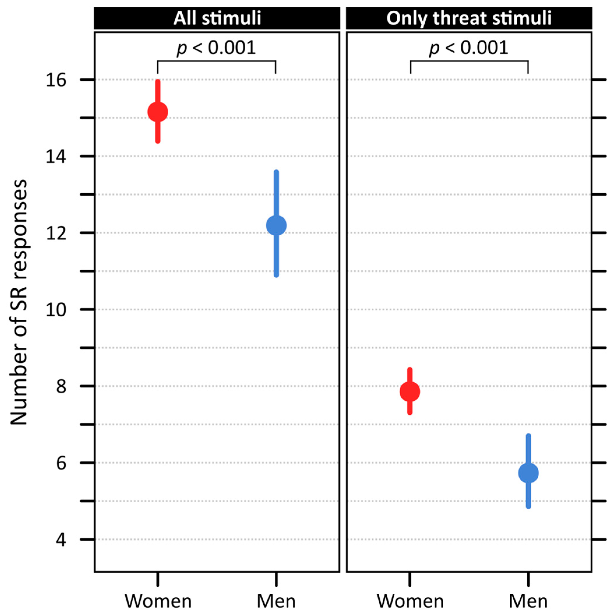
The effect of sex, however, was highly significant for both the number of responses to all stimuli (df = 1, χ2 = 18.42, p < 0.001, residual df = 111, Nagelkerke’s R2 = 0.100) and the number of responses to just the threat stimuli (df = 1, χ2 = 16.49, p < 0.001, residual df = 111, Nagelkerke’s R2 = 0.103). Model estimated means for women were 15.15 (95% CI = 14.39–15.94) reactions in total, with 7.86 (95% CI = 7.31–8.43) observed in threat trials. For men, both estimated means were significantly lower: 12.19 (95% CI = 10.90–13.58) reactions in total, with 5.73 (95% CI = 4.86–6.70) observed in threat trials (Figure 3).

2.4. Effect of Subjective Disgust Saliency on Electrodermal Responses

Finally, to test the effect of threat stimuli subjective saliency, we once again constructed a GLME model. In the model, the presence of an SR change was used as a binary response, the 7-point Likert scale rating according to elicited disgust as an explanatory variable, and the participants’ ID as a random factor. Thus, this model tested for a direct link between objectively measured electrodermal response and intensity of subjective emotion. Nonetheless, the intensity of elicited disgust did not prove statistically significant: AIC0 = 2716, AIC1 = 2716, χ2 = 2.19, p = 0.139.
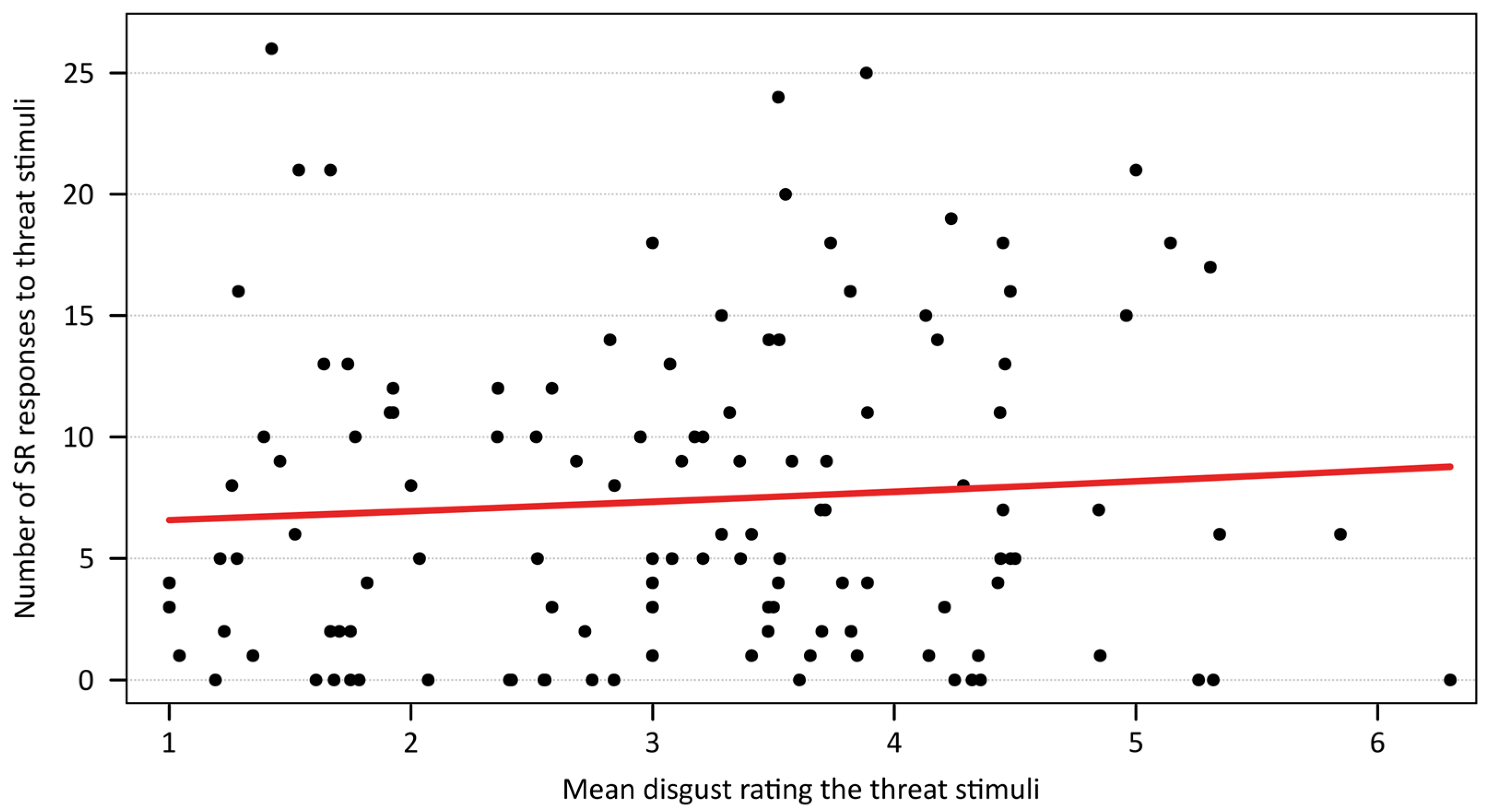
In the next step, we focused on the participants’ reactivity. In the GLM model, the number of observed SR responses for trials with threat stimuli was used as response and mean subjective disgust ratings of all threats as explanatory variable. Therefore, this model tested for a general link between electrodermal reactivity to a specific threat type and, overall, subjectively perceived disgust evoked by that type of threat. The effect of the mean disgust rating proved statistically significant (χ2 = 3.90, p = 0.048), but the amount of explained deviance was very low (Nagelkerke’s R2 = 0.032; Figure 4).

3. Discussion

The present study demonstrated that electrodermal responses to visual threat cues were best explained by the finest categorization of the stimuli into seven groups, with sneezing and disgusting animals eliciting the strongest reactions. Moreover, sex differences emerged significant, with women showing higher response amplitudes and frequencies than men. Finally, the subjective disgust ratings were not strongly associated with physiological reactivity at the trial level, though a weak overall relationship was found between average disgust rating of a stimulus type and the number of responses it elicited.

3.1. Differences Between Stimulus Categories

In this study, we examined whether the probability of an electrodermal response depends on the type of the visual threat cue. To address this, we compared different approaches to stimulus categorization: a broad distinction between ancestral and modern threats, and a more fine-grained classification that distinguished between spoiled food, “disgusting” animals, pollution, hospital environments, and preventive behaviours such as masks and sneezing. The results indicated that the most detailed categorization provided the best model fit, although only two categories—sneezing and “disgusting” animals—elicited significantly more responses than neutral controls. This finding is noteworthy, as both sneezing and parasites/worms are typical examples of contamination or pathogen-related disgust cues [26,33], just as masks and hospital environments are. However, sneezing represents a more ancestral cue, directly linked to disease transmission, whereas masks and hospitals are evolutionarily novel and likely require greater cognitive mediation. This pattern suggests a possible bias toward stronger autonomic reactivity to ancestral pathogen cues, yet it is complicated by the unexpectedly low response probability to spoiled food, which was also classified among ancestral threats.
Spoiled food, a prototypical core disgust stimulus [55,56], elicited the lowest probability of electrodermal responses in our study. This finding suggests that spoiled food may represent a somewhat distinct category of disgust cue. Unlike “disgusting” animals, which require active avoidance to prevent direct contact, spoiled food poses no immediate danger as long as it is not consumed. This interpretation is further supported by recent findings [57] showing that the proximity of a stimulus to the mouth strongly increases perceived disgust, especially for inedible (non-food) stimuli. From an adaptive perspective, it may have been advantageous not to expend energy on avoidance responses to food stimuli that could simply be ignored in contrast to the more urgent threat of involuntary oral contamination posed by invertebrates near the mouth. This highlights that the disgust system is sensitive to contextual cues. Nevertheless, spoiled food still elicited some reactions, which may reflect the evolutionary ambiguity of such cues. While completely rotten food is clearly hazardous, partially spoiled food may historically have been worth closer exploration if nutritional resources were scarce, thereby attractive for further evaluation [58]. Interestingly, it has been previously shown that spoiled food items are not visually avoided but rather gazed upon, unlike other highly disgust-evoking stimuli [59,60], and that nutritional value of depicted food items does not affect visual attention or skin conductance responses to these items [61]. In our study, however, the stimuli depicted strongly aversive, clearly spoiled food, which left little room for such ambiguity and may explain the overall low probability of responses. This pattern is consistent with the hypothesis that spoiled food does not signal an active, immediate threat.
On the other hand, “disgusting” animals proved to be particularly effective in eliciting electrodermal responses. A straightforward interpretation is that cues involving visible parasites or worms constitute highly salient pathogen threats, thereby recruiting stronger automatic defensive responses. However, previous studies have shown that small invertebrates can evoke both disgust and fear [62,63,64,65], which complicates this interpretation. This category of stimuli may therefore elicit not only a need for immediate active avoidance, in contrast to spoiled food, but also engage elements of a “fight-or-flight” response, albeit on a small scale, resulting in more pronounced sympathetic activation. Such an explanation might seem less applicable to endoparasites, since visual contact would be unlikely to trigger an evolved fear response, however, subjectively these animals showed high rating of fear [66] as well as disgust [63]. Also, the ectoparasites included in this category have been reported to elicit non-zero fear ratings [66], suggesting that a partial fear component may contribute to their strong physiological impact.
Beyond biological sources of pathogen threat such as animals and spoiled food, we also examined more modern disgust cues linked to pollution and chemical toxicity. These stimuli elicited relatively few electrodermal responses compared to other categories. Previous research on fear-evoking threats has shown that even evolutionarily novel stimuli can elicit strong subjective fear [43,44], trigger rapid direction of attention [67], and evoke automatic physiological reaction [68], suggesting that fear is flexible and readily extends to ontogenetically novel dangers ([49,69,70]; but see also [71,72]). Disgust, however, appears to have evolved along a different trajectory. Originating as an adaptation to protect against harmful ingestion (core or pathogen disgust) [40], it later broadened to encompass symbolic, social, and moral domains [73]. In line with this, statements related to pollution and toxicity threats were often rated as evoking fear and anger accompanied by moral disgust [43]. Yet, the lack of frequent skin resistance responses indicates that these cues did not trigger the strong autonomic arousal typically associated with anger. Instead, we speculate that modern contamination cues such as pollution may not immediately activate the evolved automatic defence system but rather rely on conscious appraisal and deliberate cognitive processing.
A similar interpretation can be applied to the low frequency of responses observed for the category of masks (preventive behaviours) and hospital environments. Pandemic-related visual cues are inherently more abstract than direct cues such as sneezing and, therefore, may require conscious processing rather than triggering immediate autonomic reactions. In a related study, these also evoked less salient disgust evaluation compared to visible cues of infection, i.e., sneezing [36]. This may help explain why these stimuli elicited only weak electrodermal responses in our study. Moreover, some stimuli might potentially elicit contempt. This hostile emotion is directed at a human target, stems from a sense of superiority and a negative evaluation of the target’s character, can lead to feelings of detachment, and is often tied to violations of social or communal norms [74]. However, we believe that airborne disease cues are more likely to elicit disgust rather than contempt, because disgust is primarily an emotion related to the physical body and the risk of contamination, whereas contempt is a social emotion directed at a person’s character. It is also possible that stronger reactions would occur if participants had been recently primed by an ongoing pandemic context. As our experiment was conducted 2–3 years after the COVID-19 pandemic, such contextual priming was probably already absent, which may have further reduced the salience of these cues.
We also examined the effect of reaction amplitude; however, this parameter did not differ between the categories. Therefore, the probability of reaction seems to contain the main portion of information on the participants’ responses towards various types of threats. It is worth mentioning that the amplitude distribution was substantially skewed right (skewness = 4.04), meaning that the majority of responses was rather small in their amplitude; however, a few responses were also very large. Thus, the amplitude appears to be affected more by external factors and individual setting of each participant.
Taken together, these findings suggest that the ancestral threats are rather a heterogenous category, with high avoidant reaction toward sneezing, avoidant and fearful reaction to “disgusting” animals, and disinterest for food. Ancestral pathogen cues present in stimuli evoking pathogen disgust (sneezing and parasites) thus seem to elicit electrodermal response. Spoiled food, on the other hand, evokes core or oral disgust [55,56,75], and electrodermal responses to this disgust subcategory is not so effective. Contrary, modern threats appear to work in a more homogenous way by recruiting complex cognitive processing resulting in lesser immediate responses.

3.2. Subjective Disgust Rating and Reactivity

Surprisingly, even though the categories of disgust-evoking threats differ from each other, detailed analysis of the level of disgust showed no statistically significant influence on the probability of SR response. We tested this relationship using two approaches: the first one linking the presence or absence of individual skin resistance responses to subjective ratings of the respective stimulus, and the second one comparing the participants’ overall reactivity to the average disgust rating of the entire threat category. While the latter model revealed a statistical effect, its size was negligible and unlikely to reflect a meaningful biological relationship.
The weak correspondence between subjective ratings of elicited disgust and physiological measures echoes a long-standing debate in emotion research. Early theories such as the James–Lange theory (reviewed in [76]) proposed that physiological changes actually precede and shape our emotional feelings. In this view, our emotions are, in part, an interpretation of these peripheral bodily changes. The opposing point of view is that the emotion is the first response of the thalamus after perceiving the stimulus, and the physiological reactions are rather separate response to prepare body for proper behavioural action [77]. Contemporary research indicates that the relationship might be more complex. For example, Taschereau-Dumouchel et al. [78] demonstrated that while subjective fear ratings and physiological responses were correlated, they were characterized by activity of distinct neural substrates: defensive circuits such as the amygdala and insula predicted autonomic reactivity, whereas higher-order prefrontal regions predicted subjective reports. This dissociation could explain our result, suggesting that subjective disgust evaluations may depend on conscious appraisal processes, whereas skin resistance responses primarily capture automatic sympathetic activation.

3.3. Participants Characteristics and Reactivity

In this study, we were also interested in the link between subjective experience of disgust and the evoked physiological response, as well as whether sensitivity to disgust (TDDS questionnaire) or anxiousness, i.e., trait anxiety (STAI-X2 questionnaire) influenced the participants’ reactions. Our results showed that general sensitivity to disgust did not significantly affect the probability of response to the stimuli. This finding diverges from previous work such as Rohrmann et al. [79], who reported that individuals with higher self-reported disgust sensitivity showed stronger physiological reactions to disgusting videos, suggesting a link between disgust sensitivity and bodily responses. A likely explanation for this discrepancy is the type of stimuli employed. Rohrmann et al. [79] used highly intense film clips of mutilation and burn treatment, which evoke particularly strong forms of disgust. By contrast, our study used static visual images, which typically elicit weaker and more variable responses, as also noted by [29,30]. In line with this, Croy et al. [9] demonstrated that the sensory modality of stimulus presentation can significantly alter both subjective and physiological responses, with the olfactory modality especially evoking stronger reactions compared to other modalities of stimuli. Meanwhile, subjective ratings of disgust were the highest for visual stimuli.
Stark et al. [12] emphasized that there are substantial interindividual differences in how subjective ratings of disgust translate into physiological reactivity, and that these relationships may be inconsistent across disgust domains and measures. This issue of interindividual variability in physiological responsiveness is well-documented in fear-conditioning research, where participants are frequently excluded from analyses due to insufficient electrodermal changes (reviewed in [22]). Moreover, Tuvblad et al. [80] suggested that the association between electrodermal activity and psychological processes may have a hereditary component. Thus, not only the intensity of the emotional experience is relevant, but also, because electrodermal responsiveness depends on multiple factors, interindividual variability in responsiveness is inevitable. One might nonetheless expect disgust sensitivity to translate into stronger bodily reactivity if the stimuli is more closely tied to the personal or clinical relevance of the participant. For instance, individuals with spider phobia display heightened electrodermal responses alongside elevated subjective fear when confronted with phobic stimuli [81,82]. Moreover, these measures tend to covary more strongly in ophiophobic than in non-phobic populations [83]. However, that does not necessarily seem to be the case for other disorders. Research on obsessive–compulsive disorder (OCD) shows that OCD patients consistently report elevated disgust sensitivity, but physiological measures such as skin conductance or heart rate often show weak or inconsistent correlations with these self-reports [84,85]. A recent review similarly concluded that individuals with OCD may experience strong subjective disgust without corresponding autonomic activation, reflecting a partial disconnection between conscious experience and physiological responses [86]. Taken together, these findings suggest that the absence of a robust relationship between TDDS scores and physiological reactivity in our study may not be an anomaly, but rather consistent with the broader literature indicating that subjective disposition and autonomic response do not always align.
General trait anxiety, as measured by the STAI-X2, did not significantly influence the participants’ responses. This is consistent with our earlier findings, where trait anxiety also failed to correlate with physiological reactivity [87]. Given that the STAI-X2 captures stable dispositional anxiety rather than momentary fluctuations, future studies might benefit from including measures of state anxiety (e.g., STAI-X1), which may be more sensitive to capturing context-dependent effects on autonomic responses.
Our results also revealed a robust effect of sex on electrodermal reactivity. Women were not only more reactive to both all and specifically just threat stimuli but also displayed higher average amplitudes of skin resistance changes compared to men. In this regard, existing studies are mixed. Kopacz & Smith and Rohrmann et al. [88,89] found similar tendency of women having more pronounced electrodermal responses compared to men, and even though [90] found the same trend, it did not prove significant. Several studies [91,92] did not find gender differences in the skin conductance response.

4. Materials and Methods

4.1. Participants

One hundred and twenty-three healthy participants predominantly from the Czech Republic, Slovakia, and other countries of Central Europe aged 18 to 74 (median = 22, mean = 25.29, SD = 9.67) were recruited for this experiment through poster flyers, social networks, or direct contacts established during previous projects. Since studies have shown women to have higher disgust sensitivity than men [93,94], we primarily recruited women (97, i.e., 78.86%) but did not exclude men (26, i.e., 21.14%) either. To further characterize the respondents, we asked them to fill in two questionnaires: the State-Trait Anxiety Inventory-X2 (STAI-X2; [95,96]) to assess their trait anxiety, and the Three Domains of Disgust Scale (TDDS; [33,97]), excluding the sexual disgust subscale, to measure their sensitivity to pathogen and moral disgust.

4.2. Stimuli

To investigate physiological responses to evolutionarily relevant and modern threats, a set of visual stimuli was assembled. The images were drawn from established affective picture databases, namely DIRTI [98], OASIS [99], IAPS [100], and SMID [101], and were supplemented by images obtained through web search (Pixabay, Wikimedia Commons, and Flickr) or taken by the authors of this paper (Supplementary Table S1). These stimuli were categorized based on their subject matter into four disgust-eliciting categories: “disgusting” animals (parasites and worm-like animals), spoiled food, pandemic, and toxicity and pollution. The former two represented evolutionarily relevant threats, while toxicity and pollution represented modern, ontogenetic threats. The threat of pandemic was initially categorized into modern threats, although some cues were ancestral (see Introduction). For each category, 28 coloured images were selected. The emotionally neutral stimuli (control) were photographs of leaves with uniform 20% grey background (Figure 1).
In the category of “disgusting” animals, photographs of ecto- and endoparasites (e.g., ticks, tape worms) were selected, and also potentially disease spreading animals like cockroaches and slimy worm-like animals (e.g., flatworms) were chosen to represent this category. Pictures of spiders were avoided. The category of spoiled food included photographs of rotten fruits and mouldy cooked meals. The category of toxicity and pollution depicted images related to radiation or environmental contamination such as garbage, water pollution, or oil spill. In some cases, the photos contained humans or animals. The category evoking the threat of pandemic consisted of scenes suggesting direct transmission risk (e.g., an individual sneezing near another, hospitalized patients with overt respiratory illness) as well as indirect risk cues (e.g., groups of people with and without masks in enclosed public spaces such as waiting rooms or pharmacies). The images were not modified, only resized to 1772 × 1181 pixels if needed.

4.3. Procedure

Each participant was tested individually in a controlled, quiet laboratory environment. Upon arrival, the participants received a detailed explanation of the experimental procedure, offered the opportunity to ask questions, and provided an informed consent with the procedure. Next, they were seated approximately 60 cm from the display screen (26”, Quad HD resolution 2560 × 1440 pixels, full-screen presentation), and the experimenter proceeded to attach sensors used for recording physiological responses. Continuous physiological monitoring was conducted using the VLV3 multifunctional biotelemetric system [102]; the skin resistance was measured via two dry Ag/AgCl sensors affixed to the distal phalanges of the index and middle fingers of the participant’s non-dominant hand.
The participants viewed a sequence of 28 experimental stimuli from one randomly assigned category, interchanged with neutral control images. There was always a black screen for at least 5 s between each stimulus. This presentation paradigm was adapted from previously validated methodologies [103]. To mitigate potential order effects [104], three distinct randomized presentation sequences were generated for each stimulus category, and the participants were randomly allocated to one of these versions. The total duration of the stimulus presentation block was approximately 15 min. We presented the experimental sequence with “disgusting” animals to 31 participants, spoiled food to 32 participants, pandemic to 29 participants, and toxicity and pollution to 31 participants.
Following the physiological recording session, participants were asked to provide basic demographic information (sex, age, field and level of education, email). They then completed subjective ratings for each experimental stimulus they had viewed during the session. These ratings were provided on a 7-point Likert scale, quantifying the intensity of disgust evoked by each image, where 1 indicated “no elicited disgust” and 7 indicated “very strong disgust.” Furthermore, the participants completed the above-mentioned two questionnaires, STAI-X2 and TDDS.

4.4. Data Curation

The final dataset was processed using the vlv3_prohlizec software (version 1.1) [102] (also used in Landová et al. [93]) and is available in Supplementary Tables S2 and S3. Consistent with the established psychophysiological literature [17], an immediate emotional skin resistance response was operationally defined as a discernible change in skin conductance initiated within 4 s of stimulus onset. However, for the current study, this temporal window was extended to 5 s for the potentially greater cognitive processing demands associated with the complex visual stimuli. Responses initiated beyond this 5 s window were not considered an automatic response to the stimulus since they are less likely to reflect an immediate, direct emotional reaction and more likely to involve subsequent cognitive elaboration [17]. These responses were, therefore, not recorded.
In the further step of data curation, we excluded trials during which participants considerably moved, casting doubt on whether the observed response was a result of the movement or an emotional reaction to the stimulus. For this reason, we excluded 962 out of the total of 6888 trials (13.97%). There was a considerable inter-individual variability between the participants in this regard (min = 34, max = 56, median = 49, mean = 48.18, SD = 5.69), but no substantial differences were found between different presentation categories (85, 88, 86, and 86% of valid trials for respective stimulus categories).
Finally, we excluded non-reactive participants, i.e., participants with 0 valid responses to at least one stimulus (control or threat). A total of 13 participants was excluded for this reason: 3 men and 10 women, 3 presented with “disgusting” animal experimental sequence, 2 with spoiled food, 4 with pandemics, and 2 with toxicity and pollution experimental sequences. Non-reactive participants are sometimes excluded in similar studies since they occur quit often (e.g., [12]). Nonetheless, we employed a very conservative cut-off line to make sure that the exclusion would not obscure the investigated effects.

4.5. Statistical Analysis

All statistical analyses were performed using RStudio (Version 4.3.1) [105], with generalized linear models (GLM) and generalized linear mixed-effect models (GLMM) implemented via the lme4 package [106]. Further, package performance [107] was used to compute effect sizes. With GLMM models, we successively determined effects of stimulus category, participant’s sex, and of subjective disgust rating of the stimulus, on the probability or amplitude of skin resistance (SR) change. In models for the probability of SR change, binomial distribution with logit link was used, while Gamma distribution with natural logarithm link was used in models investigating amplitude of SR change. In all models, participants’ ID was included as a random factor. The best model was chosen on the basis of the Akaike information criterion (AIC) and tested against the null model (i.e., models with no fixed factors).
To investigate the effect of participants’ characteristics on their SR responses, we summed the number of valid trials with observed SR response for each participant separately, which represented the participant’s general electrodermal reactivity. In a similar manner, we summed the number of trials with threat stimuli and observed SR response which in turn represented their reactivity to the specific threat. With GLM models (Poisson distribution, natural logarithm link), we assessed the effects of the STAI-X2 score, TDDS score, and participant’s sex, and, additionally, in separate models, the effect of mean threat disgust rating on these parameters. The best model was chosen by backward selection.

4.6. Ethical Note

All procedures performed in this study were carried out in accordance with the ethical standards of the appropriate institutional research committee, the Ethical Committees of Charles University, Faculty of Science (approval no. 2021/02, granted on 14 April 2021), the National Institute of Mental Health (no. 91/21, granted 31 March 2021), and with the 1964 Helsinki declaration and its later amendments or comparable ethical standards. Written informed consent was obtained from all participants included in the study. Additionally, written informed consent for the publication of identifying images was obtained from volunteers posing for stimuli photos.

5. Conclusions

In this study, we aimed to investigate whether various types of disgust-related threats, including typical (ancestral) as well as modern stimuli, evoke automatic physiological response measured as skin resistance change. We found that stimulus categories substantially differed in this respect. None of the modern cues—hospital environment, pandemic preventive behaviour, or pollution and toxicity—was associated with higher probability of SR response than the control. We concluded that these types of threats probably rely on complex cognitive processing resulting in lesser chance of observing an immediate response. Contrary, two out of three ancestral cues proved potent since we observed SR change associated with presentation of these stimuli quite frequently. Specifically, these were the pathogen-related stimuli, or, in other words, stimuli representing acute risk of contamination—pictures of people sneezing, including the aerosol droplets, and of disgust-evoking animals, including ecto- and endoparasites, cockroaches, and worms. Ancestral disgust-related threat of rotten or spoiled food deviate from this pattern; we speculate that spoiled food evoked slightly different type of disgust (oral or core disgust), does not represent acute threat, and hence does not trigger immediate automatic physiological response.

Supplementary Materials

The following supporting information can be downloaded at: https://www.mdpi.com/article/10.3390/physiologia5040041/s1, Table S1: Sources of experimental stimuli photos; Table S2: Data associated with this study—respondents’ characteristics; Table S3: Data associated with this study—measured skin resistance responses.

Author Contributions

Conceptualization, E.L.; Methodology, E.L. and T.H.; Formal Analysis, I.Š.; Investigation, T.H.; Data Curation, T.H. and I.Š.; Writing—Original Draft Preparation, T.H. and I.Š.; Writing—Review and Editing, D.F. and E.L.; Visualization, I.Š.; Supervision, E.L. and D.F.; Funding Acquisition, E.L. All authors have read and agreed to the published version of the manuscript.

Funding

This project was funded by the Czech Scientific Foundation (GAČR), project No. 22-13381S, awarded to EL, https://www.gacr.cz/en/ (accessed on 8 October 2025).

Institutional Review Board Statement

The study was conducted in accordance with the Declaration of Helsinki and approved by the Ethical Committees of Charles University, Faculty of Science (approval no. 2021/02, granted on 14 April 2021) and the National Institute of Mental Health (no. 91/21, granted 31 March 2021).

Informed Consent Statement

Informed consent was obtained from all subjects involved in the study. Additionally, written informed consent has been obtained from the volunteers posing for stimuli photos to publish this paper.

Data Availability Statement

The dataset generated for this study can be found in Supplementary Tables S2 and S3.

Acknowledgments

We would like to thank Šárka Peléšková and Markéta Janovcová for their advice during the initial phase of this project. Additionally, we would like to acknowledge Petr Telenský for his advice on interpretation of the results.

Conflicts of Interest

The authors declare no conflicts of interest.

References

  1. Brosch, T.; Pourtois, G.; Sander, D. The Perception and Categorisation of Emotional Stimuli: A Review. Cogn. Emot. 2010, 24, 377–400. [Google Scholar] [CrossRef]
  2. Ekman, P.; Levenson, R.W.; Friesen, W.V. Autonomic Nervous System Activity Distinguishes among Emotions. Science 1983, 221, 1208–1210. [Google Scholar] [CrossRef]
  3. Lang, P.J.; Greenwald, M.K.; Bradley, M.M.; Hamm, A.O. Looking at Pictures: Affective, Facial, Visceral, and Behavioral Reactions. Psychophysiology 1993, 30, 261–273. [Google Scholar] [CrossRef]
  4. Cannon, W.B. The Emergency Function of the Adrenal Medulla in Pain and the Major Emotions. Am. J. Physiol. Leg. Content 1914, 33, 356–372. [Google Scholar] [CrossRef]
  5. McCarty, R. Chapter 4—The Fight-or-Flight Response: A Cornerstone of Stress Research. In Stress: Concepts, Cognition, Emotion, and Behavior; Fink, G., Ed.; Academic Press: San Diego, CA, USA, 2016; pp. 33–37. ISBN 978-0-12-800951-2. [Google Scholar]
  6. McCorry, L.K. Physiology of the Autonomic Nervous System. Am. J. Pharm. Educ. 2007, 71, 78. [Google Scholar] [CrossRef] [PubMed]
  7. Bradley, M.M.; Codispoti, M.; Cuthbert, B.N.; Lang, P.J. Emotion and Motivation I: Defensive and Appetitive Reactions in Picture Processing. Emotion 2001, 1, 276–298. [Google Scholar] [CrossRef]
  8. Collet, C.; Vernet-Maury, E.; Delhomme, G.; Dittmar, A. Autonomic Nervous System Response Patterns Specificity to Basic Emotions. J. Auton. Nerv. Syst. 1997, 62, 45–57. [Google Scholar] [CrossRef]
  9. Croy, I.; Laqua, K.; Suess, F.; Joraschky, P.; Ziemssen, T.; Hummel, T. The Sensory Channel of Presentation Alters Subjective Ratings and Autonomic Responses toward Disgusting Stimuli—Blood Pressure, Heart Rate and Skin Conductance in Response to Visual, Auditory, Haptic and Olfactory Presented Disgusting Stimuli. Front. Hum. Neurosci. 2013, 7, 510. [Google Scholar] [CrossRef]
  10. Christie, I.C.; Friedman, B.H. Autonomic Specificity of Discrete Emotion and Dimensions of Affective Space: A Multivariate Approach. Int. J. Psychophysiol. 2004, 51, 143–153. [Google Scholar] [CrossRef]
  11. Kreibig, S.D. Autonomic Nervous System Activity in Emotion: A Review. Biol. Psychol. 2010, 84, 394–421. [Google Scholar] [CrossRef] [PubMed]
  12. Stark, R.; Walter, B.; Schienle, A.; Vaitl, D. Psychophysiological Correlates of Disgust and Disgust Sensitivity. J. Psychophysiol. 2005, 19, 50–60. [Google Scholar] [CrossRef]
  13. Baker, L.B. Physiology of Sweat Gland Function: The Roles of Sweating and Sweat Composition in Human Health. Temperature 2019, 6, 211–259. [Google Scholar] [CrossRef]
  14. Harker, M. Psychological Sweating: A Systematic Review Focused on Aetiology and Cutaneous Response. Ski. Pharmacol. Physiol. 2013, 26, 92–100. [Google Scholar] [CrossRef]
  15. Morris-Jones, R. Neural Control of Sweat Secretion: A Review of the Neurology and Current Treatment Options for Hyperhidrosis. Br. J. Dermatol. 2018, 178, 1233–1234. [Google Scholar] [CrossRef] [PubMed]
  16. Wilke, K.; Martin, A.; Terstegen, L.; Biel, S.S. A Short History of Sweat Gland Biology. J. Cosmet. Sci. 2007, 29, 169–179. [Google Scholar] [CrossRef] [PubMed]
  17. Boucsein, W.; Fowles, D.C.; Grimnes, S.; Ben-Shakhar, G.; Roth, W.T.; Dawson, M.E.; Filion, D.L. Society for Psychophysiological Research Ad Hoc Committee on Electrodermal Measures Publication Recommendations for Electrodermal Measurements. Psychophysiology 2012, 49, 1017–1034. [Google Scholar] [CrossRef]
  18. Arabian, H.; Schmid, R.; Wagner-Hartl, V.; Moeller, K. Analysis of EDA and Heart Rate Signals for Emotional Stimuli Responses. Curr. Dir. Biomed. Eng. 2023, 9, 150–153. [Google Scholar] [CrossRef]
  19. Vrana, S.R. The Psychophysiology of Disgust: Differentiating Negative Emotional Contexts with Facial EMG. Psychophysiology 1993, 30, 279–286. [Google Scholar] [CrossRef] [PubMed]
  20. Rymarczyk, K.; Żurawski, Ł.; Jankowiak-Siuda, K.; Szatkowska, I. Empathy in Facial Mimicry of Fear and Disgust: Simultaneous EMG-fMRI Recordings During Observation of Static and Dynamic Facial Expressions. Front. Psychol. 2019, 10, 701. [Google Scholar] [CrossRef]
  21. Gläscher, J.; Adolphs, R. Processing of the Arousal of Subliminal and Supraliminal Emotional Stimuli by the Human Amygdala. J. Neurosci. 2003, 23, 10274–10282. [Google Scholar] [CrossRef] [PubMed]
  22. Gold, A.K.; Kredlow, M.A.; Orr, S.P.; Hartley, C.A.; Otto, M.W. Skin Conductance Levels and Responses in Asian and White Participants during Fear Conditioning. Physiol. Behav. 2022, 251, 113802. [Google Scholar] [CrossRef]
  23. Sánchez-Navarro, J.P.; Martínez-Selva, J.M.; Maldonado, E.F.; Carrillo-Verdejo, E.; Pineda, S.; Torrente, G. Autonomic Reactivity in Blood-Injection-Injury and Snake Phobia. Psychosom. Res. 2018, 115, 117–124. [Google Scholar] [CrossRef] [PubMed]
  24. Schienle, A.; Stark, R.; Vaitl, D. Evaluative Conditioning: A Possible Explanation for the Acquisition of Disgust Responses? Learn. Motiv. 2001, 32, 65–83. [Google Scholar] [CrossRef]
  25. Grus, A.; Hromatko, I. Psychological and Physiological Correlates of Pathogen-Induced Disgust. In Proceedings of the 22nd Psychology Days in Zadar, Zadar, Croatia, 1–3 October 2020; Banai, I.P., Ed.; University of Zadar, Department of Psychology: Zadar, Croatia, 2022; Volume 3, pp. 29–40. [Google Scholar]
  26. Rozin, P.; Haidt, J.; McCauley, C.R. Disgust: The Body and Soul Emotion. In Handbook of Cognition and Emotion; John Wiley & Sons Ltd.: Hoboken, NJ, USA, 1999; pp. 429–445. ISBN 978-0-471-97836-7. [Google Scholar]
  27. Levenson, R.W. Autonomic Nervous System Differences among Emotions. Psychol. Sci. 1992, 3, 23–27. [Google Scholar] [CrossRef]
  28. Levenson, R.W.; Ekman, P.; Friesen, W.V. Voluntary Facial Action Generates Emotion-Specific Autonomic Nervous System Activity. Psychophysiology 1990, 27, 363–384. [Google Scholar] [CrossRef]
  29. Comtesse, H.; Stemmler, G. Cardiovascular Regulation Pattern of Contamination-Related Disgust: Consistency and Context Dependence. Psychophysiology 2016, 53, 1407–1416. [Google Scholar] [CrossRef] [PubMed]
  30. de Jong, P.J.; van Overveld, M.; Peters, M.L. Sympathetic and Parasympathetic Responses to a Core Disgust Video Clip as a Function of Disgust Propensity and Disgust Sensitivity. Biol. Psychol. 2011, 88, 174–179. [Google Scholar] [CrossRef] [PubMed]
  31. Rohrmann, S.; Hopp, H. Cardiovascular Indicators of Disgust. Int. J. Psychophysiol. 2008, 68, 201–208. [Google Scholar] [CrossRef]
  32. Olatunji, B.O.; Haidt, J.; McKay, D.; David, B. Core, Animal Reminder, and Contamination Disgust: Three Kinds of Disgust with Distinct Personality, Behavioral, Physiological, and Clinical Correlates. J. Res. Pers. 2008, 42, 1243–1259. [Google Scholar] [CrossRef]
  33. Tybur, J.M.; Lieberman, D.; Griskevicius, V. Microbes, Mating, and Morality: Individual Differences in Three Functional Domains of Disgust. J. Pers. Soc. Psychol. 2009, 97, 103–122. [Google Scholar] [CrossRef]
  34. Davey, G.C.L. Mechanisms of Disgust in Psychopathology. In The Handbook of Disgust Research: Modern Perspectives and Applications; Powell, P.A., Consedine, N.S., Eds.; Springer International Publishing: Cham, Switzerland, 2021; pp. 191–208. ISBN 978-3-030-84486-8. [Google Scholar]
  35. Ekman, P. An Argument for Basic Emotions. Cogn. Emot. 1992, 6, 169–200. [Google Scholar] [CrossRef]
  36. Janovcová, M.; Polák, J.; Končická, A.; Chomik, A.; Kaňková, Š.; Frynta, D.; Landová, E. From bugs to sickness: Disgust evaluation of ancestral, modern, and pandemic threats. Evol. Psychol. Sci. 2025, 11, 273–290. [Google Scholar] [CrossRef]
  37. Rozin, P.; Haidt, J.; McCauley, C. Disgust: The Body and Soul Emotion in the 21st Century. In Disgust and Its Disorders: Theory, Assessment, and Treatment Implications; American Psychological Association: Washington, DC, USA, 2009; pp. 9–29. ISBN 978-1-4338-0397-0. [Google Scholar]
  38. Bradshaw, H.K.; Gassen, J. The Evolution of Disgust, Pathogens, and the Behavioural Immune System. In The Handbook of Disgust Research: Modern Perspectives and Applications; Powell, P.A., Consedine, N.S., Eds.; Springer International Publishing: Cham, Switzerland, 2021; pp. 31–51. ISBN 978-3-030-84486-8. [Google Scholar]
  39. Schaller, M.; Park, J.H. The Behavioral Immune System (and Why It Matters). Curr. Dir. Psychol. Sci. 2011, 20, 99–103. [Google Scholar] [CrossRef]
  40. Darwin, C. The Expression of the Emotions in Man and Animals; John Murray: London, UK, 1872; pp. vi, 374. [Google Scholar]
  41. Olatunji, B.O.; Williams, N.L.; Tolin, D.F.; Abramowitz, J.S.; Sawchuk, C.N.; Lohr, J.M.; Elwood, L.S. The Disgust Scale: Item Analysis, Factor Structure, and Suggestions for Refinement. Psychol. Assess. 2007, 19, 281–297. [Google Scholar] [CrossRef]
  42. Shapouri, S.; Martin, L.L. Snakes vs. Guns: A Systematic Review of Comparisons Between Phylogenetic and Ontogenetic Threats. Adapt. Hum. Behav. Physiol. 2022, 8, 131–155. [Google Scholar] [CrossRef]
  43. Landová, E.; Polák, J.; Janovcová, M.; Štolhoferová, I.; Peterková, Š.; Chomik, A.; Frynta, D. Imprint of Ancestral and Modern Threats in Human Mind—Experience of Fear, Disgust, and Anger. Front. Psychol. 2025, 15, 1520224. [Google Scholar] [CrossRef]
  44. Peléšková, Š.; Polák, J.; Janovcová, M.; Chomik, A.; Sedláčková, K.; Frynta, D.; Landová, E. Human Emotional Evaluation of Ancestral and Modern Threats: Fear, Disgust, and Anger. Front. Psychol. 2024, 14, 1321053. [Google Scholar] [CrossRef]
  45. Bennett, K. Ancestral Threats vs. Modern Threats. In Encyclopedia of Evolutionary Psychological Science; Shackelford, T.K., Weekes-Shackelford, V.A., Eds.; Springer International Publishing: Cham, Switzerland, 2019; pp. 1–3. ISBN 978-3-319-16999-6. [Google Scholar]
  46. Schaller, M. The Behavioural Immune System and the Psychology of Human Sociality. Philos. Trans. R. Soc. B Biol. Sci. 2011, 366, 3418–3426. [Google Scholar] [CrossRef]
  47. Curtis, V.; de Barra, M.; Aunger, R. Disgust as an Adaptive System for Disease Avoidance Behaviour. Philos. Trans. R. Soc. B Biol. Sci. 2011, 366, 389–401. [Google Scholar] [CrossRef]
  48. Thiebaut, G.; Méot, A.; Witt, A.; Prokop, P.; Bonin, P. The Behavioral Immune System: How Does It Contribute to Our Understanding of Human Behavior? In Advances in Psychology Research; Nova Science Publishers, Inc.: Hauppauge, NY, USA, 2021; pp. 1–59. ISBN 978-1-5361-9542-2. [Google Scholar]
  49. Schaller, M.; Miller, G.E.; Gervais, W.M.; Yager, S.; Chen, E. Mere Visual Perception of Other People’s Disease Symptoms Facilitates a More Aggressive Immune Response. Psychol. Sci. 2010, 21, 649–652. [Google Scholar] [CrossRef]
  50. Giner-Sorolla, R.; Kupfer, T.; Sabo, J. Chapter Five—What Makes Moral Disgust Special? An Integrative Functional Review. In Advances in Experimental Social Psychology; Olson, J.M., Ed.; Academic Press: Cambridge, MA, USA; Oxford, UK, 2018; Volume 57, pp. 223–289. [Google Scholar]
  51. Ottaviani, C.; Mancini, F.; Petrocchi, N.; Medea, B.; Couyoumdjian, A. Autonomic Correlates of Physical and Moral Disgust. Int. J. Psychophysiol. 2013, 89, 57–62. [Google Scholar] [CrossRef]
  52. Armelagos, G.J.; Goodman, A.H.; Jacobs, K.H. The Origins of Agriculture: Population Growth during a Period of Declining Health. Popul. Environ. 1991, 13, 9–22. [Google Scholar] [CrossRef]
  53. Latham, K. Human Health and the Neolithic Revolution: An Overview of Impacts of the Agricultural Transition on Oral Health, Epidemiology, and the Human Body. Neb. Anthropol. 2013, 28, 95–102. [Google Scholar]
  54. Piret, J.; Boivin, G. Pandemics Throughout History. Front. Microbiol. 2021, 11, 631736. [Google Scholar] [CrossRef] [PubMed]
  55. Davey, G.C.L. Disgust. In Encyclopedia of Human Behavior; Ramachandran, V.S., Ed.; San Diego Press: San Diego, CA, USA, 1994; pp. 135–141. [Google Scholar]
  56. Rozin, P.; Fallon, A.E. A Perspective on Disgust. Psychol. Rev. 1987, 94, 23–41. [Google Scholar] [CrossRef]
  57. Prokop, P.; Fančovičová, J.; Šramelová, D.; Thiebaut, G.; Méot, A.; Bonin, P. Mouth Proximity Influences Perceived Disgust of Visual Stimuli. Pers. Individ. Dif. 2023, 207, 112146. [Google Scholar] [CrossRef]
  58. Ciesielski, B.G.; Armstrong, T.; Zald, D.H.; Olatunji, B.O. Emotion Modulation of Visual Attention: Categorical and Temporal Characteristics. PLoS ONE 2010, 5, e13860. [Google Scholar] [CrossRef]
  59. Armstrong, T.; Stewart, J.G.; Dalmaijer, E.S.; Rowe, M.; Danielson, S.; Engel, M.; Bailey, B.; Morris, M. I’ve Seen Enough! Prolonged and Repeated Exposure to Disgusting Stimuli Increases Oculomotor Avoidance. Emotion 2022, 22, 1368–1381. [Google Scholar] [CrossRef]
  60. Fančovičová, J.; Prokop, P.; Šramelová, D.; Thiebaut, G.; Méot, A.; Witt, A.; Bonin, P.; Medina-Jerez, W. Does Food Play a Prominent Role in Visual Attention to Disgusting Stimuli? J. Ethol. 2022, 40, 23–29. [Google Scholar] [CrossRef]
  61. Schlezingerová, N.; Málková, P.; Kocourek, M.; Telenský, P. Mild Hunger Elicits Attentional Desensitization to Visual Food Cues in Healthy, Non-Obese Individuals. Front. Psychol. 2024, 15, 1441184. [Google Scholar] [CrossRef]
  62. Gerdes, A.B.M.; Uhl, G.; Alpers, G.W. Spiders Are Special: Fear and Disgust Evoked by Pictures of Arthropods. Evol. Hum. Behav. 2009, 30, 66–73. [Google Scholar] [CrossRef]
  63. Polák, J.; Rádlová, S.; Janovcová, M.; Flegr, J.; Landová, E.; Frynta, D. Scary and Nasty Beasts: Self-Reported Fear and Disgust of Common Phobic Animals. Br. J. Psychol. 2020, 111, 297–321. [Google Scholar] [CrossRef]
  64. Arrindell, W.A. Phobic Dimensions: IV. The Structure of Animal Fears. Behav. Res. Ther. 2000, 38, 509–530. [Google Scholar] [CrossRef]
  65. Davey, G.C.L.; McDonald, A.S.; Hirisave, U.; Prabhu, G.G.; Iwawaki, S.; Jim, C.I.; Merckelbach, H.; de Jong, P.J.; Leung, P.W.L.; Reimann, B.C. A Cross-Cultural Study of Animal Fears. Behav. Res. Ther. 1998, 36, 735–750. [Google Scholar] [CrossRef] [PubMed]
  66. Prokop, P.; Fančovičová, J. The Association between Disgust, Danger and Fear of Macroparasites and Human Behaviour. Acta Ethologica 2010, 13, 57–62. [Google Scholar] [CrossRef]
  67. Fox, E.; Griggs, L.; Mouchlianitis, E. The Detection of Fear-Relevant Stimuli: Are Guns Noticed as Quickly as Snakes? Emotion 2007, 7, 691–696. [Google Scholar] [CrossRef] [PubMed]
  68. Flykt, A.; Esteves, F.; Öhman, A. Skin Conductance Responses to Masked Conditioned Stimuli: Phylogenetic/Ontogenetic Factors versus Direction of Threat? Biol. Psychol. 2007, 74, 328–336. [Google Scholar] [CrossRef]
  69. Hugdahl, K.; Johnsen, B.H. Preparedness and Electrodermal Fear-Conditioning: Ontogenetic vs Phylogenetic Explanations. Behav. Res. Ther. 1989, 27, 269–278. [Google Scholar] [CrossRef]
  70. Subra, B.; Muller, D.; Fourgassie, L.; Chauvin, A.; Alexopoulos, T. Of Guns and Snakes: Testing a Modern Threat Superiority Effect. Cogn. Emot. 2018, 32, 81–91. [Google Scholar] [CrossRef]
  71. Abado, E.; Aue, T.; Okon-Singer, H. Spider vs. Guns: Expectancy and Attention Biases to Phylogenetic Threat Do Not Extend to Ontogenetic Threat. Front. Psychol. 2023, 14, 1232985. [Google Scholar] [CrossRef]
  72. Sulikowski, D. Are Natural Threats Superior Threats? Evol. Hum. Behav. 2022, 43, 34–43. [Google Scholar] [CrossRef]
  73. Rozin, P.; Lowery, L.; Imada, S.; Haidt, J. The CAD Triad Hypothesis: A Mapping between Three Moral Emotions (Contempt, Anger, Disgust) and Three Moral Codes (Community, Autonomy, Divinity). J. Pers. Soc. Psychol. 1999, 76, 574–586. [Google Scholar] [CrossRef]
  74. Miceli, M.; Castelfranchi, C. Contempt and Disgust: The Emotions of Disrespect. J. Theor. Soc. Behav. 2018, 48, 205–229. [Google Scholar] [CrossRef]
  75. Davey, G.C.L.; Chapman, L. Disgust and Eating Disorder Symptomatology in a Non-Clinical Population: The Role of Trait Anxiety and Anxiety Sensitivity. Clin. Psychol. Psychother. 2009, 16, 268–275. [Google Scholar] [CrossRef] [PubMed]
  76. Myers, G.E. William James’s Theory of Emotion. Trans. Charles S Peirce Soc. 1969, 5, 67–89. [Google Scholar]
  77. Cannon, W.B. The James-Lange Theory of Emotions: A Critical Examination and an Alternative Theory. Am. J. Physiol. 1927, 39, 106–124. [Google Scholar] [CrossRef]
  78. Taschereau-Dumouchel, V.; Kawato, M.; Lau, H. Multivoxel Pattern Analysis Reveals Dissociations between Subjective Fear and Its Physiological Correlates. Mol. Psychiatry 2020, 25, 2342–2354. [Google Scholar] [CrossRef]
  79. Rohrmann, S.; Hopp, H.; Schienle, A.; Hodapp, V. Emotion Regulation, Disgust Sensitivity, and Psychophysiological Responses to a Disgust-Inducing Film. Anxiety Stress Coping 2009, 22, 215–236. [Google Scholar] [CrossRef]
  80. Tuvblad, C.; Gao, Y.; Isen, J.; Botwick, T.; Raine, A.; Baker, L.A. The Heritability of the Skin Conductance Orienting Response: A Longitudinal Twin Study. Biol. Psychol. 2012, 89, 47–53. [Google Scholar] [CrossRef]
  81. Knopf, K.; Pössel, P. Individual Response Differences in Spider Phobia: Comparing Phobic and Non-Phobic Women of Different Reactivity Levels. Anxiety Stress Coping 2009, 22, 39–55. [Google Scholar] [CrossRef]
  82. Wendt, J.; Lotze, M.; Weike, A.I.; Hosten, N.; Hamm, A.O. Brain Activation and Defensive Response Mobilization during Sustained Exposure to Phobia-Related and Other Affective Pictures in Spider Phobia. Psychophysiology 2008, 45, 205–215. [Google Scholar] [CrossRef]
  83. Schaefer, H.S.; Larson, C.L.; Davidson, R.J.; Coan, J.A. Brain, Body, and Cognition: Neural, Physiological and Self-Report Correlates of Phobic and Normative Fear. Biol. Psychol. 2014, 98, 59–69. [Google Scholar] [CrossRef]
  84. Serrano, M.Á.; Rosell-Clari, V.; García-Soriano, G. The Role of Perceived Control in the Psychophysiological Responses to Disgust of Subclinical OCD Women. Sensors 2019, 19, 4180. [Google Scholar] [CrossRef]
  85. Whitton, A.E.; Henry, J.D.; Grisham, J.R. Cognitive and Psychophysiological Correlates of Disgust in Obsessive-Compulsive Disorder. Br. J. Clin. Psychol. 2015, 54, 16–33. [Google Scholar] [CrossRef]
  86. Pruneti, C.; Coscioni, G.; Guidotti, S. A Systematic Review of Clinical Psychophysiology of Obsessive–Compulsive Disorders: Does the Obsession with Diet Also Alter the Autonomic Imbalance of Orthorexic Patients? Nutrients 2023, 15, 755. [Google Scholar] [CrossRef]
  87. Štolhoferová, I.; Hladíková, T.; Janovcová, M.; Peléšková, Š.; Frynta, D.; Landová, E. Subjective and Psychophysiological Response to Pictures of Ancestral and Modern Threats: Not All Evolutionary Threats Are Alike. PLoS ONE 2025. submitted. [Google Scholar]
  88. Kopacz II, F.M.; Smith, B.D. Sex Differences in Skin Conductance Measures as a Function of Shock Threat. Psychophysiology 1971, 8, 293–303. [Google Scholar] [CrossRef] [PubMed]
  89. Rohrmann, S.; Hopp, H.; Quirin, M. Gender Differences in Psychophysiological Responses to Disgust. J. Psychophysiol. 2008, 22, 65–75. [Google Scholar] [CrossRef]
  90. Bari, D.S. Gender Differences in Tonic and Phasic Electrodermal Activity Components. Sci. J. Univ. Zakho 2020, 8, 29–33. [Google Scholar] [CrossRef]
  91. Kelly, M.M.; Forsyth, J.P.; Karekla, M. Sex Differences in Response to a Panicogenic Challenge Procedure: An Experimental Evaluation of Panic Vulnerability in a Non-Clinical Sample. Behav. Res. Ther. 2006, 44, 1421–1430. [Google Scholar] [CrossRef]
  92. Wrase, J.; Klein, S.; Gruesser, S.M.; Hermann, D.; Flor, H.; Mann, K.; Braus, D.F.; Heinz, A. Gender Differences in the Processing of Standardized Emotional Visual Stimuli in Humans: A Functional Magnetic Resonance Imaging Study. Neurosci. Lett. 2003, 348, 41–45. [Google Scholar] [CrossRef]
  93. Druschel, B.A.; Sherman, M.F. Disgust Sensitivity as a Function of the Big Five and Gender. Pers. Individ. Dif. 1999, 26, 739–748. [Google Scholar] [CrossRef]
  94. Haidt, J.; McCauley, C.; Rozin, P. Individual Differences in Sensitivity to Disgust: A Scale Sampling Seven Domains of Disgust Elicitors. Pers. Individ. Dif. 1994, 16, 701–713. [Google Scholar] [CrossRef]
  95. Heretik, A.; Ritomský, A.; Novotný, V.; Pečeňák, J. Restandardizace state-trait anxiety inventory x-2—Úzkostnost jako rys—Proquest. Českoslov. psychol. 2009, 53, 587–599. [Google Scholar]
  96. Spielberger, C.D.; Gorsuch, R.L.; Lushene, R.E. Manual for the State-Trait Anxiety Inventory (Self-Evaluation Questionnaire); Consulting Psychologists Press: Palo Alto, CA, USA, 1970. [Google Scholar]
  97. Polák, J.; Landová, E.; Frynta, D. Undisguised Disgust: A Psychometric Evaluation of a Disgust Propensity Measure. Curr. Psychol. 2019, 38, 608–617. [Google Scholar] [CrossRef]
  98. Haberkamp, A.; Glombiewski, J.A.; Schmidt, F.; Barke, A. The DIsgust-RelaTed-Images (DIRTI) Database: Validation of a Novel Standardized Set of Disgust Pictures. Behav. Res. Ther. 2017, 89, 86–94. [Google Scholar] [CrossRef]
  99. Kurdi, B.; Lozano, S.; Banaji, M.R. Introducing the Open Affective Standardized Image Set (OASIS). Behav. Res. 2017, 49, 457–470. [Google Scholar] [CrossRef] [PubMed]
  100. Mikels, J.A.; Fredrickson, B.L.; Larkin, G.R.; Lindberg, C.M.; Maglio, S.J.; Reuter-Lorenz, P.A. Emotional Category Data on Images from the International Affective Picture System. Behav. Res. Methods 2005, 37, 626–630. [Google Scholar] [CrossRef] [PubMed]
  101. Crone, D.L.; Bode, S.; Murawski, C.; Laham, S.M. The Socio-Moral Image Database (SMID): A Novel Stimulus Set for the Study of Social, Moral and Affective Processes. PLoS ONE 2018, 13, e0190954. [Google Scholar] [CrossRef]
  102. Kašpar, J.; Hon, Z.; Janatová, M.; Smrčka, P.; Vítězník, M.; Hána, K. RESDB. Available online: https://isdv.upv.gov.cz/webapp/resdb.print_detail.det?pspis=PT/2014-979 (accessed on 27 July 2024).
  103. Landová, E.; Peléšková, Š.; Sedláčková, K.; Janovcová, M.; Polák, J.; Rádlová, S.; Vobrubová, B.; Frynta, D. Venomous Snakes Elicit Stronger Fear than Nonvenomous Ones: Psychophysiological Response to Snake Images. PLoS ONE 2020, 15, e0236999. [Google Scholar] [CrossRef] [PubMed]
  104. Kosonogov, V. The Effects of the Order of Picture Presentation on the Subjective Emotional Evaluation of Pictures. Psicologia 2020, 34, 171–178. [Google Scholar] [CrossRef]
  105. R Core Team. R: A Language and Environment for Statistical Computing; R Foundation for Statistical Computing: Vienna, Austria, 2023. [Google Scholar]
  106. Bates, D.; Mächler, M.; Bolker, B.; Walker, S. Fitting Linear Mixed-Effects Models Using lme4. J. Stat. Softw. 2015, 67, 1–48. [Google Scholar] [CrossRef]
  107. Lüdecke, D.; Ben-Shachar, M.S.; Patil, I.; Waggoner, P.; Makowski, D. Performance: An R Package for Assessment, Comparison and Testing of Statistical Models. J. Open Source Softw. 2021, 6, 3139. [Google Scholar] [CrossRef]
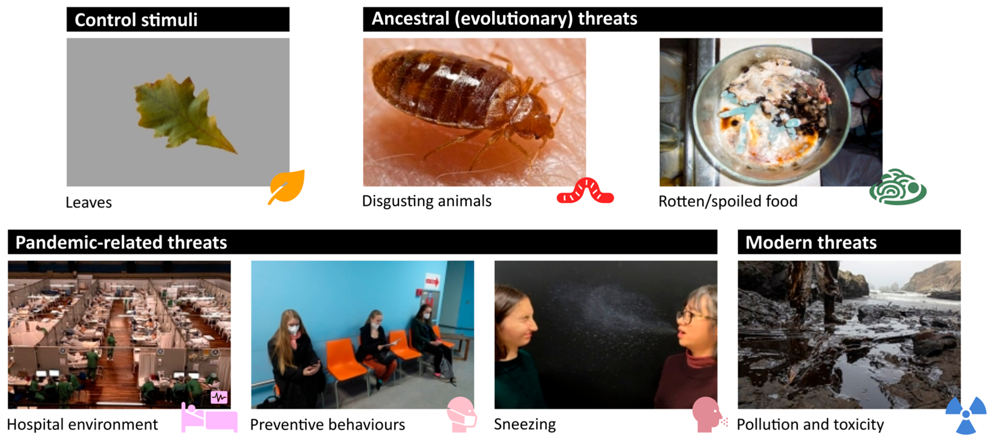
Figure 1. Examples of control and experimental stimuli and their categorization.
Physiologia 05 00041 g001
Figure 2. Mean and 95% confidence intervals of probability of SR change as a response to different stimuli categories; the pictograms from left to right represent the following stimulus categories: control, “disgusting” animals, spoiled food, hospital environment, preventive behaviours (masks), sneezing, and toxicity and pollution. Significant differences between control and threat categories are inscribed with p-values.
Physiologia 05 00041 g002
Figure 3. Effect of participants’ sex on their reactivity (the number of observed SR responses): estimated means and 95% confidence intervals, significant differences inscribed with p-values.
Physiologia 05 00041 g003
Figure 4. Effect of disgust saliency on the number of SR responses to the threat stimuli. The red line represents the mean relationship as computed with the GLM model.
Physiologia 05 00041 g004
Table 1. Probability of SR change as a response to different stimulus categories. Values on transformed (logit) scale are the direct output of the GLME model, and the values on the original scale are the estimated means and 95% confidence intervals (CI) converted to probability. Z-values and p-values refer to the test of statistically significant (non-zero) difference between control stimuli and respective categories’ p-values < 0.05 are in bold.
CategoryMean, 95% CI
(Logit Scale)
Mean, 95% CI
(Probability Scale)
z-Valuep-Value
Control−0.814 (−1.123, −0.504)0.31 (0.24, 0.38)
“Disgusting” animals−0.504 (−0.881, −0.126)0.38 (0.29, 0.47)2.390.017
Spoiled food−1.062 (−1.459, −0.664)0.26 (0.19, 0.34)−1.690.092
Hospital environment−0.638 (−1.071, −0.204)0.35 (0.26, 0.45)1.040.299
Preventive behaviours−0.920 (−1.376, −0.464)0.29 (0.20, 0.39)−0.580.565
Sneezing0.070 (−0.700, 0.840)0.52 (0.33, 0.70)2.410.016
Toxicity and pollution−1.040 (−1.421, −0.660)0.26 (0.19, 0.34)−1.720.085
Disclaimer/Publisher’s Note: The statements, opinions and data contained in all publications are solely those of the individual author(s) and contributor(s) and not of MDPI and/or the editor(s). MDPI and/or the editor(s) disclaim responsibility for any injury to people or property resulting from any ideas, methods, instructions or products referred to in the content.