Systematic Review

Systematic Review of the Role of Kv4.x Potassium Channels in Neurodegenerative Diseases: Implications for Neuronal Excitability and Therapeutic Modulation

by
Bárbara Teruel-Peña
1,
Piedad Gómez-Torres
2,
Sergio Galarreta-Aperte
2,
Nora Suleiman-Martos
3,
Isabel Prieto
1,
Manuel Ramírez-Sánchez
1,
Carmen M. Fernández-Martos
4,5,† and
Germán Domínguez-Vías
6,*,†
1
Department of Health Sciences, University of Jaén, 23071 Jaén, Spain
2
Nursing Department, Faculty of Health Sciences, Ceuta University of Granada, 51001 Ceuta, Spain
3
Nursing Department, Faculty of Health Sciences, University of Granada, 18071 Granada, Spain
4
Faculty of Pharmacy, University of San Pablo CEU, 28003 Madrid, Spain
5
Wicking Dementia Research and Education Centre, College of Health and Medicine, University of Tasmania, Hobart, TAS 7000, Australia
6
Department of Physiology, Faculty of Health Sciences, Ceuta University of Granada, 51001 Ceuta, Spain
*
Author to whom correspondence should be addressed.
These authors contributed equally as senior authors.
Physiologia 2025, 5(3), 31; https://doi.org/10.3390/physiologia5030031
Submission received: 22 August 2025 / Revised: 7 September 2025 / Accepted: 8 September 2025 / Published: 10 September 2025
(This article belongs to the Special Issue Feature Papers in Human Physiology—3rd Edition)

Abstract

Background/Objectives: The voltage-gated potassium channels of the Kv4 family (Kv4.1, Kv4.2, Kv4.3) regulate neuronal excitability and synaptic integration. The dysregulation of these channels has been linked to neurodegenerative diseases, such as Alzheimer’s disease (AD), spinocerebellar ataxias, amyotrophic lateral sclerosis (ALS), prion diseases, and Parkinson’s disease (PD). Current evidence is scattered across diverse models, and a systematic synthesis is lacking. This review seeks to compile and analyze data on Kv4 channel alterations in neurodegeneration, focusing on genetic variants, functional changes, and phenotypic consequences. Methods: A systematic search was conducted for peer-reviewed studies, including human participants, human-derived cell models, and relevant animal models. Studies were considered eligible if they investigated Kv4.1–Kv4.3 (encoded by gene encoding the Kv4.1-Kv4.3 α-subunit of voltage-gated A-type potassium channels (KCND1-KCND3)) expression, function, or genetic variants, as well as associated auxiliary subunits such as DPP6 (dipeptidyl peptidase–like protein 6) and KChIP2 (Kv channel–interacting protein 2), in neurodegenerative diseases. Both observational and experimental designs were considered. Data extraction included disease type, model, Kv4 subunit, functional or genetic findings, and key outcomes. Risk of bias was assessed in all included studies. Results: Kv4 channels exhibit significant functional and expression changes in various neurodegenerative diseases. In AD and prionopathies, reduced Kv4.1- and Kv4.2-mediated currents contribute to neuronal hyperexcitability. In spinocerebellar ataxias, KCND3 mutations cause loss- or gain-of-function phenotypes in Kv4.3, disrupting cerebellar signaling. In models of ALS and PD, Kv4 dysfunction correlates with altered neuronal excitability and can be modulated pharmacologically. Subunit modulators such as DPP6 and KChIP2 influence channel function and could represent therapeutic targets. Conclusions: Kv4 channels are crucial for neuronal excitability in multiple neurodegenerative contexts. Dysregulation through genetic or pathological mechanisms contributes to functional deficits, highlighting Kv4 channels as promising targets for interventions aimed at restoring electrical homeostasis and mitigating early neuronal dysfunction.

1. Introduction

1.1. Molecular Structure of Kv4 Channels

Ion channels are macromolecular complexes present in the plasma membrane and in intracellular organelles of all cells, participating in essential functions such as the preservation of cellular integrity, smooth muscle contraction, and the release of neurotransmitters and hormones [1]. Alterations in channel structure, function, gating, or expression give rise to channelopathies, which represent a challenge for research and therapeutic development [2,3]. This review focuses on voltage-gated potassium (Kv) channels, particularly the Kv4 or Shal subfamily, formed by Kv4.1 (KCND1), Kv4.2 (KCND2) and two splice variants of Kv4.3 (KCND3), with ~60% sequence identity [2,3]. Kv4.x channels are expressed mainly in brain, heart and muscle, where they generate transient potassium ion (K+) currents, known as the transient outward K+ current (Ito) in the heart and the transient A-type current (Ia, or also referred as IKA and IA) in neurons, with Ito and Ia being different names for the same current depending on the tissue context [4]. Thus, Kv4 channels are responsible for the outward movement of K+, which contributes to the generation of a transient outward current.
Pore-forming subunits, termed α-subunits, assemble as symmetric or heterotetrameric complexes with auxiliary subunits and scaffolding proteins. These complexes regulate surface expression, kinetics, and neuronal excitability [5,6,7,8,9]. Each subunit has six transmembrane helices (S1–S6), with cytoplasmic N- and C-terminal ends (Figure 1). The T1 domain at the N-terminus regulates assembly and tetramerization, S1–S4 constitutes the voltage sensor, and S5–S6 with the P loop forms the pore and selectivity filter. The fourth helix (S4), positively charged, responds to depolarization and generates the gating current [10]. The transmembrane region is highly conserved, except in the proximal C-terminus of S6 of Kv4.3, where an additional exon encodes 19 amino acids, giving rise to the Kv4.3L and Kv4.3S variants [11,12]. Membrane depolarization causes a movement of positively charged residues of S4 across the gated channel. This movement of charges across the membrane’s electric field mediates the actual opening of the channel and generates what is known as a gate current. The properties of the A-type current depend on the cellular context, since expression in heterologous systems reflects the intrinsic characteristics of the α subunit, but may differ from native channels [4]. In heterologous systems, Kv4.2 displays intrinsic properties characteristic of A-type K+ channels. In neurons, hippocampal studies indicate that Kv4.2 contributes to neuronal excitability and synaptic signaling. Furthermore, post-translational modification of Kv4.x, especially by kinases, dynamically modulates membrane excitability and intracellular signaling cascades [9].

1.2. Subunits and Auxiliary Proteins That Interact with Kv4

The functionality and expression of Kv4 channels is modulated by various subunits and accessory proteins (Figure 1). Kvβ subunits (Kvβ1, Kvβ2, Kvβ3) interact with Kv4 α-subunits, increasing current density, modulating inactivation and serving as chaperones for their correct membrane expression [9,13,14,15,16,17,18,19,20,21,22,23,24]. K+ channel interacting Proteins (KChIPs 1–4) are calcium-binding proteins that bind to the amino terminus of Kv4.x, increasing current density, accelerating recovery from inactivation, and stabilizing the channels at the membrane; KChIP4, however, does not promote surface expression and eliminates rapid inactivation [25,26,27,28,29,30,31,32,33]. Neuronal calcium sensor-1 (NCS-1) increases the current density and surface area of Kv4.2 and Kv4.3, modulating inactivation in a calcium ion (Ca2+)-dependent manner [34,35,36]. K+ Channel Accessory Protein (KChAP) promotes Kv4.3 expression by interacting with Kvβ1.2 [37,38,39], while dipeptidyl-peptidase-like Protein 6 (DPPX or also known as potassium channel accelerating factor (KAF)) accelerates inactivation, increases surface expression and restores native A-type current properties for Kv4.2 and Kv4.3 [40,41]. Cytoskeletal proteins such as filamin and adhesion molecules such as integrins regulate the somatodendritic localization of Kv4.2 and Kv4.3 [42,43,44]. The postsynaptic density protein 95 (PSD-95) facilitates surface expression and clustering of Kv4.2 via a PSD-95/Discs-large/ZO-1 (PDZ) domain [45,46]. Finally, in the heart, MinK-related peptide 1 (MiRP1) modulates the activation and inactivation of Kv4.2, although its neuronal relevance has not yet been confirmed [47,48,49,50,51,52]. Together, these interactors are critical components of Kv4 complexes and contribute to the regulation of A-type K+ currents in neuronal and cardiac cells.

1.3. Kv4 Channelopathies and Implications for Neuronal Function

Alterations in these channels lead to the origin of many nervous system disorders. The role of Kv4 channels in neurodegenerative processes is not yet fully understood, although alterations in their expression and function have been documented in various diseases of this type [4,53,54]. Kv4.x channels and their auxiliary subunits play a key role in the regulation of neuronal excitability through A-type currents and synaptic transmission, modulating the membrane potential, repolarization after the action potential and the release of neurotransmitters [4,53,55]. Its alterations are associated with the pathogenesis of some CNS diseases such as Alzheimer’s disease (AD), Parkinson’s disease (PD), and amyotrophic lateral sclerosis (ALS) [53,56,57].
In the brain, the expression of Kv4 channels varies according to the neuronal type and the region studied, and research is still being conducted to understand their distribution in different subcellular compartments and neuronal populations [58,59,60]. In the hippocampus, Kv4 alterations have been associated with neurodegenerative and non-neurodegenerative disorders (e.g., schizophrenia and epilepsy) [60,61,62,63].
Disorders such as certain types of epilepsy and early repolarization syndrome are due to functional or genetic alterations in Kv4 channels. In epilepsy models, Kv4.2 has been shown to target proconvulsant microRNAs, with decreased expression following seizures, an effect that is modulated by hormonal context [64]. In humans, mutations in KCND3 (Kv4.3) are associated with early repolarization syndrome, demonstrating that Kv4.3 dysfunction underlies the electrical alteration characteristic of this channelopathy [65]. Therefore, pharmacological modulation of somatic or dendritic Ia (i.e., Isa and Ito currents, respectively) could offer a potential therapeutic approach for these diseases. These findings confirm that, although these pathologies are not neurodegenerative, Kv4 channel dysfunction represents the central underlying mechanism, highlighting its critical role in the regulation of neuronal excitability and the pathophysiology of these diseases. In epilepsy, animal models have been observed to present a transient decrease in Kv4.2 in the granule cell layer of the hippocampus after seizures induced by convulsives such as pentylenetetrazol or kainate, which correlates with neuronal hyperexcitability and is mediated, at least in part, by the activation of N-methyl-D-aspartate (NMDA)-type glutamate receptors that bidirectionally regulate the phosphorylation and dephosphorylation of Kv4.2 channels, thus modulating excitability and synaptic plasticity in the hippocampus [66,67,68,69]. Alterations of the KChIP3 auxiliary subunit have also been described in human hippocampal tissue and in animal models, indicating that changes in the composition of the Kv4.x complex may contribute to epileptogenesis. Furthermore, post-translational regulation of channels, including phosphorylation by kinases such as extracellular signal–regulated kinase (ERK), Ca2+/calmodulin-dependent protein kinase II (CaMKII), protein kinase C (PKC) and protein kinase A (PKA), could rapidly modify Kv4.x activity during epileptogenesis [4]. In AD, the interaction of Kv4.x with the auxiliary subunit KChIP3 (calsenilin) and with presenilin proteins (PS1 and PS2) suggests a possible link with pathological mechanisms of the disease, although its functional contribution is still unclear [4]. Mutations in presenilins can alter Kv4.x channels through changes in KChIP3 modulation, potentially affecting neuronal excitability and signaling. This evidence suggests that Kv4.x could participate in processes of cellular plasticity and homeostasis relevant to neurodegeneration. Overall, in both epilepsy and AD, alterations in Kv4.x channels and auxiliary subunits represent a potential mechanism of neuronal dysfunction, highlighting their relevance as therapeutic targets in CNS pathologies [4,44,60,70].
In this review, we will focus exclusively on neurodegenerative diseases of the CNS, since these pathologies involve a progressive deterioration of neuronal function associated with alterations in Kv4 channels, which allows a more homogeneous analysis of mechanisms, genetic variants and A-type current dysfunction. This delimitation excludes other non-degenerative CNS disorders that, although also involving Kv4 channelopathies, present distinct and heterogeneous pathophysiological mechanisms. As a limitation, this review does not address the effects of Kv4 dysfunctions in acute, neurodevelopmental or cardiovascular diseases, nor does it evaluate therapeutic interventions in detail, which restricts the generalization of the findings outside the context of neurodegeneration.

2. Results

The results of this review are presented below, organized by groups of neurodegenerative diseases, followed by an integrative section that analyzes general mechanisms, causal relationships, and clinical implications. Table 1 provides the risk of bias (RoB) assessment together with a concise synthesis of the findings, whereas Supplementary Table S1 includes the complete dataset with detailed numerical information for further consultation. RoB for each study was assessed using predefined criteria adapted from the Newcastle-Ottawa Scale (NOS) or Systematic Review Centre for Laboratoy Animal Experimentation (SYRCLE) RoB tool, with adjustments for atypical experimental designs. The degree of risk (low, some concerns, or high) is summarized in Table 1, with detailed rationale provided in Section 4.2 and Supplementary Table S2.

2.1. Amyotrophic Lateral Sclerosis (ALS)

In ALS, alterations in Kv4 channels have been investigated primarily in patient-derived cell cultures and preclinical models. A study using human bone marrow mesenchymal stem cells (hBM-MSCs) obtained from ALS patients demonstrated a cell passage-dependent functional profile of Kv4 channels. Specifically, in early passages of hBM-MSCs, reduced expression of the Kv4.2 was observed, accompanied by a decrease in the Ia current [56]. This suggests that the ability of these cells to generate Kv4.2-dependent A-type currents deteriorates in the early stages of culture, perhaps reflecting intrinsic alterations related to the ALS condition or to cellular aging in vitro. Furthermore, in a motor neuron cell model (NSC-34 cell line), it was shown that treatment with cannabinol (CBN) can up-modulate the expression of Kv4 channel genes [71]. Specifically, exposure to CBN dose-dependently increased the expression of Kv4.1 and Kv4.2 in these cells. This finding suggests a potential neuroprotective or modulatory mechanism where cannabinoid compounds can restore or increase the expression of Kv4 channels in motor neurons, compensating for the K+ current dysfunction that could occur in ALS. It is worth noting that, to date, no known pathogenic variants in the KCND1–3 genes directly associated with ALS have been identified, so the alterations observed in Kv4.1 and Kv4.2 appear to be of a functional or regulatory rather than genomic nature.

2.2. Parkinson’s Disease (PD)

In PD, characterized by degeneration of dopaminergic neurons in the substantia nigra and accumulation of α-synuclein, the effects of this pathological protein on Kv4.3 channels have been explored. In murine models of PD that overexpress the A53T mutation of α-synuclein (SNCA), dysfunction of Kv4.3 channels has been observed in vulnerable neurons. Specifically, in dopaminergic neurons of the substantia nigra of A53T-SNCA transgenic mice, Kv4.3-mediated A current activity was markedly reduced, contributing to an increase in neuronal excitability [72]. This effect suggests that the presence of mutant α-synuclein causes a loss of Kv4.3 function that destabilizes electrical homeostasis, possibly facilitating hyperexcitability and vulnerability of dopaminergic neurons. However, the effects may be region dependent. In the dorsal motor nucleus of the vagus nerve of the A53T-SNCA model, no significant changes in the activation or inactivation properties of Kv4.3 currents of vagal motor neurons were found. Furthermore, intracellular administration of the antioxidant glutathione (GSH) was able to rescue A-type currents in neurons from control mice but had no effect on neurons from A53T-SNCA mice, indicating that in the latter, Kv4.3 currents are not susceptible to oxidative stress as expected [57]. This could imply that α-synuclein-induced Kv4.3 dysfunction depends on specific neuronal contexts (dopaminergic versus cholinergic) or on the interaction with oxidative stress pathways. No pathogenic mutations in KCND3 (or other KCNDs) directly linked to PD have been reported so far; instead, Kv4.3 modulation in this pathology seems to occur secondary to α-synuclein toxicity and the altered cellular environment.

2.3. Alzheimer’s Disease (AD)

In AD, a broad spectrum of research suggests that dysfunction of Kv4 channels—especially Kv4.2—contributes to the alteration of neuronal excitability in the presence of amyloid and tau pathology. From a genetic point of view, some studies have identified variants in Kv4 modulating genes in patients with AD. For example, missense variants in DPP6 (gene encoding an auxiliary subunit essential for Kv4.2 expression) have been found in patients with early-onset Alzheimer’s and frontotemporal dementia [73]. In experimental models, DPP6 deletion leads to reduced brain Kv4.2 levels and functional deficits in the A-type current. Brains of patients carrying DPP6 variants exhibited decreased Kv4.2 expression, suggesting a contribution to neuronal hyperexcitability [73]. This genetic link highlights a mechanism by which genotypic variants outside of KCND2 may indirectly impact Kv4.2 function in AD.
Several AD models have directly explored how amyloid peptides (Aβ) and other cellular alterations affect Kv4 channels. The 5xFAD mouse model, which overexpresses human amyloid precursor protein (APP) and presenilin-1 (PSEN1) carrying five familial AD mutations, develops early and robust amyloid pathology and cognitive decline. The RyR2-E4872Q knock-in mouse harbors a point mutation in the ryanodine receptor type 2 that enhances Ca2+ release from the endoplasmic reticulum (ER). In the 5xFAD × RyR2-E4872Q cross, combining amyloid pathology with exaggerated RyR2 activity, dysfunction of the Kv4.2 current was observed in hippocampal neurons [74]. This suggests that the Ca2+ imbalance caused by amyloid pathology may decrease the A-type current, contributing to hyperexcitability. On the other hand, early in vitro studies showed opposite effects when Aβ was applied acutely to non-AD–specific cells (e.g., cerebellar neurons, HEK cells). This approach provides mechanistic insight into early disease stages but does not fully reproduce the complexity of AD pathology, limiting direct generalization to the human condition. Exposure to the Aβ peptide (isoforms 1–40 or 1–42) can increase the activity and expression of Kv4 channels [75,80]. Rat cerebellar neurons and HEK293 cells incubated with Aβ1–40 showed increased density of A-type potassium currents and overexpression of Kv4.2 and Kv4.3. Similarly, Aβ25–35 applied to cerebellar granule neurons induced an increase in the Ia current and in the protein expression of Kv4.2 and Kv4.3 subunits, changes that could be prevented by coadministration of the modulatory neuropeptide substance P (SP) [54]. Likewise, in rats injected with Aβ25–35 fragments, an increase in Kv4.2 mRNA and protein in the cerebral cortex was reported (≈58% over controls), while Kv4.3 remained unchanged [79]. In Campolongo’s study [81], Kv4.2 protein expression increased in the hippocampus but not in the cortex, and paradoxically, treatment with SP did not restore Kv4.2 protein levels. These findings suggest that in early stages or in acute exposures, amyloid may trigger a compensatory response by increasing Kv4 channel function (possibly as an attempt to counteract incipient hyperexcitability).
However, in chronic AD contexts and advanced transgenic models, loss of Kv4 channel function predominates, associated with synaptic and dendritic hyperexcitability. In transgenic mouse models of Alzheimer’s (such as hAPP_J20 and hAPP_swe/PS1dE9), depletion of Kv4.2 protein was observed in the dendrites of hippocampal neurons, mediated by tau protein-dependent mechanisms [53]. This reduction in Kv4.2 was correlated with dendritic hyperexcitability; in fact, treatment with the antiepileptic drug levetiracetam prevented the loss of Kv4.2 and reduced hyperexcitability, highlighting the importance of this channel in stabilizing neuronal activity [53]. Similarly, in Tg2576 mice (another amyloid model) a decrease in the expression and function of Kv4.1 in granule neurons of the fascia dentata was reported [76]. This effect was linked to oxidative stress, since treatment with the antioxidant Trolox restored Kv4.1 levels and normalized neuronal excitability. This indicates that different Kv4 subunits (Kv4.1 in this case) may be affected by Alzheimer’s pathology, especially through oxidative damage, and that their rescue mitigates neuronal dysfunction. On the other hand, not all facets of AD pathophysiology involve Kv4. A study in APP knockout mice (AβPP−/−), a β-amyloid deficient model, found no alterations in Kv4.2 expression or function, concluding that the absence of Aβ per se does not modify the A-type current and, therefore, Kv4.2 does not participate in the changes in excitability when amyloid is absent [77]. This result suggests that it is the presence of Aβ, and probably also tau [53], that negatively modulates Kv4 in AD, rather than the absence of APP. Consideration of differences across models (e.g., hAPP_J20, hAPP/PS1, Tg2576, 3xTg-AD) provides important context for interpreting Kv4 alterations [53,74,76,78].
Another key mechanism is the phosphorylation of Kv4.2 in the presence of Aβ. Intracellular accumulation of Aβ42 oligomers in neurons (as occurs in the 3xTg-AD model) induced neuronal hyperexcitability due to the inhibition of A-type currents mediated by Kv4.2 phosphorylation. In this context, inhibition of glycogen synthase kinase 3 (GSK-3, kinase involved in phosphorylating Kv4.2) prevented such hyperexcitability by promoting dephosphorylation of the channel, thus restoring its function [78]. Overall, the results in AD indicate a complex picture. Initially, there may be a compensatory increase in Kv4 function, but as the disease progresses and tau, Ca2+, and oxidative stress become involved, there is a decrease in the expression or functional availability of Kv4.2/Kv4.1, contributing to neuronal dysrhythmia and cognitive impairment. To date, no pathogenic mutations in the KCND1–3 genes directly associated with AD have been identified; however, variants in modulators such as DPP6 highlight the relevance of the Kv4 pathway in susceptibility to AD.

2.4. Spinocerebellar and Episodic Ataxias

A notable group of inherited ataxias, particularly certain autosomal dominant spinocerebellar ataxias, have been linked to mutations in the KCND3 gene, which encodes the Kv4.3 subunit [82,83,84,85,86,87,88,89,90,91,92,93,94,95,96,97,98,99,102,103,104]. These include spinocerebellar ataxia types 19 and 22 (SCA19/22)—two allelic disorders caused by variants in KCND3—as well as some cases of progressive cerebellar ataxias and familial episodic ataxias. Phenotypically, patients present with progressive cerebellar ataxia that may manifest with gait instability, coordination disturbances, and dysarthria, typically beginning in young adulthood. Numerous genetic variants in KCND3 have been identified associated with these ataxias (Table 2), most of them missense or small deletions or duplications that alter the Kv4.3 protein.
Among the reported mutations are, for example, p.F227del, a phenylalanine deletion at position 227; p.V338E and p.G345V, substitutions that alter the S6 segment of the channel; p.S347W, p.W359G, p.T377M, p.R419H, p.R293_Phe295dup (duplication of the RVF sequence), among others. These variants have been identified in families and isolated cases throughout the world, including populations in Europe, Asia and Latin America. In recent case studies in Latin America, for example, de novo mutations p.Ser357Trp, p.Gly384Ser and p.Gly371Arg in KCND3 were described in patients with ataxia, consolidating the involvement of Kv4.3 in the etiology of the disease [97]. However, not all cohorts of patients with ataxia have mutations in KCND3; a study investigating patients with spinocerebellar ataxia type 13 and type 19 found no pathogenic variants in KCND3, indicating genetic heterogeneity and the existence of other causal genes in these syndromes [100].
The functional effects of Kv4.3 mutations have been analyzed by electrophysiological and expression assays in heterologous systems (HEK293, CHO-K1 cells, Xenopus oocytes) and in animal models. In general, most KCND3 mutations associated with ataxia cause a loss-of-function of the Kv4.3 channel, either by reducing the potassium current Ia that this channel conducts, by decreasing the protein expression of the channel in the membrane, or by altering its activation/inactivation properties. Kv4.3 variants p.G345V, p.S347W, and p.W359G have been shown to produce reduced Kv4.3-mediated current and lower channel expression, consistent with a loss-of-function effect [93]. Likewise, the p.F227del mutation in Kv4.3 causes reduced surface expression due to retention in the ER and Golgi, leading to decreased K+ conductance and cellular dysfunction [99]. This intracellular retention mechanism suggests a dominant negative effect, where the mutant subunit interferes with the function of the wild-type subunits, contributing to the pathogenesis of SCA22. Other mutations, such as p.Arg293_Phe295dup, which results in duplication of the RVF (arginine–valine–phenylalanine) motif described by Smets et al. [103], disrupt the channel’s voltage sensing. Functionally, this duplication caused severe changes in the kinetics of Kv4.3 activation and inactivation, significantly reducing the outward current without affecting the number of channels present on the cell surface. Interestingly, in that case, coexpression of the auxiliary protein KChIP2 was able to partially rescue the function of the mutated channel, indicating that some variants could cause channel instability that is mitigated by chaperones or auxiliary subunits.
Although most KCND3 mutations are associated with loss of function, at least one variant with the opposite effect has been identified. In a case of progressive cerebellar ataxia reported by Hsiao et al. [102], the p.R419H mutation in Kv4.3 was associated with a gain of function, where electrophysiological studies showed that this variant increases potassium current, despite not altering the expression or localization of the channel. This finding suggests that excessive Kv4.3 activity may also be deleterious to cerebellar neuronal function, possibly through excessive repolarization that affects the firing of Purkinje neurons. Indeed, in ataxias, both the abnormal decrease and increase in A currents could disturb the delicate balance of excitability in cerebellar circuits, resulting in motor symptoms.
Mutations in Kv4.3 are not the only relevant factor in ataxias. In spinocerebellar ataxia type 1 (SCA1), which is caused by a polyglutamine expansion in ataxin-1 (ATXN1) and not by ion channel mutations, alterations in Kv4 channel function have also been observed. In a transgenic mouse model of SCA1, it was found that in presymptomatic stages Purkinje potentials exhibited an increase in Kv4.3-mediated A-type current, even though Kv4.1, Kv4.2 and Kv4.3 mRNA levels were not increased in the cerebellum [101]. This suggests a possible increase in the functional availability of Kv4 (with a higher presence at the plasma membrane without a change in total expression) in the early response to the disease. This increase in A-type current could contribute to a reduction in dendritic excitability of Purkinje cells, possibly as a compensatory mechanism. Interestingly, administration of aminopyridines (K+ channel blockers, including the clinically used 4-aminopyridine —4-AP—) to these mice reversed early electrical dysfunction and improved motor symptoms, suggesting that modulating A-type currents may have a symptomatic and neuroprotective benefit in spinocerebellar ataxias [101]. Finally, it has been proposed that mutations in KCND3 could also be involved in episodic ataxias (EA), paroxysmal disorders characterized by brief episodes of cerebellar dysfunction [104]. A cohort study identified a de novo variant c.1291C > T (p.Arg431Cys) in KCND3 in a patient with episodic ataxia. Although this variant was considered potentially pathogenic, functional validation of its effect has not yet been performed, so its role in the etiology of episodic ataxia remains unclear. In summary, Kv4.3-associated ataxias show that channel variants can produce both loss and gain of function, both converging in an alteration of cerebellar neuronal excitability that clinically translates into ataxia.

2.5. Prionopathies (Prion Diseases)

Prion diseases, such as Creutzfeldt–Jakob disease, Gerstmann–Sträussler–Scheinker syndrome (GSS), and fatal familial insomnia, are neurodegenerative disorders characterized by the accumulation of pathological prions (PrPSc) derived from normal cellular prion protein (PrPC) [107]. Recent studies suggest that PrPC plays a role in modulating Kv4 channels, particularly Kv4.2, through interactions with auxiliary subunits [105]. Under physiological conditions, PrPC appears to facilitate the correct function of Kv4.2 channels by interacting with DPP6, thus contributing to the positive modulation of the A-type current in neurons. In a study using Prnp knockout mice (devoid of PrPC) and the Gerstmann–Sträussler–Scheinker (GSS) prion variant, it was observed that the absence or mutation of PrP leads to a loss of this modulation. The normal prion enhances Kv4.2 function, whereas the pathogenic GSS-associated variant loses this capacity, resulting in a decrease in Kv4.2 function in the presence of the mutant prion [105]. This indicates that prion neurotoxicity may be partly related to the disruption of Kv4.2 channel activity, which would have consequences for neuronal excitability.
Additionally, toxic fragments of the prion protein can directly affect potassium currents. Exogenous application of the synthetic prion peptide PrP(106–126) to rodent neurons significantly reduced the Kv4.2-mediated transient current Ia (~23% reduction) as well as the delayed rectifying current IK [106]. This reduction in A-type current occurred without eliminating Kv4.2 expression, which is present in a high percentage of cholinergic and GABAergic (γ-aminobutyric acid–releasing) neurons, implying an acute functional effect. The prion fragment possibly alters the kinetics or gating of Kv4.2 channels, decreasing their conductance. The consequence of this decrease in Ia would be neuronal hyperexcitability, given that the A-type current normally acts as a brake on depolarization. Taken together, the findings in prionopathies suggest that the PrPC–Kv4.2 interaction is part of normal physiology, and its perturbation by the absence of PrPC or the presence of pathological prions leads to loss of Kv4.2 function, potentially contributing to the neuronal dysfunction characteristic of these diseases. So far, no mutations in the KCND genes have been identified that are primarily responsible for prion diseases; rather, the effects on Kv4.2 derive from the alteration of PrPC and its interactions.

2.6. Integrative Analysis of Mechanisms, Causality, and Clinical Implications

Across neurodegenerative disorders, alterations in Kv4 channels converge on a common mechanism: the dysregulation of A-type K+ currents leading to abnormal neuronal excitability. Across disease groups, Kv4 involvement falls into two categories: (i) primary genetic causation in SCA19/22 via KCND3 (Kv4.3) mutations, and (ii) secondary modulation of Kv4.1, Kv4.2 and Kv4.3 in AD, PD, ALS, and prionopathies, driven by upstream pathology (Aβ/tau, α-synuclein, prion protein–DPP6 signaling, Ca2+ abnormalities, oxidative stress). While Kv4.3 predominates in SCA and is prominent in PD, Kv4.2 changes are more frequently reported in AD, ALS, and prion diseases. Also evident in these groups are changes in auxiliary proteins (DPP6 and KChIPs), which further tune channel trafficking and gating, shaping excitability in a context-dependent manner.
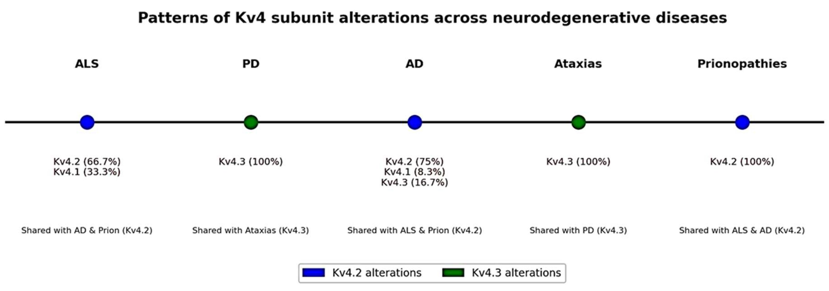
Analysis of the distribution of Kv4 subunit alterations across neurodegenerative disease models (Table 3) revealed both shared and disease-specific patterns. Kv4.2 dysfunction was the most frequent alteration overall (38.2%), predominantly observed in AD (75.0%), ALS (66.7%), and prionopathies (100%), indicating a common mechanism of disrupted A-type currents in these conditions. In contrast, Kv4.3 alterations accounted for 55.9% of all findings, but were largely confined to ataxias (100%) and PD (100%), reflecting the central role of KCND3 mutations in ataxias and α-synuclein-related modulation in PD. Kv4.1 involvement was rare (5.9%), reported only in isolated studies in ALS (33.3%) and AD (8.3%). Together, these results highlight that Kv4.2 alterations are shared among AD, ALS, and prionopathies, whereas Kv4.3 alterations predominate in ataxias and PD, underscoring disease-specific mechanisms despite a convergent impact on neuronal excitability.
In ALS and AD, downregulation of Kv4.2 and Kv4.3 or their auxiliary proteins is frequently associated with neuronal hyperexcitability, a feature implicated in excitotoxicity and disease progression [56,71,73]. However, the available evidence does not allow definitive conclusions regarding causality. In SCA19/22, pathogenic KCND3 mutations provide proof that Kv4 dysfunction can be a primary driver of disease [82,83,84,85,86,87,88,89,90,91,92,93,94,95,96,97,98,99,102,103,104], whereas in AD, PD, and ALS, Kv4 alterations may represent secondary or compensatory responses to protein aggregation [53], oxidative stress [57], or Ca2+ dysregulation [74]. From a translational perspective, several pharmacological interventions—such as 4-AP in ataxias and ALS [56,101], levetiracetam in AD [53], and cannabinoids [71] in ALS—demonstrate that Kv4 channels are not only mechanistically relevant but also clinically actionable. Together, these findings highlight Kv4 channels as a converging point between mechanistic understanding and therapeutic potential in neurodegeneration.

3. Discussion

3.1. Pathophysiological and Clinical Implications of Kv4 Dysfunction in Neurodegenerative Diseases

The collected findings outline a picture in which Kv4 potassium channels (Kv4.1, Kv4.2 and Kv4.3) play a critical role in the pathophysiology of multiple neurodegenerative diseases. In general terms, alterations in the function or expression of Kv4 channels contribute to the disruption of neuronal electrical homeostasis, mainly through the modulation of excitability. Since Kv4-regulated transient Ia currents act by dampening depolarization and controlling neuronal firing frequency, their decrease generates a tendency toward hyperexcitability that can manifest in deleterious ways: increased spontaneous activity and neurotransmitter release, susceptibility to epileptiform phenomena, dysregulation of synaptic networks and, in the long term, excitotoxic stress. This concept is reflected in several of the diseases analyzed here. For example, loss of dendritic A-type current in AD is associated with hyperexcitability and aberrant synchronicity in the hippocampus [73,74,76,78], while in PD the reduction in Kv4.3 in dopaminergic neurons could exacerbate their vulnerability by promoting excessive activity [72]. In spinocerebellar ataxias, the A-type current of Purkinje cells is fundamental for timing their output. Therefore, mutations in Kv4.3 that decrease this current compromise the precision of neuronal firing, resulting in loss of motor coordination [82,83,84,88,89,90,93,98,99,103]. Even mutations that increase current (gain of function) can be pathogenic by altering the normal pattern of excitability, indicating that there is an optimal window of Kv4 function necessary for the correct functioning of the cerebellar circuit [102].
An aspect of biological and clinical relevance is the confirmation that multiple pathogenic pathways converge on Kv4 dysfunction, which positions these channels as potential nodes for therapeutic interventions. From a clinical perspective, drugs that modulate potassium A currents could be useful in several contexts, for example, 4-AP, a Kv channel blocker used to improve synaptic conduction, showed beneficial effects in a model of ataxia (SCA1) by normalizing Purkinje excitability and improving motor function. This same drug and analogs are used in human spinocerebellar ataxias and multiple sclerosis to improve symptoms of coordination and fatigue, respectively, underlining the idea that enhancing neuronal activity by modulating Kv4 channels can rescue impaired functions [108,109,110,111,112,113,114,115,116]. In contrast, in epilepsy associated with AD, where hyperexcitability is related to the loss of Kv4 channels, the neuroprotective drug levetiracetam contributes to reducing excessive activity, favors the increase in A-type current and prevents the loss of Kv4 [53]. Similarly, interventions that target underlying mechanisms, such as the antioxidant Trolox (which reverses Kv4.1 reduction in AD), or GSK-3β inhibitors (which prevent Kv4.2 hyperphosphorylation) highlight the value of addressing secondary causes of Kv4 dysfunction along with the channels themselves [76,78]. These studies could suggest that Kv4 channels may participate in cellular stress responses, although they do not establish a general integrator role.
Despite the convergence around Kv4, there are important limitations in interpreting these findings. First, many of the data come from in vitro or animal experimental models, which, while replicating aspects of the disease, do not always capture the human complexity. Although in vitro and animal models are essential for mechanistic studies, they cannot fully reproduce human disease. Species differences in organ systems and disease mechanisms, along with the absence of systemic interactions in cell models, limit direct translation to patients [117]. Moreover, these approaches remain subject to limitations such as incomplete cell fidelity, restricted maturation, and atypical physiology, which may reduce their reliability in some applications [118]. In this line, the observed modulation of Kv4.2 by Aβ in acute cultures could differ in magnitude and consequences from the situation in a patient’s brain with decades of pathology. These findings highlight the importance of considering the specific characteristics of each AD model when interpreting Kv4 alterations. APP knockout mice (AβPP-/-) lack β-amyloid entirely and therefore serve as a negative control for Aβ-related mechanisms, showing that Kv4.2 function remains unaltered in the absence of amyloid [77]. In contrast, transgenic models that overexpress mutant APP or PSEN1 (e.g., hAPP_J20, hAPP/PS1, Tg2576, 3xTg-AD) develop progressive amyloid deposition and, in some cases, tau pathology, conditions under which Kv4 dysfunction has been reported [53,74,76,78]. Thus, the APP knockout model differs fundamentally from amyloid-overexpressing strains, underscoring that Kv4 dysregulation in AD appears to be driven by the pathological presence of Aβ and tau rather than by the absence of APP itself [53,74,76,77,78]. Similarly, the beneficial effects of drugs in murine models (such as aminopyridines in SCA1 and antioxidants in AD models) should be taken with caution until evaluated in controlled clinical trials. Second, several observations present apparent discrepancies. While some studies find increased Kv4 expression with Aβ exposure, others find decreased expression in chronic models. These differences could be attributed to different exposure times (early compensatory effects vs. late degeneration), different brain regions (cortex vs. hippocampus vs. cerebellum), or differences between models (cell lines, different transgenic animals, postmortem human tissue). Therefore, it is necessary to unify these findings by investigating intermediate stages and diverse cellular environments to understand how Kv4 dysfunction evolves throughout the course of the disease.
Another limitation is the relative scarcity of direct clinical data in some diseases. In ALS and PD, evidence on Kv4 comes from cellular or animal models, with no confirmation yet of how these channels behave in patients (whether in neurons derived from patient iPSCs, postmortem tissue recordings, or functional biomarkers). The translation of ALS findings in MSCs to neuronal relevance, or of findings in mouse models of PD to the human condition, requires confirmation. In ataxias caused by KCND3, although the genetic link is clear, it is not yet fully understood why certain specific mutations produce slightly different clinical pictures or of variable severity, nor why only Kv4.3 is involved while Kv4.2/Kv4.1 do not seem to give rise to similar phenotypes. Exploring the differential expression of Kv4 subunits in cerebellar versus cortical regions, and the presence of compensatory mechanisms (other K+ channels or their currents) could help explain these differences. Despite these limitations, the results compiled offer multiple avenues for future research. One obvious direction is the development of selective Kv4 channel modulators. More specific drugs than aminopyridines (which are relatively nonspecific and can cause adverse effects) could take advantage of the unique properties of Kv4.3 or Kv4.2 to adjust neuronal excitability more precisely. In ataxias due to KCND3 mutations, personalized medicine strategies could be designed, such as chaperone molecules or allosteric stabilizers that favor the correct localization of mutated Kv4.3 in the membrane (mitigating the effects of aberrant trafficking as in F227del) or that modulate its gating. Similarly, gene therapy could be considered in the long term to replace the function of Kv4.3 in the cerebellum if a specific and safe delivery is achieved. In diseases such as AD and PD, it is worth investigating whether hyperexcitability associated with Kv4 loss contributes to neurodegeneration or is rather an epiphenomenon. This would help clarify whether intervening in Kv4 would have a disease-modifying effect or merely a symptomatic one. On the other hand, the discovery of direct interactions between Kv4 channels and pathological proteins would provide insights into Kv4 biophysics through binding or post-translational modifications. Additionally, given that DPP6 emerged as a factor in AD and prionopathies, its role in other diseases with altered excitability could be investigated, and even whether common polymorphisms in DPP6 or KChIP2 modulate the risk or progression of neurodegenerative diseases [73,94,105].

3.2. Common Patterns and Differences Between Diseases and Models

Despite the heterogeneity of neurodegenerative and neurological disorders, common patterns emerge in the involvement of Kv4 channels (Kv4.1–Kv4.3). A transversal finding is the inverse relationship between type A current and excitability. The reduction in Kv4 function favors neuronal hyperexcitability, contributing to the pathogenesis of ALS [119], PD [72,120], AD [121], prionopathies [122] and epilepsy [62]. In ataxias linked to KCND3 (Kv4.3) mutations, the defect may be due to loss or gain of function, but always with the same outcome: dysfunction in Purkinje neurons and alteration of motor coordination [85,98].
Another key point is to differentiate between primary and secondary Kv4 alterations. In spinocerebellar ataxias (SCA19/22), mutations in KCND3 directly damage the channel [98], while in AD, PD or prionopathies, Kv4 dysfunction occurs indirectly, mediated by pathological interactions with Aβ, α-synuclein or PrPSc, as well as by factors such as oxidative stress or cofactor imbalance [54,57,76].
Auxiliary subunits and associated proteins also have a transversal role. DPP6, KChIP2 and other regulators appear in various contexts, modulating Kv4 expression, trafficking and kinetics [40,84,85,89,105]. Similarly, common cellular mechanisms such as oxidative stress, Ca2+ imbalances or phosphorylation converge in the negative regulation of these channels in different disorders [72,78].
In terms of genetics, KCND3 is the only gene in the Kv4 family consistently implicated in hereditary pathology. Missense mutations that reduce A-type currents are the predominant pattern, although there are also cases of gain of function [102]. Neither KCND1 nor KCND2 have yet been linked to defined clinical phenotypes, suggesting that their alterations could have more serious or yet unrecognized consequences [123].
In summary, Kv4 channels represent a common node in the regulation of neuronal excitability. Although pathogenetic pathways vary, ranging from direct mutations to secondary effects of the cellular environment, the result converges in an imbalance of the A-type current and, consequently, of neuronal homeostasis [62,119]. This shared pattern reinforces the idea that Kv4 constitutes a common point of vulnerability and a potential therapeutic target in various central nervous system (CNS) diseases.
The comparative analysis of Kv4 subunit alterations across neurodegenerative diseases highlights both convergence and divergence in channel dysfunction (Figure 2). Kv4.2 dysregulation emerged as a common mechanism in AD, ALS, and prionopathies, consistent with its role in mediating A-type currents that critically shape neuronal excitability. In contrast, Kv4.3 alterations were largely restricted to ataxias and PD, reflecting the direct contribution of KCND3 mutations in spinocerebellar ataxias and the modulatory effects of α-synuclein pathology in PD. Interestingly, Kv4.1 involvement was rare and disease-specific, reported only in isolated studies of ALS and AD, suggesting a more limited role in neurodegeneration. From a therapeutic perspective, these results underscore the value of distinguishing between shared versus disease-specific Kv4 mechanisms. Kv4.2 may represent a broad therapeutic target, given its consistent involvement across several neurodegenerative diseases characterized by neuronal hyperexcitability. In contrast, Kv4.3 dysfunction appears as a disease-specific marker, offering opportunities for precision strategies in ataxias and PD, where its alterations are predominant. This dichotomy supports the development of interventions that either target common pathways (Kv4.2-related excitability control) or tailored approaches focused on Kv4.3 dysfunction in genetically or pathologically defined subgroups.

3.3. Integrative Synthesis of Kv4 Alterations

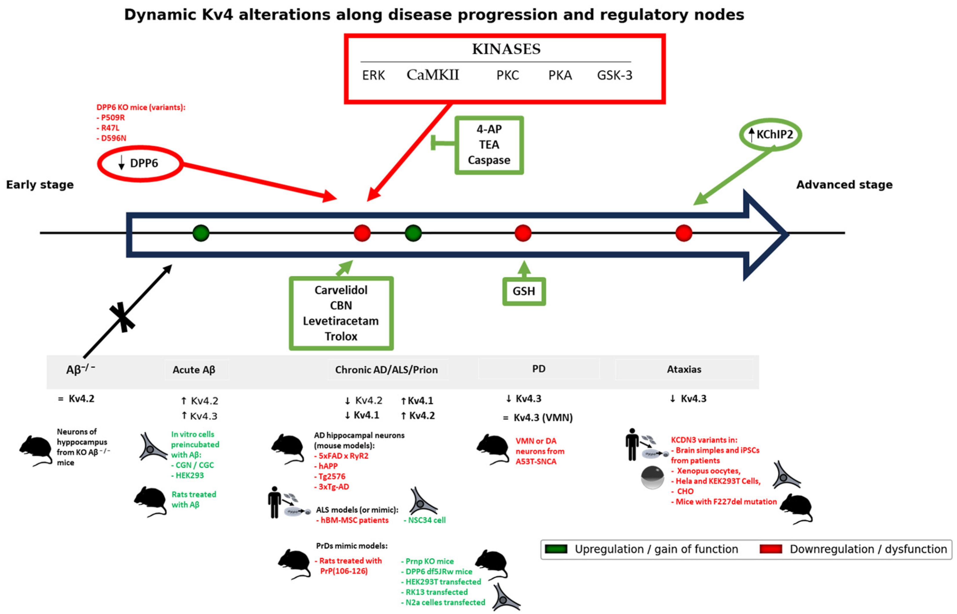
Across neurodegenerative diseases, Kv4 dysfunction can be summarized in three dimensions: (i) Summary of findings: alterations are consistently reported, with Kv4.2 involvement predominating in AD, ALS, and prionopathies, and Kv4.3 in ataxias and PD; (ii) Functional changes and pathophysiological implications: Kv4 downregulation or dysfunction leads to abnormal neuronal excitability, either as a primary effect of KCND3 mutations (ataxias) or secondary to upstream pathology such as Aβ/tau, α-synuclein, oxidative stress, or prion protein interactions; and (iii) Therapeutic relevance: pharmacological modulation of Kv4 highlights the translational potential of these channels as targets for disease modification.
This integrative schematic (Figure 3) highlights how different neurodegenerative disease models converge on Kv4 channel dysfunction while also displaying disease-specific patterns. Kv4.2 dysregulation is a shared feature of AD, ALS, and prionopathies, whereas Kv4.3 alterations predominate in PD and spinocerebellar ataxias driven by KCND3 mutations. Acute Aβ exposure in vitro produces transient increases in Kv4 activity, contrasting with the chronic downregulation seen in transgenic and patient-derived models, underscoring the importance of disease stage and model type. Importantly, auxiliary subunits (DPP6, KChIP2) and kinases (ERK, CaMKII, PKC, PKA, GSK-3) emerge as common regulatory nodes, providing mechanistic links between upstream pathology and Kv4 dysfunction. Pathogenic alterations in Kv4.3 frequently cluster in functionally relevant domains such as the S2 and S4–S6 transmembrane segments, which participate in voltage sensing and pore gating, as well as in the C-terminal region, important for protein stability and subunit interactions. Mutations in these regions (e.g., p.F227del in S2 [99], p.G345V/p.M373I in S5–S6 [93], or p.R419H in the C-terminus [102]) lead to reduced surface expression, impaired gating, or loss of current, ultimately producing neuronal hyperexcitability. In parallel, auxiliary subunits such as KChIP2 [84,85] and DPP6 [74,105] are crucial modulators of Kv4 channel trafficking and kinetics. KChIP2 can partially rescue surface expression in certain ataxia mutants, although not always restoring function, whereas DPP6 deficiency reduces Kv4.2 expression and contributes to excitability defects in AD and PrDs. Auxiliary subunits not only regulate Kv4 channel trafficking and gating but also shape their pharmacological responses. For example, DPP6 and MiRP1 differentially modulate the action of the synthetic KChIP2 ligand IQM-266 on Kv4.3 currents, shifting drug sensitivity depending on whether the channel resides in active-closed or inactivated states [124]. Together, these structural and auxiliary determinants highlight the intimate connection between Kv4 molecular architecture and neuronal dysfunction in neurodegenerative disorders.
Several compounds (e.g., levetiracetam, carvedilol, Trolox, GSH, cannabinoids) have been shown to rescue Kv4 currents or expression in experimental systems, supporting the translational potential of Kv4 modulation as a therapeutic avenue. Beyond neurodegenerative models, additional evidence supports the translational potential of K+ channel modulators such as 4-AP. In traumatic brain injury (TBI), acute 4-AP administration reduced axonal damage, demyelination, and cytoskeletal disruption and decreased Kv1.2 dispersion, although conduction deficits persisted [125]. Similarly, in experimental autoimmune encephalomyelitis (EAE), a model of multiple sclerosis, 4-AP did not alter disease incidence or progression but significantly improved mobility and coordination by ameliorating conduction failure [126,127]. These findings underscore that Kv channel inhibition can yield symptomatic benefits and partial structural protection, even in the absence of strong disease-modifying effects, thereby reinforcing the rationale for considering Kv4 channels as therapeutic targets. Cannabinoids, through CB1 receptor activation, also modulate Kv4.2-containing A-type currents by shifting their inactivation properties in a sex-specific manner. This indicates that cannabinoid regulation of Kv4.2 is not simply a reduction in current magnitude but rather a biophysical modulation of channel gating, which may contribute to neuronal inhibition and could extend to neurodegenerative contexts where Kv4.2 dysfunction is implicated [127]. In addition, Trolox, a vitamin E derivative with antioxidant properties, can counteract arachidonic acid–induced inhibition of Kv4.2 currents. While intracellular Trolox only slows current suppression, extracellular application prevents the reduction in current amplitude and shifts activation to more positive voltages [128]. These findings suggest that Trolox may protect Kv4.2 function against lipid-mediated oxidative stress, thereby stabilizing neuronal excitability and synaptic transmission. Together, these pharmacological examples illustrate proof-of-concept that Kv4 modulation is not only mechanistically relevant but also offers translational potential, paving the way for targeted therapeutic strategies in neurodegenerative diseases.

4. Materials and Methods

4.1. Literature Search and Selection Criteria

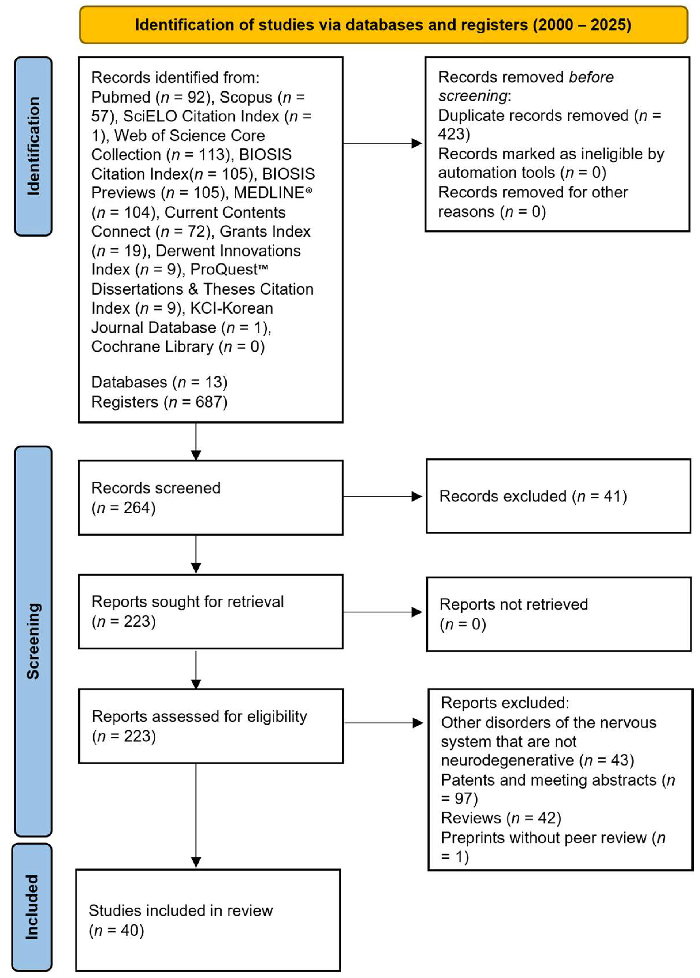
This review focuses exclusively on neurodegenerative diseases of the CNS, excluding other CNS disorders such as neurodevelopmental or mental illnesses, trauma, epilepsy, neoplasia, or infectious diseases. Identification of studies was carried out through a multiengine literature search of the PubMed, Scopus, SciELO Citation Index, Web of Science Core Collection, Biosis Citation Index, Biosis Previews, Medline, Current Contents Connect, Grants Index, Derwent Innovations Index, ProQuest Dissertations & Theses Citation Index, KCI-Korean Journal Database, and Cochrane Library databases by 3 independent researchers (S.G.-A., N.S.-M., and G.D.-V.). The review was registered in PROSPERO with ID: CRD420251119426. The following search strategy was used to identify potentially eligible studies and was adapted to the different databases consulted: (Potassium Channel AND ((Kv4.1 OR KCND1) OR (Kv4.2 OR KCND2) OR (Kv4.3 OR KCND3)) AND ((amyotrophic lateral sclerosis OR ALS) OR Parkinson OR Alzheimer OR Huntington OR Ataxia* OR dementia* OR (Central Nervous System Diseases) NOT Neoplasms)). All potentially eligible studies were retrieved, and their bibliographies were manually searched for additional studies. Only available full-text articles written in English or Spanish and published from January 1, 2000, to the date of PROSPERO registration (4 August 2025) were included. Studies evaluating human participants with neurodegenerative disorders of the central nervous system in which variants in KCND1, KCND2, or KCND3 (potassium channels Kv4.1–4.3) were identified, providing clinical, genetic, or functional data, were included. Functional or genetic analyses of these variants were considered, including humanized cellular or animal models carrying human mutations. Studies on other channels or variants unrelated to neurodegeneration, research in non-humanized models, review and meta-analyses articles, unreviewed preprints, in silico predictions without experimental validation, incomplete articles, data published only in abstract form, duplicate studies or written in another language that is not part of the inclusion requirements were excluded. Comparators included healthy individuals, wild-type models, or humanized models without pathogenic variants. Observational and interventional studies evaluating Kv4 channel function and their clinical impact were included. The research question was formulated following PICO: population (patients with variants in KCND1–3), intervention/exposure (functional or genetic analysis), comparator (healthy individuals or humanized models without variants) and outcomes (impact on Kv4 function and clinical phenotypes). Preferred reporting items for systematic reviews and meta-analyses (PRISMA) guidelines were followed for the methodology used in the study [129]. The detailed screening protocol for the studies is depicted in Figure 4.

4.2. Assessment of the Methodological Quality and Risk of Bias of the Included Studies

The methodological quality and RoB of the 40 included studies were thoroughly assessed using study-specific tools (Tables S2 and S3, Figures S1 and S2). For observational studies in humans (e.g., patient series, clinical genetic studies), the NOS (Figure S3) [130] was used. For preclinical animal studies, the SYRCLE (Figure S4) [131] RoB tool was used. Finally, atypical designs, such as single-case reports or case series, were qualitatively assessed following guidelines proposed in the literature [132], considering domains such as selection (representativeness of the case), adequate accuracy of observations, causality (plausibility that the intervention/exposure explains the outcome, without other explanations), and completeness of results reporting.

4.3. Verification of Genetic Variants

The genetic variants included in this review were verified by consulting the dbSNP (National Center for Biotechnology Information, Bethesda, MD, USA; https://www.ncbi.nlm.nih.gov/snp/, accessed 19 August 2025) and ClinVar—Genomic Variation as it Relates to Human Health (National Library of Medicine, Bethesda, MD, USA; https://www.ncbi.nlm.nih.gov/clinvar/, accessed 19 August 2025) databases to ensure correct identification of nucleotide changes and their effects on the encoded proteins.

5. Conclusions

In conclusion, current evidence positions Kv4 potassium channels as important regulators of neuronal activity whose alteration, due to genetic mutations or adverse pathological environments, contributes to a variety of neurological disorders. Their presence in different cell and brain types, as well as their modulation by accessory proteins, makes them vulnerable sites but also promising targets. A deeper understanding of how each subunit (Kv4.1, Kv4.2, Kv4.3) participates in the physiology of specific brain circuits and how it is affected in the disease will allow not only a better understanding of the pathogenesis of ALS, PD, AD, ataxias, and prionopathies, but also the design of interventions aimed at restoring the excitatory–inhibitory balance in these conditions, with a view to developing treatments that improve neuronal function and the quality of life of patients.

Supplementary Materials

The following supporting information can be downloaded at: https://www.mdpi.com/article/10.3390/physiologia5030031/s1, Table S1: Comprehensive_Kv4_variants; Table S2: Table of assessment of methodological quality and risk of bias of the included studies. Table S3. Studies grouped by level of methodological quality. Figure S1. Assessment of the overall methodological quality of the included studies. Figure S2. Frequency of studies with weaknesses (moderate/high risk of bias) in each assessed domain. Figure S3. Distribution of studies according to Newcastle-Ottawa (NOS) tool. Figure S4. Distribution of studies according to SYRCLE tool.

Author Contributions

Conceptualization, I.P., C.M.F.-M. and G.D.-V.; methodology, N.S.-M., P.G.-T. and G.D.-V.; software, S.G.-A.; validation, N.S.-M., P.G.-T., and S.G.-A.; formal analysis, G.D.-V.; investigation, B.T.-P. and G.D.-V.; resources, G.D.-V.; data curation, B.T.-P. and G.D.-V.; writing—original draft preparation, G.D.-V.; writing—review and editing, G.D.-V.; visualization, I.P., M.R.-S., P.G.-T. and S.G.-A.; supervision, C.M.F.-M. and G.D.-V.; project administration, G.D.-V.; funding acquisition, G.D.-V. All authors have read and agreed to the published version of the manuscript.

Funding

This publication is part of the project with Ref. No. CE-10-UGR24, funded by the Autonomous City of Ceuta in its commitment to research (2024); this work also presents results that were supported by grant code PPJIA2022.09, “Plan Propio de Investigación y Transferencia de la Universidad de Granada, 2022: Programa 20. Proyectos de Investigación Precompetitivos para Jóvenes Investigadores. Modalidad 20.a. Proyectos para jóvenes doctores” from University of Granada (Spain).

Institutional Review Board Statement

Not applicable.

Informed Consent Statement

Not applicable.

Data Availability Statement

Not applicable.

Conflicts of Interest

The authors declare no conflicts of interest.

References

  1. Kim, J.B. Channelopathies. Korean J. Pediatr. 2014, 57, 1–18. [Google Scholar] [CrossRef]
  2. Isbrandt, D.; Leicher, T.; Waldschütz, R.; Zhu, X.; Luhmann, U.; Michel, U.; Sauter, K.; Pongs, O. Gene Structures and Expression Profiles of Three Human KCND (Kv4) Potassium Channels Mediating A-Type Currents I(TO) and I(SA). Genomics 2000, 64, 144–154. [Google Scholar] [CrossRef]
  3. Decher, N.; Barth, A.S.; Gonzalez, T.; Steinmeyer, K.; Sanguinetti, M.C. Novel KChIP2 Isoforms Increase Functional Diversity of Transient Outward Potassium Currents. J. Physiol. 2004, 557, 761–772. [Google Scholar] [CrossRef]
  4. Birnbaum, S.G.; Varga, A.W.; Yuan, L.L.; Anderson, A.E.; Swepatt, J.D.; Schrader, L.A. Structure and Function of Kv4-Family Transient Potassium Channels. Physiol. Rev. 2004, 84, 803–833. [Google Scholar] [CrossRef] [PubMed]
  5. Ruppersberg, J.P.; Schröter, K.H.; Sakmann, B.; Stocker, M.; Sewing, S.; Pongs, O. Heteromultimeric Channels Formed by Rat Brain Potassium-Channel Proteins. Nature 1990, 345, 535–537. [Google Scholar] [CrossRef] [PubMed]
  6. Isacoff, E.Y.; Jan, Y.N.; Jan, L.Y. Evidence for the Formation of Heteromultimeric Potassium Channels in Xenopus Oocytes. Nature 1990, 345, 530–534. [Google Scholar] [CrossRef]
  7. Strang, C.; Cushman, S.J.; DeRubeis, D.; Peterson, D.; Pfaffinger, P.J. A Central Role for the T1 Domain in Voltage-Gated Potassium Channel Formation and Function. J. Biol. Chem. 2001, 276, 28493–28502. [Google Scholar] [CrossRef]
  8. Nanao, M.H.; Zhou, W.; Pfaffinger, P.J.; Choe, S. Determining the Basis of Channel-Tetramerization Specificity by X-Ray Crystallography and a Sequence-Comparison Algorithm: Family Values (FamVal). Proc. Natl. Acad. Sci. USA 2003, 100, 8670–8675. [Google Scholar] [CrossRef] [PubMed]
  9. Levin, G.; Chikvashvili, D.; Singer-Lahat, D.; Peretz, T.; Thornhill, W.B.; Lotan, I. Phosphorylation of a K+ Channel Alpha Subunit Modulates the Inactivation Conferred by a Beta Subunit. Involv. Cytoskelet. J. Biol. Chem. 1996, 271, 29321–29328. [Google Scholar] [CrossRef]
  10. Horn, R. A New Twist in the Saga of Charge Movement in Voltage-Dependent Ion Channels. Neuron 2000, 25, 511–514. [Google Scholar] [CrossRef]
  11. Kong, W.; Po, S.; Yamagishi, T.; Ashen, M.D.; Stetten, G.; Tomaselli, G.F. Isolation and Characterization of the Human Gene Encoding Ito: Further Diversity by Alternative MRNA Splicing. Am. J. Physiol. 1998, 275, H1963–H1970. [Google Scholar] [CrossRef]
  12. Gonzalez, W.G.; Pham, K.; Miksovska, J. Modulation of the Voltage-Gated Potassium Channel (Kv4.3) and the Auxiliary Protein (KChIP3) Interactions by the Current Activator NS5806. J. Biol. Chem. 2014, 289, 32201–32213. [Google Scholar] [CrossRef]
  13. Deschênes, I.; Tomaselli, G.F. Modulation of Kv4.3 Current by Accessory Subunits. FEBS Lett. 2002, 528, 183–188. [Google Scholar] [CrossRef]
  14. England, S.K.; Uebele, V.N.; Shear, H.; Kodali, J.; Bennett, P.B.; Tamkun, M.M. Characterization of a Voltage-Gated K+ Channel Beta Subunit Expressed in Human Heart. Proc. Natl. Acad. Sci. USA 1995, 92, 6309–6313. [Google Scholar] [CrossRef]
  15. Fink, M.; Duprat, F.; Lesage, F.; Heurteaux, C.; Romey, G.; Barhanin, J.; Lazdunski, M. A New K+ Channel Beta Subunit to Specifically Enhance Kv2.2 (CDRK) Expression. J. Biol. Chem. 1996, 271, 26341–26348. [Google Scholar] [CrossRef]
  16. Heinemann, S.H.; Rettig, J.; Wunder, F.; Pongs, O. Molecular and Functional Characterization of a Rat Brain Kv Beta 3 Potassium Channel Subunit. FEBS Lett. 1995, 377, 383–389. [Google Scholar] [CrossRef]
  17. Leicher, T.; Bähring, R.; Isbrandt, D.; Pongs, O. Coexpression of the KCNA3B Gene Product with Kv1.5 Leads to a Novel A-Type Potassium Channel. J. Biol. Chem. 1998, 273, 35095–35101. [Google Scholar] [CrossRef] [PubMed]
  18. Nagaya, N.; Papazian, D.M. Potassium Channel Alpha and Beta Subunits Assemble in the Endoplasmic Reticulum. J. Biol. Chem. 1997, 272, 3022–3027. [Google Scholar] [CrossRef] [PubMed]
  19. Nakahira, K.; Shi, G.; Rhodes, K.J.; Trimmer, J.S. Selective Interaction of Voltage-Gated K+ Channel Beta-Subunits with Alpha-Subunits. J. Biol. Chem. 1996, 271, 7084–7089. [Google Scholar] [CrossRef] [PubMed]
  20. Pérez-García, M.T.; López-López, J.R.; González, C. Kvbeta1.2 Subunit Coexpression in HEK293 Cells Confers O2 Sensitivity to Kv4.2 but not to Shaker Channels. J. Gen. Physiol. 1999, 113, 897–907. [Google Scholar] [CrossRef]
  21. Rhodes, K.J.; Monaghan, M.M.; Barrezueta, N.X.; Nawoschik, S.; Bekele-Arcuri, Z.; Matos, M.F.; Nakahira, K.; Schechter, L.E.; Trimmer, J.S. Voltage-Gated K+ Channel Beta Subunits: Expression and Distribution of Kv Beta 1 and Kv Beta 2 in Adult Rat Brain. J. Neurosci. 1996, 16, 4846–4860. [Google Scholar] [CrossRef]
  22. Shi, G.; Nakahira, K.; Hammond, S.; Rhodes, K.J.; Schechter, L.E.; Trimmer, J.S. Beta Subunits Promote K+ Channel Surface Expression through Effects Early in Biosynthesis. Neuron 1996, 16, 843–852. [Google Scholar] [CrossRef]
  23. Trimmer, J.S. Regulation of Ion Channel Expression by Cytoplasmic Subunits. Curr. Opin. Neurobiol. 1998, 8, 370–374. [Google Scholar] [CrossRef]
  24. Yang, E.K.; Alvira, M.R.; Levitan, E.S.; Takimoto, K. Kvbeta Subunits Increase Expression of Kv4.3 Channels by Interacting with Their C Termini. J. Biol. Chem. 2001, 276, 4839–4844. [Google Scholar] [CrossRef] [PubMed]
  25. An, W.F.; Bowlby, M.R.; Betty, M.; Cao, J.; Ling, H.P.; Mendoza, G.; Hinson, J.W.; Mattsson, K.I.; Strassle, B.W.; Trimme, J.S.; et al. Modulation of A-Type Potassium Channels by a Family of Calcium Sensors. Nature 2000, 403, 553–556. [Google Scholar] [CrossRef]
  26. Bähring, R.; Dannenberg, J.; Peters, H.C.; Leicher, T.; Pongs, O.; Isbrandt, D. Conserved Kv4 N-Terminal Domain Critical for Effects of Kv Channel-Interacting Protein 2.2 on Channel Expression and Gating. J. Biol. Chem. 2001, 276, 23888–23894. [Google Scholar] [CrossRef]
  27. Deschênes, I.; DiSilvestre, D.; Juang, G.J.; Wu, R.C.; An, W.F.; Tomaselli, G.F. Regulation of Kv4.3 Current by KChIP2 Splice Variants: A Component of Native Cardiac Ito? Circulation 2002, 106, 423–429. [Google Scholar] [CrossRef]
  28. Holmqvist, M.H.; Cao, J.; Hernandez-Pineda, R.; Jacobson, M.D.; Carroll, K.I.; Sung, M.A.; Betty, M.; Ge, P.; Gilbride, K.J.; Brown, M.E.; et al. Elimination of Fast Inactivation in Kv4 A-Type Potassium Channels by an Auxiliary Subunit Domain. Proc. Natl. Acad. Sci. USA 2002, 99, 1035–1040. [Google Scholar] [CrossRef] [PubMed]
  29. Morohashi, Y.; Hatano, N.; Ohya, S.; Takikawa, R.; Watabiki, T.; Takasugi, N.; Imaizumi, Y.; Tomita, T.; Iwatsubo, T. Molecular Cloning and Characterization of CALP/KChIP4, a Novel EF-Hand Protein Interacting with Presenilin 2 and Voltage-Gated Potassium Channel Subunit Kv4. J. Biol. Chem. 2002, 277, 14965–14975. [Google Scholar] [CrossRef]
  30. Patel, S.P.; Campbell, D.L.; Morales, M.J.; Strauss, H.C. Heterogeneous Expression of KChIP2 Isoforms in the Ferret Heart. J. Physiol. 2002, 539, 649–656. [Google Scholar] [CrossRef] [PubMed]
  31. Rosati, B.; Grau, F.; Rodriguez, S.; Li, H.; Nerbonne, J.M.; McKinnon, D. Concordant Expression of KChIP2 MRNA, Protein and Transient Outward Current throughout the Canine Ventricle. J. Physiol. 2003, 548, 815–822. [Google Scholar] [CrossRef]
  32. Rosati, B.; Pan, Z.; Lypen, S.; Wang, H.S.; Cohen, I.; Dixon, J.E.; McKinnon, D. Regulation of KChIP2 Potassium Channel Beta Subunit Gene Expression Underlies the Gradient of Transient Outward Current in Canine and Human Ventricle. J. Physiol. 2001, 533, 119–125. [Google Scholar] [CrossRef]
  33. Shibata, R.; Misonou, H.; Campomanes, C.R.; Anderson, A.E.; Schrader, L.A.; Doliveira, L.C.; Carroll, K.I.; Sweatt, J.D.; Rhodes, K.J.; Trimmer, J.S. A Fundamental Role for KChIPs in Determining the Molecular Properties and Trafficking of Kv4.2 Potassium Channels. J. Biol. Chem. 2003, 278, 36445–36454. [Google Scholar] [CrossRef] [PubMed]
  34. Guo, W.; Li, H.; Aimond, F.; Johns, D.C.; Rhodes, K.J.; Trimmer, J.S.; Nerbonne, J.M. Role of Heteromultimers in the Generation of Myocardial Transient Outward K+ Currents. Circ. Res. 2002, 90, 586–593. [Google Scholar] [CrossRef]
  35. Guo, W.; Malin, S.A.; Johns, D.C.; Jeromin, A.; Nerbonne, J.M. Modulation of Kv4-Encoded K(+) Currents in the Mammalian Myocardium by Neuronal Calcium Sensor-1. J. Biol. Chem. 2002, 277, 26436–26443. [Google Scholar] [CrossRef]
  36. Nakamura, T.Y.; Pountney, D.J.; Ozaita, A.; Nandi, S.; Ueda, S.; Rudy, B.; Coetzee, W.A. A Role for Frequenin, a Ca2+-Binding Protein, as a Regulator of Kv4 K+-Currents. Proc. Natl. Acad. Sci. USA 2001, 98, 12808–12813. [Google Scholar] [CrossRef]
  37. Kuryshev, Y.A.; Gudz, T.I.; Brown, A.M.; Wible, B.A. KChAP as a Chaperone for Specific K+ Channels. Am. J. Physiol. Cell Physiol. 2000, 278, C931–C941. [Google Scholar] [CrossRef] [PubMed]
  38. Kuryshev, Y.A.; Wible, B.A.; Gudz, T.I.; Ramirez, A.N.; Brown, A.M. KChAP/Kvbeta1.2 Interactions and Their Effects on Cardiac Kv Channel Expression. Am. J. Physiol. Cell Physiol. 2001, 281, C290–C299. [Google Scholar] [CrossRef] [PubMed]
  39. Wible, B.A.; Yang, Q.; Kuryshev, Y.A.; Accili, E.A.; Brown, A.M. Cloning and Expression of a Novel K+ Channel Regulatory Protein, KChAP. J. Biol. Chem. 1998, 273, 11745–11751. [Google Scholar] [CrossRef]
  40. Nadal, M.S.; Ozaita, A.; Amarillo, Y.; Vega-Saenz De Miera, E.; Ma, Y.; Mo, W.; Goldberg, E.M.; Misumi, Y.; Ikehara, Y.; Neubert, T.A.; et al. The CD26-Related Dipeptidyl Aminopeptidase-like Protein DPPX Is a Critical Component of Neuronal A-Type K+ Channels. Neuron 2003, 37, 449–461. [Google Scholar] [CrossRef]
  41. Zagha, E.; Ozaita, A.; Chang, S.Y.; Nadal, M.S.; Lin, U.; Saganich, M.J.; McCormack, T.; Akinsanya, K.O.; Qi, S.Y.; Rudy, B. DPP10 Modulates Kv4-Mediated A-Type Potassium Channels. J. Biol. Chem. 2005, 280, 18853–18861. [Google Scholar] [CrossRef] [PubMed]
  42. Petrecca, K.; Miller, D.M.; Shrier, A. Localization and Enhanced Current Density of the Kv4.2 Potassium Channel by Interaction with the Actin-Binding Protein Filamin. J. Neurosci. 2000, 20, 8736–8744. [Google Scholar] [CrossRef] [PubMed]
  43. Sheng, M.; Tsaur, M.L.; Nung Jan, Y.; Yeh Jan, L. Subcellular Segregation of Two A-Type K+ Channel Proteins in Rat Central Neurons. Neuron 1992, 9, 271–284. [Google Scholar] [CrossRef]
  44. Vasilyev, D.V.; Barish, M.E. Regulation of an Inactivating Potassium Current (IA) by the Extracellular Matrix Protein Vitronectin in Embryonic Mouse Hippocampal Neurones. J. Physiol. 2003, 547, 859–871. [Google Scholar] [CrossRef] [PubMed]
  45. Wong, W.; Newell, E.W.; Jugloff, D.G.M.; Jones, O.T.; Schlichter, L.C. Cell Surface Targeting and Clustering Interactions between Heterologously Expressed PSD-95 and the Shal Voltage-Gated Potassium Channel, Kv4.2. J. Biol. Chem. 2002, 277, 20423–20430. [Google Scholar] [CrossRef]
  46. Hsueh, Y.P.; Sheng, M. Requirement of N-Terminal Cysteines of PSD-95 for PSD-95 Multimerization and Ternary Complex Formation, but not for Binding to Potassium Channel Kv1.4. J. Biol. Chem. 1999, 274, 532–536. [Google Scholar] [CrossRef]
  47. Abbott, G.W.; Butler, M.H.; Bendahhou, S.; Dalakas, M.C.; Ptacek, L.J.; Goldstein, S.A.N. MiRP2 Forms Potassium Channels in Skeletal Muscle with Kv3.4 and Is Associated with Periodic Paralysis. Cell 2001, 104, 217–231. [Google Scholar] [CrossRef]
  48. Abbott, G.W.; Sesti, F.; Splawski, I.; Buck, M.E.; Lehmann, M.H.; Timothy, K.W.; Keating, M.T.; Goldstein, S.A.N. MiRP1 Forms IKr Potassium Channels with HERG and Is Associated with Cardiac Arrhythmia. Cell 1999, 97, 175–187. [Google Scholar] [CrossRef]
  49. Schroeder, B.C.; Waldegger, S.; Fehr, S.; Bleich, M.; Warth, R.; Greger, R.; Jentsch, T.J. A Constitutively Open Potassium Channel Formed by KCNQ1 and KCNE3. Nature 2000, 403, 196–199. [Google Scholar] [CrossRef]
  50. Tinel, N.; Diochot, S.; Borsotto, M.; Lazdunski, M.; Barhanin, J. KCNE2 Confers Background Current Characteristics to the Cardiac KCNQ1 Potassium Channel. EMBO J. 2000, 19, 6326–6330. [Google Scholar] [CrossRef]
  51. Wulfsen, I.; Hauber, H.P.; Schiemann, D.; Bauer, C.K.; Schwarz, J.R. Expression of MRNA for Voltage-Dependent and Inward-Rectifying K Channels in GH3/B6 Cells and Rat Pituitary. J. Neuroendocr. Neuroendocrinol. 2000, 12, 263–272. [Google Scholar] [CrossRef]
  52. Zhang, M.; Jiang, M.; Tseng, G.N. MinK-Related Peptide 1 Associates with Kv4.2 and Modulates Its Gating Function: Potential Role as Beta Subunit of Cardiac Transient Outward Channel? Circ. Res. 2001, 88, 1012–1019. [Google Scholar] [CrossRef]
  53. Hall, A.M.; Throesch, B.T.; Buckingham, S.C.; Markwardt, S.J.; Peng, Y.; Wang, Q.; Hoffman, D.A.; Roberson, E.D. Tau-Dependent Kv4.2 Depletion and Dendritic Hyperexcitability in a Mouse Model of Alzheimer’s Disease. J. Neurosci. 2015, 35, 6221–6230. [Google Scholar] [CrossRef] [PubMed]
  54. Pieri, M.; Amadoro, G.; Carunchio, I.; Ciotti, M.T.; Quaresima, S.; Florenzano, F.; Calissano, P.; Possenti, R.; Zona, C.; Severini, C. SP Protects Cerebellar Granule Cells against Beta-Amyloid-Induced Apoptosis by down-Regulation and Reduced Activity of Kv4 Potassium Channels. Neuropharmacology 2010, 58, 268–276. [Google Scholar] [CrossRef] [PubMed]
  55. Ping, Y.; Waro, G.; Licursi, A.; Smith, S.; Vo-Ba, D.A.; Tsunoda, S. Shal/K(v)4 Channels Are Required for Maintaining Excitability during Repetitive Firing and Normal Locomotion in Drosophila. PLoS ONE 2011, 6, e16043. [Google Scholar] [CrossRef]
  56. Park, K.S.; Choi, M.R.; Jung, K.D.; Kim, S.H.; Kim, H.Y.; Kim, K.S.; Cha, E.-J.; Kim, Y.; Chai, Y.G. Diversity of Ion Channels in Human Bone Marrow Mesenchymal Stem Cells from Amyotrophic Lateral Sclerosis Patients. Korean J. Physiol. Pharmacol. 2008, 12, 337–342. [Google Scholar] [CrossRef]
  57. Lasser-Katz, E.; Simchovitz, A.; Chiu, W.-H.; Oertel, W.H.; Sharon, R.; Soreq, H.; Roeper, J.; Goldberg, J.A. Mutant α-Synuclein Overexpression Induces Stressless Pacemaking in Vagal Motoneurons at Risk in Parkinson’s Disease. J. Neurosci. 2017, 37, 47–57. [Google Scholar] [CrossRef]
  58. Dabrowska, J.; Rainnie, D.G. Expression and Distribution of Kv4 Potassium Channel Subunits and Potassium Channel Interacting Proteins in Subpopulations of Interneurons in the Basolateral Amygdala. Neuroscience 2010, 171, 721–733. [Google Scholar] [CrossRef] [PubMed]
  59. Alfaro-Ruíz, R.; Aguado, C.; Martín-Belmonte, A.; Moreno-Martínez, A.E.; Luján, R. Expression, Cellular and Subcellular Localisation of Kv4.2 and Kv4.3 Channels in the Rodent Hippocampus. Int. J. Mol. Sci. 2019, 20, 246. [Google Scholar] [CrossRef]
  60. Tsaur, M.L.; Sheng, M.; Lowenstein, D.H.; Jan, Y.N.; Jan, L.Y. Differential Expression of K+ Channel MRNAs in the Rat Brain and Down-Regulation in the Hippocampus Following Seizures. Neuron 1992, 8, 1055–1067. [Google Scholar] [CrossRef]
  61. Breen, M.S.; Dobbyn, A.; Li, Q.; Roussos, P.; Hoffman, G.E.; Stahl, E.; Chess, A.; Sklar, P.; Li, J.B.; Devlin, B.; et al. Global Landscape and Genetic Regulation of RNA Editing in Cortical Samples from Individuals with Schizophrenia. Nat. Neurosci. 2019, 22, 1402–1412. [Google Scholar] [CrossRef]
  62. Bernard, C.; Anderson, A.; Becker, A.; Poolos, N.P.; Deck, H.; Johnston, D. Acquired Dendritic Channelopathy in Temporal Lobe Epilepsy. Science 2004, 305, 532–535. [Google Scholar] [CrossRef]
  63. MacDonald, J.F.; Belrose, J.C.; Xie, Y.F.; Jackson, M.F. Nonselective Cation Channels and Links to Hippocampal Ischemia, Aging, and Dementia. Adv. Exp. Med. Biol. 2013, 961, 433–447. [Google Scholar] [CrossRef]
  64. Tiwari, D.; Rajathi, V.; Rymer, J.K.; Beasley, L.N.; McGann, A.M.; Bunk, A.T.; Parkins, E.V.; Rice, M.F.; Smith, K.E.; Ritter, D.M.; et al. Estradiol- and Progesterone-Associated Changes in MicroRNA-Induced Silencing and Reduced Antiseizure Efficacy of an Antagomir in Female Mice. eNeuro 2023, 10, ENEURO.0047-22.2023. [Google Scholar] [CrossRef]
  65. Choubey, M.; Bansal, R.; Siddharthan, D.; Naik, N.; Sharma, G.; Saxena, A. Early Repolarization Syndrome, Epilepsy, and Atrial Fibrillation in a Young Girl with Novel KCND3 Mutation Managed with Quinidine. J. Cardiovasc. Electrophysiol. 2022, 33, 1312–1315. [Google Scholar] [CrossRef]
  66. Tiwari, D.; Schaefer, T.L.; Schroeder-Carter, L.M.; Krzeski, J.C.; Bunk, A.T.; Parkins, E.V.; Snider, A.; Danzer, R.; Williams, M.T.; Vorhees, C.V.; et al. The Potassium Channel Kv4.2 Regulates Dendritic Spine Morphology, Electroencephalographic Characteristics and Seizure Susceptibility in Mice. Exp. Neurol. 2020, 334, 113437. [Google Scholar] [CrossRef]
  67. Gross, C.; Yao, X.; Engel, T.; Tiwari, D.; Xing, L.; Rowley, S.; Danielson, S.W.; Thomas, K.T.; Jimenez-Mateos, E.M.; Schroeder, L.M.; et al. MicroRNA-Mediated Downregulation of the Potassium Channel Kv4.2 Contributes to Seizure Onset. Cell Rep. 2016, 17, 37–45. [Google Scholar] [CrossRef] [PubMed]
  68. Hu, J.H.; Malloy, C.; Hoffman, D.A. P38 Regulates Kainic Acid-Induced Seizure and Neuronal Firing via Kv4.2 Phosphorylation. Int. J. Mol. Sci. 2020, 21, 5921. [Google Scholar] [CrossRef] [PubMed]
  69. Mulholland, P.J.; Chandler, L.J. Inhibition of Glutamate Transporters Couples to Kv4.2 Dephosphorylation through Activation of Extrasynaptic NMDA Receptors. Neuroscience 2010, 165, 130–137. [Google Scholar] [CrossRef] [PubMed]
  70. Hong, Y.M.; Jo, D.G.; Lee, M.C.; Kim, S.Y.; Jung, Y.K. Reduced Expression of Calsenilin/DREAM/KChIP3 in the Brains of Kainic Acid-Induced Seizure and Epilepsy Patients. Neurosci. Lett. 2003, 340, 33–36. [Google Scholar] [CrossRef]
  71. Trainito, A.; Muscarà, C.; Gugliandolo, A.; Chiricosta, L.; Salamone, S.; Pollastro, F.; Mazzon, E.; D’Angiolini, S. Cannabinol (CBN) Influences the Ion Channels and Synaptic-Related Genes in NSC-34 Cell Line: A Transcriptomic Study. Cells 2024, 13, 1573. [Google Scholar] [CrossRef]
  72. Subramaniam, M.; Althof, D.; Gispert, S.; Schwenk, J.; Auburger, G.; Kulik, A.; Fakler, B.; Roeper, J. Mutant α-Synuclein Enhances Firing Frequencies in Dopamine Substantia Nigra Neurons by Oxidative Impairment of A-Type Potassium Channels. J. Neurosci. 2014, 34, 13586–13599. [Google Scholar] [CrossRef] [PubMed]
  73. Cacace, R.; Heeman, B.; Van Mossevelde, S.; De Roeck, A.; Hoogmartens, J.; Van Rijk, P.; Gossye, H.; Van Vos, K.; De Coster, W.; Strazisar, M.; et al. Loss of DPP6 in Neurodegenerative Dementia: A Genetic Player in the Dysfunction of Neuronal Excitability. Acta Neuropathol. 2019, 137, 901–918. [Google Scholar] [CrossRef] [PubMed]
  74. Yao, J.; Sun, B.; Institoris, A.; Zhan, X.; Guo, W.; Song, Z.; Liu, Y.; Hiess, F.; Boyce, A.K.J.; Ni, M.; et al. Limiting RyR2 Open Time Prevents Alzheimer’s Disease-Related Neuronal Hyperactivity and Memory Loss but Not β-Amyloid Accumulation. Cell Rep. 2020, 32, 108169. [Google Scholar] [CrossRef]
  75. Plant, L.D.; Webster, N.J.; Boyle, J.P.; Ramsden, M.; Freir, D.B.; Peers, C.; Pearson, H.A. Amyloid Beta Peptide as a Physiological Modulator of Neuronal ‘A’-Type K+ Current. Neurobiol. Aging 2006, 27, 1673–1683. [Google Scholar] [CrossRef] [PubMed]
  76. Kim, K.-R.; Kim, Y.Y.; Jeong, H.-J.; Kang, J.-S.; Lee, S.H.S.-H.S.H.; Kim, Y.Y.; Lee, S.H.S.-H.S.H.; Ho, W.-K. Impaired Pattern Separation in Tg2576 Mice Is Associated with Hyperexcitable Dentate Gyrus Caused by Kv4.1 Downregulation. Mol. Brain 2021, 14, 62. [Google Scholar] [CrossRef]
  77. Li, Y.; Wang, J.-Z.; Deng, Y.-M.; Wang, K.; Yang, L.; Long, C. Amyloid-β Protein Precursor Regulates Electrophysiological Properties in the Hippocampus via Altered Kv1.4 Expression and Function in Mice. J. Alzheimers Dis. 2023, 92, 1241–1256. [Google Scholar] [CrossRef]
  78. Scala, F.; Fusco, S.; Ripoli, C.; Piacentini, R.; Li Puma, D.D.; Spinelli, M.; Laezza, F.; Grassi, C.; D’Ascenzo, M. Intraneuronal Aβ Accumulation Induces Hippocampal Neuron Hyperexcitability through A-Type K+ Current Inhibition Mediated by Activation of Caspases and GSK-3. Neurobiol. Aging 2015, 36, 886–900. [Google Scholar] [CrossRef]
  79. Pan, Y.; Xu, X.; Tong, X.; Wang, X. Messenger RNA and Protein Expression Analysis of Voltage-Gated Potassium Channels in the Brain of Aβ25-35-Treated Rats. J. Neurosci. Res. 2004, 77, 94–99. [Google Scholar] [CrossRef]
  80. Kerrigan, T.L.; Atkinson, L.; Peers, C.; Pearson, H.A. Modulation of ‘A’-Type K+ Current by Rodent and Human Forms of Amyloid β Protein. Neuroreport 2008, 19, 839–843. [Google Scholar] [CrossRef]
  81. Campolongo, P.; Ratano, P.; Ciotti, M.T.; Florenzano, F.; Nori, S.L.; Marolda, R.; Palmery, M.; Rinaldi, A.M.; Zona, C.; Possenti, R.; et al. Systemic Administration of Substance P Recovers Beta Amyloid-Induced Cognitive Deficits in Rat: Involvement of Kv Potassium Channels. PLoS ONE 2013, 8, e78036. [Google Scholar] [CrossRef]
  82. Ågren, R.; Geerdink, N.; Brunner, H.G.; Paucar, M.; Kamsteeg, E.-J.; Sahlholm, K. An E280K Missense Variant in KCND3/Kv4.3—Case Report and Functional Characterization. Int. J. Mol. Sci. 2023, 24, 10924. [Google Scholar] [CrossRef]
  83. Duarri, A.; Lin, M.-C.A.; Fokkens, M.R.; Meijer, M.; Smeets, C.J.L.M.; Nibbeling, E.A.R.; Boddeke, E.; Sinke, R.J.; Kampinga, H.H.; Papazian, D.M.; et al. Spinocerebellar Ataxia Type 19/22 Mutations Alter Heterocomplex Kv4.3 Channel Function and Gating in a Dominant Manner. Cell Mol. Life Sci. 2015, 72, 3387–3399. [Google Scholar] [CrossRef]
  84. Reis, M.C.; Mandler, L.; Kang, J.-S.; Oliver, D.; Halaszovich, C.; Nolte, D. A Novel KCND3 Variant in the N-Terminus Impairs the Ionic Current of Kv4.3 and Is Associated with SCA19/22. J. Cell Mol. Med. 2024, 28, e70039. [Google Scholar] [CrossRef] [PubMed]
  85. Duarri, A.; Jezierska, J.; Fokkens, M.; Meijer, M.; Schelhaas, H.J.; den Dunnen, W.F.A.; van Dijk, F.; Verschuuren-Bemelmans, C.; Hageman, G.; van de Vlies, P.; et al. Mutations in Potassium Channel Kcnd3 Cause Spinocerebellar Ataxia Type 19. Ann. Neurol. 2012, 72, 870–880. [Google Scholar] [CrossRef]
  86. Li, M.; Liu, F.; Hao, X.; Fan, Y.; Li, J.; Hu, Z.; Shi, J.; Fan, L.; Zhang, S.; Ma, D.; et al. Rare KCND3 Loss-of-Function Mutation Associated With the SCA19/22. Front. Mol. Neurosci. 2022, 15, 919199. [Google Scholar] [CrossRef]
  87. Kurihara, M.; Ishiura, H.; Sasaki, T.; Otsuka, J.; Hayashi, T.; Terao, Y.; Matsukawa, T.; Mitsui, J.; Kaneko, J.; Nishiyama, K.; et al. Novel De Novo KCND3 Mutation in a Japanese Patient with Intellectual Disability, Cerebellar Ataxia, Myoclonus, and Dystonia. Cerebellum 2018, 17, 237–242. [Google Scholar] [CrossRef]
  88. Hsiao, C.-T.; Fu, S.-J.; Cheng, K.-M.; Lo, H.; Tang, C.-Y.; Chan, C.-C.; Jeng, C.-J. Restoration of Shal/K(V)4 Proteostasis and Motor Function in a Drosophila Model of Spinocerebellar Ataxia Type 19/22. Cell Mol. Life Sci. 2025, 82, 181. [Google Scholar] [CrossRef] [PubMed]
  89. Arancibia, F.; Martin, F.; Ruiz-Fuentes, J.; Diaz, E.; Hermosilla, T.; Gonzalez, W.; Simon, F.; Avila-Jaque, D.; Luna-Álvarez, M.; Dávila Ortiz de Montellano, D.J.; et al. Functional Characterization of Two KCND3 Variants Associated with SCA 19/22 Ataxia in Latin American Families. Biol. Res. 2025, 58, 18. [Google Scholar] [CrossRef] [PubMed]
  90. Paucar, M.; Ågren, R.; Li, T.; Lissmats, S.; Bergendal, Å.; Weinberg, J.; Nilsson, D.; Savichetva, I.; Sahlholm, K.; Nilsson, J.; et al. V374A KCND3 Pathogenic Variant Associated With Paroxysmal Ataxia Exacerbations. Neurol. Genet. 2021, 7, e546. [Google Scholar] [CrossRef]
  91. Duarri, A.; Nibbeling, E.; Fokkens, M.R.; Meijer, M.; Boddeke, E.; Lagrange, E.; Stevanin, G.; Brice, A.; Durr, A.; Verbeek, D.S. The L450P Mutation in KCND3 Brings Spinocerebellar Ataxia and Brugada Syndrome Closer Together. Neurogenetics 2013, 14, 257–258. [Google Scholar] [CrossRef]
  92. Marina, M.L.C.; Fraile, P.Q.; Marmiesse, A.F.; Cruz, N.G.; del Valle, F.M. De novo sporadic mutation in the KCND3 gene in a patient with early onset of chronic ataxia. Rev. Neurol. 2019, 68, 398–399. [Google Scholar] [CrossRef]
  93. Zanni, G.; Hsiao, C.-T.; Fu, S.-J.; Tang, C.-Y.; Capuano, A.; Bosco, L.; Graziola, F.; Bellacchio, E.; Servidei, S.; Primiano, G.; et al. Novel KCND3 Variant Underlying Nonprogressive Congenital Ataxia or SCA19/22 Disrupt KV4.3 Protein Expression and K plus Currents with Variable Effects on Channel Properties. Int. J. Mol. Sci. 2021, 22, 4986. [Google Scholar] [CrossRef] [PubMed]
  94. Hsiao, C.-T.; Fu, S.-J.; Liu, Y.-T.; Lu, Y.-H.; Zhong, C.-Y.; Tang, C.-Y.; Soong, B.-W.; Jeng, C.-J. Novel SCA19/22-Associated KCND3 Mutations Disrupt Human K(V) 4.3 Protein Biosynthesis and Channel Gating. Hum. Mutat. 2019, 40, 2088–2107. [Google Scholar] [CrossRef]
  95. Contaldi, E.; Gallo, S.; Corrado, L.; D’Alfonso, S.; Magistrelli, L. Parkinsonism in SCA19/22: Dopamine Transporter Imaging in an Italian Family Harboring a Novel Mutation. Cerebellum 2024, 23, 1226–1230. [Google Scholar] [CrossRef]
  96. Paucar, M.; Bergendal, Å.; Gustavsson, P.; Nordenskjöld, M.; Laffita-Mesa, J.; Savitcheva, I.; Svenningsson, P.; Bergendal, A.; Gustavsson, P.; Nordenskjold, M.; et al. Novel Features and Abnormal Pattern of Cerebral Glucose Metabolism in Spinocerebellar Ataxia 19. Cerebellum 2018, 17, 465–476. [Google Scholar] [CrossRef] [PubMed]
  97. Avila-Jaque, D.; Martin, F.; Bustamante, M.L.; Álvarez, M.L.; Fernández, J.M.; de Montellano, D.J.D.O.; Pardo, R.; Varela, D.; Miranda, M. The Phenotypic Spectrum of Spinocerebellar Ataxia Type 19 in a Series of Latin American Patients. Cerebellum 2024, 23, 1727–1732. [Google Scholar] [CrossRef]
  98. Lee, Y.-C.; Durr, A.; Majczenko, K.; Huang, Y.-H.; Liu, Y.-C.; Lien, C.-C.; Tsai, P.-C.; Ichikawa, Y.; Goto, J.; Monin, M.-L.; et al. Mutations in KCND3 Cause Spinocerebellar Ataxia Type 22. Ann. Neurol. 2012, 72, 859–869. [Google Scholar] [CrossRef] [PubMed]
  99. Hung, H.-C.; Lin, J.-H.; Teng, Y.-C.; Kao, C.-H.; Wang, P.-Y.; Soong, B.-W.; Tsai, T.-F. A Dominant Negative Kcnd3 F227del Mutation in Mice Causes Spinocerebellar Ataxia Type 22 (SCA22) by Impairing ER and Golgi Functioning. J. Pathol. 2025, 265, 57–68. [Google Scholar] [CrossRef]
  100. Tada, Y.; Kume, K.; Matsuda, Y.; Kurashige, T.; Kanaya, Y.; Ohsawa, R.; Morino, H.; Tabu, H.; Kaneko, S.; Suenaga, T.; et al. Genetic Screening for Potassium Channel Mutations in Japanese Autosomal Dominant Spinocerebellar Ataxia. J. Hum. Genet. 2020, 65, 363–369. [Google Scholar] [CrossRef]
  101. Hourez, R.; Servais, L.; Orduz, D.; Gall, D.; Millard, I.; D’EXaerde, A.d.K.; Cheron, G.; Orr, H.T.; Pandolfo, M.; Schiffmann, S.N. Aminopyridines Correct Early Dysfunction and Delay Neurodegeneration in a Mouse Model of Spinocerebellar Ataxia Type 1. J. Neurosci. 2011, 31, 11795–11807. [Google Scholar] [CrossRef]
  102. Hsiao, C.-T.; Tropea, T.F.; Fu, S.-J.; Bardakjian, T.M.; Gonzalez-Alegre, P.; Soong, B.-W.; Tang, C.-Y.; Jeng, C.-J. Rare Gain-of-Function KCND3 Variant Associated with Cerebellar Ataxia, Parkinsonism, Cognitive Dysfunction, and Brain Iron Accumulation. Int. J. Mol. Sci. 2021, 22, 8247. [Google Scholar] [CrossRef]
  103. Smets, K.; Duarri, A.; Deconinck, T.; Ceulemans, B.; van de Warrenburg, B.P.; Züchner, S.; Gonzalez, M.A.; Schüle, R.; Synofzik, M.; Van der Aa, N.; et al. First de Novo KCND3 Mutation Causes Severe Kv4.3 Channel Dysfunction Leading to Early Onset Cerebellar Ataxia, Intellectual Disability, Oral Apraxia and Epilepsy. BMC Med. Genet. 2015, 16, 51. [Google Scholar] [CrossRef]
  104. Choi, K.-D.; Kim, J.-S.; Kim, H.-J.; Jung, I.; Jeong, S.-H.; Lee, S.-H.; Kim, D.U.; Kim, S.-H.; Choi, S.Y.; Shin, J.-H.; et al. Genetic Variants Associated with Episodic Ataxia in Korea. Sci. Rep. 2017, 7, 13855. [Google Scholar] [CrossRef]
  105. Mercer, R.C.C.; Ma, L.; Watts, J.C.; Strome, R.; Wohlgemuth, S.; Yang, J.; Cashman, N.R.; Coulthart, M.B.; Schmitt-Ulms, G.; Jhamandas, J.H.; et al. The Prion Protein Modulates A-Type K+ Currents Mediated by Kv4.2 Complexes through Dipeptidyl Aminopeptidase-like Protein 6. J. Biol. Chem. 2013, 288, 37241–37255. [Google Scholar] [CrossRef] [PubMed]
  106. Alier, K.; Li, Z.; MacTavish, D.; Westaway, D.; Jhamandas, J.H. Ionic Mechanisms of Action of Prion Protein Fragment PrP(106–126) in Rat Basal Forebrain Neurons. J. Neurosci. Res. 2010, 88, 2217–2227. [Google Scholar] [CrossRef]
  107. Baldwin, K.J.; Correll, C.M. Prion Disease. Semin. Neurol. 2019, 39, 428–439. [Google Scholar] [CrossRef]
  108. Wilke, C.; Pellerin, D.; Mengel, D.; Traschütz, A.; Danzi, M.C.; Dicaire, M.J.; Neumann, M.; Lerche, H.; Bender, B.; Houlden, H.; et al. GAA-FGF14 Ataxia (SCA27B): Phenotypic Profile, Natural History Progression and 4-Aminopyridine Treatment Response. Brain 2023, 146, 4144–4157. [Google Scholar] [CrossRef] [PubMed]
  109. Clément, G.; Puisieux, S.; Pellerin, D.; Brais, B.; Bonnet, C.; Renaud, M. Spinocerebellar Ataxia 27B (SCA27B), a Frequent Late-Onset Cerebellar Ataxia. Rev. Neurol. 2024, 180, 410–416. [Google Scholar] [CrossRef]
  110. Jensen, H.B.; Ravnborg, M.; Dalgas, U.; Stenager, E. 4-Aminopyridine for Symptomatic Treatment of Multiple Sclerosis: A Systematic Review. Ther. Adv. Neurol. Disord. 2014, 7, 97–113. [Google Scholar] [CrossRef] [PubMed]
  111. Dietrich, M.; Koska, V.; Hecker, C.; Göttle, P.; Hilla, A.M.; Heskamp, A.; Lepka, K.; Issberner, A.; Hallenberger, A.; Baksmeier, C.; et al. Protective Effects of 4-Aminopyridine in Experimental Optic Neuritis and Multiple Sclerosis. Brain 2020, 143, 1127–1142. [Google Scholar] [CrossRef] [PubMed]
  112. Ghorbanpour, S.; Rahimibarghani, S.; Rohani, S.; Rastkar, M.; Ghajarzadeh, M. Fampridine for Gait Imbalance in Patients with Multiple Sclerosis (MS): A Systematic Review and Meta-Analysis. Neurol. Sci. 2023, 44, 3059–3069. [Google Scholar] [CrossRef]
  113. Lecat, M.; Decavel, P.; Magnin, E.; Lucas, B.; Gremeaux, V.; Sagawa, Y. Multiple Sclerosis and Clinical Gait Analysis before and after Fampridine: A Systematic Review. Eur. Neurol. 2017, 78, 272–286. [Google Scholar] [CrossRef]
  114. Elbini, I.; Neili, N.-E. Potassium Channels at the Crossroads of Neuroinflammation and Myelination in Experimental Models of Multiple Sclerosis. Biochem. Biophys. Res. Commun. 2023, 653, 140–146. [Google Scholar] [CrossRef]
  115. Toljan, K.; Aboseif, A.; Amin, M. Efficacy of Pharmacologic Treatments for Fatigue in Multiple Sclerosis: A Systematic Review and Meta-Analysis. Mult. Scler. Relat. Disord. 2025, 96, 106352. [Google Scholar] [CrossRef]
  116. Leussink, V.I.; Montalban, X.; Hartung, H.P. Restoring Axonal Function with 4-Aminopyridine: Clinical Efficacy in Multiple Sclerosis and Beyond. CNS Drugs 2018, 32, 637–651. [Google Scholar] [CrossRef]
  117. Kawaguchi, N.; Nakanishi, T. Animal Disease Models and Patient-IPS-Cell-Derived In Vitro Disease Models for Cardio-vascular Biology—How Close to Disease? Biology 2023, 12, 468. [Google Scholar] [CrossRef]
  118. Lei, T.; Zhang, X.; Fu, G.; Luo, S.; Zhao, Z.; Deng, S.; Li, C.; Cui, Z.; Cao, J.; Chen, P.; et al. Advances in Human Cellular Mechanistic Understanding and Drug Discovery of Brain Organoids for Neurodegenerative Diseases. Ageing Res. Rev. 2024, 102, 102517. [Google Scholar] [CrossRef] [PubMed]
  119. Wainger, B.J.; Kiskinis, E.; Mellin, C.; Wiskow, O.; Han, S.S.W.; Sandoe, J.; Perez, N.P.; Williams, L.A.; Lee, S.; Boulting, G.; et al. Intrinsic Membrane Hyperexcitability of ALS Patient-Derived Motor Neurons. Cell Rep. 2014, 7, 1–11. [Google Scholar] [CrossRef] [PubMed]
  120. Chen, X.; Feng, Y.; Quinn, R.J.; Pountney, D.L.; Richardson, D.R.; Mellick, G.D.; Ma, L. Potassium Channels in Parkinson’s Disease: Potential Roles in Its Pathogenesis and Innovative Molecular Targets for Treatment. Pharmacol. Rev. 2023, 75, 758–788. [Google Scholar] [CrossRef]
  121. Bhoi, R.; Mitra, T.; Tejaswi, K.; Manoj, V.; Ghatak, S. Role of Ion Channels in Alzheimer’s Disease Pathophysiology. J. Membr. Biol. 2025, 258, 187. [Google Scholar] [CrossRef]
  122. Wulf, M.A.; Senatore, A.; Aguzzi, A. The Biological Function of the Cellular Prion Protein: An Update. BMC Biol. 2017, 15, 34. [Google Scholar] [CrossRef]
  123. Jerng, H.H.; Pfaffinger, P.J. Modulatory Mechanisms and Multiple Functions of Somatodendritic A-Type K+ Channel Auxiliary Subunits. Front. Cell Neurosci. 2014, 8, 82. [Google Scholar] [CrossRef] [PubMed]
  124. de Benito-Bueno, A.; Socuellamos, P.G.; Merinero, Y.G.; Cercos, P.; Izquierdo, C.; Daniel-Mozo, M.; Marín-Olivero, I.; Perez-Lara, A.; Gonzalez-Vera, J.A.; Orte, A.; et al. Modulation of KV4.3-KChIP2 Channels by IQM-266: Role of DPP6 and KCNE2. Int. J. Mol. Sci. 2022, 23, 9170. [Google Scholar] [CrossRef] [PubMed]
  125. Radomski, K.L.; Zi, X.; Lischka, F.W.; Noble, M.D.; Galdzicki, Z.; Armstrong, R.C. Acute Axon Damage and Demyelination Are Mitigated by 4-Aminopyridine (4-AP) Therapy after Experimental Traumatic Brain Injury. Acta Neuropathol. Commun. 2022, 10, 67. [Google Scholar] [CrossRef] [PubMed]
  126. Göbel, K.; Wedell, J.H.; Herrmann, A.M.; Wachsmuth, L.; Pankratz, S.; Bittner, S.; Budde, T.; Kleinschnitz, C.; Faber, C.; Wiendl, H.; et al. 4-Aminopyridine Ameliorates Mobility but not Disease Course in an Animal Model of Multiple Sclerosis. Exp. Neurol. 2013, 248, 62–71. [Google Scholar] [CrossRef]
  127. Tang, S.L.; Tran, V.; Wagner, E.J. Sex Differences in the Cannabinoid Modulation of an A-Type K+ Current in Neurons of the Mammalian Hypothalamus. J. Neurophysiol. 2005, 94, 2983–2986. [Google Scholar] [CrossRef]
  128. Angelova, P.R.; Müller, W.S. Arachidonic Acid Potently Inhibits Both Postsynaptic-Type Kv4.2 and Presynaptic-Type Kv1.4 IA Potassium Channels. Eur. J. Neurosci. 2009, 29, 1943–1950. [Google Scholar] [CrossRef]
  129. Liberati, A.; Altman, D.G.; Tetzlaff, J.; Mulrow, C.; Gøtzsche, P.C.; Ioannidis, J.P.A.; Clarke, M.; Devereaux, P.J.; Kleijnen, J.; Moher, D. The PRISMA Statement for Reporting Systematic Reviews and Meta-Analyses of Studies That Evaluate Healthcare Interventions: Explanation and Elaboration. BMJ 2009, 339, b2700. [Google Scholar] [CrossRef]
  130. Lo, C.K.L.; Mertz, D.; Loeb, M. Newcastle-Ottawa Scale: Comparing Reviewers’ to Authors’ Assessments. BMC Med. Res. Methodol. 2014, 14, 45. [Google Scholar] [CrossRef]
  131. Hooijmans, C.R.; Rovers, M.M.; De Vries, R.B.M.; Leenaars, M.; Ritskes-Hoitinga, M.; Langendam, M.W. SYRCLE’s Risk of Bias Tool for Animal Studies. BMC Med. Res. Methodol. 2014, 14, 43. [Google Scholar] [CrossRef] [PubMed]
  132. Murad, M.H.; Sultan, S.; Haffar, S.; Bazerbachi, F. Methodological Quality and Synthesis of Case Series and Case Reports. BMJ Evid. Based Med. 2018, 23, 60–63. [Google Scholar] [CrossRef] [PubMed]
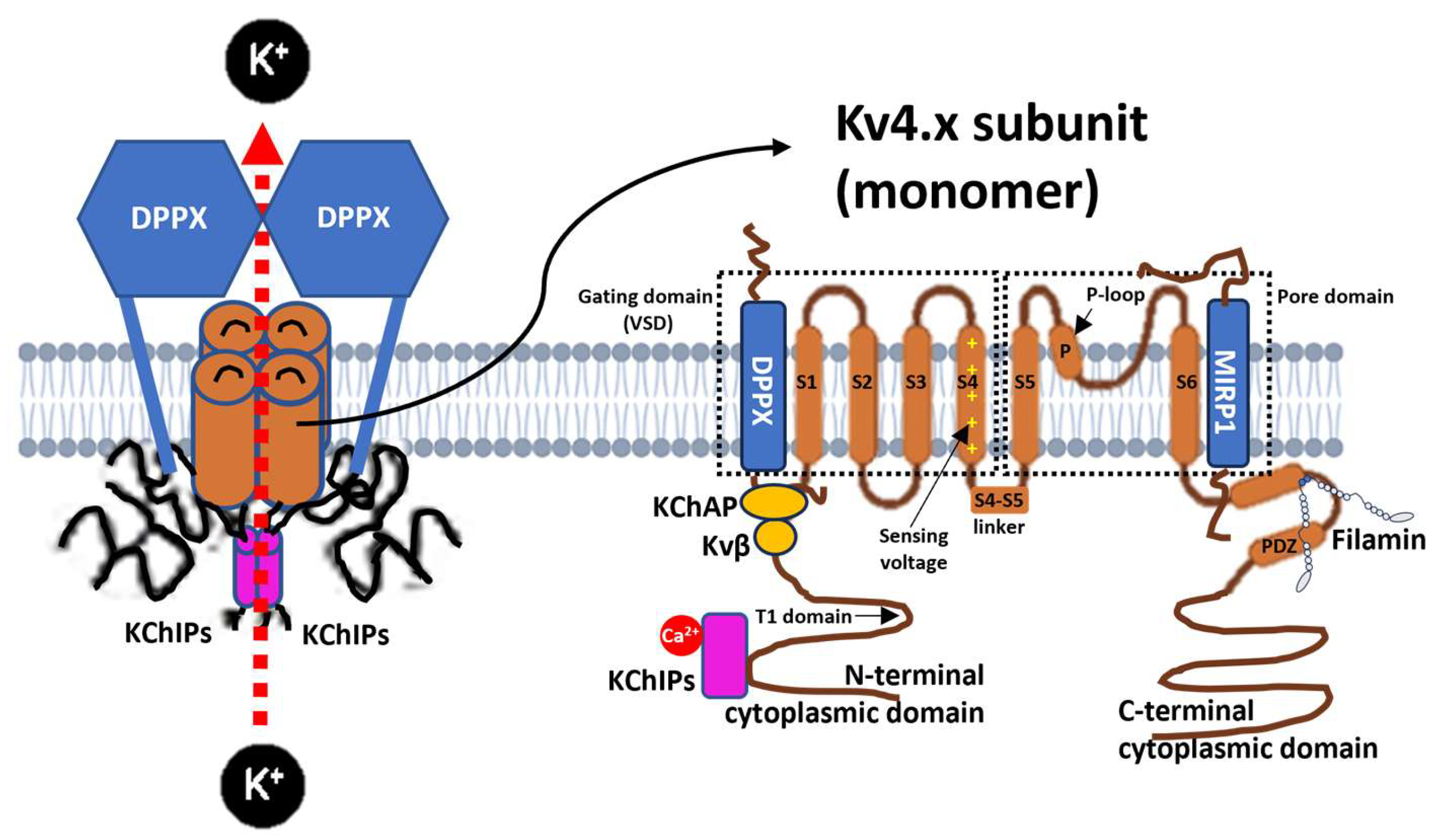
Figure 1. Transmembrane structure of the Kv4.x voltage-gated potassium channels. Schematic diagram of the Kv4 channel in its tetrameric form and the representation of one of its monomers with the six transmembrane segments (S1–S6) and the interacting subunits and auxiliary proteins. DPPX: dipeptidyl-peptidase-like protein 6. KChAP: K+ Channel Accessory Protein. KChIPs: K+ channel-interacting proteins. MiRP1: MinK-related peptide 1. VSD: voltage-sensing domain. Source: Created by the authors based on the reviewed data.
Physiologia 05 00031 g001
Figure 2. Schematic representation of Kv4 subunit alterations across neurodegenerative disease groups. The timeline illustrates the predominant alterations reported in Table 2, highlighting both shared and disease-specific patterns. Kv4.2 dysfunction is common to Alzheimer’s disease (AD), amyotrophic lateral sclerosis (ALS), and prionopathies, whereas Kv4.3 alterations predominate in spinocerebellar ataxias (SCA/EA) and Parkinson’s disease (PD). Kv4.1 involvement was rare and restricted to isolated studies in ALS and AD. Percentages indicate the proportion of studies reporting alterations within each disease group.
Physiologia 05 00031 g002
Figure 3. Dynamic Kv4 alterations along disease progression and regulatory nodes. The timeline depicts changes in Kv4 subunits across neurodegenerative disease models: upregulation/gain of function (green) and downregulation/dysfunction (red). Acute Aβ exposure transiently enhances Kv4.2 and Kv4.3 currents, whereas chronic AD, ALS, and prion models consistently show Kv4.2 downregulation. PD and ataxias primarily involve Kv4.3 dysfunction (KCND3 mutations). Regulatory nodes include auxiliary subunits (DPP6 deficient, KChIP2 coexpression) and kinases (ERK, CaMKII, PKC, PKA, GSK-3). Pharmacological interventions (levetiracetam, carvedilol, cannabinoids, Trolox, GSH, 4-AP, TEA) are shown to partially rescue Kv4 currents or expression. Icons indicate experimental models, including mouse transgenics, patient-derived cells, and in vitro preparations. 4-AP, 4-aminopyridine. Aβ, amyloid-β peptide. AD, Alzheimer’s disease. ALS, amyotrophic lateral sclerosis. APP, amyloid precursor protein. CaMKII, Ca2+/calmodulin-dependent protein kinase II. CBN, cannabidiol. CGC, cerebellar granule cells. CGN, cerebellar granule neurons. DA, dopaminergic neurons. DPP6, dipeptidyl-peptidase-like protein-6. ERK, extracellular signal-regulated kinase. GSH, glutathione. GSK-3, glycogen synthase kinase-3. iPSC, induced pluripotent stem cells. KChIP2, Kv channel-interacting protein 2. PD, Parkinson’s disease. PKA, protein kinase A. PKC, protein kinase C. PrDs, prion diseases. PSEN1, presenilin-1. SCA, spinocerebellar ataxia. TEA, tetraethylammonium. VMN, vagus motor neurons. Arrows: red arrows indicate downregulation/dysfunction pathways; green arrows indicate rescue or protective interventions. Symbols: ↑, upregulation or gain of function. ↓, downregulation or loss of function. =, no change.
Physiologia 05 00031 g003
Figure 4. Systematic review flow diagram.
Physiologia 05 00031 g004
Table 1. Findings of genetic and functional variants of Kv4 channels (Kv4.1–Kv4.3) in neurodegenerative diseases and movement disorders.
CitationDisease
(Study Design)
Participants and/or ModelComparatorOutcome VariableKey FindingsRoB
Amyotrophic Lateral Sclerosis (ALS)
Park et al., 2008 [56]ALS
(Experimental study)
hBM-MSC (from ALS patients, in vitro)K+ blocker (4-AP, TEA)Kv4.x current and KCND2 mRNA ↓ KCND2 expression and Ito current detected in hBM-MSCs.
Kv4.2-dependent profile varied with passage.
High
Trainito et al., 2024 [71]ALS
(Experimental study)
NSC-34 (motor neuron line) Untreated vs. CBN KCND1 and KCND2 mRNA↑ KCND1 and KCND2 expression.
CBN increases KCND1 and KCND2 expression in a dose-dependent manner.
Low
Parkinson’s Disease (PD)
Subramaniam et al. (2014) [72]PD
(Experimental study)
DA neurons in the substantia nigra (SN) from A53T-α-syn mouseWTKv4.3 function↓ Function of Kv4.3.
Mutant α-syn induced Kv4.3 dysfunction and enhanced neuronal excitability in DA neurons.
Some concerns
Lasser-Katz et al. (2017) [57]PD
(Experimental study)
DMV neurons (A53T-α-syn mouse)WT mice and treated with glutation (GSH intervention)Kv4.3 currents= Function of Kv4.3.
Kv4.3 currents unchanged in A53T-α-syn; GSH rescued currents in WT but not mutant mice, suggesting DMV neurons are less vulnerable to oxidative stress.
Low
Alzheimer’s disease (AD)
Cacace et al. (2019) [73]AD
(Observational study of EOAD and FTD)
DPP6 KO mice; patient brain (variants p.R509R, p.R47L, p.D596N)WT/patient carriersKv4.2 protein expression and function↓ Kv4.2 expression and function with DPP6 loss.
Variants linked to reduced Kv4.2 and impaired excitability.
Low
Yao et al. (2020) [74]AD
(Experimental study)
CA1 neurons from 5xFAD × RyR2-E4872Q miceWT and R-carvedilol interventionKv4.2 current and function↓ Kv4.2 current.
RyR2 hyperactivation showed dysfunction of the Kv4.2 channel. Carvedilol rescued Kv4.2 impairment.
Some concerns
Plant et al. (2006) [75]AD
(Experimental study)
Rat cerebellar granule neurons; HEK293 (Aβ1–40/42)N/AKv4.2/Kv4.3 current and protein expression↑ Expression and function of Kv4.2 and Kv4.3.
The Aβ peptide modulates Kv4 channel activity in culture.
Low
Hall et al. (2015) [53]AD
(Experimental study)
CA1/dentate neurons (hAPPJ20, hAPP/PS1dE9 mice)WT; and hAPP model with levetiracetam interventionKv4.2 protein expression↓ Kv4.2 expression
in dendrites (rather than in somas). Aβ load correlated with reduced Kv4.2. Levetiracetam prevents the loss of Kv4.2 protein.
Some concerns
Kim et al. (2021) [76]AD
(Experimental study)
Dentate gyrus neurons (Tg2576 mice)WT and with Trolox interventionKv4.1 mRNA, protein expression and function↓ Kv4.1 expression and function. Trolox restores Kv4.1 expression, normalizes excitability, and restores Kv4.1 levels.Some concerns
Li et al. (2023) [77]AD
(Experimental study)
Hippocampal neurons (APP−/− mice)WT Kv4.2 protein expression, LFP, glutamatergic neuron firing= Kv4.2 expression.
APP loss did not affect Kv4.2 expression or function; neuronal excitability unaltered.
Low
Scala et al. (2015) [78]AD
(Experimental study)
Hippocampal neurons in a 3xTg-AD miceIntracellular incubation of Aβ42 and treatment with caspase and glycogen synthase kinase 3 (GSK-3) as an interventionA-type K+ currents and Kv4.2 phosphorylation↓ Kv4.2 function.
Intracellular Aβ42 accumulation induces hyperexcitability dependent on the inhibition of Ia mediated by Kv4.2 phosphorylation. Dephosphorylation of Kv4.2 by GSK-3 inhibition prevents this increase in excitability.
Low
Pan et al. (2004) [79]AD
(Experimental study)
Rats treated with Aβ25-35N/AKv4.2 and Kv4.3 mRNA and protein expression ↑ Kv4.2 expression and = Kv4.3 expression.
Aβ25-35 increases Kv4.2 mRNA (58%) in cortex. Kv4.2 protein analysis increased (42%) in cortex and (5%) hippocampus. No change in Kv4.3 mRNA.
Some concerns
Kerrigan et al. (2008) [80]AD
(Experimental study)
Rat CGN and HEK293 cells treated with Aβ1-40CGN WTK+ currents (IK and Ia) and Kv4.2 mRNA expression↑ Kv4.2 expression and function. Aβ1-40 modulates K+ currents, increased Kv4.2 expression and altered Kv4.2 inactivation in CGNs.Some concerns
Pieri et al. (2010) [54]AD
(Experimental study)
Rat CGCs treated with Aβ25-35CGC SP interventionK+ currents and Kv4.2 protein expression↑ Kv4.2 and↑Kv4.3 expression and function.
Aβ25–35 increased Ia current and the protein expression of Kv4.2 and Kv4.3. SP reversed these effects, normalizing current and expression.
Some concerns
Campolongo et al. (2013) [81]AD
(Experimental study)
Rats with intracerebroventricular Aβ25-35 Intervention with SP Kv4.2 protein expression↑ Kv4.2 expression (only hippocampus).
SP treatment did not rescue changes in hippocampus or cortex.
Some concerns
Ataxia
Ågren et al. (2023) [82]SCA19/22
(Case report)
Family clinical case with atypical SCA19/22 and Xenopus oocytes WT and his variant: Kv4.3 WT, WT/E280KFamily clinical evaluation, KCND3 variant identification, and electrophysiological characterization of the channel in oocytesAltered Kv4.3 function.
The E280K variant causes a positive shift in the activation and inactivation potentials without altering the maximum current amplitude, associated with developmental delay, but without ataxia or parkinsonism in carriers.
High
Duarri et al. (2015) [83]SCA19/22
(Experimental study)
SCA19/22-mutant Kv4.3 HeLa cells WT Current amplitude or gating properties, and protein Kv4.3 expression↓ Kv4.3 expression and altered function.
T352P, M373I, S390N and ΔF227 mutations in Kv4.3 alter its function and localization.
Low
Reis et al. (2024) [84]SCA19/22
(Experimental study)
Patient and CHO cells WT and variant p.D152G, with or without KChIP2b as interventionKCND3 variant identification and functional characterization by electrophysiology ↓ Kv4.3 function.
Variant p.D152G does not change gating but reduces ionic current.
Coexpression with KChIP2b mitigates negative effect.although it does not reach statistical significance. Voltage dependence unaffected.
High
Duarri et al. (2012) [85]SCA19/22
(Experimental study)
Post-mortem brain tissue of SCA19 patients and HeLa cells (Kv4.3 mutants)WT and intervention with KChIP2b KCND3 variant identification, protein expression, trafficking and function ↓ Kv4.3 expression and function. Kv4.3 mutants (T352P, M373I, S390N) of HeLa cells showed absent or reduced surface expression due to ER retention, protein instability, and loss of function.
KChIP2 subunit rescues localization in T352P and M373I, but without restoring function. The mislocalization of the S390N was not rescued by coexpression of KChIP2b.
High
Li et al. (2022) [86]SCA19/22
(Experimental study)
iPSC of SCA19/22 patients Healthy controlsKCND3 variant identification and Kv4.3 protein expression ↓ Kv4.3 expression.
It was identified T377M mutation in patients.
iPSC with T377M reduces functional protein, mRNA unchanged, and associated with enrichment of ER stress.
Some concerns
Kurihara et al. (2018) [87]SCA19/22
(Case report)
Patients with early-onset cerebellar ataxiaN/AKCND3 variant identification? Kv4.3 expression or function. Novel KCND3 mutation (c.1150G > A, p.G384S).High
Hsiao et al. (2025) [88]SCA19/22
(Preclinical study)
Patient fibroblasts (C317Y, P375S), Drosophila, HEK293T, Xenopus oocytesControlKv4.3 protein and locomotor function↓ Expression and function in Kv4.3 variants.
Kv4.3 proteostasis defects underlie SCA19/22, and overexpression of human Kv4.3 rescued locomotor impairment. Human Kv4.3 variants (V338E and P375S) reduce Shal expression in flies and decrease K+ currents in Xenopus.
Some concerns
Arancibia et al. (2025) [89]SCA19/22
(Genetic cohort study)
Patients and AD293 cellsWT AD293 and coexpressed with KChIP2 as interventionKCND3 variant identification and current ↓Kv4.3 function.
Kv4.3 variants (G371R and S357W) either prevent or do not generate current. Coexpression with KChIP2 partially failed to rescue.
High
Paucar et al. (2021) [90]SCA19/22
(Experimental study)
Family case, HEK293T cells and Xenopus oocytesHEK293T cells transfected with V374A variant as interventionClinical evaluations, K+ currents, KCND3 variant identification and membrane expression ↓ Kv4.3 function and = Kv4.3 expression.
V374A mutant reduces K+ current, with normal membrane expression, associated with paroxysmal ataxia.
High
Duarri et al. (2013) [91]SCA19/22 and
Brugada syndrome
(Case Report)
Patients and in vitro cells (HeLa and HEK293T)WT and intervention with coexpressed KChIP2 KCND3 variant identification and current analysis ↑ Kv4.3 (p.L450P) and ? Kv4.3 (p.P614S) function.
Identification of ataxia L450P variant with gain of function in Kv4.3 and a P614S variant not significantly different of WT.
Low
Carrasco-Marina et al. (2019) [92]SCA19/22) (Case Report)Pediatric patient with early-onset chronic ataxiaN/AKCND3 variant identification and in silico functional analysisAltered Kv4.3 function.
Gly371Arg alters the function with non-conservative substitution in the channel pore region.
High
Zanni et al. (2021) [93]SCA19/22) (Case Report)Patients with non-progressive congenital ataxia and in vitro cells (Xenopus oocytes, HEK293T cells)WT KCND3 variant identification, protein expression and current↓ Kv4.3 expression and function.
G345V, S347W, and W359G variants produce loss of function through reduced expression and currents.
Some concerns
Hsiao et al. (2019) [94]SCA19/22) (Case Report)Patients and in vitro cells (Xenopus oocytes, HEK293T cells)WT and intervention with KChIP2 coexpressedKCND3 variant identification, protein expression and current↓ Kv4.3 expression and function. New mutations identified: C317Y, P375S, V338E and T377M, with loss of function not restored by coexpression with KChIP2 and reduction in total Kv4.3 protein expression in the variants.Low
Contaldi et al. (2024) [95]SCA19/22) (Case Report)Patient N/AKCND3 variant identification? Kv4.3 expression or function. Identified Ser346Phe mutation. Effect on function unclear.High
Paucar et al. (2018) [96]SCA19, allelic with SCA22 (Case Report)Family caseN/AKCND3 variant identification? KCND3 function or expression.
Identified T377M mutation.
High
Avila-Jaque et al. (2024) [97]SCA19
(Case Report)
Patients N/AKCND3 variant identification? Kv4.3 expression and function.
Pathogenic variants from different Latin American populations reported: Ser357Trp, Gly384Ser, Gly371Arg.
High
Lee et al. (2012) [98]SCA22
(Experimental study)
Patients and in vitro cells (HEK-293T transfected)WTKCND3 variant identification, protein expression and current ↓ Kv4.3 expression and function.
Multiple KCND3 mutations (F227del, G345V, V338E, T377M).
F227del disrupted protein localization and decreases K+ currents.
Low
Hung et al. (2025) [99]SCA22
(Experimental study)
Knock-in mice (F227del mutation)WTCurrent Kv4.3 and protein localization↓ Kv4.3 expression and function. F227del mutation causes SCA22 through a dominant negative mechanism that reduces function, affecting localization to the plasma membrane, protein accumulation, and causing dysfunctions in the ER and Golgi.Some concerns
Tada et al. (2020) [100]SCA13, SCA19
(Clinical Trial)
PatientsN/AKCND3 variant identification? Kv4.3 function and 0 pathogenic variants in KCND3.
No pathogenic mutations identified.
Low
Hourez et al. (2011) [101]SCA1
(Comparative Study)
SCA1 Transgenic mouse model and in vitro cerebellar slicesWT and with aminopyridines interventionmRNA expression and currents of Kv4 channels= Kv4.1, = Kv4.2, = Kv4.3 (10× more in membrane surface) expressions.
No increase in the expression of Kv4 in SCA1 mice. Increased Ia in presymptomatic PCs mediated by Kv4.3.
Aminopyridines partially reversed electrical dysfunction.
Some concerns
Hsiao et al. (2021) [102]Progressive cerebellar ataxia
(Case Report)
Patients and HEK293T cellsWTKCND3 variant identification, protein expression and current ↑ Kv4.3 function and = Kv4.3 expression.
Identified R419H mutation in the patient, which is predicted to be disease-causing but does not affect Kv4.3 localization or quantity, although increases the K+ current.
High
Smets et al. (2015) [103]Early cerebellar ataxia
(Case Report)
Patient and in vitro cells (HeLa and CHO-K1 cells)WT and intervention with KChIP2 coexpression KCND3 variant identification, protein expression and current ↓ Kv4.3 function and = Kv4.3 expression.
p.Arg293_Phe295dup mutation causes changes in the gating and inactivation properties of the channel, with reduction in output current.
Kv4.3 detected at cell surface but reduced by cycloheximide and rescued by KChIP2 coexpression.
High
Choi et al. (2017) [104]EA
(Genetic cohort study)
PatientsN/AKCND3 variant identification? Kv4.3 function.
Identified Arg431Cys variant, but functional validation is required.
Some concerns
Prion diseases
Mercer et al. (2013) [105]PrDS (GSS) (Experimental study)Prnp0/0 KO mice, DPP6 df5J/Rw mice, and in vitro transfected cell lines (HEK293T, RK13, and N2a)WTKv4.2 current and protein interactions↑ Kv4.2 function.
The PrPC prion modulates Ia through DPP6, but the mutant prion variant (GSS form) loses Kv4.2 function.
Low
Alier et al. (2010) [106]PrDs
(Experimental study)
RatsControlKv4.2 current and mRNA Kv4.2 expression↓ Kv4.2 function.
Application of the PrP(106-126) fragment reduced Ia by 23.5% and IK currents. mRNA Kv4.2 expressed in 75% of cholinergic and 60% of GABAergic neurons, altering excitability.
Some concerns
Footnotes: Aβ, amyloid-β. Aβ42, Amyloid-β 1–42. AD, Alzheimer’s Disease. ALS, amyotrophic lateral sclerosis. APP, amyloid precursor protein. CA1/DG: hippocampal subregions (Cornu Ammonis 1; Dentate gyrus). CBN, cannabinol. CHO cells, Chinese hamster ovary cells. CGC, cerebellar granule cells. CGN, cerebellar granule neurons. DA, dopaminergic. DMV, dorsal motor nucleus of the vagus. DPP6, dipeptidyl peptidase–like protein 6. EA, episodic ataxia. EOAD, early-onset Alzheimer’s disease. ER, endoplasmic reticulum. FTD, frontotemporal dementia. GSH, glutathione. GSK-3, glycogen synthase kinase 3. GSS, Gerstmann–Sträussler–Scheinker Disease. Ia, A-type K+ current. IK, delayed rectifier. iPSCs, induced pluripotent stem cells. KChIP2b, K+ channel-interacting protein 2b. KCND1, gene encoding Kv4.1. KCND2, gene encoding Kv4.2. KCND3, gene encoding Kv4.3. KO, knockout. Kv4.x, subunits of A-type voltage-gated K+ channels encoded by KCNDx. LFP, local field potential. LTP, long-term potentiation. PCs, Purkinje cells. PD, Parkinson’s Disease. N/A, not applicable. NSC-34, motor neuron-like cell line. PrDs, prion diseases. PSEN1, presenilin-1. RoB, risk of bias. SCA1, spinocerebellar ataxia type 1. SCA13, spinocerebellar ataxia type 13. SCA19, spinocerebellar ataxia type 19. SCA22, spinocerebellar ataxia type 22. SCA19/22, spinocerebellar ataxia 19/22. SP, substance P. WT, Wild-Type. 3xTg-AD mice, triple-transgenic AD mice (mutant APP, PSEN1, and tau). α-syn, α-synuclein. Symbols: ↑, upregulation or gain of function. ↓, downregulation or loss of function. =, no change. ?, uncertain or inconsistent results.
Table 2. Variants for the KCNDx gene according to the Human Genome Variation Society (HGVS) standard.
KCND3 Variantc. Nucleotide Change (HGVS)p. Amino Acid ChangeAccession Number (NM)dbSNP rsIDAssociated AtaxiaAffected Transmembrane Segment
C317Yc.950G > Ap.Cys317TyrNM_001378969.1rs1571939905SCA19/22S4-S5
D152Gc.455A > Gp.Asp455GlyN/AN/ASCA19/22NH2
E280Kc.838G > Ap.Glu280LysNM_001378969.1rs2101995916SCA19/22S3-S4
F227delc.679_681delTTCp.Phe227delNM_001378969.1rs397515475SCA19/22S2
G345Vc.1034G > Tp.Gly345ValNM_001378969.1rs797045634SCA19/22S5-S6
G384Sc.1150G > Ap.Gly384SerNM_001378969.1rs1664632655SCA19/22S6
G371Rc.1111G > Ap.Gly371ArgNM_001378969.1rs1057521793SCA19/22S5-S6
L450Pc.1348C > Tp.Leu450PheNM_001378969.1rs150401343SCA19/22COOH
M373Ic.1119G > Ap.Met373IleNM_001378969.1rs397515477SCA19/22S5-S6
P375Sc.1123C > Tp.Pro375SerNM_001378969.1rs1571636508SCA19/22S5-S6
P614Sc.1840C > Tp.Pro633SerNM_001378970.1N/ASCA19/22COOH
R419Hc.1256G > Ap.Arg419HisNM_001378969.1rs774338559Progressive cerebelar ataxiaCOOH
R431Cc.1291C > Tp.Arg431CysNM_001378969.1rs777183510SCA19/22COOH
S347Wc.1040C > Gp.Ser347TrpN/AN/ASCA19/22S5-S6
S357Wc.1070C > Gp.Ser357TrpNM_001378969.1rs867628133SCA19/22S5-S6
S390Nc.1169G > Ap.Ser390AsnNM_001378969.1rs397515478SCA19/22S6
T352Pc.1054A > Cp.Thr352ProNM_001378969.1rs397515476SCA19/22S5-S6
T377Mc.1130C > Tp.Thr377MetNM_001378969.1rs1571636501SCA19/22S5-S6
V338Ec.1013T > Cp.Val338GluNM_001378969.1rs1571939827SCA19/22S5
V374Ac.1121T > CN/AN/AN/ASCA19/22S5-S6
W359Gc.1075T > Gp.Trp359GlyN/AN/ASCA19/22S5-S6
N/Ac.1037C > Tp.Ser346PheN/AN/ASCA19/22N/A
N/Ac.877_885dupCGCGTCTTCp.Arg293_Phe295dupNM_001378969.1rs2525698722SCA19/22S4-S5
Footnotes: Genetic variant data consulted in dbSNP and ClinVar (NCBI, NLM, 2025). N/A: Not applicable or data not available. dbSNP: Single Nucleotide Polymorphism database. ClinVar: Genomic Variation as it Relates to Human Health. HGVS: Human Genome Variation Society. Key: “c.” = coding DNA change (HGVS); “p.” = protein (amino acid) change; NM_ = RefSeq transcript; rsID = dbSNP identifier.
Table 3. Frequency of Kv4 subunit alterations reported in neurodegenerative diseases from a total of 40 results.
Neurodegenerative DiseaseN Studies with Alterations of KvKv4.1 (n, %)Kv4.2 (n, %)Kv4.3 (n, %)
ALS31 (33.3%)2 (66.7%)0 (0%)
PD10 (0%)0 (0%)1 (100%)
AD121 (8.3%)9 (75.0%)2 (%)
Ataxias (SCA/EA)160 (0%)0 (0%)16 (6.7%)
Prionopathies20 (0%)2 (100%)0 (0%)
Total342 (5.9%)13 (38.2%)19 (55.9%)
Footnotes: Frequency of Kv4 subunit alterations reported in neurodegenerative disease models. Only studies reporting functional or expression changes were included, whereas studies with no change or uncertain results were excluded. Values are shown as absolute counts and percentages relative to the number of studies with alterations in each disease group.
Disclaimer/Publisher’s Note: The statements, opinions and data contained in all publications are solely those of the individual author(s) and contributor(s) and not of MDPI and/or the editor(s). MDPI and/or the editor(s) disclaim responsibility for any injury to people or property resulting from any ideas, methods, instructions or products referred to in the content.