Review

Personalized Management of Stomatognathic Pain: A Narrative Review

1
Section of Dentistry, Department of Clinical, Surgical, Diagnostic and Pediatric Sciences, University of Pavia, 27100 Pavia, Italy
2
Maxillo-Facial Surgery Unit, Fondazione Istituto di Ricovero e Cura a Carattere Scientifico Policlinico San Matteo, 27100 Pavia, Italy
3
Department of Clinical and Experimental Sciences Physical and Rehabilitation Medicine, University of Brescia, 25123 Brescia, Italy
4
Department of Continuity of Care and Vulnerability, Physical and Rehabilitation Medicine Unit, Azienda Socio-Sanitaria Territoriale Spedali Civili di Brescia, 25123 Brescia, Italy
*
Author to whom correspondence should be addressed.
Hygiene 2026, 6(2), 28; https://doi.org/10.3390/hygiene6020028 (registering DOI)
Submission received: 10 April 2026 / Revised: 27 May 2026 / Accepted: 28 May 2026 / Published: 30 May 2026
(This article belongs to the Special Issue Advance in Oral Hygiene and Oral Health)

Abstract

Pain is a complex experience that encompasses both physical and emotional dimensions. Stomatognathic Pain (SP) affects the anatomical and functional structures of the stomatognathic system, which is involved in essential activities such as mastication, deglutition, and phonation. Consequently, SP can significantly impair patients’ quality of life, affecting physiological, psychological, and social well-being. Precision Medicine (PM) is an emerging patient-centered approach that enables healthcare providers to tailor diagnostic and therapeutic strategies according to each patient’s biochemical, physiological, environmental, and behavioral profile. The aim of this narrative review is to highlight PM tools for the diagnosis and treatment of SP and to explore their contribution to personalized management. A literature search was conducted in the PubMed and Scopus databases using the keywords “orofacial pain”, “stomatognathic pain”, “precision medicine”, “pain management”, “pain assessment”, “pain treatment”, and “patient-centered care”. Current evidence suggests that PM can improve SP management by identifying pathogenic mechanisms, clarifying disease etiology, and integrating advanced molecular and digital technologies. Emerging approaches, including genetic profiling, biomarker analysis, artificial intelligence–based diagnostics, targeted therapies, individualized rehabilitation, and behavioral interventions, may support personalized treatment and facilitate the development of patient-centered care models, ultimately reducing the burden of SP and improving clinical outcomes and quality of life.

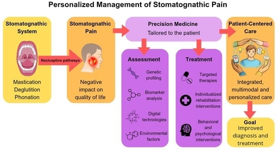
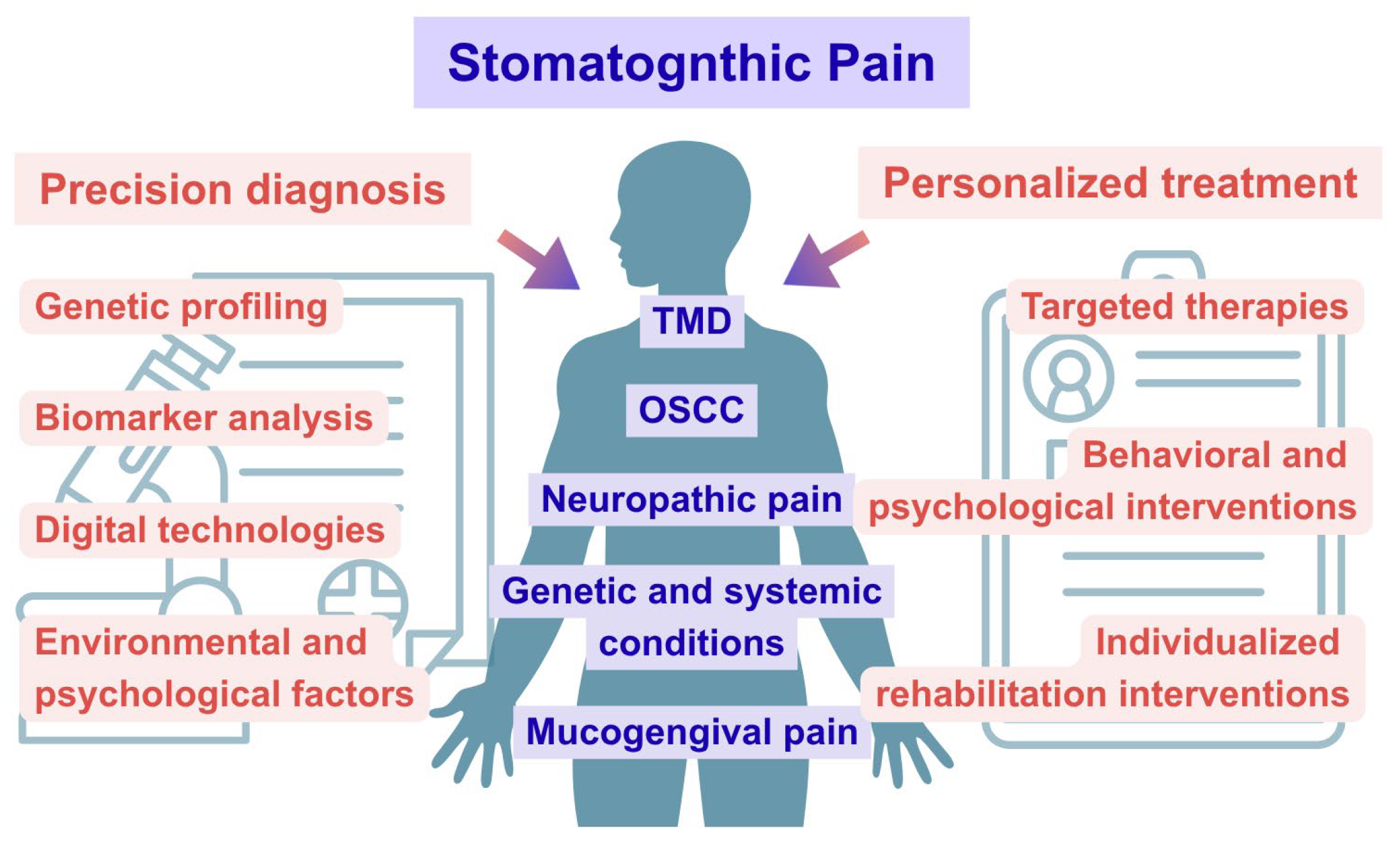
Graphical Abstract

1. Introduction

The Stomatognathic System (SS) comprises a wide array of anatomical and functional structures, located in the oral and craniofacial cavities [1]: teeth, periodontium, salivary glands, mandibular and maxillary bones, temporomandibular joint, masticatory muscles, and neurovascular network [2]. SS is involved in essential functions such as mastication, deglutition, phonation, respiration, and facial aesthetics [3]. This anatomical complex has functional relationships with posture, the mechanical processes of feeding and eating behavior, and the nervous system; consequently, disorders of the SS can affect these interrelated mechanisms [4,5,6].
Stomatognathic pain (SP) is a complex physical and emotional experience that affects the structures of the SS. Typically, SP is caused by dental and periodontal diseases, oral cancer or musculoskeletal and neuropathological disorders [7,8]. SP can also be experienced in acute and chronic manifestations [9]. Acute pain typically arises from specific stimuli, such as trauma, dental procedures, or surgery. Chronic pain is frequently associated with idiopathic and neuropathic conditions, such as burning mouth syndrome and trigeminal neuralgia, as well as temporomandibular disorders (TMD), that encompass a spectrum of disorders affecting the jaw muscles, temporomandibular joint, and associated structures [10,11]. SP significantly impacts patients’ quality of life because it is associated with healthcare expenses, loss of productivity, reduced psychological and oral health-related well-being [9]. Effective pain assessment and management is therefore essential.
Precision Medicine (PM) is an approach that tailors interventions to the patient’s unique biochemical, physiological, environmental, and behavioral profile [12]. Advanced diagnostic and therapeutic tools employed in PM include genome sequencing, biomarker profiling, imaging technologies, analysis of epigenetic mechanisms, assessment of environmental and behavioral factors and the use of digital tools such as mobile applications and wireless monitoring devices [12]. PM is closely linked to the concept of personalized medicine, which aims to individualize care based on each patient’s unique profile [13]. Therefore, a personalized management of SP should ideally provide the safest, most efficacious and most efficient diagnostics and therapies [14]. Technology improvements in PM can lead toward a patient-centered approach to disease and pain control [15].
Patient-Centered Care (PCC) is a model of care delivery that contributes to improved health outcomes, reduced healthcare costs and greater patient satisfaction, by addressing the patient’s values, needs and desires [16].
Therefore, PM offers a promising approach for the diagnosis and treatment of pathological conditions affecting the SS that may cause SP, incorporating a PCC methodology through tailored and personalized management. Currently, there is a gap in the literature regarding the personalized treatment of SP. Although several advanced diagnostic and prognostic tools have recently been introduced, there is still no standardized protocol, and the available evidence remains fragmented. Consequently, a personalized approach to the management of this burdensome condition has not yet been clearly established.
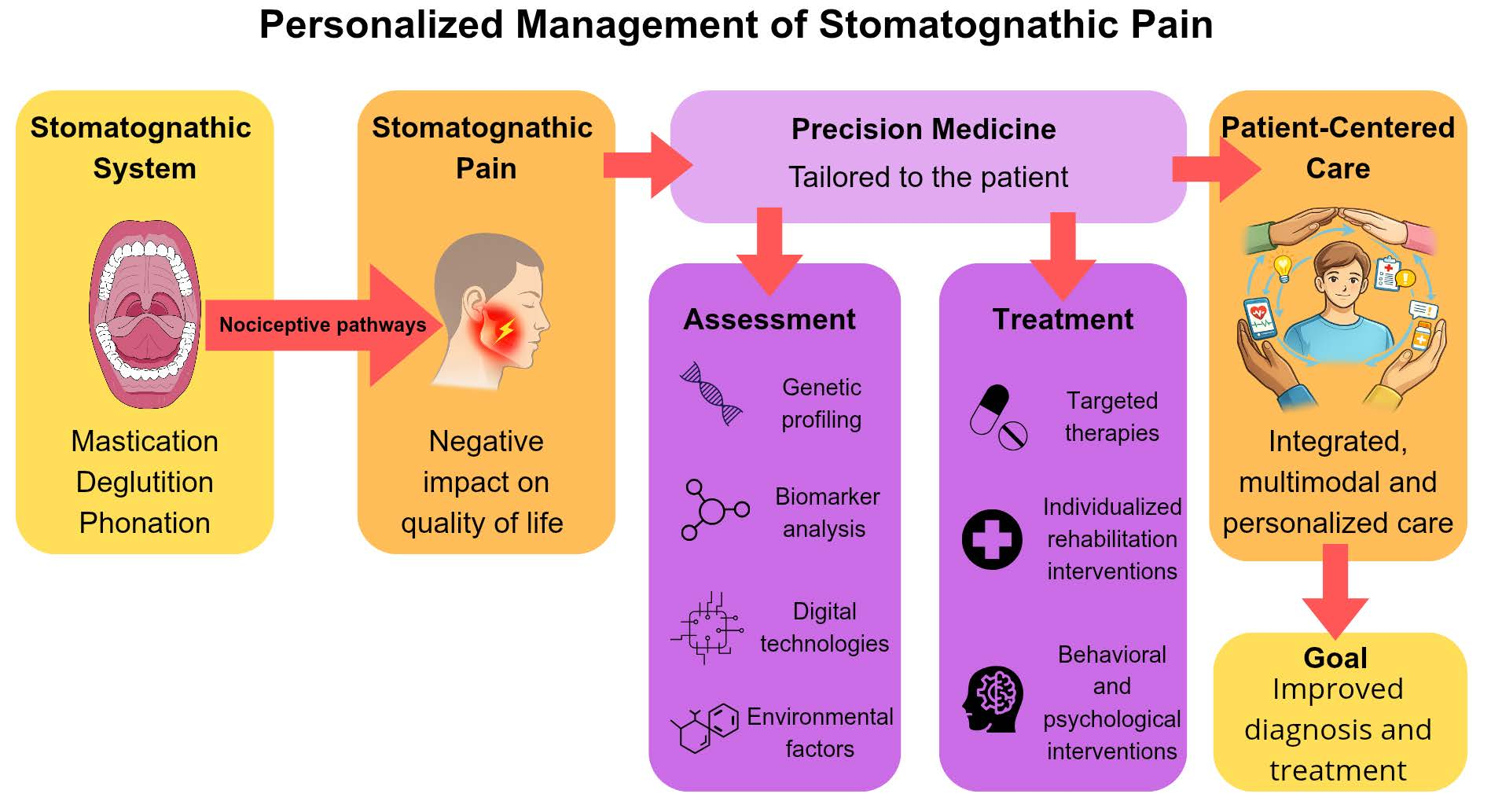
This review aims to present and evaluate the diagnostic methodologies and therapeutic strategies that facilitate personalized management of SP, promoting a patient-centered approach to clinical care. The conceptual framework of this review is summarized in Figure 1.

2. Materials and Methods

To identify evidence on PM tools for the personalized management of SP, a literature search was conducted in the PubMed and Scopus databases. The search strategy was designed to retrieve relevant studies using the following keywords and their combinations: “orofacial pain”, “stomatognathic pain”, “precision medicine”, “pain management”, “pain assessment”, “pain treatment” and “patient-centered care”. The search was conducted between February 2025 and November 2025.
Study selection was performed according to predefined inclusion and exclusion criteria. The inclusion criteria comprised: (i) article types including narrative reviews, systematic reviews, meta-analyses, and clinical trials; and (ii) articles published from 2004 onwards. The exclusion criteria included: (i) case reports; (ii) articles published before 2004; and (iii) retracted articles.
The identified records were screened for relevance based on title, abstract, and full-text evaluation when necessary. This methodological approach enabled the identification of relevant evidence regarding PM strategies for personalized pain management.

3. Neurophysiology of Stomatognathic Pain

3.1. Trigeminal Sensory System

The trigeminal sensory system consists of the trigeminal nerve (CNV), the trigeminal ganglion, and the trigeminal sensory nuclei [17]. The CNV divides into three branches and each of them covers roughly one-third of the craniofacial dermatome [18]. The trigeminal ganglion (or semilunar or Gasserian ganglion) is located in the middle cranial fossa at the base of the skull. The trigeminal sensory nuclei are located in the brainstem [17].

3.1.1. Trigeminal Nerve Anatomy and Function

The CNV partially innervates the craniofacial region. The Gasserian ganglion is a sensory ganglion located in Meckel cave [19]. The trigeminal ganglion contains the peripheral axons of pseudo-unipolar primary afferent neurons and gives origin to three branches: the ophthalmic, the maxillary and the mandibular branches [20].
The CNV is a mixed nerve, meaning that it is responsible for both sensory and motor functions in the craniofacial region. Sensory branches provide sensory input to the skin, oral and nasal cavities, cornea, teeth, temporomandibular joint, parts of the tongue, and parts of the brain’s protective layers. It also detects touch, pain, and temperature. The motor branches control chewing muscles and small muscles like the mylohyoid, tensor veli palatini, and tensor tympani [21].

3.1.2. Trigeminal Sensory Nuclei

The trigeminal sensory nucleus is a long column of cells, located in the dorsolateral brainstem. This group includes the mesencephalic nucleus and the sensory trigeminal complex, which gathers the principal trigeminal nucleus and the spinal trigeminal nucleus [22].
Each subnucleus is dedicated to interpreting specific types of sensory input, such as gentle touch, pressure, temperature changes and pain. The processed sensory information is then relayed to various regions of the central nervous system, playing a crucial role in sensing stimuli from the craniofacial region [17].

3.2. Pain Transmission and Modulation

3.2.1. Nociceptive Pathways

Trigeminal primary afferent fibers extend into peripheral tissues, where they function as sensory receptors. These low-threshold mechanoreceptors are mainly linked to A-beta or A-delta fibers, while free nerve endings act as nociceptors, detecting harmful stimuli. These nociceptors include small myelinated (A-delta) fibers and slower unmyelinated (C) fibers, with A-beta fibers occasionally playing a nociceptive role [23]. A-delta and C fibers are activated by chemical, mechanical or thermal stimuli to protect the injured area until it can heal; they can also respond to non-noxious stimuli such as cooling, warming, or light touch [23,24].
Upon repeated exposure to noxious stimuli, the peripheral nociceptors transfer repetitive stimuli to the primary neurons located in the Gasserian ganglion. These pain signals are then relayed to the secondary neurons within the trigeminal nuclei. From there, the signals are projected via the ventral trigeminus-thalamic tract to the tertiary neurons in the thalamus [25]. The ventro-posteromedial and ventro-posterolateral nuclei of the thalamus are somatotopically arranged and receive input from the contralateral half of the head and body, thus enabling the localization of nociceptive stimuli. In contrast, the intralaminar and dorsal thalamic nuclei are primarily involved in affective-motivational components of nociception. Finally, the neurons within thalamus nuclei project to various areas of the cerebral cortex, including the primary and secondary somatosensory cortices, in order to obtain localization and qualitative and quantitative perception of noxious stimuli [22].

3.2.2. Pain Modulation Mechanisms

Noxious stimuli also activate a descending pain regulation system [26]. The periaqueductal gray acts as an integration center, receiving descending nociceptive inputs from higher brain areas and projecting them to the rostral ventromedial medulla; it also activates an endogenous pain-inhibitory system [23,26]. The rostral ventromedial medulla, which receives descending projections from periaqueductal gray, posteromedial thalamic nucleus, locus coeruleus, hypothalamus, can exert both excitatory and inhibitory control over pain. Nociceptive perception results from the joint regulation of ascending and descending pathways, which are normally balanced under physiological conditions. However, a disruption in this balance may lead to pathological pain, as in cases of nerve injury or inflammation, which induce molecular alterations within the trigeminal nervous system [26].

3.2.3. Peripheral and Central Sensitization

Persistent or repeated noxious stimulation triggers the release of various chemical mediators, including glutamate, gamma-aminobutyric acid (GABA), serotonin, noradrenaline and neuropeptides [23]. This cascade promotes peripheral sensitization, which is characterized by increased neuronal excitability, reduced thresholds and the emergence of background activity [23,24]. Clinically, peripheral sensitization manifests as hypersensitivity and static hyperalgesia [24,27]. Peripheral sensitization acts as a defensive mechanism, by reducing risk of repeated injury [27]. Peripheral sensitization can also trigger central sensitization in the spinal cord, while trigeminal central sensitization occurs in the caudalis nuclei [24,28]. This involves the upregulation of ionotropic and metabotropic glutamate receptors, down regulation of GABA receptors, alterations in sodium and potassium channels, and neuroinflammatory changes [24]. Central sensitization is driven by mechanisms of central neural plasticity [29]. Clinically, central sensitization is expressed through allodynia—when nociceptive neurons start responding to non-nociceptive stimuli—hypersensitivity, secondary hyperalgesia to punctate or pressure stimuli—which is increased sensitivity to a stimulus that is normally painful—aftersensations and enhanced temporal summation. Additionally, central sensitization promotes the amplification of nociceptive input and transition from acute to chronic pain states [24]. This mechanism is particularly relevant in SP, especially chronic SP conditions [27].

3.3. Biopsychosocial Aspects of Pain

Both acute and chronic SP significantly impact patients’ quality of life, affecting physical health, mental well-being and social functioning. Pain can lead to heightened worry, anxiety and fear, potentially amplifying the perception of pain and initiating a vicious cycle of suffering that hinders effective pain management. A correlation has been identified between pain and psychological conditions such as depression, anxiety, and chronic stress [30].
It has also been suggested that persistent SP, whether accompanied by an identifiable organic pathology or not, may reflect underlying psychological factors and serve as a significant source of emotional distress and disruption to daily life [31].
These complex mechanisms play an important role in the onset of SP, and both biochemical, physiological and psychological aspects of pain have been investigated to develop new PM tools for a better understanding and management of this condition.

4. Precision Medicine in Stomatognathic Pain Assessment

In the literature, numerous tools are available for pain assessment. Pain can be evaluated in terms of intensity, localization, quality, and behavior. Pain intensity can be measured using the Visual Analogue Scale, the Numerical Rating Scale, the Verbal Rating Scale, the Faces Pain Scale, and the Relief Assessment Scale. For pain localization, pain maps are useful. The McGill Pain Questionnaire can be used to assess pain quality by evaluating pain intensity and localization, as well as sensory and affective aspects. Pain behavior is usually assessed with a multidimensional questionnaire and a pain diary, which allows daily tracking of SP. It is also important to evaluate quality of life, psychological status, and sleep disorders. The Oral Health Impact Profile is used to assess the impact of oral health on quality of life. For psychological assessment, commonly used tools include the Hamilton Anxiety Rating Scale, the Hamilton Depression Rating Scale, the State–Trait Anxiety Inventory, the Beck Depression Inventory, the Ten-Item Personality Inventory, and the Pain Catastrophizing Scale. For sleep disorder assessment, the Epworth Sleepiness Scale and the Pittsburgh Sleep Quality Index are commonly used. Using these tools allows clinicians to perform a multidimensional assessment of SP, which is essential for effective pain management and improved patient outcomes [32].
Even though these tools remain clinically valuable, their main limitations are the lack of objectivity and patient-specific personalization. Since pain is a subjective and multidimensional experience, pain scales may not adequately capture the emotional, psychological, and functional dimensions associated with pain experience. Moreover, these tools are not universally applicable across different patient populations, as the interpretation of these scales may vary according to age, cognitive status, cultural background and communication abilities [33]. For example, BaHammam et al. (2022) reported that existing SP measurements for disabled and frail adults have not been adequately and comprehensively validated to establish strong evidence regarding their measurement properties, feasibility, and interpretability [34]. Consequently, clinicians face substantial challenges in selecting the most appropriate assessment tool, emphasizing the need for more comprehensive and personalized approaches to pain evaluation.
Recent research highlights how the study of genetic polymorphisms, specific biomarkers, and digital technologies can provide clinicians with more objective methods for pain assessment, thereby enabling targeted therapies and more effective management of the different types of SP arising from the multiple conditions that can affect the SS.

4.1. Genetic Profiling

Since the advent of next-generation sequencing, genome analysis has become dramatically faster, cheaper, and more precise. These advances have made it possible to uncover a wide range of genetic alterations—including single nucleotide variants and structural changes—that underlie disease risk, progression, and treatment response. Together, these innovations highlight genomics as a transformative tool for improving diagnosis, prognosis, personalized treatment strategies, thereby contributing to the more effective management of pain-related conditions [35].
Carvalho et al. (2020) highlighted that genetic polymorphisms related to opioid (OPRM1), catecholaminergic (COMT), inflammatory (TNFa) and dopaminergic pathways (DRD2) are significantly associated with pressure and thermal pain sensitivity in the orofacial region. This suggests that genetic factors should be taken into account for an accurate interpretation of SP sensitivity [36].
Cruz et al. (2022) conducted a systematic review of genetic studies investigating both TMD and primary headaches. Among the genes explored, COMT, ESR1, and MTHFR were the only ones found in association studies of both conditions. COMT variants were frequently linked to pain sensitivity and susceptibility in TMD, as COMT gene encodes the COMT enzyme, which degrades numerous neurotransmitters and is highly associated with chronic pain syndromes, though their role in headaches was less consistent. ESR1 polymorphisms, related to estrogen signaling, showed associations with both TMD and migraine, supporting a shared hormonal influence and the female predominance observed in both disorders. MTHFR, while studied in relation to vascular and metabolic mechanisms in migraine and to pain in TMD, showed inconclusive evidence for either condition. Overall, the review highlighted that despite some overlap, particularly in ESR1, current evidence for shared genetic risk between TMD and headaches remains limited and requires further research [11,37].
Additionally, the study of gene expression can uncover differential expression patterns associated with malignant transformation, enhancing the understanding of the molecular mechanisms involved in disease progression and potentially helping to identify factors related to cancer-associated pain, as well as targets for early intervention and prevention. The article by Carinci et al. (2005) investigated the molecular mechanisms underlying the progression of tongue squamous cell carcinoma by analyzing gene expression profiles across normal tissue, dysplasia, non-metastatic tumors, and metastatic tumors. Using cDNA microarray analysis, the authors identified specific genes whose expression changes as the disease progresses, highlighting potential biomarkers involved in tumor development and metastasis. They found that early progression from dysplasia to carcinoma was associated with alterations in genes related to oncogenesis, transcriptional regulation, and cell-cycle control, while metastatic progression involved genes associated with cell motility, intercellular adhesion, extracellular matrix remodeling, and apoptosis regulation. Overall, the study suggests that these genetic markers may improve the early identification of high-risk pre-malignant lesions and help predict metastatic potential, contributing to better diagnosis and prognosis of oral tongue cancer [38].

4.2. Biomarker Analysis

Proteomics, transcriptomics, and metabolomics capture dynamic biological states and provide multidimensional insights into disease processes. Proteomics tracks proteins, revealing changes in abundance, modification, and interaction that underpin disease processes. Transcriptomics measures RNA transcripts, shedding light on which genes are actively expressed and how gene regulation shifts during disease or treatment. Metabolomics assesses small molecules and metabolic pathways, reflecting real-time biochemical activity and phenotype. Taken together, these omics layers provide a more comprehensive insight into disease mechanisms, biomarker identification, and potential therapeutic targets [35].
Niculescu et al. (2019) present evidence on universal biomarkers for pain. In high pain states, G Protein Subunit Gamma 7 with roles in signal transduction, Contractin 1, with roles in neuronal cell adhesion, Coiled-Coil Domain-Containing 144B and Microfibril-Associated Protein 3 (MFAP3) are decreased in blood. In high pain states, lymphocyte antigen 9, with immunomodulatory roles, and guanylate-binding protein 1, with interferon-induced signaling roles, are increased in blood. These findings help reach an objective diagnosis through biomarker evaluation [39].
Shrivastava et al. (2021) explored different biomarkers associated with pain in patients with TMD. Key biomarkers investigated for assessing pain in TMD include inflammatory cytokines, such as IL-1β, IL-6, TNF-α and PGE2, which are frequently elevated in synovial fluid and associated with pain severity and joint inflammation. Matrix metalloproteinases (MMP-2, MMP-7, MMP-9) reflect extracellular matrix degradation and may signal degenerative changes, while oxidative stress markers like malondialdehyde (MDA) point to tissue injury processes. Neuropeptides such as substance P indicate nociceptive activation, though technical challenges limit their clinical applicability. Emerging evidence on microRNAs and genetic variants suggests potential for personalized approaches, but further validation is needed before translation into clinical practice [40].
Periodontal inflammation, particularly gingivitis, is associated with SP and discomfort, while advanced periodontitis is linked to increased physical pain and a significant negative impact on oral health–related quality of life [41]. The systematic review of Scribante et al. (2024) highlights that salivary cortisol levels are elevated in chronic periodontitis and correlate with greater disease severity—such as deeper probing depths, higher plaque indices, and increased gingival inflammation—suggesting that cortisol may link stress and inflammatory responses in periodontal disease and serve as a non-invasive biomarker for periodontal status [42]. In the work of D’Agostino et al. (2024), it was highlighted that pro-inflammatory interleukins, such as IL-1β, IL-6, and IL-17, play a major role in promoting connective tissue breakdown and alveolar bone loss, while anti-inflammatory cytokines, such as IL-10, help limit this damage. Since periodontitis is a chronic inflammatory disease in which bacterial plaque triggers an immune response, interleukins play a significant role in its progression. In their study, the authors found that in individuals with Type 1 Diabetes, hyperglycemia enhances the inflammatory response, increasing interleukin production and making periodontal tissues more vulnerable to destruction. This creates a bidirectional relationship in which periodontitis can worsen metabolic control, while diabetes accelerates periodontal disease progression, highlighting interleukins as both biomarkers and potential therapeutic targets [43].
SP can also be caused by oral squamous cell carcinoma (OSCC). Pain is one of the most frequent symptoms reported by patients with OSCC. Although pain is often the dominant symptom, it typically appears only once lesions have reached a considerable size, prompting patients to seek medical care. As a result, early-stage carcinomas frequently remain undetected due to the absence of symptoms [44]. In this context, biomarkers may be useful for assessing OSCC prognosis and for improving understanding of cancer-related pain mechanisms. The review of Pereira et al. (2017) shows that microRNAs, in particular miR-125b, miR-181 and miR-339, may influence pain pathways by modulating inflammatory processes and endogenous pain-control systems, potentially contributing to peripheral and central sensitization in OSCC patients [45]. The review of Pellegrini et al. (2025) examines the current evidence on salivary biomarkers as non-invasive prognostic indicators in OSCC. The authors report that several proteomic, transcriptomic, and metabolomic salivary markers—most notably miR-423-5p, AKR1B10, and 3-methylhistidine—are associated with disease progression, survival outcomes, and tumor aggressiveness [46].
Although these techniques may appear invasive and still require further scientific validation before being implemented in clinical practice, the study of biomarkers in painful SS conditions—TMD, periodontitis, OSCC—may help to better elucidate pain mechanisms in patients with chronic pain who do not benefit from commonly used therapeutic approaches, thereby contributing to the identification of more effective treatments.

4.3. Digital Technologies

In recent years, the development of new artificial intelligence (AI) technologies has transformed the medical model, improving diagnostic accuracy and treatment outcomes, facilitating early detection of clinical conditions, and advancing drug development, medical management and medical education [47,48,49,50].
According to the review of El-Tallawy et al. (2024), machine learning algorithms and AI are increasingly being integrated into pain assessment to provide objective, continuous, and personalized measures of pain that complement or even go beyond subjective self-reports. Techniques such as computer vision (analyzing facial expressions and body movement), deep learning (e.g., convolutional neural network, recurrent neural networks and multilayer perceptron for processing medical images and electroencephalogram to extract patterns and assess pain levels objectively), and natural language processing (for analyzing verbal or written pain reports) are combined in multimodal models to estimate pain intensity, classify pain types, and predict treatment outcomes. This is obtained with the use of high-tech tools, such as wearable devices and mobile applications, virtual reality and augmented reality, smart home technology and Internet of Things Integration, which allows multiple devices to exchange information, to provide real-time pain monitoring through electronic diaries. The core advantages are greater objectivity and consistency, faster and more efficient analysis of large and complex datasets and the ability to tailor assessments and interventions to individual patients. Moreover, AI approaches make it feasible to assess pain even in non-communicative populations (e.g., dementia or unconscious patients), detect early changes or exacerbations, and support remote monitoring or telemedicine settings [51].
Kreiner and Vilora (2022) developed a multilayer perceptron to diagnose SP. The novel AI was built as a multilayer perceptron, designed to mimic brain function. It used clinical data (symptoms, pain details, and related factors) as inputs, represented in yes/no form. The network was trained with a backpropagation algorithm to adjust its connections and reduce errors, resulting in a structure with 18 input neurons, 5 hidden layers, and 1 output layer. Then, the ability of the novel AI and the general dental practitioners to diagnose clinical cases in the area of SP and TMD was compared. Their model proved to have a higher diagnostic accuracy than that of general dental clinicians, regarding TMD, neuropathic, neurovascular and referred cardiac pain, thus being helpful in life-threatening situations [52].
Similarly, recent works by Clark et al. (2025) and Burchiel et al. (2025) have proposed new models to support the assessment of SP by applying machine-learning techniques to structured clinical data. In the first model, Clark and colleagues implemented a naïve Bayesian inference system that processes dynamically entered patient history and examination features to output probabilities for a spectrum of orofacial diagnoses. In the second framework, Burchiel and co-authors trained a supervised machine-learning model using questionnaire responses and directed physical exam variables to discriminate between TMD and trigeminal neuralgia, achieving around 90% accuracy. Together, these findings underscore the potential of AI to bring more objective, data-driven insight into pain diagnosis [53,54]. This evidence provides promising prospects for the use of AI in SP management. Although AI represents a powerful tool and current findings are encouraging, it cannot, at present, replace the role of the clinician. New and more specific models need to be developed to support and integrate with clinical practice and further evidence regarding the relationship between pain assessment and AI is required to establish a new framework that can be applied in daily practice to better understand the type of pain experienced by patients.

4.4. Environmental and Psychological Factors

Epigenetics studies chemical modifications to DNA and histones that regulate gene activity without altering the DNA sequence. These modifications are influenced by environmental factors and can shape how genes are switched on or off. Epigenomics has become increasingly important in understanding how the environment contributes to disease risk, progression, and treatment [35].
The study of Ao et al. (2024) identified differentially methylated regions in genes FMOD, PM20D1, ZNF718, ZFP57 and RNF39, associated with chronic painful TMD. Many of these methylation changes appeared early, in the acute phase, and persisted or changed in line with whether patients recovered or developed chronic pain. The methylation status at these loci also correlated with nearby genetic variation and with expression of neighboring genes, suggesting epigenetic regulation helps mediate which individuals go on to have persistent versus resolving TMD pain [55].
It is important to note that gender and the psychosocial environment can influence pain perception, and recognizing these differences may help clinicians adopt more specific approaches for the management of SP. Gender-related differences in pain perception arise from the interplay of biological factors—particularly hormonal influences—while psychosocial determinants shaped by gender-role expectations also play a significant role, with women consistently demonstrating greater pain sensitivity and lower pain tolerance across studies [56].
The study by Calabria et al. (2024) shows that gender differences in pain related to burning mouth syndrome are influenced by sociodemographic and lifestyle factors. For example, lower education and unmarried status in women are associated with increased pain severity, while alcohol use and depression in men are correlated with poorer pain quality. These findings highlight the need for gender-responsive approaches to burning mouth syndrome management [57].
The study of Bamashmous et al. (2025) finds that women who have given birth report significantly higher actual pain during dental procedures than women who have not, a difference potentially linked to childbirth-related neuroplastic changes; this suggests that motherhood may alter pain processing and invites tailored, mother-sensitive pain-management strategies in dental care [58].
Although SP can arise from musculoskeletal, cancer-related, mucogingival, and genetic disorders or alterations, PM tools can support clinicians in accurately diagnosing these conditions and identifying their underlying mechanisms, thereby narrowing the therapeutic window and enabling targeted management through a tailored and effective approach.

5. Personalized Treatment Strategies for Stomatognathic Pain

Given the possibility of a precise and personalized assessment of SP, it is possible to tailor precision interventions for its treatment and management. Personalized management can be achieved through pharmacogenetic approaches, as well as by designing specific interventions for pain control and by modulating behavioral and psychological strategies. These strategies not only help treat and control SP, but also enable a comprehensive patient-centered approach, guiding patients through pain management and helping to alleviate its psychosocial burden.
In their review, Raman et al. (2023) argue that SP, being multifactorial and highly variable across patients, is poorly served by one-size-fits-all treatments. Their findings support the notion that precise, individualized interventions based on biological profiling could improve outcomes in orofacial pain management [59].

5.1. Targeted Therapies

Given that SP is multifactorial and often influenced by comorbidities, treatment must be carefully tailored to each patient: this supports the importance of a personalized pharmaceutical approach that balances efficacy, safety, and long-term outcomes [60].
Recent literature highlights a range of strategies—from pharmacological agents to regenerative and molecular interventions—that can be tailored to individual patient characteristics to improve efficacy, safety, and long-term outcomes.
The review by Rahmatipour et al. (2025) highlights that effective pain management with Botulinum Toxin Type A depends heavily on individualized treatment. Rather than applying a uniform protocol, therapy is optimized by tailoring both the dose and injection sites to the patient’s specific pain distribution, muscle involvement, and comorbidities. Stratifying patients by pain phenotype (e.g., neuropathic versus nociceptive patterns) further refines candidate selection and improves outcomes. Botulinum Toxin is most effective when integrated into a multimodal, personalized care plan—alongside physiotherapy, neuromodulation, or pharmacologic strategies—ensuring a comprehensive, patient-centered approach [61].
Ren et al. (2023) argue that mesenchymal stem cells (MSCs) hold considerable promise in treating chronic pain, including SP, due to their immunomodulatory, regenerative, and paracrine properties. MSCs derived from bone marrow, adipose tissue, umbilical cord or dental tissue have been shown in preclinical models to reduce hyperalgesia, suppress neuroinflammation, and modulate neural excitability. Importantly, their analgesic effects are mediated not by engraftment but by secreted factors such as exosomes, which dampen inflammation and modulate neural activity. These therapies may be further enhanced by “priming” MSCs (e.g., with inflammatory stimuli) to boost their analgesic potential. Patient selection, tissue source, cell dose, delivery method, and safety must all be carefully considered, underscoring the central role of personalized medicine in stem-cell-based pain management [62].
Andrade et al. (2025) focus on drugs that specifically target Transient Receptor Potential Vanilloid 1 (TRPV1) for the treatment of SP. TRPV1, a nociceptive ion channel sensitized by inflammatory mediators and physicochemical changes, promotes pain via Ca2+ influx and neurotransmitter release. Targeting TRPV1 with agonists (to induce desensitization) or antagonists (to block activation) disrupts peripheral sensitization and reduces nociceptive signaling. Clinical studies with TRPV1 modulators demonstrate meaningful pain relief in orofacial and neuropathic conditions, suggesting that this approach may provide more precise analgesia than conventional therapies [63].
In the review of Bonomini et al. (2023), it emerged that neurotrophins such as NGF and BDNF play dual roles in maintaining neuronal health and facilitating nociceptive signaling, making them promising therapeutic targets. Modulating their pathways could shift their function from pain promotion to tissue repair, positioning neurotrophins as potential personalized therapies for SP. However, further research is needed to clarify mechanisms and confirm clinical applicability [64].
Zhang et al. (2025) emphasize the therapeutic potential of targeting P2X receptors (particularly P2X3, P2X4, and P2X7), which are expressed in peripheral nerves, trigeminal ganglia, and glial cells. These receptors contribute to pain transmission through ATP-mediated depolarization, neuroinflammation, and microglial activation. Pharmacological blockade of P2X receptors can suppress pain signaling, reduce sensitization, and diminish pro-inflammatory cytokine release, thereby offering a more targeted approach with potentially fewer off-target effects than conventional analgesics [65].
Overall, the evidence emphasizes the importance of targeted, mechanism-based therapies for SP. By integrating knowledge of pain pathways with patient-specific characteristics, these strategies lay the groundwork for truly personalized SP management, although further studies are needed to validate these results and standardize new therapeutic protocols.

5.2. Individualized Rehabilitation Interventions

Specific exercise-based interventions have been shown to play an important role in the management of SP, reinforcing the value of personalized and tailored treatment strategies.
In a randomized controlled trial, Calixtre et al. (2019) demonstrated that a targeted program combining upper cervical mobilization with cranio-cervical flexor training led to significant and clinically meaningful reductions in SP and headache impact among women with TMD. These improvements, achieved over a 5-week intervention, underscore the therapeutic relevance of sustained, mechanism-driven exercise targeting cervical–trigeminal interactions, even though no significant gains were observed in mandibular function or pressure pain thresholds [66].
Complementing these findings, de Oliveira-Souza et al. (2024) showed that an 8-week neck motor control exercise program produced greater and longer-lasting benefits than manual therapy or placebo, including reductions in pain, improved jaw function, and enhanced oral health–related quality of life, with effects persisting for at least three months [67].
Together, these studies emphasize that specific, mechanism-based exercises addressing cervical contributions to TMD are effective and highlight the importance of individualized, targeted interventions as a cornerstone of personalized rehabilitation for SP. Further standardized rehabilitation protocols are needed to strengthen the evidence base and improve the reproducibility and generalizability of these therapeutic approaches.
Additionally, the combination of other therapeutic modalities may aid in pain management when conventional interventions do not provide the desired results. For example, the study by Bugshan et al. (2022) found that adding acupuncture to oral orthotic appliance therapy significantly reduced patient-reported pain in refractory cases of primary chronic myofascial head and neck pain, suggesting that acupuncture may serve as a beneficial complementary therapy for managing persistent TMD-related and myofascial pain, when conventional appliance therapy alone is insufficient [68].

5.3. Behavioral and Psychological Interventions

The burden of SP can substantially impair patients’ quality of life, influencing not only physical functioning but also behavioral and psychological well-being [69]. Also, SP affects oral health-related quality of life in children, having a negative impact on children’s daily functioning and well-being [70].
Evidence from the literature highlights that the benefits of behavior-theory–based interventions can substantially support preventive dental care and promote lasting oral health improvements [71].
A review by Noma et al. (2020) reported that cognitive-behavioral therapy (CBT), whether applied independently or in combination with intraoral appliances, stress management, or biofeedback, is generally associated with reductions in pain intensity, functional interference, and psychological distress, along with improvements in quality of life. While results are promising, the effectiveness of CBT may vary depending on pain location and patient characteristics, and long-term benefits are not always consistent. Nonetheless, CBT-based approaches appear to be a key component of SP management, particularly when incorporated into a comprehensive, mechanism-oriented treatment plan [72].
Behavioral interventions can also be effective in the management of SP in pediatric patients. In this context, Sangalli et al. (2021) emphasized the biopsychosocial impact of chronic SP in children and adolescents, including TMD, headaches, and neuropathic pain. Such conditions can significantly disrupt daily functioning, mood, sleep, and development. Psychological distress, maladaptive coping strategies, and family dynamics often exacerbate symptoms. Evidence supports the use of multidisciplinary, individualized interventions—integrating education, behavioral therapies, physiotherapy, and lifestyle modifications—as first-line strategies. The authors also highlight the need for further pediatric-specific research to refine tailored treatment approaches that address both the physical and psychosocial dimensions of chronic SP [73].
In the systematic review and meta-analysis by Alzubaidi et al. (2024), behavioral interventions were identified as effective strategies for reducing intra-operative SP in children undergoing routine dental procedures. Techniques such as Tell–Show–Do (TDS), distraction methods, and relaxation strategies were associated with significantly lower pain scores during dental procedures. These interventions act primarily by reducing anxiety and improving cooperation, which in turn modulates pain perception. Therefore, behavioral approaches represent a low-risk, easily implementable, and evidence-based component of pediatric SP management [74].
Similarly, Zhang et al. (2025) reported in their systematic review that most of the included studies demonstrated favorable effects of both CBT and behavioral modification interventions in alleviating pediatric dental anxiety, as measured by validated anxiety scales and physiological parameters. However, methodological limitations across the trials were noted, warranting cautious interpretation of the findings [75].
Overall, this evidence highlights the importance of behavioral management in alleviating both pain and anxiety, representing a valuable adjunct in pediatric pain management.
In the literature, there is growing evidence supporting the use of AI for behavioral management, particularly in pediatric dentistry. This body of evidence lays the groundwork for new opportunities to apply AI in SP management, potentially extending its use to adult patients as well, despite representing emerging developments rather than established clinical standards. According to the review of Acharya et al. (2024), AI is increasingly being applied to behavior management in pediatric dentistry through personalized, data-driven approaches that reduce anxiety, fear, and pain. AI-powered tools—particularly virtual reality, emotion recognition systems, gamification strategies, and intelligent virtual assistants—enhance distraction, provide child-friendly education, monitor emotional states in real time, and tailor interventions to individual needs. These technologies have demonstrated effectiveness in improving cooperation, reducing preoperative and procedural anxiety, and fostering positive dental experiences, highlighting AI’s potential to transform behavioral guidance and PCC in children [76].
Similarly, in the randomized clinical trial of Vitale et al. (2025) involving children aged 5–10, adding AI-generated animated videos to the traditional TSD technique significantly reduced dental anxiety compared with TSD alone, as shown by lower anxiety scores, suggesting that AI-based instructional tools can enhance behavior management in pediatric dental patients [77].
Also, the trial by Bahrololoomi et al. (2024) found that the use of virtual reality headsets during primary mandibular molar pulpotomy significantly reduced both self-reported dental anxiety and pain in children aged 6–8 years compared with procedures performed without virtual reality distraction. Although physiological measures such as pulse rate did not differ significantly between conditions, these findings suggest that immersive VR distraction can effectively reduce children’s perceived pain and anxiety during dental treatment [78].
Additionally, anxiety and pain can be managed using smartphones, opening new possibilities for SP management. According to the review by Pascadopoli et al. (2023), smartphone applications are increasingly used in oral health care for the prevention, management, and monitoring of dental conditions across various age groups. Mobile health applications can support oral hygiene education, therapeutic adherence, disease prevention and control, dental anxiety management, and improvements in health-related quality of life; however, evidence of their clinical effectiveness is still emerging and varies according to application type and measured outcomes [79].
As SP is a complex experience that imposes both physical and psychological burdens on patients, it is essential to tailor and design treatments that can fully support patients in managing painful conditions. PM tools should be integrated to achieve a personalized approach to the management of these conditions.

6. Patient-Centered Care Implementation

As highlighted in this review, placing the patient at the center of care is essential for understanding and effectively managing SP. Recent advances in the literature emphasize the value of modern technologies and innovative approaches in supporting this shift. For example, in a randomized trial of patients with chronic back pain, Piette et al. (2022) tested an AI-guided version of cognitive behavioral therapy, known as AI-CBT-CP, that dynamically adjusted therapy intensity and delivery mode (e.g., phone, brief sessions, or asynchronous voice response) according to daily patient feedback. Compared with standard therapist-delivered CBT, AI-CBT-CP achieved noninferior outcomes at 3 months and modestly better functional improvement at 6 months, while requiring less than half of the therapist time. By tailoring interventions in real time, incorporating patient input and optimizing resource use, AI demonstrates the potential of patient-centered approaches to enhance accessibility, personalize care, reduce clinician burden and improve patient engagement and outcomes. Developing a similar model for patients with SP could be highly valuable, given the condition’s significant impact on quality of life [80].
In parallel, designing a structured framework for implementing PCC in this population may also be beneficial. Santana et al. (2018) proposed a conceptual framework for PCC based on Donabedian’s model of structure, process, and outcomes. According to this model, robust structures—such as a culture of PCC, co-designed educational and prevention programs, supportive environments, and systems for health information and performance monitoring—are necessary to support effective processes. These processes include compassionate and respectful communication, patient engagement in self-management, and continuity of care across providers. In turn, these processes lead to improved access, stronger patient-reported outcomes, and overall better care experiences [81].
These studies lay the foundation for the future development of a PCC model applicable to patients suffering from SP. The process begins with effective communication by first-line clinicians and a comprehensive understanding of the patient’s condition, which can guide the selection of the most appropriate diagnostic examinations and tools to achieve an accurate and precise diagnosis. Based on this precise diagnosis, clinicians can then design a personalized treatment plan that includes not only pharmacological and physical interventions but also behavioral strategies, guiding the patient step by step toward stabilization or recovery. A PCC model may support patient recovery, reduce the burden of pain, and improve patients’ quality of life. Table 1 summarizes the evidence found in this review, from diagnosis to treatment and a patient-centric approach.

7. Limitations

Although the presented results are promising, it should be acknowledged that AI tools, as well as new molecular targets and targeted pharmacotherapies, still require further implementation and investigation before their full integration into clinical practice. In contrast, behavioral and rehabilitation interventions have shown encouraging results and strong applicability in clinical practice, although standardized intervention protocols are still needed.
Also, the manuscript treats diverse evidence types: observational studies, systematic reviews, guidelines, without distinguishing their relative strength. The level of evidence of the manuscript is heterogeneous; therefore, further high-quality clinical studies and systematic reviews are needed to draw deeper guidelines.

8. Conclusions

In this review, we highlighted the current state of the literature on personalized assessment and treatment of SP. We found that some studies have focused on identifying the underlying mechanisms of different types of SP and developing new approaches for accurate diagnosis. Our findings also summarized the existing strategies for personalizing treatment of SP, both by drug administration and physical interventions and behavioral aspects. All these resourceful tools can help tailoring management to the patient’s profile, laying ground for PCC, from diagnosis to treatment.
Although there is evidence of progress, future research should place greater emphasis on developing more personalized approaches to pain management and on designing protocols for individualized, multimodal treatments. Such efforts are essential to fully realize a PCC model and to reduce the overall burden of SP.

Author Contributions

Conceptualization, A.S., M.M. and P.Z.; methodology, A.S.; validation, A.S. and P.Z.; formal analysis, A.G. and D.S.; investigation, A.G. and A.S.; resources, A.G., A.S. and P.Z.; data curation, A.G.; writing—original draft preparation, A.G.; writing—review and editing, A.S., M.M. and P.Z.; visualization, A.S. and M.M.; supervision, A.S., M.M. and P.Z.; project administration, A.S., D.S. and M.M. All authors have read and agreed to the published version of the manuscript.

Funding

This research received no external funding.

Data Availability Statement

The original contributions presented in this study are included in the article. Further inquiries can be directed to the corresponding author.

Conflicts of Interest

The authors declare no conflicts of interest.

Abbreviations

The following abbreviations are used in this manuscript:
SSStomatognathic system
SPStomatognathic pain
PMPrecision medicine
PCCPatient-centered care
CNVTrigeminal nerve
GABAGamma-aminobutyric acid
TMDTemporomandibular disorders
MFAP3Microfibril-Associated Protein 3
OSCCOral squamous cell carcinoma
AIArtificial intelligence
MSCsMesenchymal stem cells
TRPV1Transient receptor potential vanilloid 1
CBTCognitive-behavioral therapy
TDSTell-show-do

References

  1. Zieliński, G.; Filipiak, Z.; Ginszt, M.; Matysik-Woźniak, A.; Rejdak, R.; Gawda, P. The Organ of Vision and the Stomatognathic System-Review of Association Studies and Evidence-Based Discussion. Brain Sci. 2021, 12, 14. [Google Scholar] [CrossRef]
  2. Cuccia, A.; Caradonna, C. The Relationship between the Stomatognathic System and Body Posture. Clinics 2009, 64, 61–66. [Google Scholar] [CrossRef] [PubMed]
  3. Yoshida, K. Movement Disorders of the Stomatognathic System: A Blind Spot between Dentistry and Medicine. Dent. Med. Probl. 2024, 61, 613–625. [Google Scholar] [CrossRef]
  4. Michalakis, K.; Papagiannaki, M.; Hirayama, H.; Tsouknidas, A. Mini-Review: Pathways of Postural Disturbances Tracing to the Stomatognathic System. Neurosci. Lett. 2024, 836, 137889. [Google Scholar] [CrossRef]
  5. Hunger Malek-Zadeh, C.; Moriguchi Watanabe, L.; Salgado Junior, W.; Batista Vasconcelos, P.; Cecilio Hallak Regalo, S.; Barbosa Nonino, C. Evaluation of Stomatognathic System Parameters After Bariatric Surgery. Obes. Surg. 2022, 32, 374–380. [Google Scholar] [CrossRef] [PubMed]
  6. Wu, Y.; Lan, Y.; Mao, J.; Shen, J.; Kang, T.; Xie, Z. The Interaction between the Nervous System and the Stomatognathic System: From Development to Diseases. Int. J. Oral Sci. 2023, 15, 34. [Google Scholar] [CrossRef]
  7. Badel, T.; Zadravec, D.; Bašić Kes, V.; Smoljan, M.; Kocijan Lovko, S.; Zavoreo, I.; Krapac, L.; Anić Milošević, S. Orofacial pain—diagnostic and therapeutic challenges. Acta Clin. Croat. 2019, 58, 82–89. [Google Scholar] [CrossRef]
  8. Reyes-Gibby, C.C.; Anderson, K.O.; Merriman, K.W.; Todd, K.H.; Shete, S.S.; Hanna, E.E. Survival Patterns in Squamous Cell Carcinoma of the Head and Neck: Pain as an Independent Prognostic Factor for Survival. J. Pain Off. J. Am. Pain Soc. 2014, 15, 1015–1022. [Google Scholar] [CrossRef]
  9. Randall, C.L.; McNeil, D.W.; Shaffer, J.R.; Crout, R.J.; Weyant, R.J.; Marazita, M.L. Fear of Pain Mediates the Association between MC1R Genotype and Dental Fear. J. Dent. Res. 2016, 95, 1132–1137. [Google Scholar] [CrossRef]
  10. Häggman-Henrikson, B.; Lobbezoo, F.; Durham, J.; Peck, C.; List, T. The voice of the patient in orofacial pain management. J. Evid.-Based Dent. Pract. 2022, 22, 101648. [Google Scholar] [CrossRef] [PubMed]
  11. Busse, J.W.; Casassus, R.; Carrasco-Labra, A.; Durham, J.; Mock, D.; Zakrzewska, J.M.; Palmer, C.; Samer, C.F.; Coen, M.; Guevremont, B.; et al. Management of Chronic Pain Associated with Temporomandibular Disorders: A Clinical Practice Guideline. BMJ 2023, 383, e076227. [Google Scholar] [CrossRef]
  12. Goetz, L.H.; Schork, N.J. Personalized Medicine: Motivation, Challenges, and Progress. Fertil. Steril. 2018, 109, 952–963. [Google Scholar] [CrossRef] [PubMed]
  13. Garcia, I.; Kuska, R.; Somerman, M.J. Expanding the Foundation for Personalized Medicine. J. Dent. Res. 2013, 92, S3–S10. [Google Scholar] [CrossRef]
  14. Hung, K.F.; Yeung, A.W.K.; Bornstein, M.M.; Schwendicke, F. Personalized Dental Medicine, Artificial Intelligence, and Their Relevance for Dentomaxillofacial Imaging. Dento Maxillo Facial Radiol. 2023, 52, 20220335. [Google Scholar] [CrossRef]
  15. Seyhan, A.A.; Carini, C. Are Innovation and New Technologies in Precision Medicine Paving a New Era in Patients Centric Care? J. Transl. Med. 2019, 17, 114. [Google Scholar] [CrossRef] [PubMed]
  16. Constand, M.K.; MacDermid, J.C.; Dal Bello-Haas, V.; Law, M. Scoping Review of Patient-Centered Care Approaches in Healthcare. BMC Health Serv. Res. 2014, 14, 271. [Google Scholar] [CrossRef]
  17. Kim, H.K.; Chung, K.-M.; Xing, J.; Kim, H.Y.; Youn, D.-H. The Trigeminal Sensory System and Orofacial Pain. Int. J. Mol. Sci. 2024, 25, 11306. [Google Scholar] [CrossRef]
  18. Klein, A.H. The Orotrigeminal System. Handb. Clin. Neurol. 2019, 164, 205–216. [Google Scholar] [CrossRef]
  19. Kc, E.; Islam, J.; Park, Y.S. Trigeminal Ganglion Itself Can Be a Viable Target to Manage Trigeminal Neuralgia. J. Headache Pain 2022, 23, 150. [Google Scholar] [CrossRef]
  20. Messlinger, K.; Balcziak, L.K.; Russo, A.F. Cross-Talk Signaling in the Trigeminal Ganglion: Role of Neuropeptides and Other Mediators. J. Neural Transm. 2020, 127, 431–444. [Google Scholar] [CrossRef] [PubMed]
  21. Moayedi, M.; Hodaie, M. Trigeminal Nerve and White Matter Brain Abnormalities in Chronic Orofacial Pain Disorders. Pain Rep. 2019, 4, e755. [Google Scholar] [CrossRef]
  22. Terrier, L.-M.; Hadjikhani, N.; Destrieux, C. The Trigeminal Pathways. J. Neurol. 2022, 269, 3443–3460. [Google Scholar] [CrossRef]
  23. Chichorro, J.G.; Porreca, F.; Sessle, B. Mechanisms of Craniofacial Pain. Cephalalgia Int. J. Headache 2017, 37, 613–626. [Google Scholar] [CrossRef] [PubMed]
  24. De Ridder, D.; Adhia, D.; Vanneste, S. The Anatomy of Pain and Suffering in the Brain and Its Clinical Implications. Neurosci. Biobehav. Rev. 2021, 130, 125–146. [Google Scholar] [CrossRef] [PubMed]
  25. Rotpenpian, N.; Yakkaphan, P. Review of Literatures: Physiology of Orofacial Pain in Dentistry. eNeuro 2021, 8, ENEURO.0535-20.2021. [Google Scholar] [CrossRef]
  26. Liu, Y.-J.; Li, Y.-L.; Fang, Z.-H.; Liao, H.-L.; Zhang, Y.-Y.; Lin, J.; Liu, F.; Shen, J.-F. NMDARs Mediate Peripheral and Central Sensitization Contributing to Chronic Orofacial Pain. Front. Cell. Neurosci. 2022, 16, 999509. [Google Scholar] [CrossRef] [PubMed]
  27. Jessri, M.; Sultan, A.S.; Tavares, T.; Schug, S. Central Mechanisms of Pain in Orofacial Pain Patients: Implications for Management. J. Oral Pathol. Med. Off. Publ. Int. Assoc. Oral Pathol. Am. Acad. Oral Pathol. 2020, 49, 476–483. [Google Scholar] [CrossRef]
  28. Xie, Y. Glial Involvement in Trigeminal Central Sensitization. Acta Pharmacol. Sin. 2008, 29, 641–645. [Google Scholar] [CrossRef]
  29. Hu, W.; Zhang, K.; Zhang, J. Atypical Trigeminal Neuralgia: A Consequence of Central Sensitization? Med. Hypotheses 2010, 75, 65–66. [Google Scholar] [CrossRef]
  30. Madhavi, A.; Sujatha, M.M.; Mazhar, M.; Pabba, K.; Lavanya, G.; Gupta, A. Evaluating the Influence of Acute and Chronic Orofacial Pains on the Overall Comprehensive Quality of Life. Cureus 2024, 16, e63625. [Google Scholar] [CrossRef]
  31. Alrashdan, M.S.; Alkhader, M. Psychological Factors in Oral Mucosal and Orofacial Pain Conditions. Eur. J. Dent. 2017, 11, 548–552. [Google Scholar] [CrossRef]
  32. Scribante, A.; Pellegrini, M.; Pulicari, F.; Ghizzoni, M.; Modugno, F.P.; Spadari, F. Pain Assessment in Oral Medicine through Its Different Dimensions: A Comprehensive Review. Dent. J. 2023, 11, 246. [Google Scholar] [CrossRef]
  33. Robinson, C.L.; Phung, A.; Dominguez, M.; Remotti, E.; Ricciardelli, R.; Momah, D.U.; Wahab, S.; Kim, R.S.; Norman, M.; Zhang, E.; et al. Pain Scales: What Are They and What Do They Mean. Curr. Pain Headache Rep. 2024, 28, 11–25. [Google Scholar] [CrossRef]
  34. BaHammam, F.A.; McCracken, G.I.; Wassall, R.; Durham, J.; Abdulmohsen, B. Measurement Properties, Interpretability and Feasibility of Instruments Measuring Oral Health and Orofacial Pain in Dependent Adults: A Systematic Review. BMC Oral Health 2022, 22, 208. [Google Scholar] [CrossRef]
  35. Babu, M.; Snyder, M. Multi-Omics Profiling for Health. Mol. Cell. Proteom. MCP 2023, 22, 100561. [Google Scholar] [CrossRef]
  36. Carvalho Soares, F.F.; Poluha, R.L.; De la Torre Canales, G.; Costa, Y.M.; Nascimento, G.G.; Rodrigues Conti, P.C.; Bonjardim, L.R. Effect of Genetic Polymorphisms on Pain Sensitivity in the Orofacial Region: A Systematic Review. J. Oral Facial Pain Headache 2020, 34, 353–363. [Google Scholar] [CrossRef]
  37. Cruz, D.; Monteiro, F.; Paço, M.; Vaz-Silva, M.; Lemos, C.; Alves-Ferreira, M.; Pinho, T. Genetic Overlap between Temporomandibular Disorders and Primary Headaches: A Systematic Review. Jpn. Dent. Sci. Rev. 2022, 58, 69–88. [Google Scholar] [CrossRef]
  38. Carinci, F.; Lo Muzio, L.; Piattelli, A.; Rubini, C.; Chiesa, F.; Ionna, F.; Palmieri, A.; Maiorano, E.; Pastore, A.; Laino, G.; et al. Potential Markers of Tongue Tumor Progression Selected by cDNA Microarray. Int. J. Immunopathol. Pharmacol. 2005, 18, 513–524. [Google Scholar] [CrossRef] [PubMed]
  39. Niculescu, A.B.; Le-Niculescu, H.; Levey, D.F.; Roseberry, K.; Soe, K.C.; Rogers, J.; Khan, F.; Jones, T.; Judd, S.; McCormick, M.A.; et al. Towards Precision Medicine for Pain: Diagnostic Biomarkers and Repurposed Drugs. Mol. Psychiatry 2019, 24, 501–522. [Google Scholar] [CrossRef] [PubMed]
  40. Shrivastava, M.; Battaglino, R.; Ye, L. A Comprehensive Review on Biomarkers Associated with Painful Temporomandibular Disorders. Int. J. Oral Sci. 2021, 13, 23. [Google Scholar] [CrossRef] [PubMed]
  41. Ferreira, M.C.; Dias-Pereira, A.C.; Branco-de-Almeida, L.S.; Martins, C.C.; Paiva, S.M. Impact of Periodontal Disease on Quality of Life: A Systematic Review. J. Periodontal Res. 2017, 52, 651–665. [Google Scholar] [CrossRef]
  42. Scribante, A.; Pellegrini, M.; Ghizzoni, M.; Pulicari, F.; Giannì, A.B.; Spadari, F. Exploring the Potential Clinical Applications of Salivary Cortisol in the Diagnosis and Management of Cushing’s Syndrome, Diabetes, Depression, and Periodontal Disease: A Systematic Review. Open Dent. J. 2024, 18, 1–15. [Google Scholar] [CrossRef]
  43. D’Agostino, S.; Valentini, G.; Dolci, M. Exploring Interleukin Levels in Type 1 Diabetes and Periodontitis: A Review with a Focus on Childhood. Children 2024, 11, 238. [Google Scholar] [CrossRef] [PubMed]
  44. Bagan, J.; Sarrion, G.; Jimenez, Y. Oral Cancer: Clinical Features. Oral Oncol. 2010, 46, 414–417. [Google Scholar] [CrossRef] [PubMed]
  45. Pereira, C.M.; Sehnem, D.; da Fonseca, E.O.; Barboza, H.F.G.; de Carvalho, A.C.P.; DaSilva, A.F.M.; Moura-Neto, V.; DosSantos, M.F. miRNAs: Important Targets for Oral Cancer Pain Research. BioMed Res. Int. 2017, 2017, 4043516. [Google Scholar] [CrossRef]
  46. Pellegrini, M.; Pascadopoli, M.; Faretta, M.R.; Nobili, A.; Martínez, C.P.-A.; Spadari, F.; Scribante, A. Salivary Biomarkers as Prognostic Tools in Oral Squamous Cell Carcinoma: A Systematic Review of Survival and Progression Outcomes. Dent. J. 2025, 13, 479. [Google Scholar] [CrossRef]
  47. Liu, P.-R.; Lu, L.; Zhang, J.-Y.; Huo, T.-T.; Liu, S.-X.; Ye, Z.-W. Application of Artificial Intelligence in Medicine: An Overview. Curr. Med. Sci. 2021, 41, 1105–1115. [Google Scholar] [CrossRef] [PubMed]
  48. Kim, H.; Song, J.-S.; Shin, T.J.; Kim, Y.-J.; Kim, J.-W.; Jang, K.-T.; Hyun, H.-K. Image Segmentation of Impacted Mesiodens Using Deep Learning. J. Clin. Pediatr. Dent. 2024, 48, 52–58. [Google Scholar] [CrossRef]
  49. Wang, C.; Yang, J.; Liu, H.; Yu, P.; Jiang, X.; Liu, R. Co-Mask R-CNN: Collaborative Learning-Based Method for Tooth Instance Segmentation. J. Clin. Pediatr. Dent. 2024, 48, 161–172. [Google Scholar] [CrossRef]
  50. Al-Namankany, A. Influence of Artificial Intelligence-Driven Diagnostic Tools on Treatment Decision-Making in Early Childhood Caries: A Systematic Review of Accuracy and Clinical Outcomes. Dent. J. 2023, 11, 214. [Google Scholar] [CrossRef]
  51. El-Tallawy, S.N.; Pergolizzi, J.V.; Vasiliu-Feltes, I.; Ahmed, R.S.; LeQuang, J.K.; El-Tallawy, H.N.; Varrassi, G.; Nagiub, M.S. Incorporation of “Artificial Intelligence” for Objective Pain Assessment: A Comprehensive Review. Pain Ther. 2024, 13, 293–317. [Google Scholar] [CrossRef] [PubMed]
  52. Kreiner, M.; Viloria, J. A Novel Artificial Neural Network for the Diagnosis of Orofacial Pain and Temporomandibular Disorders. J. Oral Rehabil. 2022, 49, 884–889. [Google Scholar] [CrossRef]
  53. Clark, G.T.; Vistoso Monreal, A.; Veas, N.; Loeb, G.E. Artificial Intelligence Algorithm for Real-Time Diagnostic Assist in Orofacial Pain. J. Am. Dent. Assoc. 2025, 156, 664–673. [Google Scholar] [CrossRef] [PubMed]
  54. Burchiel, K.J.; Bergman, S.; McGehee, M.; Obayashi, J. An Artificial Intelligence Tool for the Diagnosis of Facial Pain. Neurosurgery 2025, 97, 993–1002. [Google Scholar] [CrossRef]
  55. Ao, X.; Parisien, M.; Fillingim, R.B.; Ohrbach, R.; Slade, G.D.; Diatchenko, L.; Smith, S.B. Whole-Genome Methylation Profiling Reveals Regions Associated with Painful Temporomandibular Disorders and Active Recovery Processes. Pain 2024, 165, 1060–1073. [Google Scholar] [CrossRef]
  56. Archey, M.; Goldey, K.; Crockett, E.; Boyette-Davis, J. An Investigation of the Effects of Testosterone and Behavioral Expressions of Pain on Sex/Gender Differences in Pain Perception. Psychol. Rep. 2019, 122, 826–840. [Google Scholar] [CrossRef]
  57. Calabria, E.; Canfora, F.; Leuci, S.; Coppola, N.; Pecoraro, G.; Giudice, A.; Antonelli, A.; Aria, M.; D’Aniello, L.; Mignogna, M.D.; et al. Gender Differences in Pain Perception among Burning Mouth Syndrome Patients: A Cross-Sectional Study of 242 Men and 242 Women. Sci. Rep. 2024, 14, 3340. [Google Scholar] [CrossRef]
  58. Bamashmous, M.S.; Khogeer, L.N.; Gadi, R.; Ashi, H. Impact of Maternal Status on Dental Pain Perception in Adult Women: A Cross-Sectional Study. Open Dent. J. 2025, 19, e18742106366841. [Google Scholar] [CrossRef]
  59. Raman, S.; Ikutame, D.; Okura, K.; Matsuka, Y. Targeted Therapy for Orofacial Pain: A Novel Perspective for Precision Medicine. J. Pers. Med. 2023, 13, 565. [Google Scholar] [CrossRef] [PubMed]
  60. Guan, G.; Polonowita, A.K.; Mei, L.; Polonowita, D.A.; Polonowita, A.D. Chronic Orofacial Pain and Pharmacological Management: A Clinical Guide. Oral Surg. Oral Med. Oral Pathol. Oral Radiol. 2025, 140, e1–e21. [Google Scholar] [CrossRef]
  61. Rahmatipour, H.; Shabestari, S.M.; Benisi, S.Z.; Samadikhah, H. Pioneering Pain Management with Botulinum Toxin Type A: From Anti-Inflammation to Regenerative Therapies. Heliyon 2025, 11, e42350. [Google Scholar] [CrossRef]
  62. Ren, K.; Vickers, R.; Murillo, J.; Ruparel, N.B. Revolutionizing Orofacial Pain Management: The Promising Potential of Stem Cell Therapy. Front. Pain Res. 2023, 4, 1239633. [Google Scholar] [CrossRef]
  63. Andrade, A.C.M.; Molina Esquivel, N.; Goldschmied Rossel, F.; Benso, B. TRPV1-Target Drugs for the Treatment of Orofacial Pain. Front. Pharmacol. 2025, 16, 1568109. [Google Scholar] [CrossRef]
  64. Bonomini, F.; Favero, G.; Castrezzati, S.; Borsani, E. Role of Neurotrophins in Orofacial Pain Modulation: A Review of the Latest Discoveries. Int. J. Mol. Sci. 2023, 24, 12438. [Google Scholar] [CrossRef]
  65. Zhang, J.; Ju, T.; Qin, Y.; Hou, M.; Gao, L.; Wu, L.-A. Therapeutic Targeting of P2X Receptors for Orofacial Pain. Brain Res. Bull. 2025, 224, 111301. [Google Scholar] [CrossRef] [PubMed]
  66. Calixtre, L.B.; Oliveira, A.B.; de Sena Rosa, L.R.; Armijo-Olivo, S.; Visscher, C.M.; Alburquerque-Sendín, F. Effectiveness of Mobilisation of the Upper Cervical Region and Craniocervical Flexor Training on Orofacial Pain, Mandibular Function and Headache in Women with TMD. A Randomised, Controlled Trial. J. Oral Rehabil. 2019, 46, 109–119. [Google Scholar] [CrossRef]
  67. de Oliveira-Souza, A.I.S.; do Valle Sales, L.R.; de Fontes Coutinho, A.D.; de Oliveira, D.A.; Armijo-Olivo, S. Effectiveness of an 8-Week Neck Exercise Training on Pain, Jaw Function, and Oral Health-Related Quality of Life in Women with Chronic Temporomandibular Disorders: A Randomized Controlled Trial. J. Oral Facial Pain Headache 2024, 38, 40–51. [Google Scholar] [CrossRef]
  68. Bugshan, A.S.; Natto, Z.; Maloney, G.E.; Farag, A.M. Complementary Effect of Electro Acupuncture on Refractory Head and Neck Myofascial Pain: A Retrospective Investigation of Patient-Reported Outcomes. Open Dent. J. 2022, 16, e187421062207050. [Google Scholar] [CrossRef]
  69. Oghli, I.; List, T.; Su, N.; Häggman-Henrikson, B. The Impact of Oro-Facial Pain Conditions on Oral Health-Related Quality of Life: A Systematic Review. J. Oral Rehabil. 2020, 47, 1052–1064. [Google Scholar] [CrossRef] [PubMed]
  70. Lembacher, S.; Hofer, V.; Bekes, K. The Impact of Dental Pain on the Oral Health-Related Quality of Life (OHRQoL) of Preschool Children in Austria. J. Clin. Med. 2023, 12, 5906. [Google Scholar] [CrossRef]
  71. Sreenivasan, P.K.; Nandlal, B.; Shashikumar, P.; Shivamallu, A.B. Oral Hygiene Improvements by a Novel Zinc Toothpaste—Results from a 6-Week Randomized Clinical Study amongst Community-Dwelling Adults. Hygiene 2024, 4, 374–384. [Google Scholar] [CrossRef]
  72. Noma, N.; Watanabe, Y.; Shimada, A.; Usuda, S.; Iida, T.; Shimada, A.; Tanaka, Y.; Oono, Y.; Sasaki, K. Effects of Cognitive Behavioral Therapy on Orofacial Pain Conditions. J. Oral Sci. 2020, 63, 4–7. [Google Scholar] [CrossRef]
  73. Sangalli, L.; Gibler, R.; Boggero, I. Pediatric Chronic Orofacial Pain: A Narrative Review of Biopsychosocial Associations and Treatment Approaches. Front. Pain Res. 2021, 2, 790420. [Google Scholar] [CrossRef]
  74. Alzubaidi, M.A.; Drummond, B.K.; Wu, J.; Jones, A.; Aggarwal, V.R. Interventions to Reduce Intra-Operative and Post-Operative Pain Associated with Routine Dental Procedures in Children: A Systematic Review and Meta-Analysis. Dent. J. 2024, 12, 163. [Google Scholar] [CrossRef]
  75. Zhang, C.; Zhang, D.; Wang, W. Application of Cognitive Behavioral Therapy and Behavioral Modification Therapy in Pediatric Dental Anxiety: A Systematic Review. J. Clin. Pediatr. Dent. 2025, 49, 21–29. [Google Scholar] [CrossRef]
  76. Acharya, S.; Godhi, B.S.; Saxena, V.; Assiry, A.A.; Alessa, N.A.; Dawasaz, A.A.; Alqarni, A.; Karobari, M.I. Role of Artificial Intelligence in Behavior Management of Pediatric Dental Patients-a Mini Review. J. Clin. Pediatr. Dent. 2024, 48, 24–30. [Google Scholar] [CrossRef] [PubMed]
  77. Vitale, M.C.; Pascadopoli, M.; Zampetti, P.; Balbi, A.; Scribante, A. Reducing Dental Anxiety in Children through Tell-Show-Do Technique vs. Additional Instructions with an Artificial Intelligence-Based Animated Video: Randomized Clinical Trial. J. Clin. Pediatr. Dent. 2025, 49, 38–46. [Google Scholar] [CrossRef]
  78. Bahrololoomi, Z.; Zein Al-Din, J.; Maghsoudi, N.; Sajedi, S. Efficacy of Virtual Reality Distraction in Reduction of Pain and Anxiety of Pediatric Dental Patients in an Iranian Population: A Split-Mouth Randomized Crossover Clinical Trial. Int. J. Dent. 2024, 2024, 1290410. [Google Scholar] [CrossRef]
  79. Pascadopoli, M.; Zampetti, P.; Nardi, M.G.; Pellegrini, M.; Scribante, A. Smartphone Applications in Dentistry: A Scoping Review. Dent. J. 2023, 11, 243. [Google Scholar] [CrossRef]
  80. Piette, J.D.; Newman, S.; Krein, S.L.; Marinec, N.; Chen, J.; Williams, D.A.; Edmond, S.N.; Driscoll, M.; LaChappelle, K.M.; Kerns, R.D.; et al. Patient-Centered Pain Care Using Artificial Intelligence and Mobile Health Tools: A Randomized Comparative Effectiveness Trial. JAMA Intern. Med. 2022, 182, 975–983. [Google Scholar] [CrossRef]
  81. Santana, M.J.; Manalili, K.; Jolley, R.J.; Zelinsky, S.; Quan, H.; Lu, M. How to Practice Person-Centred Care: A Conceptual Framework. Health Expect. 2018, 21, 429–440. [Google Scholar] [CrossRef]
Figure 1. Conceptual framework of personalized management of stomatognathic pain through precision medicine diagnostic and prognostic tools.
Hygiene 06 00028 g001
Table 1. Personalized management of stomatognathic pain conditions.
SP Condition/PathologyUnderlying MechanismsPrecision Diagnostic ToolsPersonalized Therapeutic StrategiesPCC ImplicationsClinical Applicability
TMDInflammation, peripheral and central sensitization, cervical–trigeminal interactions, altered pain modulationGenetic profiling, inflammatory cytokines, MMPs, oxidative stress biomarkers, AI-assisted diagnostic models, methylation profilingBotulinum toxin type A, targeted exercised-based rehabilitation, oral appliances, CBTImproved pain control, better jaw function, individualized rehabilitation, improved quality of lifeRehabilitation, oral appliances and CBT are clinically applicable; AI and biomarker-based approaches still require validation
Neuropathic SPNeural hyperexcitability, altered neurotransmission, central sensitization, neuroplastic changesGenetic polymorphisms, AI-based diagnostic systems, pain biomarkers, clinical questionnairesTRPV1-targeted drugs, P2X receptor antagonists, neuromodulators, CBTEarlier diagnosis, mechanism-based therapy, reduced chronic SP burdenCBT and clinical assessment tools are applicable in practice, while targeted drugs and AI diagnostics remain emerging approaches
Periodontal and mucogingival painChronic inflammation, microbial dysbiosis, stress-mediated immune responseSalivary biomarkers, inflammatory markers, proteomics, transcriptomics, metabolomics profilingAnti-inflammatory approaches, regenerative therapies, behavioral support for adherenceEarly intervention, non-invasive monitoring, improved disease controlSalivary biomarkers are promising for non-invasive monitoring, but omics-based and regenerative approaches need further validation
OSCC-related painTumor-induced inflammation, nerve invasion, peripheral and central sensitizationSalivary proteomics, transcriptomics, metabolomics, microRNA profilingPrecision pharmacotherapy, personalized oncologic pain management, supportive behavioral careImproved prognosis assessment, tailored pain control, better patient supportSalivary biomarkers and microRNA profiling are promising prognostic tools, though still mainly investigational
Pediatric SPAnxiety-mediated pain amplification, behavioral dysregulationBehavioral assessment tools, AI-assisted emotional recognition, digital monitoringTSD technique, distraction techniques, virtual reality, AI-generated educational tools, CBTReduced anxiety and pain perception, improved compliance, enhanced care experienceBehavioral interventions and virtual reality are already feasible in clinical practice; AI-based systems are still emerging
Genetic and systemic conditions affecting the SSHereditary molecular alterations, dysregulated protein expression, altered pain susceptibilityGenomics, transcriptomics, proteomics, metabolomics profilingStem-cell therapy, neurotrophin modulation, precision pharmacotherapyLong-term personalized care, mechanism-specific interventionsOmics profiling supports personalized assessment, while stem-cell and neurotrophin therapies remain experimental
Abbreviations: AI: Artificial Intelligence; CBT: Cognitive-Behavioral Therapy; OSCC: Oral Squamous Cell Carcinoma; SP: Stomatognathic Pain; SS: Stomatognathic System; TMD: Temporomandibular Disorders; TSD: Tell-Show-Do.
Disclaimer/Publisher’s Note: The statements, opinions and data contained in all publications are solely those of the individual author(s) and contributor(s) and not of MDPI and/or the editor(s). MDPI and/or the editor(s) disclaim responsibility for any injury to people or property resulting from any ideas, methods, instructions or products referred to in the content.