Skip to Content
  • Article
  • Open Access

8 August 2026

Screening of Microalgae Strains Capable of Surviving Under High Copper Concentrations and Testing Their Potential for Colonizing Contaminated Substrates


and
1
Institute of North Industrial Ecology Problems—Separate Subdivision of Federal Research Centre ‘Kola Science Centre’, Apatity 184209, Russia
2
Polar-Alpine Botanic Garden-Institute—Separate Subdivision of Federal Research Centre ‘Kola Science Centre’, Apatity 184209, Russia
*
Author to whom correspondence should be addressed.
Phycology2026, 6(3), 91;https://doi.org/10.3390/phycology6030091 

(registering DOI)

Abstract

The Murmansk Region (the Russian Arctic) faces severe environmental degradation due to heavy metal (HM) pollution from copper–nickel smelting, resulting in vast industrial barrens with elevated concentrations of copper (Cu) and nickel (Ni). Conventional phytostabilization methods are often ineffective or costly, necessitating alternative bioremediation strategies. This study evaluates the potential of microalgae and cyanobacteria for revegetating HM-contaminated substrates. Six strains of Nostoc-like morphotypes and three green microalgae were tested for Cu2+ tolerance (0.5–15 mg/L). While most strains exhibited growth inhibition at ≥3 mg/L Cu2+, Atlanticothrix sp. KPABG-154445, isolated from Tolbachik Volcano, showed positive growth at 2 mg/L Cu2+ under the tested conditions and recovering metabolic activity post-exposure. In sorption experiments, non-viable biomass achieved 68% Cu2+ removal at 2 mg/L, outperforming actively growing cultures. A microcosm experiment using copper-spiked nepheline slime (simulating mining waste) revealed Atlanticothrix sp. KPABG-154445’s ability to colonize nutrient-poor substrates, forming biocrusts covering 42% of the surface within one month, even under Cu2+ contamination (10 mg/kg). These findings highlight cyanobacteria, particularly strains such as KPABG-154445, as promising agents for the bioremediation of Arctic industrial barrens, leveraging their dual capacity for heavy metal tolerance and biocrust formation.

1. Introduction

The rapid expansion of industrial activities has escalated heavy metal (HM) pollution to a critical environmental challenge. These toxic elements accumulate in biological systems, inducing physiological disruptions and posing significant threats to ecosystem integrity and human health [1]. The Murmansk Region, situated in the Russian Arctic, exemplifies this issue due to its high industrialization levels. As the region’s economic cornerstone, mineral extraction and processing—particularly non-ferrous metallurgy—have profoundly impacted central Murmansk’s ecosystems. The copper–nickel production complex operated by Kola Mining and Metallurgical Company (JSC) in Monchegorsk represents a primary pollution source, emitting substantial HM loads through its ore processing and metal refining operations.
Continuous smelter emissions since the 1950s have created an expansive industrial barren surrounding Monchegorsk, with documented soil HM concentrations reaching extreme levels [2,3,4]. Numerous studies have documented the effects of heavy metal deposition on ecosystems in this region [5,6,7,8,9,10,11,12,13,14]. Extensive research has characterized the ecological consequences of this pollution, including a complete forest ecosystem collapse in immediate impact zones, total eradication of ground vegetation [15], and formation of a 200 km2 industrial desert [16]—among the world’s largest affected areas [17].
Additional remediation measures are necessary in heavily contaminated areas due to the soil’s high toxicity and elevated HM concentrations.
In the Murmansk Region, the primary remediation strategy for contaminated and waste sites involves phytostabilization—cultivating plants to immobilize contaminants [18]. However, large areas remain untreated due to the prohibitive costs of current technologies or extreme substrate toxicity.
A key challenge in reclaiming waste heaps is their adverse physicochemical properties: low organic content, poor water retention, and persistent HM contamination, all of which hinder natural vegetation recovery. Given these constraints, exploring heavy metal-resistant microalgae and cyanobacterial strains for slag and barren soil revegetation presents a promising alternative.
Native communities dominated by cyanobacteria and microalgae represent the most promising candidate pool for the initial stages of industrial barren remediation [19]. Owing to their unique combination of photosynthesis and molecular nitrogen fixation, these organisms rank among the most widespread and ecologically significant primary producers in the Murmansk region’s ecosystems [20,21,22], and they are capable of colonizing bare substrates. Moreover, the formation of cyanobacterial extracellular matrices promotes biofilm development and substrate stabilization.
The ability to colonize nutrient-poor substrates containing high concentrations of HMs imposes stringent species selection requirements, necessitating the identification of suitable strains for remediation technology development.
Cyanobacteria possess two key characteristics that make them particularly valuable for bioremediation applications: (1) rapid growth and scalability for mass cultivation, and (2) efficient biomass production for substrate inoculation. Their frequent occurrence in metal-contaminated environments likely stems from evolved mechanisms for metal accumulation and intracellular detoxification [23,24,25,26,27,28,29,30,31,32,33].
Therefore, this study aims to screen microalgae and cyanobacterial strains for copper tolerance and to evaluate their potential for the bioremediation of contaminated Arctic substrates. The specific tasks include: (1) determining copper tolerance thresholds among nine strains; (2) selecting the most promising strain; (3) assessing its copper sorption capacity; (4) testing its ability to colonize nepheline sand and form biocrusts under copper contamination; (5) substantiating microalgae and/or cyanobacteria as promising agents for the remediation of industrial barrens.

2. Materials and Methods

2.1. Research Objects and Microbial Strains

The study focused on microalgal strains from two collections. Green algae Scenedesmus sp., Microthamnion sp., and Monoraphidium sp. were obtained from the Microorganism Ecology Laboratory of the Institute of North Industrial Ecology Problems. These strains were identified based on morphological characteristics. Six cyanobacterial strains Nostoc-like morphotypes were provided by the Polar-Alpine Botanical Garden-Institute culture collection (KPABG) (https://isling.org/cyano, accessed on 30 June 2026) (Table 1).
Table 1. Strain collection with isolation sources and environmental conditions.
The media, stock solutions, and glassware were sterilized by autoclaving at 0.5 atmospheres overpressure for 30 min. The glass flasks used for culturing in media with elevated copper concentrations were additionally treated with a 10% nitric acid solution to remove trace amounts of metals.
Before the experiment, the cultures were transferred into flasks containing 50 mL of Z8 liquid medium [34]. The cultures were maintained under artificial illumination with a 16:8 light:dark photoperiod, at a temperature of 22 °C, and a light intensity of 35 μmol photons m−2 s−1.
For the toxicity test, a microalgae culture one month after subculturing was used. Sterile 100 mL flasks were filled with 50 mL of medium containing different copper concentrations. Inoculation was performed under aseptic conditions by adding 1 mL of algal inoculum (108–109 cells/mL) to the test solution.
A copper concentration gradient (ranging from 0.5 to 15 mg/L, calculated as copper ions) was prepared by adding different volumes of a stock solution obtained by dissolving 300 mg of CuSO4·5H2O per liter of distilled water to the nutrient medium.
In the first phase of the experiment, strain responses were tested at concentrations of 3–15 mg/L, while in the second phase, the range was 0.5–2 mg/L. The experiment was conducted in duplicate. A sterile medium without copper sulfate addition served as the control.
Survival capacity was assessed after one month based on three combined criteria: (1) increase in visible biomass (reproductive ability); (2) growth recovery after copper removal and subculturing in fresh Z8 medium (to exclude bacteriostatic effects); (3) chlorophyll autofluorescence (a viability indicator) [35]. Chlorophyll fluorescence was detected after exposure using an Olympus CX-41 microscope (Olympus, Hachioji, Japan) equipped with a ProgRes CT3 camera (Jenoptik, Jena, Germany). Morphological changes were monitored using an AxioScope A1 microscope (Carl Zeiss, Jena, Germany) equipped with an Olympus DP23 camera.
The data were converted into a binary format. Binary conversion was based on the three combined criteria stated above. A strain was scored “+” only if all three replicates were positive.

2.2. Determination of Sorption Capacity

The selected strain was cultivated in liquid Z8 medium containing copper concentrations ranging from 0.01 to 2 mg/L. The experiment was conducted in triplicate. Following the one-month growth period, 9 mL aliquots of culture medium were aseptically transferred to sterile plastic centrifuge tubes. Biomass was harvested by centrifugation at 5000× g for 6 min. The resulting supernatant was collected for subsequent metal analysis. Copper ion concentrations in the supernatant samples were quantified using graphite furnace atomic absorption spectrometry (GFAAS) with electrothermal atomization. Measurements were performed on a Shimadzu AA7000G spectrometer (Shimadzu Corporation, Kyoto, Japan) in accordance with Russian standard analytical method PND F 14.1:2:4.140-98 (“Measurement of metal content in water and wastewater by atomic absorption spectrometry”). All analytical procedures were conducted at the Shared Research Facilities Center of the Kola Science Centre, Russian Academy of Sciences.
Percentage removal efficiency (%R) were calculated using the following equation:
% R = C 0 C f C 0 × 100
where: C0—initial concentration of heavy metal (mg/L); Cf—final concentration after adsorption (mg/L).

2.3. Cultivation in Microcosms with Nepheline Sand

To assess the ability of cyanobacterial strains to colonize nutrient-deficient technogenic substrates characteristic of industrial barrens, an experiment was conducted using processing wastes from apatite–nepheline ore production.
The beneficiation tailings represent fine-grained fractions (0.10–0.25 mm) containing ~59% nepheline (Na3K(Al4Si4O16)), ~7.7% aegirine (NaFe3+(Si2O6)), and ~7.6% natrolite (Na2Al2Si3O10 × 2(H2O)) [36].
The nepheline sand was collected from the tailings of apatite–nepheline ore processing at the Apatite–Nepheline Beneficiation Plant II. Petri dishes (90 mm diameter) were filled with sterilized nepheline sand mixed with copper sulfate to achieve the following copper ion concentrations: 2, 3, 4, and 10 mg/kg of dry sand weight. After drying and evaporation of excess moisture, 2.5 mL of the strain Atlanticothrix sp. KPABG-154445 suspension (0.4 mg dry weight) was inoculated. The experiment was conducted in triplicate, with uncontaminated sand serving as the negative control.
Cyanobacterial cultures were grown under controlled artificial illumination conditions (as specified in the previous section) for a duration of one month. Throughout the experimental period, the cultures received periodic hydration with sterile distilled water according to the following irrigation schedule: the first week, 5 mL of aliquots administered twice a week (on Mondays and Thursdays); the second week, 2.5 mL of aliquots administered twice weekly; subsequent weeks, 1 mL of aliquots administered twice a week until the end of the experiment. The substrate was maintained in a moist condition, and the total irrigation volume did not exceed the average monthly precipitation of the target field site, ensuring that the proposed technology would be viable under natural conditions without additional water input.
Following the cultivation period, comprehensive photographic documentation of the biocrust formations was obtained using a Canon EOS 5D Mark III digital camera system (Canon, Tokyo, Japan). The acquired images were subsequently processed and analyzed using Adobe Photoshop CS5 software (Adobe Inc., San Jose, CA, USA) to quantitatively assess biocrust growth dynamics and development patterns.
Following digital image analysis, we obtained measurements of the total surface area of the Petri dish, as well as the area covered by cyanobacterial biofilm. Both values were recorded in pixels. The percentage of surface colonization was calculated using the following formula:
Colonization percentage = (Biofilm area/Total dish area) × 100
Statistical analysis was performed in the R programming language using the Posit Cloud environment (https://posit.cloud/ accessed on 30 June 2026). The data were first checked for normal distribution using the Shapiro–Wilk test and for homogeneity of variances using Levene’s test. Variance analysis was then conducted using one-way ANOVA. When significant differences were found, the Tukey HSD post hoc test was applied. All tests were performed with a 95% confidence interval.

3. Results

3.1. Selection of Copper Concentrations and Strain Tolerance Screening

Tolerance to varying copper concentrations was assessed in nine microbial strains, comprising three green algae and six cyanobacterial isolates of Nostoc-like morphotypes, all obtained from terrestrial ecosystems (Table 1).
Exposure to elevated copper ion concentrations (3–15 mg/L) exerted a pronounced inhibitory effect on microalgal development (Table 2). Growth was completely suppressed in all tested strains, with no observable recovery following subculture in copper-free medium, suggesting irreversible cellular damage. This finding was further corroborated by fluorescence microscopy, which detected a complete loss of chlorophyll autofluorescence—and, consequently, photosynthetic activity—in treated samples.
Table 2. Growth response of microalgal strains to copper exposure in liquid culture.
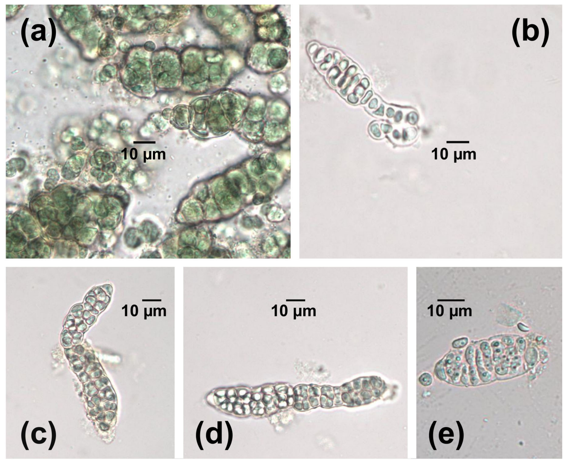
All studied strains exhibited significant morphological changes when exposed to 3 mg/L copper compared to control conditions, including reduced cell volume and partial or complete pigment loss. In Microthamnion sp. (Figure 1a,b), surviving cells displayed protoplast-wall separation, with cellular remnants surrounding the damaged structures. Scenedesmus sp. (Figure 1c,d) showed particularly severe copper-induced damage, likely attributable to combined osmotic stress and cellular dehydration [37].
Figure 1. Morphology of microalgae strains cultivated under high copper concentrations: (a) Microthamnion sp., control sample; (b) Microthamnion sp. exposed to 3 mg/L Cu2+ after one month of cultivation; (c) Scenedesmus sp., control sample; (d) Scenedesmus sp. exposed to 3 mg/L Cu2+ after one-month exposure. Scale bar = 10 μm.
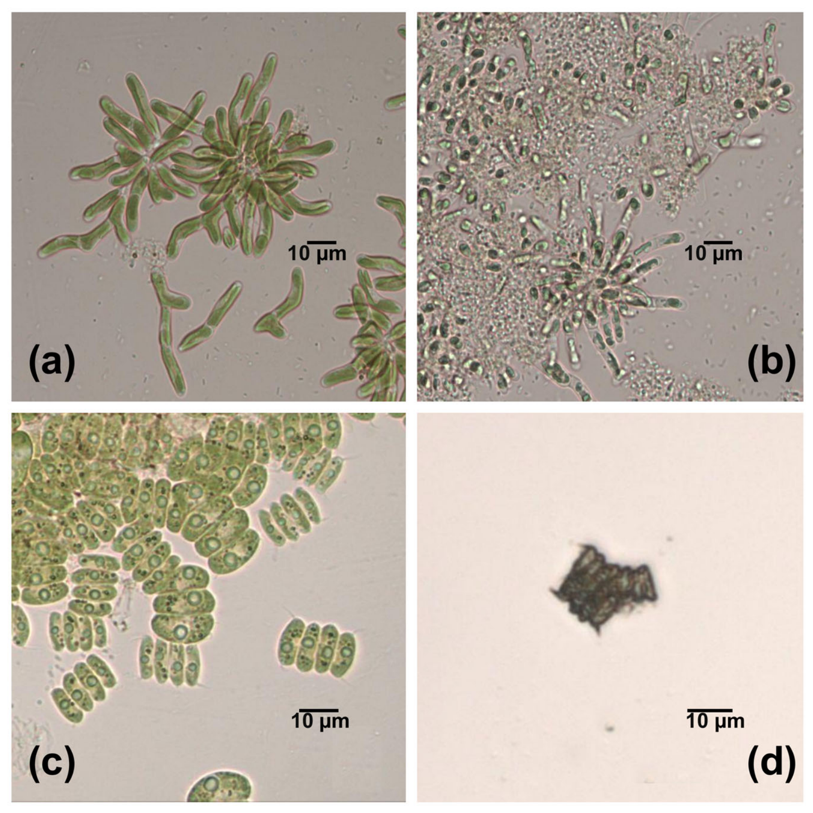
Cyanobacterial strains exhibited comparable sensitivity to copper exposure. The strain Atlanticothrix sp. KPABG-154446 (Figure 2) demonstrated progressive cellular degradation across all tested copper concentrations. While the overall colony morphology was maintained due to the protective mucilaginous sheath, microscopic analysis revealed substantial cellular damage with progressive loss of structural integrity. Partial staining of cellular contents was noted at 15 mg/L.
Figure 2. Morphology of cyanobacteria Atlanticothrix sp. KPABG-154446 strain cultivated under high copper concentrations: (a) control (no copper treatment), (be) copper treatment at concentrations of 3, 6, 9, and 15 mg/L, respectively. Scale bar = 10 μm.
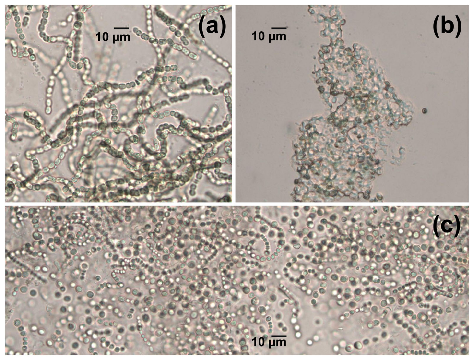
The strain Atlanticothrix sp. KPABG-154445 (Figure 3) demonstrated distinct morphological responses to different copper concentrations. Exposure to 3 mg/L Cu2+ induced the formation of disorganized cell clusters, while at 15 mg/L Cu2+, the cells-maintained dimensions comparable to the control and retained their filamentous thallus structure.
Figure 3. Morphology of Atlanticothrix sp. KPABG-154445 strain: (a) control (no copper treatment); (b) 3 mg/L Cu2+ concentration; (c) 15 mg/L Cu2+ concentration. Scale bar = 10 μm.
These observations informed the design of subsequent experiments, which examined lower copper concentrations (0.5–2 mg/L) and included a broader range of cyanobacterial strains (Table 2).
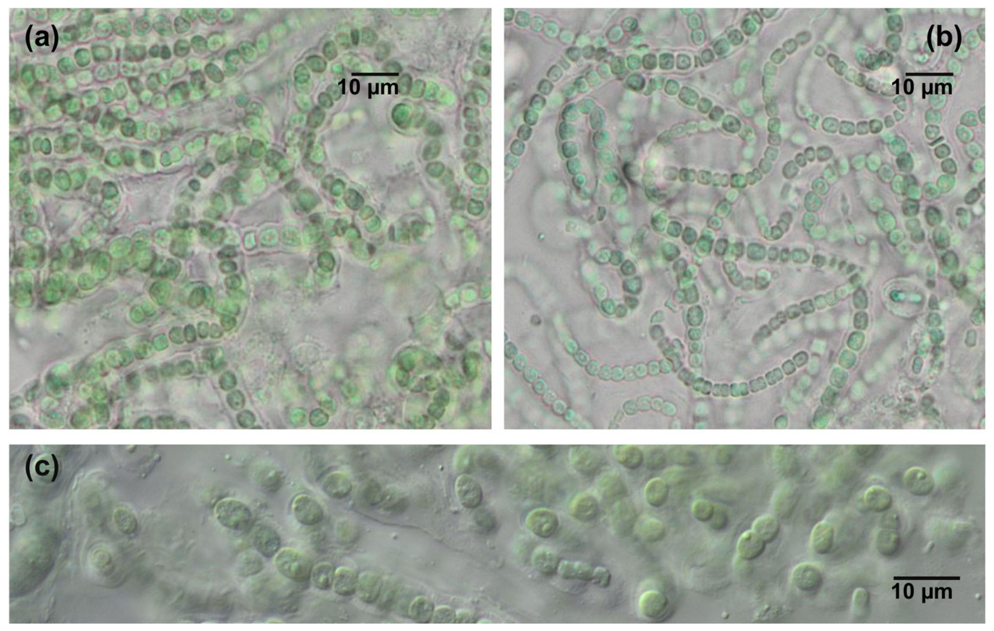
Several strains of Nostoc-like morphotypes (KPABG-4176, KPABG-610010, KPABG-610016, KPABG-154445, and KPABG-154446) exhibited limited biomass accumulation at copper concentrations of 1–2 mg/L. However, all strains displayed evident stress symptoms by the experimental endpoint and failed to resume growth upon transfer to copper-free medium. Among the tested strains, Atlanticothrix sp. KPABG-154445, isolated from fumarolic environments of Tolbachik Volcano (Kamchatka), demonstrated positive growth at 2 mg/L Cu2+. Growth resumption within the medium was observed three months post-experiment (Figure 4). The strain maintained under initial conditions for 10 months exhibited persistent pigmentation (Figure 4b).
Figure 4. Morphology of the copper-adapted strain Atlanticothrix sp. KPABG-154445: (a) culture after 1 month of growth in medium containing 2 mg/L copper; (b) culture after 10 months of growth in medium with 2 mg/L copper; (c) control: culture after 1 month in BG-11 medium without copper supplementation. Scale bar = 10 μm.
Microscopic examination of Atlanticothrix sp. KPABG-154445 revealed that cell integrity was maintained under copper exposure. At 2 mg/L Cu2+ (Figure 4), the strain exhibited morphological adaptations primarily characterized by reduced cell size, along with moderately decreased proliferative capacity, while viability was sustained.
The strain required three months to attain visible growth throughout the entire medium volume, whereas the control culture achieved a comparable density within one month under identical conditions.

3.2. Copper Removal Efficiency by Atlanticothrix sp. KPABG-154445 Strain

To determine the strain’s sensitivity, its viability was evaluated on media containing copper concentrations ranging from 0.01 to 2 mg/L over one month. Following this cultivation period, residual copper content in the solution was measured, enabling quantitative assessment of the strain’s sorption capacity.
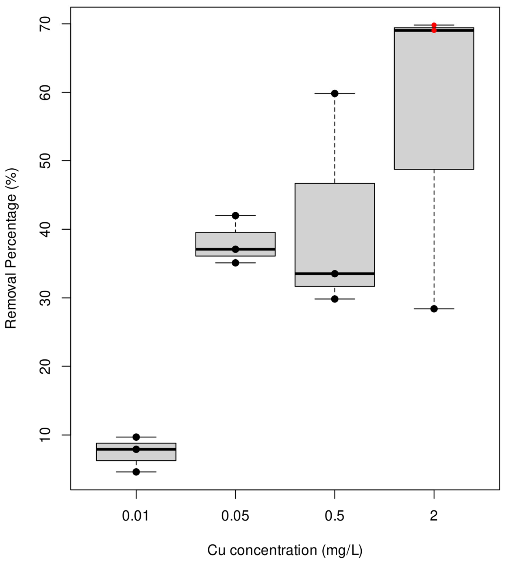
Notably, at lower concentrations (0.01–0.5 mg/L), the culture demonstrated normal growth comparable to control samples. However, at the maximum tested concentration (2 mg/L), growth was observed in only one of three replicates, indicating the strain’s tolerance threshold. This allowed comparison of copper removal efficiency between viable and non-viable biomass at 2 mg/L copper (Figure 5).
Figure 5. Comparative copper removal efficiency by biomass of Atlanticothrix sp. KPABG-154445 strain from Z8 medium. Values represent mean percentage of Cu2+ removed after one-month exposure (n = 3). Red notches indicate samples with dead biomass.
Maximum copper sorption (68 ± 1.6%) occurred in samples containing 2 mg/L where no culture growth was observed, while actively growing cultures at lower concentrations (0.01–0.5 mg/L) showed significantly lower metal removal rates (7.3–40% respectively).
Given its demonstrated stress tolerance, the Atlanticothrix sp. KPABG-154445 strain was selected for subsequent experiments involving nepheline sludge revegetation.

3.3. Formation of Fouling on Nepheline Sand

One of the promising methods for stabilizing mobile fine-grained nepheline sludge involves the formation of biological soil crusts (biocrusts) using cyanobacteria. Capable of active surface growth, cyanobacteria stabilize the substrate through exopolysaccharide production that binds sand particles, thereby increasing resistance to wind erosion.
In this study, we evaluated the Atlanticothrix sp. KPABG-154445 strain for addressing two interconnected objectives: (1) testing its capacity for surface colonization of nepheline slag to prevent dust formation, and (2) assessing biocrust resistance to chemical pollutants characteristic of industrial waste deposits. Copper sulfate (10 mg/L Cu2+ equivalent) served as a model contaminant.
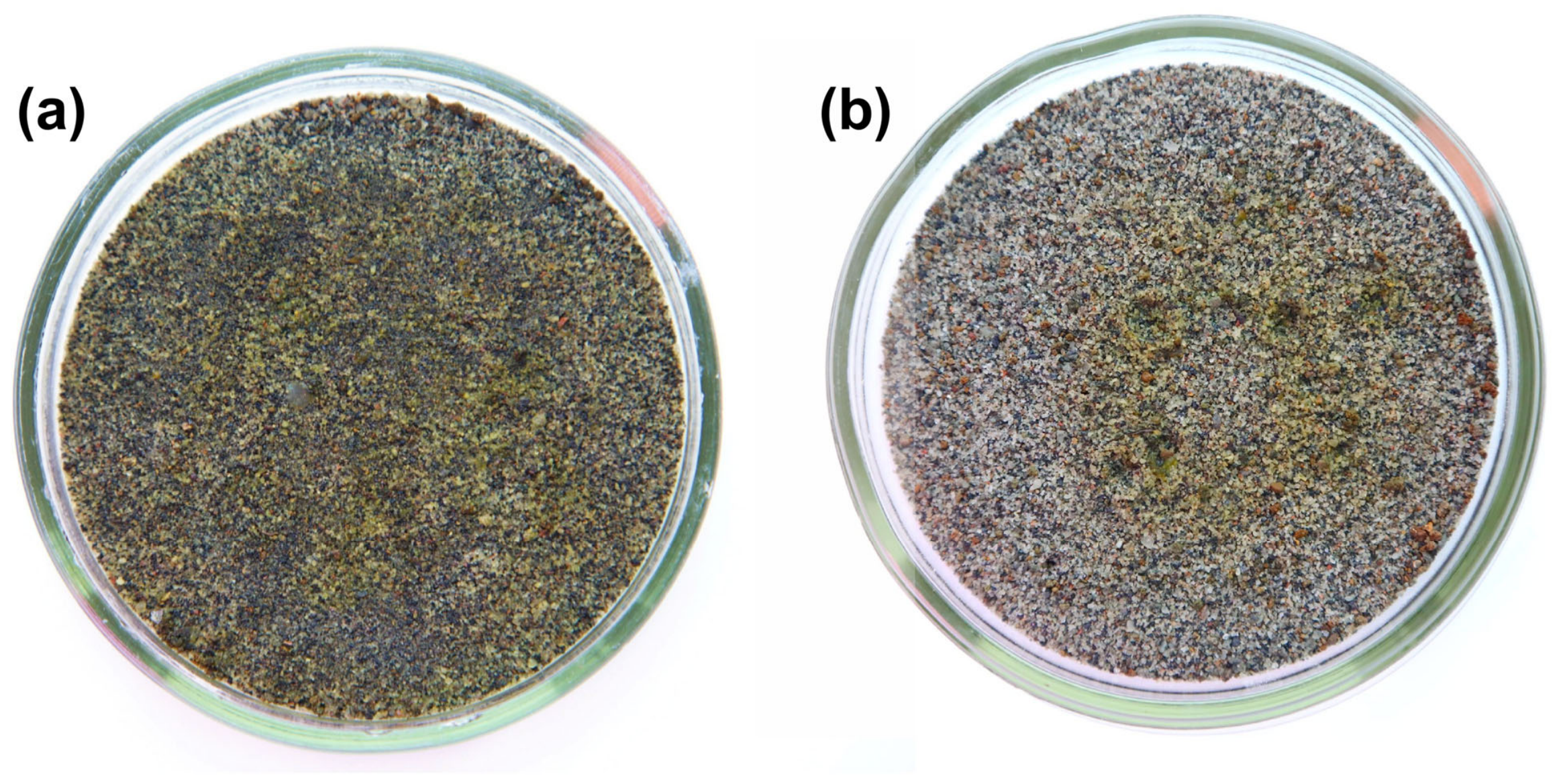
The Atlanticothrix sp. KPABG-154445 strain demonstrated substantial colonization potential on nepheline sand substrates. Under humid conditions, active biomass growth over one month resulted in coverage of up to 42% of the dish surface containing copper-contaminated sand, as determined by image analysis (Figure 6). Cyanobacterial colonies developed in all replicates, with coverage metrics indicating successful adaptation to the nepheline sand’s specific chemical composition (Table 3). Notably, copper sulfate supplementation showed no statistically significant growth inhibition. Conversely, sustained biomass production confirmed the strain’s tolerance to this contamination level.
Figure 6. Atlanticothrix sp. KPABG-154445 crust on the nepheline sand bed materials: (a) 10 mg/kg Cu2+ concentration; (b) 0 mg/kg Cu2+ concentration.
Table 3. The percent of square of Atlanticothrix sp. KPABG-154445 crust on the nepheline sand bed materials at different Cu2+ concentrations.

4. Discussion

4.1. Summary of Key Findings

Copper was chosen as the model toxicant because it is generally regarded as more toxic to cyanobacteria than nickel, and therefore likely constitutes the primary limiting factor for microbial growth in polymetallic-contaminated sites [38,39,40]. Although metal mixtures may occasionally exert synergistic or antagonistic effects [41,42], screening against the most restrictive metal offers a conservative foundation for evaluating strain tolerance.
The selection of copper ion concentrations for strain testing was based on established regulatory standards. According to Kuznetsov et al. [43], the maximum permissible copper concentration in soils, accounting for various toxicity indicators, is 3.0 mg/kg. The World Health Organization (WHO) has been studying HM impact on human health on regular basis based on which WHO have already ascertained toxicity threshold limits for copper in 2.0 mg/L and the U.S. Environmental Protection Agency in 1.3 mg/L [27]. However, field measurements in the study area have revealed significantly higher copper levels: up to 29.87 mg/kg in soil samples [6] and up to 1.688 mg/L in percolation water [12]. In the present study, we examined copper concentrations ranging from 0.5 to 15 mg/L to encompass both environmentally relevant and elevated exposure scenarios.
Copper (Cu) is an essential micronutrient for microalgae and cyanobacteria, where it functions as a cofactor in key proteins involved in photosynthesis and antioxidant defense [44,45]. However, at elevated concentrations, Cu becomes toxic, exhibiting algicidal effects by disrupting metal homeostasis and inducing oxidative stress via the production of reactive oxygen species (ROS) [46]. Due to the considerable morphological and physiological diversity within the Cyanobacteria and green algae, the thresholds for Cu deficiency or toxicity vary considerably among strains. For instance, the reproduction rates of most cyanobacteria are reduced at cupric ion activities above 10−12 M (approximately 6 × 10⁻⁵ mg/L), whereas most eukaryotic algae maintain maximum reproduction rates at 10−11 M (approximately 6 × 10⁻⁴ mg/L). For unicellular cyanobacteria, concentrations of 0.032–0.064 mg/L represent the threshold for metabolic impairment [47]. Studies have demonstrated that exposure within this range induces pronounced oxidative stress and disrupts electron transport in photosystem II. Approximately 0.635 mg/L Cu2+ constitutes a critical threshold for filamentous and nitrogen-fixing cyanobacteria (e.g., Nostoc muscorum). While filamentous forms are generally more resistant due to their mucilaginous sheaths, concentrations at this level have been shown to decrease chlorophyll-a content by ~38%, degrade phycobiliproteins, and suppress nitrogenase activity [48].
These two microbial groups were selected due to their contrasting adaptive strategies to heavy metal exposure. Green algae have evolved specialized systems for intracellular Cu storage, efficiently sequestering the metal in vacuoles, mitochondria, and chloroplasts [49,50]. In contrast, cyanobacterial metabolism demonstrates particular sensitivity to membrane integrity, which serves as a primary target for reactive oxygen species [51]. These organisms also differ in their exudation of metal-complexing ligands, where cyanobacteria uniquely produce strong siderophores like hydroxamates and schizokinen primarily for iron acquisition but which also complex other toxic metals, whereas green algae typically excrete weaker organic acids and ligands, with siderophore production being less common or emphasized [52]. In the cyanobacterium Synechococcus sp., rapid cellular uptake of heavy metals has been observed, with significant Cu absorption occurring within the first minute of exposure, followed by slower uptake over the next 15–20 min [24,53]. This kinetic pattern reflects the involvement of two distinct mechanisms: a rapid initial phase, attributed to passive biosorption onto the cell surface, and a slower subsequent phase, corresponding to active intracellular accumulation. Although we did not directly measure these processes, this biphasic behavior is consistent with the general model of metal interaction with microbial cells [54]. Additionally, Synechococcus sp. can efflux internalized Cu, likely to maintain homeostasis and counteract its toxicity [24].
Cell growth, being intrinsically linked to metabolic activity, is commonly employed to assess heavy metal toxicity as it integrates multiple essential processes like photosynthesis, respiration, and nutrient uptake [55]. The observed growth inhibition at concentrations exceeding 3 mg/L Cu2+ aligns with established literature findings. For Desmodesmus sp., exposure to 2 mg/L Cu2+ at pH 4 significantly affected photosynthetic pigments, while a tenfold concentration increase (20 mg/L) caused markedly pronounced inhibition [56]. Exposure of Scenedesmus sp. cultures to copper solutions at concentrations of 0.16 and 0.64 mg/L for 48 h resulted in concentration-dependent growth inhibition, which persisted without recovery during the 12 h period after transfer to the original copper-free medium [55]. The freshwater green alga Monoraphidium arcuatum exhibited even greater sensitivity, demonstrating 50% growth inhibition at 0.0011 mg/L Cu2+ [40]. The toxic effects of metals principally arise from their affinity for protein sulfhydryl groups, which can lead to either structural denaturation or the replacement of vital cofactors [57].
Overall, the documented sensitivity to copper supports the use of copper sulfate at concentrations of 0.25–2 mg/L as an algaecide for controlling algal growth in water bodies [58]. These concentrations effectively suppress phytoplankton development. However, higher doses—despite their pronounced algicidal effect—are not a viable solution due to the substantial ecological risks they pose to aquatic ecosystems.
Microalgal stress indicators can manifest in diverse forms, including alterations in cell morphology [59]. The morphological response appears non-specific to particular heavy metals due to shared toxicity mechanisms. For instance, light microscopy can reveal plasmolysis as well as changes in cell dimensions—both increases and decreases in volume [37]. These effects occur because sublethal copper concentrations induce ROS formation, which damages membranes and organelles, ultimately disrupting osmotic balance [60].
Initial screening results revealed low microalgal tolerance—growth was completely inhibited at copper concentrations as low as 3 mg/L, significantly limiting their bioremediation potential for heavily contaminated sites. These findings reinforce the understanding that copper tolerance is strain-specific and governed by unique molecular mechanisms, highlighting the necessity for comprehensive testing of each individual population [61,62].
While microalgal copper absorption capacity is typically assessed through short-term aqueous experiments [63], such approaches often fail to account for long-term microbial adaptation processes. In this study, we evaluated the copper removal capacity of Atlanticothrix sp. KPABG-154445 through prolonged stress exposure experiments.
In this study, the Atlanticothrix sp. KPABG-154445 strain was subjected to repeated stress conditions. The Atlanticothrix sp. KPABG-154445 strain, maintained for 10 months in a copper-supplemented medium (2 mg/L Cu2+), formed morphologically typical colonies with preserved pigmentation, showing no visible differences from copper-free controls. The persistent pigmentation reflects three key physiological characteristics: structurally intact thylakoid membranes, operational chlorophyll biosynthesis pathways, and maintained cellular compartmentalization—collectively indicating robust cellular homeostasis during extended cultivation. Short-term toxicity tests, as commonly used in metal ecotoxicology [61,62], may underestimate the adaptive potential of tolerant strains. While certain cyanobacterial species have demonstrated tolerance to copper concentrations up to 12 μM in experimental studies [63], the isolated Atlanticothrix sp. KPABG-154445 strain exhibits markedly superior survival capacity at these elevated metal levels.
Tripathi et al. [64] reported that metal-treated algal assemblages showed progressive recovery following transfer to metal-free medium, with initial signs of recovery observed within 5–7 days. Nevertheless, the extent of recovery was substantially constrained: Cu-treated cultures recovered to only 52% of the control biovolume. This differential recovery highlights the persistent toxicity of copper and corroborates our observation that the majority of tested strains failed to resume growth after transfer to copper-free medium following extended exposure. In marked contrast to the limited recovery documented by Tripathi et al. [64] for Cu-exposed communities, our Atlanticothrix sp. KPABG-154445 strain demonstrated remarkable recovery capacity, resuming growth in copper-free medium three months after Cu-exposure. These contrasting observations suggest that although community-level recovery from copper stress is typically slow and incomplete, select cyanobacterial strains possess exceptional resilience mechanisms that warrant further investigation.
The copper tolerance of Atlanticothrix sp. KPABG-154445 may be attributed to its origin. The Tolbachik Volcano region features extreme climatic conditions and naturally high copper concentrations in bedrock. Studies have demonstrated that microbial communities within biofilms in this area actively participate in biomineralization processes, extracting and accumulating calcium and heavy metals (Cu, Pb) from basaltic rocks [65]. Specifically, calcium and copper oxalates detected in lichen crusts provide evidence of these organisms’ adaptive capacity to metal-rich environments. Atlanticothrix sp. KPABG-154445 likely evolved its tolerance mechanisms through prolonged adaptation to such extreme conditions. It is also likely that the culture of this strain experienced the described effect, where initial exposure to copper not only has a direct toxic effect but also significantly alters the copper tolerance of cyanobacteria upon repeated exposure [66].
Evaluation of copper concentration reduction in Atlanticothrix sp. KPABG-154445 cultivation experiments suggests two complementary hypotheses: (1) Dead biomass exhibits superior copper ion biosorption capacity compared to living cells. (2) Viable cells may regulate environmental metal levels through active efflux mechanisms, representing an adaptive strategy of Atlanticothrix sp. KPABG-154445 to high copper contamination.
Metal efflux has been previously characterized as a potential resistance mechanism. Pandey et al. [67] described a Cu2+-tolerant Nostoc calcicola BREB strain capable of growth at copper concentrations 8-fold higher than the lethal dose for wild-type strains (5 μM). Remarkably, this resistant strain exhibited 22% lower Cu2+ uptake compared to copper-sensitive cells. The efflux activity was light-dependent, requiring photosynthetic ATP generation, and significantly decreased when maintained in darkness [67].
The molecular basis of such systems was elucidated by D.H. Nies, who identified heavy metal efflux transporters in prokaryotes [68]. Comparative genomic analysis revealed Nostoc sp. possesses 8 RND proteins, 3 CDF transporters, and 13 additional transport system proteins—substantially more than Synechocystis sp. (6 RND, 1 CDF, and 9 others, respectively).
RND and CDF proteins constitute bacterial transmembrane transporters that mediate extrusion of diverse substrates, including heavy metals and cations. These systems are crucial for maintaining metal homeostasis and conferring resistance.
Nevertheless, cyanobacteria of the Nostoc-like morphotypes show significant potential as bioremediation agents. Live biomass of Nostoc linckia was employed in three 12-day treatment cycles for polymetallic contamination removal, achieving over 80% copper elimination efficiency in the Cu-Fe system by the third cycle [69]. Similarly, Nostoc muscorum isolated from a mining site removed 80% of copper within 30 h of cultivation at concentrations of 5–10 mg/L Cu2+ [70].
A comparative analysis of cyanobacterial adsorption capacities reveals substantial interspecies variation. Reported values range from 8.73 to 12.62 mg/g for various Anabaena species [71] to considerably higher levels in other genera. Nostoc PCC 7936 and Aulosira fertilissima showed intermediate capacities of 79 mg/g [72] and 21.77 mg/g [73], respectively. The highest adsorption was observed in Cyanospira capsulata—at 143 mg/g [74]—and Spirulina platensis, which exhibited the maximum capacity at 272.1 mg/g [75].
Our studied Atlanticothrix sp. KPABG-154445 strain demonstrated considerable copper removal capacity from aqueous solutions, consistent with previous findings. However, practical application for remediation of technogenic landscapes (tailings ponds, waste dumps) requires further investigation of Nostoc’s metal accumulation capacity under terrestrial growth conditions.
The growth capacity of Atlanticothrix sp. KPABG-154445 strain on nepheline slag surfaces was tested to evaluate its potential for dust suppression in mineral processing tailings. In the Murmansk region, wind-driven dispersion of fine particulate matter from mining and processing waste poses significant environmental hazards. These airborne particles contaminate soils, water bodies, and atmospheric air across considerable distances, leading to ecosystem degradation, agricultural land deterioration, and adverse health effects for nearby urban populations [76].
Concurrently, we assessed the cyanobacterial crust’s resistance to copper contamination typical of industrial waste piles. As copper represents a characteristic pollutant from metallurgical operations, its presence in disturbed soils exacerbates wind erosion vulnerability. This dual approach enables comprehensive evaluation of both the efficacy and long-term stability of the proposed biological reclamation method under conditions mimicking actual industrial waste deposits.
The Atlanticothrix sp. KPABG-154445 strain demonstrated successful colonization of slag substrates at copper contamination levels of 10 mg/L, indicating significantly higher soil tolerance compared to liquid culture experiments. These findings highlight the importance of determining copper inhibition thresholds in complex nepheline slag systems. This represents a promising avenue for future research.
Terrestrial ecosystems serve as a reservoir of resistant strains; therefore, anthropogenic habitats are particularly relevant targets for bioprospecting. Furthermore, cyanobacteria already occupy a niche in such environments, where they contribute to substrate stabilization and toxicity reduction through metal absorption. This observation is supported by literature describing cyanobacterial community formation under elevated heavy metal concentrations.
In mesocosm experiments with the cyanobacterium Leptolyngbya sp. in an arsenic-contaminated area, recorded as accumulation in the biocrust up to 269.57 mg/kg after 80 days of cultivation [77]. A detailed investigation in South Sardinia (Italy) explored the composition of cyanobacterial populations in biocrusts from mining zones [78]. The authors identified 56 cyanobacterial morphotypes, with representatives of the genus Nostoc found at all study sites, including areas with high levels of copper contamination (89–303 mg/kg). The experimental data demonstrating the ability of isolated strains to remediate copper from aqueous solutions (up to 89%) are particularly noteworthy. Together, these findings support further research into the diversity and adaptation mechanisms of cyanobacteria under technogenic pollution.
The biomass of the inoculum used in this study (0.4 mg dry weight per 90 mm diameter plate) is substantially lower than that reported in comparable studies. For example, Chamizo et al. [79] and Román et al. [80] used 40 mg dry weight per plate and demonstrated that Nostoc commune covered up to 60% of the sand surface under periodic moistening Similarly, Nostoc flagelliforme growing on sand bed materials gradually expanded and covered the entire plate surface within 60 days [81]. By day 100, a dense crust up to 2 mm in height had formed [81].
Environments characterized by chronic copper stress represent a reservoir of strains with potential biotechnological applications. In this study, the Nostoc-like strain colonized nepheline sludge, covering 42% of the substrate within one month, demonstrating its potential for restoring degraded soils in technogenic landscapes (e.g., tailings and waste dumps). Biocrust formation alters nutrient profiles and water transport in substrates, influencing plant growth [82], making Nostoc-based biocrusts a promising tool for remediating disturbed ecosystems, including desertification control.

4.2. Research Limitations and Future Research Directions

This study has a few key limitations. We deliberately aimed for a simple, straightforward protocol suitable for routine screening of cyanobacterial strains for copper tolerance. Since the primary objective was to identify a survival threshold and select promising copper-resistant candidates, rather than to calculate precise median inhibitory concentrations (IC50) over a short exposure period, the quantitative dataset obtained in this study remains limited.
It should also be noted that the non-viable biomass used in the sorption experiments was not obtained through a dedicated pretreatment protocol, but rather resulted from the failure of the selected strain to survive at 2 mg/L Cu2+ during the second round of exposure. This concentration appears to represent a borderline toxicity threshold for the tested strain, which explains why we refer to positive growth rather than full tolerance at this level.
Our laboratory results demonstrate that cyanobacterial growth on nepheline sand can be sustained with distilled water only, suggesting that the substrate itself provides sufficient mineral nutrients for initial colonization and biofilm formation. However, it remains an open question whether a single inoculation would be sufficient to establish a self-sustaining biocrust, or whether repeated applications would be necessary to compensate for initial cell mortality and environmental stress. Given the harsh climatic conditions of the Arctic, including freeze–thaw cycles, high winds, and low temperatures, we anticipate that one or more supplementary inoculations may be required to ensure successful establishment.

5. Conclusions

This study provides a screening of microalgae and cyanobacterial strains for copper tolerance and evaluates their potential for bioremediation of Arctic technogenic substrates. Among the nine strains tested, only the cyanobacterium Atlanticothrix sp. KPABG-154445, isolated from the copper-enriched fumarolic fields of Tolbachik Volcano (Kamchatka Peninsula), demonstrated positive growth at 2 mg/L Cu2+, with sustained viability and recovery of metabolic activity after extended exposure. The strain also exhibited long-term adaptation, maintaining pigmentation and morphological integrity after 10 months in copper-supplemented medium, underscoring its resilience to metal stress.
Sorption experiments revealed that non-viable biomass of this strain achieved up to 68% copper removal at 2 mg/L, significantly outperforming actively growing cultures. This finding suggests that dead or pretreated biomass may serve as an effective biosorbent. In microcosm experiments using copper-spiked nepheline sand, the strain successfully colonized the nutrient-poor substrate and formed biocrusts covering up to 100% (42% in average) of the surface within one month, even at copper concentrations of 10 mg/kg, with no statistically significant growth inhibition. This dual capacity—heavy metal tolerance combined with biocrust formation—positions Atlanticothrix sp. KPABG-154445 as a promising candidate for the initial stages of bioremediation of industrial barrens in the Arctic.
However, several challenges remain. The transition from laboratory-scale microcosms to full-scale field application requires further investigation, particularly regarding the long-term survival and self-sustaining capacity of biocrusts under harsh Arctic conditions, including freeze–thaw cycles, high winds, and UV radiation. Future studies should also quantify biomass-specific metal uptake (mg/g) to enable direct comparison with other strains, assess tolerance to nickel and other co-occurring metals in polymetallic mixtures, and evaluate the performance of the strain in real-world field trials.
Our findings highlight the potential of cyanobacteria, particularly strains adapted to extreme metal-rich environments, as versatile tools for bioremediation and soil restoration in contaminated Arctic and subarctic regions. The combination of metal tolerance, biosorption capacity, and biocrust-forming ability makes Atlanticothrix sp. KPABG-154445 a valuable resource for developing nature-based solutions to mitigate the environmental impact of industrial activities in the Russian Arctic.

Author Contributions

Conceptualization, J.N. and D.D.; methodology, J.N. and D.D.; software, J.N.; validation, D.D.; formal analysis, J.N. and D.D.; investigation, J.N. and D.D.; resources, D.D.; data curation, D.D.; writing—original draft preparation, J.N. and D.D.; writing—review and editing, D.D.; visualization, J.N.; supervision, D.D.; project administration, D.D.; funding acquisition, D.D. All authors have read and agreed to the published version of the manuscript.

Funding

This research was funded by the Russian Science Foundation, grant number 25-14-20011, https://rscf.ru/project/25-14-20011/, accessed on 30 June 2026.

Institutional Review Board Statement

Not applicable.

Data Availability Statement

The data presented in this study are openly available in “L.” information system (https://isling.org/cyano, accessed on 30 June 2026.

Conflicts of Interest

The authors declare no conflicts of interest.

Abbreviations

The following abbreviations are used in this manuscript:
HMHeavy metal
KPABGPolar-Alpine Botanical Garden-Institute culture collection
WHOWorld Health Organization
ROSReactive oxygen species

References

  1. Franchitto, N.; Gandia-Mailly, P.; Georges, B.; Galinier, A.; Telmon, N.; Ducassé, J.L.; Rougé, D. Acute Copper Sulphate Poisoning: A Case Report and Literature Review. Resuscitation 2008, 78, 92–96. [Google Scholar] [CrossRef] [PubMed]
  2. Dumontet, S.; Dinel, H.; Levesque, P. The Distribution of Pollutant Heavy Metals and Their Effect on Soil Respiration and Acid Phosphatase Activity in Mineral Soils of the Rouyn-Noranda Region, Québec1. Sci. Total Environ. 1992, 121, 231–245. [Google Scholar] [CrossRef]
  3. Freedman, B.; Hutchinson, T.C. Pollutant inputs from the atmosphere and accumulations in soils and vegetation near a nickel–copper smelter at Sudbury, Ontario, Canada. Can. J. Bot. 1980, 58, 108–132. [Google Scholar] [CrossRef]
  4. Rebele, F.; Surma, A.; Kuznik, C.; Bornkamm, R.; Brej, T. Heavy Metal Contamination of Spontaneous Vegetation and Soil around the Copper Smelter “Legnica”. Acta Soc. Bot. Pol. 2014, 62, 53–57. [Google Scholar] [CrossRef]
  5. Chernenkova, T.W.; Butusov, O.B.; Sytchev, W.W.; Koneva, G.G.; Kabirov, R.R.; Stepanov, A.M.; Kuperman, R.G.; Kataev, G.D. Forest Ecosystems of Kola Peninsula Under Atmospheric Pollution Influence of Smelters; Russian Academy of Sciences: St. Petersburg, Russia, 1995. [Google Scholar]
  6. Dvornikov, Y.; Slukovskaya, M.; Gurinov, A.; Vasenev, V. Soil Type and Content of Macro-Elements Determine Hotspots of Cu and Ni Accumulation in Soils of Subarctic Industrial Barren: Inference from a Cascade Machine Learning. Environ. Pollut. 2025, 377, 126457. [Google Scholar] [CrossRef] [PubMed]
  7. Evdokimova, G.A.; Kalabin, G.V.; Mozgova, N.P. Contents and Toxicity of Heavy Metals in Soils of the Zone Affected by Aerial Emissions from the Severonikel Enterprise. Eurasian Soil Sci. 2011, 44, 237–244. [Google Scholar] [CrossRef]
  8. Hagner, O.; Rigina, O. Detection of Forest Decline in Monchegorsk Area. Remote Sens. Environ. 1998, 63, 11–23. [Google Scholar] [CrossRef]
  9. Kashulina, G.; De Caritat, P.; Reimann, C. Snow and Rain Chemistry around the “Severonikel” Industrial Complex, NW Russia: Current Status and Retrospective Analysis. Atmos. Environ. 2014, 89, 672–682. [Google Scholar] [CrossRef]
  10. Kozlov, M.V.; Haukioja, E.; Bakhtiarov, A.V.; Stroganov, D.N.; Zimina, S.N. Root versus Canopy Uptake of Heavy Metals by Birch in an Industrially Polluted Area: Contrasting Behaviour of Nickel and Copper. Environ. Pollut. 2000, 107, 413–420. [Google Scholar] [CrossRef] [PubMed]
  11. Kozlov, M.V.; Haukioja, E.; Yarmishko, V.T. Aerial Pollution in Kola Peninsula. In Proceedings of the International Workshop, St. Petersburg, Russia, 14–16 April 1992; p. 417. [Google Scholar]
  12. Lindroos, A.; Derome, J.; Nikonov, V.; Niska, K. Influence of Sulphur and Heavy Metal Emissions from Monchegorsk, Northwest Russia, on Percolation Water Quality in Pinus sylvestris Stands. Scand. J. For. Res. 1996, 11, 97–103. [Google Scholar] [CrossRef]
  13. Lyanguzova, I.V.; Goldvirt, D.K.; Fadeeva, I.K. Spatiotemporal Dynamics of the Pollution of Al–Fe-Humus Podzols in the Impact Zone of a Nonferrous Metallurgical Plant. Eurasian Soil Sci. 2016, 49, 1189–1203. [Google Scholar] [CrossRef]
  14. Reimann, C.; Ayras, M.; Chekushin, V.; Bogatyrev, I.; Boyd, R.; de Caritat, P.; Dutter, R.; Finne, T.E.; Halleraker, J.H.; Jaeger, O.; et al. Environmental Geochemical Atlas of the Central Barents Region; Geological Survey of Norway: Trondheim, Norway, 1998. [Google Scholar]
  15. Koptsik, G.N.; Koptsik, S.V.; Smirnova, I.E. Alternative Remediation Technologies for Technogenic Wastelands in the Kola Subarctic. Eurasian Soil Sci. 2016, 49, 1375–1391. [Google Scholar] [CrossRef]
  16. Kalabin, G.; Evdokimova, G. Ecology and Nature Protection of the Kola North, Educational Textbook; Kola Scientific Center: Apatity, Russia, 1993. [Google Scholar]
  17. Kozlov, M.V.; Zvereva, E.L. Industrial Barrens: Extreme Habitats Created by Non-Ferrous Metallurgy. Rev. Environ. Sci. Bio/Technol. 2007, 6, 231–259. [Google Scholar] [CrossRef]
  18. Slukovskaya, M.V.; Ivanova, L.A.; Kremenetskaya, I.P.; Gorbacheva, T.T.; Drogobuzhskaya, S.V.; Lashchuk, V.V.; Markovskaya, E.F. Rehabilitation of Industrial Barren in Arctic Region Using Mining Wastes. Open Ecol. J. 2018, 11, TOERJ-11-1. [Google Scholar] [CrossRef]
  19. Rocha, F.; Esteban Lucas-Borja, M.; Pereira, P.; Muñoz-Rojas, M. Cyanobacteria as a Nature-Based Biotechnological Tool for Restoring Salt-Affected Soils. Agronomy 2020, 10, 1321. [Google Scholar] [CrossRef]
  20. Davydov, D.A. Cyanoprokaryota and Their Role in the Process of Nitrogen Fixation in Terrestrial Ecosystems of the Murmansk Region; Geos: Moscow, Russia, 2010. [Google Scholar]
  21. Davydov, D.; Vilnet, A. Cyanobacterial Assemblages Inhabiting the Apatity Thermal Power Plant Fly Ash Dumps in the Russian Arctic. Microorganisms 2025, 13, 1762. [Google Scholar] [CrossRef] [PubMed]
  22. Davydov, D.A.; Redkina, V.V. Algae and Cyanoprokaryotes on Naturally Overgrowing Ash Dumps of the Apatity Thermal Power Station (Murmansk Region). Proc. Karelian Res. Cent. Russ. Acad. Sci. 2021, 51–68. [Google Scholar] [CrossRef]
  23. Avery, S.V.; Codd, G.A.; Gadd, G.M. Caesium Accumulation and Interactions with Other Monovalent Cations in the Cyanobacterium Synechocystis PCC 6803. Microbiology 1991, 137, 405–413. [Google Scholar] [CrossRef]
  24. Croot, P.L.; Bengt, K.; Van Elteren, J.T.; Kroon, J.J. Uptake and Efflux of 64Cu by the Marine Cyanobacterium Synechococcus (WH7803). Limnol. Oceanogr. 2003, 48, 179–188. [Google Scholar] [CrossRef]
  25. Ghorbani, E.; Nowruzi, B.; Nezhadali, M.; Hekmat, A. Metal Removal Capability of Two Cyanobacterial Species in Autotrophic and Mixotrophic Mode of Nutrition. BMC Microbiol. 2022, 22, 58. [Google Scholar] [CrossRef] [PubMed]
  26. Jensen, T.E.; Baxter, M.; Rachlin, J.W.; Jani, V. Uptake of Heavy Metals by Plectonema boryanum (Cyanophyceae) into Cellular Components, Especially Polyphosphate Bodies: An X-Ray Energy Dispersive Study. Environ. Pollut. Ser. A Ecol. Biol. 1982, 27, 119–127. [Google Scholar] [CrossRef]
  27. Kalita, N.; Baruah, P.P. Cyanobacteria as a Potent Platform for Heavy Metals Biosorption: Uptake, Responses and Removal Mechanisms. J. Hazard. Mater. Adv. 2023, 11, 100349. [Google Scholar] [CrossRef]
  28. Mehta, S.K.; Gaur, J.P. Use of Algae for Removing Heavy Metal Ions From Wastewater: Progress and Prospects. Crit. Rev. Biotechnol. 2005, 25, 113–152. [Google Scholar] [CrossRef] [PubMed]
  29. Rai, P.K.; Tripathi, B.D. Removal of Heavy Metals by the Nuisance Cyanobacteria Microcystis in Continuous Cultures: An Eco-Sustainable Technology. Environ. Sci. 2007, 4, 53–59. [Google Scholar] [CrossRef]
  30. Singh, J.S.; Singh, D.P.; Dixit, S. Cynobacteria: An Agent of Heavy Metal Removal. In Bioremediation of Pollutants; IK International Publisher: New Delhi, India, 2011; pp. 223–243. [Google Scholar]
  31. Verma, S.K.; Singh, S.P. Factors Regulating Copper Uptake in a Cyanobacterium. Curr. Microbiol. 1990, 21, 33–37. [Google Scholar] [CrossRef]
  32. Whitton, B.A.; Gale, N.L.; Wixson, B.G. Chemistry and Plant Ecology of Zinc-Rich Wastes Dominated by Blue-Green Algae. Hydrobiologia 1981, 83, 331–341. [Google Scholar] [CrossRef]
  33. Whitton, B.A.; Shehata, F.H.A. Influence of Cobalt, Nickel, Copper and Cadmium on the Blue-Green Alga Anacystis nidulans. Environ. Pollut. Ser. A Ecol. Biol. 1982, 27, 275–281. [Google Scholar] [CrossRef]
  34. Kotai, J. Instructions for Preparation of Modified Nutrient Solution Z8 for Algae. Nor. Inst. Water Res. 1972, 11, 1–5. [Google Scholar] [CrossRef]
  35. Solovchenko, A.; Lukyanov, A.; Vasilieva, S.; Lobakova, E. Chlorophyll Fluorescence as a Valuable Multitool for Microalgal Biotechnology. Biophys. Rev. 2022, 14, 973–983. [Google Scholar] [CrossRef] [PubMed]
  36. Brylyakov, Y.E.; Vasileva, N.Y.; Petrovskiy, A.A. Prospects for the Integrated Use of Apatite-Nepheline Ores. Gorn. Zhurnal 1999, 9, 42–45. [Google Scholar]
  37. Wang, H.; Sathasivam, R.; Ki, J.-S. Physiological Effects of Copper on the Freshwater Alga Closterium ehrenbergii Meneghini (Conjugatophyceae) and Its Potential Use in Toxicity Assessments. ALGAE 2017, 32, 131–137. [Google Scholar] [CrossRef]
  38. Debelius, B.; Forja, J.M.; Lubián, L.M. Toxicity of Copper, Nickel and Zinc to Synechococcus Populations from the Strait of Gibraltar. J. Mar. Syst. 2011, 88, 113–119. [Google Scholar] [CrossRef]
  39. Filová, A.; Fargašová, A.; Molnárová, M. Cu, Ni, and Zn Effects on Basic Physiological and Stress Parameters of Raphidocelis subcapitata Algae. Environ. Sci. Pollut. Res. 2021, 28, 58426–58441. [Google Scholar] [CrossRef] [PubMed]
  40. McKnight, K.S.; Gissi, F.; Adams, M.S.; Stone, S.; Jolley, D.; Stauber, J. The Effects of Nickel and Copper on Tropical Marine and Freshwater Microalgae Using Single and Multispecies Tests. Environ. Toxicol. Chem. 2023, 42, 901–913. [Google Scholar] [CrossRef] [PubMed]
  41. Gao, C.; Gao, L.; Duan, P.; Wu, H.; Li, M. Evaluating Combined Toxicity of Binary Heavy Metals to the Cyanobacterium Microcystis: A Theoretical Non-Linear Combined Toxicity Assessment Method. Ecotoxicol. Environ. Saf. 2020, 187, 109809. [Google Scholar] [CrossRef] [PubMed]
  42. Nawaz, A.; Šotek, P.E.; Molnárová, M. Reciprocal Effects of Metal Mixtures on Phytoplankton. Phycology 2024, 4, 117–138. [Google Scholar] [CrossRef]
  43. Kuznetsov, A.M.; Fesyun, A.P.; Samokhvalov, S.G.; Makhonka, E.P. Metodicheskiye Ukazaniya Po Opredeleniyu Tyazhelykh Metallov v Pochvakh Selskokhozyaystvennykh Ugodiy i Produktakh Rastenievodstva [Guidelines for Determining Heavy Metals in Soils of Agricultural Lands and Crop Products]; Tsentral’nyi Institut Agrokhimicheskogo Obsluzhivaniya Selskohozyaystva (TsINAO): Moscow, Russia, 1992. [Google Scholar]
  44. De la Cerda, B.; Castielli, O.; Durán, R.V.; Navarro, J.A.; Hervás, M.; De la Rosa, M.A. A Proteomic Approach to Iron and Copper Homeostasis in Cyanobacteria. Brief. Funct. Genom. Proteom. 2008, 6, 322–329. [Google Scholar] [CrossRef] [PubMed]
  45. Quigg, A. Micronutrients. In The Physiology of Microalgae; Borowitzka, M.A., Beardall, J., Raven, J.A., Eds.; Springer International Publishing: Cham, Switzerland, 2016; pp. 211–231. [Google Scholar]
  46. Arunakumara, K.K.I.U.; Zhang, X. Heavy Metal Bioaccumulation and Toxicity with Special Reference to Microalgae. J. Ocean Univ. China 2008, 7, 60–64. [Google Scholar] [CrossRef]
  47. Oder, J.C.; Sateles, T.E.; De Souza, L.B.; Nunes-Nesi, A.; Araújo, W.L.; Alvarenga-Lucius, L. Metabolic Responses, Uptake, and Export of Copper in Cyanobacteria. Biology 2025, 14, 798. [Google Scholar] [CrossRef] [PubMed]
  48. Jardim, W.F.; Pearson, H.W. Copper Toxicity to Cyanobacteria and Its Dependence on Extracellular Ligand Concentration and Degradation. Microb. Ecol. 1985, 11, 139–148. [Google Scholar] [CrossRef] [PubMed]
  49. Danouche, M.; El Ghachtouli, N.; El Baouchi, A.; El Arroussi, H. Heavy Metals Phycoremediation Using Tolerant Green Microalgae: Enzymatic and Non-Enzymatic Antioxidant Systems for the Management of Oxidative Stress. J. Environ. Chem. Eng. 2020, 8, 104460. [Google Scholar] [CrossRef]
  50. Monteiro, C.M.; Castro, P.M.L.; Malcata, F.X. Metal Uptake by Microalgae: Underlying Mechanisms and Practical Applications. Biotechnol. Prog. 2012, 28, 299–311. [Google Scholar] [CrossRef] [PubMed]
  51. Mikula, P.; Zezulka, S.; Jancula, D.; Marsalek, B. Metabolic Activity and Membrane Integrity Changes in Microcystis aeruginosa—New Findings on Hydrogen Peroxide Toxicity in Cyanobacteria. Eur. J. Phycol. 2012, 47, 195–206. [Google Scholar] [CrossRef]
  52. Gaur, J.P.; Rai, L.C. Heavy Metal Tolerance in Algae. In Algal Adaptation to Environmental Stresses; Rai, L.C., Gaur, J.P., Eds.; Springer: Berlin/Heidelberg, Germany, 2001; pp. 363–388. [Google Scholar]
  53. Quigg, A.; Reinfelder, J.R.; Fisher, N.S. Copper Uptake Kinetics in Diverse Marine Phytoplankton. Limnol. Oceanogr. 2006, 51, 893–899. [Google Scholar] [CrossRef]
  54. Danouche, M.; El Ghachtouli, N.; El Arroussi, H. Phycoremediation Mechanisms of Heavy Metals Using Living Green Microalgae: Physicochemical and Molecular Approaches for Enhancing Selectivity and Removal Capacity. Heliyon 2021, 7, e07609. [Google Scholar] [CrossRef] [PubMed]
  55. Tripathi, B.N.; Gaur, J.P. Physiological Behavior of Scenedesmus sp. during Exposure to Elevated Levels of Cu and Zn and after Withdrawal of Metal Stress. Protoplasma 2006, 229, 1–9. [Google Scholar] [CrossRef] [PubMed]
  56. Buayam, N.; Davey, M.P.; Smith, A.G.; Pumas, C. Effects of Copper and pH on the Growth and Physiology of Desmodesmus sp. AARLG074. Metabolites 2019, 9, 84. [Google Scholar] [CrossRef] [PubMed]
  57. Rocha, G.S.; Parrish, C.C.; Espíndola, E.L.G. Effects of Copper on Photosynthetic and Physiological Parameters of a Freshwater Microalga (Chlorophyceae). Algal Res. 2021, 54, 102223. [Google Scholar] [CrossRef]
  58. Ramadan, T. Algae Control Solves Aesthetic Problems. Opflow 2000, 26, 1–4. [Google Scholar] [CrossRef]
  59. Elisabeth, B.; Rayen, F.; Behnam, T. Microalgae Culture Quality Indicators: A Review. Crit. Rev. Biotechnol. 2021, 41, 457–473. [Google Scholar] [CrossRef] [PubMed]
  60. Zeeshan, M.; Murugadas, A.; Ghaskadbi, S.; Rajendran, R.B.; Akbarsha, M.A. ROS Dependent Copper Toxicity in Hydra -Biochemical and Molecular Study. Comp. Biochem. Physiol. Part C Toxicol. Pharmacol. 2016, 185–186, 1–12. [Google Scholar] [CrossRef] [PubMed]
  61. Cavalletti, E.; Romano, G.; Palma Esposito, F.; Barra, L.; Chiaiese, P.; Balzano, S.; Sardo, A. Copper Effect on Microalgae: Toxicity and Bioremediation Strategies. Toxics 2022, 10, 527. [Google Scholar] [CrossRef] [PubMed]
  62. Levy, J.L.; Stauber, J.L.; Jolley, D.F. Sensitivity of Marine Microalgae to Copper: The Effect of Biotic Factors on Copper Adsorption and Toxicity. Sci. Total Environ. 2007, 387, 141–154. [Google Scholar] [CrossRef] [PubMed]
  63. Wang, M.; Gui, H.; Chen, J.; Li, C.; Wang, C.; Chen, C.; Zhao, C.; Li, Y. Experimental Study on Removal of Iron, Manganese and Copper from Water by Microalgae. Pol. J. Environ. Stud. 2022, 31, 1847–1855. [Google Scholar] [CrossRef] [PubMed]
  64. Tripathi, B.N.; Singh, V.; Kim, B.H.; Gaur, J.P. Recovery of Freshwater Algal Assemblage from Copper and Zinc Toxicity in a Semi-Continuous Culture System. Ecotoxicology 2026, 35, 105. [Google Scholar] [CrossRef] [PubMed]
  65. Vereshchagin, O.S.; Frank-Kamenetskaya, O.V.; Vlasov, D.Y.; Zelenskaya, M.S.; Rodina, O.A.; Chernyshova, I.A.; Himelbrant, D.E.; Stepanchikova, I.S.; Britvin, S.N. Microbial Biomineralization under Extreme Conditions: Case Study of Basaltic Rocks, Tolbachik Volcano, Kamchatka, Russia. CATENA 2023, 226, 107048. [Google Scholar] [CrossRef]
  66. Shavyrina, O.B.; Gapochka, L.D.; Azovskii, A.I. Development of Tolerance for Copper in Cyanobacteria Repeatedly Exposed to Its Toxic Effect. Biol. Bull. Russ. Acad. Sci. 2001, 28, 183–187. [Google Scholar] [CrossRef]
  67. Pandey, P.K.; Singh, S.P.; Bisen, P.S. Cu2+ Efflux and Its Regulation in Cu-resistant (Cur) Cyanobacterium Nostoc calcicola BRÉB. J. Basic Microbiol. 1997, 37, 355–360. [Google Scholar] [CrossRef]
  68. Nies, D.H. Efflux-Mediated Heavy Metal Resistance in Prokaryotes. FEMS Microbiol. Rev. 2003, 27, 313–339. [Google Scholar] [CrossRef] [PubMed]
  69. Cepoi, L.; Zinicovscaia, I.; Valuta, A.; Codreanu, L.; Rudi, L.; Chiriac, T.; Yushin, N.; Grozdov, D.; Peshkova, A. Peculiarities of the Edaphic Cyanobacterium Nostoc linckia Culture Response and Heavy Metal Accumulation from Copper-Containing Multimetal Systems. Toxics 2022, 10, 113. [Google Scholar] [CrossRef] [PubMed]
  70. Hazarika, J.; Pakshirajan, K.; Sinharoy, A.; Syiem, M.B. Bioremoval of Cu(II), Zn(II), Pb(II) and Cd(II) by Nostoc muscorum Isolated from a Coal Mining Site. J. Appl. Phycol. 2015, 27, 1525–1534. [Google Scholar] [CrossRef]
  71. Tien, C.-J.; Sigee, D.C.; White, K.N. Copper Adsorption Kinetics of Cultured Algal Cells and Freshwater Phytoplankton with Emphasis on Cell Surface Characteristics. J. Appl. Phycol. 2005, 17, 379–389. [Google Scholar] [CrossRef]
  72. De Philippis, R.; Paperi, R.; Sili, C.; Vincenzini, M. Assessment of the Metal Removal Capability of Two Capsulated Cyanobacteria, Cyanospira capsulata and Nostoc PCC7936. J. Appl. Phycol. 2003, 15, 155–161. [Google Scholar] [CrossRef]
  73. Singh, A.; Mehta, S.K.; Gaur, J.P. Removal of Heavy Metals from Aqueous Solution by Common Freshwater Filamentous Algae. World J. Microbiol. Biotechnol. 2007, 23, 1115–1120. [Google Scholar] [CrossRef]
  74. Micheletti, E.; Colica, G.; Viti, C.; Tamagnini, P.; De Philippis, R. Selectivity in the Heavy Metal Removal by Exopolysaccharide-Producing Cyanobacteria. J. Appl. Microbiol. 2008, 105, 88–94. [Google Scholar] [CrossRef] [PubMed]
  75. Vannela, R.; Verma, S.K. Co2+, Cu2+, and Zn2+ Accumulation by Cyanobacterium Spirulina platensis. Biotechnol. Prog. 2006, 22, 1282–1293. [Google Scholar] [CrossRef] [PubMed]
  76. Eker, H. Investigation of the Usability of Industrial Mining Wastes in Agriculture. Front. Environ. Sci. 2023, 11, 1248188. [Google Scholar] [CrossRef]
  77. Mao, Q.; Xie, Z.; Pei, F.; Irshad, S.; Issaka, S.; Randrianarison, G. Indigenous Cyanobacteria Enhances Remediation of Arsenic-Contaminated Soils by Regulating Physicochemical Properties, Microbial Community Structure and Function in Soil Microenvironment. Sci. Total Environ. 2023, 860, 160543. [Google Scholar] [CrossRef] [PubMed]
  78. Pagli, C.; Chamizo, S.; Migliore, G.; Rugnini, L.; De Giudici, G.; Braglia, R.; Canini, A.; Cantón, Y. Isolation of Biocrust Cyanobacteria and Evaluation of Cu, Pb, and Zn Immobilisation Potential for Soil Restoration and Sustainable Agriculture. Sci. Total Environ. 2024, 946, 174020. [Google Scholar] [CrossRef] [PubMed]
  79. Chamizo, S.; Mugnai, G.; Rossi, F.; Certini, G.; De Philippis, R. Cyanobacteria Inoculation Improves Soil Stability and Fertility on Different Textured Soils: Gaining Insights for Applicability in Soil Restoration. Front. Environ. Sci. 2018, 6, 49. [Google Scholar] [CrossRef]
  80. Román, J.R.; Roncero-Ramos, B.; Rodríguez-Caballero, E.; Chamizo, S.; Cantón, Y. Effect of Water Availability on Induced Cyanobacterial Biocrust Development. CATENA 2021, 197, 104988. [Google Scholar] [CrossRef]
  81. Chen, X.; Jia, S.; Wang, Y.; Wang, N. Biological Crust of Nostoc flagelliforme (Cyanobacteria) on Sand Bed Materials. J. Appl. Phycol. 2011, 23, 67–71. [Google Scholar] [CrossRef]
  82. Rabiei, A.; Zomorodian, S.M.A.; O’Kelly, B.C. Reducing the Erodibility of Sandy Soils Engineered by Cyanobacteria Inoculation: A Laboratory Investigation. Sustainability 2023, 15, 3811. [Google Scholar] [CrossRef]
Disclaimer/Publisher’s Note: The statements, opinions and data contained in all publications are solely those of the individual author(s) and contributor(s) and not of MDPI and/or the editor(s). MDPI and/or the editor(s) disclaim responsibility for any injury to people or property resulting from any ideas, methods, instructions or products referred to in the content.

Article Metrics

Citations

Article Access Statistics

Multiple requests from the same IP address are counted as one view.