Article

A Temperature-Controlled Fluorescence Fingerprint for Identifying Pseudo-nitzschia hasleana in Harmful Algal Blooms

1
Institute of Automation and Control Processes, Far Eastern Branch, Russian Academy of Sciences, st. Radio 5, Vladivostok 690041, Russia
2
A.V. Zhirmunsky National Scientific Center of Marine Biology, Far Eastern Branch, Russian Academy of Sciences, st. Palchevskogo 17, Vladivostok 690041, Russia
3
Laboratory of Marine Microbiology, Institute of the World Ocean, Far Eastern Federal University, Ajax 10, Vladivostok 690922, Russia
*
Author to whom correspondence should be addressed.
Phycology 2025, 5(4), 52; https://doi.org/10.3390/phycology5040052
Submission received: 11 August 2025 / Revised: 23 September 2025 / Accepted: 24 September 2025 / Published: 1 October 2025
(This article belongs to the Collection Harmful Microalgae)

Abstract

Harmful algal blooms (HABs) caused by toxic species such as Pseudo-nitzschia hasleana pose significant risks to marine ecosystems and human health. This study investigates the effects of heating rate on the fluorescence temperature curves (FTCs) of P. hasleana and compares them with non-toxic species (Phaeodactylum tricornutum and Picochlorum maculatum) to design a reliable detection method. An increasing heating rate leads to a change in the temperature spectrum of the fluorescence of the studied algae and to increasing differences between them. During the study, the FTCs were measured in the temperature range of 20–80 °C and at heating rates of 1, 2, 3, and 6°/min. The results showed that P. hasleana exhibited a distinct local fluorescence maximum at 45–55 °C when heated at a rate of 3 °C/min or more, which was absent in non-toxic species. Additionally, rapid heating (6 °C/min) preserved fluorescent pigment–protein complexes, yielding four-fold higher fluorescence intensity at 70–80 °C compared to slower rates. There were no such changes for the microalgae P. maculatum and P. tricornutum. The results of this study make it possible to increase the efficiency of detecting hazardous microalgae using non-invasive optical monitoring methods. These findings demonstrate that controlled heating protocols can enhance the species-specific identification of toxic microalgae, offering a practical tool for early HAB detection.

1. Introduction

Analysis of the laser-induced fluorescence (LIF) spectra of microalgae is a widely used indirect method for assessing the state of marine ecosystems [1,2]. This method allows one to estimate the total volume of microalga biomass and dissolved organic matter, but it does not allow one to determine the species composition of the samples under study. The LIF spectra of microalgae are mainly determined by the fluorescence of chlorophyll-a, which is present in each alga and slightly depends on the pigment composition. Because of this, it is not possible to qualitatively distinguish microalgae by their fluorescence using only spectra. In our work [3,4,5], it was proposed to use the temperature dependence of the fluorescence spectra (FTS) of microalgae to determine the species, as well as the dependence of the maximum chlorophyll-a fluorescence intensity on temperature, called the fluorescence temperature curve (FTC). In addition, we can note that measuring LIF requires more complex and expensive equipment than measuring the FTC, which can be constructed based on the fluorescence signal measured using a photodiode, which is advantageous for continuous monitoring devices.
It is obvious that it is not possible to completely solve the problem of determining the species composition of microalgae in a seawater sample using an indirect method such as analysis of LIF temperature spectra [2]. At the same time, this is not required for a number of practical problems. It is known that the greatest problems for the ecology of water bodies are created by the seasonal increase in the concentration of certain types of microalgae, which is typical for a given water area. Among these species, microalgae stand out in particular, which have the ability to synthesize and secrete toxins [6,7,8], which are dangerous to humans and inhabitants of the water area. Such harmful algal blooms (HABs) often cause an ecological catastrophe in the aquatic ecosystem [7,9]. Among the toxins that can be produced during the HAB process are cytotoxins, neurotoxins, hepatotoxins, dermatoxins, etc. Since blooming occurs under certain combinations of environmental conditions [10,11], it is usually caused by a type of algae for which such conditions are optimal [12]. When monitoring harmful blooms, it is important to be able to detect potentially harmful microalgae in water at low concentrations before they cause disasters. This requires continuous monitoring or short intervals between measurements.
The use of FTS and FTC expands the capabilities of the LIF method, as it allows us to consider the relationship between the fluorescent signal and the processes of internal regulation and the mechanisms of adaptation to external conditions in microalgae. For example, the sensitivity and adaptability of microalgae to existence under different temperature conditions have created unique mechanisms that regulate physiological processes in them, both for individual groups of microalgae and, in some cases, for individual cultures [12,13,14]. Controlled temperature changes make it possible to directly influence these processes through the photosynthetic apparatus and the state of pigment–protein complexes, which is reflected in changes in fluorescence spectra [15,16]. It is important to note that the heating rate affects the time of exposure of pigment–protein complexes to temperature, during which they are modified or destroyed [16,17,18] depending on the mechanisms of the adaptation of specific microalgae to external conditions, which can be reflected in the form of an FTC.
Previous work [4] has shown that, for some cultures at a heating rate of 1 °C/min, a similarity in their FTCs is observed, sometimes even in the case when the cultures belong to different phyla of microalgae and have different organizations of pigment–protein complexes [19,20,21,22]. Similarity in the form of the FTC was found in the potentially toxic diatom Pseudo-nitzschia hasleana [23,24], whose blooming is observed in the seas of the Pacific basin [25,26], and non-toxic green Picochlorum maculatum and diatom Phaeodactylum tricornutum [27,28].
The diatom microalga P. hasleana produces domoic acid. This neurotoxin can accumulate in aquatic organisms, which leads to the poisoning and death of fish, birds, and marine mammals [29]. In humans, domoic acid causes severe poisoning known as amnestic shellfish poisoning (ASP) [30]. The toxin acts as a glutamate agonist and has excitotoxicity in the central nervous system of vertebrates and other organs rich in glutamate receptors [31]. Clinical manifestations in humans who have eaten shellfish containing domoic acid include gastrointestinal upset, confusion, disorientation, seizures, short-term memory loss, and, in the most severe cases, death.
The aim of this study is to investigate the effect of heating rate on the FTC of the toxic diatom P. hasleana to identify species differences from non-toxic microalgae, such as P. tricornutum and P. maculatum. In this work, we establish automated fluorescence-based methods for environmental monitoring, specifically to enhance the identification capability of these methods, which is important for solving the pressing problem of HAB detection.

2. Materials and Methods

The microalga Pseudo-nitzschia hasleana Lundholm, 2012 (MBRU_PH18) was cultured on f/2 medium, and Picochlorum maculatum (Butcher) Henley, Hironaka, Guillou, M.Buchheim, J.Buchheim, M.Fawley & K.Fawley, 2004 (MBRU _NM-86) and Phaeodactylum tricornutum Bohlin (MBRU_PT-85) were grown in Goldberg medium. Preparation of media was performed according to local standards [32]. Samples of cultures were provided by the collection of the Institute of Marine Biology FEB RAS (https://marbank.dvo.ru/index.php/en/, accessed on 11 August 2025). EasyFlask Nunclone Delta Thermo FS (USA, Waltham, MA, USA) flasks seeded with cultures were placed on a Binder climate controller (Tuttlingen, Germany) at a temperature of 16 °C, with illumination parameters of 3500 lux and periodic illumination of 12 h light/12 h dark. The number of cultures was monitored once every two days in a Goryaev chamber using the EVOS M5000 visualization system (Thermo Scientific, Waltham, MA, USA).
The microalga monocultures, pre-cooled to a temperature of 17 °C, were heated at a constant rate to 90 °C. For this purpose, the culture was placed in a transparent 10 × 10 mm quartz cuvette and installed in a temperature-controlled chamber. Heating was performed by means of a Peltier element through the bottom of the cuvette. To ensure a uniform temperature distribution over the sample volume, the water in the cuvette was stirred with a magnetic stirrer at a speed of 150 rpm. The fluorescence of the samples was measured using an orthogonal scheme. The laser radiation and the fluorescence signal of the sample were transmitted through a quartz optical fiber with a diameter of 600 μm and a numerical aperture of 0.22. Fluorescence excitation was performed using a Melles-Griot laser (445 nm, 500 mW; Carlsbad, CA, USA). The intensity of the Raman scattering of water at a wavelength of 525 nm was used as a reference signal for monitoring the stability of the laser radiation. The fluorescence spectra were measured using a Newton EM CCD camera in accumulation mode (10 spectra with an exposure of 0.2 s) with the camera cooled to −60 °C. Broadband fluorescence spectra were measured in the range of 500 to 760 nm. The range was set using a Shamrock 303i monochromator (focal length 303 mm, aperture F/4) with a 300 line/mm diffraction grating, which allows for achieving a spectral resolution of 0.16 nm. The temperature in the chamber was changed linearly (using the PID control) at a rate of 0.5 °C/min, 1 °C/min, 2 °C/min, 3 °C/min, or 6 °C/min. The sample temperature was measured using an immersion thermocouple probe (T-type); the probe was polled every 300 ms. Fluorescence spectra were measured in the sample temperature range of 20 to 80 °C, with a temperature change of 1 °C. Laser excitation was carried out continuously during the spectrum accumulation process.
The measured spectra were smoothed using the Savitzky–Golay filter with a window width of 15 frames and a polynomial degree equal to one. In each obtained spectrum, the search for the maximum intensity was performed in the wavelength range of 660–740 nm. Then, the dependence of the found intensity of the maximum fluorescence on temperature was constructed, which is the FTC. To compare the fluorescence of different samples, we normalized the FTC intensity in the studied temperature range to the maximum intensity value. The normalized fluorescence temperature curves (NFTCs) obtained were used in this work.

3. Results

3.1. Study of the Influence of Heating Rate on the Characteristics of the Fluorescent Signal of P. hasleana

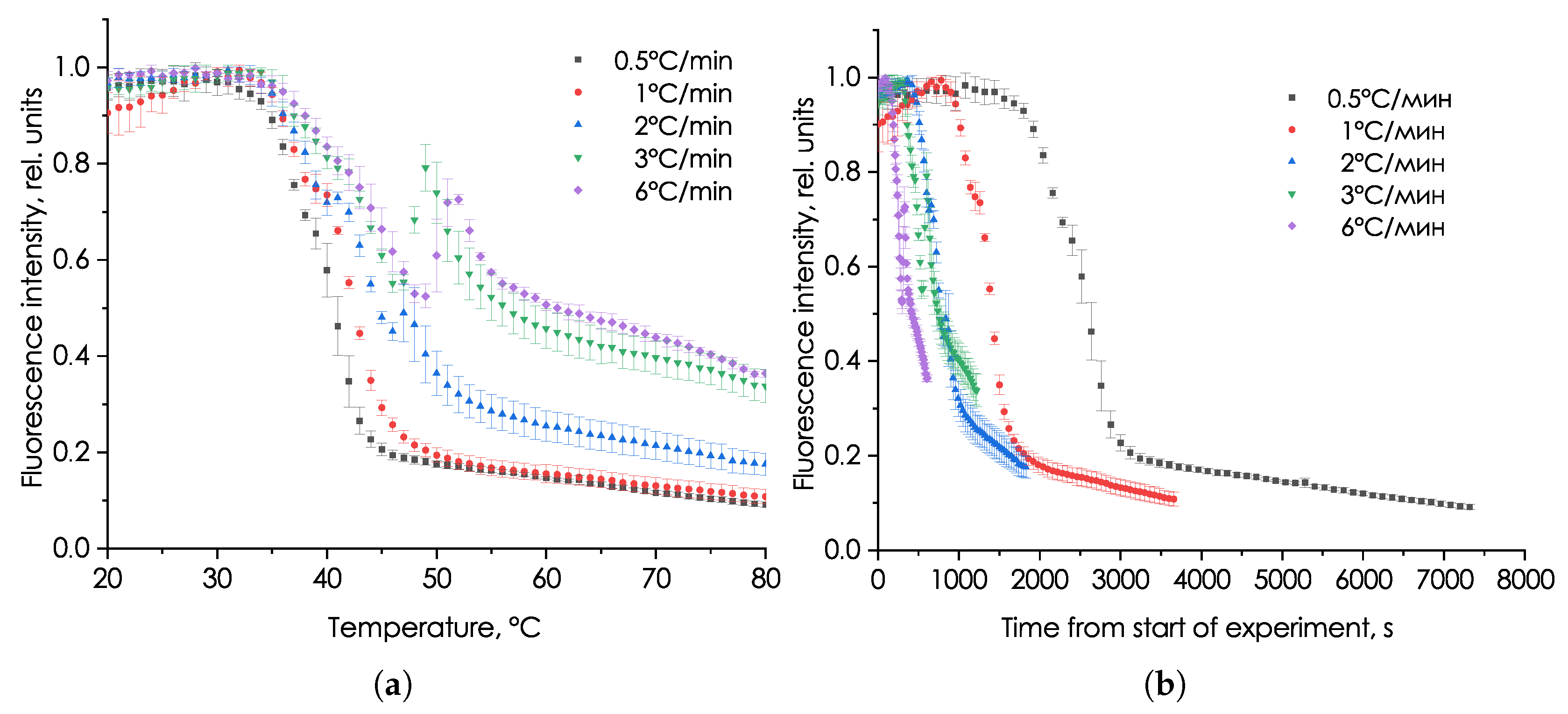
The results of the NFTC measurements of the P. hasleana during the linear heating of samples at different rates are presented in Figure 1.
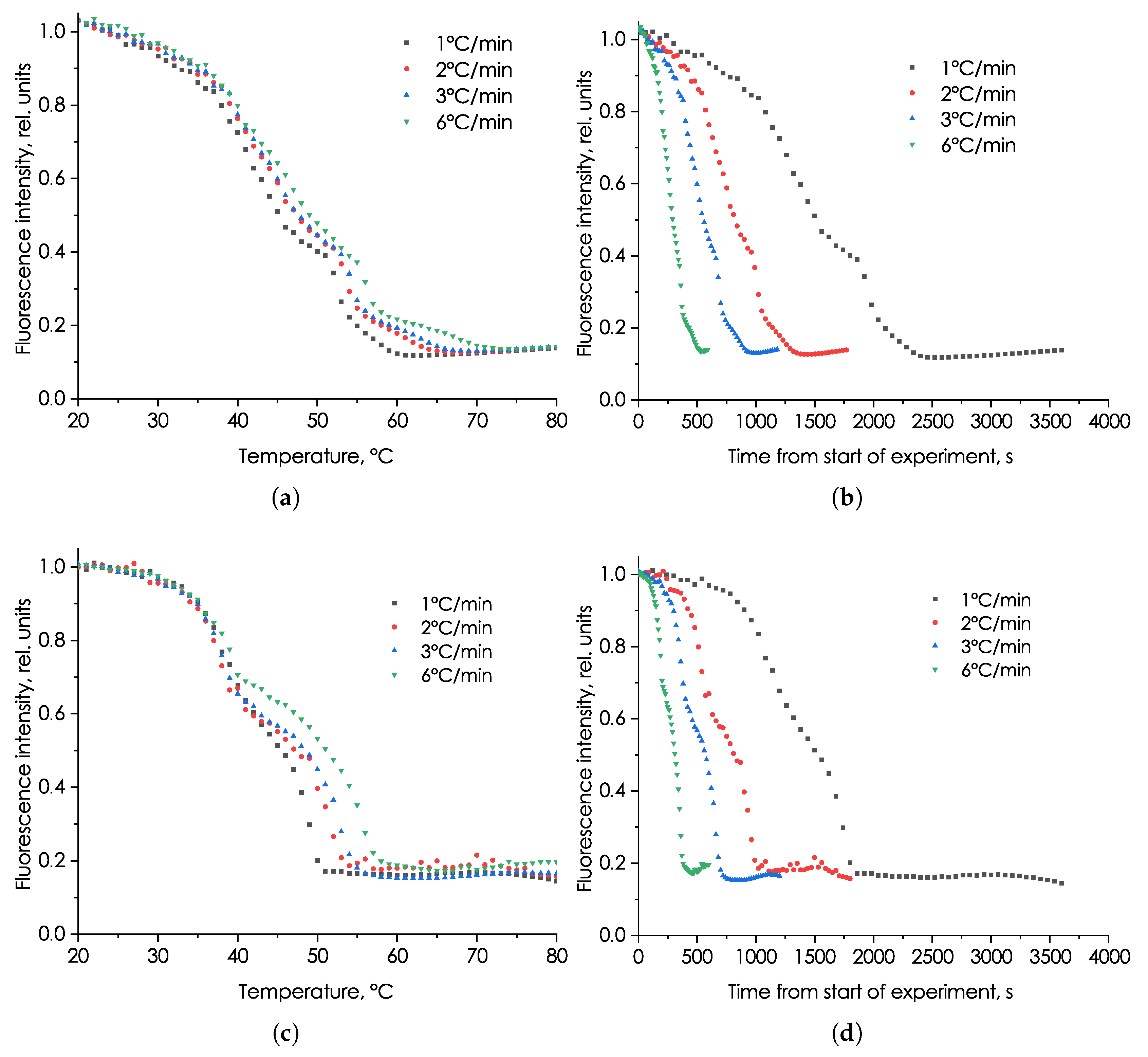
Figure 1a–e show the dependence of the NFTC shape on the heating rate of microalga cultures. For identical samples, a smooth character and small scatter of the NFTC shape are observed. This confirms that any changes that occur in the NFTC when the heating rate is changed are associated with the rate and are not due to the heterogeneity of the studied samples or the low stability of the fluorescence parameters during their storage. For ease of analysis, we will consider the averaged NFTC for each experiment on one graph in Figure 2.
In the initial section of the NFTC (Figure 2a) at temperatures up to 35 °C, the fluorescence intensity of the samples is independent of the heating rate. At this stage, microalgae are not subjected to strong stress, and their protective mechanisms can ensure the preservation of structural and biophysical parameters. A further increase in temperature leads to thermal damage to the microalgae, which is reflected in a decrease in the fluorescence intensity. At heating rates of 0.5 and 1 °C/min, a continuous decrease in fluorescence is observed without local peaks. At the same time, for the NFTC measured at heating rates of 2 and 3 °C/min, from a temperature of 46 °C, and for the NFTC measured at a heating rate of 6 °C/min, from a temperature of 48 °C, a pronounced local fluorescence peak is observed with further heating. That is, there is a clear dependence of the NFTC shape of this microalga on the heating rate, and there are characteristic threshold temperatures.
In Figure 1b, it is seen that the samples heated to 80 °C at a higher rate have a higher fluorescence intensity at high temperatures than the samples heated to this temperature at a lower rate. This allows us to assume that the destruction processes have significant inertia and depend not only on the temperature, but also on the time spent at this temperature. This assumption helps explain why, when heating at rates of 1 and 0.5 °C/min, no local peak in fluorescence intensity is observed in the NFTC. Thus, rapid heating (2–6 °C/min) allows a certain stage of fluorescence reduction to be passed in a short time and a certain amount of fluorescent pigment–protein complexes, which provide a local increase in fluorescence at high temperatures, to be preserved. With slow heating, no increase in fluorescence is observed as a result of the complete uniform destruction of all fluorescent complexes.
The experiments carried out showed the influence of the heating rate on the form of the NFTC of the microalga P. hasleana, which makes it possible in further studies to indirectly evaluate the internal processes occurring in the photosynthetic apparatus under the influence of temperature.

3.2. Study of the Effect of Heating Rate on the Fluorescent Signal Characteristics of Microalga Phaeodactylum tricornutum and Picochlorum maculatum

The dependence of the NFTC of the diatom microalga P. tricornutum and the green microalga P. maculatum on the heating parameters is shown in Figure 3.
These algae are characterized by a stable fluorescence maximum in the temperature range up to 35 °C. When they are heated above 35 °C, a uniform decrease in fluorescence intensity is observed, which ends at temperatures of 50 and 60 °C, respectively, for P. tricornutum and P. maculatum algae. Figure 3c,d show that the shape of the NFTC of the microalgae studied does not practically depend on the heating rate. The small dependence is mainly reflected in the change in the slope of the intensity decrease with a change in the heating rate. The most important result obtained when heating P. tricornutum and P. maculatum cultures at different rates is the fact that their NFTCs do not show local peaks at any heating rate.

4. Discussion

Our experiments have shown that the use of different heating rates allows one to change the degree of difference between the NFTC of the microalgae (Figure 4). The cardinal differences between the NFTC of P. hasleana and the others begin to appear at a heating rate of 3 °C/min and higher, which is primarily due to the presence of a local fluorescence peak and a significant excess of fluorescence intensity at high temperatures. In our opinion, this indicates that, in this microalga, at these heating rates, mechanisms are triggered that prevent the standard destruction of pigment–protein complexes.
As can be seen in Figure 4, for the value of the NFTC and, consequently, for the pigment composition within the three microalgae, there are two stable states regardless of the heating rate: (1) at initial temperatures, when all pigment–protein complexes are normal; (2) at high temperatures, when all pigment–protein complexes are destroyed. Between these states, there is a transition process which is characterized by a rapid change in the fluorescence intensity and is significantly dependent on the heating rate and time. This transition, as we believe, is associated with either a reduction in the number of fluorescence centers or a decrease in the quantum yield of the fluorescence in them. In our experiments, fluorescence is excited with closed reaction centers of photosynthesis.
Consequently, the change in fluorescence intensity for P. tricornutum, P. maculatum, and P. hasleana cultures, obtained by us in experiments, directly depends on the state of the light-harvesting complex and peripheral antennae and is not associated with energy transformations in reaction centers [33,34]. The appearance of a local fluorescence peak in the P. hasleana culture at heating rates of 3 °C/min in the region of 45 °C may be associated with the release of highly fluorescent chlorophyll-a [16]. At the same time, high heating rates do not allow microalgae to receive such a heat dose [18] that would be sufficient for complete “oxidation” of this chlorophyll-a during the experiment. At the same time, no additional peak in fluorescence intensity is observed at all for P. tricornutum and P. maculatum cultures, which means that there is no release of highly fluorescent chlorophyll-a during protein denaturation, or this release is completely compensated for by chlorophyll “oxidation” in decomposing cultures, which is due to the different biochemical states of the studied microalgae [20]. This confirms the need for further studies of the biochemistry of microalgae at different stages of heating performed at different rates.
Another important distinctive feature of the studied microalgae is the fluorescence intensity at high temperatures, namely the fluorescence level that is observed with completely destroyed peripheral antennae and the light-harvesting complex. For P. tricornutum and P. maculatum, the time to reach the minimum fluorescence level depends on the heating rate (Figure 3b,d). With rapid heating, these cultures reach the minimum fluorescence in less time but at a slightly higher temperature, which can be explained by the inertia of the destruction processes, which have their own speed. Unlike these two cultures, the P. hasleana does not reach the minimum value during the experiment with increasing heating rate. The fluorescence intensity of the rapidly heated culture at 80 °C is four times higher than the intensity of the slowly heated culture. This suggests that, due to rapid heating, the biochemical compounds in the cells of P. hasleana do not have time to completely break down before they have the opportunity to combine into more heat-resistant compounds. Thus, accelerated heating is manifested only in the NFTC culture of P. hasleana, which is apparently associated with its unique biochemical composition.
Having analyzed the NFTCs measured at different rates, shown in Figure 4, we can confidently conclude that changing the heating rate alters the degree of mutual similarity between the NFTCs. The degree of similarity can be estimated using the least-squares method, which is often used as a criterion for assessing the quality of approximation. The similarity between the curves is greater when the least-squares sum (LLS) is minimal:
LLS = n ( A n B n ) 2 min
where A n is the intensity at the NFTC of the A-th microalga, corresponding to the n-th temperature; B n is the intensity at the NFTC of the B-th microalga, corresponding to the n-th temperature.
Calculating the degree of mutual similarity revealed that the maximum similarity between the NFTCs for the P. hasleana and P. tricornutum pair (LSS = 0.23) and the P. hasleana and P. maculatum pair (LSS = 0.26) is observed at a heating rate of 2 °C/min. Increasing the heating rate to 3 and 6 °C/min reduces the similarity and increases the LSS to 2.38 for the P. hasleana and P. tricornutum pair and to 2.54 for the P. hasleana and P. maculatum pair. There are no significant differences in LSS for heating rates of 3 and 6 °C/min. At a heating rate of 1 °C/min, the LSS for the P. hasleana and P. tricornutum pair is 0.89, and for the P. hasleana and P. maculatum pair, it is 0.64. This once again confirms the possibility of increasing the difference between the NFTCs with a change in rate. Using a heating rate of 6 °C/min will increase the difference between the NFTCs by almost 10 times compared to the minimum and will significantly reduce the time spent on measurements.

5. Conclusions

This study demonstrates that the fluorescence response of Pseudo-nitzschia hasleana to controlled heating provides a reliable biomarker for distinguishing this potential toxic alga from non-toxic species. The heating rate is an important parameter for inducing species-specific fluorescence signatures such as an FTC. The appearance of a local fluorescence maximum (45–55 °C) at heating rates of 3 °C/min and higher serves as a unique identifier of P. hasleana, which is absent in P. tricornutum and P. maculatum. Thermal stress resistance in P. hasleana manifests through the increase in fluorescent intensity (at 80 °C) under rapid heating (6 °C/min), suggesting adaptive biochemical mechanisms not present in non-toxic counterparts. It was also noted that, at the initial stages of heating (up to 35 °C), the NFTCs of all three microalgae differ little and are practically independent of the heating rate, from which it can be concluded that temperatures up to 35 °C are not critical for these microalgae and did not affect their internal state during the experiment.
Our results contribute to the development of environmental monitoring by proposing a way to improve existing microalga fluorescence analysis instruments. This method, as demonstrated in the article, has the potential to be developed and improve identification quality. Thus, whereas fluorescence meters previously only allowed for estimating the chlorophyll-a concentration in water, the proposed method will improve the accuracy of this concentration determination and enable the identification of dominant species. These improvements are undoubtedly important for HAB monitoring. The NFTC measurement technique is easily transferred to existing meters, including automated and autonomous systems. Therefore, our measurement method can be used, for example, on buoy stations, enabling 24/7 monitoring and the creation of early HAB warning systems, supporting microalga and environmental research. The ability to increase the NFTC measurement speed will reduce the time between measurements and, if necessary, significantly improve the temporal resolution of monitoring. Furthermore, as our work demonstrates, this speed will increase the differences between the signals of P. hasleana, P. tricornutum, and P. maculatum, improving the quality of the identification method. Our fluorescence studies reveal differences between microalgae that require deeper study at the biochemical level, posing new challenges for the scientific community.

Author Contributions

Conceptualization, A.P.; methodology, A.P.; validation, T.D. and T.O.; formal analysis, A.L.; investigation, A.P., T.D., and A.L.; resources, T.D. and T.O.; data curation, S.V. and A.L.; writing—original draft preparation, A.P.; writing—review and editing, S.V. and T.D.; supervision, S.V. and T.O.; funding acquisition, S.V. and T.O. All authors have read and agreed to the published version of the manuscript.

Funding

This work was supported by the Russian Science Foundation within the framework of the program “Optical methods for detecting the birth and propagation of harmful microalgae blooms and for identifying toxic microalgae species in marine areas” [grant number 23-77-00004]. The work on the cultivation and investigation of the microalgae was supported by the Russian Federal Service for Hydrometeorology and Environmental Monitoring [grant number 169-15-2023-002].

Institutional Review Board Statement

Not applicable.

Informed Consent Statement

Not applicable.

Data Availability Statement

The data presented in this study are available on request from the corresponding author.

Acknowledgments

The team of authors expresses great gratitude to the staff of the Marine Biobank (https://marbank.dvo.ru/index.php, accessed on 11 August 2025) for providing samples of microalgae for conducting studies of their fluorescent properties.

Conflicts of Interest

The authors declare no conflicts of interest. The funders had no role in the design of the study; in the collection, analyses, or interpretation of data; in the writing of the manuscript; or in the decision to publish the results.

References

  1. Taniguchi, M.; Lindsey, J.S. Absorption and Fluorescence Spectral Database of Chlorophylls and Analogues. Photochem. Photobiol. 2021, 97, 136–165. [Google Scholar] [CrossRef]
  2. Sá, M.; Ferrer Ledo, N.; Gao, F.; Bertinetto, C.; Jansen, J.; Crespo, J.; Wijffels, R.; Barbosa, M.J.; Galinha, C. Perspectives of fluorescence spectroscopy for online monitoring in microalgae industry. Microb. Biotechnol. 2022, 15, 1824–1838. [Google Scholar] [CrossRef]
  3. Voznesenskiy, S.S.; Gamayunov, E.L.; Popik, A.Y.; Markina, Z.V.; Orlova, T.Y. Temperature dependence of the parameters of laser-induced fluorescence and species composition of phytoplankton: The theory and the experiments. Algal Res. 2019, 44, 101719. [Google Scholar] [CrossRef]
  4. Popik, A.Y.; Gamayunov, E.L.; Voznesenskiy, S.S.; Markina, Z.M.; Orlova, T.Y. The study of fluorescence features of microalgae from the genus Pseudo-nitzschia and the possibility of their detection in water. Algal Res. 2022, 64, 102662. [Google Scholar] [CrossRef]
  5. Popik, A.; Voznesenskiy, S.; Dunkai, T.; Gamayunov, E.; Orlova, T.; Leonov, A.; Zinov, A. Methodology of Measuring and Using Fluorescent Parameters of Microalgae. Bull. Russ. Acad. Sci. Phys. 2024, 88, S399–S407. [Google Scholar] [CrossRef]
  6. Santi Delia, A.; Caruso, G.; Melcarne, L.; Caruso, G.; Parisi, S.; Laganà, P. Biological Toxins from Marine and Freshwater Microalgae. In Microbial Toxins and Related Contamination in the Food Industry; Caruso, G., Caruso, G., Laganà, P.L., Santi Delia, A., Parisi, S., Barone, C., Melcarne, L., Mazzù, F., Eds.; Springer International Publishing: Cham, Switzerland, 2015; pp. 13–55. [Google Scholar] [CrossRef]
  7. Blossom, H.E.; Markussen, B.; Daugbjerg, N.; Krock, B.; Norlin, A.; Hansen, P.J. The cost of toxicity in microalgae: Direct evidence from the dinoflagellate Alexandrium. Front. Microbiol. 2019, 10, 1065. [Google Scholar] [CrossRef]
  8. Wang, S.Y.; Bi, W.H.; Li, X.Y.; Zhang, B.J.; Fu, G.W.; Jin, W.; Jiang, T.J.; Zhao, J.; Shi, W.J.; Zhang, Y.F. A detection method of typical toxic mixed red tide algae in Qinhuangdao based on three-dimensional fluorescence spectroscopy. Spectrochim. Acta. Part A Mol. Biomol. Spectrosc. 2023, 298, 122704. [Google Scholar] [CrossRef]
  9. Al-Hussieny, A.A. Algae Toxins and Their Treatment. In Progress in Microalgae Research; Zepka, L.Q., Jacob-Lopes, E., Deprá, M.C., Eds.; IntechOpen: Rijeka, Croatia, 2022; Chapter 6; pp. 1–16. [Google Scholar] [CrossRef]
  10. Anderson, D.M.; Cembella, A.D.; Hallegraeff, G.M. Progress in understanding harmful algal blooms: Paradigm shifts and new technologies for research, monitoring, and management. Annu. Rev. Mar. Sci. 2012, 4, 143–176. [Google Scholar] [CrossRef]
  11. Oyeku, O.G.; Mandal, S.K. Historical occurrences of marine microalgal blooms in Indian peninsula: Probable causes and implications. Oceanologia 2021, 63, 51–70. [Google Scholar] [CrossRef]
  12. Kholssi, R.; Lougraimzi, H.; Moreno-Garrido, I. Effects of global environmental change on microalgal photosynthesis, growth and their distribution. Mar. Environ. Res. 2023, 184, 105877. [Google Scholar] [CrossRef]
  13. Teoh, M.L.; Chu, W.L.; Phang, S.M. Effect of temperature change on physiology and biochemistry of algae: A review. Malays. J. Sci. 2010, 29, 82–97. [Google Scholar] [CrossRef]
  14. Singh, S.; Singh, P. Effect of temperature and light on the growth of algae species: A review. Renew. Sustain. Energy Rev. 2015, 50, 431–444. [Google Scholar] [CrossRef]
  15. Kouřil, R.; Ilík, P.; Tomek, P.; Nauš, J.; Poulíčková, A. Chlorophyll fluorescence temperature curve on Klebsormidium flaccidum cultivated at different temperature regimes. J. Plant Physiol. 2001, 158, 1131–1136. [Google Scholar] [CrossRef]
  16. Lípová, L.; Krchňák, P.; Komenda, J.; Ilík, P. Heat-induced disassembly and degradation of chlorophyll-containing protein complexes in vivo. Biochim. Et Biophys. Acta-Bioenerg. 2010, 1797, 63–70. [Google Scholar] [CrossRef] [PubMed]
  17. Kochubey, S.M.; Shevchenko, V.V.; Kazantsev, T.A. Changes of the antenna of photosystem i induced by short-term heating. Biochem. (Moscow) Suppl. Ser. A Membr. Cell Biol. 2013, 7, 67–77. [Google Scholar] [CrossRef]
  18. Ladjimi, M.T.; Labavić, D.; Guilbert, M.; Anquez, F.; Pruvost, A.; Courtade, E.; Pfeuty, B.; Thommen, Q. Dynamical thermal dose models and dose time-profile effects. Int. J. Hyperth. 2019, 36, 721–729. [Google Scholar] [CrossRef]
  19. Sturm, S.; Engelken, J.; Gruber, A.; Vugrinec, S.; G Kroth, P.; Adamska, I.; Lavaud, J. A novel type of light-harvesting antenna protein of red algal origin in algae with secondary plastids. BMC Evol. Biol. 2013, 13, 159. [Google Scholar] [CrossRef]
  20. Engelken, J.; Brinkmann, H.; Adamska, I. Taxonomic distribution and origins of the extended LHC (light-harvesting complex) antenna protein superfamily. BMC Evol. Biol. 2010, 10, 233. [Google Scholar] [CrossRef]
  21. Pi, X.; Zhao, S.; Wang, W.; Liu, D.; Xu, C.; Han, G.; Kuang, T.; Sui, S.F.; Shen, J.R. The pigment-protein network of a diatom photosystem II–light-harvesting antenna supercomplex. Science 2019, 365, eaax4406. [Google Scholar] [CrossRef]
  22. Green, B.R.; Anderson, J.M.; Parson, W.W. Photosynthetic Membranes and Their Light-Harvesting Antennas. In Light-Harvesting Antennas in Photosynthesis. Advances in Photosynthesis and Respiration; Green, B.R., Parson, W.W., Eds.; Springer: Dordrecht, The Netherlands, 2003; Chapter 1; pp. 1–28. [Google Scholar] [CrossRef]
  23. Trainer, V.L.; Bates, S.S.; Lundholm, N.; Thessen, A.E.; Cochlan, W.P.; Adams, N.G.; Trick, C.G. Pseudo-nitzschia physiological ecology, phylogeny, toxicity, monitoring and impacts on ecosystem health. Harmful Algae 2012, 14, 271–300. [Google Scholar] [CrossRef]
  24. Lundholm, N.; Bates, S.S.; Baugh, K.A.; Bill, B.D.; Connell, L.B.; Léger, C.; Trainer, V.L. Cryptic and pseudo-cryptic diversity in diatoms-with descriptions of pseudo-nitzschia hasleana sp. Nov. And p. Fryxelliana sp. Nov.(1). J. Phycol. 2012, 48, 436–454. [Google Scholar] [CrossRef]
  25. Aleksanin, A.I.; Kim, V.; Orlova, T.Y.; Stonik, I.V.; Shevchenko, O.G. Phytoplankton of the Peter the Great Bay and its remote sensing problem. Oceanology 2012, 52, 219–230. [Google Scholar] [CrossRef]
  26. Stonik, I.V. Long-term variations in species composition of bloom-forming toxic pseudo-nitzschia diatoms in the north-western sea of japan during 1992–2015. J. Mar. Sci. Eng. 2021, 9, 568. [Google Scholar] [CrossRef]
  27. Martin-Jézéquel, V.; Tesson, B. Phaeodactylum tricornutum polymorphism: An overview. In Advances in Algal Cell Biology; De Gruyter Brill: Berlin, Germany, 2013; pp. 43–80. [Google Scholar] [CrossRef]
  28. Butler, T.; Kapoore, R.V.; Vaidyanathan, S. Phaeodactylum tricornutum: A Diatom Cell Factory. Trends Biotechnol. 2020, 38, 606–622. [Google Scholar] [CrossRef] [PubMed]
  29. Stonik, I.V.; Orlova, T.Y. The Seasonal Accumulation of Amnesic Toxin (Domoic Acid) in Commercial Bivalves Mytilus trossulus Gould, 1850 and Mizuhopecten yessoensis Jay, 1850 in Vostok Bay, Sea of Japan. Russ. J. Mar. Biol. 2020, 46, 56–58. [Google Scholar] [CrossRef]
  30. Bates, S.S.; Hubbard, K.A.; Lundholm, N.; Montresor, M.; Leaw, C.P. Pseudo-nitzschia, Nitzschia, and domoic acid: New research since 2011. Harmful Algae 2018, 79, 3–43. [Google Scholar] [CrossRef]
  31. Lefebvre, K.A.; Robertson, A. Domoic acid and human exposure risks: A review. Toxicon 2010, 56, 218–230. [Google Scholar] [CrossRef]
  32. Garcia, A.R.; Filipe, S.B.; Fernandes, C.; Estevão, C.; Ramos, G. Algal Culturing Techniques; Elsevier Academic Press: Amsterdam, The Netherlands, 2005; p. 578. [Google Scholar]
  33. Pospišil, P.; Skotnica, J.; Nauš, J. Low and high temperature dependence of minimum F0 and maximum FM chlorophyll fluorescence in vivo. Biochim. Et Biophys. Acta (BBA)-Bioenerg. 1998, 1363, 95–99. [Google Scholar] [CrossRef]
  34. Kouřil, R.; Lazár, D.; Ilík, P.; Skotnica, J.; Krchňák, P.; Nauš, J. High-Temperature Induced Chlorophyll Fluorescence Rise in Plants at 40–50 °C: Experimental and Theoretical Approach. Photosynth. Res. 2004, 81, 49–66. [Google Scholar] [CrossRef]
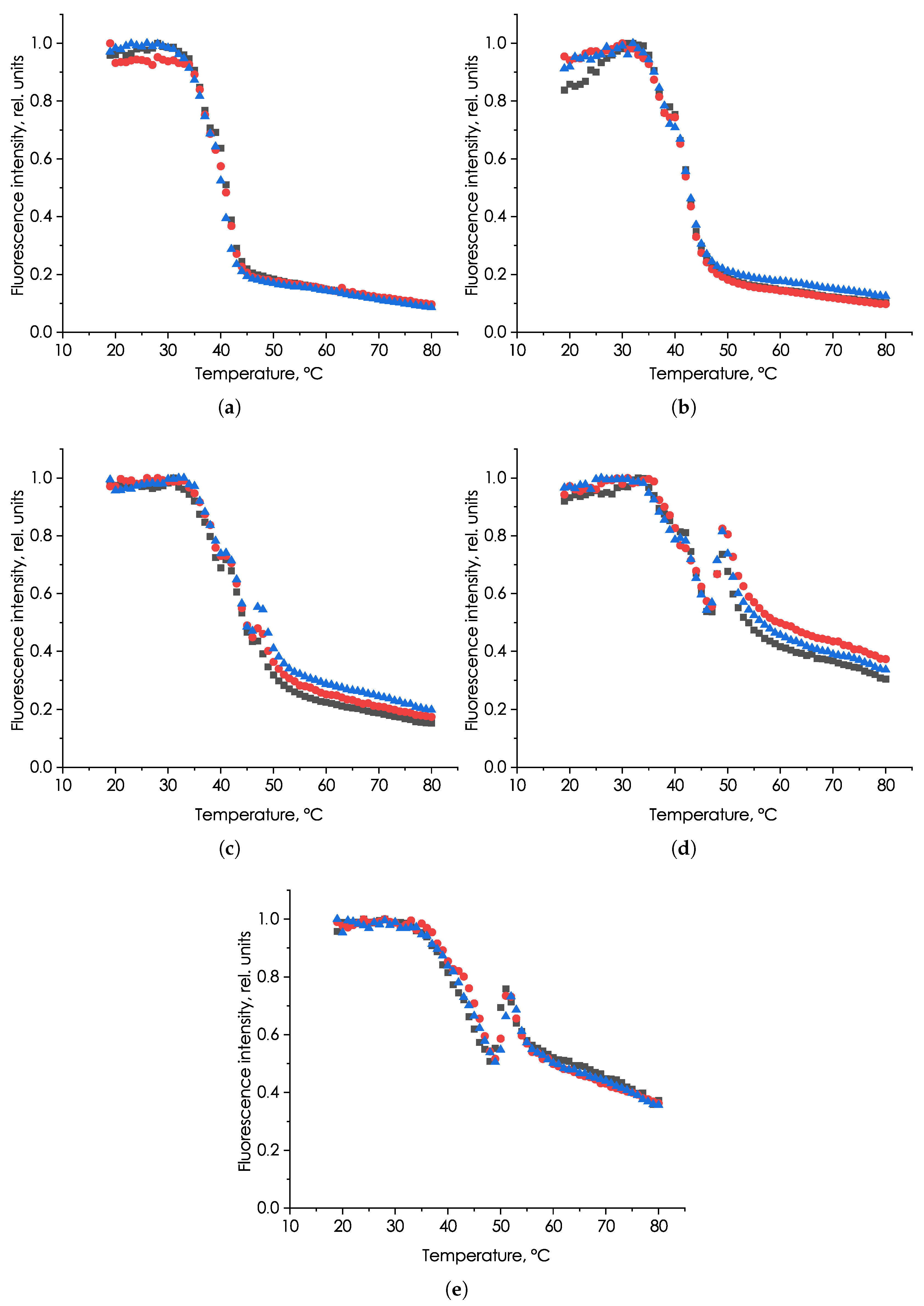
Figure 1. Dependences of the NFTCs of microalga P. hasleana on heating rate: (a) 0.5 °C/min; (b) 1 °C/min; (c) 2 °C/min; (d) 3 °C/min; (e) 6 °C/min. Colored markers show the fluorescent signals of different replicates.
Phycology 05 00052 g001
Figure 2. Dependence of the fluorescence intensity of Pseudo-nitzschia hasleana at different heating rates (a) on the temperature of the environment and (b) on the heating time.
Phycology 05 00052 g002
Figure 3. Dependence of the fluorescence intensity of Picochlorum maculatum and Phaeodactylum tricornutum at different heating rates (1–6 °C/min): (a) P. maculatum and the temperature of the environment; (b) P. maculatum and the heating time; (c) P. tricornutum and the temperature of the environment; (d) P. tricornutum and the heating times.
Phycology 05 00052 g003
Figure 4. NFTC of microalgae Pseudo-nitzschia hasleana, Phaeodactylum tricornutum, and Picochlorum maculatum obtained at different heating rates: (a) 1 °C/min; (b) 2 °C/min; (c) 3 °C/min; (d) 6 °C/min. In the figure, the fluorescence of P. hasleana (black square), P. tricornutum (red circle), and P. maculatum (blue triangle) is indicated with color markers.
Phycology 05 00052 g004
Disclaimer/Publisher’s Note: The statements, opinions and data contained in all publications are solely those of the individual author(s) and contributor(s) and not of MDPI and/or the editor(s). MDPI and/or the editor(s) disclaim responsibility for any injury to people or property resulting from any ideas, methods, instructions or products referred to in the content.