Article

Enhancement of Lipids Content in Chlorella sp. Under Phosphorus Limitation and Heavy Metal Addition for Biodiesel Production

by
Napisa Pattharaprachayakul
1,2,
Ramachandran Sivaramakrishnan
3 and
Aran Incharoensakdi
1,4,*
1
Laboratory of Cyanobacterial Biotechnology, Department of Biochemistry, Faculty of Science, Chulalongkorn University, Bangkok 10330, Thailand
2
Program in Biotechnology, Faculty of Science, Chulalongkorn University, Bangkok 10330, Thailand
3
Algal Biotechnology Lab, Department of General Surgery, Saveetha Medical College and Hospital, Saveetha Institute of Medical and Technical Sciences (SIMATS), Chennai 602105, India
4
Academy of Science, Royal Society of Thailand, Bangkok 10300, Thailand
*
Author to whom correspondence should be addressed.
Phycology 2025, 5(3), 49; https://doi.org/10.3390/phycology5030049
Submission received: 8 July 2025 / Revised: 8 August 2025 / Accepted: 3 September 2025 / Published: 19 September 2025

Abstract

Microalgae are photosynthetic microorganisms that could be used as potential microbial cell factories by directly converting CO2 into valuable bioproducts and biofuels. This study aims to improve the production of biofuel from the isolated green alga Chlorella sp., in terms of an increase in its lipid content and its conversion to fatty acid methyl esters (FAMEs) when the cells are grown under the influence of phosphorus (P) limitation and heavy metal addition. The results show that the highest content of lipids, at 68.9%, was achieved within one day under 0% P with a 17 µM cobalt addition. Moreover, supplementation with a low Pb concentration increased cell growth even under P limitation, but under this condition, its lipid content was decreased after seven days of growth. The lipids of Chlorella sp. were transesterified to produce FAMEs. The overall biodiesel properties of the obtained FAMEs were of acceptable quality according to the standards (ASTM and EN). Additionally, the energy conversion from light energy to lipids was shown to be in the range of 10–16% conversion efficiency within seven days. Hence, the physiological modification of Chlorella sp. culture by phosphorus limitation coupled with the addition of a low concentration of heavy metals enabled the improvement of lipid content, with the subsequent transesterification resulting in the production of biodiesel with acceptable quality.

1. Introduction

Recently, the concern of biofuels to replace fossil fuels has become a worldwide issue, with regard to the economic and environmental aspects. The rising cost of petroleum oil is not a problem at present; however, the use of fossil fuel leads to increases in greenhouse gases, especially carbon dioxide [1]. The demand for diesel is higher in the markets compared with that for gasoline, and there is also no need for a modified engine [2]. Biodiesel research and development was a major focus of the policies that started in 2008 and ended in 2022, which aimed to replace the use of biodiesel derived from crude palm oil to biodiesel derived from other suitable feedstocks [3]. Thus, the alternative feedstocks for biodiesel production can vary depending on how economic crops are utilized in each country.
Microalgae are considered to be more advantageous than plant products because they have higher growth rates, produce zero CO2, and do not require arable land for cell growth. Notably, microalgae are attractive because of their ability to raise their lipid accumulation under various conditions of nitrogen (N) and phosphorus (P) limitation [1,4]. N deprivation significantly increases the lipid content but negatively affects the biomass formation of Chlorella sp. [5]. On the other hand, P limitation, which is less studied than N-deprivation, could elevate the lipid content and unsaturated fatty acids in some microalgae species, including Chlorella sp. [6,7,8]. An increase in unsaturated fatty acids is desirable for biodiesel production applications, and could be achieved in microalgae cultivated under P limitation [9,10]. Moreover, P limitation could provide the added benefit of minimizing the content of P inside the cells, especially when cell lipids are utilized for biodiesel production. The phosphorus found in biodiesel comes from the phospholipids in the oil used as the feedstock, which could disrupt engine systems by damaging the catalytic converters; consequently, a maximum phosphorus level of 10 mg/kg biodiesel has been established by the ASTM 6751 standard [11].
Furthermore, micronutrients, such as small amounts of metal ions, also participate in microalgae metabolic functions as co-enzymes or energy carriers [12]. In general, heavy metals can be toxic to microalgae, although the addition of heavy metals at low concentrations has been found to enhance the lipid content of microalgae with different trends and patterns [13]. Several studies have reported that the oxidative stress due to heavy metals, especially Fe and Co in microalgae, leads to an increase in the lipid content [14,15,16,17]. There is no study yet examining the effect of Pb, which is not present in the BG11 medium, in combination with P limitation. Therefore, the current study specifically aims to explore (1) how P limitation in combination with heavy metal addition affects the lipid content in Chlorella sp. and (2) the properties of biodiesel derived from lipids of cells under optimal conditions, under the hypothesis that the reduced level of P inside the cells could contribute to an improvement in biodiesel properties.

2. Materials and Methods

2.1. Microalgal Cultivation and Biomass Determination

Chlorella sp. was isolated from natural brackish water in Thailand. This isolated Chlorella strain had a higher lipid content than another strain isolated from stone quarry pond water in a previous study [18]. The starting optical density at 680 nm of the culture was 0.5 in 800 mL BG-11 medium [19]. All samples were cultured under constant cool white light of 40 µmol m−2 s−1, with shaking at 150 rpm, 30 °C.
The cultivation up to 7 days was anticipated to induce lipid accumulation in microalgae. The Fe, FeCl3·6H2O and Co, Co(NO3)2·6H2O were supplied to the medium at 6 µM and 17 µM, respectively. Another heavy metal, Pb, at 10 µM Pb(NO3)2 was supplied to P-deprived, 50% P-, and 100% P-containing media. The biomass was determined by measuring the cell dry weight gravimetrically, as previously described [18]. Biomass was reported as grams of dried biomass per liter of culture.

2.2. Total Lipid Extraction and Determination

Microalgal total lipids were extracted by n-hexane, adapted from Halim, et al. [20]. Briefly, 5–10 mg lyophilized biomass was suspended in 800 µL n-hexane. Total lipids were subsequently extracted in the hexane phase after shaking at ambient temperature overnight, followed by centrifugation at 6000× g for 2 min. The upper layer was transferred to a new pre-weighed glass vial. The residual hexane was evaporated for 2–3 h or until a constant weight of total lipids was obtained. The lipid content was reported as the percentage of dry cell weight (% of DCW), whereas lipid production was reported as grams per liter of culture.

2.3. Transesterification and Biodiesel Properties’ Analysis

Transesterification and fatty acid profile analysis were adapted from IUPAC [21] and AOAC [22]. Briefly, dried lipid samples were firstly esterified into methyl ester by saponification with 0.5 N methanolic NaOH and then transesterified with 14% BF3 in methanol (v/v) at 80 °C. After cooling the solution, heptane and saturated NaCl were added to the solution to improve the efficiency of separation by forcing smaller fatty acid esters into the heptane phase (upper layer). After transesterification, the fatty acid methyl esters (FAMEs) were analyzed by gas chromatography (GC), as described by Limsuwatthanathamrong, et al. [23]. Individual FAMEs were identified by comparison with C10-C18 FAME standards (Sigma-Aldrich, St. Louis, MO, USA). The GC chromatographic temperature program was set as follows: initial temperature of 150 °C, increasing to 180 °C at 10 °C min−1, to 200 °C at 5 °C min−1, to 205 °C at 0.5 °C min−1, held at 205 °C for 2 min, and finally, increased to 250 °C at 5 °C min−1 and held there for 5 min (total run time of 30 min).
According to Sivaramakrishnan and Incharoensakdi [24], the determination of biodiesel characteristics had been characterized by equations published earlier. The saponification value (SV) and iodine value (IV) were determined by the equations from Francisco, et al. [25]. The cetane number (CN) was determined by equations published by Ramos, et al. [26]. The cloud point (CP) was calculated using the equation from Sarin, et al. [27]. KV, density, and the higher heating value (HHV) were determined by equations published by Ramírez-Verduzco, et al. [28].

2.4. Determination of Antioxidative Components

The H2O2 content of Pb-treated cells was determined by treating the cells with 1 M potassium iodide (KI). The experiments were performed according to Alexieva, et al. [29], and the absorbance was read at 390 nm using a UV spectrophotometer (Hitachi, Tokyo, Japan). The content was determined using the standard curve of H2O2 concentration. For malondialdehyde (MDA) content determination, 1 ml of 20% trichloroacetic acid containing 0.5% thiobarbituric acid was mixed with 0.5 mL supernatant. The reaction mixture was incubated in a boiling water bath for 15 min and centrifuged at 2790× g for 10 min. The samples were analyzed using a spectrophotometer by measuring absorbance at 450, 532, and 600 nm, and the values obtained were used to calculate the MDA content using the equation below [30].
M D A = 6.45 × A 532 A 600 ( 0.56 × A 450 ) W
where MDA is a level of malondialdehyde in the sample (µmol g−1 fresh weight); and W is the weight of the fresh sample taken (g).
The lysed cell suspension was used for the determination of superoxide dismutase (SOD) and catalase (CAT) activity following the kit protocol (Sigma Aldrich, St. Louis, MO, USA).

2.5. Energy Conversion Efficiency Analysis

According to Ren, et al. [31], the potential of lipid production had been determined under various microalgal culture modes by calculating the total energy conversion efficiency (TECE) from light to lipids according to the following equation:
T E C E   ( % ) = H V   o f   e x t r a c t e d   l i p i d s i n p u t   l i g h t   e n e r g y × 100
where the heating value (HV) of lipids is estimated as 36.3 kJ g−1, though the value could be varied depending on different microalgal species. The input light energy was calculated by the equations reported by Ren, et al. [31] and Lips, et al. [32], and it is estimated as 18.88 kJ.

2.6. Statistical Analysis

The data are presented as the mean of three replicate values, with the error bars showing standard deviations (means ± SD, n = 3). Statistical significance (p < 0.01–0.1) was analyzed by t-test comparisons using GraphPad software (GraphPad Prism 9).

3. Results and Discussion

3.1. Effect of Phosphorus Deprivation on Lipid Production

The isolated Chlorella sp. in this study had a lipid content of 35–42% during seven days of growth (Figure 1A), which was within the range reported for other Chlorellas (2–55%) [33]. In this study, we chose stationary-phase Chlorella sp. as the initial cells for lipid production due to the high lipid content under stress conditions [34,35]. The lipid contents in cells under normal and under P deprivation condition were not different during the first three days, whereas after seven days, the lipid content was increased to 56% in cells under P deprivation, which was higher than the control with approximately 35% (Figure 1B). This value is higher than those in some previous Chlorella studies under P deprivation, i.e., Chlorella sp. BUM11008 (31.9%) [5], C. zofingiensis (44.7%) [36], C. pyrenoidosa (32.77%) [37], and Chlorella sp. (13.9%) [38]. Thus, the results imply that different Chlorella strains had different responses to P deprivation with regard to lipid production [35], suggesting that the effect of P deprivation is not strain-specific. The response of microalgae to P deprivation is rather complicated, involving a complex interplay of metabolic shifts among the synthesis and breakdown of various macromolecules.
Phosphorus plays an important role in ATP and NADPH production, which is required to drive lipid synthesis and photosynthetic activity [39]. Therefore, P deprivation could decrease the chlorophyll content and consequently lead to a low biomass [40]. Nucleic acid synthesis is also suppressed under P deprivation, leading to reduced cell division and biomass formation. As expected, the biomass did not increase under P deprivation on day 7 (Figure 1A), resulting in lower lipid production than the control with 100% P (Figure 1C). It is worth mentioning that under P deprivation, there was an alteration of the lipid composition, e.g., diacylglyceryl-trimethylhomoserine (DGTS), diacylglyceryl-hydroxymethyltrimethyl-β-alanine (DGTA), and diacylglyceryl-carboxyhydroxy-methylcholine (DGCC), in plastid membranes of several green microalgae, including Chlorella [6,41]. For example, the cultivation of a green alga Monodus subterraneus under P deprivation also reduced the phospholipid but increased triacylglycerols (TAGs), mainly digalactosyldiacylglycerol (DGDG) [42]. According to Alipanah, et al. [43], the down-regulation of the fatty acid synthesis pathway was observed in diatom Phaeodactylum tricornutum under P deprivation, which led to the decrease in membrane lipid synthesis and cell division. Additionally, the up-regulation of phospholipid:diacylglycerol acyltransferase (PDAT) and phospholipase genes was detected, which increases the phospholipid degradation forming diacylglycerol (DAG) and phosphatidic acid (PA), leading to high TAG accumulation. The overall results of this study suggest that the Chlorella sp. could accumulate a high lipid content under P deprivation. However, further optimization of nutrients is required to increase the biomass under this condition, to increase lipid production.

3.2. Effect of Phosphorus Deprivation and Heavy Metal Addition on Lipid Production

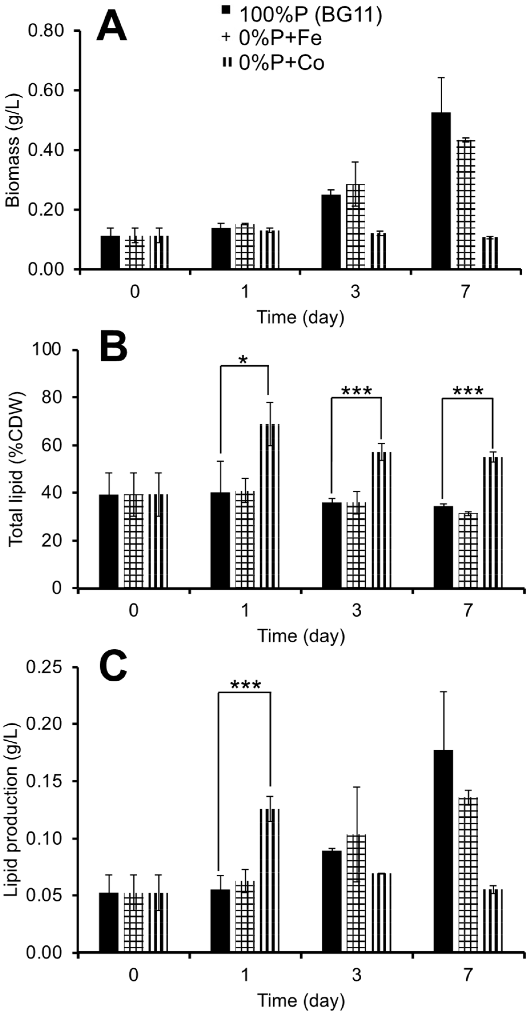
The key macro- and micronutrients, such as phosphate and some metal ions, in BG11 medium could alter the lipid content in various microalgae, including Chlorella [44]. In this study, we combined both P deprivation and metal addition, Fe and Co, in Chlorella cultures to modulate the biomass and lipid content. Fe was used to increase the lipid content in microalgae [45]. Interestingly, the addition of a suitable concentration of Fe not only improved the lipid content but could also increase the cell growth in green algae [24,46]. In this study, we added FeCl3 as an extra iron source because of its strong influence on the lipid content in green algae [47]. Under P deprivation, which caused an inhibition of the cell growth, as shown in Figure 1A, the addition of Fe increased cell biomass, whereas no apparent change in biomass occurred upon the addition of Co (Figure 2A). Notably, a sufficient Fe amount could prevent a severe drop in biomass production in several microalgae, even under the essential nutrient limitation (N, P) [14,47,48].
Under P deprivation, the addition of Co did not increase the biomass (Figure 2A), but it increased the lipid content to 68.9% within one day, whereas Fe addition hardly changed the lipid content (Figure 2B). Lipid production was highest on day 1 with Fe addition, and on day 3 with Co addition; however, the lipid production of the two treated cells was lower than that of the control on day 7 (Figure 2C) due to the lower biomass of the metal-stressed cells.
In C. sorokiniana, Fe and Mg play significant roles in photosynthesis, and thus the maximum quantum efficiency of photosystem II (Fv/Fm) was improved under a metal stress condition [49]. Consequently, the chlorophyll content was increased when the metal concentrations (Fe, Mg, Ca) were also increased. Additionally, the green alga Ankistrodesmus faculatus KJ671624 was studied on biomass to determine the lipid content under various stress combinations, i.e., N, P, and Fe, where it was shown that the highest lipid content (59.6%) was achieved under N limitation (750 mg/L), P deprivation, and Fe addition (9 mg/L), whereas the biomass declined by approximately 50% [48]. Besides the P deprivation, P limitation could also increase the lipid content. It has been reported that the addition of 32 µM phosphorus to a Chlorella sp. culture could enhance the lipid content to 23.60% [8]. Singh et al. [49] investigated the effects of nitrogen and phosphorus stress on both the lipid content and biomass productivity in microalgae. They found the reduction in biomass and lipid productivity under the limited P condition due to the a of essential macronutrients leading to negative effects on cell growth and cell division. In addition, the heavy metal stress was found to induce cell growth and a lipid content even under a macronutrient limitation condition. In the present study, Fe and Co affected cell growth and the lipid content differently in Chlorella sp. under P stress (0% P). Fe and Co stimulated and inhibited cell growth, respectively (Figure 2A), whereas Co, not Fe, increased the lipid content (Figure 2B). This suggests that Chlorella sp. employed different mechanisms to respond to different metal ions present in the growth medium. It is noted that the Co concentration used in this study was 100 times that present in normal BG 11.
Microalgae utilize appropriate mechanisms to take up various kinds of metals and further detoxify them. Different mechanisms may be used by Chlorella to deal with the presence of Fe and Co. The absorption of Fe and Co and the transport into the cells can be managed by different transporters, and this can lead to different levels of ROS generation caused by Fe and Co. Although the present study revealed no lethal toxicity of the cells in seven days of exposure to metals, it remains to be further investigated whether metals, especially Co, could result in cell death from long-term exposure, including the effect on the lipid content of the cells.
In this study, 17 µM cobalt nitrate (100× of normal BG11) was applied to the P-deprived BG11, which resulted in an improvement of the lipid content (Figure 2B) with low biomass (Figure 2A). Similarly, it was reported that Co at a low concentration (≈3 µM) could increase the lipid content and reduce the biomass of C. vulgaris [15]. Moreover, Li, et al. [50] also found that ≥10 µM of Co inhibited the cell growth of marine microalga Pavlova viridis but markedly increased antioxidant enzymes’ activities and non-enzyme antioxidative substances. On the other hand, C. pyrenoidosa showed a rapid decrease in Fv/Fm under 0.1–10 mM of Co treatment within 30 min [51]. The results of the present study are in line with previous studies, which showed that Co can induce toxicity in Chlorella and other green microalgae, especially at high concentrations [50,51].
The oxidative stress indicators under Fe3+ and Co2+ treatments were not analyzed in this study. However, it has previously been reported that Chlorella sp. showed a high tolerance under treatment with 34 µM of cobalt, and cells showed elevated levels of stress biomarkers [52]. The concentration of Co used in the reported study (34 µM) was in the same order of magnitude, which was not much different from that in our study (17 µM). For Fe, the addition of iron up to 10 µM increased final cell densities by nearly 2-fold while 1 mM iron was toxic [53]. Moreover, Fe treatment in our study showed no growth inhibition (Figure 2A), and it was thus unlikely to cause oxidative stress.

3.3. Effect of Phosphorus Limitation and Lead-Induced Oxidative Stress on Lipid Production

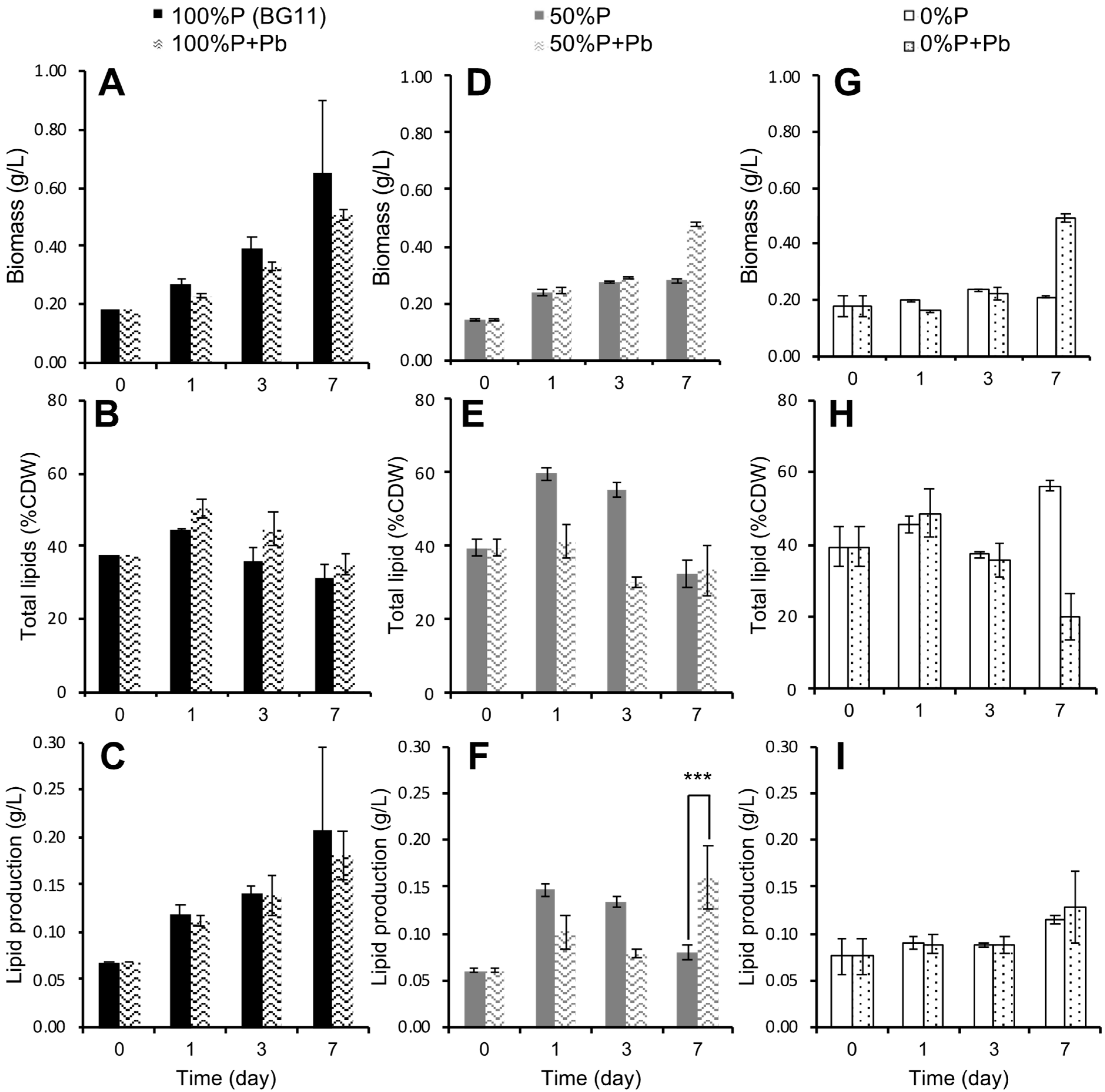
Pb at a suitable concentration has been reported to trigger the increase in lipid content and growth in microalgae [54,55]. In this study, the concentration of Pb at 10 µM was used based on previous reports showing that microalgae, despite slight growth inhibition, could survive up to 500 µM [55] and 100 µM Pb [54]. The levels of P (100%, 50%) and P deprivation (0%) were tested in combination with the stress by Pb with the aim to increase the lipid content (Figure 3). The biomass of the control without Pb2+ was highest on day 7 in 100% P compared to that at 50 and 0% P (Figure 3A,D,G). On day 7 under 50 and 0% P, the presence of Pb stimulated the increase in biomass (Figure 3D,G). Pb could stimulate biomass production when phosphate is limited. The lipid content was slightly increased under 100% P with added Pb compared to the control without Pb (Figure 3B). However, Pb was unable to increase the lipid content under 50 and 0% P (Figure 3E,H). The 100% P without Pb showed the highest lipid production on day 7 (Figure 3C), whereas the lipid production decreased under 50 and 0% P without Pb on day 7 (Figure 3F,I) due to their low biomass. Although under 50% P, a similar lipid content was observed on day 7 with or without Pb (Figure 3E), the presence of Pb under 50% P increased lipid production compared to the control by 2-fold on day 7 (Figure 3F) due to its higher biomass than the control. Thus, the combination of P limitation (50% P) and Pb addition could increase lipid production, even though its lipid content was not increased, as can be seen in Figure 3E. It is noted that under 0% P on day 7, Pb caused a higher biomass, but a lower lipid content compared to the control (Figure 3G,H). This led to relatively unchanged lipid production regardless of the presence of Pb (Figure 3I).
Besides the P deprivation, P limitation could also increase the lipid content. It has been reported that the addition of 32 µM phosphorus to the Chlorella sp. culture could enhance the lipid content to 23.60% [8]. Singh et al. [49] investigated the effects of nitrogen and phosphorus stress on both the lipid content and biomass productivity in microalgae. They found a reduction in biomass and lipid productivity under the limited P condition due to the lack of essential macronutrients, leading to negative effects on cell growth and cell division. In addition, the heavy metal stress was found to induce the cell growth and lipid content even under the macronutrient limitation condition. In the present study, Fe and Co affected cell growth and the lipid content differently in Chlorella sp. under P stress (0% P). Fe and Co stimulated and inhibited cell growth, respectively (Figure 2A), whereas Co, not Fe, increased the lipid content (Figure 2B). This suggests that Chlorella sp. employed different mechanisms to respond to different metal ions present in the growth medium. It is noted that the Co concentration used in this study was 100 times that present in normal BG 11. A similar study reported that the microalga Botryococcus braunii could survive a high concentration of cobalt (50 × normal level) [56].
On the other hand, unharmful levels of Pb ranging from 0.01 to 10 µM could increase pigment contents in microalgae [57]. This could explain why the cells could grow better under P limitation with Pb addition. Unlike Fe and Co, Pb is a non-essential micronutrient for microalgae growth, and it is found to cause toxicity to the environment. Therefore, the lower range of Pb at 0.01–500 µM has been studied in microalgae [54,57]. Increasing the concentration of Pb from 1 to 100 µM decreased the growth of C. vulgaris, whereas the cell number and chlorophyll content at 1 µM Pb were not much different from the control [54]. Different microalgae showed different responses to Pb stress. The cell growth and contents of pigments were more suppressed at Pb ≥ 0.1 µM in A. obliquus than in C. vulgaris [55]. Recently, Pham et al. [58] reported that Scenedesmus sp. had the maximum lipid contents of 31.1% and 30.8% when treated with 0.5 and 1 mg/L of Pb, respectively. In the present study, 0.1–10 µM of Pb was used. The maximum lipid content was observed at 1 µM Pb, and at 10 µM Pb, a decrease in biomass occurred, which was in agreement with a previous study [54]. Hence, the Pb toxicity and the increase in the lipid content in Chlorella might be attributed to the Pb-induced oxidative stress.
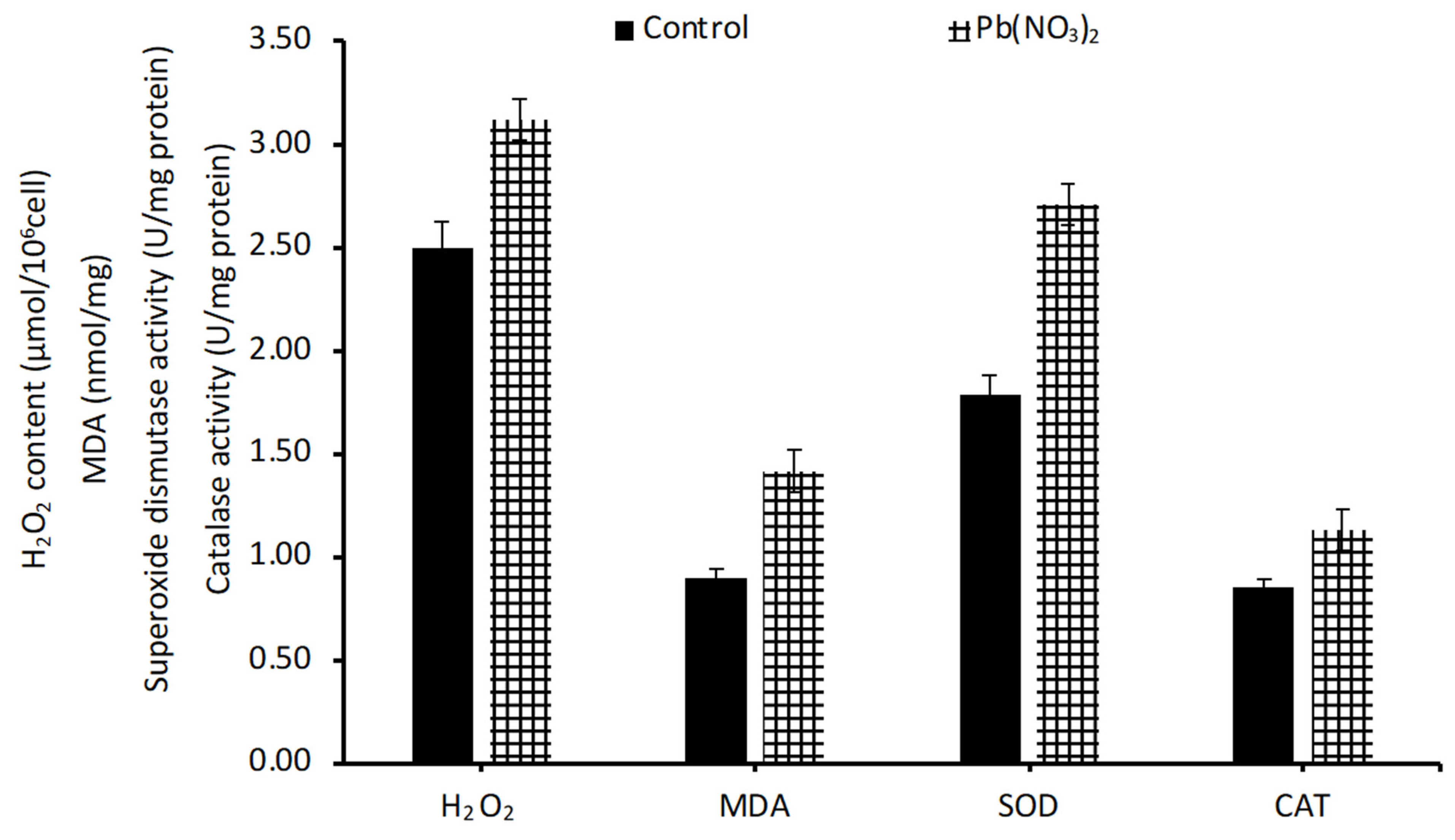
Oxidative stress treatment could improve the lipid content. After 24 h of treatment with Pb, the H2O2 level was elevated when compared to the control (Figure 4). Pb-induced oxidative stress showed some positive effects on the lipid content. During oxidative stress, a reduction in O2 occurs during chloroplast electron transport to form H2O2 [59]. The presence of reactive oxygen species (ROS) stimulates the signaling molecules and triggers the physiological responses and cell growth [60]. In the present study, it is clear that the Pb treatment increases the H2O2 level as a response to oxidative stress.
During oxidative stress, polyunsaturated fatty acids are oxidized and generate MDA, which is considered an oxidative stress marker. Cell walls are damaged, and the cells try to adapt by means of lipid peroxidation [61]. In the present study, the MDA level was increased after Pb treatment (Figure 4), which confirms that the cells are able to defend against the oxidative stress.
Apart from the increase in H2O2 and MDA, cells also produce antioxidant enzymes to mitigate the effects of ROS. The antioxidant enzymes superoxide dismutase (SOD) and catalase (CAT) were analyzed and are shown in Figure 4. SOD is an important metalloenzyme, which efficiently scavenges the superoxide molecules. In the present study, Pb treatment increased the ROS, and the cells were well protected against ROS by SOD and CAT enzymes. The enzyme activity of both enzymes was enhanced after the oxidative stress treatment by Pb. Due to the action of SOD and CAT enzymes, ROS-mediated electrons are directed towards lipid synthesis to neutralize the effect of ROS, and this was supported by the result showing the increased lipid content after the Pb treatment (Figure 4). Oxidative stress is a consequence of an imbalance between the production of reactive oxygen species (ROS) and the antioxidant system, which includes the function of various antioxidant enzymes. Oxidative stress regulates the expression and activity of lipid metabolism-related enzymes, leading to the increase in fatty acid synthesis. Our previous study in Chlorella sp. showed an increase in ACC activity, the enzyme for fatty acid synthesis, as well as the up-regulation of some important genes in the fatty synthesis pathway, under oxidative stress caused by UV radiation and H2O2 [62]. The up- and down-regulation of some genes in the fatty acid synthesis pathway can lead to a change in the SFA/UFA ratio.

3.4. Fatty Acid Profiles and Biodiesel Properties Under Pb Stress Conditions

The fatty acid compositions and biodiesel properties were determined from Chlorella FAMEs after 24 h of treatment with different P and Pb concentrations (Table 1). The ratio of saturated fatty acids (SFAs) and unsaturated fatty acids (UFAs) in Chlorella was high because of the high contents of palmitic acid (C16:0) and stearic acid (C18:0). Other microalgae showed lower values of this ratio (SFA:UFA) due to the high contents of C18:2 and C18:3 along with the low content of C20:0 (≤0.01%) [17]. In this study, we observed a reduction in decanoic acid (C10:0) and stearic acid (C18:0), but an increase in palmitic acid (54.57%) upon the decrease in P from 100 to 0%, especially when coupled with Pb addition. Under 0% P, the UFAs were higher and different in composition compared with other conditions, where a high content of palmitoleic acid (C16:1) was found. Thus, P limitation could increase UFAs in Chlorella sp., especially monounsaturated fatty acids (MUFAs), which is in agreement with previous studies [5,6]. Additionally, we also attempted to test the nitrogen limitation and found the high amount of UFAs in both, with and without Pb. However, the low levels of P could improve biodiesel quality by reducing damage to catalytic converters in operational systems [63]. It should be mentioned that the shifts in fatty acid levels (e.g., C16:0, C18:0, C16:1) in Chlorella sp. caused by oxidative stress were also observed in our previous study, whereby different levels of genes involved in fatty acid synthesis and desaturation were expressed in cells under oxidative stress [64]. Next, we determined the key properties which affect the quality of biodiesel. SV, IV, CN, CP, KV, density, and HHV were analyzed according to Hoekman, et al. [64], and the results are shown in Table 1.
The saponification value, SV, is a measure of the average molecular weight of all fatty acids present. The maximum SV of 2.37 g KOH/g lipid was obtained under the 100% P with Pb supplementation condition. The Pb addition resulted in a higher SV than that without Pb under each P-limited condition. Thus, the addition of 10 ± µM Pb could positively alter the SV in all the three concentrations of P, i.e., 100, 50 and 0% P, with the SV values of 10.14, 4.78, and 4.17 g KOH/g, respectively.
The iodine value, IV, is a measure of total unsaturation of biodiesel, and this value is used to represent oxidative stability [65]. The increase in IV could negatively affect engine performance because a high content of alkyl double bonds leads to the formation of insoluble sediments. According to the European biodiesel standard, the IV is limited to 120 g I2/100 g. The results showed that the values obtained from all conditions are within the limits. In all the cases, the value was not significantly different, except under 0% P without Pb, which showed the highest value of 38.75 g I2/100 g.
The cetane number, CN, is an important property determining the combustion quality and ignition delay time. A high CN ensures the ignition properties and a good engine performance as well as a reduction in white smoke formation from the engine. The results demonstrated that all of the samples had values higher than the minimum value of the American Society for Testing and Materials (ASTM) and European standards. The lowest CN was found in samples with 0% P, whereas the highest CN was also found in P-deprived samples with Pb addition. However, the range of CN value is not significantly different in each condition. Notably, Chlorella sp. in this study had a higher CN (62.93–67.18) than that of Chlorella sp. in other previous studies [66], which meant that it could be blended at higher concentrations with petroleum diesel [17]. Thus, this Chlorella sp. is a good candidate for biodiesel production.
The cloud point, CP, is the temperature at which the fuel starts to appear cloudy and confirms the wax crystal formation, which blocks the filters and fuel lines of the vehicle [67]. No limit ranges are given in ASTM due to the fact that the climate conditions in the United States vary considerably. However, a lower range of CP is more suitable. In this study, the range was rather high in each condition, except in 100% P with Pb addition (8.78 °C). Additionally, the decrease in P concentrations could lead to an increase in CP, where the highest CP was detected at 0% P with Pb addition. However, this Chlorella sp. oil still had a high CP when compared with other microalgae oil [68]. A high number of double bonds located near the ends of carbon chains would likely contribute to a high CP value of the oil [68].
The higher heating value, HHV, is the amount of heat released after the complete combustion, which is a unit quantity of fuel combustion into H2O and CO2 [68]. The HHV could be decreased when double bonds and shorter-chain fatty acids increase. However, the selection between the lower and higher HHV compounds as biodiesel sources remains unclear. In general, the HHV of microalgae biodiesel was found to be in the range of 36.6–40.4 MJ/kg, which is normally lower than that obtained from commercial diesel fuel (45.62–46.48 MJ/kg) [17,67]. The HHVs of all samples were between 38 and 39 MJ/kg, which were within the range reported previously [17].
Kinematic viscosity, KV, is an essential value used to present the resistance of biodiesel flow in fuel injection systems at low temperatures. The samples with cis double bonds generally have higher KV than samples with trans double bonds [65]. Moreover, the unsaturated FAMEs have lower viscosities than the saturated FAMEs. Hence, the high KV could cause poor vaporization and atomization of the engine. For ASTM values, the KV range is 1.9–6.0, whereas the KV range in Europe is 3.5–5.0. The results demonstrated that KV values of samples under all conditions are within the limits of both EN and ASTM. Interestingly, the KV of this Chlorella sp. FAME showed a lower value (3.18–3.70 mm2/s) than those of biodiesels derived from several crops and some microalgae, e.g., N. oculate, D. brasiliensis, and B. braunii braunii [17,69].
It is noted that Pb and P do not directly affect the values of biodiesel properties. The compositions of the obtained lipids after Pb and P treatment contribute to the quality of biodiesel. Palmitic acid is the major component (54.57%) of Chlorella sp. under Pb and 0% P in this study (Table 1). Palmitic acid is a suitable component for biodiesel production and ensures good biodiesel properties, as previously reported in Aphanothece halophytica grown under white light-emitting diodes [70].

3.5. Total Energy Conversion Efficiency

How algal cells cultured under normal and stress conditions can convert the substrate into lipids is determined by their total energy conversion efficiency (TECE). However, under stress conditions, the TECEs of cultivations were lower than that under a normal condition due to the lower biomass production. The highest TECEs were 26.70 ± 1.52% and 19.66 ± 6.28% when cultivating in normal BG11 with Pb, and 0% P with Fe addition, respectively (Table 2). Interestingly, the TECEs in the present study were higher than that in Scenedesmus sp. cultured under a mixotrophic condition (14.6%) [31]. Interestingly, either nutrient limitation or heavy metal stress of microalgae could allow the improvement of the energy conversion even under autotrophic cultivation.

4. Conclusions

Microalgae as s feedstock for biodiesel production are clearly more effective and productive than vegetable oils used in terms of environmental and sustainable aspects. Besides the benefit of microalgae for the reduction in CO2 emissions, microalgae can also survive under heavy metal stress or limited nutrient conditions, e.g., wastewater or harsh environments. In this study, the highest lipid content of Chlorella sp. was obtained when the cells were cultivated under 17 µM Co combined with 0% P. Chlorella sp. showed a good biomass production of 0.49 g/L and lipid production of 0.13 g/L when we added 10 µM Pb combined with 0% P. Thus, 10 µM of Pb was not harmful to Chlorella sp. but could favorably trigger cell growth. The fatty acid profiles revealed an increase in UFAs under sole P deprivation with abundant MUFAs, i.e., C16:1. Under Pb stress, there was no significant enhancement in PUFAs, whereas the biodiesel quality was better than that under a non-Pb condition. A good quality of biodiesel was obtained from Chlorella sp. under 10 µM Pb addition combined with 0% P. In addition, the biodiesel properties were all in the ranges within the EN 14214 and ASTM D6751 standards [11]. This study is the first to report on Pb stress with P limitation in Chlorella for biodiesel production, in which the reduction in phosphorus could improve the quality of biodiesel. Hence, this study suggests the application of Chlorella sp. as a high-potential feedstock for biodiesel production. However, the usefulness of the data in this study requires further in-depth investigation—in particular, the long-term effect of metal toxicity on lipid production as well as the amount of metal accumulating in the cells. The molecular mechanisms regarding the effect of metals on the genes involved in fatty acid synthesis also need to be further investigated. Studies on metal accumulation in Chlorella will be beneficial in terms of the bioremediation of metal-contaminated environments, allowing the use of the organism to treat wastewater containing heavy metals. Nevertheless, more extensive studies with the use of a consortium of various organisms are needed in real-world applications due to the presence of an array of mixed heavy metals in wastewater.

Author Contributions

Conceptualization, N.P. and A.I.; Methodology, N.P. and R.S.; Validation, A.I.; Investigation, N.P. and R.S.; Writing—Original Draft, N.P.; Writing—Review and Editing, A.I.; Supervision, A.I.; Project Administration, A.I.; Funding Acquisition, A.I. All authors have read and agreed to the published version of the manuscript.

Funding

This research received no external funding.

Data Availability Statement

The original contributions presented in this study are included in the article. Further inquiries can be directed to the corresponding author(s).

Acknowledgments

N.P. is supported by the Royal Golden Jubilee Ph.D. program (PHD/0086/2558) and the 90th Anniversary of Chulalongkorn University Ratchadaphiseksomphot Endowment Fund (46/2563).

Conflicts of Interest

The authors declare no conflicts of interest.

References

  1. Chisti, Y. Biodiesel from microalgae. Biotechnol. Adv. 2007, 25, 294–306. [Google Scholar] [CrossRef]
  2. Bloyd, C. Thailand Alternative Fuels Update 2017; United State Department of Energy: Washington, DC, USA, 2017. [Google Scholar]
  3. Sakulsuraekkapong, J.; Thepa, S.; Pairintra, R. Improvement of biodiesel’s policy in Thailand. Energy Sources Part B Econ. Plan. Policy 2018, 13, 158–164. [Google Scholar] [CrossRef]
  4. Kumar, K.S.; Dahms, H.-U.; Won, E.-J.; Lee, J.-S.; Shin, K.-H. Microalgae–A promising tool for heavy metal remediation. Ecotoxicol. Environ. Saf. 2015, 113, 329–352. [Google Scholar]
  5. Praveenkumar, R.; Shameera, K.; Mahalakshmi, G.; Akbarsha, M.A.; Thajuddin, N. Influence of nutrient deprivations on lipid accumulation in a dominant indigenous microalga Chlorella sp., BUM11008: Evaluation for biodiesel production. Biomass Bioenergy 2012, 37, 60–66. [Google Scholar]
  6. Sharma, K.K.; Schuhmann, H.; Schenk, P.M. High lipid induction in microalgae for biodiesel production. Energies 2012, 5, 1532–1553. [Google Scholar] [CrossRef]
  7. El-Sheek, M.; Rady, A.J.P. Effect of phosphorus starvation on growth, photosynthesis and some metabolic processes in the unicellular green alga Chlorella kessleri. Phyton 1995, 35, 139–151. [Google Scholar]
  8. Liang, K.; Zhang, Q.; Gu, M.; Cong, W. Effect of phosphorus on lipid accumulation in freshwater microalga Chlorella sp. J. Appl. Phycol. 2013, 25, 311–318. [Google Scholar] [CrossRef]
  9. Stansell, G.R.; Gray, V.M.; Sym, S.D. Microalgal fatty acid composition: Implications for biodiesel quality. J. Appl. Phycol. 2012, 24, 791–801. [Google Scholar] [CrossRef]
  10. Yodsuwan, N.; Chamchuang, N.; Puchcha, Y.; Sirisansaneeyakul, S.J.K.J. Outdoor photoautotrophic cultivation of Chlorella sp. TISTR 8990 in nitrogen-and phosphorus-minimal media for lipid accumulations. Kasetsart J. 2015, 49, e91. [Google Scholar]
  11. Silitonga, A.S.; Masjuki, H.H.; Mahlia, T.M.I.; Ong, H.C.; Chong, W.T.; Boosroh, M.H. Overview properties of biodiesel diesel blends from edible and non-edible feedstock. Renew. Sustain. Energy Rev. 2013, 22, 346–360. [Google Scholar] [CrossRef]
  12. da Silva Ferreira, V.; Sant’Anna, C. The effect of physicochemical conditions and nutrient sources on maximizing the growth and lipid productivity of green microalgae. Phycol. Res. 2017, 65, 3–13. [Google Scholar] [CrossRef]
  13. Saavedra, R.; Muñoz, R.; Taboada, M.E.; Vega, M.; Bolado, S. Comparative uptake study of arsenic, boron, copper, manganese and zinc from water by different green microalgae. Bioresour. Technol. 2018, 263, 49–57. [Google Scholar] [CrossRef]
  14. Liu, Z.-Y.; Wang, G.-C.; Zhou, B.-C. Effect of iron on growth and lipid accumulation in Chlorella vulgaris. Bioresour. Technol. 2008, 99, 4717–4722. [Google Scholar] [CrossRef] [PubMed]
  15. Battah, M.; El-Ayoty, Y.; Abomohra, A.E.-F.; El-Ghany, S.A.; Esmael, A. Effect of Mn2+, Co2+ and H2O2 on biomass and lipids of the green microalga Chlorella vulgaris as a potential candidate for biodiesel production. Ann. Microbiol. 2015, 65, 155–162. [Google Scholar] [CrossRef]
  16. Carfagna, S.; Lanza, N.; Salbitani, G.; Basile, A.; Sorbo, S.; Vona, V. Physiological and morphological responses of lead or cadmium exposed Chlorella sorokiniana 211-8K (Chlorophyceae). SpringerPlus 2013, 2, 147. [Google Scholar] [CrossRef] [PubMed]
  17. Islam, M.A.; Magnusson, M.; Brown, R.J.; Ayoko, G.A.; Nabi, M.N.; Heimann, K. Microalgal species selection for biodiesel production based on fuel properties derived from fatty acid profiles. Energies 2013, 6, 5676–5702. [Google Scholar] [CrossRef]
  18. Sivaramakrishnan, R.; Incharoensakdi, A. Enhancement of total lipid yield by nitrogen, carbon, and iron supplementation in isolated microalgae. J. Phycol. 2017, 53, 855–868. [Google Scholar] [CrossRef]
  19. Rippka, R.; Deruelles, J.; Waterbury, J.B.; Herdman, M.; Stanier, R.Y. Generic assignments, strain histories and properties of pure cultures of cyanobacteria. Microbiology 1979, 111, 1–61. [Google Scholar] [CrossRef]
  20. Halim, R.; Gladman, B.; Danquah, M.K.; Webley, P.A. Oil extraction from microalgae for biodiesel production. Bioresour. Technol. 2011, 102, 178–185. [Google Scholar] [CrossRef]
  21. IUPAC. Standard Methods for the Analysis of Oils, Fats and Derivatives; Pergamon Press: Paris, France, 1979; pp. 2503–2526. [Google Scholar]
  22. AOAC. Official Methods of Analysis of AOAC International; Association of Official Analytical Chemists: Washington, DC, USA, 1995. [Google Scholar]
  23. Limsuwatthanathamrong, M.; Sooksai, S.; Chunhabundit, S.; Noitung, S.; Ngamrojanavanich, N.; Petsom, A. Fatty acid profile and lipid composition of farm-raised and wild-caught sand worms, Perinereis nuntia, the diet for marine shrimp broodstock. Asian J. Anim. Sci. 2012, 6, 65–75. [Google Scholar]
  24. Sivaramakrishnan, R.; Incharoensakdi, A. Higher efficiency of microalgal biorefinery is achieved with integrated than one-way method. Fuel 2021, 300, 120988. [Google Scholar] [CrossRef]
  25. Francisco, É.C.; Neves, D.B.; Jacob-Lopes, E.; Franco, T.T. Microalgae as feedstock for biodiesel production: Carbon dioxide sequestration, lipid production and biofuel quality. J. Chem. Technol. Biotechnol. 2010, 85, 395–403. [Google Scholar] [CrossRef]
  26. Ramos, M.J.; Fernández, C.M.; Casas, A.; Rodríguez, L.; Pérez, Á. Influence of fatty acid composition of raw materials on biodiesel properties. Bioresour. Technol. 2009, 100, 261–268. [Google Scholar] [CrossRef] [PubMed]
  27. Sarin Sarin, A.; Arora, R.; Singh, N.P.; Sarin, R.; Malhotra, R.K.; Kundu, K. Effect of blends of Palm-Jatropha-Pongamia biodiesels on cloud point and pour point. Energy 2009, 34, 2016–2021. [Google Scholar] [CrossRef]
  28. Ramírez-Verduzco, L.F.; Rodríguez-Rodríguez, J.E.; Jaramillo-Jacob, A.d.R. Predicting cetane number, kinematic viscosity, density and higher heating value of biodiesel from its fatty acid methyl ester composition. Fuel 2012, 91, 102–111. [Google Scholar] [CrossRef]
  29. Alexieva, V.; Sergiev, I.; Mapelli, S.; Karanov, E. The effect of drought and ultraviolet radiation on growth and stress markers in pea and wheat. Plant Cell Environ. 2001, 24, 1337–1344. [Google Scholar] [CrossRef]
  30. Szechyńska-Hebda, M.; Skrzypek, E.; Dąbrowska, G.; Biesaga-Kościelniak, J.; Filek, M.; Wędzony, M. The role of oxidative stress induced by growth regulators in the regeneration process of wheat. Acta Physiol. Plant. 2007, 29, 327–337. [Google Scholar] [CrossRef]
  31. Ren, H.-Y.; Liu, B.-F.; Kong, F.; Zhao, L.; Xie, G.-J.; Ren, N.-Q. Energy conversion analysis of microalgal lipid production under different culture modes. Bioresour. Technol. 2014, 166, 625–629. [Google Scholar] [CrossRef]
  32. Lips, D.; Schuurmans, J.M.; Branco dos Santos, F.; Hellingwerf, K.J. Many ways towards ‘solar fuel’: Quantitative analysis of the most promising strategies and the main challenges during scale-up. Energy Environ. Sci. 2018, 11, 10–22. [Google Scholar] [CrossRef]
  33. Shuba Eyasu, S.; Kifle, D. Microalgae to biofuels: ‘Promising’ alternative and renewable energy, review. Renew. Sustain. Energy Rev. 2018, 81, 743–755. [Google Scholar] [CrossRef]
  34. Yang, J.; Li, X.; Hu, H.; Zhang, X.; Yu, Y.; Chen, Y. Growth and lipid accumulation properties of a freshwater microalga, Chlorella ellipsoidea YJ1, in domestic secondary effluents. Appl. Energy 2011, 88, 3295–3299. [Google Scholar] [CrossRef]
  35. Zhang, Q.; Hong, Y. Effects of stationary phase elongation and initial nitrogen and phosphorus concentrations on the growth and lipid-producing potential of Chlorella sp. HQ. J. Appl. Phycol. 2014, 26, 141–149. [Google Scholar] [CrossRef]
  36. Feng, P.; Deng, Z.; Fan, L.; Hu, Z. Lipid accumulation and growth characteristics of Chlorella zofingiensis under different nitrate and phosphate concentrations. J. Biosci. Bioeng. 2012, 114, 405–410. [Google Scholar] [CrossRef] [PubMed]
  37. Li, Y.; Han, F.; Xu, H.; Mu, J.; Chen, D.; Feng, B.; Zeng, H. Potential lipid accumulation and growth characteristic of the green alga Chlorella with combination cultivation mode of nitrogen (N) and phosphorus (P). Bioresour. Technol. 2014, 174, 24–32. [Google Scholar] [CrossRef] [PubMed]
  38. Anto, S.; Pugazhendhi, A.; Mathimani, T. Lipid enhancement through nutrient starvation in Chlorella sp. and its fatty acid profiling for appropriate bioenergy feedstock. Biocatal. Agric. Biotechnol. 2019, 20, 101179. [Google Scholar] [CrossRef]
  39. Roopnarain, A.; Gray, V.M.; Sym, S.D. Phosphorus limitation and starvation effects on cell growth and lipid accumulation in Isochrysis galbana U4 for biodiesel production. Bioresour. Technol. 2014, 156, 408–411. [Google Scholar] [CrossRef]
  40. Carstensen, A.; Herdean, A.; Schmidt, S.B.; Sharma, A.; Spetea, C.; Pribil, M.; Husted, S. The impacts of phosphorus deficiency on the photosynthetic electron transport chain. Plant Physiol. 2018, 177, 271–284. [Google Scholar] [CrossRef]
  41. Kalisch, B.; Dörmann, P.; Hölzl, G. DGDG and glycolipids in plants and algae. In Lipids in Plant and Algae Development; Springer: Cham, Switzerland, 2016; pp. 51–83. [Google Scholar]
  42. Khozin-Goldberg, I.; Cohen, Z. The effect of phosphate starvation on the lipid and fatty acid composition of the fresh water eustigmatophyte Monodus subterraneus. Phytochemistry 2006, 67, 696–701. [Google Scholar] [CrossRef]
  43. Alipanah, L.; Winge, P.; Rohloff, J.; Najafi, J.; Brembu, T.; Bones, A.M. Molecular adaptations to phosphorus deprivation and comparison with nitrogen deprivation responses in the diatom Phaeodactylum tricornutum. PLoS ONE 2018, 13, e0193335. [Google Scholar] [CrossRef]
  44. Ghafari, M.; Rashidi, B.; Haznedaroglu, B.Z. Effects of macro and micronutrients on neutral lipid accumulation in oleaginous microalgae. Biofuels 2018, 9, 147–156. [Google Scholar] [CrossRef]
  45. Das, P.; Ibrahim Thaher, M.; Abdul Quadir Mohd Abdul Hakim, M.; Al-Jabri, H.M.S.J.; Alghasal, G.S.H.S. Optimization of iron dosage for microalgal biomass production as a feedstock for biofuel. Biofuels 2019, 12, 569–577. [Google Scholar] [CrossRef]
  46. Ren, H.-Y.; Liu, B.-F.; Kong, F.; Zhao, L.; Xie, G.-J.; Ren, N.-Q. Enhanced lipid accumulation of green microalga Scenedesmus sp. by metal ions and EDTA addition. Bioresour. Technol. 2014, 169, 763–767. [Google Scholar] [CrossRef]
  47. Rizwan, M.; Mujtaba, G.; Lee, K. Effects of iron sources on the growth and lipid/carbohydrate production of marine microalga Dunaliella tertiolecta. Biotechnol. Bioprocess Eng. 2017, 22, 68–75. [Google Scholar] [CrossRef]
  48. Singh, P.; Guldhe, A.; Kumari, S.; Rawat, I.; Bux, F. Investigation of combined effect of nitrogen, phosphorus and iron on lipid productivity of microalgae Ankistrodesmus falcatus KJ671624 using response surface methodology. Biochem. Eng. J. 2015, 94, 22–29. [Google Scholar] [CrossRef]
  49. Singh, P.; Kumari, S.; Guldhe, A.; Singh, G.; Bux, F. ACCase and rbcL gene expression as a function of nutrient and metal stress for enhancing lipid productivity in Chlorella sorokiniana. Energy Convers. Manag. 2017, 148, 809–819. [Google Scholar] [CrossRef]
  50. Li, M.; Zhu, Q.; Hu, C.-w.; Chen, L.; Liu, Z.-l.; Kong, Z.-m. Cobalt and manganese stress in the microalga Pavlova viridis (Prymnesiophyceae): Effects on lipid peroxidation and antioxidant enzymes. J. Environ. Sci. 2007, 19, 1330–1335. [Google Scholar] [CrossRef] [PubMed]
  51. Plekhanov, S.E.; Chemeris, Y.K. Early toxic effects of zinc, cobalt, and cadmium on photosynthetic activity of the green alga Chlorella pyrenoidosa Chick S-39. Biol. Bull. Russ. Acad. Sci. 2003, 30, 506–511. [Google Scholar] [CrossRef]
  52. Kashyap, M.; Anand, V.; Ghosh, A.; Kiran, B. Superintending Scenedesmus and Chlorella sp. with lead and cobalt tolerance governed via stress biomarkers. Water Supply 2021, 21, 2387–2399. [Google Scholar] [CrossRef]
  53. Wan, M.; Jin, X.; Rosenberg, J.N.; Yu, G.; Nie, Z.; Oyler, G.A.; Betenbaugh, M.J. The effect of iron on growth, lipid accumulation, and gene expression profile of the freshwater microalga Chlorella sorokiniana. Bioenerg. Biofuels 2014, 98, 9473–9481. [Google Scholar] [CrossRef]
  54. Bajguz, A. Suppression of Chlorella vulgaris growth by cadmium, lead, and copper stress and its restoration by endogenous brassinolide. Arch. Environ. Contam. Toxicol. 2011, 60, 406–416. [Google Scholar] [CrossRef]
  55. Piotrowska-Niczyporuk, A.; Bajguz, A.; Talarek, M.; Bralska, M.; Zambrzycka, E. The effect of lead on the growth, content of primary metabolites, and antioxidant response of green alga Acutodesmus obliquus (Chlorophyceae). Environ. Sci. Pollut. Res. 2015, 22, 19112–19123. [Google Scholar] [CrossRef]
  56. Cheng, P.; Wang, J.; Liu, T. Effect of cobalt enrichment on growth and hydrocarbon accumulation of Botryococcus braunii with immobilized biofilm attached cultivation. Bioresour. Technol. 2015, 177, 204–208. [Google Scholar] [CrossRef]
  57. Piotrowska-Niczyporuk, A.; Bajguz, A.; Zambrzycka-Szelewa, E. Response and the detoxification strategies of green alga Acutodesmus obliquus (Chlorophyceae) under lead stress. Environ. Exp. Bot. 2017, 144, 25–36. [Google Scholar] [CrossRef]
  58. Pham, T.-L.; Dao, T.-S.; Bui, H.N.; Pham, T.K.N.; Ngo, T.T.H.; Bui, H.M. Lipid production combined with removal and bioaccumulation of Pb by Scenedesmus sp. green alga. Pol. J. Environ. Stud. 2020, 29, 1785–1791. [Google Scholar] [CrossRef] [PubMed]
  59. Dat, J.; Vandenabeele, S.; Vranová, E.; Van Montagu, M.; Inzé, D.; Van Breusegem, F. Dual action of the active oxygen species during plant stress responses. Cell. Mol. Life Sci. 2000, 57, 779–795. [Google Scholar] [CrossRef]
  60. Piotrowska-Niczyporuk, A.; Bajguz, A. The effect of natural and synthetic auxins on the growth, metabolite content and antioxidant response of green alga Chlorella vulgaris (Trebouxiophyceae). Plant Growth Regul. 2014, 73, 57–66. [Google Scholar] [CrossRef]
  61. Sivaramakrishnan, R.; Incharoensakdi, A. Enhancement of lipid production in Scenedesmus sp. by UV mutagenesis and hydrogen peroxide treatment. Bioresour. Technol. 2017, 235, 366–370. [Google Scholar] [CrossRef]
  62. Sivaramakrishnan, R.; Incharoensakdi, A. UV mutagenesis followed by hydrogen peroxide treatment ameliorates lipid production and omega-3 fatty acids levels in Chlorella sp. Algal Res. 2023, 74, 103195. [Google Scholar] [CrossRef]
  63. Zezza, T.R.C.; Castilho, M.d.S.; Stradiotto, N.R. Determination of phosphorus in biodiesel using 1:12 phosphomolybdic modified electrode by cyclic voltammetry. Fuel 2012, 95, 15–18. [Google Scholar] [CrossRef]
  64. Hoekman, S.K.; Broch, A.; Robbins, C.; Ceniceros, E.; Natarajan, M. Review of biodiesel composition, properties, and specifications. Renew. Sustain. Energy Rev. 2012, 16, 143–169. [Google Scholar] [CrossRef]
  65. Yaakob, Z.; Narayanan, B.N.; Padikkaparambil, S.; Unni, K.S.; Akbar, P.M. A review on the oxidation stability of biodiesel. Renew. Sustain. Energy Rev. 2014, 35, 136–153. [Google Scholar] [CrossRef]
  66. Deshmukh, S.; Kumar, R.; Bala, K. Microalgae biodiesel: A review on oil extraction, fatty acid composition, properties and effect on engine performance and emissions. Fuel Process. Technol. 2019, 191, 232–247. [Google Scholar] [CrossRef]
  67. Demirbas, A. Progress and recent trends in biodiesel fuels. Energy Convers. Manag. 2009, 50, 14–34. [Google Scholar] [CrossRef]
  68. Sajjadi, B.; Raman, A.A.A.; Arandiyan, H. A comprehensive review on properties of edible and non-edible vegetable oil-based biodiesel: Composition, specifications and prediction models. Renew. Sustain. Energy Rev. 2016, 63, 62–92. [Google Scholar] [CrossRef]
  69. Atadashi, I.M.; Aroua, M.K.; Aziz, A.A. High quality biodiesel and its diesel engine application: A review. Renew. Sustain. Energy Rev. 2010, 14, 1999–2008. [Google Scholar] [CrossRef]
  70. Thongtha, S.; Kittiwongwattana, C.; Incharoensakdi, A.; Phunpruch, S. Light emitting diode illumination enhances biomass, pigment and lipid production in halotolerant cyanobacterium Aphanothece halophytica. Phycology 2025, 5, 12. [Google Scholar] [CrossRef]
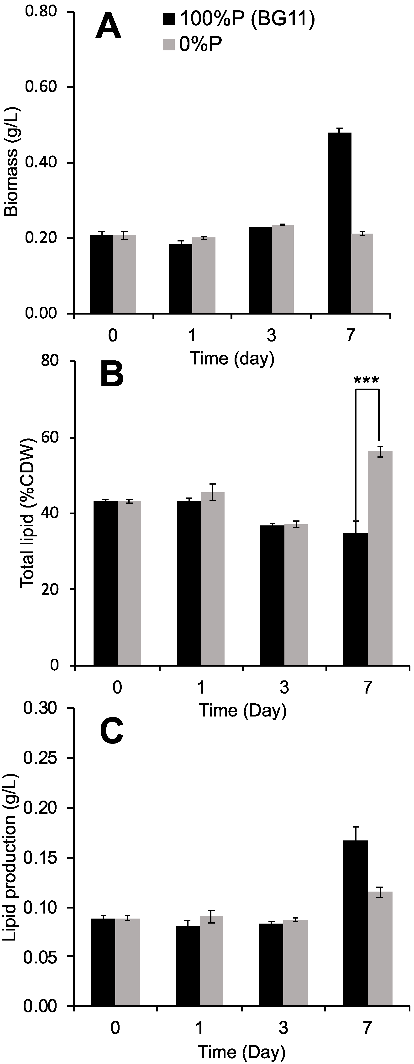
Figure 1. Cultivation of Chlorella sp. under normal BG11 and P-deprived BG11 conditions. (A) Biomass (g/L), (B) total lipid (%DCW), (C) lipid production (g/L). All the data are expressed as mean ± standard deviation from cultures that were run in triplicate (*** p < 0.001).
Phycology 05 00049 g001
Figure 2. Cultivation of Chlorella sp. under P deprivation in BG11 with added metals (Fe and Co). (A) Biomass (g/L), (B) total lipid (%DCW), (C) lipid production (g/L). All the data are expressed as mean ± standard deviation from cultures that were run in triplicate (* p < 0.1; *** p < 0.001).
Phycology 05 00049 g002
Figure 3. Cultivation of Chlorella sp. under P limitation in BG11 with added Pb: 100% P (AC), 50% P (DF), and 0% P (GI). All the data are expressed as mean ± standard deviation from cultures that were run in triplicate (*** p < 0.001).
Phycology 05 00049 g003
Figure 4. H2O2 and MDA levels and SOD and CAT activities in 10 µM Pb-treated cells. All the data are expressed as mean ± standard deviation from cultures that were run in triplicate.
Phycology 05 00049 g004
Table 1. Fatty acid profiles and biodiesel properties as per ASTM D6751 and EN14214 [11] under combined stress conditions with and without Pb and various P levels.
Fatty Acid Compositions (%)
Types of fatty acidsBG-11BG-11 with Pb
100% a50% a0% a100% a50% a0% a
Decanoic acid (C10:0)8.21 ± 0.377.66 ± 0.31n.d. *24.30 ± 0.8011.16 ± 0.332.56 ± 0.11
Myristic acid (C14:0)11.86 ± 0.3610.88 ± 0.30≤0.013.33 ± 0.1111.13 ± 0.2812.02 ± 0.35
Palmitic acid (C16:0)42.27 ± 1.9945.86 ± 1.8649.24 ± 1.9526.19 ± 1.0538.59 ± 1.7554.57 ± 2.05
Palmitoleic acid (C16:1)15.52 ± 0.629.32 ± 0.3933.36 ± 1.505.27 ± 0.1517.15 ± 0.807.61 ± 0.33
Stearic acid (C18:0)17.94 ± 0.8116.68 ± 0.7811.36 ± 0.3525.24 ± 0.8118.01 ± 0.8211.89 ± 0.34
Oleic acid (C18:1)≤0.016.92 ± 0.31≤0.0110.59 ± 0.35≤0.012.30 ± 0.10
Others b2.90 ± 0.102.68 ± 0.116.02 ± 0.245.08 ± 0.223.95 ± 0.129.05 ± 0.35
SFA *83.28 ± 2.4281.07 ± 2.1860.59 ± 2.3079.06 ± 2.3178.88 ± 2.3181.04 ± 2.51
UFA *16.72 ± 0.6518.93 ± 0.8039.41 ± 1.8120.94 ± 0.9021.12 ± 0.8218.96 ± 0.78
FactorsENASTMBiodiesel properties
SV (mg KOH/g)NANA226.50 ± 10.61224.14 ± 9.82215.32 ± 9.85236.64 ± 10.18228.92 ± 10.73219.49 ± 9.55
IV (g I2/100g)120NA16.57 ± 0.7617.95 ± 0.8138.75 ± 1.7419.35 ± 0.7520.69 ± 0.8317.74 ± 0.51
CN>51>4766.67 ± 2.9166.61 ± 2.8862.93 ± 2.8265.00 ± 2.6865.49 ± 3.0167.18 ± 3.09
CP (°C)NANA18.82 ± 0.8519.13 ± 0.8820.91 ± 0.848.78 ± 0.3315.31 ± 0.6623.71 ± 1.14
HHV (MJ/kg)NANA38.90 ± 1.5638.97 ± 1.6239.21 ± 1.8138.56 ± 1.7138.82 ± 1.8639.09 ± 1.90
Viscosity (mm2/s)3.5–5.01.9–6.03.47 ± 0.163.54 ± 0.173.70 ± 0.173.18 ± 0.163.37 ± 0.163.68 ± 0.18
Density (g/cm3)0.86–0.90NA0.87 ± 0.040.87 ± 0.040.87 ± 0.050.87 ± 0.040.87 ± 0.040.87 ± 0.04
n.d. * = not detected, SFA * = saturated fatty acid, UFA * = unsaturated fatty acid. a Phosphorus concentration. b C18:2 and C18:3 total %. NA = not available. The data are the means ± SD of triplicate experiments.
Table 2. Energy conversion efficiency from light to lipids under different conditions.
ConditionsTotal Lipids (g/L)HV of Lipids (kJ)Input Light Energy (kJ)TECE (%)
100% P (BG11)0.09 ± 0.0013.14 ± 0.0618.8815.84 ± 0.71
0% P0.09 ± 0.0033.25 ± 0.1018.8817.19 ± 0.51
0% P + Co0.06 ± 0.012.35 ± 0.1918.8812.44 ± 1.03
0% P + Fe0.10 ± 0.033.71 ± 1.1918.8819.66 ± 6.28
100% P + Pb0.14 ± 0.0085.04 ± 0.2918.8826.70 ± 1.52
50% P + Pb0.09 ± 0.0013.16 ± 0.0718.8816.73 ± 0.36
0% P + Pb0.08 ± 0.012.85 ± 0.3718.8815.08 ± 1.93
Disclaimer/Publisher’s Note: The statements, opinions and data contained in all publications are solely those of the individual author(s) and contributor(s) and not of MDPI and/or the editor(s). MDPI and/or the editor(s) disclaim responsibility for any injury to people or property resulting from any ideas, methods, instructions or products referred to in the content.