Article

Alkalinema pantanalense and Roholtiella edaphica (Cyanobacteria): Two New Species Records for Egypt

by
Rania M. Mahmoud
1,
Mostafa M. El-Sheekh
2,*,
Asmaa A. Adawy
1 and
Abdullah A. Saber
3,*
1
Botany Department, Faculty of Science, Fayoum University, Fayoum 63514, Egypt
2
Botany Department, Faculty of Science, Tanta University, Tanta 31527, Egypt
3
Botany Department, Faculty of Science, Ain Shams University, Abbassia Square, Cairo 11566, Egypt
*
Authors to whom correspondence should be addressed.
Phycology 2025, 5(3), 46; https://doi.org/10.3390/phycology5030046
Submission received: 19 July 2025 / Revised: 7 September 2025 / Accepted: 11 September 2025 / Published: 15 September 2025

Abstract

Our current knowledge of the cyanobacterial diversity in Egypt is still underestimated During our routine study on Egyptian cyanobacteria, two interesting and morphologically cryptic strains were isolated from streams of Bahr Yussef and Qarun Lake, one of the oldest lakes in the world, located at the Faiyum depression, Egypt. We applied the polyphasic approaches, combining the state-of-the-art morphotaxonomy, 16S rRNA gene phylogenies, and ecological preferences to precisely unravel the taxonomic positions of these two cyanobacterial strains. Based on a combination of their morphotaxonomic traits and 16S rRNA phylogenetic assessment, we identified them as Alkalinema pantanalense (Leptolyngbyaceae, Leptolyngbyales) and Roholtiella edaphica (Nostocaceae, Nostocales). Both species are considered new cyanobacterial records for Egypt and the African continent based on the available literature. From an ecological standpoint, both species are eutraphentic, where they could tolerate relatively elevated concentrations of NO3, NH4+ (in particular for R. edaphica), and silicates, reflecting eutrophication signs in the ecosystems they colonize. This study adds to the limited molecular information available on the Egyptian cyanobacteria, and also highlights the need for re-investigation of Egyptian cyanobacteria, using polyphasic approaches, to better understand their taxonomy and ecology.

1. Introduction

Cyanobacteria are autotrophic gram-negative bacteria with heterogeneous genera and species. Cyanobacteria inhabit diverse ecosystems, ranging from freshwater to marine and even extreme terrestrial environments [1]. Morphological diagnostic traits and life-cycle stages have long been used to clarify their taxonomic positions, although these criteria are currently insufficient to distinguish species complexes and cryptic species and genera [2,3,4]. Over the past decades, numerous studies addressed the necessity of polyphasic approaches, combining morphological, ecological, and eco-physiological traits to precisely identify different species and infraspecies of cyanobacteria [5,6,7,8]. With the aid of sequencing 16S rRNA and 16S–23S rRNA internal transcribed spacer (ITS) genes, as well as the application of modern taxonomy standards for describing novel species, rapid changes in cyanobacterial taxonomy have been taking place [9,10,11,12].
The family Leptolyngbyaceae is distinguished by having thin trichomes, 1.5–2.5 (–4.5) μm wide, and cylindrical cells with facultatively constricted cross-walls [10]. Applying the polyphasic approach, several new and cryptic genera, which are morphologically very similar to the genus Leptolyngbya Anagnostidis & Komárek, have been detected. Among them, the genus Alkalinema Vieira Vaz et al. was described in alkaline and saline wetlands in the Pantanal, Brazil [13]. Taxonomically, Alkalinema is a monotypic genus with the type species Alkalinema pantanalense Vieira Vaz et al., which is characterized by having trichomes which are frequently organized in ornate (interwoven) mats in liquid medium. The color of the culture ranges from red to brown. Sheath is usually absent, but the trichomes have diffluent mucilages and are non-motile. Cell content is homogeneous, and the shape ranges from isodiametric to rectangular, 2.0–4.1 µm long × 1.7–2.2 µm broad. Apical cells are rounded–conical or narrowed. Reproduction usually occurs by hormogonia formation.
While conducting their broader survey on the hidden diversity of terrestrial cyanobacteria, Bohunicka et al. [14] discovered the new genus Roholtiella Bohunická, Pietrasiak & J.R. Johansen (Nostocaceae, Nostocales) from soil localities in the USA, Russia, and Czech Republic. Morphologically, the genus Roholtiella shares some taxonomic traits with the genus Calothrix C.Agardh ex Bornet & Flahault, but it is still distinguished by having a series of arthrospores and existing without a tapering terminal hair. Ecologically, Roholtiella is usually found in fresh water and soils. Phylogenetically, Roholtiella is also close to the genus Calochaete Hauer, Bohunická & Mühlsteinová, but they are still different in the conserved structures of the 16S–23S ITS regions. So far, there are only six taxonomically accepted species of the genus Roholtiella [15]: R. bashkiriorum Gaysina & Bohunická, R. edaphica Bohunická & Lukesová, R. fluviatilis J.R. Johansen & Gaysina, R. mojavensis N. Pietrasiak & J.R. Johansen [14], R. mixta Abdullin & A.A. Gontcharov [16], and R. volcanic Gaysina et al. [17].
Previous studies of cyanobacteria in Egypt have been largely depended on classical morphotaxonomy systems. These studies have documented the presence of various cyanobacterial species in diverse habitats, including the Nile River and its delta [18,19,20]. However, this traditional taxonomic approach most often overlooks the genetic diversity and ecological preferences of cyanobacteria, leading to an incomplete understanding of their accurate taxonomic positions and distribution. Recent research on Egyptian cyanobacteria has uncovered the presence of some species new to science, and has highlighted the need for integrating molecular techniques, such as DNA sequencing and phylogenetic analysis, and modern taxonomy standards to obtain a more accurate and comprehensive picture of the diversity of cyanobacteria [1,21,22]. For instance, the recent study conducted by Hentschke et al. [23] on a filamentous cyanobacterial strain that was isolated from a benthic mat in an irrigation canal in the Nile discovered the novel species Pegethrix niliensis G.S. Hentschke. Qasem and other co-workers [24] also discovered the new cyanobacterial species Desikacharya aegyptiaca Qasem, El-Gamal, Mahfouz, A.A. Saber & Salah El Din from the Egyptian soils. Generally, taxonomic knowledge about Egyptian, and also African [25,26,27], cyanobacteria is still understudied [22,28]. Komárek [28] pinpointed that the diversity of cyanobacteria in North Africa includes several cryptic species complexes, and these taxa require more detailed investigations, including genetic, ecophysiological, and phenotypic taxonomy systems.
This paper documents the existence of Alkalinema pantanalense and Roholtiella edaphica as new cyanobacterial species records for Egypt in streams of Bahr Yussef and Qarun Lake, respectively, in the Faiyum depression, Egypt. Morphotaxonomy, ecology, and 16S rRNA phylogeny of both species are provided.

2. Materials and Methods

2.1. Sampling and Isolation of Cyanobacterial Strains

Our two strains were isolated from water samples collected in two different loci in the El-Faiyum governorate, Egypt (Figure 1). The strain Alkalinema pantanalense RMFY20 was isolated on August 2020 from Bahr Yussef (29°16′32.5″ N, 30°49′21.6″ E). Bahr Yussef is a small canal which feeds water northwards into the Faiyum Oasis, parallel with the Nile. The second strain, Roholtiella edaphica AR5, was isolated in July 2021 from the eastern sector of Qarun Lake, near the outlet of El-Batts drain (29°30′14.0″ N, 30°50′36.6″ E). Qarun Lake is a historical lake located in the Faiyum Oasis, ~80 km southwest of Cairo, which is nowadays just a small portion of its previous size. Each cyanobacterial strain was isolated using the streaking technique from the cyanobacterial and microalgal cultures grown from each ecosystem. A culture flask containing MBL medium [29] was used for the cultivation of A. pantanalense RMFY20 at 25 ± 1 °C, while BG-110 medium [30] was used for R. edaphica AR5 at 29 ± 1 °C. Both strains were kept under a 16:8 h light:dark photocycle using white fluorescent light (40 µmol photons m–2 s−1).

2.2. Hydrochemical Analyses

The seasonal physical and chemical parameters of water samples of the two ecosystems studied were measured following the protocols adapted by the American Public Health Association Standard Methods [31,32]. Water conductivity and pH were measured in situ. Ammonium (NH4+) concentrations were determined using the indophenol method following the method adapted by Bremner and Shaw [33]. Nitrite (NO2) was assessed using a colorimetric method [34]. Nitrate (NO3) was measured according to the protocol described by Mullin and Riley [35]. Orthophosphates (PO43−) were measured following the stannous chloride method. Silicate was estimated using the molybdosilicate method [36].

2.3. DNA Extraction, PCR Amplification, Sequencing, and Phylogenetic Analysis

DNA extraction and PCR amplification were carried out using the primers WAW8F (5′-AGAGTTTGATCCTGGCTCAG-3′) and WAW23SR (5′-TTTGCGGCCGCTGTGTGCCTAGGTATCC-3′) to amplify the 16S rRNA gene, as described by Casamatta et al. [37]. For phylogenetic assessment, BLAST searches were performed using all of the 16S rRNA sequences generated against the NCBI core nucleotide database. The phylogenetic relationships of the studied strains were inferred using Bayesian inference implemented in MrBayes 3.2.7a arm. The nucleotide substitution model GTR+I+G (General Time Reversible model with a proportion of invariable sites and gamma-distributed rate heterogeneity) [38] was selected. Two independent runs were performed, each with four Markov chains (one cold, three heated) for 5,000,000 generations, sampling every 1000 generations. A burn-in fraction of 25% was applied, and convergence was assessed by examining the average standard deviation of split frequencies (<0.01) and the potential scale reduction factor (PSRF ≈ 1.0). A 50% majority-rule consensus tree was generated with posterior probabilities (PP) mapped onto the nodes. This was applied as the best-fit model of nucleotide substitution, and 1000 bootstrap replications were used to assess how robust the inferred phylogenetic connections were. A total of 16S rRNA gene sequences of the cyanobacterial strains were submitted to the NCBI GenBank database under accession numbers OR469049 and OR610310 for Alkalinema pantanalense RMFY20 and Roholtiella edaphica AR5, respectively.

2.4. Morphological Analyses

For morphological analyses, each strain was examined and microphotographed using a BEL® photonics biological microscope (BEL® Engineering, Monza, Italy) fitted with a Canon Powershot G12 digital camera. The specimens were identified following the relevant taxonomic characteristics in Vieira Vaz et al. [13] for Alkalinema pantanalense RMFY20, and in Bohunická et al. [14] for Roholtiella edaphica AR5. In each cyanobacterial strain, 20–30 measurements of each taxonomic characteristic were carried out at various positions of the slides prepared.

3. Results

3.1. Hydrochemical Characterization

In general, and during the study period, Qarun Lake water contained a higher concentration of NH4+ ions (750 µg.L−1) than Bahr Yussef (89 µg.L−1). However, average concentrations of silicates (1900 µg.L−1) and PO43− (200 µg.L−1) in Bahr Yussef were distinctly higher than those in Qarun Lake (1206 and1531 µg.L−1, respectively) (Figure 2). Both ecosystems were in general characterized by having alkaline water, with an average pH value of 8.5 at Qarun Lake and 7.8 at Bahr Yussef. The average water temperature gradients were 33.1 °C at Qarun Lake and 30 °C for Bahr Yussef, reflecting the warm conditions typical for these aquatic ecosystems.

3.2. Taxonomic Descriptions

3.2.1. Alkalinema Pantanalense Vieira Vaz et al. [13] (Figure 3a–h)

Description: Trichomes grow free-floating in liquid medium with reddish to brownish color. Trichomes solitary or organized in interwoven mats. Each trichome has a thin sheath. Cells are isodiametric or longer, with a homogeneous content and reddish to brownish, 1.5–2.5 µm wide × 2.0–3.0 µm long. End cell rounded to narrowly short conical. Reproduction only occurs by hormogonia.
Distribution in Egypt: This is the first record of this cyanobacterial species in Egypt where it was found in Bahr Yussef, the Faiyum depression, Egypt.
Figure 3. Light micrographs of Alkalinema pantanalense: (a) macroscopic interwoven mats growing in liquid medium, (b) details of trichomes and sheaths, (c) trichome with rounded end cell, (d,h) trichomes with narrowly short conical end cells, (eg) hormogonia formation and trichome disintegration. Scale bars: 10 µm.
Phycology 05 00046 g003

3.2.2. Roholtiella edaphica Bohunická & Lukesová [14] (Figure 4a–s)

Description: Thallus olive-green to brown-olive, with short-to-long heteropolar filaments which are usually single and rarely false-branched. Sheath colorless or slightly brown, thin and firm, sometimes widened in older filaments, closed at the ends or becoming diffluent towards the ends, or open when the arthrospores are released. Trichomes 5.5–11.0 μm wide, gradually tapered and constricted at cross-walls, sometimes swollen at the base where the widest cells are basal or rarely intercalary. Cells are generally shorter than wide or sometimes slightly longer than wide, barrel-shaped to nearly spherical, or spherical–compressed, 2.5–9.0 μm long. Cell content is smooth or finely granulated. End cells conical or conical rounded, 4.0–7.0 μm long × 3.0–5.5 μm wide. Heterocytes, both intercalary and terminal, are pale yellow or yellow. Intercalary heterocytes are usually barrel-shaped and shorter than wide, but terminal heterocytes are hemispherical to spherical, 3.0–8.5 μm long × 5.0–10.0 μm wide. Reproduction occurs by the formation of short hormogonia and arthrospores. Arthrospores are usually released from the end of the filaments by dissociation, 4.5–9.0 μm long × 6.0–11.5 μm wide.
Distribution in Egypt: The finding of this cyanobacterial species in Qarun Lake represents the first documented instance of its existence in Egypt.
Figure 4. Light micrographs of Roholtiella edaphica: (ac) hormogonia, (dg) young filaments with basal heterocytes. Note the beginning of false branching (arrowhead) and swollen bases in (e). (hj) Filaments with intercalary heterocytes, (kp,r) details of tapered mature filaments. Note the diffluent sheath at the filament end (m), and the firm, widened sheath at the filament base (o). (q) Mature filament with a false branching at the intercalary compressed heterocyte, (s) formation and releasing of arthrospores. Scale bars: 10 µm, except (a) 20 µm.
Phycology 05 00046 g004

3.3. Phylogenetic Assessment

The Bayesian phylogenetic tree (Figure 5) based on the 16S rRNA gene sequences resolved the studied taxa into well-supported clades. The GTR+I+G model provided a robust fit to the data, and most nodes were supported by high posterior probabilities (≥0.95).
A sample was taken every 1000 generations in two simultaneous runs of 5 million generations, with the first 25% being destroyed as burn-in. Strong phylogenetic resolution was shown by the consensus tree’s high posterior probability support across the majority of internal nodes, with values ranging from 95 to 100% for most branches.
With posterior probability of 100% (PP = 100), the isolate Roholtiella edaphica RAFY13 grouped securely within the Roholtiella clade alongside reference strains (R. edaphica AR2–AR6, AR39, AR45, JOH4, JOH39, and RU1). In a similar manner, Alkalinema pantanalense RMFY20 was assigned to the Alkalinema lineage, forming a group comprising reference strains (DHCY212 and DHCY213) and type strains (A. pantanalense CENA528–CENA531) that had 100% posterior support. These positions provide strong phylogenetic evidence for the two isolates affiliated with the genera Roholtiella and Alkalinema, respectively, and validate their identities as R. edaphica and A. pantanalense. In line with earlier taxonomic frameworks, the tree also retrieved the more general relationships between the groups Nostocaceae, Tolypothrichaceae, and Leptolyngbyaceae.
Overall, the phylogeny confirms the placement of the newly isolated strains within their respective genera and supports their affiliation as new records for Egypt.
To complement the tree topology in Figure 6, a heatmap of patristic distances (substitutions per site) was generated using branch length information from the consensus tree. This matrix representation highlighted the relative genetic divergence among taxa, with closely related strains forming blocks of low distance values (blue), while more distantly related taxa exhibited higher distances (yellow-to-green). Together, the Bayesian phylogeny and patristic distance heatmap provide strong evidence for the accurate placement of the studied isolates within their respective genera and reveal the degree of evolutionary divergence among the sampled cyanobacteria.
To exclude potential affinity with Calochaete, we included representative Calochaete 16S sequences in our phylogeny. Bayesian inference grouped the isolate firmly within Roholtiella (posterior probability = 1.00), consistent with morphology and supporting its identification as Roholtiella. We note that 16S–23S ITS would provide additional resolution, and we plan to pursue it in future work.

4. Discussion

Our strain of Alkalinema pantanalense was placed in a well-supported cluster mainly composed of A. pantanalense sequences based on the 16S rRNA phylogenetic assessment. Morphometric diagnostic features of the Egyptian A. pantanalense strain examined also coincide with the original description provided by Vieira Vaz et al. [13]: thallus morphology, cell outline and dimensions, and mode of reproduction. However, our A. pantanalense strain is distinguished by the presence of a sheath around each trichome. This taxonomic trait has not been reported in the description of the holotype [13]. Supporting our findings, dela Cerna and Geraldino [39] isolated six sheathed strains of A. pantanalense from a hot spring in Negros Oriental (Philippines), and confirmed their identifications using an integrated approach of morphological and molecular data. During their phylogenetic and taxonomic study on the diversity of homocytous cyanobacteria, Genuário et al. [40] also documented the presence of a sheath in trichomes of the strain A. pantanalense CMAA1554 isolated from Amazonian rivers. We think that the presence and/or absence of the sheath in A. pantanalense is a facultative characteristic belonging to the environmental conditions, and its presence in the Egyptian strain is a type of phenotypic adaptation to the environment ‘ecomorphotype’. From an ecological standpoint, our A. pantanalense was isolated from the alkaline nutrient-rich freshwater stream of Bahr Yussef. In general, it has been reported that A. pantanalense can be found in a wide ecological niche, i.e., saline–alkaline Pantanal wetlands in Brazil [13], phytoplanktonic or epilithic species living on gravel in freshwaters in South Korea, China, Brazil, and Sri Lanka [40,41,42,43], and even in cryoconite holes in Antarctica [44], indicating its high adaptive environmental capability and extensive ecological distribution. Based on the available literature, this study provides the first record of A. pantanalense in Egypt and the African continent.
Given that the Roholtiella edaphica clade includes the Egyptian strain, with typical R. edaphica morphology, and other established strains of the species, we, therefore, maintain the conservative identification of the Egyptian material as R. edaphica. From an ecological point of view, this study pinpointed that R. edaphica is frequent in the planktonic assemblages of the alkaline nutrient-rich Qarun Lake. However, this species was first described from Sahara soil in the USA by Bohunicka et al. [14], and future research studies reported its presence in the terrestrial cyanobacterial diversity of Eurasia [45]. This study highlights that R. edaphica has a high degree of ecological plasticity and resilience to changing environmental conditions, where it can occur in mineral- and nutrient-rich ecosystems such as Qarun Lake, in addition to its well-known ecological preferences for the soils. Due to its specific reproductive structures that can resist prolonged desiccation, we think that this cyanobacterial species can be ecologically adapted to these harsh environmental conditions.
We think that both A. pantanalense and R. edaphica have been transferred from the Sahara Desert across the Atlantic Ocean into both North and South America. Supporting our hypothesis, a recent study conducted by Johansen et al. [46] described the genetically close cyanobacterial species Pseudoacaryochloris cystiformans Jusko & J.R. Johansen and P. abyssiniae Melaku Mesfin & J.R. Johansen from the San Nicolas Island and Ethiopia, respectively, supporting the hypothesis of the dispersal of cyanobacteria from arid soils in Africa and their establishment in the Americas, followed by genetic lineage separation in the immigrant populations. Wind is indeed the most likely vector for dispersal of the soil microbes [47], and cyanobacteria have been emphasized to be present in atmospheric samples [48]. It has been hypothesized that dust particles originating in the Sahara Desert in Africa have been found in both North and South America [49,50]. The same conclusion can be applied for cyanobacteria, where they could make a similar journey across the Atlantic Ocean [46]. Conclusively, and based on our integrative polyphasic study and the available literature, both A. pantanalense and R. edaphica are considered new cyanobacterial species for Egypt and Africa.

5. Conclusions

In conclusion, reporting the two cyanobacterial species Alkalinema pantanalense and Roholtiella edaphica in Egypt not only partially contributed to our better understanding of the hidden and cryptic diversity of cyanobacteria in Egypt and Africa, but also expanded our limited knowledge about their ecological preferences. Both species seem to tolerate the relatively high concentrations of nutrients and silicates, reflecting the eutrophication process in the ecosystems they colonize.

Author Contributions

Conceptualization, R.M.M. and A.A.S.; methodology and experiments, R.M.M. and A.A.A.; visualization, R.M.M. and A.A.S.; writing—original draft preparation. R.M.M. and A.A.S.; writing—review and editing, M.M.E.-S.; supervision, M.M.E.-S. All authors have read and agreed to the published version of the manuscript.

Funding

This research received no external funding.

Data Availability Statement

The original data presented in this study are included in the article.

Acknowledgments

The authors are very grateful to their home universities for carrying out this research.

Conflicts of Interest

The authors declare that they have no conflicts of interest relevant to the content of this manuscript.

References

  1. Saber, A.A.; El-Refaey, A.A.; Saber, H.; Singh, P.; van Vuuren, S.J.; Cantonati, M. Cyanoprokaryotes and algae: Classification and habitats. In Handbook of Algal Biofuels: Aspects of Cultivation, Conversion and Biorefinery; El-Sheekh, M., Abomohra, A., Eds.; Elsevier: Amsterdam, The Netherland, 2022; pp. 1–38. [Google Scholar]
  2. Komárek, J.; Anagnostidis, K. Süßwasserflora von Mitteleuropa. Cyanoprokaryota. Oscillatoriales; Elsevier Spektrum Akademischer Verlag: München, Germany, 2005; Volume 19, p. 759. [Google Scholar]
  3. Komárek, J.; Kaštovský, J.; Ventura, S.; Turicchia, S.Š. The cyanobacterial genus Phormidesmis. Algol. Stud. 2009, 129, 41–59. [Google Scholar] [CrossRef]
  4. Whitton, B.A. Cyanobacteria (Cyanophyta). In The Freshwater Algal Flora of the British Isles: An Identification Guide to Freshwater and Terrestrial Algae, 2nd ed.; John, D.M., Whitton, B.A., Brook, A.J., Eds.; Cambridge University Press: Cambridge, UK, 2011; pp. 31–158. [Google Scholar]
  5. Andreote, A.P.D.; Vieira Vaz, M.G.; Genuário, D.B.; Barbiero, L.; Rezende-Filho, A.T.; Fiore, M.F. Nonheterocytous cyanobacteria from Brazilian saline-alkaline lakes. J. Phycol. 2014, 50, 675–684. [Google Scholar] [CrossRef]
  6. Komárek, J. A polyphasic approach for the taxonomy of cyanobacteria: Principles and applications. Eur. J. Phycol. 2016, 51, 346–353. [Google Scholar] [CrossRef]
  7. Komárek, J. Taxonomic review of cyanobacteria 2021/2022 according to polyphasic evaluation. Fottea 2023, 23, 141–148. [Google Scholar] [CrossRef]
  8. Kaštovský, J. Welcome to the jungle!: An overview of modern taxonomy of cyanobacteria. Hydrobiologia 2024, 851, 1063–1077. [Google Scholar] [CrossRef]
  9. Johansen, J.R.; Kovacik, L.; Casamatta, D.A.; Fučíková, K.; Kaštovský, J. Utility of 16S-23S ITS sequence and secondary structure for recognition of intrageneric and intergeneric limits within cyanobacterial taxa: Leptolyngbya corticola sp. nov. (Pseudanabaenaceae, Cyanobacteria). Nova Hedwig. 2011, 92, 283–302. [Google Scholar] [CrossRef]
  10. Strunecký, O.; Ivanova, A.P.; Mareš, J. An updated classification of cyanobacterial orders and families based on phylogenomic and polyphasic analysis. J. Phycol. 2023, 59, 12–51. [Google Scholar] [CrossRef]
  11. Labrada, N.A.; McGovern, C.A.; Thomas, A.L.; Hurley, A.C.; Mooney, M.R.; Casamatta, D.A. The CIMS (Cyanobacterial ITS motif slicer) for molecular systematics. Fottea 2024, 24, 23–26. [Google Scholar] [CrossRef]
  12. Ginn, B.L.; Lau, C.Y.; Asma, N.A.; Serri, N.A.; Broady, P.; Convey, P.; Muangmai, N.; Merican, F. Desmonostoc cockrellii sp. nov. (Nostocales, Cyanobacteria): A new record of a subaerophytic species from the Habitat Penang Hill, Malaysia. Fottea 2025, 25, 91–104. [Google Scholar] [CrossRef]
  13. Vieira Vaz, M.G.; Genuário, D.B.; Andreote, A.P.; Malone, C.F.; Sant’Anna, C.L.; Barbiero, L.; Fiore, M.F. Pantanalinema gen. nov. and Alkalinema gen. nov.: Novel pseudanabaenacean genera (Cyanobacteria) isolated from saline-alkaline lakes. Int. J. Syst. Evol. Microbiol. 2015, 65, 298–308. [Google Scholar]
  14. Bohunická, M.; Pietrasiak, N.; Johansen, J.R.; Gómez, E.B.; Hauer, T.; Gaysina, L.A.; Lukesova, A. Roholtiella, gen. nov. (Nostocales, Cyanobacteria)—A tapering and branching cyanobacteria of the family Nostocaceae. Phytotaxa 2015, 197, 84–103. [Google Scholar] [CrossRef]
  15. Guiry, M.D.; Guiry, G.M.; AlgaeBase. World-Wide Electronic Publication;National University of Ireland, Galway. 2025. Available online: http://www.algaebase.org (accessed on 1 July 2025).
  16. Abdullin, S.R.; Nikulin, V.Y.; Nikulin, A.Y.; Manyakhin, A.Y.; Bagmet, V.B.; Suprun, A.R.; Gontcharov, A.A. Roholtiella mixta sp. nov. (Nostocales, Cyanobacteria): Morphology, molecular phylogeny, and carotenoid content. Phycologia 2021, 60, 73–82. [Google Scholar] [CrossRef]
  17. Gaysina, L.A.; Johansen, J.R.; Saraf, A.; Allaguvatova, R.Z.; Pal, S.; Singh, P. Roholtiella volcanica sp. nov., a new species of cyanobacteria from Kamchatkan Volcanic Soils. Diversity 2022, 14, 620. [Google Scholar] [CrossRef]
  18. Shaaban, A.S. Freshwater Algae of Egypt. In Biological Diversity of Egypt; Egyptian Environmental Affairs Agency: Cairo, Egypt, 1994; p. 150. [Google Scholar]
  19. Hamed, A.F. Survey of distribution and diversity of blue-green algae (Cyanobacteria) in Egypt. Acta Bot. Hung. 2005, 47, 117–136. [Google Scholar] [CrossRef]
  20. Hamed, A.F. Biodiversity and distribution of blue-green algae/cyanobacteria and diatoms in some of the Egyptian water habitats in relation to conductivity. Aust. J. Basic Appl. Sci. 2008, 2, 1–21. [Google Scholar]
  21. Saber, A.A.; Cantonati, M.; Mareš, J.; Anesi, A.; Guella, G. Polyphasic characterization of Westiellopsis prolifica (Cyanobacteria) from the El-Farafra Oasis (Western Desert, Egypt). Phycologia 2017, 56, 697–709. [Google Scholar] [CrossRef] [PubMed]
  22. Saber, A.A.; El-Sheekh, M.; Nikulin, A.Y.; Cantonati, M.; Saber, H. Taxonomic and ecological observations on some algal and cyanobacterial morphospecies new for or rarely recorded in either Egypt or Africa. Egypt. J. Bot. 2021, 61, 283–301. [Google Scholar] [CrossRef]
  23. Hentschke, G.S.; Ciancas Jiménez, J.C.; Hoepfner, C.; Guzmán, D.; Mesquita, M.J.; Vasconcelos, V. The extremophile Eurychoronema bolivianum gen. et sp. nov. (Nodosilineales, Cyanobacteria) and Leptolyngbya aquatica comb. nov. Phycologia 2024, 63, 510–519. [Google Scholar] [CrossRef]
  24. Qasem, W.M.; El-Gamal, A.D.; Mahfouz, A.Y.; Saber, A.A.; Salah El Din, R.A. Desikacharya aegyptiaca sp. nov. (Nostocaceae, Cyanobacteria) PQ309038, a novel species isolated from Egyptian soil. Egypt. J. Bot. 2025, 65, 509–517. [Google Scholar]
  25. Mehda, S.; Muñoz-Martín, M.Á.; Oustani, M.; Hamdi-Aïssa, B.; Perona, E.; Mateo, P. Microenvironmental conditions drive the differential cyanobacterial community composition of biocrusts from the Sahara Desert. Microorganisms 2021, 9, 487. [Google Scholar] [CrossRef]
  26. Mehda, S.; Muñoz-Martín, M.A.; Oustani, M.; Hamdi-Aïssa, B.; Perona, E.; Mateo, P. Lithic cyanobacterial communities in the polyextreme Sahara Desert: Implications for the search for the limits of life. Environ. Microbiol. 2022, 24, 451–474. [Google Scholar] [CrossRef]
  27. Stoyneva-Gartner, M.P.; Gartner, G.; Uzunov, B.; Descy, J.-P.; Okello, W. Cyanocystopsis kitagatae gen. et sp. nov. (Cyanoprokaryota/Cyanobacteria) from the tropical lake Kitagata (Uganda, Africa). Wulfenia 2021, 28, 51–65. [Google Scholar]
  28. Komárek, J. Areas of distribution in Cyanobacteria; specificity of the cyanoprokaryotic microflora in the Mediterranean region. Bocconea 2003, 16, 341–354. [Google Scholar]
  29. Nichols, H.W. MBL medium-CSIRO modification adapted for freshwater algae. In Handbook of Phycological Methods; Guillard, R.R.L., Stein, J.R., Eds.; Cambridge University Press: Cambridge, UK, 1973. [Google Scholar]
  30. Rippka, R.; Deruelles, J.; Waterbury, J.B.; Herdman, M.; Stanier, R.Y. Generic assignments, strain histories and properties of pure cultures of cyanobacteria. J. Gen. Microbiol. 1979, 111, 1–61. [Google Scholar] [CrossRef]
  31. APHA. Standard Methods for the Examination of Water and Wastewater, 20th ed.; American Public Health Association: Washington, DC, USA, 2001. [Google Scholar]
  32. Chapman, H.D.; Pratt, P.F. Methods of Analysis for Soil, Plants and Water; Division of Agricultural Sciences, University of California: Oakland, CA, USA, 1978. [Google Scholar]
  33. Bremner, J.M.; Shaw, K. Determination of ammonia and nitrate in soil. J. Agric. Sci. 1955, 46, 320–328. [Google Scholar] [CrossRef]
  34. Barnes, H.; Folkard, A.R. The determination of nitrites: A comparison of existing techniques. Analyst 1951, 76, 711–716. [Google Scholar] [CrossRef]
  35. Mullin, J.B.; Riley, J.P. The spectrophotometric determination of nitrate in natural waters, with particular reference to sea-water. Anal. Chim. Acta. 1955, 12, 464–480. [Google Scholar] [CrossRef]
  36. Krausse, G.L.; Schelske, C.L.; Davis, C.O. Comparison of three wet-alkaline methods of digestion of biogenic silica in water. Freshw. Biol. 1983, 13, 73–81. [Google Scholar] [CrossRef]
  37. Casamatta, D.A.; Johansen, J.R.; Vis, M.L.; Broadwater, S.T. Molecular and morphological characterization of ten polar and near-polar strains within the Oscillatoriales (Cyanobacteria). J. Phycol. 2005, 41, 421–438. [Google Scholar] [CrossRef]
  38. Tamura, K.; Peterson, D.; Peterson, N.; Stecher, G.; Nei, M.; Kumar, S. MEGA5: Molecular evolutionary genetics analysis using maximum likelihood, evolutionary distance, and maximum parsimony Methods. Mol. Biol. Evol. 2011, 28, 2731–2739. [Google Scholar] [CrossRef] [PubMed]
  39. dela Cerna, V.A.M.V.; Geraldino, P.J.L. First record of Alkalinema sp. (Cyanobacteria) thermophilic strains in Negros Oriental, Philippines. Philipp. J. Sci. 2024, 153, 1017–1026. [Google Scholar] [CrossRef]
  40. Genuário, D.B.; Vieira Vaz, M.G.M.; de Melo, I.S. Phylogenetic insights into the diversity of homocytous cyanobacteria from Amazonian rivers. Mol. Phylogenet. Evol. 2017, 116, 120–135. [Google Scholar] [CrossRef] [PubMed]
  41. Chen, M.Y.; Teng, W.K.; Zhao, L.; Han, B.P.; Song, L.R.; Shu, W.S. Comparative genomics reveals insights into cyanobacterial evolution and habitat adaptation. ISME J. 2021, 15, 211–227. [Google Scholar] [CrossRef] [PubMed]
  42. Hossain, M.F.; Bowange, R.W.T.M.R.; Kumara, K.L.W.; Magana-Arachchi, D.N.; Ratnayake, R.R. First record of cyanobacteria species: Cephalothrix komarekiana, from Tropical Asia. Environ. Eng. Res. 2021, 26, 200040. [Google Scholar] [CrossRef]
  43. Lee, O.M. Newly recorded genera and species, Pantanalinema rosaneae and Alkalinema pantanalense (Leptolyngbyaceae, Cyanobacteria) isolated in Korea. J. Species Res. 2022, 11, 10–21. [Google Scholar]
  44. Segawa, T.; Yonezawa, T.; Edwards, A.; Akiyoshi, A.; Tanaka, S.; Uetake, J.; Irvine-Fynn, T.; Fukui, K.; Li, Z.; Takeuchi, N. Biogeography of cryoconite forming cyanobacteria on polar and Asian glaciers. J. Biogeogr. 2017, 44, 2849–2861. [Google Scholar] [CrossRef]
  45. Gaysina, L.A.; Bohunická, M.; Hazuková, V.; Johansen, J.R. Biodiversity of terrestrial cyanobacteria of the South Ural region. Cryptogam. Algol. 2018, 39, 167–198. [Google Scholar] [CrossRef]
  46. Johansen, J.R.; Jusko, B.M.; Mesfin, M.; Lunkis, M.A.; Wain, A.; Hoyer, W.F.; Hasenstab-lehman, K.E. Pseudoacaryochloris (Acaryochloridaceae, Cyanobacteria) species from Africa and North America: A disjunct distribution suggesting transatlantic wind dispersal. West. North Am. Nat. 2025, 85, 193–204. [Google Scholar] [CrossRef]
  47. Marshall, W.A.; Chalmersh, M.O. Airborne dispersal of Antarctic terrestrial algae and cyanobacteria. Ecography 1997, 20, 585–594. [Google Scholar] [CrossRef]
  48. Després, V.R.; Huffman, J.A.; Burrows, S.M.; Hoose, C.; Safatov, A.S.; Buryak, G.; Fröhlich-Nowoisky, J.; Elbert, W.; Andreae, M.O.; Pöschl, U.; et al. Primary biological aerosol particles in the atmosphere: A review. Tellus B Chem. Phys. Meteorol. 2012, 64, 15598. [Google Scholar] [CrossRef]
  49. Engelstaedter, S.; Tegen, I.; Washington, R. North African dust emissions and transport. Earth-Sci. Rev. 2006, 79, 73–100. [Google Scholar] [CrossRef]
  50. Prospero, J.M.; Delany, A.C.; Carlson, T.N. The discovery of African dust transport to the Western Hemisphere and the Saharan air layer: A history. Bull. Am. Meteorol. Soc. 2021, 102, e1239–e1260. [Google Scholar] [CrossRef]
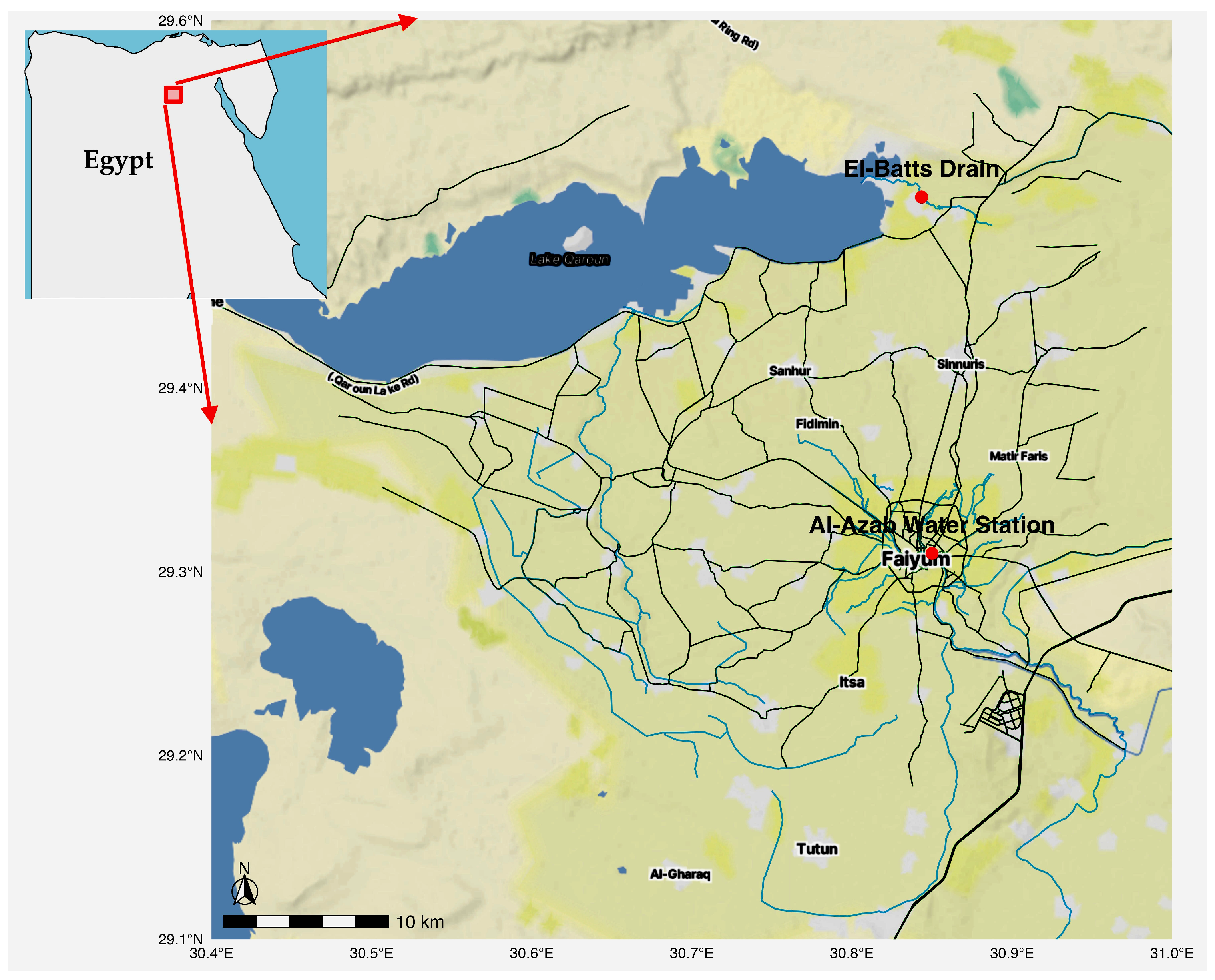
Figure 1. Locations of the sampling sites where the cyanobacterial strains were isolated.
Phycology 05 00046 g001
Figure 2. Boxplots showing average concentrations of the hydrochemical variables of Bahr Yussef and Qarun Lake in the present study.
Phycology 05 00046 g002
Figure 5. Bayesian consensus phylogenetic tree of the studied cyanobacterial strains based on 16S rRNA gene sequences. The analysis was performed in MrBayes under the GTR+I+G substitution model, with two runs of four chains each for 5,000,000 generations. Posterior probabilities (≥0.95) are shown at the nodes. The newly sequenced strains Roholtiella edaphica RAFY13 and Alkalinema pantanalense RMFY20 are highlighted in bold red color.
Phycology 05 00046 g005
Figure 6. Comparative visualization of phylogenetic relationships among cyanobacterial strains. (Left) Heatmap of patristic distances (substitutions per site) based on branch lengths of the Bayesian consensus tree, with warmer colors (yellow–green) indicating greater divergence and cooler colors (blue–purple) reflecting closer relationships. (Right) Bayesian consensus phylogenetic tree inferred from 16S rRNA gene sequences under the GTR+I+G substitution model in MrBayes (5,000,000 generations, two runs, four chains). Posterior probabilities (≥95%) are shown at nodes. The newly sequenced strains Roholtiella edaphica RAFY13 and Alkalinema pantanalense RMFY20 are highlighted in bold red color.
Phycology 05 00046 g006
Disclaimer/Publisher’s Note: The statements, opinions and data contained in all publications are solely those of the individual author(s) and contributor(s) and not of MDPI and/or the editor(s). MDPI and/or the editor(s) disclaim responsibility for any injury to people or property resulting from any ideas, methods, instructions or products referred to in the content.