Article

Morphology and Reproduction of Acanthophora spicifera (Ceramiales: Rhodophyta)

by
Cynthia Mariana Hernández-Casas
1,2,
Rosa María Pineda-Mendoza
2,
Ángela Catalina Mendoza-González
1,
Gerardo Zúñiga
2 and
Luz Elena Mateo-Cid
1,*
1
Instituto Politécnico Nacional, Escuela Nacional de Ciencias Biológicas, Departamento de Botánica, Laboratorio de Ficología, Prolongación de Carpio y Plan de Ayala, Casco de Santo Tomás, Miguel Hidalgo, Mexico City 11340, Mexico
2
Instituto Politécnico Nacional, Escuela Nacional de Ciencias Biológicas, Departamento de Zoología, Laboratorio de Variación Biológica y Evolución, Prolongación de Carpio y Plan de Ayala, Casco de Santo Tomás, Miguel Hidalgo, Ciudad de México 11340, Mexico
*
Author to whom correspondence should be addressed.
Phycology 2025, 5(3), 45; https://doi.org/10.3390/phycology5030045
Submission received: 10 July 2025 / Revised: 27 August 2025 / Accepted: 10 September 2025 / Published: 12 September 2025

Abstract

Rhodomelaceae is the largest red algae family, with 158 genera and more than 1000 described nominal species. In particular, Acanthophora (Rhodomelaceae) is a red alga with erect thalli that arises from stoloniferous branches or holdfast discs, with cylindrical main axes and spine-like branchlets. The life cycle of members of this genus has been partially described; however, the female gamete (carpogonium) has not been described. Here, we present a complete description of each stage in the life cycle of Acanthophora. Thalli of this species were collected from 27 localities in the Gulf of Mexico between 2021 and 2024 and placed in a 5% formaldehyde solution in seawater. Reproductive structures were measured and characterized under stereo and optical microscopes. A total of 62 thalli were collected, of which 10 were carposporophytes, 12 male gametophytes, 1 female gametophyte, 16 vegetative thalli, and 23 tetrasporophytic thalli. A detailed description of the shape and size of the reproductive structures is presented. We documented carpogonium for the first time. The evidence here presented contributes to the description of the life cycle of the genus Acanthophora, in which structures forgotten in current works are recovered, which is of great help in the comparative phycology of the Rhodomelaceae family and Ceramiales order.

1. Introduction

The Class Florideophyceae, with forty orders and 7311 species, is the largest within Rhodophyta [1,2,3,4]. Florideophytes are of great economic importance due to their cell-wall-derived compounds such as agar and carrageenan [5]. This class exhibits a specialized triphasic life history, which consists of a haploid gametophyte phase responsible for sexual reproduction, and two diploid sporophyte phases [6]. The evolutionary success of this group is due to evolutionary innovations such as two additional phases in the life cycle and the presence of carposporophytes and tetrasporophytes [5].
The Order Ceramiales belongs to this class, characterized by the presence of an auxiliary cell that facilitates the development of the carposporophyte [7]. This order is composed of five families, namely Callithamniaceae, Delesseriaceae, Ceramiaceae, Wrangeliaceae, and Rhodomelaceae [4,8], which are closely related [9,10,11].
The Rhodomelaceae is the largest family of the red algae; almost half of all known Florideae belong to this family. Currently, it includes 158 genera and more than 1000 nominal species [4]. This family is monophyletic, and it is closely related to Dasyaceae and Delesseriaceae. The family Rhodomelaceae shows a highly conserved reproductive structure and postfertilization development [7,12]. Fritsch [13] suggested that both Rhodomelaceae and Ceramiales form an ancestral group whose representatives have a structurally complex vegetative system that has evolved in relatively recent times. The Rhodomelaceae species present a well-developed procarp that consists of a carpogonium, which is born on a carpogonial branch, associated with sterile cells and protective cells known as a pericarp. This structure is a unique and key morphological feature for their classification [5,6,14,15,16]. The pericarp initiates before the fertilization of the carpogonium, which achieves its mature condition only after the diploid nucleus reaches the auxiliary cell development of the gonimoblast [6].
The study of the life cycle of Florideophyceae, especially the developmental of reproductive structures in male and female gametophytes as well as tetrasporophytes, is relevant because it provides a much better understanding of the diversification of red algal genera and families [5]. However, the life cycle has been studied in less than 5% of species in the family Rhodomelaceae, including the genera Laurencia [17,18,19,20,21], Chondria [22,23], and Polysiphonia [24,25,26,27]. This life cycle is characterized by being trigenetic, in which the tetrasporophyte and gametophyte are isomorphic, and the gametophyte is dioecious [28,29]; however, some of these phases have not been observed or described in many of the members.
The genus Acanthophora is a red algae group with erect thalli arising from stoloniferous branches or holdfast discs, cylindrical branched main axes, and spine-like branchlets [28,30,31]. Acanthophora spicifera is a native species to Florida, which has been recorded as invasive on the islands of the central Pacific since 1952 and in La Paz Bay since 2006 [32,33]. It has a high degree of morphological plasticity which, combined with its sexual and asexual reproduction, its ability to regenerate, and its adaptability to various environmental conditions, has made it a successful invasive species. Among the negative effects caused by this species, the damage produced to the coral and the displacement of native species, such as Sargassum spp. and other genera, have been highlighted [34]. However, it can also have positive effects, as it promotes the recruitment of sessile organisms by offering stable microhabitats over time. In addition, given that it colonizes most of the rock substrate, it produces a large amount of biomass that could have applications in areas such as biomedicine, given that the main polysaccharide produced by this species is λ-carrageenan, which has antiviral and anti-inflammatory properties [34,35].
These red algae have morphological and vegetative characteristics (e.g., cartilaginous texture), allowing easy fragmentation of the thalli by wave action [29]. These fragments readily adhere to suitable substrates to give rise to new thalli [30,36]. The life cycle of members of this genus has partially been described using drawings, but the female gamete (carpogonium) has not been described [28]. Here, we present a complete description of each of the stages within the Acanthophora life cycle, contributing to knowledge of the reproductive cycle of the members of this genus in the Rhodomelaceae family.

2. Materials and Methods

A total of 62 specimens of Acanthophora spicifera were collected between 2021 and 2024 from 27 localities in the Gulf of Mexico (Figure 1, Supplementary Table S1). Thalli were collected manually with a spatula in the intertidal zone and placed in a 5% formaldehyde solution in seawater to be transported to the laboratory.
Reproductive structures were observed under a Leica S9E stereomicroscope (Leica Microsystems, Wetzlar, Germany). Transverse and longitudinal sections were cut for both cystocarps and tetrasporangial branches, and small portions of the branches containing these structures were collected in the case of spermatia. Additionally, transverse sections of the carposporophytic thalli were taken to locate the carpogonium. All slides were stained with Harris hematoxylin and mounted in Karo® honey (ACH Foods, Mexico City, Mexico) with phenol to avoid the growth of bacteria, fungi, or yeasts [37].
The slides were observed under a Leica DM500 microscope (Leica Microsystems, Wetzlar, Germany), and structures were measured and characterized. Photographs were taken on a VE-BC3 PLUS optical microscope (Velab Co., Pharr, TX, USA), and digital images were edited and assembled on plates using GIMP v 3.0.4.
The shape and size of the reproductive structures were described according to the work by De Jong et al. [30].

3. Results

Of the thalli reviewed, 10 (16.12%) were carposporophytes, 12 (19.3%) male gametophytes, 1 (1.6%) female gametophyte, 16 (24.19%) vegetative thalli, and 23 (38.79) tetrasporophytic thalli.

3.1. Morphological Description

Acanthophora spicifera (Vahl) Børgesen, Botanisk Tidsskrift 30: pp. 177–207, Figure 18. 1910 TYPE: St. Croix, Virgin Islands Vahl, 1802 [38].
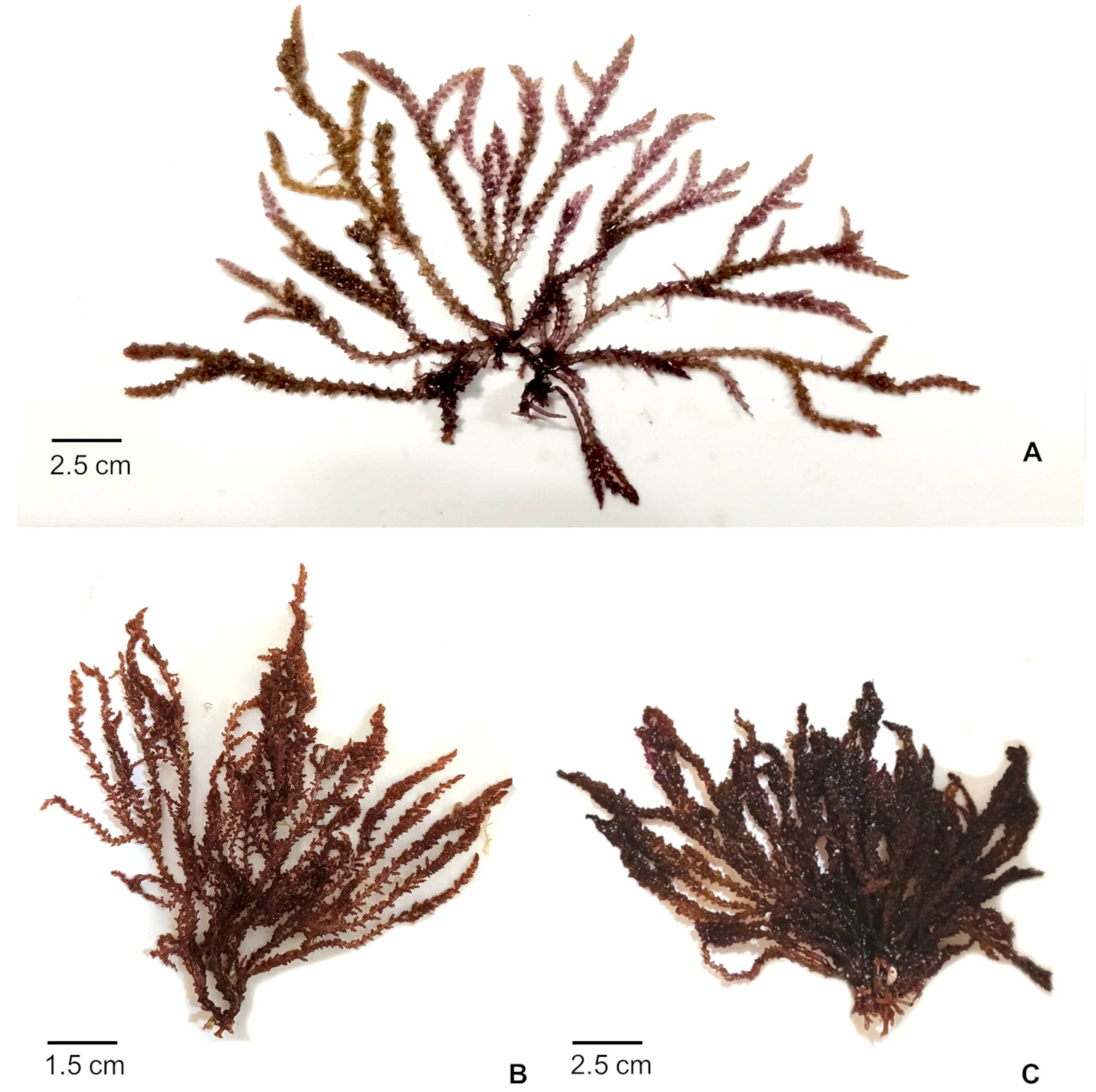
Thalli were erect, with abundant branching. The first is dichotomous, and subsequent ones are alternate and irregular, up to 12 cm high, with purple to reddish coloration. They were adhered to the substrate by means of fixation discs and stoloniferous branches, present cylindrical axes, branches with determined growth, and thorn-like twigs (Figure 2). Anatomically, they consist of a uniaxial axis (30–80 μm in diameter), surrounded by five pericentral cells (130–170 μm high and 150–240 μm wide), with a pith composed of two, scarcely three layers of cells and a layer of rounded-polygonal cells forming the cortex (10–35 μm high and 12–30 μm wide).

3.1.1. Male Gametophyte

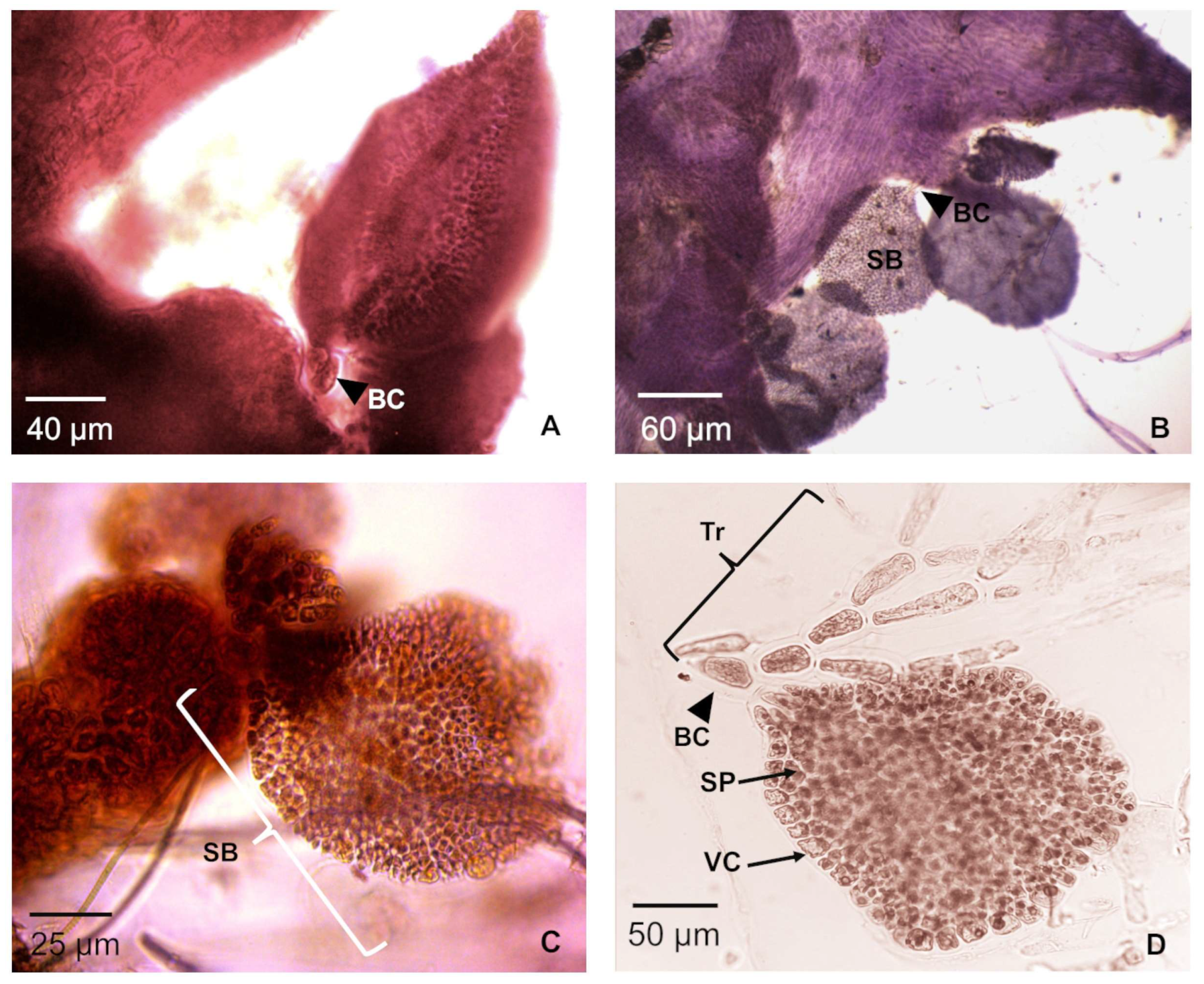
Thalli are pink-violet in color, 4–11 cm tall, and 1–1.5 mm wide in the main axes (Figure 2A). The spermatangial bodies are flattened discs formed from trichoblasts, presenting a layer of vegetative cells (10 μm wide and 7 μm high) surrounding the spermatios (5 μm in diameter) (Figure 3A–D).

3.1.2. Female Gametophyte with Carposporophyte

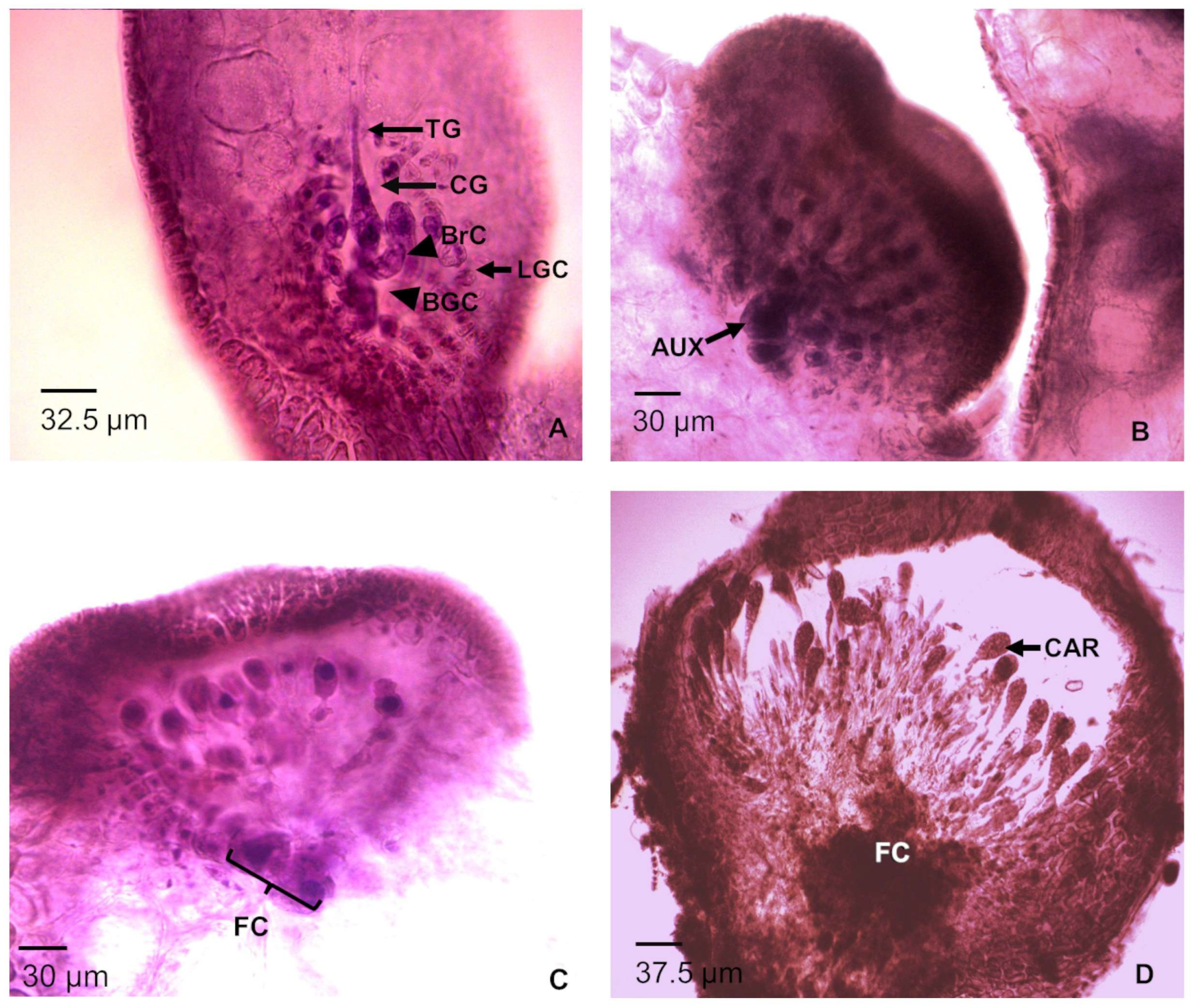
The thallus is pink-violet, up to 11 cm tall, and 1.5 mm in diameter on the main axis (Figure 2B). The carpogonium is formed from a fertile trichoblast that transforms into the carpogonial branch. Figure 4A shows the mature carpogonial branch formed by the carpogonium (12.5 µm high and 21.5 μm wide) from which the trichogonium (32.5 μm long) arises, in addition to the three cells of the carpogonial branch. The rest of the cells are the lateral and basal sterile cell group. This construction is maintained until the time of fertilization.
After fertilization, the trichogyne detaches from the carpogonium, and the cells of the carpogonial branch begin to fuse and disappear as the procarp completes its formation (Figure 4B). The auxiliary cell forms in the apical part of the supporting cell (Figure 4C). Both cells subsequently fuse and form fusion cells. From this cell, the gonimoblastic filaments develop, which gives rise to carpospores (Figure 4C).
Cystocarps form upward from the middle of the thallus. They arise from spine-like branchlets, and are sessile, globose, and urn-shaped with an ostiole at the apical part; they are 950 μm high and 880 μm wide. Mature carpospores are clavate in shape and 37.5 μm long (Figure 4D).

3.1.3. Tetrasporophytes

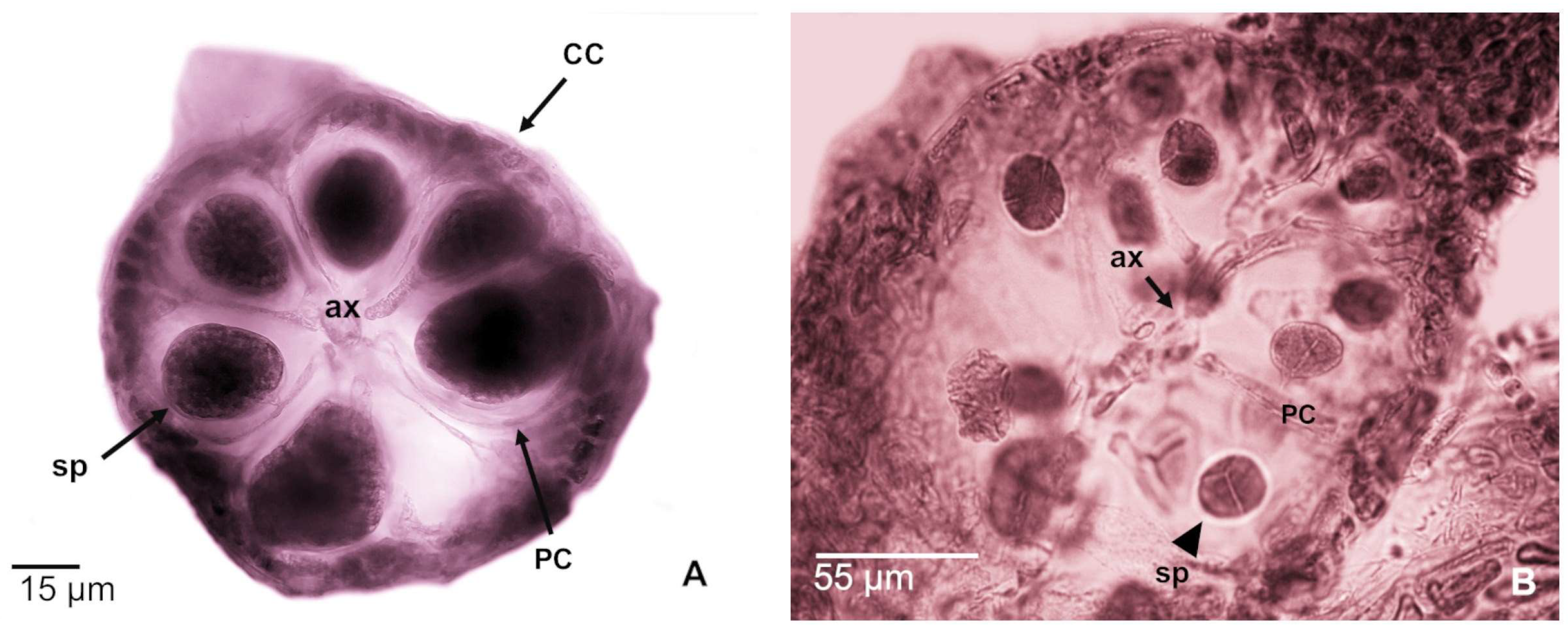
Thalli are violet to purple in color, 4 to 12 cm high, and 1 mm in diameter in the main axes. The tetrasporangial stichidia are formed on the last-order branches, which may have one or two spines. The tetrasporangia are arranged throughout the branch and grow from the pericentral cells, forming a small peduncle. Tetrasporangia are tetrahedral, measuring 42.5 × 50 µm when mature (Figure 5A,B).

4. Discussion

In the present study, the dominant phase was tetrasporophytic, which coincides with the findings reported by Buchan-Antalan and Trono [28] who recorded a higher proportion of tetrasporophytic thalli compared to the rest of the generations for Acanthophora spicifera from Bacoor Bay, Philippines. Santelices [39] mentioned that spore formation and their rapid dispersal require less energy expenditure compared to gametophytes, which occur when conditions are not optimal. Several authors have reported similar results associated with the dominance of a reproductive phase due to seasonal changes in environmental factors [40,41,42,43,44]. Thus, it is considered that the tetrasporophytic phase has a greater survival rate compared to the gametophytic or other reproductive strategies [45,46].
All gametophytes found in this study were dioecious, which agrees with that documented by Cecere [47], who found that the sexual reproduction of the species A. nayadiformis is very similar to the life cycle of members of genera Polysiphonia and Chondria [22].
In A. spicifera, spermatangial bodies are formed from modified trichoblasts, developed as spermatangial bodies surrounded by a vegetative cell layer. Scagel [6] describes trichoblasts as characteristic structures of the Rhodomelaceae family, which are not found in the other families. The trichoblasts are key for the formation of most reproductive structures; even in some genera such as Polysiphionia and Falkenbergiella, whose trichoblasts are produced only when the plants become fertile. On the other hand, Böker-Tôrres et al. [22] mentioned that the male gametes of Chondria curvilineata are formed in spermatangial bodies, with a layer of sterile cells surrounding them, developing from the base of modified trichoblasts, and when fully mature, they are orbicular, pedunculated bodies. Buchan-Antalan and Trono [28] and Cecere et al. [47] described similar reproductive structures in A. spicifera and A. nayadiformis, indicating that the spermatangial bodies are biconcave discs formed from trichoblasts, as occurs in Polysiphonia confusa, in which stichidia is associated with the initial cell of the trichoblasts [27]. This coincides with what was described by Scagel [6], who mentions that in trichoblasts occur a fusion of the spermatangial axes, which results in the formation of plate-shaped fertile axes. Although Acanthophora, Polysiphonia, and Ceramium are classified within the same order (Ceramiales), they present important differences in male gamete development. For example, Polysiphonia confusa forms elongated stichidia, and there is not a layer of sterile cells covering them, while in Ceramium, the spermatia are formed by structures called spermatangia, which are developed from cortical cells forming one or two terminal spermatia [12].
Currently, there is no documented record of the female gamete of Acanthophora. However, given that it belongs to the order Ceramiales, it is assumed that it is formed by a trichogyne, a carpogonium (fertile cell), carpogonial branch cell, and the support cell. Some of these elements have been observed in other genera of the same order, such as Laurencia and Chondria [17,20,22,23]; however, to our knowledge, there is no graphic evidence of a complete carpogonium. Our report is the first documented record of a female gamete, which expands our knowledge of the reproductive characteristics of the genus Acanthophora.
In Acanthophora spicifera, the procarp is composed of both basal and lateral sterile cells, in addition to the carpogonium and the tricogyne. Scagel [6] mentions that Rhodomelaceae is characterized by this constitution, but it is not constant in all families of the Ceramiales, since some genera of Delesseriaceae and Ceramiaceae do not have this structure.
Although Chondria is considered the sister genus of Acanthophora, a structural difference between these genera is the cystocarp form [30], which is the development after fertilization in Rhodomelaceae. This structure in Acanthophora is sessile and formed axially to the central axis, whereas in Chondria, the cystocarp is formed adaxially with an urn shape. Furthermore, the cystocarp floor in Acanthophora is much larger than in Chondria [22,48]. The same condition is observed in Polysiphonia, a genus of the same family, in which the development of the cystocarp is variable. In this genus, the cystocarp is drop-shaped, and the carpospores are formed from short gonimoblast filaments, unlike that observed in Acanthophora, in which longer gonimoblast filaments have been observed and carpospores are formed in the apical part [26,49]. Laurencia and Acanthophora have morphologically similar cystocarps, but those of Laurencia are smaller [20].
As for the tetrasporangia, Buchan-Atalan and Trono [28] reported that A. spicifera forms tetrasporangial stichidia at the apex of the determined branches. Our results confirm the formation of tetrasporangial stichidia in this species; however, unlike the results of previous studies, the tetrasporangia were observed in the distal part of the pericentral cells and scattered throughout the stichidium; a similar pattern was also recorded in Chondria [22]. Although these genera belong to the same family, the formation of reproductive structures of the same generation vary widely among genera. For example, the tetrasporangia in the genus Palisada is arranged in a spiral at the apex of the fertile branch, and not in stichidia, as in Acanthophora [16]. The tetrasporangia in Polysiphonia is formed from the pericentral cells, but they are arranged in a straight line or spiral with respect to the uniaxial axis [23]. These stiquids are complex structures where tetrasporangia are usually protected and can be formed from pericentral cells, although, in some cases, they are formed from cortical cells. This more complex evolutionary characteristic is present in the families Delesseriaceae, Dasyaceae, and Rhodomelaceae, while the family Ceramiaceae lacks protective cells in both the gonimoblast and tetrasporangium, which is why it is considered the most primitive family [6].

5. Conclusions

The evidence presented in this study contributes to the description of the life cycle of the genus Acanthophora, in which structures forgotten in the current literature are recovered. These are of great help in the comparative phycology of the Rhodomelaceae family and Ceramiales.

Supplementary Materials

The following supporting information can be downloaded at: https://www.mdpi.com/article/10.3390/phycology5030045/s1. Table S1. Information about the sample sites of Acanthophora spicifera in the present study.

Author Contributions

Conceptualization, C.M.H.-C., R.M.P.-M. and L.E.M.-C.; methodology, C.M.H.-C., L.E.M.-C. and Á.C.M.-G.; software, C.M.H.-C. and R.M.P.-M.; validation, G.Z., L.E.M.-C., R.M.P.-M. and Á.C.M.-G.; formal analysis, C.M.H.-C., L.E.M.-C., G.Z. and R.M.P.-M.; investigation, R.M.P.-M., C.M.H.-C., Á.C.M.-G., L.E.M.-C. and G.Z.; resources, L.E.M.-C., Á.C.M.-G.; R.M.P.-M. and G.Z. data curation, C.M.H.-C., R.M.P.-M., Á.C.M.-G., L.E.M.-C. and G.Z.; writing—original draft preparation, R.M.P.-M., C.M.H.-C., Á.C.M.-G., L.E.M.-C. and G.Z.; writing—review and editing, C.M.H.-C., R.M.P.-M., Á.C.M.-G., L.E.M.-C. and G.Z. All authors have read and agreed to the published version of the manuscript.

Funding

This proyect was funding by Instituto Politécnico Nacional (SIP-20210885, 20220254, 20230767, 20231466 and 20241358), which provided financial assistance, facilities, and equipment necessary for the development of this study.

Acknowledgments

This work was part of CMHC PhD thesis. She thanks to the Secretaría de Ciencia, Humanidades, Tecnología e Innovación (SECIHTI) for the scholarship (802136), and Luz Elena Mateo-Cid and Ángela Catalina Mendoza-González thank to Comisión de Operación y Fomento de Actividades Académicas (COFAA) and Programa de Estímulos al Desempeño de los Investigadores (EDI), Instituto Politécnico Nacional (IPN). for fellowships granted.

Conflicts of Interest

The authors declare no conflicts of interest.

References

  1. Saunders, G.W.; Hommersand, M.H. Assessing red algal supraordinal diversity and taxonomy in the context of contemporary systematic data. Am. J. Bot. 2004, 91, 1494–1507. [Google Scholar] [CrossRef]
  2. Yoon, H.S.; Müller, K.M.; Sheath, R.G.; Ott, F.D.; Bhattacharya, D. Defining the Major Lineages of Red Algae (Rhodophyta). J. Phycol. 2006, 42, 482–492. [Google Scholar] [CrossRef]
  3. Graham, L.E.; Graham, J.M.; Wilcox, L.W. Algae; Benjamin Cummings: San Francisco, CA, USA, 2009. [Google Scholar]
  4. Guiry, M.D.; Guiry, G.M. AlgaeBase Version 4.2. World-Wide Electronic Publication. National University of Ireland, Galway. 2025. Available online: http://www.algaebase.org (accessed on 20 May 2025).
  5. Yang, E.; Boo, S.; Bhattacharya, D.; Sauders, G.W.; Knoll, A.H.; Fredericq, S.; Graf, L.; Yoon, H.S. Divergence time estimates and the evolution of major lineages in the florideophyte red algae. Sci. Rep. 2016, 6, 21361. [Google Scholar] [CrossRef]
  6. Scagel, R.F. A morphological study of some dorsiventral Rhodomelaceae. Univ. Calif. Publ. Bot. 1953, 27, 1–108. [Google Scholar]
  7. Hommersand, M.H. The morphology and classification of some Ceramiaceae and Rhodomelaceae. Univ. Calif. Publ. Bot. 1963, 35, 165–366. [Google Scholar]
  8. Wynne, M.J. Checklist of benthic marine algae of the tropical and subtropical Western Atlantic: Fifth revision. Nova Hedwig. Beih. 2022, 153, 180. [Google Scholar]
  9. Kylin, H. Entwicklungsgescllichtliche Florideenstudien. Ibid 1928, 24, 127. [Google Scholar]
  10. Kylin, H. 6ber die Entwicklungsgescllichte der FIorideen. Ibid 1930, 26, 104. [Google Scholar]
  11. Papenfus, G.F. Structure and taxonomy of Taenioma; including a discussion on the phylogeny of the Ceramiales. Madroño 1994, 7, 193–214. [Google Scholar]
  12. Choi, H.G.; Kraft, G.T.; Lee, I.K.; Saunders, G.W. Phylogenetic analyses of anatomical and nuclear SSU rDNA sequence data indicate that the Dasyaceae and Delesseriaceae (Ceramiales, Rhodophyta) are polyphyletic. Eur. J. Phycol. 2002, 37, 551–569. [Google Scholar] [CrossRef]
  13. Fritsch, F.E. Present-day classification of algae. Bot. Rev. 1944, 10, 233–277. [Google Scholar] [CrossRef]
  14. Kim, M.S.; Maggs, E.A.; Mc Ivor, L.; Guiry, M.D. Reappraisal of the type species of Polysiphonia (Rhodomelaceae, Rhodophyta). Eur. J. Phycol. 2000, 35, 83–92. [Google Scholar] [CrossRef]
  15. Choi, S.J.; Park, E.J.; Endo, H.; Kitade, Y.; Saga, N. Inheritance pattern of chloroplast and mitochondrial genomes in artificial hybrids of Porphyra yezoensis (Rhodophyta). Fish. Sci. 2008, 74, 822–829. [Google Scholar] [CrossRef]
  16. Sentíes, A.; Dreckmann, K.M. Biodiversidad de las macroalgas marinas de la familia Rhodomelaceae (Rhodophyta) en México. Rev. Mex. Biodivers. 2014, 85, S62–S68. [Google Scholar] [CrossRef]
  17. Nam, K.W.; Saito, Y.; Sohn, C.H. Vegetative structure and reproduction of Laurencia nipponica Yamada (Rhodomelaceae, Rhodophyta). Korean J. Phycol. 1991, 6, 83–92. [Google Scholar]
  18. Furnari, G.; Serio, D. The reproductive structures of the Mediterranean alga Laurencia pelagosae (Ceramiales, Rhodophyta). Eur. J. Phycol. 1993, 28, 141–143. [Google Scholar] [CrossRef]
  19. Nam, K.W.; Saito, Y. Vegetative and reproductive anatomy of some Laurencia (Ceramiales, Rhodophyta) species with a description of L. maris-rubri sp. nov. from the Red Sea. Phycologia 1995, 34, 157–165. [Google Scholar] [CrossRef]
  20. Nam, K.W.; Choi, H.G.; Lee, S.; Park, E.J.; Kang, K.H.; Kim, Y.S. Vegetative and reproductive development of Laurencia venusta (Ceramiales, Rhodophyta). Cryptogam. Algae 2000, 21, 97–110. [Google Scholar] [CrossRef]
  21. Kyaw, S.P.P.; Soe-Htun, U. Studies on the developmental morphology and life history in culture of Laurencia sp. 1 (Ceramiales, Rhodophyta) from Myanmar. J. Aquac. Mar. Biol. 2018, 7, 246–251. [Google Scholar] [CrossRef]
  22. Böker-Tôrres, M.; Horta, P.A.; Salles, J.; Ouriques, L.; Fujii, M.T.; Bouzon, Z. Morfologia e reprodução de Chondria curvilineata F. S. Collins & Hervey (Rhodomelaceae, Rhodophyta), uma adição à flora brasileira. Acta Bot. Bras. 2009, 23, 73–78. [Google Scholar] [CrossRef]
  23. Williams, T.M.; Stacy, A.; Krueger-Hadfield, K.; Hill-Spanik, K.M.; Kosaki, R.K.; Stoeckel, S.; Spalding, H.L. The reproductive system of the cryptogenic alga Chondria tumulosa (Florideophyceae) at Manawai, Papahānaumokuākea Marine National Monument. Phycologia 2024, 63, 36–44. [Google Scholar] [CrossRef]
  24. Kim, M.S.; Lee, I.K. Morphology and reproduction of Polysiphonia yendoi Segi (Rhodomelaceae, Rhodophyta) in Korea. Algae 1997, 12, 73–81. [Google Scholar]
  25. Rindi, F.; Guiry, M.D.; Cinelli, F. Morphology and reproduction of the adventive Mediterranean rhodophyte Polysiphonia setacea. Hydrobiologia 1999, 398/399, 91–100. [Google Scholar] [CrossRef]
  26. Kim, M.S.; Lee, I.K.; Guiry, M. Vegetative and Reproductive Morphology of Polysiphonia lanosa (Rhodomelaceae, Rhodophyta) from Ireland. Bot. Mar. 2002, 45, 293–302. [Google Scholar] [CrossRef]
  27. Aguilar-Rosas, R.; Aguilar-Rosas, L.E.; Pedroche, F.F. Descripción de talos espermatangiales y combinación de fases en Polysiphonia confusa (Rhodomelaceae, Rhodophyta). Rev. Mex. Biodivers. 2006, 77, 1–6. [Google Scholar]
  28. Buchan-Antalan, T.; Trono, G.C. The morphology, growth and seasonality in the reproductive states of Acanthophora spicifera (Vahl) Boergesen in Bacoor Bay. Nat. Appl. Sci. Bull. 1983, 35, 18–27. [Google Scholar]
  29. Kilar, J.A.; McLachlan, J.L. Ecological studies of the algae, Acanthophora spicifera (Vahl) Boerg, (Ceramiales: Rhodophyta): Vegetative fragmentation. J. Exp. Mar. Biol. Ecol. 1986, 104, 1–21. [Google Scholar] [CrossRef]
  30. De Jong, Y.S.D.M.; Hitipew, C.; Prud’homme van Reine, W.F. A taxonomic, phylogenetic and biogeographic study of the genus Acanthophora (Rhodomelaceae, Rhodophyta). Blumea 1999, 44, 217–249. [Google Scholar]
  31. García-Soto, G.C.; López-Bautista, J.M. Acanthophora dendroides Harvey (Rhodomelaceae), a new record for the Atlantic and Pacific oceans. Check List 2019, 15, 509–514. [Google Scholar] [CrossRef]
  32. Ávila, E.; Méndez-Trejo, M.; Riosmena-Rodríguez, R.; López-Vivas, J.; Sentíes, A. Epibiotic traits of the invasive red seaweed Acanthophora spicifera in la paz bay, south Baja California (Eastern pacific). Mar. Ecol. 2012, 33, 470–480. [Google Scholar] [CrossRef]
  33. Mendoza-Becerril, M.A.; Pedroche, F.F.; Estrada-González, M.C.; Serviere-Zaragoza, E. Records of the non-native alga Acanthophora spicifera (Rhodophyta) and their colonial epibionts in La Paz Bay, Gulf of California. Biodivers. Data J. 2023, 11, e114262. [Google Scholar] [CrossRef]
  34. Mendez-Trejo, C.; Riosmena-Rodríguez, R.; Ávila, E.; López-Vivas, J.M.; Sentíes, A. Evaluación de la invasión de Acanthophora spicifera (Rhodophyta) sobre la epifauna en Bahía de La Paz, B.C.S. In Especies Invasoras Acuáticas: Casos de estudio en Ecosistemas de México; Low-Pfeng, A.M., Quijón, P.A., Peters-Recagno, E.M., Eds.; Secretaria de Medio Ambiente y Recursos Naturales (SEMARNAT): Mexico City, Mexico, 2012; pp. 433–456. [Google Scholar]
  35. Schnöller, V.C.G. Caracterización química del alga roja Acanthophora spicifera (M. Vahl) Børgesen (Ceramiales: Rhodophyta) en La Bahía De La Paz, Baja California Sur, México. Cienc. Mar. 2016, 46, 165–176. [Google Scholar] [CrossRef]
  36. O’Doherty, D.C.; Sherwood, A.R. Genetic population structure of the Hawaiian alien invasive seaweed Acanthophora spicifera (Rhodophyta) as revealed by DNA Sequencing and ISSR Analyses. Pac. Sci. 2007, 61, 223–234. [Google Scholar] [CrossRef]
  37. Ávila-Ortiz, A.; Mateo-Cid, L.E.; Mendoza-González, A.C. Caracterización morfológica de Padina boergesenii (Dictyotaceae, Phaeophyceae) en la costa mexicana del Golfo de México y mar Caribe. Polibotánica 2011, 31, 1–20. [Google Scholar]
  38. Borgesen, F. Some new or little known West Indian Florideae. II. Botanisk Tidsskrift 1910, 30, 177–207. [Google Scholar]
  39. Santelices, B. Ecología de Algas Marinas Bentónicas. Efectos de factores ambientales. Ediciones Universidad Católica de Chile.; Universidad Católica de Chile: Santiago, Chile, 1977; 488p. [Google Scholar]
  40. Mateo-Cid, L.E.; Mendoza-González, A.C. Algas marinas bénticas de la Isla Cozumel, Quintana Roo, México. Acta Bot. Mex. 1991, 16, 57–87. [Google Scholar] [CrossRef]
  41. Cecere, E.; Saracino, O.D.; Fanelli, M.; Petrocelli, A. Phenology of two Acanthophora najadiformis (Rhodophyta, Ceramiales) populations in the Ionian Sea (Mediterranean Sea). Bot. Mar. 2000, 43, 109–117. [Google Scholar] [CrossRef]
  42. Magalhães, L.A.; de Castro-Nunes, J.M. Aportación al conocimiento fenológico de las rodofíceas marinas de la playa de Guarajuba (Camaçari, Bahía) Brasil. Bot. Complut. 2002, 26, 17–34. [Google Scholar]
  43. Mateo-Cid, L.E.; Mendoza-González, A.C.; Ávila-Ortiz, A.G.; Díaz Martínez, S. Algas marinas bentónicas del litoral de Campeche, México. Acta Bot. Mex. 2013, 104, 53–92. [Google Scholar] [CrossRef]
  44. Mateo-Cid, L.E.; Mendoza-González, A.C.; Hernández-Casas, C.M. Diversity of brown algae (Ochrophyta, Phaeophyceae) of Sian Ka’an reserve Biosphere, Mexican Caribbean. Pak. J. Bot. 2019, 51, 361–366. [Google Scholar] [CrossRef]
  45. Ardito, S.; Gómez, S. Patrón fenológico de una población de Gelidium serrulatum J. Agardh (Rhodophyta, Gelidiales) en la localidad de Taguao, Estado Vargas, Venezuela. Acta Bot. Venez. 2005, 28, 101–111. [Google Scholar]
  46. Mateo-Cid, L.E.; Mendoza-González, A.C.; García-López, D.Y.; Hernández-Casas, C.M.; Méndez-Guzmán, I. Diversidad de algas marinas bentónicas del litoral de Veracruz, México. Acta Bot. Mex. 2024, 131, e2316. [Google Scholar] [CrossRef]
  47. Cecere, E.; Perrone, C.; Petrocelli, A. Acanthophora nayadiformis (Ceramiales, Rhodophyta) in the Gulf of Taranto. Giorn. Bot. Ital. 1994, 128, 1082–1084. [Google Scholar]
  48. Lee, Y.P.; Yoon, S.Y. Taxonomy of Chondria (Rhodophyta) in Korea. Algae 1996, 11, 107–139. [Google Scholar]
  49. Bustamante, D.E.; Ramírez, M.E. El género Polysiphonia sensu lato, en la costa norte y centro-sur de Chile (18–41°S) (Rhodophyta, Rhodomelaceae). Bol. Mus. Nac. Hist. Nat. Chile 2009, 58, 31–50. [Google Scholar]
Figure 1. Sampling sites distributed along the Gulf of Mexico and Mexican Caribbean (Qgis v 3.30.1).
Phycology 05 00045 g001
Figure 2. Acanthophora spicifera thallus habit. (A) Male gametophyte collected in Payucan, Campeche October 2021. (B) Female gametophyte and carposporophyte; collected at Penacho del Indio, Veracruz, August 2021. (C) Tetrasporophyte collected at Punta Xen, Campeche, October 2021.
Phycology 05 00045 g002
Figure 3. Development of the spermatangial bodies of Acanthophora spicifera (Payucan, Campeche October 2021). (A) Immature spermatangial bodies with elongated shape, arising from the modification of the trichoblast. (B) Disc-shaped spermatangial bodies, growing from the basal cell of the trichoblast. (C) Close-up of mature spermatangial bodies. (D) Mature spermatangial bodies formed from a trichoblast, showing a layer of vegetative cells surrounding the spermatios. BC: basal cell, VC: vegetative cell, SP: spermatios, SB: spermatangial body, Tr: trichoblast.
Phycology 05 00045 g003
Figure 4. Acanthophora spicifera Slide of the gametophytic thallus (Penacho del Indio, Veracruz). (A) Female gamete formed by the carpogonium, trichogyne, as well as the cells of the carpogonial branch and sterile cells. (B) Post-fertilization events where the cells fuse to form the auxiliary cell and maturation of the procarp. (C) Formation of the gonimoblastic filaments. (D) Mature cystocarp showing the floor of the cystocarp from which the gonimoblastic filaments arise, at the apex of these, the mature carpospores are formed in a nailed form. TG: trichogyne, CG: carpogonium, BrC: carpogonial branch, LGC: sterile cells, BGC: basal sterile cell, AUX: auxiliar cell, FC: fusion cell, CAR: carpospores.
Phycology 05 00045 g004
Figure 5. Acanthophora spicifera tetrasporophyte (Chabihau, Yucatan May 2022). (A) Cross section of tetrasporangial stichidium showing tetrahedral tetraspores. (B) Longitudinal section of the tetrasporangial stichidium. The tetraspores are observed distributed along the entire length of the stichidium. Tetraspores are formed from pericentral cells. sp: tetrasporangium, ax: axial cell, PC: pericentral cell, CC: cortical cell.
Phycology 05 00045 g005
Disclaimer/Publisher’s Note: The statements, opinions and data contained in all publications are solely those of the individual author(s) and contributor(s) and not of MDPI and/or the editor(s). MDPI and/or the editor(s) disclaim responsibility for any injury to people or property resulting from any ideas, methods, instructions or products referred to in the content.