Article

Bioactivity Screening of Extracts from Icelandic Seaweeds for Potential Application in Cosmeceuticals

by
Sophie Jensen
1,*,
Júlía Karítas Helgadóttir
2 and
Rósa Jónsdóttir
1
1
Matís, Vínlandsleið 12, 113 Reykjavík, Iceland
2
Alvotech Iceland, Sæmundargata 15-19, 101 Reykjavík, Iceland
*
Author to whom correspondence should be addressed.
Phycology 2024, 4(4), 576-588; https://doi.org/10.3390/phycology4040031
Submission received: 8 August 2024 / Revised: 18 September 2024 / Accepted: 2 October 2024 / Published: 15 October 2024

Abstract

:
Seaweed is a great source of biologically active metabolites which could prove interesting in cosmeceutical applications. In this study, seven Icelandic seaweed species (Ascophyllum nodosum, Alaria esculenta, Laminaria hyperborea, Laminaria digitata, Saccharina latissima, Palmaria palmata, and Schizymenia jonssonii) were screened for total polyphenol content, antioxidant properties, and inhibition of skin-degrading enzymes. Antioxidant assays included DPPH (2,2-diphenyl-1-picrylhydrazyl), reducing power, and ORAC (oxygen radical absorbance capacity). In most assays, A. nodosum extracts were the most active. A. nodosum extracts also showed the strongest inhibition of the skin-degrading enzymes elastase and collagenase at low concentrations, demonstrating its skin-protective qualities. To further investigate the activity, A. nodosum was subsequently extracted with solvents with increasing polarity into seven different extracts. Compared to other extracts, the extracts obtained by extraction with acetone and methanol showed the highest activity in all assays. Extracts obtained with room-temperature water and 85 °C water also demonstrated moderate to high activities. The outcomes of this study support the potential utilization of the brown seaweed A. nodosum as a source of natural ingredients in cosmeceuticals.

1. Introduction

The skin is the largest organ of the body and is widely exposed to the outer environment. Skin aging is a slow and complex process resulting from the passage of time, which is influenced by genetic factors, as well as extrinsic factors such as UV radiation and pollution. These factors will mainly cause thinning of the dermis and epidermis, resulting in wrinkles, dryness, and loss of elasticity, due to the reduction in collagen and elastic fibers [1]. To counteract skin aging, several methods have been developed, but the main research focus has been on reducing oxidative stress and extracellular matrix degradation by applying antioxidants and anti-collagenase and anti-elastase agents [2,3,4,5,6].
Cosmetic products are intended for topical applications and are formulated with active ingredients, excipients, and additives. The largest cosmetic product category worldwide is skincare, which comprises a wide variety of products expected to improve or alter skin functions and appearance. Cosmeceuticals [7] are a combination of cosmetics and pharmaceuticals with biologically active ingredients to have medicinal or drug-like benefits to improve skin health [8,9,10].
The cosmetic market is competitive but extensive, so the cosmetic industry is ever-expanding, which has led to the use of synthetic compounds for economic benefits. However, high-end cosmetics with natural ingredients are becoming more attractive to the industry since consumers are demanding cosmetic formulations containing naturally occurring compounds with proven activity [11,12,13]. The cosmeceutical sector of cosmetic products is highly innovative and always looking for active molecules that serve better characteristics and to create new possibilities [10,14]. Natural bioactive substances can derive from diverse sources, where plant-derived ingredients have been very popular and widely used as cosmeceuticals. However, terrestrial plants are generally limited by slow growth and variable chemical composition [15,16].
Seaweed has been used since ancient times as a source of chemically unique and highly bioactive secondary metabolites [17]. It can be grown rapidly and in large quantities [18]. Although primary metabolites such as fucoidans, agars, carrageenans, alginates, and laminarin play an important role in the bioactivity of seaweed, recent data have shown that the content of some secondary metabolites, especially phenolic compounds, largely determines the bioactive potential of seaweeds, which could prove interesting in cosmeceutical applications [19,20]. Seaweeds have therefore become interesting for cosmeceutical product development, being rich in bioactive compounds with therapeutic benefits (skin tone/whitening, increasing skin radiance, decreasing skin wrinkling, and providing skin anti-aging benefits) [21]. Variability in chemical composition can be limiting for seaweed applicability, which is mainly related to season but also varies with species and location and within populations [22,23,24,25]. Temperature has significant effects on the growth and development of macroalgae as well as the biosynthesis of bioactive components [26]. Arctic and sub-Arctic regions have very specific climate conditions that may give local algal species unique chemical attributes.
There are already several studies reporting on the extraction of bioactive compounds from seaweed [27], mainly from tropical or temperate regions. However, limited information is available on the bioactivity of Icelandic seaweed species, especially S. jonssonii, which has never before been investigated for its potential skin-protecting properties.
In this study, extracts from Icelandic seaweed species were screened for skin-protecting bioactivity to identify ingredients for potential application in cosmeceuticals. The extracts were obtained either by ultrasound-assisted extraction (UAE) or maceration and screened for total polyphenol content (TPC) and potential antioxidant activity by means of oxygen radical absorbance capacity (ORAC), DPPH (2,2-diphenyl-1-picrylhydrazyl) radical scavenging activity, and reducing power (RP) as well as inhibitory activity against the skin-degrading enzymes collagenase and elastase. Further investigation was guided by activity in the assays for possibilities to be incorporated into cosmeceutical skin applications.

2. Materials and Methods

2.1. Seaweed Collection and Pre-Processing

Wild or cultivated biomass of seven seaweed species was collected in Iceland (Table 1). The wild collected species were identified by Símon Már Sturluson at Íslensk bláskel & sjávargróður. P. palmata and the land-based cultivated species S. jonssonii was identified by Bjarni G. Bjarnason at Hyndla ehf. About 3 kg wet weight of each species was collected from at least five plants (individuals) of each species. The freshly collected seaweed was rinsed with clean seawater to remove salt precipitates, epiphytes, and sand. Excess seawater was drained from the samples before they were put in closed plastic bags and frozen. The seaweed samples were kept frozen during the 1 or 2 h transport period to the laboratory at Matís. The samples were lyophilized for 72 h, homogenized into a powder using an IKA A10 Analysenmühle (IKA Labortechnik, Jahne & Kunkel, Staufen, Germany), and stored in sealed plastic containers at room temperature prior to extraction.

2.2. Chemicals

Milli-Q H2O (MQW) was used for the extractions. Other solvents used for extraction and assays were purchased from Sigma-Aldrich: Dimethyl sulfoxide (Cat. 276855), petroleum ether ≥ 90% (Cat. 32299), acetone ≥ 99.9% (Cat. 67-64-1), sodium hydroxide (Cat. 06203). Methanol (Cat. 34860), ethanol > 99.98% (Cat. 32221), and hydrochloric acid (Cat. 30721) were acquired from Honeywell.

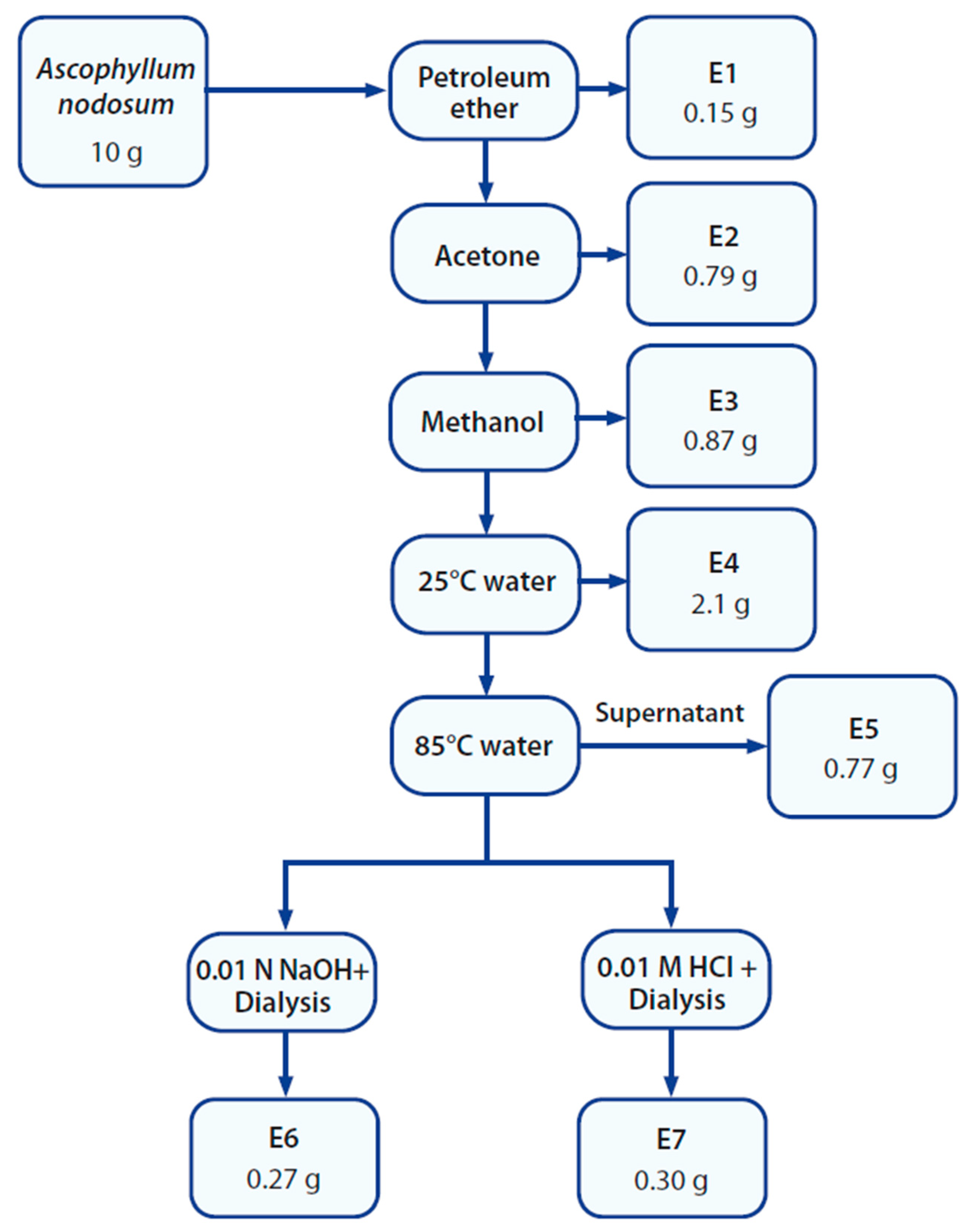
2.3. Preparation of Extracts

Water extracts were prepared from seven seaweed species by weighing 5 g of dry seaweed powder accurately into a 500 mL Erlenmeyer flask and mixing it with 400 mL of MQW. Extraction was obtained by means of maceration on a magnetic stirrer for 24 h, followed by centrifugation (Eppendorf centrifuge 5810R) for 20 min at 1258× g. The supernatant was decanted, frozen, and lyophilized. Extraction yield was calculated, and dry extracts were stored in a −20 °C freezer until analysis. Obtained extracts were screened for total polyphenol content and antioxidant activity as well as for collagenase and elastase inhibitory effects.
Further subsequent extraction of A. nodosum was performed according to Figure 1. Extracts E1 (petroleum ether), E2 (acetone), and E3 (methanol) were obtained using organic solvents and ultrasonic-assisted (Branson 3510E-DTH, MO, USA, 100 W, 42 kHz) extraction at 45 °C for 30 min, followed by filtration (Whatman 4 paper, 150 mm). The solid residue was allowed to dry before adding the next solvent. The solvent was evaporated on a rotary evaporator (Heidolph, Laborota 4001) and finally dried under nitrogen gas to obtain three crude extracts (E1, E2, E3).
The air-dried residue biomass was macerated with 300 mL MQW for 24 h followed by centrifugation (Beckman Coulter, TJ-25 centrifuge) at 1258× g for 20 min. The supernatant was decanted, frozen, and lyophilized to obtain water extract E4. The pellet was air-dried and extracted with 85 °C MQW for 120 min, followed by centrifugation at 1258× g for 20 min. The supernatant was freeze-dried to obtain extract E5. The remaining pellet was split into two equal parts and macerated with either 0.01 M NaOH or 0.01 M HCl for 24 h. Followed by centrifugation, the supernatants were subjected to dialysis (Sigma-Aldrich, cellulose membrane, average diameter 49 mm, MW cut-off 1 kDa) overnight and then lyophilized to obtain extracts E6 and E7 according to Figure 1.

2.4. Bioassays

Seaweed extracts were dissolved in either DMSO or MQW to 10 mg/mL concentration stock solutions. These stock solutions were then diluted further and used in the different assays. For antioxidant assays, the solvent was 15% DMSO, as DMSO can interfere and donate electrons at certain concentrations. The difference between the results was compared by ANOVA test for repeated measures, and Tukey–Kramer’s post hoc test for multiple comparisons. Statistical calculations were carried out using NCSS 2000 (NCSS, Kaysville, UT, USA). A p value < 0.05 was considered statistically significant.

2.4.1. Total Polyphenol Content (TPC) Assay

TPC was determined according to the method by Singleton and Rossi [28] adapted to microplate (Thermo Scientific™ Nunc, Cat. 269620, Thermo Fischer Scientific, Rochester, NY, USA) format and some modifications. Briefly, 20 µL of sample or standard was mixed with 100 µL of 0.2 N Folin–Ciocalteu reagent (Sigma-Aldrich, Cat. F9252, Sigma Aldrich Chemie GmBH, Steinheim, Switzerland) and allowed to stand at room temperature for 5 min. Then, 80 µL of 7.5% Na2CO3 (Sigma-Aldrich ≥ 99.0%, Cat. S2127) was added, heated for 10 s at 550 W in a microwave (Daewoo, KOR-636T, Prokaria, Courbevoie, France), and incubated for 30 min at room temperature under constant agitation, 320 Mot/1 min (IKA® KS 130 basic, IKA, Staufen, Germany). Absorbance was read at 720 nm with a microplate reader (Multiskan Sky, Thermo Fisher, Life Technologies Holding Pte Ltd., Singapore). The standard curves contained 6 concentrations of gallic acid (Sigma-Aldrich 97.5–102.5%, Cat. G7384) and 6 concentrations of phloroglucinol (Sigma-Aldrich ≥ 99.0%, Cat. 79330) between 0 and 100 µg/mL.
Results were expressed as g gallic acid equivalents (GAE) or phloroglucinol equivalents (PGE) per 100 g of extract.

2.4.2. DPPH Assay

DPPH (2,2-diphenyl-1-picrylhydrazyl) radical scavenging activity was determined as recommended by Sharma and Bhat [29]. A 2 mM 2,2-diphenyl-1-picrylhydrazyl (Sigmal-Aldrich, Cat. D9132) methanol solution was prepared and diluted 1:10 in 70% ethanol. Samples were dissolved in either MQW or 15% DMSO, prepared in triplicate and 4 concentrations. On a microplate (Thermo NUNC, Cat. 269620, Germany), a 150 µL sample was mixed with 50 µL of 0.2 mM DPPH or 70% ethanol solution, for control. The blank was 150 µL of sample solvent and 50 µL of DPPH and a control blank consisted of 150 µL of sample solvent and 50 µL of 70% ethanol. The plate was shaken for 30 min at 320 Mot/1 min, protected from light (IKA® KS 130 basic), and absorbance was measured at 520 nm (Multiskan Sky, Thermo Fisher). The control sample, blank, and control blank were used to remove background noise and increase the inter- and intra-accuracy of the assay. DPPH logarithmic curves were generated from concentration vs. percent radical inhibition plots and used to calculate the IC50 values in mg/mL ± standard error of the mean (SEM).
Results expressed as IC50 values (mg/mL) were compiled from one run, with every extract run in four concentrations and each concentration in triplicate.

2.4.3. ORAC Assay

The ORAC (oxygen radical absorbance capacity) assay was performed in triplicate with three concentrations and followed protocols from Huang et al. [30] and Ganske F. and Dell E.J. [31] with slight modifications. Briefly, 60 µL of 10 nM fluorescein solution (Sigma-Aldrich, Cat. BCCG9496) in 10 mM phosphate buffer pH 7.4 (Sigma-Aldrich, Cat. 30412 and S9638) was added to 10 µL of standard or sample solution on a microplate (black, flat bottom, 96-well EIA/RIA plate, REF:3694, Costar). After 10 min incubation at 37 °C, the reaction was started by adding 30 µL of 120 mM AAPH (2,2′-Azobis(2-methylpropionamidine) dihydrochloride, Sigma-Aldrich, Cat. 440914) solution. Fluorescence (ex/em 485 nm/520 nm) was acquired every minute for 100 min in a microplate reader (POLARstar Optima, BMG Labtech, Ortenberg, Germany). A 5 mM Trolox (Sigma-Aldrich, Cat. 238813) solution was prepared in methanol and diluted with buffer. For the ORAC assay measuring hydrogen donation, the results were expressed as average µmol Trolox equivalents [32] per gram of extract.
The average normalized ORAC values for the various seaweed samples were calculated as mean values ± SD (n = 3).

2.4.4. Reducing Power Assay

Depending on extract solubility, the solvents used to dissolve the samples were either water or 7.5% DMSO. Then, 63 µL of 6.8 pH phosphate buffer (Sigma-Aldrich, Na2HPO4 2H2O 99.5%, Cat. 04272, and NaH2PO4 H2O 98.0–102%, Cat. S9638), 63 µL of 1% potassium ferricyanide (Sigma-Aldrich, Cat. 244023) and 13 µL of sample/stock were added to the plate. The microplate was incubated for 30 min at 50 °C before 63 µL of 10% trichloroacetic acid (Sigma-Aldrich, Cat. 27242) and 40 µL of 0.1% ferric chloride (Sigma-Aldrich, Cat. 157740) were added to each plate (Thermo NUNC, Cat. 269620, Germany). Lastly, absorbance was measured at 720 nm (Multiskan Sky, Thermo Fisher). Samples and standards were tested in quadruplicate with two different concentrations.
The reducing power was calculated using a normalized L-ascorbic acid (Sigma-Aldrich 99.0%, Cat. A7506) standard curve made from six data points, and the reduction of Fe2+ to Fe3+ was expressed as mg L-ascorbic acid equivalents (AAEq)/g extract.

2.4.5. Collagenase Inhibition Assay

The collagenase inhibition assay was performed according to the instructions provided with the collagenase activity colorimetric assay kit (MAK293-1KT, Sigma Aldrich, Saint Louis, MO, USA). Seaweed stock solutions were diluted to 10, 5, and 1 mg/mL in MQW or 15% DMSO and all solutions and reagents were added (10 µL) to a clear microplate (Thermo NUNC, Cat. 269620, Germany) according to the assay kit instructions. The microplate was incubated for 10 min at room temperature. After the incubation, 100 µL of the substrate mix was added to each well. The microplate was read in kinetic mode (reading every 30 s) at 345 nm using a microplate reader Multiskan (Multiskan Sky, Thermo Fisher) at 37 °C for 30 min.
IC50 values (mg/mL) and standard error of the mean (SEM) for anti-collagenase activity were determined with logarithmic regression. Results were expressed as the mean values of experiments performed in triplicate.

2.4.6. Elastase Inhibition Assay

The elastase inhibition assay was performed according to instructions of the EnzChek elastase assay kit (Invitrogen, E12056, Thermo Fisher Scientific, Waltham, MA, USA). The elastase enzyme was dissolved in MQW to 1 U/mL and stored at −20 °C. For the assay, the enzyme was diluted in buffer to 1 µU/mL. Elastase inhibitor N-methoxysuccinyl-Ala-Ala-Pro-Val-chloromethyl ketone was dissolved in DMSO to a 10 mg/mL concentration and stored at −20 °C. For the assay, the inhibitor was diluted in buffer to 0.33 mg/mL (resulting in 0.016 mM in well). A 200 nM sodium azide (Sigma-Aldrich, USA) solution was prepared in MQW and stored at room temperature. DQ elastin substrate was diluted to 200 mM in 90% MQW and 10% 200 nM sodium azide and stored at −20 °C in aliquots. For the assay, one aliquot was thawed and diluted to 20 mM DQ elastin. Seaweed extract stock solutions were diluted to 10, 5, and 1 mg/mL in their respective solvents. According to the assay kit instructions, all solutions were added to a black 96-well microplate (Costar, CLS3915-100EA, Corning, NY, USA) except the DQ elastin solution. The plate was incubated for 10 min at 37 °C, and after incubation, 50 µL of DQ elastin was added to each well. The excitation was read at 485 nm and the emission at 520 nm for 60 min in the microplate reader POLARstar Optima.
IC50 values (mg/mL) and standard error of the mean (SEM) for anti-elastase activity were determined with logarithmic regression. Results were calculated as the mean values of sample concentrations and control samples run in triplicate (n = 3).

3. Results and Discussion

3.1. Extraction Yield

Extraction yield for the dried extracts ranged from 1% to 57%, as can be seen in Table 2 and Table 3. In general, seaweed contains low amounts of lipids, ranging from 0.6 to 4.15% [33,34], while polysaccharides as the major component can account for up to 76% of the dry weight [35], depending on the species.
The extraction yields obtained in this study (Table 2 and Table 3) were reflected by this, where the lowest yield (1%) was obtained with the least polar solvent (petroleum ether) and generally increased with increasing solvent polarity.
Novel technologies for extraction of bioactive compounds from marine algae have been extensively reviewed [36]. Ultrasound-assisted extraction (UAE) has been demonstrated to have many benefits. It facilitates extraction by acoustic cavitation, which results in the disruption of cell walls and rupturing of cells to release their content for solubilization [37,38]. This can result in higher yields and shorter extraction time than conventional methods [39,40]. It is simple and cost-effective and can be an efficient alternative to traditional extraction techniques, such as maceration [36]. In addition, it is a relatively environmentally friendly and mild method [41], suitable for extracting thermolabile compounds, and it can be easily combined with other technologies [42,43]. UAE has been reported for the extraction of bioactive compounds from A. nodosum before [42].

3.2. Evaluation of Skin-Protecting Properties of Icelandic Seaweed Species

3.2.1. Total Polyphenol Content (TPC)

The total polyphenol content results (Table 2 and Table 3) are expressed as gram (g) gallic acid equivalents (GAE) or phloroglucinol equivalents (PGE) per 100 g extract and were <2% w/w for most seaweed water extracts, except for A. nodosum. A. nodosum acetone (E2) and methanol (E3) extracts (Table 3) contained substantially higher amounts of polyphenols than the water extracts, and the acetone extract had a higher TPC than the gallic acid standard when measured at the same concentration. Using acetone as the extraction solvent has been shown to increase TPC in seaweed extracts [44,45]. This is because phenolic compounds are generally more soluble in polar organic solvents (e.g., acetone, ethanol, and methanol) than in water. It has been found that 70% aqueous acetone or ethyl acetate are the most efficient polyphenol extractants [46,47,48].
Brown seaweed is well known to be rich in phlorotannins [35]. In this study, the Fucales, A. nodosum, contained the highest level of phenolic compounds compared to the Laminariales (A. esculenta, L. digitata, L. hyperborea, and S. latissima). This has also been shown by earlier findings for the same species [49,50,51] and is considered to be linked to the growth position of the seaweeds on the shore, with a progressive decrease in TPC from species living at the mid-tide level (like A. nodosum and F. vesiculosus) to those living at the low-tide level, and a minimum in the subtidal L. digitata [49].
Since the antioxidant properties of phenolic compounds found in seaweeds are often associated with phenolic content [52], it is relevant to have information about chemical composition variability for reproducibility. Numerous seasonal studies on brown seaweeds in specific sites have been reported [50] and species-specific variations have been observed in snapshot measurements [53]. Roleda et al. [54] investigated temporal and spatial variations in polyphenol content of three edible seaweed species, and variation in polyphenols was largely species-specific and seasonal. But spatial variation was not observed within the biogeographic region studied.

3.2.2. Antioxidant Activity

Amongst the water extracts from different seaweed species, the A. nodosum extract could be distinguished with the highest content of phenolic compounds (TPC mean values 10 g GAE/100 g and 11 g PGE/100 g) and exhibited the highest antioxidant activity (Table 2). Seaweed extracts with high TPC have previously been observed to be potent radical scavengers [44,45,55,56,57,58].
Previous studies have shown that using a single assay to determine and compare the in vitro antioxidant activity of different compounds or extracts obtains limited results, since the active compounds may have different antioxidant mechanisms [59]. Therefore, the antioxidant activity was evaluated herein using both a DPPH assay and an ORAC assay, as well as the ferricyanide method of measuring reducing power.
In the three assays, A. nodosum was the most active compared to the other tested seaweed species, suggesting that compounds from this species may be particularly effective at preventing oxidative damage. A. nodosum water extract demonstrated significantly higher electron donation capacity than water extracts from other seaweed species. Other seaweed extracts also had lower reducing power, and the lowest radical quenching effect was seen for L. digitata and P. palmata.
A. nodosum E2 (acetone) and E3 (methanol) extracts showed increased activity in all assays compared to the crude A. nodosum water extract. Acetone and methanol have been demonstrated as efficient solvents to extract phenolic and flavonoid compounds [60,61]. The acetone (E2) and methanol (E3) extracts had the highest DPPH radical inhibition and the highest capacity for hydrogen donation (ORAC) (Table 3). In general, high correlation is found between TPC of seaweed extracts and their scavenging capacity against DPPH and peroxyl radicals, indicating an important role of algal polyphenols as chain-breaking antioxidants [44]. Positive correlation has also been found between phlorotannins and flavonoids and antioxidant power [62]. However, although the TPC values of the A. nodosum E1 (petroleum ether) and E5 (85 °C water) extracts were low, they demonstrated high DPPH radical scavenging effects (IC50 values = 0.2 and 0.08 mg/mL, respectively). This suggests that other components besides phenolic compounds (e.g., sulphated polysaccharides and pigments) [63,64,65,66,67] could be responsible for the antioxidant activity.
Overall, the A. nodosum acetone extract (E2) was the most active, followed by the methanol extract (E3). The use of acetone as extraction solvent has earlier been shown to give higher scavenging activity compared to water extracts [44].

3.2.3. Inhibition of Skin-Degrading Enzymes

Enzyme inhibitory activity was assessed after exposure to seaweed water extracts. The A. nodosum water extract was the only extract exerting activity against collagenase with an IC50 value of 0.1 mg/mL ± 0.03 (SEM). Water extracts from other species lacked collagenase inhibitory activity. In an earlier study, methanol and ethanol extracts of A. esculenta and S. latissima species both showed low or no collagenase inhibitory activity [68]. However, 68–91% inhibition of collagenase was found when A. esculenta was extracted with hot water and a pulsed electric field [45]. The differences between studies may arise not only from species differences and seasonal variation but may also depend on extraction solvents and extraction methods [69,70,71]. The evaluation of the bioactivity of extracts should also consider different mechanisms of action and groups of compounds. As extracts are complex matrices, the synergetic effect of molecules may dramatically impact the final bioactive capacity.
The A. nodosum water extract also exhibited the highest inhibitory activity against elastase (IC50 = 0.04 mg/mL ± 0.01 (SEM)) compared to A. esculenta (IC50 = 1.5 mg/mL ± 0.25 (SEM)) and L. hyperborea (IC50 = 0.17 mg/mL ± 0.04 (SEM)). Water extracts from other species lacked elastase inhibitory activity.
Based on these results, A. nodosum was chosen as the species for further investigation (Table 4). An increased inhibitory effect against collagenase and elastase was seen when A. nodosum was extracted subsequently and was highest for the acetone extract (E2). High anti-elastase activity has been demonstrated before for A. nodosum acetone extract [51]. However, anti-collagenase activity has previously not been demonstrated for A. nodosum extracts. The sulphated polysaccharide ascophyllan isolated from A. nodosum has been demonstrated both in vitro and in vivo as a potent bioactive compound [72,73,74,75,76,77,78,79].
Polysaccharides have been reported to play a key role in collagenase and elastase inhibition. Sulphated polysaccharides that were derived from the brown seaweed Hizikia fusiforme potentially inhibited collagenase and elastase by regulating different pathways in HDF cells radiated by UVB [80]. Fucoidans obtained from Chnoospora minima and Sargassum polycystum showed elastase and collagenase inhibition activities in a dose-dependent manner [74]. Furthermore, Moon et al. [81,82] studied how fucoidans inhibit UVB-induced MMP-1 promoter expression and the downregulation of type I procollagen synthesis in human skin fibroblasts. Kim et al. [83] investigated the in vitro inhibitory effects of Ecklonia cava-derived phlorotannin on MMP activity and observed its complete inhibition of bacterial collagenase-1 activity.

4. Conclusions

The results of this bioactivity screening of extracts from seven different Icelandic seaweed species (A. esculenta, A. nodosum, L. hyperborea, L. digitata, S. latissima, P. palmata, and S. jonsonii) demonstrated the potential skin-protecting properties of the brown seaweed A. nodosum. Overall, this study indicated that A. nodosum water extract possesses antioxidant activity as well as inhibition of the skin-degrading enzymes collagenase and elastase. The A. nodosum water extract exhibited the highest inhibitory activity against elastase, followed by A. esculenta and L. hyperborea. Water extracts from other species lacked elastase and collagenase inhibitory activity.
Further subsequent extraction of A. nodosum demonstrated increased bioactivity. The highest TPC, antioxidant activity, and enzyme inhibition were found for A. nodosum acetone extract, demonstrating that the type of extractant greatly impacts the bioactivity of seaweed extracts. High TPC correlated with high DPPH and ORAC values, indicating that algal polyphenols were mainly responsible for the free radical scavenging activities of the extracts and acetone was more efficient for polyphenol extraction than water. However, other co-extracted active compounds such as pigments, polysaccharides, or proteins and peptides may also contribute to the overall skin-protecting effects.
It has been demonstrated that A. nodosum extract has potential as a natural ingredient for use in cosmeceuticals and has, from the species included herein, the most promising extract for further testing in cosmetic products. The A. nodosum acetone extract could also be of interest for further investigation as a highly bioactive cosmetic ingredient, e.g., serum.
Future work with further fractionation and purification of active components could increase their activity and other potential health benefits and may promote their use as natural sources of antioxidants. Formulation studies, product development, and in vivo intervention studies are needed for potential commercialization.

Author Contributions

Conceptualization: S.J. and R.J.; methodology: S.J., R.J. and J.K.H.; validation: S.J., R.J. and J.K.H.; formal analysis: J.K.H. and S.J.; investigation: S.J. and J.K.H.; resources: S.J. and J.K.H.; data curation: S.J., R.J. and J.K.H.; writing—original draft preparation: S.J. and J.K.H.; writing—review and editing: S.J. and R.J.; supervision, S.J. and R.J.; project administration, S.J. and R.J.; funding acquisition, R.J. All authors have read and agreed to the published version of the manuscript.

Funding

This research was funded by the ERA-NET Co-fund Blue Bioeconomy and the European Union’s Horizon 2020 research and innovation program under grant agreement No 817992.

Institutional Review Board Statement

Not applicable.

Informed Consent Statement

Not applicable.

Data Availability Statement

The data that support the findings of this study are available from the corresponding author [Jensen, Sophie] upon reasonable request.

Acknowledgments

We thank Símon Már Sturluson at Íslensk bláskel & sjávargróður, Bjarni Bjarnason, and the staff at Hyndla for their help collecting and identifying seaweed species.

Conflicts of Interest

The authors declare no conflicts of interest.

References

  1. Couteau, C.; Coiffard, L. Phycocosmetics and other marine cosmetics, specific cosmetics formulated using marine resources. Mar. Drugs 2020, 18, 322. [Google Scholar] [CrossRef] [PubMed]
  2. Tobin, D.J. Introduction to skin aging. J. Tissue Viability 2017, 26, 37–46. [Google Scholar] [CrossRef] [PubMed]
  3. Russell-Goldman, E.; Murphy, G.F. The pathobiology of skin aging: New insights into an old dilemma. Am. J. Pathol. 2020, 190, 1356–1369. [Google Scholar] [CrossRef]
  4. Wong, Q.Y.A.; Chew, F.T. Defining skin aging and its risk factors: A systematic review and meta-analysis. Sci. Rep. 2021, 11, 22075. [Google Scholar] [CrossRef]
  5. Ganceviciene, R.; Liakou, A.I.; Theodoridis, A.; Makrantonaki, E.; Zouboulis, C.C. Skin anti-aging strategies. Derm.-Endocrinol. 2012, 4, 308–319. [Google Scholar] [CrossRef]
  6. Farage, M.A.; Miller, K.W.; Elsner, P.; Maibach, H.I. Characteristics of the aging skin. Adv. Wound Care 2013, 2, 5–10. [Google Scholar] [CrossRef] [PubMed]
  7. Kilgman, A. Cosmeceuticals: A broad-spectrum category between cosmetics and drugs. In Cosmeceuticals and Active Cosmetics, 2nd ed.; Elsner, P., Maibach, H.I., Eds.; CRC Press: Boca Raton, FL, USA, 2005; pp. 1–9. [Google Scholar]
  8. Ferreira, M.S.; Resende, D.; Lobo, J.M.S.; Sousa, E.; Almeida, I.F. Marine ingredients for sensitive skin: Market overview. Mar. Drugs 2021, 19, 464. [Google Scholar] [CrossRef] [PubMed]
  9. Kalasariya, H.S.; Patel, N.B.; Yadav, A.; Perveen, K.; Yadav, V.K.; Munshi, F.M.; Yadav, K.K.; Alam, S.; Jung, Y.K.; Jeon, B.H. Characterization of fatty acids, polysaccharides, amino acids, and minerals in marine macroalga Chaetomorpha crassa and evaluation of their potentials in skin cosmetics. Molecules 2021, 26, 7515. [Google Scholar] [CrossRef]
  10. Kalasariya, H.S.; Yadav, V.K.; Yadav, K.K.; Tirth, V.; Algahtani, A.; Islam, S.; Gupta, N.; Jeon, B.H. Seaweed-based molecules and their potential biological activities: An eco-sustainable cosmetics. Molecules 2021, 26, 5313. [Google Scholar] [CrossRef]
  11. Jesumani, V.; Du, H.; Aslam, M.; Pei, P.; Huang, N. Potential use of seaweed bioactive compounds in skincare—A review. Mar. Drugs 2019, 17, 688. [Google Scholar] [CrossRef]
  12. Kim, S.K. Marine cosmeceuticals. J. Cosmet. Dermatol. 2014, 13, 56–67. [Google Scholar] [CrossRef] [PubMed]
  13. Sanjeewa, K.K.A.; Kim, E.-A.; Son, K.-T.; Jeon, Y.-J. Bioactive properties and potentials cosmeceutical applications of phlorotannins isolated from brown seaweeds: A review. J. Photochem. Photobiol. B Biol. 2016, 162, 100–105. [Google Scholar] [CrossRef] [PubMed]
  14. Kim, S.-K.; Ravichandran, Y.D.; Khan, S.B.; Kim, Y.T. Prospective of the cosmeceuticals derived from marine organisms. Biotechnol. Bioprocess Eng. 2008, 13, 511–523. [Google Scholar] [CrossRef]
  15. Alves, A.; Sousa, E.; Kijjoa, A.; Pinto, M. Marine-derived compounds with potential use as cosmeceuticals and nutricosmetics. Molecules 2020, 25, 2536. [Google Scholar] [CrossRef] [PubMed]
  16. Siavash, H.C.; Fadzilah, A.A.M.; Mohamad, R.S. Cosmeceutical value of herbal extracts as natural ingredients and novel technologies in anti-aging. J. Med. Plants Res. 2011, 5, 3074–3077. [Google Scholar]
  17. El-Beltagi, H.S.; Mohamed, A.A.; Mohamed, H.I.; Ramadan, K.M.A.; Barqawi, A.A.; Mansour, A.T. Phytochemical and potential properties of seaweeds and their recent applications: A review. Mar. Drugs 2022, 20, 342. [Google Scholar] [CrossRef]
  18. Agrawal, S.; Adholeya, A.; Barrow, C.J.; Deshmukh, S.K. Marine fungi: An untapped bioresource for future cosmeceuticals. Phytochem. Lett. 2018, 23, 15–20. [Google Scholar] [CrossRef]
  19. Salehi, B.; Sharifi-Rad, J.; Seca, A.M.L.; Pinto, D.; Michalak, I.; Trincone, A.; Mishra, A.P.; Nigam, M.; Zam, W.; Martins, N. Current trends on seaweeds: Looking at chemical composition, phytopharmacology, and cosmetic applications. Molecules 2019, 24, 4182. [Google Scholar] [CrossRef]
  20. Jayawardhana, H.H.A.C.K.; Jayawardena, T.U.; Sanjeewa, K.K.A.; Liyanage, N.M.; Nagahawatta, D.P.; Lee, H.-G.; Kim, J.-I.; Jeon, Y.-J. Marine Algal Polyphenols as Skin Protective Agents: Current Status and Future Prospectives. Mar. Drugs 2023, 21, 285. [Google Scholar] [CrossRef]
  21. López-Hortas, L.; Flórez-Fernández, N.; Torres, M.D.; Ferreira-Anta, T.; Casas, M.P.; Balboa, E.M.; Falqué, E.; Domínguez, H. Applying seaweed compounds in cosmetics, cosmeceuticals and nutricosmetics. Mar. Drugs 2021, 19, 552. [Google Scholar] [CrossRef]
  22. Grozdanić, N.; Zdunić, G.; Šavikin, K.; Đuričić, I.; Kosanić, M.; Macic, V.; Matić, I.; Stanojkovic, T. Seasonal variation in biopharmaceutical activity and fatty acid content of endemic Fucus virsoides algae from Adriatic Sea. Acta Pol. Pharm.-Drug Res. 2019, 76, 833–844. [Google Scholar] [CrossRef] [PubMed]
  23. Barbosa, M.; Fernandes, F.; Pereira, D.M.; Azevedo, I.C.; Sousa-Pinto, I.; Andrade, P.B.; Valentão, P. Fatty acid patterns of the kelps Saccharina latissima, Saccorhiza polyschides and Laminaria ochroleuca: Influence of changing environmental conditions. Arab. J. Chem. 2020, 13, 45–58. [Google Scholar] [CrossRef]
  24. Sappati, P.K.; Nayak, B.; VanWalsum, G.P.; Mulrey, O.T. Combined effects of seasonal variation and drying methods on the physicochemical properties and antioxidant activity of sugar kelp (Saccharina latissima). J. Appl. Phycol. 2019, 31, 1311–1332. [Google Scholar] [CrossRef]
  25. Manns, D.; Nielsen, M.M.; Bruhn, A.; Saake, B.; Meyer, A.S. Compositional variations of brown seaweeds Laminaria digitata and Saccharina latissima in Danish waters. J. Appl. Phycol. 2017, 29, 1493–1506. [Google Scholar] [CrossRef]
  26. Konstantin, B.; Anastasia, P.; Nikolay, I.; Daria, P. Seasonal variations in the chemical composition of Arctic brown macroalgae. Algal Res. 2023, 72, 103112. [Google Scholar] [CrossRef]
  27. Jang, H.; Lee, J.; Park, Y.-K.; Lee, J.-Y. Exploring the health benefits and concerns of brown seaweed consumption: A comprehensive review of bioactive compounds in brown seaweed and its potential therapeutic effects. J. Agric. Food Res. 2024, 17, 101215. [Google Scholar] [CrossRef]
  28. Singleton, V.L.; Rossi, J.A. Colorimetry of total phenolics with phosphomolybdic-phosphotungstic acid reagents. Am. J. Enol. Vitic. 1965, 16, 144–158. [Google Scholar] [CrossRef]
  29. Sharma, O.P.; Bhat, T.K. DPPH antioxidant assay revisited. Food Chem. 2009, 113, 1202–1205. [Google Scholar] [CrossRef]
  30. Huang, D.; Ou, B.; Hampsch-Woodill, M.; Flanagan, J.A.; Prior, R.L. High-throughput assay of oxygen radical absorbance capacity (ORAC) using a multichannel liquid handling system coupled with a microplate fluorescence reader in 96-well format. J. Agric. Food Chem. 2002, 50, 4437–4444. [Google Scholar] [CrossRef]
  31. Ganske, F.; Dell, E. ORAC Assay on the FLUOstar OPTIMA to Determine Antioxidant Capacity. BMG LABTECH Application Note 148. Rev. 12/2006. Available online: https://www.pharmaceutical-technology.com/wp-content/uploads/sites/24/2017/09/196.pdf?utm_campaign=Verdict%20Whitepaper%20downloads&utm_medium=email&_hsenc=p2ANqtz--_qMD3rndRfHPZPZhZGZn0XEu7TPzioOoC3hswqewfmfzBvkkUpX20GN_3njY2Murs1l5psfOP8P8nGLXFmzX7ktsxGQ&_hsmi=75927857&utm_content=75927857&utm_source=hs_automation (accessed on 1 August 2024).
  32. Yano, S.; Mori, M.; Teramoto, N.; Iisaka, M.; Suzuki, N.; Noto, M.; Kaimoto, Y.; Kakimoto, M.; Yamada, M.; Shiratsuchi, E.; et al. Preparation of photocrosslinked fish elastin polypeptide/microfibrillated cellulose composite gels with elastic properties for biomaterial applications. Mar. Drugs 2015, 13, 338–353. [Google Scholar] [CrossRef]
  33. El Maghraby, D.M.; Fakhry, E.M. Lipid content and fatty acid composition of Mediterranean macro-algae as dynamic factors for biodiesel production. Oceanologia 2015, 57, 86–92. [Google Scholar] [CrossRef]
  34. Rodrigues, D.; Freitas, A.C.; Pereira, L.; Rocha-Santos, T.A.; Vasconcelos, M.W.; Roriz, M.; Rodríguez-Alcalá, L.M.; Gomes, A.M.; Duarte, A.C. Chemical composition of red, brown and green macroalgae from Buarcos bay in Central West Coast of Portugal. Food Chem. 2015, 183, 197–207. [Google Scholar] [CrossRef] [PubMed]
  35. Holdt, S.L.; Kraan, S. Bioactive compounds in seaweed: Functional food applications and legislation. J. Appl. Phycol. 2011, 23, 543–597. [Google Scholar] [CrossRef]
  36. Kadam, S.U.; Tiwari, B.K.; O’Donnell, C.P. Application of novel extraction technologies for bioactives from marine algae. J. Agric. Food Chem. 2013, 61, 4667–4675. [Google Scholar] [CrossRef]
  37. Tiwari, B.K. Ultrasound: A clean, green extraction technology. TrAC Trends Anal. Chem. 2015, 71, 100–109. [Google Scholar] [CrossRef]
  38. Cikoš, A.-M.; Jokić, S.; Šubarić, D.; Jerković, I. Overview on the application of modern methods for the extraction of bioactive compounds from marine macroalgae. Mar. Drugs 2018, 16, 348. [Google Scholar] [CrossRef]
  39. Bordoloi, A.; Goosen, N. Green and integrated processing approaches for the recovery of high-value compounds from brown seaweeds. In Advances in Botanical Research; Elsevier: Amsterdam, The Netherlands, 2020; Volume 95, pp. 369–413. [Google Scholar]
  40. Gomez, L.; Alvarez, C.; Zhao, M.; Tiwari, U.; Curtin, J.; Garcia-Vaquero, M.; Tiwari, B. Innovative processing strategies and technologies to obtain hydrocolloids from macroalgae for food applications. Carbohydr. Polym. 2020, 248, 116784. [Google Scholar] [CrossRef] [PubMed]
  41. van Sittert, D.; Lufu, R.; Mapholi, Z.; Goosen, N.J. Ultrasound-assisted extraction of alginate from Ecklonia maxima with and without the addition of alkaline cellulase–factorial and kinetic analysis. J. Appl. Phycol. 2024, 36, 2781–2793. [Google Scholar] [CrossRef]
  42. Kadam, S.U.; Tiwari, B.K.; Smyth, T.J.; O’Donnell, C.P. Optimization of ultrasound assisted extraction of bioactive components from brown seaweed Ascophyllum nodosum using response surface methodology. Ultrason. Sonochemistry 2015, 23, 308–316. [Google Scholar] [CrossRef]
  43. Chemat, F.; Khan, M.K. Applications of ultrasound in food technology: Processing, preservation and extraction. Ultrason. Sonochemistry 2011, 18, 813–835. [Google Scholar] [CrossRef]
  44. Wang, T.; Jónsdóttir, R.; Ólafsdóttir, G. Total phenolic compounds, radical scavenging and metal chelation of extracts from Icelandic seaweeds. Food Chem. 2009, 116, 240–248. [Google Scholar] [CrossRef]
  45. Castejón, N.; Thorarinsdottir, K.A.; Einarsdóttir, R.; Kristbergsson, K.; Marteinsdóttir, G. Exploring the potential of Icelandic seaweeds extracts produced by aqueous pulsed electric fields-assisted extraction for cosmetic applications. Mar. Drugs 2021, 19, 662. [Google Scholar] [CrossRef] [PubMed]
  46. Koivikko, R.; Loponen, J.; Honkanen, T.; Jormalainen, V. Contents of soluble, cell-wall-bound and exuded phlorotannins in the brown alga Fucus vesiculosus, with implications on their ecological functions. J. Chem. Ecol. 2005, 31, 195–212. [Google Scholar] [CrossRef] [PubMed]
  47. Waterman, P.G.; Mole, S. Analysis of Phenolic Plant Metabolites; Blackwell Scientific Publications: Oxford, UK, 1994. [Google Scholar]
  48. Susano, P.; Silva, J.; Alves, C.; Martins, A.; Gaspar, H.; Pinteus, S.; Mouga, T.; Goettert, M.I.; Petrovski, Ž.; Branco, L.B.; et al. Unravelling the dermatological potential of the brown seaweed Carpomitra costata. Mar. Drugs 2021, 19, 135. [Google Scholar] [CrossRef] [PubMed]
  49. Connan, S.; Goulard, F.; Stiger, V.; Deslandes, E.; Gall, E.A. Interspecific and temporal variation in phlorotannin levels in an assemblage of brown algae. Bot. Mar. 2004, 47, 410–416. [Google Scholar] [CrossRef]
  50. Schiener, P.; Black, K.D.; Stanley, M.S.; Green, D.H. The seasonal variation in the chemical composition of the kelp species Laminaria digitata, Laminaria hyperborea, Saccharina latissima and Alaria esculenta. J. Appl. Phycol. 2015, 27, 363–373. [Google Scholar] [CrossRef]
  51. Gager, L.; Connan, S.; Molla, M.; Couteau, C.; Arbona, J.-F.; Coiffard, L.; Cérantola, S.; Stiger-Pouvreau, V. Active phlorotannins from seven brown seaweeds commercially harvested in Brittany (France) detected by 1H NMR and in vitro assays: Temporal variation and potential valorization in cosmetic applications. J. Appl. Phycol. 2020, 32, 2375–2386. [Google Scholar] [CrossRef]
  52. Connan, S.; Deslandes, E.; Gall, E.A. Influence of day–night and tidal cycles on phenol content and antioxidant capacity in three temperate intertidal brown seaweeds. J. Exp. Mar. Biol. Ecol. 2007, 349, 359–369. [Google Scholar] [CrossRef]
  53. Heffernan, N.; Brunton, N.P.; FitzGerald, R.J.; Smyth, T.J. Profiling of the Molecular Weight and Structural Isomer Abundance of Macroalgae-Derived Phlorotannins. Mar. Drugs 2015, 13, 509–528. [Google Scholar] [CrossRef]
  54. Roleda, M.Y.; Marfaing, H.; Desnica, N.; Jónsdóttir, R.; Skjermo, J.; Rebours, C.; Nitschke, U. Variations in polyphenol and heavy metal contents of wild-harvested and cultivated seaweed bulk biomass: Health risk assessment and implication for food applications. Food Control 2019, 95, 121–134. [Google Scholar] [CrossRef]
  55. Meng, W.; Sun, H.; Mu, T.; Garcia-Vaquero, M. Extraction, purification, chemical characterization and antioxidant properties in vitro of polyphenols from the brown macroalga Ascophyllum nodosum. Algal Res. 2023, 70, 102989. [Google Scholar] [CrossRef]
  56. Blanc, N.; Hauchard, D.; Audibert, L.; Gall, E.A. Radical-scavenging capacity of phenol fractions in the brown seaweed Ascophyllum nodosum: An electrochemical approach. Talanta 2011, 84, 513–518. [Google Scholar] [CrossRef] [PubMed]
  57. Liu, H.; Gu, L. Phlorotannins from brown algae (Fucus vesiculosus) inhibited the formation of advanced glycation endproducts by scavenging reactive carbonyls. J. Agric. Food Chem. 2012, 60, 1326–1334. [Google Scholar] [CrossRef] [PubMed]
  58. Farasat, M.; Khavari-Nejad, R.A.; Nabavi, S.M.; Namjooyan, F. Antioxidant properties of two edible green seaweeds from Northern Coasts of the Persian Gulf. J. Nat. Pharm. Prod. 2013, 8, 47–52. [Google Scholar]
  59. Makori, S.I.; Mu, T.-H.; Sun, H.-N. Profiling of polyphenols, flavonoids and anthocyanins in potato peel and flesh from four potato varieties. Potato Res. 2022, 65, 193–208. [Google Scholar] [CrossRef]
  60. Afrin, F.; Ahsan, T.; Mondal, M.N.; Rasul, M.G.; Afrin, M.; Silva, A.A.; Yuan, C.; Shah, A.K.M.A. Evaluation of antioxidant and antibacterial activities of some selected seaweeds from Saint Martin’s Island of Bangladesh. Food Chem. Adv. 2023, 3, 100393. [Google Scholar] [CrossRef]
  61. O’Sullivan, A.M.; O’Callaghan, Y.C.; O’Grady, M.N.; Queguineur, B.; Hanniffy, D.; Troy, D.J.; Kerry, J.P.; O’Brien, N.M. In vitro and cellular antioxidant activities of seaweed extracts prepared from five brown seaweeds harvested in spring from the West Coast of Ireland. Food Chem. 2011, 126, 1064–1070. [Google Scholar] [CrossRef]
  62. Obluchinskaya, E.D.; Pozharitskaya, O.N.; Zakharov, D.V.; Flisyuk, E.V.; Terninko, I.I.; Generalova, Y.E.; Smekhova, I.E.; Shikov, A.N. The biochemical composition and antioxidant properties of Fucus vesiculosus from the Arctic region. Mar. Drugs 2022, 20, 193. [Google Scholar] [CrossRef]
  63. Sanger, G.; Wonggo, D.; Lita, Y.; Dotulong, V. Pigments constituents, phenolic content and antioxidant activity of brown seaweed Sargassum sp. IOP Conf. Ser. Earth Environ. Sci. 2022, 1033, 012057. [Google Scholar] [CrossRef]
  64. Garcia-Perez, P.; Lourenço-Lopes, C.; Silva, A.; Pereira, A.G.; Fraga-Corral, M.; Zhao, C.; Xiao, J.; Simal-Gandara, J.; Prieto, M.A. Pigment composition of nine brown algae from the Iberian Northwestern Coastline: Influence of the extraction solvent. Mar. Drugs 2022, 20, 113. [Google Scholar] [CrossRef]
  65. Ajisaka, K.; Yokoyama, T.; Matsuo, K. Structural characteristics and antioxidant activities of fucoidans from five brown seaweeds. J. Appl. Glycosci. 2016, 63, 31–37. [Google Scholar] [CrossRef]
  66. Obluchinskaya, E.D.; Pozharitskaya, O.N.; Gorshenina, E.V.; Daurtseva, A.V.; Flisyuk, E.V.; Generalova, Y.E.; Terninko, I.I.; Shikov, A.N. Ascophyllum nodosum (Linnaeus) Le Jolis from Arctic: Its biochemical composition, antiradical potential, and human health risk. Mar. Drugs 2024, 22, 48. [Google Scholar] [CrossRef]
  67. Obluchinskaya, E.D.; Pozharitskaya, O.N.; Shikov, A.N. In vitro anti-inflammatory activities of fucoidans from five species of brown seaweeds. Mar. Drugs 2022, 20, 606. [Google Scholar] [CrossRef] [PubMed]
  68. Zhang, S. Extraction and Characterization of Bioactive Natural Products from Brown Seaweed Alaria esculenta. Master’s Thesis, Norwegian University of Science and Technology, Trondheim, Norway, 2022. [Google Scholar]
  69. Yan, X.; Nagata, T.; Fan, X. Antioxidative activities in some common seaweeds. Plant Foods Hum. Nutr. 1998, 52, 253–262. [Google Scholar] [CrossRef] [PubMed]
  70. Amaro, H.M.; Pagels, F.; Tavares, T.G.; Costa, I.; Sousa-Pinto, I.; Guedes, A.C. Antioxidant and anti-inflammatory potential of seaweed extracts as functional ingredients. Hydrobiology 2022, 1, 469–482. [Google Scholar] [CrossRef]
  71. Obluchinskaya, E.D.; Pozharitskaya, O.N. The efficacy of two methods for extracting fucoidan from frozen Arctic algae thalli: Chemical composition, kinetic study and process optimization. J. Appl. Phycol. 2024, 36, 1413–1432. [Google Scholar] [CrossRef]
  72. Abu, R.; Jiang, Z.; Ueno, M.; Isaka, S.; Nakazono, S.; Okimura, T.; Cho, K.; Yamaguchi, K.; Kim, D.; Oda, T. Anti-metastatic effects of the sulfated polysaccharide ascophyllan isolated from Ascophyllum nodosum on B16 melanoma. Biochem. Biophys. Res. Commun. 2015, 458, 727–732. [Google Scholar] [CrossRef] [PubMed]
  73. Abu, R.; Jiang, Z.; Ueno, M.; Okimura, T.; Yamaguchi, K.; Oda, T. In vitro antioxidant activities of sulfated polysaccharide ascophyllan isolated from Ascophyllum nodosum. Int. J. Biol. Macromol. 2013, 59, 305–312. [Google Scholar] [CrossRef]
  74. Jiang, Z.; Hama, Y.; Yamaguchi, K.; Oda, T. Inhibitory effect of sulphated polysaccharide porphyran on nitric oxide production in lipopolysaccharide-stimulated RAW264. 7 macrophages. J. Biochem. 2012, 151, 65–74. [Google Scholar] [CrossRef]
  75. Jiang, Z.; Okimura, T.; Yamaguchi, K.; Oda, T. The potent activity of sulfated polysaccharide, ascophyllan, isolated from Ascophyllum nodosum to induce nitric oxide and cytokine production from mouse macrophage RAW264. 7 cells: Comparison between ascophyllan and fucoidan. Nitric Oxide 2011, 25, 407–415. [Google Scholar] [CrossRef]
  76. Jiang, Z.; Okimura, T.; Yokose, T.; Yamasaki, Y.; Yamaguchi, K.; Oda, T. Effects of sulfated fucan, ascophyllan, from the brown alga Ascophyllum nodosum on various cell lines: A comparative study on ascophyllan and fucoidan. J. Biosci. Bioeng. 2010, 110, 113–117. [Google Scholar] [CrossRef] [PubMed]
  77. Nakayasu, S.; Soegima, R.; Yamaguchi, K.; Oda, T. Biological activities of fucose-containing polysaccharide ascophyllan isolated from the brown alga Ascophyllum nodosum. Biosci. Biotechnol. Biochem. 2009, 73, 961–964. [Google Scholar] [CrossRef] [PubMed]
  78. Zhang, W.; Du, J.-Y.; Jiang, Z.; Okimura, T.; Oda, T.; Yu, Q.; Jin, J.-O. Ascophyllan purified from Ascophyllum nodosum induces Th1 and Tc1 immune responses by promoting dendritic cell maturation. Mar. Drugs 2014, 12, 4148–4164. [Google Scholar] [CrossRef] [PubMed]
  79. Nakano, K.; Kim, D.; Jiang, Z.; Ueno, M.; Okimura, T.; Yamaguchi, K.; Oda, T. Immunostimulatory activities of the sulfated polysaccharide ascophyllan from Ascophyllum nodosum in in vivo and in vitro systems. Biosci. Biotechnol. Biochem. 2012, 76, 1573–1576. [Google Scholar] [CrossRef] [PubMed]
  80. Wang, L.; Lee, W.; Oh, J.Y.; Cui, Y.R.; Ryu, B.; Jeon, Y.-J. Protective effect of sulfated polysaccharides from celluclast-assisted extract of Hizikia fusiforme against ultraviolet B-Induced skin damage by regulating NF-κB, AP-1, and MAPKs signaling pathways in vitro in human dermal fibroblasts. Mar. Drugs 2018, 16, 239. [Google Scholar] [CrossRef]
  81. Moon, H.J.; Lee, S.R.; Shim, S.N.; Jeong, S.H.; Stonik, V.A.; Rasskazov, V.A.; Zvyagintseva, T.; Lee, Y.H. Fucoidan inhibits UVB-induced MMP-1 expression in human skin fibroblasts. Biol. Pharm. Bull. 2008, 31, 284–289. [Google Scholar] [CrossRef]
  82. Moon, H.J.; Lee, S.H.; Ku, M.J.; Yu, B.C.; Jeon, M.J.; Jeong, S.H.; Stonik, V.A.; Zvyagintseva, T.N.; Ermakova, S.P.; Lee, Y.H. Fucoidan inhibits UVB-induced MMP-1 promoter expression and down regulation of type I procollagen synthesis in human skin fibroblasts. Eur. J. Dermatol. 2009, 19, 129–134. [Google Scholar] [CrossRef]
  83. Kim, M.-M.; Van Ta, Q.; Mendis, E.; Rajapakse, N.; Jung, W.-K.; Byun, H.-G.; Jeon, Y.-J.; Kim, S.-K. Phlorotannins in Ecklonia cava extract inhibit matrix metalloproteinase activity. Life Sci. 2006, 79, 1436–1443. [Google Scholar] [CrossRef]
Figure 1. An overview of the subsequent extraction of A. nodosum. Residual dry biomass: 6.48 g.
Phycology 04 00031 g001
Table 1. Seaweed species collected, collection date, collection site, and harvesting source.
SpeciesCollection DateCollection SiteHarvesting
Alaria esculenta25 April 2021Breiðafjörður 1Wild
Ascophyllum nodosum25 April 2021BreiðafjörðurWild
Laminara digitata21 August 2021BreiðafjörðurWild
Laminara hyperborea21 August 2021BreiðafjörðurWild
Palmaria palmata28 May 2021Grindavík 2Wild
Schizymenia jonssonii22 August 2021Reykjanes 3Cultivated
Saccharina latissima25 April 2021BreiðafjörðurWild
1 Breiðafjörður, Iceland (65°04′40″ N and 22°45′20″ W). 2 Grindavík, Iceland (63°84′93.88″ N, −22°30′81.33″ W). 3 Land-based cultivation in Reykjanes, Iceland (63°82′099″ N and −22°50′99.2″ W).
Table 2. Dry extract yield, total polyphenol content (TPC), and antioxidant activity (ORAC, DPPH, and reducing power (RP)) of seaweed water extracts. Results are shown as mean values ± standard deviation (SD) or standard error of the mean (SEM). N/A = No available results. Different letters within a column indicate a statistically significant difference (p < 0.05).
Assays
Seaweed SpeciesYield (%)
Dry Weight
TPC
(g GAE/100 g
±SD)
(n = 8)
TPC
(g PGE/100 g
±SD)
(n = 8)
ORAC
(µmol TEq/g
±SD)
(n = 3)
DPPH IC50 Value
(mg/mL
±SEM)
(n = 3)
RP
(mg AAEq/g
±SD)
(n = 8)
A. esculenta361.0 c ± 0.050.70 bc ± 0.04107 b ± 73.0 ± 0.145.2 cd ± 1.0
A. nodosum2910 a ± 0.811 a ± 0.9869 a ± 600.18 b ± 0.01130 a ± 8.1
L. digitata500.30 d ± 0.030.30 c ± 0.06N/AN/A1.7 d ± 0.07
L. hyperborea571.6 b ± 0.41.1 b ± 0.359 b ± 21.4 b ± 0.0618 b ± 2.8
P. palmata320.70 cd ± 0.060.50 c ± 0.0439 b ± 4N/A2.4 d ± 0.05
S. jonssonii320.90 c ± 0.20.60 bc ± 0.149 b ± 84.9 a ± 1.233.7 d ± 0.5
S. latissima410.60 cd ± 0.060.40 c ± 0.0545 b ± 32.8 ± 0.468.5 c ± 0.4
Table 3. Dry extract yield, total polyphenol content (TPC), and antioxidant activity (ORAC, DPPH, and reducing power (RP)) of A. nodosum extracts. Results are shown as mean values ± standard deviation (SD) or standard error of the mean (SEM). Different letters within a column indicate a statistically significant difference (p < 0.05).
Assays
A. nodosum ExtractYield (%)
Dry Weight
TPC
(g GAE/100 g
±SD)
(n = 8)
TPC
(g PGE/100 g
±SD)
(n = 8)
ORAC
(µmol TEq/g
±SD)
(n = 3)
DPPH IC50 Value
(mg/mL
±SEM)
(n = 3)
RP
(mg AAEq/g
±SD)
(n = 16)
E1 (Petroleum ether extract)12.2 c ± 0.11.6 cd ± 0.0150 f ± 4.60.20 c ± 0.034 c ± 3.0
E2 (Acetone extract)5110 a ± 3.283 a ± 2.42640 a ± 380.050 c ± 0.0410 a ± 43
E3 (Methanol extract)677 b ± 7.558 b ± 5.72120 b ± 2550.80 bc ± 0.1300 b ± 26
E4 (Water extract)144.5 c ± 0.24.8 c ± 0.31580 c ± 571.5 ± 0.347 c ± 7.2
E5 (85 °C water extract)53.3 c ± 0.43.6 cd ± 0.51070 d ± 430.080 bc ± 0.0433 c ± 13
E6 (0.01 M NaOH extract)23.0 c ± 0.32.8 cd ± 0.3547 e ± 176.1 ab ± 0.328 c ± 6.3
E7 (0.01 M HCl extract)21.3 c ± 0.11.7 d ± 0.1359 ef ± 594.1 a ± 0.414 c ± 2.6
Table 4. Concentrations of A. nodosum extracts required to achieve 50% inhibition of skin-degrading enzymes collagenase and elastase, showing mean IC50 (mg/mL) ± SEM, (n = 3). Results are compiled from one run, with every sample run in at least four concentrations and in triplicates. N/A = No available results. Different letters within a column indicate a statistically significant difference (p < 0.05).
Inhibition of CollagenaseInhibition of Elastase
A. nodosum ExtractIC50 Value
(mg/mL ± SEM)
IC50 Value
(mg/mL ± SEM)
E1 (Petroleum ether extract)N/A0.38 ab ± 0.02
E2 (Acetone extract)0.02 b ± 0.000.004 d ± 0.00
E3 (Methanol extract)0.03 b ± 0.00<0.01 ± NA
E4 (Water extract)0.14 b ± 0.020.17 c ± 0.01
E5 (85 °C water extract)0.57 a ± 0.230.24 b ± 0.02
E6 (0.01 M NaOH extract)N/A0.33 a ± 0.03
E7 (0.01 M HCl extract)N/A0.32 a ± 0.02
Disclaimer/Publisher’s Note: The statements, opinions and data contained in all publications are solely those of the individual author(s) and contributor(s) and not of MDPI and/or the editor(s). MDPI and/or the editor(s) disclaim responsibility for any injury to people or property resulting from any ideas, methods, instructions or products referred to in the content.

