Abstract
Benthic dinoflagellates of the Coolia genus have been associated with cytotoxicity and lethal and sublethal effects on marine species. This study aimed to assess the harmful effects of C. cf. canariensis phylogroup II (PII) and C. malayensis strains through bioassays. Experimental exposures (24, 48, and 72 h) of Artemia salina nauplii to Coolia species (330–54,531 cells mL−1) were performed independently. When a concentration-dependent response was achieved, additional experiments were carried out to evaluate the cell-free medium toxicity. The two Coolia species were harmful to Artemia nauplii, inducing significant mortality and sublethal responses. Coolia cf. canariensis PII was the most toxic species, inducing significant lethality at lower concentrations and shorter exposure times, followed by C. malayensis. Only the survival curves achieved after 24 and 48 h of exposure to C. cf. canariensis PII fitted to a concentration–response curve with valid LC50s of 18,064 and 19,968 cells mL−1, respectively. Moreover, extracellular compounds (i.e., culture filtrates) of C. cf. canariensis PII induced significant mortality to nauplii after 48 and 72 h. The toxicity of C. cf. canariensis PII was demonstrated for the first time using bioassays, and it was surprisingly higher than that of the C. malayensis strain, which was previously demonstrated to induce biological activity at the cellular and subcellular levels. Our findings highlight the harmful and lethal effects induced by Coolia cells and the importance of bioassays for toxicity assessments.
Keywords:
Artemia test; bioassays; benthic dinoflagellate; harmful algae; lethality test; LC50; phycotoxins 1. Introduction
Benthic dinoflagellates are microalgal cells that can support highly complex ecosystems, appearing attached to substrates such as macroalgae [1,2]. These groups of microalgae are globally distributed with greater species diversity in tropical and subtropical regions [3]. The genera Ostreopsis Schmidt (1902), Prorocentrum Ehrenberg (1834), Coolia Meunier (1919), Gambierdiscus Adachi & Fukuyo (1979), and Amphidinium Claraparède & Lachmann (1859) are the main representatives of the epi-benthic dinoflagellate assemblage and can co-occur at high densities in marine systems [2]. Some benthic dinoflagellates can synthesize toxic compounds, such as species from the genera Gambierdiscus, Prorocentrum, and Ostreopsis [4]. Some of these intracellular toxins are very potent and persistent in the food chain, causing harmful effects to marine life [4,5,6,7] and human health through the consumption of contaminated seafood [8].
The dinoflagellate genus Coolia is widely distributed in temperate and tropical regions [9]. Currently, this genus includes eight described species: Coolia monotis [10,11], C. tropicalis [12], C. areolate [13], C. canariensis [14], C. malayensis [15], C. santacroce and C. palmyrensis [16], and C. guanchica [17]. Coolia malayensis has been found to be broadly distributed in temperate and tropical waters in the Atlantic, Pacific, and Indian oceans and the Mediterranean Sea, while C. tropicalis, C. canariensis, and C. palmyrensis are found to occur in the tropical areas of both the Atlantic and Pacific oceans [14,16,18,19,20,21,22,23,24]. Coolia canariensis is considered a species complex with cryptic diversity [16,18] and is phylogenetically divided into four clades [23]. While C. canariensis phylogroup III has a broad geographic distribution, strains from phylogroups I, II, and IV are currently known to occur only at one location [14,19,23], and phylogroup II is composed solely of one strain isolated from Trindade Island in Brazil [19].
Coolia species are not often related to the formation of high-biomass blooms, but a few blooms of C. monotis have been recorded in the Mediterranean Sea with maximum abundances of 1.5 × 10⁴ cells L−1 in the Egyptian coast and 2.5 × 10⁶ cells L−1 in the Gulf of Gabes [25]. In the oceanic Trindade Island (South Atlantic Ocean, Brazil), the abundance of the Coolia genus ranged from 1.0 × 10² cells gFW−1 of the macroalgae Dictyota mertensii to 2.6 × 10³ cells gWW−1 of the macroalgae Canistrocarpus cervicornis [26]. Currently, five Coolia lineages are known to produce toxins: C. canariensis phylogroup IV, C. tropicalis, C. malayensis, C. palmyrensis, and C. santacroce [16,21,23,27,28,29]. Cooliatoxin, a yessotoxin analog, was the first toxin reported from a Coolia tropicalis strain [26]. Five compounds composed of less oxygen, compared to cooliatoxin and other analogs of yessotoxin were detected in C. malayensis from Okinawa in Japan [9]. Recently, a yessotoxin analog was detected in strains of C. malayensis, C. canariensis (phylogroup IV), and C. palmyrensis isolated from Guam, while other analogs were only found in C. malayensis and C. canariensis phylogroup IV [23]. The potent toxin 44-methylgambierone (i.e., maitotoxin-3 or MTX3), previously detected in some species of Gambierdiscus and Fukuyoa, was found in C. malayensis from Australia and New Zealand and C. tropicalis from Australia, Cook Islands, Brazil, Guam, and Hong Kong [21,30,31].
Hemolytic activity [31,32,33,34], cytotoxicity [16,34], hypothermia, and respiratory failure in mice [27,35] have been registered after exposure to extracts and/or lysates of Coolia species (e.g., C. tropicalis, C. palmyrensis, C. santacroce, and C. malayensis). Changes in the behavior of brine shrimps (nauplii and adults) and abnormal development in the pluteus larvae of sea urchins have been induced by Coolia exposure [21,23,36]. Exposure to C. tropicalis lysates has been shown to be lethal to medaka fish larva (Oryzias melastigma) by inducing hemolysis-associated toxicities and reducing fish heart rates [31]. Sublethal concentrations of C. tropicalis lysates have caused changes in the behavior and physiological performance of medaka larvae, as well as abnormalities in the early development of fish with changes in the expression of genes associated with apoptosis, inflammatory response, oxidative stress, and energy metabolism [37]. Moreover, crude extracts of a C. malayensis strain from Brazil (UNR-02) induced toxicity at the cellular and sub-cellular levels, leading to cell mass decrease and a significant depression in mitochondrial oxidative phosphorylation efficiency, which increased its susceptibility [38].
Coolia toxicity was previously considered species-specific [16], and intraspecific variability in toxin production is recognized in C. malayensis and C. tropicalis [2,23,39]. Considering interspecific toxicity, C. malayensis is generally reported as more toxic in bioassays than other species of the genus [23,36]. However, the induction of biological activity and toxicity to marine organisms is not directly related to the presence of detectable known intracellular toxins on Coolia isolates [23]. The present study aimed to assess the toxicity of two Coolia species (C. cf. canariensis phylogroup II (PII) and C. malayensis) isolated from tropical marine systems in the Brazilian coast and oceanic islands. Experimental exposures of Artemia salina nauplii to Coolia species at environmentally relevant concentrations (i.e., found in natural environments) were performed using laboratory bioassays. When the exposure to Coolia cells induced a concentration-dependent effect on nauplii survival, additional assays were performed to evaluate the toxicity of the cell-free medium (i.e., filtrates) of cultured strains to the brine shrimps. This study has direct implications for the United Nations Sustainable Development targets (Goal 14—Life Below Water) by increasing the research efforts in marine sciences.
2. Materials and Methods
2.1. Dinoflagellates Cultures
Clonal cultures of C. cf. canariensis phylogroup II strain UNR-25 (GenBank MK109023) were isolated from Trindade Island, Brazil (20°29′22.2″ S, 29°20′04.2″ W), while C. malayensis strain UNR-02 (GenBank MK109022) was isolated from Armação dos Búzios, Rio de Janeiro state, Brazil (22°45′18″ S, 41°54′07″ W). These strains were maintained at the Marine Microalgae Culture Collection from the Federal University of the State of Rio de Janeiro (UNIRIO); detailed information concerning sampling and cells isolation is described in [19].
Coolia cultures were maintained at the exponential growth phase in filtered (glass-fiber filter, Millipore AP-40, Millipore Brazil, São Paulo, Brazil) seawater (FSW) with salinity of 34 and supplemented with L1 enrichment medium [40]. All stock cultures were kept in a temperature-controlled cabinet at 24 ± 2 °C, with a 12:12 h dark–light cycle and photon flux density of 60 μmol m−2s−1 provided by cool-white fluorescent tubes. Photosynthetically active radiation was measured with a QSL-100 quantum sensor (Biospherical Instruments, San Diego, CA, USA). Cell abundance was determined at the beginning of the experiments. Samples were preserved with neutral Lugol’s iodine solution for cell counts (n = 3) using a Sedgewick-rafter chamber and observation in an inverted light microscope (Primovert, Zeiss, Göttingen, Germany).
2.2. Experimental Design
The maximum concentrations of Coolia cells detected in environmental samples from Brazil [26] and the Mediterranean Sea [25] were considered in the determination of the abundance range used in the bioassays with environmentally relevant concentrations of dinoflagellate cells. Just before the incubations, eight different cellular concentrations of each Coolia species were established by successive dilutions (factor of 2) of stock cultures in FSW. When necessary, cultures were concentrated by filtration in a polyamide mesh (mesh size = 10 µm) and resuspended in smaller volumes of FSW to reach a higher cellular concentration. Considering the maximum concentrations achieved in cultures, the ranges of cellular concentration were 330 to 42,270 cells mL−1 for C. cf. canariensis PII and 926 to 54,531 cells mL−1 for C. malayensis.
2.2.1. Test Organism
Dried Artemia salina cysts (approximately 1 g) (Maramar, Brazil) were hatched in FSW (filtered in glass-fiber filter, Millipore AP-40, Millipore Brazil) with salinity of 34. FSW was aerated and maintained at 25 ± 1 °C under a 12:12 light–dark cycle. Newly hatched larvae (instar stages II and III) were sorted using a Pasteur pipette after 72 h of incubation for the bioassays. All the procedures applied in Artemia bioassays considered previous studies [41], using nauplii hatched from commercial cysts from the same lot and geographical origin, controlled abiotic conditions, and a standard test protocol for all the assays to reduce variability.
2.2.2. Toxicity Tests
Bioassays were performed in 6-well plates containing ten A. salina nauplii in 10 mL of FSW (negative control) or FSW with Coolia cells (i.e., test solutions) by well [5]. Independent assays (true replicates) were performed for each species of Coolia (C. cf. canariensis and C. malayensis). In total, twelve control replicates (nauplii with only FSW) and four independent replicates by concentration were performed in each treatment (i.e., Coolia species).
Additionally, cell-free medium assays were performed to detect effects of extracellular compounds only when exposure to Coolia cells induced a concentration-dependent effect on nauplii survival. Cell-free medium was obtained by filtration using a glass-fiber filter (Millipore AP-40, Millipore Brazil) of Coolia cf. canariensis PII culture at the exponential growth phase with 126,000 total cells. Cell-free medium incubations were performed in 6-well plates immediately after filtration, using 10 mL of filtrate solution or FSW supplemented with L1-enriched medium (control) and ten nauplii by well. Cell-free medium treatment was performed in six independent replicates, and the control was performed using four replicates.
Nauplii survival (i.e., the number of alive individuals) was monitored after 24, 48, and 72 h of incubation with Coolia cells using a stereoscope microscope (Leica EZ4 HD). Additionally, changes in nauplii behavior (e.g., immobility, agitation, and swimming) were described during the incubations. Experiments were performed in a temperature-controlled cabinet at 24 ± 2 °C, with a 12:12 h light–dark cycle and photon flux density of 60 μmol m−2s−1 provided by cool-white fluorescent tubes.
2.3. Data Analysis
The survival proportion (i.e., the number of alive individuals at tx/individuals at t0) was calculated by independent replicates at each exposure time. The arithmetic mean of independent replicates by the concentration of each Coolia species and negative controls were used to calculate the cumulative percentage of mortality. Data transformation was applied before statistical analysis using arcsine of the square root (proportion data) to conform the parametric test assumptions. A one-way analysis of variance (ANOVA) was performed to evaluate the influence of different concentrations of each Coolia species on the survival proportion of A. salina nauplii by exposure time (i.e., 24, 48, and 72 h), and Tukey’s test was applied a posteriori. T-tests for independent samples were applied to compare the survival proportion of nauplii between the control and cell-free medium treatment after 24, 48, and 72 h of incubation. Kolmogorov–Smirnov and Levene tests were applied to assess the normality and homogeneity of variance, respectively, of data distribution. When the assumptions of the parametric test were not met, the non-parametric Kruskal–Wallis test was performed, followed by multiple comparisons. Parametric and non-parametric tests were considered statistically significant when p ≤ 0.05. Statistical analyses were performed using the software Statistica 10 (StatSoft).
The LC50 and 95% of confidence intervals (CIs) were determined using the survival data of replicates corrected by the mean of control data. A four-parameter logistic equation (variable slope) with the least-squares fitting method was applied after the log-transformation of the x-axis values (Coolia concentrations). The non-linear regression was carried out using the GraphPad Prism 5.01 software. LC50 results were validated if the fitted concentration–response curves had an R2 ≥ 0.80 and if the percent fitting error of the LC50 (FE, in percentage) was <40%. The % FE was calculated by the equation [42]:
where SE = standard error.
% FE = SE Log LC50 × Ln10 × 100
3. Results
3.1. Nauplii Exposure to C. cf. canariensis PII
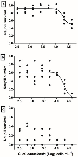
The exposure to C. cf. canariensis cells induced a significant effect on A. salina nauplii survival after 24 h (ANOVA, F(8,44) = 11.98, p ≤ 0.001), 48 h (ANOVA, F(8,44) = 33.17, p ≤ 0.001), and 72 h (Kruskal–Wallis, H(8,44) = 33.57, p ≤ 0.001) of exposure. Even exposure to the lower concentration of C. cf. canariensis tested (330 cells mL−1) was enough to induce significant nauplii mortality after 24 h of exposure (Table 1). Moreover, all tested concentrations of the benthic dinoflagellate C. cf. canariensis PII induced sublethal effects on A. salina nauplii, such as abnormal swimming activity and mobility impairment. The movement of A. salina exposed to the higher dinoflagellate concentration (10,565 cells mL−1) was extremely reduced, and nauplii showed slow appendage beats and the absence of displacement in the water column, possibly related to mucilage secreted by dinoflagellate cells at high concentrations.

Table 1.
Cumulative mortality (%) of Artemia salina nauplii exposed to Coolia cf. canariensis PII after 24, 48, and 72 h. Data comprise the mean values of ten A. salina individuals from each independent replicate of control and eight dinoflagellate concentrations.
The survival data of A. salina nauplii showed a non-linear concentration-dependent response after 24 h and 48 h of exposure to cells of C. cf. canariensis PII; however, no clear concentration–response curve was observed after 72 h of exposure (Figure 1). Considering the adjustment to the four-parameter logistic model, LC50 values were only determined for 24 h and 48 h of exposure. The LC50 values were validated according to the previously described criteria, the fitting error (FE) of LC50s did not exceed 25.7%, and fitted dose–response curves did not have an R2 lower than 0.829 (Table 2).

Figure 1.
Survival (in proportion of negative controls) of Artemia salina nauplii exposed to Coolia cf. canariensis PII (Log, cells mL−1) after 24 h (a), 48 h (b), and 72 h (c) of incubation. Data are shown as the values of independent replicates by concentration (n = 4), in black circles (●), and fit curve of validated assays (a,b) in black solid line. Since survival data after 72 h of exposure did not adjust to the four-parameter logistic model, there was no fit curve to be included in the graph (c). LC50 and 95% of confidence intervals are shown in Table 2.

Table 2.
Validated results of concentration–response LC50 and 95% of confidence interval (CI) (cells mL−1), with R2 and fitting error (FE, %) values, after Artemia salina nauplii exposure to cells of Coolia cf. canariensis PII.
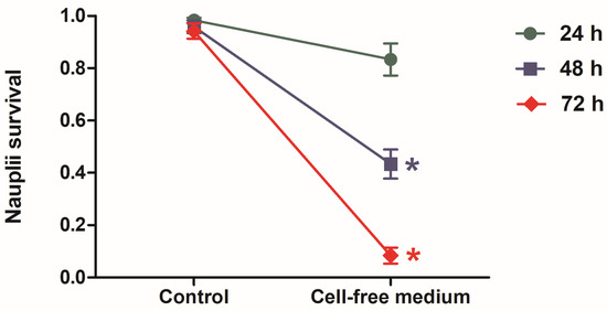
Since the exposure to the cells of C. cf. canariensis PII induced a significant concentration-dependent effect on nauplii survival, additional assays were performed to test the toxicity of BEC to nauplii. Exposure to cell-free medium, after the filtration of C. cf. canariensis PII culture with 126,000 total cells, induced a significant lethal effect on nauplii after incubation for 48 h (t-test, t-value = 7.239, df = 8, p< 0.0001) and 72 h (t-test, t-value = 9.705, df = 8, p < 0.0001) (Figure 2). No significant difference between the control group (only FSW) and cell-free medium treatment was detected after 24 h of incubation (t-test, t-value = 1.699, df = 8, p = 0.128) (Figure 2).

Figure 2.
Mean ± SD values of survival (in proportion of alive individuals) after incubation of Artemia salina nauplii with FSW (control) and cell-free medium (extracellular compounds) for 24 h (●), 48 h (■), and 72 h (♦). * Corresponds to treatments significantly different from the control (t-test, p < 0.0001). Cell-free medium was obtained by the filtration of C. cf. canariensis PII culture with 126,000 total cells.
3.2. Nauplii Exposure to C. malayensis
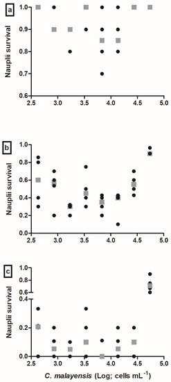
Exposure to C. malayensis induced a significant effect on A. salina nauplii survival after 24 h (Kruskal–Wallis, H(8,44) = 29.012, p = 0003), 48 h (Kruskal–Wallis, H(8,44) = 35.686, p < 0.0001), and 72 h (Kruskal–Wallis, H(8,44) = 33.936, p < 0.0001). Nauplii mortality was mostly detected at intermediary concentrations of C. malayensis (e.g., 1704–13,633 cells mL−1) after 48 h and 72 h of exposure (Table 3). Moreover, exposure to cells of C. malayensis induced sublethal effects on A. salina nauplii, which showed abnormal swimming activity and mobility impairment, mostly at higher dinoflagellate concentrations, possibly related to mucilage secreted by dinoflagellate cells at high concentrations.

Table 3.
Cumulative mortality (%) of Artemia salina nauplii exposed to Coolia malayensis after 24, 48, and 72 h. Data comprise the mean values of ten A. salina individuals from each independent replicate of control and eight dinoflagellate concentrations.
No marked concentration–response curve was observed for the survival data of A. salina nauplii exposed to C. malayensis for the concentrations tested (426–54,531 cells mL−1); thus, it was not possible to determine LC50 values for C. malayensis. Independently of exposure time, a decrease in the tendency of nauplii to survive was shown at lower concentrations (426 and 1704 cells mL−1), followed by a stabilization or increase in survival response at intermediary and higher concentrations (Figure 3). Exposure to the benthic dinoflagellate C. malayensis induced changes in A. salina nauplii behavior, which showed abnormal swimming activity, mobility impairment, and agitation. Most of the effects were noticed at higher concentrations associated with reductions in nauplii mobility, possibly related to mucilage secreted by dinoflagellate cells at high concentrations.

Figure 3.
Survival (in proportion of negative controls) of Artemia salina nauplii exposed to Coolia malayensis (Log, cells mL−1) after 24 h (a), 48 h (b), and 72 h (c) of incubation. Data are shown as values of independent replicates by concentration (n = 4), in black circles (●), and mean values by concentration (in grey squares, ■).
4. Discussion
In the present study, significant toxicity was induced by the strains of Coolia cf. canariensis PII and C. malayensis. Exposure to cells of C. cf. canariensis PII induced significant lethality to A. salina nauplii, even at the lower tested concentration (330 cells mL−1) and shorter exposure time (24 h). Moreover, exposure to cells of this strain also induced sublethal effects on nauplii, and extracellular compounds (i.e., cell-free medium) induced lethal effects on nauplii after 48 and 72 h of exposure. Coolia cf. canariensis PII is composed solely of the strain tested in the current study, isolated from the oceanic Trindade Island in Brazil [19]. Little information is available in the literature on the biological activity of strains from other phylogroups of the C. canariensis species complex. Yessotoxin and its analogs were not detected in three strains from phylogroup I and III isolated from the Canary Islands (Spain), and the extracts of one strain, which was injected intraperitoneally into mice, did not induce toxicity [14]. Bioassays performed with extracts of strains of C. cf. canariensis PIII from Hong Kong did not show significant lethality to nauplii of Artemia franciscana [36]. Moreover, direct exposure to cells of strains of C. cf. canariensis PIII from the Bay of Biscay (Spain) did not induce lethality to pluteus larvae of the sea urchin Heliocidaris crassispina [29]. Recently, yessotoxin analogs were detected in strains of C. cf. canariensis phylogroup IV isolated from Guam; however, their extracts did not induce biological activity and toxicity in Artemia bioassays [23]. Genetic differences separate strains from the C. cf. canariensis species complex in four phylogroups [23] which may also present differences in their biological activity and toxicity.
Surprisingly, the strain of C. cf. canariensis PII was more toxic than the strain of C. malayensis tested in the present study, considering both the exposure time and the minimum cellular concentration necessary to induce nauplius lethality. The lowest concentration of C. malayensis that induced a significant lethal effect was 852 cells mL−1 after 72 h of exposure, while 48 h of exposure was the minimum time that induced significant mortality at 1704 cells mL−1. An increase in the survival response of nauplii was detected at higher C. malayensis concentrations (independently of exposure time), which is possibly a hormetic effect (i.e., an overcompensation response to a disruption in homeostasis) [43]. Hormesis is a highly frequent phenomenon independently of a tested stressor, biological endpoint, and experimental model system [44]. It is not expected that a determined intensity of a specific stressor (e.g., a toxicant concentration) could induce similar hormetic responses in different biological systems; however, a hormetic effect was previously detected in A. salina individuals exposed to the chemical compound bisphenol A [45]. In contrast to our findings, C. malayensis is usually more toxic than other Coolia species [23,36], although [21] found a significant higher mortality rate of A. salina adults exposed to a strain of C. tropicalis that synthesizes gambierone (MTX3) at the maximum algal biomass tested (16,000 ng C mL−1) compared to C. malayensis (19,300 ng C mL−1), C. santacroce (15,000 ng C mL−1), and C. palmyrensis (11,500 ng C mL−1) after 72 h of incubations. In a previous study, the crude extract of the C. malayensis strain UNR-02 (i.e., the same used in the current study) induced a significant cell mass decrease in HepG2 and H9c2(2-1) cell lines at equivalent dinoflagellate concentrations of 5000–10,000 and 313–10,000 cells mL−1, respectively, after 72 h of exposure [38]. Additionally, at the subcellular level, C. malayensis crude extract induced changes to mitochondrial membrane potential generation and fluctuations associated with the induction of mitochondrial permeability transition [38], reinforcing the concept that the extract of this strain is toxic at the subcellular level and in cellular-based assays.
In the present study, only the exposure to C. cf. canariensis PII induced a concentration-dependent effect on nauplii survival with validated LC50 values of 18,064 and 19,968 cells mL−1 for 24 h and 48 h of exposure, respectively. Since exposure to Coolia malayensis did not fit to a concentration–response curve, it was not possible to determine LC50 values. There is no information concerning LC50 results obtained in bioassays using the direct exposure of marine organisms to Coolia spp. cells as frequently occurs in natural marine environments, where species from this dinoflagellate genus are widely distributed in tropical and temperate regions [9]. The results obtained from the direct exposure to Coolia cells, applied in the current study, are not directly comparable to LC50 results obtained in previous studies that performed the bioassays with Coolia lysates [36] and extracts [23]. A milder effect is expected to occur in individuals exposed to live dinoflagellate cells, without direct contact with intracellular toxins. C. malayensis lysates from strains S020 and Ve051 isolated from Hong Kong induced mortality significantly different from the control in larvae of brine shrimp and sea urchins, at concentrations up to 0.05 mg mL−1, while lysates of C. tropicalis (strain S002) induced mortality significantly different from the control of sea urchin larvae at 0.075 mg mL−1 [36]. The tested concentrations of lysates from C. cf. canariensis phylogroup III (strains W039 and Ve011) and C. palmyrensis (strains S017 and W085) did not significantly affect the survival of sea urchin larvae compared to the control [36]. The LC50,48h values of algal lysates of Coolia species was estimated for A. franciscana nauplii incubated with two strains of C. malayensis (0.086–0.117 mg mL−1), as well as for H. crassispina pluteus larvae incubated with two strains of C. malayensis (0.016–0.046 mg mL−1), C. tropicalis (0.029–0.038 mg mL−1), C. palmyrensis (0.023–0.049 mg mL−1), and C. cf. canariensis phylogroup III (0.064–0.082 mg mL−1) [36]. The toxicity of water-soluble extracts of Coolia species was tested in Artemia bioassays, and lethal and sublethal effects were detected in nauplii exposed to extracts of C. malayensis, while C. palmyrensis and C. cf. canariensis phylogroup IV did not exhibit a noxious effect [23]. A hydrophilic algal extract of a C. tropicalis strain, which produces 44-methylgambierone, induced a concentration effect on medaka fish larvae survival with an LC50,96h of 0.062 mg mL−1 [31].
Behavioral changes in aquatic organisms during bioassays may be an indicator of sublethal responses induced by toxic benthic dinoflagellates [5,7,46]. All the behavioral changes (i.e., abnormal swimming activity, mobility impairment, agitation, and slow appendages beats) detected in A. salina nauplii exposed to Coolia species seem to be related to noxious effects induced by dinoflagellate toxicity. Another possibility is that the large amount of mucus secreted by Coolia cells at high abundances may have caused behavioral alterations in A. salina nauplii, particularly when exposed to C. cf. canariensis PII, which induced more sublethal effects. Extracts of C. malayensis strains isolated from the Pacific Ocean have induced mobility impairment in exposed Artemia after 8 h, as well as a severe reduction in motility after 24 h and swimming inability after 30 h [23]. Abnormal behavior in brine shrimp (e.g., imbalanced swimming and/or lack of mobility) was detected after exposure to Coolia lysates [36]. Abnormal swimming has been reported in tintinids after exposure to the planktonic Alexandrium species [47,48]. Exposure to A. fundyense decreased the mobility of crab larvae [49], and paralysis was reported in copepod nauplii treated with exudates of A. tamiyavanichii [50]. Our findings highlighted the lethal and sublethal effects of two Coolia species to Artemia nauplii and more potent effects induced by C. cf. canariensis PII, which was not tested before.
Additionally, extracellular compounds released by C. cf. canariensis PII in cell-free medium (culture filtrate) with a high cellular concentration (126,000 total cells) induced significant mortality of nauplii after 48 and 72 h of incubation. Several noxious effects of bioactive extracellular compounds (BECs) produced and released by dinoflagellate species to aquatic organisms have been described in the literature [51,52,53,54]. In a previous study, filtrates of C. guanchica did not induce noxious effects on A. salina [17]. For the first time, in the present study, significant lethality was detected on Artemia nauplii after 48 and 72 h of incubations with culture filtrate (cell-free medium) of C. cf. canariensis PII, suggesting the production of compounds with allelopathic potential. It is important to highlight that most of the toxic effects detected on Artemia nauplii seem to have been induced by their direct contact with Coolia cells, likely caused by the ingestion of toxic cells. The length of C. malayensis and C. cf. canariensis PII is, on average, 24.5 µm [19], and therefore, these cells could have been grazed by Artemia nauplii during the assays. In a previous study, with other three benthic dinoflagellate genera (Prorocentrum, Ostreopsis, and Gambierdiscus), the exposure to culture filtrates (extracellular compounds) did not affect brine shrimp survivorship, and the lethality of A. salina adults was directly related to their feeding on dinoflagellate cells [5].
Phenotypic plasticity and differences in toxicity can be influenced by several factors, such as nutrient limitation and different environmental (or culture) conditions, which may induce not only differences in cellular toxin content, but also in toxin profile [55]. Differences in the profile of compounds synthesized by these dinoflagellates may also cause intraspecific variations in toxicity (i.e., among strains of the same Coolia species) [23,36]. Toxic compounds were already detected in five Coolia lineages—C. canariensis phylogroup IV, C. tropicalis, C. malayensis, C. palmyrensis, and C. santacroce [16,21,23,27,28,29]. However, biological activity and toxicity to marine organisms are not directly related to the presence of known Coolia toxins [23]. The findings of harmful and lethal effects, particularly caused by C. cf. canariensis PII in A. salina nauplii, reinforce the need for further studies to determine and identify the compounds that may be synthesized by Coolia species and their association with toxic effects in bioassays.
5. Conclusions
In the present study, for the first time, the toxicity of C. cf. canariensis PII was detected using Artemia bioassays. A concentration-dependent response on nauplii survival was induced by C. cf. canariensis PII exposure after 24 and 48 h. More potent effects on nauplii survival were detected after exposure to C. cf. canariensis PII with a shorter time (24 h) and concentration (330 cells mL−1). Moreover, extracellular compounds produced by C. cf. canariensis PII and released in culture medium induced lethality to nauplii, suggesting an allelopathic potential of this strain in the environment. Coolia malayensis also induced lethal and sublethal effects on Artemia nauplii after 48 and 72 h of exposure to concentrations of 1704 and 852 cells mL−1, respectively. Our study highlights that short-term exposure to Coolia cells may induce harmful effects and lethality in a marine model species.
Author Contributions
Conceptualization: A.M., R.A.F.N. and S.M.N.; methodology: A.M., S.M.N. and R.A.F.N.; validation: A.M. and R.A.F.N.; formal analysis: R.A.F.N.; investigation: A.M. and R.A.F.N.; writing—original draft: A.M. and R.A.F.N.; writing—review and editing: A.M., S.M.N. and R.A.F.N.; visualization: A.M., S.M.N. and R.A.F.N.; funding acquisition: S.M.N. and R.A.F.N. All authors have read and agreed to the published version of the manuscript.
Funding
This study was financially supported by Foundation Carlos Chagas Filho Research Support of the State of Rio de Janeiro (FAPERJ) through research grants to S.M. Nascimento (SEI-260003/002134/2021) and to R.A.F. Neves (E-26/201.283/2021), and by the Brazilian National Council for Scientific and Technological Development (CNPq) through the research grant to R.A.F. Neves (PQ2; 306212/2022-6). This study was funded in part by the Coordenação de Aperfeiçoamento de Pessoal de Nível Superior—Brasil (CAPES)—Finance Code 001—through a master’s scholarship (A. Miralha).
Institutional Review Board Statement
Not applicable.
Informed Consent Statement
Not applicable.
Data Availability Statement
The data presented in this study are available in the manuscript.
Acknowledgments
The authors are grateful to Joel Campos de Paula (UNIRIO) and to anonymous reviewers for their comments and suggestions for manuscript improvement.
Conflicts of Interest
The authors declare no conflict of interest.
References
- Zingone, A.; Berdalet, E.; Bienfang, P.; Enevoldsen, H.; Evans, J.; Kudela, R.; Testers, P. Harmful Algae in Benthic Systems: A GEOHAB Core Research Program. Cryptogam. Algol. 2012, 33, 225–230. [Google Scholar] [CrossRef]
- Leaw, C.P.; Tan, T.H.; Lim, H.C.; Teng, S.T.; Yong, H.L.; Smith, K.F.; Rhodes, L.; Wolf, M.; Holland, W.C.; Vandersea, M.W.; et al. New scenario for speciation in the benthic dinoflagellate genus Coolia (Dinophyceae). Harmful Algae 2016, 55, 137–149. [Google Scholar] [CrossRef] [PubMed]
- Hoppenrath, M.; Murray, S.A.; Chomerat, N.; Horiguchi, T. Marine Benthic Dinoflagellates—Unveiling Their Worldwide Biodiversity, 1st ed.; Senckenberg, Kleine Senckenberg-Reihe: Frankfurt, Germany, 2014; p. 276. [Google Scholar]
- Neves, R.A.F.; Rodrigues, E.T. Harmful Algal Blooms: Effect on Coastal Marine Ecosystems. In Life Below Water, Encyclopedia of the UN Sustainable Development Goals; Leal Filho, W., Azul, A.M., Brandli, L., Lange Salvia, A., Wall, T., Eds.; Springer Nature: Basel, Switzerland, 2020; pp. 1–31. [Google Scholar] [CrossRef]
- Neves, R.A.F.; Fernandes, T.; Santos, L.N.; Nascimento, S.M. Toxicity of benthic dinoflagellates on grazing, behavior and survival of the brine shrimp Artemia salina. PLoS ONE 2017, 12, e0175168. [Google Scholar] [CrossRef] [PubMed]
- Neves, R.A.F.; Contins, M.; Nascimento, S.M. Effects of the toxic benthic dinoflagellate Ostreopsis cf. ovata on fertilization and early development of the sea urchin Lytechinus Variegatus. Mar. Environ. Res. 2018, 135, 11–17. [Google Scholar] [CrossRef] [PubMed]
- Neves, R.A.F.; Santiago, T.C.; Carvalho, W.F.; Silva, E.S.; da Silva, P.M.; Nascimento, S.M. Impacts of the toxic benthic dinoflagellate Prorocentrum lima on the brown mussel Perna perna: Shell-valve closure response, immunology, and histopathology. Mar. Environ. Res. 2019, 146, 35–45. [Google Scholar] [CrossRef]
- Neves, R.A.F.; Nascimento, S.M.; Santos, L.N. Harmful algal blooms and shellfish in the marine environment: An overview of the main molluscan responses, toxin dynamics, and risks for human health. Environ. Sci. Pollut. Res. 2021, 28, 55846–55868. [Google Scholar] [CrossRef] [PubMed]
- Wakeman, K.C.; Yamaguchi, A.; Roy, M.C.; Jenke-Kodama, H. Morphology, phylogeny and novel chemical compounds from Coolia malayensis (Dinophyceae) from Okinawa, Japan. Harmful Algae 2015, 44, 8–19. [Google Scholar] [CrossRef]
- Meunier, A. Microplankton de la mer Flamande. 3 Les Peridiniens. Mem. Mus. R. Hyst. Nat. Belg. 1919, 8, 3–116. [Google Scholar]
- Faust, M.A. Observations on the morphology and sexual reproduction of Coolia monotis (Dinophyceae). J. Phycol. 1992, 28, 94–104. [Google Scholar] [CrossRef]
- Faust, M.A. Observation of sand-dwelling toxic dinoflagellates (Dinophyceae) from widely differing sites, including two new species. J. Phycol. 1995, 31, 996–1003. [Google Scholar] [CrossRef]
- Ten-Hage, L.; Delaunay, N.; Pichon, V.; Couté, A.; Puiseux-Dao, S.; Turquet, J. Okadaic acid production from the marine benthic dinoflagellate Prorocentrum arenarium Faust (Dinophyceae) isolated from Europa Island Coral Reef Ecosystem (SW Indian Ocean). Toxicon 2000, 38, 1043–1054. [Google Scholar] [CrossRef] [PubMed]
- Fraga, S.; Penna, A.; Bianconi, I.; Paz, B.; Zapata, M. Coolia canariensis sp. nov. (Dinophyceae), a new nontoxic epiphytic benthic dinoflagellate from the Canary Islands. J. Phycol. 2008, 44, 1060–1070. [Google Scholar] [CrossRef] [PubMed]
- Leaw, C.P.; Lim, P.T.; Cheng, K.W.; Ng, B.K.; Usup, G. Morphology and molecular characterization of a new species of thecate benthic dinoflagellate, Coolia malayensis sp. nov. (Dinophyceae). J. Phycol. 2010, 46, 162–171. [Google Scholar] [CrossRef]
- Karafas, S.; York, R.; Tomas, C. Morphological and genetic analysis of the Coolia monotis species complex with the introduction of two new species, Coolia santacroce. Harmful Algae 2015, 46, 18–33. [Google Scholar] [CrossRef]
- David, H.; Laza-Martinez, A.; Rodriguez, F.; Fraga, S.; Orive, E. Coolia guanchica sp. nov. (Dinophyceae) a new epi-benthic dinoflagellate from the Canary Islands (NE Atlantic Ocean). Eur. J. Phycol. 2020, 55, 76–88. [Google Scholar] [CrossRef]
- David, H.; Laza-Martínez, A.; García-Etxebarria, K.; Riobó, P.; Orive, E. Characterization of Prorocentrum elegans and Prorocentrum levis (Dinophyceae) from the Southeastern Bay of Biscay by morphology and molecular phylogeny. J. Phycol. 2014, 50, 718–726. [Google Scholar] [CrossRef]
- Nascimento, S.M.; Silva, R.A.F.; Oliveira, F.; Fraga, S.; Salgueiro, F. Morphology and molecular phylogeny of Coolia tropicalis, Coolia malayensis and a new lineage of the Coolia canariensis species complex (dinophyceae) isolated from Brazil. Eur. J. Phycol. 2019, 54, 484–496. [Google Scholar] [CrossRef]
- Larsson, M.E.; Smith, K.F.; Doblin, M.A. First description of the environmental niche of the epibenthic dinoflagellate species Coolia palmyrensis, C. malayensis, and C. tropicalis (Dinophyceae) from Eastern Australia. J. Phycol. 2019, 55, 565–577. [Google Scholar] [CrossRef]
- Tibiriçá, C.E.J.D.A.; Sibat, M.; Fernandes, L.F.; Bilien, G.; Chomérat, N.; Hess, P.; Mafra, L.L., Jr. Diversity and toxicity of the genus Coolia Meunier in Brazil, and detection of 44-Methyl Gambierone in Coolia tropicalis. Toxins 2020, 12, 327. [Google Scholar] [CrossRef]
- Abdennadher, M.; Zouari, A.B.; Medhioub, W.; Penna, A.; Hamza, A. Characterization of Coolia Spp. (Gonyaucales, Dinophyceae) from Southern Tunisia: First record of Coolia malayensis in the Mediterranean Sea. Algae 2021, 36, 175–193. [Google Scholar] [CrossRef]
- Phua, Y.H.; Roy, M.C.; Lemer, S.; Husnik, F.; Wakeman, K.C. Diversity and Toxicity of pacific strains of the benthic dinoflagellate Coolia (Dinophyceae), with a look at the Coolia canariensis species complex. Harmful Algae 2021, 109, 102120. [Google Scholar] [CrossRef]
- Zhang, H.; Lü, S.; Cen, J.; Li, Y.; Li, Q.; Wu, Z. Morphology and molecular phylogeny of three species of Coolia (Dinophyceae) from Hainan Island, South China Sea. J. Oceanol. Limnol. 2021, 39, 1020–1032. [Google Scholar] [CrossRef]
- Tsikoti, C.; Genitsaris, S. Review of harmful algal blooms in the coastal Mediterranean Sea, with a focus on Greek waters. Diversity 2021, 13, 396. [Google Scholar] [CrossRef]
- Nascimento, S.M. Abundance of Coolia Genus in the Oceanic Trindade Island (South Atlantic Ocean, Brazil); Personal Communication; Federal University of the State of Rio de Janeiro: Rio de Janeiro, Brazil, 2023. [Google Scholar]
- Holmes, M.J.; Lewis, R.J.; Jones, A.; Hoy, A.W.W. Cooliatoxin, the first toxin from Coolia monotis (Dinophyceae). Nat. Toxins 1995, 3, 355–362. [Google Scholar] [CrossRef] [PubMed]
- Rhodes, L.L.; Thomas, A.E. Coolia monotis (Dinophyceae): A toxic epiphytic microalgal species found in New Zealand. New Zeal. J. Mar. Freshw. Res. 1997, 31, 139–141. [Google Scholar] [CrossRef]
- Laza-Martinez, A.; Orive, E.; Miguel, I. Morphological and Genetic Characterization of benthic dinoflagellates of the genera Coolia, Ostreopsis and Prorocentrum from the South-Eastern Bay of Biscay. Eur. J. Phycol. 2011, 46, 45–65. [Google Scholar] [CrossRef]
- Murray, J.S.; Nishimura, T.; Finch, S.C.; Rhodes, L.L.; Puddick, J.; Harwood, D.T.; Larsson, M.E.; Doblin, M.A.; Leung, P.; Yan, M.; et al. The role of 44-Methylgambierone in ciguatera fish poisoning: Acute toxicity, production by marine microalgae and its potential as a biomarker for Gambierdiscus Spp. Harmful Algae 2020, 97, 101853. [Google Scholar] [CrossRef] [PubMed]
- Yan, M.; Leung, P.T.Y.; Gu, J.; Lam, V.T.T.; Murray, J.S.; Harwood, D.T.; Wai, T.-C.; Lam, P.K.S. Hemolysis associated toxicities of benthic dinoflagellates from Hong Kong waters. Mar. Pollut. Bull. 2020, 155, 111114. [Google Scholar] [CrossRef]
- Nakajima, I.; Oshima, Y.; Yasumoto, T. Toxicity of benthic dinoflagellates found in coral reef-II. Toxicity of benthic dinoflagellates in Okinawa. Nippon. Suisan Gakkaishi 1981, 47, 1029–1033. [Google Scholar] [CrossRef]
- Pagliara, P.; Caroppo, C. Toxicity assessment of Amphidinium carterae, Coolia cfr. monotis and Ostreopsis cfr. ovata (Dinophyta) isolated from the Northern Ionian Sea (Mediterranean Sea). Toxicon 2012, 60, 1203–1214. [Google Scholar] [CrossRef]
- Mendes, M.C.Q.; Nunes, J.M.C.; Fraga, S.; Rodríguez, F.; Franco, J.M.; Riobó, P.; Branco, S.; Menezes, M. Morphology, molecular phylogeny and toxinology of Coolia and Prorocentrum strains isolated from the tropical South Western Atlantic Ocean. Bot. Mar. 2019, 62, 125–140. [Google Scholar] [CrossRef]
- Rhodes, L.; Adamson, J.; Suzuki, T.; Briggs, L.; Garthwaite, I. Toxic marine epiphytic dinoflagellates, Ostreopsis siamensis and Coolia monotis (Dinophyceae), in New Zealand. N. Z. J. Mar. Freshw. Res. 2000, 34, 371–383. [Google Scholar] [CrossRef]
- Leung, P.T.Y.; Yan, M.; Yiu, S.K.F.; Lam, V.T.T.; Ip, J.C.H.; Au, M.W.Y.; Chen, C.Y.; Wai, T.C.; Lam, P.K.S. Molecular phylogeny and toxicity of harmful benthic dinoflagellates Coolia (Ostreopsidaceae, Dinophyceae) in a sub-tropical marine ecosystem: The first record from Hong Kong. Mar. Pollut. Bull. 2017, 124, 878–889. [Google Scholar] [CrossRef] [PubMed]
- Gu, J.; Yan, M.; Leung, P.T.Y.; Tian, L.; Lam, V.T.T.; Cheng, S.H.; Lam, P.K.S. Toxicity effects of hydrophilic algal lysates from Coolia tropicalis on marine medaka larvae (Oryzias melastigma). Aquat. Toxicol. 2021, 234, 105787. [Google Scholar] [CrossRef]
- Varela, A.T.; Neves, R.A.F.; Nascimento, S.M.; Oliveira, P.J.; Pardal, M.A.; Rodrigues, E.T.; Moreno, A.J. Mitochondrial impairment and cytotoxicity effects induced by the marine epibenthic dinoflagellate Coolia malayensis. Environ. Toxicol. Pharmacol. 2020, 77, 103379. [Google Scholar] [CrossRef] [PubMed]
- Rhodes, L.; Smith, K.; Papiol, G.G.; Adamson, J.; Harwood, T.; Munday, R. Epiphytic dinoflagellates in sub-tropical New Zealand, in particular the genus Coolia Meunier. Harmful Algae 2014, 34, 36–41. [Google Scholar] [CrossRef]
- Guillard, R.R.J. Culture methods. In Manual on Harmful Marine Microalgae; DIOC Manual and Guides; Hallegraeff, G.M., Anderson, D.M., Cembella, A.D., Eds.; UNESCO: Paris, France, 1995; Volume 33, pp. 45–56. [Google Scholar]
- Nunes, B.S.; Carvalho, F.D.; Guilhermino, L.M.; Van Stappen, G. Use of the genus Artemia in ecotoxicity testing. Environ. Pollut. 2006, 144, 453–462. [Google Scholar] [CrossRef]
- Beck, B.; Chen, Y.-F.; Dere, W.; Devanarayan, V.; Eastwood, B.J.; Farmen, M.W.; Iturria, S.J.; Iversen, P.W.; Kahl, S.D.; Moore, R.A.; et al. Assay Operations for SAR Support; Eli Lilly & Company and the National Center for Advancing Translational Sciences: Bethesda, MD, USA, 2004.
- Calabrese, E.J. Evidence that hormesis represents an “overcompensation” response to a disruption in homeostasis. Ecotoxicol. Environ. Saf. 1999, 42, 135–137. [Google Scholar] [CrossRef]
- Calabrese, E.J.; Baldwin, L.A. Hormesis: A generalizable and unifying hypothesis. Crit. Rev. Toxicol. 2001, 31, 353–424. [Google Scholar] [CrossRef]
- Naveira, C.; Rodrigues, N.; Santos, F.S.; Santos, L.N.; Neves, R.A.F. Acute toxicity of bisphenol a (bpa) to tropical marine and estuarine species from different trophic groups. Environ. Pollut. 2021, 268, 115911. [Google Scholar] [CrossRef]
- Neves, R.A.F.; Nascimento, S.M.; Santos, L.N. Sublethal fish responses to short-term food chain transfer of DSP toxins: The role of somatic condition. J. Exp. Mar. Bio. Ecol. 2020, 524, 151317. [Google Scholar] [CrossRef]
- Hansen, P.J.; Cembella, A.D.; Moestrup, Ø. The marine dinoflagellate Alexandrium ostenfeldii: Paralytic shellfish toxin concentration, composition, and toxicity to a tintinnid ciliate. J. Phycol. 1992, 28, 597–603. [Google Scholar] [CrossRef]
- Fulco, V.K. Harmful effects of the toxic dinoflagellate Alexandrium tamarense on the tintinnids Favella taraikaensis and Eutintinnus sp. J. Mar. Biol. Assoc. UK 2007, 87, 1085–1088. [Google Scholar] [CrossRef]
- Sulkin, S.; Hinz, S.; Rodriguez, M. Effects of exposure to toxic and non-toxic dinoflagellates on oxygen consumption and locomotion, in stage 1 larvae of the crabs Cancer oregonensis and C. magister. Mar. Biol. 2003, 142, 205–211. [Google Scholar] [CrossRef]
- Silva, N.J.; Tang, K.W.; Lopes, R.M. Effects of microalgal exudates and intact cells on subtropical marine zooplankton. J. Plankton Res. 2013, 35, 855–865. [Google Scholar] [CrossRef]
- Borcier, E.; Morvezen, R.; Boudry, P.; Miner, P.; Charrier, G.; Laroche, J.; Hegaret, H. Effects of bioactive extracellular compounds and paralytic shellfish toxins produced by Alexandrium minutum on growth and behaviour of juvenile great scallops Pecten maximus. Aquat. Toxicol. 2017, 184, 142–154. [Google Scholar] [CrossRef]
- Pan, L.; Chen, J.; Shen, H.; He, X.; Li, G.; Song, X.; Zhou, D.; Sun, C. Profiling of extracellular toxins associated with diarrhetic shellfish poison in Prorocentrum lima culture medium by high-performance liquid chromatography coupled with mass spectrometry. Toxins 2017, 9, 308. [Google Scholar] [CrossRef] [PubMed]
- Castrec, J.; Soudant, P.; Payton, L.; Tran, D.; Miner, P.; Lambert, C.; Le Goïc, N.; Huvet, A.; Quillien, V.; Boullot, F.; et al. Bioactive extracellular compounds produced by the dinoflagellate Alexandrium minutum are highly detrimental for oysters. Aquat. Toxicol. 2018, 199, 188–198. [Google Scholar] [CrossRef]
- Long, M.; Tallec, K.; Soudant, P.; Lambert, C.; Le Grand, F.; Sarthou, G.; Jolley, D.; Hégaret, H. A rapid quantitative fluorescence-based bioassay to study allelochemical interactions from Alexandrium minutum. Environ. Pollut. 2018, 242, 1598–1605. [Google Scholar] [CrossRef]
- Neely, T.; Campbell, L. A modified assay to determine hemolytic toxin variability among Karenia clones isolated from the Gulf of Mexico. Harmful Algae 2006, 5, 592–598. [Google Scholar] [CrossRef]
Disclaimer/Publisher’s Note: The statements, opinions and data contained in all publications are solely those of the individual author(s) and contributor(s) and not of MDPI and/or the editor(s). MDPI and/or the editor(s) disclaim responsibility for any injury to people or property resulting from any ideas, methods, instructions or products referred to in the content. |
© 2023 by the authors. Licensee MDPI, Basel, Switzerland. This article is an open access article distributed under the terms and conditions of the Creative Commons Attribution (CC BY) license (https://creativecommons.org/licenses/by/4.0/).