Review

Lacustrine Cyanobacteria, Algal Blooms and Cyanotoxins in East Africa: Implications for Human and Ecological Health Protection

by
Timothy Omara
1,2,*,
Christine Betty Nagawa
3,
Christine Kyarimpa
4,
Stefan Böhmdorfer
2,
Thomas Rosenau
2,
Solomon Omwoma Lugasi
5,
Henry Matovu
6,
Silver Odongo
7 and
Patrick Ssebugere
7,8,9,*
1
Food Safety Laboratories, Chemistry Division, Testing Department, Standards Directorate, Uganda National Bureau of Standards, Bweyogerere Industrial and Business Park, Kampala P.O. Box 6329, Uganda
2
Institute of Chemistry of Renewable Resources, Department of Chemistry, University of Natural Resources and Life Sciences Vienna (BOKU), Konrad-Lorenz-Straße 24, 3430 Tulln, Austria
3
Department of Forestry, Biodiversity and Tourism, College of Agricultural and Environmental Sciences, Makerere University, Kampala P.O. Box 7062, Uganda
4
Department of Chemistry, Faculty of Science, Kyambogo University, Kampala P.O. Box 1, Uganda
5
Department of Physical Sciences, Jaramogi Oginga Odinga University of Science and Technology, Bondo P.O. Box 210-40601, Kenya
6
Department of Chemistry, Gulu University, Gulu P.O. Box 166, Uganda
7
Department of Chemistry, College of Natural Sciences, Makerere University, Kampala P.O. Box 7062, Uganda
8
Department of Cell Toxicology, Helmholtz Centre for Environmental Research-UFZ, 04318 Leipzig, Germany
9
Department of Analytical Environmental Chemistry, Helmholtz Centre for Environmental Research-UFZ, 04318 Leipzig, Germany
*
Authors to whom correspondence should be addressed.
Phycology 2023, 3(1), 147-167; https://doi.org/10.3390/phycology3010010
Submission received: 24 January 2023 / Revised: 6 February 2023 / Accepted: 8 February 2023 / Published: 15 February 2023

Abstract

:
Advected cyanobacteria, algal blooms and cyanotoxins have been increasingly detected in freshwater ecosystems. This review gives an insight into the present state of knowledge on the taxonomy, dynamics, toxic effects, human and ecological health implications of cyanobacteria, algal blooms and cyanotoxins in the East African Community lakes. The major toxigenic microalgae in East African lakes include Microcystis, Arthrospira, Dolichospermum, Planktolyngbya and Anabaenopsis species. Anatoxin-a, homoanatoxin-a, microcystins (MCs), cylindrospermopsin and nodularin have been quantified in water from below method detection limits to 81 µg L−1, with peak concentrations characteristically reported for the wet season. In whole fish, gut, liver and muscles, MCs have been found at concentrations of 2.4 to 1479.24 μg kg−1, which can pose human health risks to a daily consumer. While there have been no reported cases of cyanotoxin-related poisoning in humans, MCs and anatoxin-a (up to 0.0514 μg kg−1) have been identified as the proximal cause of indiscriminate fish kills and epornitic mortality of algivorous Phoeniconaias minor (lesser flamingos). With the unequivocal increase in climate change and variability, algal blooms and cyanotoxins will increase in frequency and severity, and this will necessitate swift action towards the mitigation of nutrient-rich pollutants loading into lakes in the region.

Graphical Abstract

1. Introduction

Industrialization has been the key driver of economic growth and inclusive prosperity because it does not only foster economic and infrastructural development, but also enhances the realization of some vital targets enshrined in the 2015–2030 Sustainable Development Goals [1,2]. This is evident from the employment opportunities it creates, improved working conditions, optimal resource use [3] and the innovations that have led to nascent and environmentally benign (greener) production technologies [4,5]. Despite this, there are various environmental challenges that have been associated with industrialization. One of the most pronounced concerns has been the occurrence of cyanobacteria (CYB), harmful algal blooms (CYBHAB) and cyanotoxins (phycotoxins) in aquatic ecosystems, attributed to anthropogenic pollution and accelerated climate change [6].
Algal blooms were initially not considered to be harmful because they largely occurred in summer or typically dry months [7]. Lately, rising temperatures have doubled or tripled algal bloom incidences in coastal countries [7]. At present, CYBHAB are at the forefront of toxicological research because of the negative effects that they can exert on both ecosystems and humans. Cyanobacterial phytoplankton blooms are contextualized “as noticeable, CYBHAB with appreciable effects, including scum formation, marked discoloration of surface waters as well as fish, human, or other invertebrate mortalities” [8]. In other words, CYBHAB occur when algal densities surpass baseline population levels. More than 5000 species of microalgae are classified, but only 300 species (or less) may induce algal blooms that are toxic [9]. Despite the spontaneous occurrence of various microalgae in bloom conditions, CYB or blue-green algae are of primary concern because they can produce toxic metabolites (cyanotoxins). Cyanotoxins are mostly produced by CYB, dinoflagellates and benthic diatoms from genera Dolichospermum, Microcystis, Arthrospira, Anabaenopsis, Alexandrium, Protogonyaulax, Gymnodinium, Dinophysis, Gonyaulax, Prorocentrum, Pseudonitzchia and Pyrodinium [10].
CYBHAB is a globally recognized phenomenon, and several incidences have been reported in more than 25 countries, including India, Italy, Germany, Netherlands, Sweden, Greece, USA, China, Madagascar, Algeria, Ghana, Ethiopia, Zimbabwe, Botswana, Egypt, Cameroon, Mozambique, Uganda, Sudan, Kenya, Senegal, Burkina Faso, Lesotho, Morocco, Nigeria, South Africa, Tanzania and Tunisia [7]. It is believed that the incidences of CYBHAB and algal toxins production will continue to increase dramatically, plausibly due to anthropogenic nutrient loading and global warming with its impact on the vertical stratification of lakes [11,12]. Despite the central body of knowledge that CYBHAB tend to be recurrent in high nutrient-load water resources, there is accumulating evidence that blooms also occur in oligotrophic ecosystems [11]. Moreover, strong re-oligotrophication (that increases light in the metalimnion) may favor CYBHAB where metalimnetic bloom-forming CYB are present [11]. The high-nutrient paradigm as the major driver of CYBHAB is also challenged [12], because CYB have undergone adaptations so that they can thrive even under the harshest environmental conditions.
The East African Community (hereafter EAC), comprising seven sovereign states (Democratic Republic of Congo, Burundi, South Sudan, Rwanda, Tanzania, Uganda and Kenya), is bountifully blessed with water resources (Figure 1). The region’s water demands are, to a greater extent, fulfilled by eutrophic water bodies, which, regrettably, have been under intense land use charges and pollution pressure, and this could lead to increased incidences of CYB and CYBHAB [13,14,15]. The close proximity between human settlements, industries, cities, ports and most water bodies [16] lead to the recognition that CYBHAB may be a common occurrence. To this end, various event-driven studies in the region have been undertaken on the phytoplankton composition, occurrence of CYB and cyanotoxins in lacustrine ecosystems. Three reviews in regional and continental contexts have been published on CYB and cyanotoxins, with the EAC in focus. The first of its kind was by Ndlela et al. [7], who gave an overview of CYBHAB incidences and the research strides in Africa. Later, Kimambo et al. [17] synthesized data pertaining to the presence of CYBHAB in inland aquatic ecosystems of the United Republic of Tanzania, and the linkages with the country’s climatic conditions. The latest collation by Olokotum et al. [14] examined the socioecological causes and consequences of CYBHAB in Lake Victoria (L. Victoria), the largest lentic water resource in the EAC. With the inclusion of the Democratic Republic of Congo (DRC) into the EAC in 2022, more knowledge on CYB, CYBHAB and cyanotoxins could be revealed as it shares some lakes with Rwanda (Lake Kivu), Uganda (Lake Albert and Lake Edward) and Tanzania (Lake Tanganyika). We built on the foregoing reviews to examine the current understanding of the taxonomy, algal bloom dynamics, toxicity, human and ecological health implications of CYB, CYBHAB and algal toxins in the EAC. Cyanotoxins are still considered to be contaminants of emerging concern, because their toxicity mechanisms and effects on humans and ecosystems are not fully understood [10].

2. Materials and Methods

Electronic search on the occurrence of CYB, algal blooms and cyanotoxins in EAC lakes were performed in Web of Science Core Collection®, Scopus®, Science Direct, Google Scholar®, Springer Link, Taylor and Francis Online, Scientific Electronic Library Online, PubMed, Google search engine and EAC university repositories from August 2022 to December 2022. The specific search terms used were cyanobacteria, blue-green algae, algal bloom, cyanobacterial bloom, harmful algal bloom, CyanoHABs, HABs, microcystins, saxitoxin, anatoxin, cylindrospermopsin, nodularin, brackish water and the specific names of the EAC partner states or major lakes such as Lake Victoria, Lake Tanganyika, Lake Kivu, Lake Naivasha and Lake Nakuru. Reports from websites of all Nile basin organizations were searched. The review considered articles dated until December 2022.

3. Occurrence of Cyanobacteria, Algal Blooms and Phycotoxins in EAC Lakes

Toxic and non-toxic CYB are photosynthetic prokaryotes that occur naturally in terrestrial as well as aquatic ecosystems [10]. They are typically larger than normal bacterial cells, and their inherent mass production of phycobilin pigment confers upon them a bluish tint at high concentrations, hence their naming as blue-green algae [7]. They are Gram-negative bacteria that may be filamentous, unicellular or multicellular (occuring as colonies), contingent on the prevailing conditions. Under suitable environmental conditions that afford competitive advantages (e.g., alkaline pH, buoyancy, high sunlight-for conversion of ferric ion to ferrous ion, moderate temperature, i.e., 20 °C to 30 °C (10 °C in winter for Planktothrix rubescens), nutrients phosphorous and nitrogen, and water column stability), CYB are capable of proliferating and forming CYBHAB or scums in the upper sunlit layers. Such unsightly scums and blooms contain malodorous compounds such as geosmin and methylisoborneol, which are responsible for the aesthetically unpleasant taste of CYB-contaminated water. While the biology and ecology of CYB has been a subject of intensive research globally, there is a paucity of clearly articulated information regarding factors and processes that regulate toxin production in most cyanobacterial species [12]. In lentic freshwater resources (such as L. Victoria, Lake Tanganyika and Lake Kivu in the EAC), the occurrence of CYB is favored by climate variability, anthropogenic activities, hydrological shifts and high nutrient loads [19]. In part (for Ugandan lakes such as Mburo and Kachera), loading is from influx of nutrient-rich hippopotamus and cattle dung wastes [20].
Cyanobacteria are usually associated with the production of nocive cyanotoxins. The great diversity and high metabolic potential of CYB implies that there are other unknown or at least little studied cyanotoxins. According to CyanoMetDB (a comprehensive database of cyano-metabolites), at least 2000 molecules, including more than 300 microcystin congeners, are already known [21]. Cyanotoxins are contaminants of emerging concern that are potentially (eco)toxic. They can adversely impact ecosystem services provided by water resources by depleting oxygen, altering food webs, species assemblages and poisoning animals and humans [22]. Examples of cyanotoxins include cyclic hepatotoxic peptides (microcystins, nodularins), dermatoxic, cytotoxic, genotoxic or neurotoxic alkaloids, polyketides and amino acids (lyngbyatoxin-a, cylindrospermopsins, anatoxins, saxitoxins, aetokthonotoxin, lipopolysaccharides (endotoxins), guanitoxin, beta-N-methylamino-L-alanine and aplysiatoxins) [22].
The frequently encountered cyanotoxins are anatoxins, cylindrospermopsin (CYN), nodularins (NODs), saxitoxin, and microcystins (MCs), but the most-studied members are MCs (the -LR variant). Thus, in addition to anatoxin-a (ATX), they are the main cyanotoxins that garnered early scientific interest in Eastern Africa (Figure 2). In the EAC, toxigenic freshwater CYB and CYBHAB have been implicated in the reoccurrence of eutrophic and hypoxic conditions in L. Victoria [14,23,24,25,26,27]. Table S1 shows a summary of reports on the occurrence and abundance of CYB and other phytoplankton, their dominant species and MCs in EAC lakes. These are discussed per country in the following.

3.1. DRC

In the Congolese part of the oligotrophic Lake Tanganyika, the occurrence of CYB (Dolichospermum flosaquae, Anabaenopsis species and Limnococcus limneticus), along with Nitzschia asterionelloides (Bacillariophyta), has been reported [28,29]. These diazotrophic CYB were implicated in the CYBHAB witnessed in 1955 and 2018, but cyanotoxin concentrations of water sampled from the lake have not been established.
Another unique lake in DRC is Lake Kivu, a deep oligotrophic and meromictic water resource. Though it contains copious volumes of exploitable methane, CYB (Synechococcus species and Planktolyngbya limnetica) dominate the phytoplankton biomass in this lake, followed by pennate diatoms (Nitzschia bacata and Fragilaria danica) [30,31]. These reports resonate well with that of Hecky and Kling [32], who showed that CYB and chlorophytes (with biomass contents that are higher than in the neighboring Lake Tanganyika) dominated in Lake Kivu. Their report, however, pointed to the presence of additional CYB Lyngbya circumcreta West, Anabaenopsis, Cylindrospermopsis and Raphidiopsis species. Sarmento et al. [33] found a strikingly contradicting result, with pennate diatoms being more abundant than CYB in Lake Kivu. Nevertheless, picocyanobacterial Synechococcus species was found to still form a significant proportion of the annual autotrophic plankton of the lake [34]. Further, Urosolenia and Microcystis genera are dominant holopelagic species under certain stratification scenarios [33]. The dominance of diatoms in Lake Kivu thus seems to occur solely during dryer periods, when deep mixing occurs [35].

3.2. Kenya

With regard to cyanotoxins, one of the groundbreaking reports was by Ballot et al. [36] who found intracellular ATX (0.3 to 9 and 5 to 223 µg g−1 dry weight (DW)) and MCs (16 to 155, and 130 to 4593 µg MC-LR eq. g−1) in seston samples from Lake Bogoria and Lake Nakuru. Interestingly, phycotoxins were not detected (UDT) in Lake Elmenteita cyanobacterial samples. Later, MCs (2.2 μg MC-YR g−1 DW) and ATX (0.3 μg g−1 DW) were traced in axenic Arthrospira fusiformis (A. fusiformis) from Lake Sonachi, though the same species along with Anabaenopsis species from Lake Simbi had no detectable phycotoxins [37]. The authors similarly found cyanotoxins at concentrations of 1.6 to 12.0 μg MC-YR g−1 DW (in Lake Sonachi) and 19.7 to 39.0 μg MC-YR g−1 DW (in Lake Simbi) for MCs, and 0.5 to 2.0 and UDT to 1.4 μg g−1 DW for ATX, respectively. Kotut et al. [38] similarly reported MCs (2.2 and 15.02 μg g−1 DW) in A. fusiformis (synonym: Spirulina fusiformis) growing in Lakes: Sonachi and Bogoria. ATX occurred at levels of 0.3 to 10.38 μg g−1 DW in A. fusiformis sampled from lakes Sonachi, Bogoria and Nakuru, despite them being UDT in cultures from L. Simbi and L. Elmenteita. All these reports corroborate previous observations that strongly suggested the dominance of A. fusiformis in the volcanic crater and meromictic soda lakes of Kenya [39,40,41]. In later investigations, Kenyan lakes Bogoria, Nakuru, Oloidien and Elmenteita had more than 97% scum of A. fusiformis. As expected, MCs occurred at 16 to 155 µg MC-LR eq L−1 in Lake Bogoria and 130 to 4593 µg MC-LR eq L−1 in Lake Nakuru [36,42,43,44]. These reports were divergent from earlier findings of Vareschi [45], where CYB in Lake Nakuru were reported to be dominated by Spirulina platensis.
Other reports on CYB in Kenya are from its L. Victoria part (bays, gulfs and satellite lakes), where CYB (>35%) and diatoms (>30%) of Microcystis, Merismopedia and Dolichospermum species are the primary phytoplankton (Table S1). As far as cyanotoxins are concerned, Sitoki and others [46] detailed the incidence of MCs in L. Victoria water. They concluded that the levels varied greatly between seasons. This comes in concordance with later inferences of other researchers [47,48] who investigated MCs contamination of water and fish consumed by fisher communities of Winam Gulf, Homa Bay, Kisumu, Siaya and Busia counties of L. Victoria (Kenya). Regrettably, up to 30% of water from these points exceeded the regulatory set value (1.0 µg L−1) of the WHO [48]. The study suggested that CYBHAB pose potential year-round health risks to riparian communities [48].
Recently, a team of researchers collated the insights and awareness of L. Victoria shore community on MCs toxicity [47]. The authors appreciated that more than 70% of the fisherfolk are conversant with the toxic effects of MCs, and showed the urgency required to mitigate them. An earlier investigation [49] echoed that higher average values of MCs (5 to 109 µg kg−1) occurred in fish from Nyanza Gulf (Kisumu Bay) compared with those from Rusinga channel water (14 µg kg−1). These reports reaffirmed that CYBHAB are recurrent in L. Victoria.

3.3. Tanzania

Previous studies conducted about lakes Big Momela, Embagai and Manyara indicated that CYB (>50%), mostly Anabaenopsis elenkenii, A. fusiformis (Lake Big Momela), A. fusiformis, Oscillatoria, Hantzschia (Lake Embagai), Oscillatoria jenensis and Pseudoanabaena terebriformis (Lake Manyara), were dominant in the phytoplankton [50,51]. Similarly, CYB (specifically A. fusiformis) have been prevalent in Momela Lakes and Lake Natron with MCs detected in water at concentrations of 0.1–4.5 μg mL−1 of scum in the latter [52].
For L. Victoria, the occurrence of CYB (upto 82%) was quantified in several parts of the southern part. Miles et al. [53] found putative MCs analogues in extracts of a cyanobacterial bloom from Mwanza Gulf but did not quantify them. On 27 islands of Ukerewe district, MCs (0.0028 to 0.0102 μg L−1) were reported [54]. Other studies in L. Victoria (several bays, open water and Gulfs) have found MCs (up to 13 µg MC-LR eq L−1; Table S1). An incidence of multiple cyanotoxins: CYN (0.004 to 0.01 μg L−1), NODs (0.010 μg L−1) and MCs (0.0028 to 0.0118 μg L−1) in water from L. Victoria has been communicated [54]. The report emphasized that multiple and repeated exposure to phycotoxins could amplify their toxicity and/or adverse effects.

3.4. Uganda

From the available literature, Uganda has the highest number of reports about CYB in several lakes. Of these, Western Uganda crater lakes (Kyaninga, Saaka, Nyabikere, Nyinambuga, Munyayange, Kikorongo, Maseche, Murumuli, Bunyampaka, Katwe, Bagusa, Nyamunuka, Mwamba, Katanda, Karolero, Kerere, Kacuba, Mwengenyi, Kyerbwato, Katanda, Kanyamukali, Nkugute, Kyanga, Mirambi, Nyanswiga, Kitere, Chibwera, Lugembe, Nyanswiga, Kamweru (Figure 3), Nyahirya, Nyabikere, Kyasanduka, Kifuruka, Wandakara, Nyamusingire, Nyungu and Katinda) were found to contain CYB (35% to 100%), primarily of the genera Planktolyngbya, Microcystis, Anabaena and Cylindrospermopsis. No determination of cyanotoxins was performed [55,56].
Other interesting studies are available on the Albertine lakes (Edward and George) [57]. Species from Raphidiopsis and Anabaenopsis genera are the primary community in Lake Edward, though Aphanocapsa, Merismopedia, Microcystis, Aphanothece and Anathece genera are also present. According to several authors, shallow Ugandan lakes near Mount Rwenzori (Lake George, Lake Edward and Lake Mburo) are eutrophic, with Microcystis species being the most abundant CYB [57,58,59,60,61]. Lake Mburo was earlier reported to have more than 90% of its phytoplanktonic community as CYB [62,63]. Later, Okello et al. [59] found that MCs (majorly MC-LR) occurred from UDT to 3.5 μg L−1 in the foregoing shallow lakes, which were lower than 0.5 to 10.2 μg L−1 analogously investigated for Lake Shaka. Another report by Poste [64] highlighted that several fish species from Ugandan lakes contained MCs at 0.0005 to 0.198 μg kg−1 in filleted muscle tissues, 0.0034 to 1.189 μg kg−1 in gutted and beheaded fish, and 0.0028 to 0.8987 μg kg−1 in whole fish samples (Table 1).
In the Ugandan part of L. Victoria, Microcystis, Dolichospermum and Cylindrospermopsis species are the prevalent CYB (>80%) (Table S1). Cyanotoxin analyses have reported concentrations of UDT to 93 µg L−1 of MCs in water from Murchison Bay, Napoleon gulf and open lake water. Worth citing are pioneering studies in Murchison Bay where MCs were quantified in Oreochromis niloticus (Nile tilapia fish), unveiling that the concentrations in biota and aqueous phase were correlated. The study highlighted that there has been an increase in MCs-producing CYB in the lake which are plausibly ingested by fish, agreeing with previous research findings [61,65,66]. The maximum concentration of total MCs reported for guts, liver and muscles of phytoplanktivorous Oreochromis niloticus (Nile tilapia) and Lates niloticus (Nile perch) from Murchison Bay of L. Victoria (1.86 to 1479.24 μg kg−1) is slightly higher than those from other Ugandan lakes such as Lake Mburo (73.10 to 1312 μg kg−1) [65]. A study published in 2022 unveiled for the first time the occurrence of homoanatoxin-a (HTX; <0.04 HTX L−1 in water from an inshore station of Murchison Bay [67], along with MCs (0.15–11.7 µg MC-LR eq L−1). At recreation sites, MCs (0.180 to 14.800 µg MC-LR eq L−1) equally occurred. The study demonstrated that whereas CYB were eliminated by water treatment, MCs remained detectable in water during and post-treatment (0.14 µg L−1) [67]. This shows that remediation of cyanotoxins in water from L. Victoria will require more efficient technologies to avoid exposing the local population to potential effects of MCs.
At this point, it can be suggested that shallow lakes in Uganda exhibit less seasonality in their CYB composition when compared with satellite lakes and others in the main L. Victoria basin. Unlike in oligotrophic lakes in the region, the CYB dynamics (spatial and temporal variations in prevalent cyanobacterial genera) in L. Victoria are, however, inconsistent in its different parts. This may be related to external anthropogenic influences, especially nutrient loading, because the lake receives a cocktail of pollutants from different countries.

3.5. Rwanda

The only report on CYB in a Rwandese Lake (Lake Muhazi) showed that it contains mainly Microcystis aeruginosa, followed by the dinoflagellate Cerutium hirundinellu [68]. These are ingested by Nile tilapia present in the lake [69], suggesting the need to establish the concentrations of cyanotoxins in water and fish from this lake.
Overall, volcanic and tectonic lakes in the East African Great Rift Valley possess distinguished extents of hydrological connections. Volcanicity in the region resulted in endorheic basins whose bedrock, groundwater connection and climate have favored schizohaline water formation [70]. These, in turn, have contributed to the dominance of CYB, and occurrence of CYBHAB and cyanotoxins. The literature reveals that toxigenic microalgae recorded from EAC lakes are Dolichospermum, Microcystis, Arthrospira, Planktolyngbya and Anabaenopsis species. The prevalence of CYBHAB and cyanotoxins in EAC lakes is of concern due to potential bioaccumulation and trophic transfer in zooplanktivorous and carnivorous fish species [61,66]. Moreover, the observed levels of MCs in whole fish, gut, liver and muscles (2.4 to 1479.24 μg kg−1) could pose human health risks to a daily consumer, as the WHO daily intake limit of MCs in fish is 0.04 µg kg−1 [65].
In L. Victoria, cyanobacterial biomasses and MCs levels in water from gulfs and bays comparatively surpasses their levels in the open lake water, with Microcystis and Dolichospermum species being the most prevalent CYB genera. Further, CYBHAB in the lake has increased costs associated with water treatment, e.g., National Water and Sewerage Cooperation Uganda reported increased chlorine demand for water treatment, unpleasant odors and tastes in untreated water supplies, and clogging of pumps and filters. Fishermen have reported that CYB has hampered fishing operations on the lake [71]. Anecdotal reports point that portions of the lake covered by CYB were observed to have small dead fish, whereas larger fish from such brackish waters are often weak and stressed. Earlier (in 1984), indiscriminate fish die-offs were witnessed in L. Victoria (Kenya), and this was plausibly connected with CYBHAB [72]. Similar mass mortalities were observed in 1991 for fish in Lake Magadi, Kenya [73], and this event was anticipated to have been caused by reduction in the algae Spirulina platensis. These effects, according to a recent report [67], may increase in severity in the coming decades. For example, MCs detected from Murchison Bay of L. Victoria now range from 0.20 to 15.00 µg MC-LR eq L−1, which is higher than those reported previously (0.20 to 0.70 and UDT to 1.6 µg MC-LR eq L−1 between 2004 and 2005, and then 2007 to 2008) [66,74], possibly due to the doubling of the mean Microcystis biovolume [67]. The EAC recreational waters are not often screened for pelagic cyanobacterial species, implying that the sanitary activities in contaminated lakes may expose both humans and animals to the potential negative effects of CYBHAB and cyanotoxins.

4. Toxicity, Human and Ecological Health Implications of Cyanotoxins in EAC Lakes

4.1. MCs

MCs are hepatotoxins, majorly produced as secondary metabolites of planktonic cyanobacterial species from genera such as Microcystis, Cylindrospermopsis, Anabaena, Oscillatoria (Planktothrix), Anabaenopsis, Nostoc, Arthrospira, Hapalosiphon, Limnothrix, Lyngbya, Phormidium, Rivularia, Synechocystis and Synechococcus [75]. Acute effects such as nausea, diarrhea, dermal, eye and throat irritations have been associated with their ingestion. Chronic exposure to MCs culminates in hepatic necrosis, retarded growth, reduced reproduction potential and, ultimately, death in fish and humans. The neurotoxicity of MCs is also known, but this applies specifically to invertebrates without livers [76]. In addition, exposure to MCs is associated with colorectal and primary liver cancer, with MC-LR receiving classification as a possible human carcinogen (group 2B) [77]. For humans, exposure to MCs occurs principally through the ingestion of contaminated aquatic organisms (e.g., fish) or water, as well as through the recreational use of water. Upon ingestion and absorption into the liver by organic anion transport proteins, MCs inhibit protein phosphatases, thereby selectively distorting cytoskeleton formation, degrading hepatic ultrastructure in eukaryotic cells, resulting in hepatic failure, intrahepatic hemorrhage and shock [78,79].
In EAC, MCs and ATX were implicated in the death of Phoeniconaias minor Geoffroy Saint-Hilaire 1798 (lesser flamingos) [80]. The pink birds (Figure 4a) feed on A. fusiformis [81], which confers upon them the pink plumage following the accumulation of ingested cyanobacterial pigments [82]. While this phenomenon is not new (e.g., in the Greater flamingos and Western Tanager [83,84]), it should be anticipated that other nutrition-based compounds may become bioaccumulated in lesser flamingos, e.g., potentially toxic metals. Event-driven reports of lesser flamingo die-offs are available for soda lakes such as Bogoria and Nakuru of Kenya [85,86], Momela, Natron, Rishateni, Manyara and Empakai Crater of Tanzania [13,51,87,88] (Table 2). Some of these reports substantiated the anecdotal claims by quantifying MCs and ATX levels in carcasses of the birds. Krienitz et al. extended the hypothesis further and examined the concentration of MCs and ATX in Lake Bogoria, in the surrounding hot springs and in flamingo birds [89]. They concluded that the cyanotoxins from the hot-spring mats could be responsible for the mass mortalities of the birds because: (i), there were evident cyanobacterial cells, fragments in hot spring mats, and elevated levels of ATX and MCs (0.00434 and 0.000196 µg kg−1); and (ii) there were clinically indisputable signs of flamingo intoxications. Such intoxication with the biotoxins could plausibly have been caused by direct or indirect intake of CYB or their cells [89]. Wings, breasts and head feathers of the flamingos reportedly had ATX and MCs concentrations ranging from UDT to 0.03 MC-LR eq μg kg−1 [90]. Moreover, amino acid neurotoxins β-N-methylamino-L-alanine (0.0035 μg kg−1 DW) and 2,4-diaminobutyric acid (0.0085 μg kg−1 DW) were recently quantified in lesser flamingo feathers from Lake Nakuru [91].
While it is still debated that MCs may be a potential initiator of avian botulism, other probable causes of the unnatural mass death of wild birds include avian tuberculosis [96], cholera, botulism, heavy metals [95,106], pesticide residues, or combinations of these [85,98,103,107,108]. Indeed, mycobacteriosis was reported in lesser flamingos from Lake Nakuru, Kenya [109]. Nevertheless, anatoxins and MCs (at concentrations higher than reported in EAC flamingos) have been associated with avian mortalities [98,101,102,110,111]. From an ecological perspective, lesser flamingos are “Near Threatened” supported by the limited and ungazetted nesting areas, as well as the reduced bird populations [112,113,114]. At present, they reportedly breed in only five sites: one in EAC (Lake Natron, with at least 75% of the breeding birds), Etosha and Makgadikgadi Pans in Namibia and Botswana, and Purabcheria Salt and Zinzuwadia Pans of India [115]. An attempt has been made to hand-rear lesser flamingos so as to restore and conserve this rare species of birds [116], but there is no available literature with any registered success.

4.2. Anatoxin-a

ATX is toxicologically known as Very Fast Death Factor for its fast lethal effect in animals, which could be related to its high rate of absorption into the gastrointestinal tract [117]. ATX is a secondary bicyclic amine alkaloid with peracute neurotoxic effects. Its discovery and identification in the 1960s and 1972 from CYB (Anabaena flos-aquae) followed the mortality of cattle herds that ingested contaminated water from Saskatchewan Lake in Ontario [117]. It is known to be biosynthesized by CYB from Arthrospira, Anabaena, Microcystis, Planktothrix, Oscillatoria, Aphanizomenon and Cylindrospermum genus [117].
Exposure to ATX (through ingestion of contaminated water or dried algal crusts, accidental swallowing/inhalation) has been associated with burning, tingling, respiratory paralysis and dysrhythmias, which are fatal. ATX antagonizes the activity of neuronal α4β2 and α4 nicotinic acetylcholine receptors (nAchRs) of the central nervous system and (α1)2βγδ muscle-type nAchRs of the neuromuscular junction [118]. With an affinity >20 times that of acetylcholine, ATX has the same effect as the former when it binds with nAchRs, i.e., it induces a conformational effect on the receptor, opening the channel pore to permit the passage of ions (Ca2+ and Na+) into the neuron. This culminates into cell depolarization, the generation of action potentials and thus muscle contraction. During ATX-mediated toxicity, the acetylcholine neurotransmitter does not dissociate from the nAchRs, resulting into irreversible inhibition and blockage of neuromuscular transmission [119]. This inhibitory effect generally accumulates the neurotransmitter within the synaptic cleft, eventually causing paralysis, asphyxiation and death, specifically if respiratory muscles are affected. In the epornitic mortalities of algivorous EAC lesser flamingos (such as in Lake Bogoria; Figure 4b), clinical symptoms have included opisthotonus, supporting that such die-offs are (at least in part) due to ATX intoxication. Other than the foregoing, ATX possess modulatory effects on nAChRs, which can result in the release of dopamine and noradrenaline [120].

4.3. Homoanatoxin-a

HATX being structurally a higher homologue of ATX has the same toxic effects as ATX. In addition to its nicotinic agonistic effects, HATX also upregulates acetylcholine release from cholinergic nerves [121]. This may explain why the potency of HATX is greater than that of ATX. Mortalities from CYBHAB with HATX are rare, but a report of dog neurotoxicosis from New Zealand (where the animals ingested CYB from Hutt River, lower North Island with 4400 µg kg−1 wet weight of HATX) has been published [122].

4.4. Cylindrospermopsin

CYN is a hydrophilic potentially hepatotoxic and immunotoxic cyclic guanidinium alkaloid, with characteristic tricyclic hydroxymethyl uracil [76]. It has some analogues such as deoxy-CYN (lacking an oxygen atom), demethoxy-CYN and 7-epiCYN (difference in the orientation of hydroxyl group) isolated in CYB Cylindrospermopsis raciborskii. The discovery of CYN toxicity happened when more than 100 children from Palm Island in Queensland, Australia suffered from unprecedented gastroenteritis and hepatomegaly. The ordeal was finally found to be due to the ingestion of CYN in contaminated water with CYBHAB of C. raciborskii [123]. However, CYN is also produced by other CYB, including Aphanizomenon flos-aquae, Anabaena species (bergii, and lapponica), Aphanizomenon ovalisporum, Lyngbya wollei, Raphidiopsis curvata Oscillatoria (Planktothrix) species and Umezakia natans [75]. With guideline values of 0.5 to 3 μg L−1 in drinking water across continents, CYN is the second-most-studied cyanotoxin known to target the liver, kidneys, heart, spleen, ovary, eye, lung, T lymphocytes, neutrophils and vascular endothelium [124]. CYN elicit toxicity through inhibition of protein synthesis, which can also occur at subtoxic concentrations [125]. Other toxicologists stated that CYN (with its inherent reactive guanidine) could be largely toxic through the induction of DNA wreckage and disruption of the kinetochore spindle. This could possibly result in chromosome loss, aneugenic and clastogenic effects [126]. Chichova et al. [124] found that CYN elicited moderate toxicity in human intestinal epithelial cells with suppression of cellular regeneration of the epithelial layer. CYN shows hepatotoxic, nephrotoxic, and cytotoxic effects, suggesting potential carcinogenicity. The neurotoxic potential of CYN has also been cited, though this could be a direct consequence of its cytotoxicity. To this end, the full underlying mechanisms of CYN toxicity needs to be elucidated [76].
In the EAC, there are no toxicity reports on CYN, which may be due to the absence of robust data on this cyanotoxin. There are, however, episodes of human and animal CYN-related poisoning from other countries. The most notable human poisoning is the 1979 Solomon dam gastroenteritis and hepatomegaly incidence in children from Palm Island [123]. The mortality of a cow and three calves after drinking water from McKinley Shire dam, Northern Queensland (Australia) was also reported. The animals had severe abdominal and thoracic haemorrhagic effusion, hyperaemic mesentery, pale and swollen liver, extremely distended gall bladder with dark yellow bile and epicardial haemorrhages [127]. In the subsequent 21 days, another eight animals (two cows and six calves) died, and analyses implicated CYN in C. raciborskii as the cause [127]. In Lake Aleksandrovac (Serbia), indiscriminate fish deaths due to the ingestion of CYN (range: 1.91 and 24.28 µg L−1) were reported [128]. This report may point to the need to establish CYN levels in EAC lakes where indiscriminate fish deaths have been reported, as CYN may be a contributing factor in addition to MCs.

4.5. Nodularins

Nodularins, a class of hepatotoxic non-ribosomal cyclic pentapeptides, possess toxicity mechanisms similar to those of MCs [129]. They are structurally analogous to MCs, but differentiable from MCs in their amino acid components (Figure 2). To date, ten naturally occurring variants (isoforms) of NODs have been discovered, but nodularin-R (with Z amino acid = arginine) is the most common, most commercially available and most studied variant. The toxicity of NODs mainly targets the liver, but they also accumulate in the intestines, blood and kidneys [130]. Upon ingestion, NODs diffuse from the proximal and distal ileum into the liver [131], where they inhibit active sites of serine/threonine protein phosphatases (PP) namely: 1 (PP-1), 2A (PP-2A) and 3 (PP-3). A non-covalent interaction occurs at first with the side chain (ADDA part) and a free D-glutamyl carboxyl group in the cyclic structure of the PP, followed by the inhibition of the phosphatase activities. NODs–phosphatase complexes (NODs-PP-1 and NODs-PP-2A) are formed with exceptionally stable bonds. Thus, the key difference between NODs and MCs in their toxicity via protein phosphatases inhibition is that the former binds non-covalently to phosphatases, while the latter forms a covalent bond [130].
Furthermore, NODs also elicit toxicity through formation of superoxide and hydroxyl radicals (reactive oxygen species) according to a yet incompletely elucidated pathway [130]. Their tumor-promoting activity is, on the other hand, mediated through the induced gene expression of TNF-alpha and proto-oncogenes, the exact mechanism of which is yet to be unraveled. In addition, the deactivation of the resultant tumor suppressor gene products (retinoblastoma and p53) progresses via phosphorylation, and this inevitably promotes tumorigenesis [132]. Overall, the cascade of reactions following NOD ingestion causes cellular disorganizations and damages, apoptosis, necrosis, loss of cell integrity, DNA fragmentation and strand breaks, intrahepatic bleeding and rapid blistering of hepatocytes which results in blood pooling and doubling of the liver weight [133]. Thus, mortalities associated with NOD poisoning is mediated through hemorrhagic shocks, which occurs in a few hours when ingested at high concentrations [134].
There are no toxicity events involving NODs in the EAC. Nevertheless, animal (cattle, dog, sheep, horse, pig and guinea pig) NOD-poisoning-related mortalities have been reported in other parts of the world. For example, hepatotoxicosis of a South African dog following the ingestion of NODs (0.00000347 µg kg−1 DW) was reported [135]. Main et al. [136] recorded 52 sheep deaths in South Western Australia from drinking water contaminated with NODs from Nodularia spurnigena. These reports emphasize that more studies on this cyanotoxin are warranted in EAC lakes.

5. Conclusions and Recommendations

CYB, CYBHAB and cyanotoxins have increased in EAC lacustrine ecosystems. Dolichospermum, Microcystis, Arthrospira, Planktolyngbya and Anabaenopsis species are the major groups of toxigenic CYB prevalent in EAC lakes producing ATX, HATX, MCs, CYN and NODs. Shallow EAC lakes exhibit less seasonality in their CYB composition, with Microcystis being the CYB producing MCs under shallow and eutrophic lacustrine conditions. The only direct ecological effects of cyanotoxins in EAC lakes is indiscriminate fish deaths and mass die-offs of lesser flamingos. With the unequivocal increase in climate change and variability, it is inferred that CYBHAB and cyanotoxins will increase in frequency and severity. This calls for urgent action to mitigate nutrient-rich pollutants loading into water resources and the expansion of CYBHAB from eutrophic lakes to the surrounding marine environments. The (eco)toxicological relevance of co-production of phycotoxins should be assessed in the EAC because such exposure may amplify the toxicological outcomes in aquatic biota and humans. As some CYB encountered in EAC lakes produce other cyanotoxins (such as β-N-methylamino-L-alanine and saxitoxins), studies targeting these cyanobacterial metabolites should be initiated. While there are no reports of cyanotoxin poisoning of humans in the EAC, future studies should examine the risk of hepatocellular cancer, the ingestion of CYB and mycotoxin-contaminated water and foods, and hepatitis virus, which were earlier linked to increased primary liver cancer cases in Asia. Another potential relationship with microplastics should be assessed because they are known to accumulate toxins and amplify their toxicity.

Supplementary Materials

The following supporting information can be downloaded at: https://www.mdpi.com/article/10.3390/phycology3010010/s1, Table S1. Phytoplankton composition and cyanotoxins in East African Community lakes.

Author Contributions

Conceptualization, T.O.; methodology, T.O., C.B.N., H.M., S.O. and P.S.; software, T.O.; writing—original draft preparation, T.O.; writing—review and editing, C.B.N., C.K., S.B., T.R., S.O.L., H.M., S.O. and P.S.; supervision, C.B.N., C.K. and P.S. All authors have read and agreed to the published version of the manuscript.

Funding

This review was inspired by a doctoral research project supported by the Austrian Partnership Programme in Higher Education and Research (APPEAR) under APPEAR Project: Environmental Chemistry for Sustainable Development (ECSDevelop) for Timothy Omara. APPEAR is a programme of the Austrian Development Cooperation (ADC) and is implemented by Austria’s Agency for Education and Internationalization (OeAD-GmbH), OEZA Project No. 0894-01/2020. Patrick Ssebugere’s research fellowship at Helmholtz Centre for Environmental Research-UFZ was supported by the Alexander von Humboldt Foundation (UGA-1185413-GF-E). The article processing charge for this article was in part supported by the University of Natural Resources and Life Sciences, Vienna (BOKU) through its Institutional Open Access Program (No. 2207756).

Institutional Review Board Statement

Not applicable.

Informed Consent Statement

Not applicable.

Data Availability Statement

This is a review article, and no raw data were collected.

Acknowledgments

Ivan Kahwa (Mbarara University of Science and Technology, Uganda) is acknowledged for his insights on crater lakes in Western Uganda, and for the picture of Lake Kamweru presented in this review. The graphical abstract was created with BioRender.com.

Conflicts of Interest

The authors declare no conflict of interest.

References

  1. Opoku, E.E.O.; Yan, I.K.-M. Industrialization as driver of sustainable economic growth in Africa. J. Int. Trade Econ. Dev. 2019, 28, 30–56. [Google Scholar] [CrossRef]
  2. Naudé, W. Industrialization under Medieval Conditions? Global Development after COVID-19. Discussion Working paper, IZA DP No. 13829. 2020. Available online: https://docs.iza.org/dp13829.pdf (accessed on 26 November 2022).
  3. Elfaki, K.E.; Handoyo, R.D.; Ibrahim, K.H. The Impact of Industrialization, Trade Openness, Financial Development, and Energy Consumption on Economic Growth in Indonesia. Economies 2021, 9, 174. [Google Scholar] [CrossRef]
  4. Bongomin, O.; Ocen, G.G.; Nganyi, E.O.; Musinguzi, A.; Omara, T. Exponential Disruptive Technologies and the Required Skills of Industry 4.0. J. Eng. 2020, 2020, 4280156. [Google Scholar] [CrossRef] [Green Version]
  5. United Nations Industrial Development Organization. Industrialization as the Driver of Sustained Prosperity; United Nations Industrial Development Organization: Vienna, Austria, 2020; Available online: https://www.unido.org/sites/default/files/files/2020-04/UNIDO_Industrialization_Book_web4.pdf (accessed on 26 November 2022).
  6. Band-Schmidt, C.; Durán-Riveroll, L.; Bustillos-Guzmán, J.; Leyva-Valencia, I.; López-Cortés, D.; Núñez-Vázquez, E.J.; Hernández-Sandoval, F.; Ramírez-Rodríguez, D. Paralytic Toxin Producing Dinoflagellates in Latin America: Ecology and Physiology. Front. Mar. Sci. 2019, 6, 42. [Google Scholar] [CrossRef] [Green Version]
  7. Ndlela, L.L.; Oberholster, P.J.; Van Wyk, J.H.; Cheng, P.H. An overview of cyanobacterial bloom occurrences and research in Africa over the last decade. Harmful Algae 2016, 60, 11–26. [Google Scholar] [CrossRef]
  8. Kat, M. Special meeting on causes, dynamics and effects of exceptional marine blooms and related events. Int. Counc. Explor. Sea C 1984, 3. [Google Scholar]
  9. Padmakumar, K.B.; Menon, N.R.; Sanjeevan, V.N. Is Occurrence of Harmful Algal Blooms in the Exclusive Economic Zone of India on the Rise? Int. J. Oceanogr. 2012, 2012, 263946. [Google Scholar] [CrossRef] [Green Version]
  10. Klijnstra, M.D.; Faassen, E.J.; Gerssen, A. A Generic LC-HRMS Screening Method for Marine and Freshwater Phycotoxins in Fish, Shellfish, Water, and Supplements. Toxins 2021, 13, 823. [Google Scholar] [CrossRef]
  11. Jacquet, S.; Briand, J.-F.; Leboulanger, C.; Avois-Jacquet, C.; Oberhaus, L.; Tassin, B.; Vinçon-Leite, B.; Paolini, G.; Druart, J.-C.; Anneville, O.; et al. The proliferation of the toxic cyanobacterium Planktothrix rubescens following restoration of the largest natural French lake (Lac du Bourget). Harmful Algae 2005, 4, 651–672. [Google Scholar] [CrossRef] [Green Version]
  12. Reinl, K.L.; Brookes, J.D.; Carey, C.C.; Harris, T.D.; Ibelings, B.W.; Morales-Williams, A.M.; Yokota, K.; Zhan, Q. Cyanobacterial blooms in oligotrophic lakes: Shifting the high-nutrient paradigm. Freshw. Biol. 2021, 66, 1846–1859. [Google Scholar] [CrossRef]
  13. Lihepanyama, D.L.; Ndakidemi, P.A.; Treydte, A.C. Spatio–TemporalWater Quality Determines Algal Bloom Occurrence and Possibly Lesser Flamingo (Phoeniconaias minor) Presence in Momella Lakes, Tanzania. Water 2022, 14, 3532. [Google Scholar] [CrossRef]
  14. Olokotum, M.; Mitroi, V.; Troussellier, M.; Semyaloa, R.; Bernarde, C.; Montuelle, B.; Okello, W.; Quiblier, C.; Humbert, J.-F. A review of the socioecological causes and consequences of cyanobacterial blooms in Lake Victoria. Harmful Algae 2020, 96, 101829. [Google Scholar] [CrossRef] [PubMed]
  15. Saulnier-Talbot, É.; Gregory-Eaves, I.; Simpson, K.; Efitre, J.; Nowlan, T.; Taranu, Z.; Chapman, L. Small Changes in Climate Can Profoundly Alter the Dynamics and Ecosystem Services of Tropical Crater Lakes. PLoS ONE 2014, 9, e86561. [Google Scholar] [CrossRef] [PubMed] [Green Version]
  16. Baguma, G.; Musasizi, A.; Twinomuhwezi, H.; Gonzaga, A.; Nakiguli, C.K.; Onen, P.; Angiro, C.; Okwir, A.; Opio, B.; Otema, T.; et al. Heavy Metal Contamination of Sediments from an Exoreic African Great Lakes’ Shores (Port Bell, Lake Victoria), Uganda. Pollutants 2022, 2, 407–421. [Google Scholar] [CrossRef]
  17. Kimambo, O.N.; Gumbo, J.R.; Chikoore, H. The occurrence of cyanobacteria blooms in freshwater ecosystems and their link with hydro-meteorological and environmental variations in Tanzania. Heliyon 2019, 5, e01312. [Google Scholar] [CrossRef] [PubMed] [Green Version]
  18. Ayugi, B.; Dike, V.; Ngoma, H.; Babaousmail, H.; Mumo, R.; Ongoma, V. Future Changes in Precipitation Extremes over East Africa Based on CMIP6 Models. Water 2021, 13, 2358. [Google Scholar] [CrossRef]
  19. Kaggwa, M.; Straubinger-Gansberger, N.; Schagerl, M. Cyanotoxins in small artificial dams in Kenya utilised for cage fish farming—A threat to local people? Afr. J. Aquat. Sci. 2018, 43, 123–129. [Google Scholar] [CrossRef]
  20. Mbabazi, D.; Orach-Meza, F.L.; Makanga, B.; Hecky, R.E.; Balirwa, J.; Ogutu-Ohwayo, R.; Verburg, P.; Namuleno, G.; Muhumuza, E.; Luyiga, J. Trophic structure and energy flow in fish communities of two lakes of the Lake Victoria basin. Uganda J. Agric. Sci. 2004, 9, 348–359. [Google Scholar]
  21. Jones, M.R.; Pinto, E.; Torres, M.A.; Dörr, F.; Mazur-Marzec, H.; Szubert, K.; Tartaglione, L.; Dell’Aversano, C.; Miles, C.O.; Beach, D.G. CyanoMetDB, a comprehensive public database of secondary metabolites from cyanobacteria. Water Res. 2021, 196, 117017. [Google Scholar] [CrossRef]
  22. Gerssen, A.; Gago-Martinez, A. Emerging marine biotoxins. Toxins 2019, 11, 314. [Google Scholar] [CrossRef] [Green Version]
  23. Simiyu, B.; Amukhuma, H.; Sitoki, L.; Okello, W.; Kurmayer, R. Interannual variability of water quality conditions in the Nyanza Gulf of Lake Victoria, Kenya. J. Great Lakes Res. 2022, 48, 97–109. [Google Scholar] [CrossRef]
  24. Kimambo, O.N.; Gumbo, J.R.; Chikoore, H.; Msagati, T. Harmful Algal Blooms in Aquaculture Systems in Ngerengere Catchment, Morogoro, Tanzania: Stakeholder’s Experiences and Perception. Int. J. Environ. Res. Public Health 2021, 18, 4928. [Google Scholar] [CrossRef] [PubMed]
  25. Cornelissen, I.; Silsbe, G.; Verreth, J.; van Donk, E.; Nagelkerke, L. Dynamics and limitations of phytoplankton biomass along a gradient in Mwanza Gulf, southern Lake Victoria (Tanzania). Freshw. Biol. 2014, 59, 127–141. [Google Scholar] [CrossRef] [Green Version]
  26. Kondowe, B.; Masese, F.; Raburu, P.; Singini, W.; Walumona, R. Water quality and ecology of Lake Kanyaboli, Kenya: Current status and historical changes. Lakes Reserv. Res. Manag. 2022, 27, e12401. [Google Scholar] [CrossRef]
  27. Hecky, R.E.; Bugenyi, F.; Ochumba, P.; Talling, J.; Mugidde, R.; Gophen, M.; Kaufman, L. Deoxygenation of the deep water of Lake Victoria, East Africa. Limnol. Oceanogr. 1994, 39, 1476–1481. [Google Scholar] [CrossRef] [Green Version]
  28. Cocquyt, C.; Plisnier, P.-D.; Mulimbwa, N.; Nshombo, M. Unusual massive phytoplankton bloom in the oligotrophic Lake Tanganyika. Plant Ecol. Evol. 2021, 154, 351–361. [Google Scholar] [CrossRef]
  29. Ehrenfels, B.; Bartosiewicz, M.; Mbonde, A.; Baumann, K.; Dinkel, C.; Junker, J.; Kamulali, T.; Kimirei, I.; Niederdorfer, R.; Odermatt, D.; et al. Diazotrophic Cyanobacteria are Associated With a Low Nitrate Resupply to Surface Waters in Lake Tanganyika. Front. Environ. Sci 2021, 9, 716765. [Google Scholar] [CrossRef]
  30. Sarmento, H.; Darchambeau, F.; Descy, J. Phytoplankton of Lake Kivu; Descy, J.P., Darchambeau, F., Schmid, M., Eds.; Lake Kivu. Aquatic Ecology Series; Springer: Dordrecht, The Netherlands, 2012; Volume 5, pp. 67–83. [Google Scholar]
  31. Rugema, E.; Darchambeau, F.; Sarmento, H.; Stoyneva-Gärtner, M.; Leitao, M.; Thiery, W.; Latli, A.; Descy, J.P. Long-term change of phytoplankton in Lake Kivu: The rise of the greens. Freshw. Biol. 2019, 64, 1940–1955. [Google Scholar] [CrossRef]
  32. Hecky, R.; Kling, H. Phytoplankton ecology of the great lakes in the rift valleys of Central Africa. Arch. Hydrobiol. Beih. Ergeb. Limnol. 1987, 25, 197–228. [Google Scholar]
  33. Sarmento, H.; Leitao, M.; Stoyneva, M.; Couté, A.; Compère, P.; Isumbisho, M.; Descy, J. Diversity of pelagic algae of Lake Kivu (East Africa). Cryptogam. Algol. 2007, 28, 245–269. [Google Scholar]
  34. Sarmento, H.; Unrein, F.; Isumbisho, M.; Stenuite, S.; Gasol, J.; Descy, J. Abundance and distribution of picoplankton in tropical, oligotrophic Lake Kivu, eastern Africa. Freshw. Biol. 2008, 53, 756–771. [Google Scholar] [CrossRef]
  35. Sarmento, H.; Isumbisho, M.; Descy, J.-P. Phytoplankton ecology of Lake Kivu (eastern Africa). J. Plankton Res. 2006, 28, 815–829. [Google Scholar] [CrossRef]
  36. Ballot, A.; Krienitz, L.; Kotut, K.; Wiegand, C.; Metcalf, J.S.; Codd, G.A.; Pflugmacher, S. Cyanobacteria and cyanobacterial toxins in three alkaline Rift Valley lakes of Kenya—Lakes Bogoria, Nakuru and Elmenteita. J. Plankton Res. 2004, 26, 925–935. [Google Scholar] [CrossRef] [Green Version]
  37. Ballot, A.; Krienitz, L.; Kotut, K.; Wiegand, C.; Pflugmacher, S. Cyanobacteria and cyanobacterial toxins in the crater lakes Sonachi and Simbi, Kenya. Harmful Algae 2005, 4, 139–150. [Google Scholar] [CrossRef]
  38. Kotut, K.; Ballot, A.; Krienitz, L. Toxic cyanobacteria and their toxins in standing waters of Kenya: Implications for water resource use. J. Water Health 2006, 4, 3–18. [Google Scholar] [CrossRef]
  39. Tuite, C.H. Standing crop densities and distribution of Spirulina and benthic diatoms in East African alkaline saline lakes. Freshw. Biol. 1981, 11, 345–360. [Google Scholar] [CrossRef]
  40. Melack, J.M. Photosynthesis and growth of Spirulina platensis (Cyanophyta) in an equatorial lake (Lake Simbi, Kenya). Limnol. Oceanogr. 1979, 24, 753–760. [Google Scholar] [CrossRef]
  41. Finlay, B.J.; Curds, C.R.; Bamforth, S.S.; Bafort, J.M. Ciliated protozoa and other microorganisms from two African soda lakes (Lake Nakuru and Lake Simbi, Kenya). Arch. Protistenkd. 1987, 133, 81–91. [Google Scholar] [CrossRef]
  42. Dadheech, P.K.; Glöckner, G.; Casper, P.; Kotut, K.; Mazzoni, C.J.; Mbedi, S.; Krienitz, L. Cyanobacterial diversity in the hot spring, pelagic and benthic habitats of a tropical soda lake. FEMS Microbiol. Ecol. 2013, 85, 389–401. [Google Scholar] [CrossRef] [Green Version]
  43. Kibichii, S.; Shivoga, W.A.; Muchiri, M.; Enanga, E.; Miller, S.N. Seasonality in water quality and its influence on the abundance and distribution of phytoplankton and chironomid larvae in Lake Nakuru, Kenya. Int. Ver. Für Theor. Und Angew. Limnol. Verh. 2008, 30, 333–338. [Google Scholar] [CrossRef]
  44. Krienitz, L.; Kotut, K. Fluctuating algal food populations and the occurrence of lesser flamingos (Phoeniconaias minor) in three Kenyan rift valley lakes. J. Phycol. 2010, 46, 1088–1096. [Google Scholar] [CrossRef]
  45. Vareschi, E. The ecology of Lake Nakuru (Kenya). Oecologia 1978, 32, 11–35. [Google Scholar] [CrossRef]
  46. Sitoki, L.; Kurmayer, R.; Rott, E. Spatial variation of phytoplankton composition, biovolume, and resulting microcystin concentrations in the Nyanza Gulf (L. Victoria, Kenya). Hydrobiologia 2012, 691, 109–122. [Google Scholar] [CrossRef] [Green Version]
  47. Githukia, C.; Onyango, D.; Lusweti, D.; Ramkat, R.; Kowenje, C.; Miruka, J.; Lung’ayia, H.; Orina, P. An Analysis of Knowledge, Attitudes and Practices of Communities in Lake Victoria, Kenya on Microcystin Toxicity. Open J. Ecol. 2022, 12, 198–210. [Google Scholar] [CrossRef]
  48. Roegner, A.; Sitoki, L.; Weirich, C.; Corman, J.; Owage, D.; Umami, M.; Odada, E.; Miruka, J.; Ogari, Z.; Smith, W.; et al. Harmful Algal Blooms Threaten the Health of Peri-Urban Fisher Communities: A Case Study in Kisumu Bay, Lake Victoria, Kenya. Expo. Health 2020, 12, 835–848. [Google Scholar] [CrossRef]
  49. Simiyu, B.; Oduor, S.; Rohrlack, T.; Sitoki, L.; Kurmayer, R. Microcystin content in phytoplankton and in small fish from Eutrophic Nyanza Gulf, L. Victoria, Kenya. Toxins 2018, 10, 275. [Google Scholar] [CrossRef] [PubMed] [Green Version]
  50. Hamisi, M.; Lugomela, C.; Lyimo, T.; Bergman, B.; Díez, B. Plankton composition, biomass, phylogeny and toxin genes in Lake Big Momela, Tanzania. Afr. J. Aquat. Sci. 2017, 42, 109–121. [Google Scholar] [CrossRef]
  51. Lugomela, C.; Pratap, H.B.; Mgaya, Y.D. Cyanobacteria Blooms: A Possible Cause of Mass Mortality of Lesser Flamingos in Lake Manyara and Lake Big Momela, Tanzania. Harmful Algae 2006, 5, 534–541. [Google Scholar] [CrossRef]
  52. Nonga, H.E.; Mdegela, R.H.; Sandvik, M.; Lie, E.; Miles, C.O.; Skaare, J.U. Cyanobacteria and cyanobacterial toxins in the alkaline-saline Lakes Natron and Momela, Tanzania. Tanzan. Vet. Assoc. Proc. 2017, 32, 108–116. [Google Scholar]
  53. Miles, C.O.; Sandvik, M.; Nonga, H.E.; Rundberget, T.; Wilkins, A.L.; Rise, F.; Ballot, A. Identification of microcystins in a Lake Victoria cyanobacterial bloom using LC-MS with thiol derivatization. Toxicon 2013, 70, 21–31. [Google Scholar] [CrossRef]
  54. Mchau, G.; Machunda, R.; Kimanya, M.; Makule, E.; Gong, Y.; Mpolya, E.; Meneely, J.; Elliott, C.; Greer, B. First Report of the Co-occurrence of Cylindrospermopsin, Nodularin and Microcystins in the Freshwaters of Lake Victoria, Tanzania. Expo. Health 2021, 13, 185–194. [Google Scholar] [CrossRef]
  55. Busobozi, E. Eutrophication in Ugandan Crater Lakes. A Case Study of Six Crater Lakes Located in Kabarole District Western Uganda. Master’s Thesis, University of Canterbury Christchurch, Christchurch, New Zealand, 2017. [Google Scholar]
  56. Nankabirwa, A.; De Crop, W.; Van der Meeren, T.; Cocquyt, C.; Plisnier, P.-D.; Balirwa, J.; Verschuren, D. Phytoplankton communities in the crater lakes of western Uganda, and their indicator species in relation to lake trophic status. Ecol. Indic. 2019, 107, 105563. [Google Scholar] [CrossRef]
  57. Stoyneva-Gärtner, M.P.; Morana, C.; Borges, A.V.; Okello, W.; Bouillon, S.; Deirmendjian, L.; Lambert, T.; Roland, F.; Nankabirwa, A.; Nabafu, E.; et al. Diversity and ecology of phytoplankton in Lake Edward (East Africa): Present status and long-term changes. J. Great Lakes Res. 2020, 46, 741–751. [Google Scholar] [CrossRef] [Green Version]
  58. Ganf, G.G. Phytoplankton Biomass and Distribution in a Shallow Eutrophic Lake (Lake George, Uganda). Oecologia 1974, 16, 9–29. [Google Scholar] [CrossRef]
  59. Okello, W.; Ostermaier, V.; Portmann, C.; Gademann, K.; Kurmayer, R. Spatial isolation favours the divergence in microcystin net production by Microcystis in Ugandan freshwater lakes. Water Res. 2010, 44, 2803–2814. [Google Scholar] [CrossRef]
  60. Kamanyi, J.R.; Ogwang, O.; Twongo, E. Plankton identified in stomach contents of Oreochromis niloticus (Pisces, Cichlidae) and the water system of Lakes Edward, George, and Kazinga channel—Uganda. Afr. J. Trop. Hydrobiol. Fish 1996, 7, 49–54. [Google Scholar] [CrossRef]
  61. Nyakoojo, C.; Byarujali, S.M. An ecological study of two shallow, equatorial lakes: Lake Mburo and Lake Kachera, Uganda. Afr. J. Ecol. 2010, 48, 860–864. [Google Scholar] [CrossRef]
  62. Kayiira, D. Algal Community of Lake Mburo and Murchison Bay, Lake Victoria. Master’s Thesis, Makerere University, Kampala, Uganda, 2007. [Google Scholar]
  63. Byarujali, S.M. Phytoplankton production in L. Mburo—Western Uganda. In Proceedings of the First Conference on Ecology and Sustainable Natural Resource Management for Development, Mweya, Queen Elizabeth National Park, Uganda, 27 February–3 March 1995; pp. 284–290. [Google Scholar]
  64. Poste, A.E. Microcystin in Ugandan Lakes: Production Dynamics, Accumulation in Fish, and Risk Evaluation. Ph.D. Thesis, University of Waterloo, Waterloo, ON, Canada, 2010. [Google Scholar]
  65. Nyakairu, G.; Nagawa, C.; Mbabazi, J. Assessment of cyanobacteria toxins in freshwater fish: A case study of Murchison Bay (Lake Victoria) and Lake Mburo, Uganda. Toxicon 2010, 55, 939–946. [Google Scholar] [CrossRef]
  66. Semyalo, R.; Rohrlack, T.; Naggawa, C.; Nyakairu, G.W. Microcystin concentrations in Nile Tilapia (Oreochromis niloticus) caught from Murchison Bay, L. Victoria and Lake Mburo: Uganda. Hydrobiologia 2009, 638, 235–244. [Google Scholar] [CrossRef]
  67. Olokotum, M.; Humbert, J.-F.; Quiblier, C.; Okello, W.; Semyalo, R.; Troussellier, M.; Marie, B.; Baumann, K.; Kurmayer, R.; Bernard, C. Characterization of Potential Threats from Cyanobacterial Toxins in Lake Victoria Embayments and during Water Treatment. Toxins 2022, 14, 664. [Google Scholar] [CrossRef]
  68. Mukankomeje, R.; Plisnier, P.-D.; Descy, J.-P.; Massaut, L. Lake Muzahi, Rwanda: Limnological features and phytoplankton production. Hydrobiologia 1993, 257, 107–120. [Google Scholar] [CrossRef]
  69. Mukankomeje, R.; Laviolette, F.; Descy, J.-P. Régime alimentaire de Tilapia, Oreochromis niloticus, du Lac Muhazi (Rwanda). Ann. De Limnol. 1994, 30, 297–312. [Google Scholar] [CrossRef] [Green Version]
  70. Fazi, S.; Butturini, A.; Tassi, F.; Amalfitano, S.; Venturi, S.; Vazquez, E.; Clokie, M.; Wanjala, S.W.; Pacini, N.; Harper, D.M. Biogeochemistry and biodiversity in a network of saline–alkaline lakes: Implications of ecohydrological connectivity in the Kenyan Rift Valley. Ecohydrol. Hydrobiol. 2018, 18, 96–106. [Google Scholar] [CrossRef]
  71. Okello, W.; Portmann, C.; Erhard, M.; Gademann, K.; Kurmayer, R. Occurrence of microcystin-producing cyanobacteria in Ugandan freshwater habitats. Environ. Toxicol. 2010, 25, 367–438. [Google Scholar] [CrossRef] [Green Version]
  72. Ochumba, P.B.O.; Kibbara, D.I. Observations on blue-green algal blooms in the open waters of Lake Victoria, Kenya. Afr. J. Ecol. 1989, 27, 23–34. [Google Scholar] [CrossRef]
  73. Muruga, B.N.; Wagacha, J.M.; Kabaru, J.M.; Amugune, N.; Duboise, S.M. Effect of physicochemical conditions on growth rates of cyanobacteria species isolated from Lake Magadi, a soda lake in Kenya. WebPub J. Sci. Res. 2014, 2, 41–50. [Google Scholar]
  74. Okello, W.; Kurmayer, R. Seasonal development of cyanobacteria and microcystin production in Ugandan freshwater lakes. Lakes Reserv. Res. Manag. 2011, 16, 123–135. [Google Scholar] [CrossRef]
  75. Rastogi, R.; Madamwar, D.; Incharoensakdi, A. Bloom Dynamics of Cyanobacteria and Their Toxins: Environmental Health Impacts and Mitigation Strategies. Front. Microbiol 2015, 6, 1254. [Google Scholar] [CrossRef] [Green Version]
  76. Diez-Quijada, L.; Benítez-González, M.; Puerto, M.; Jos, A.; Cameán, A.M. Immunotoxic Effects Induced by Microcystins and Cylindrospermopsin: A Review. Toxins 2021, 13, 711. [Google Scholar] [CrossRef]
  77. International Agency for Research on Cancer (IARC). Microcystin-LR. IARC Monographs on the Identification of Carcinogenic Hazards to Humans. Available online: https://monographs.iarc.who.int/list-of-classifications (accessed on 2 December 2022).
  78. Otero, P.; Silva, M. The role of toxins: Impact on human health and aquatic environments. In the Pharmacological Potential of Cyanobacteria; Academic Press: Cambridge, MA, USA, 2022; pp. 173–199. [Google Scholar]
  79. WHO. Cyanobacterial Toxins: Microcystins. Background Document for Development of WHO Guidelines for Drinking-Water Quality and Guidelines for Safe Recreational Water Environments; World Health Organization: Geneva, Switzerland, 2020; WHO/HEP/ECH/WSH/2020.6; Available online: https://apps.who.int/iris/bitstream/handle/10665/338066/WHO-HEP-ECH-WSH-2020.6-eng.pdf (accessed on 2 December 2022).
  80. Codd, G.A.; Metcalf, J.S.; Morrison, L.F.; Krienitz, L.; Ballot, A.; Pflugmacher, S.; Wiegand, C.; Kotut, K. Susceptibility of flamingos to cyanobacterial toxins via feeding. Vet. Rec. 2003, 152, 722–723. [Google Scholar]
  81. Nowicka-Krawczyk, P.; Mühlsteinová, R.; Hauer, T. Detailed characterization of the Arthrospira type species separating commercially grown taxa into the new genus Limnospira (Cyanobacteria). Sci. Rep. 2019, 9, 36831. [Google Scholar] [CrossRef] [PubMed] [Green Version]
  82. Fox, D.L.; McBeth, J.W. Some dietary carotenoids and blood-carotenoid levels in flamingos. Comp. Biochem. Physiol. 1970, 34, 707–713. [Google Scholar] [CrossRef]
  83. Fox, D.; Smith, V.E.; Wolfson, A.A. Carotenoid selectivity in blood and feathers of lesser (African), Chilean and greater (European) flamingos. Comp. Biochem. Physiol. 1967, 23, 225–232. [Google Scholar] [CrossRef] [PubMed]
  84. Lewis, D.; Metallinos-Katsaras, E.; Grivetti, L. Coturnism: Human Poisoning By European Migratory Quail. J. Cult. Geogr. 1987, 7, 51–65. [Google Scholar] [CrossRef]
  85. Koenig, R. The pink death: Die-offs of the lesser flamingo raise concern. Science 2006, 313, 1724–1725. [Google Scholar] [CrossRef] [PubMed]
  86. Ballot, A.; Pflugmacher, S.; Wiegand, C.; Kotut, K.; Krause, E.; Metcalf, J.S.; Morrison, L.F.; Codd, G.A.; Krienitz, L. Cyanobacterial toxins, a further contributory cause of mass deaths of flamingos at Kenyan Rift Valley lakes. In Abstracts of the Xth International Conference on Harmful Algae; Tradewinds Conference Center: St. Pete Beach, FL, USA, 2002; p. 20. [Google Scholar]
  87. Nonga, H.; Sandvik, M.; Miles, C.; Lie, E.; Mdegela, R.; Mwamengele, G.; Semuguruka, W.; Skaare, J. Possible involvement of microcystins in the unexplained mass mortalities of Lesser Flamingo (Phoeniconaias minor Geoffroy) at Lake Manyara in Tanzania. Hydrobiologia 2011, 678, 167–178. [Google Scholar] [CrossRef]
  88. Fyumagwa, R.D.; Bugwesa, Z.; Mwita, M.; Kihwele, E.S.; Nyaki, A.; Mdegela, R.H.; Mpanduji, D.G. Cyanobacterial toxins and bacterial infections are the possible causes of mass mortality of lesser flamingos in Soda lakes in northern Tanzania. Res. Opin. Anim. Vet. Sci. 2013, 3, 1–6. [Google Scholar]
  89. Krienitz, L.; Ballot, A.; Kotut, K.; Wiegand, C.; Pütz, S.; Metcalf, J.S.; Codd, G.A.; Pflugmacher, S. Contribution of hot spring cyanobacteria to the mysterious deaths of Lesser Flamingos at Lake Bogoria, Kenya. FEMS Microbiol. Ecol. 2003, 43, 141–148. [Google Scholar] [CrossRef]
  90. Metcalf, J.S.; Morrison, L.F.; Krienitz, L.; Ballot, A.; Krause, E.; Kotut, K.; Pütz, S.; Wiegand, C.; Pflugmacher, S.; Codd, G.A. Analysis of the cyanotoxins anatoxin-a and microcystins in Lesser Flamingo feathers. Toxicol. Environ. Chem. 2006, 88, 159–167. [Google Scholar] [CrossRef]
  91. Metcalf, J.S.; Banack, S.A.; Kotut, K.; Krienitz, L.; Codd, G.A. Amino acid neurotoxins in feathers of the Lesser Flamingo, Phoeniconaias minor. Chemosphere 2013, 90, 835–839. [Google Scholar] [CrossRef] [PubMed]
  92. Riley, B. “Lake That Turns Animals to Stone” Not so Deadly as Photos Suggest. 2013. Available online: https://www.nationalgeographic.com/science/article/lake-that-turns-animals-to-stone-not-so-deadly-as-photos-suggest (accessed on 4 December 2022).
  93. Straubinger-Gansberger, N.; Gruber, M.; Kaggwa, M.N.; Lawton, L.; Oduor, S.O.; Schagerl, M. Sudden flamingo deaths in Kenyan Rift Valley lakes. Wildl. Biol. 2014, 20, 185–189. [Google Scholar] [CrossRef]
  94. Krienitz, L.; Ballot, A.; Casper, P.; Codd, G.A.; Kotut, K.; Metcalf, J.S.; Morrison, L.F.; Pflugmacher, S. Contribution of toxic cyanobacteria to massive deaths of lesser flamingos at saline-alkaline lakes of Kenya. Int. Ver. Limnol. 2005, 29, 783–786. [Google Scholar] [CrossRef]
  95. Ndetei, R.; Muhandiki, V.S. Mortalities of Lesser Flamingos in Kenyan Rift Valley saline lakes and the implications for sustainable management of the lakes. Lakes Reserv. Res. Manag. 2005, 10, 51–58. [Google Scholar] [CrossRef]
  96. Kock, N.D.; Kock, R.A.; Wambua, J.; Kamau, G.J.; Mohan, K. Mycobacterium avium related epizootic in free ranging Lesser Flamingos. Kenya J. Wildl. Dis. 1999, 35, 297–300. [Google Scholar] [CrossRef] [Green Version]
  97. Kihwele, E. Seasonal Variations in the Abundance of Lesser Flamingos (Phoeniconaiais minor) in Relation to Some Limnological Parameters in Lake Manyara, Tanzania. Master’s Thesis, University of Dar es Salaam, Dar es Salaam, Tanzania, 2010. [Google Scholar]
  98. Foss, A.J.; Miles, C.O.; Samdal, I.A.; Løvberg, K.E.; Wilkins, A.L.; Rise, F.; Jaabæk, J.A.H.; McGowan, P.C.; Aubel, M.T. Analysis of free and metabolized microcystins in samples following a bird mortality event. Harmful Algae 2018, 80, 117–129. [Google Scholar] [CrossRef]
  99. Carmichael, W.W.; Li, R. Cyanobacteria toxins in the Salton Sea. Saline Syst. 2006, 2, 5. [Google Scholar] [CrossRef] [Green Version]
  100. Alonso-Andicoberry, C.; García-Villada, L.; Lopez-Rodas, V.; Costas, E. Catastrophic mortality of flamingos in a Spanish national park caused by cyanobacteria. Vet. Rec. 2002, 151, 706–707. [Google Scholar]
  101. Matsunaga, H.; Harada, K.I.; Senma, M.; Ito, Y.; Yasuda, N.; Ushida, S.; Kimura, Y. Possible cause of unnatural mass death of wild birds in a pond in Nishinomiya, Japan: Sudden appearance of toxic cyanobacteria. Nat. Toxins 1999, 7, 81–84. [Google Scholar] [CrossRef]
  102. Henriksen, P.; Carmichael, W.W.; An, J.; Moestrup, Ø. Detection of an anatoxin-a (s)-like anticholinesterase in natural blooms and cultures of cyanobacteria/blue-green algae from Danish lakes and in the stomach contents of poisoned birds. Toxicon 1997, 35, 901–913. [Google Scholar] [CrossRef]
  103. Sileo, L.; Grootenhuise, J.G.; Tuite, G.H.; Hopcraft, H.D. Microbacteriosis in the Lesser Flamingo of Lake Nakuru, Kenya. J. Wildl. Dis. 1979, 15, 387–390. [Google Scholar] [CrossRef]
  104. Kairu, J.K. Heavy metals residue in birds of Lake Nakuru, Kenya. Afr. J. Ecol. 1996, 34, 397–400. [Google Scholar] [CrossRef]
  105. Saltwork Consultants Pty Ltd. Lake Nakuru’s Flamingo Connection—Cycles of Feast and Famine in Schizohaline Waters. Available online: https://www.saltworkconsultants.com/lake-nakuru-flamingo-connection-cycles-of-feast-and-famine-in-schizohaline-waters/ (accessed on 1 December 2022).
  106. Nelson, Y.M.; Thampy, R.J.; Motellin, G.K.; Raini, J.A.; Disante, C.J.; Lion, L.W. Model for trace metal exposure in filter feeding flamingo at an alkaline rift valley lake, Kenya. Environ. Toxicol. Chem. 1998, 17, 2302–2309. [Google Scholar] [CrossRef]
  107. Miller, E.; Brunner, E.; Driscoll, C.; McGowan, P. Botulism.Or Is It? Wildl. Rehabil. Bull. 2013, 31, 1–12. [Google Scholar] [CrossRef]
  108. WWF–LNCDP. Annual Report. World Wide Fund for Nature–Lake Nakuru Conservation and Development Project (WWF–LNCDP); WWF–LNCDP: Nakuru, Kenya, 1994. [Google Scholar]
  109. IUCN. Flamingo. Bulletin of the IUCN-SSC/Wetlands International. 2015. Available online: https://www.wetlands.org/wp-content/uploads/2015/11/Flamingo-Newsletter-14-2006.pdf (accessed on 1 December 2022).
  110. Papadimitriou, T.; Katsiapi, M.; Vlachopoulos, K.; Christopoulos, A.; Laspidou, C.; Moustaka-Gouni, M.; Kormas, K. Cyanotoxins as the “common suspects” for the Dalmatian pelican (Pelecanus crispus) deaths in a Mediterranean reconstructed reservoir. Envirn. Pollut. 2018, 234, 779–787. [Google Scholar] [CrossRef] [PubMed]
  111. Driscoll, C.; McGowan, P.; Miller, E.; Carmichael, W. Case Report: Great blue heron (Ardea herodias) morbidity and mortality investigation in Maryland’s Chesapeake Bay. In Proceedings of the Southeast Fish and Wildlife Conference, Baltimore, MD, USA, 24 October 2002. [Google Scholar]
  112. Kumssa, T.; Bekele, A. Current Population Status and Activity Pattern of Lesser Flamingos (Phoeniconaias minor) andGreater Flamingo (Phoenicopterus roseus) in Abijata-Shalla Lakes National Park (ASLNP), Ethiopia. Int. J. Biodivers. 2014, 2014, 295362. [Google Scholar] [CrossRef] [Green Version]
  113. Kihwele, E.; Lugomela, C.; Howell, K. Temporal Changes in the Lesser Flamingos Population (Phoenicopterus minor) in Relation to Phytoplankton Abundance in Lake Manyara, Tanzania. Open J. Ecol. 2014, 4, 145–161. [Google Scholar] [CrossRef] [Green Version]
  114. Childress, B.; Hughes, B.; Harper, D.; van den Bossche, W. East African flyway and key site network of the Lesser Flamingo (Phoenicopterus minor) documented through satellite tracking. J. Afr. Ornithol. 2009, 78, 463–468. [Google Scholar] [CrossRef]
  115. Childress, B.; Nagy, S.; Hughes, B. International Single Species Action Plan for the Conservation of the Lesser Flamingo (Phoeniconaias minor); CMS Technical Series No. 18, AEWA Technical Series No. 34; AEWA: Bonn, Germany, 2008. [Google Scholar]
  116. Fiorucci, L.; Grande, F.; Macrelli, R.; Schnitzer, P.; Crosta, L. Hand-Rearing of Three Lesser Flamingo Chicks (Phoeniconaias minor). Animals 2020, 10, 1251. [Google Scholar] [CrossRef]
  117. Botana, L.; James, K.; Crowley, J.; Duphard, J.; Lehane, M.; Furey, A. Anatoxin-a and Analogues: Discovery, Distribution, and Toxicology. In Phycotoxins: Chemistry and Biochemistry; Blackwell Publishing: Hoboken, NJ, USA, 2007; pp. 141–158. [Google Scholar]
  118. Aráoz, R.; Molgó, J.; Tandeau de Marsac, N. Neurotoxic cyanobacterial toxins. Toxicon 2010, 56, 813–828. [Google Scholar] [CrossRef]
  119. Osswald, J.; Rellán, S.; Gago, A.; Vasconcelos, V. Toxicology and detection methods of the alkaloid neurotoxin produced by cyanobacteria, anatoxin-a. Environ. Int. 2007, 33, 1070–1089. [Google Scholar] [CrossRef]
  120. Soliakov, L.; Gallagher, T.; Wonnacott, S. Anatoxin-a-evoked [3H]dopamine release from rat striatal synaptosomes. Neuropharmacology 1995, 34, 1535–1541. [Google Scholar] [CrossRef] [PubMed]
  121. Aas, P.; Eriksen, S.; Kolderup, J.; Lundy, P.; Haugen, J.E.; Skulberg, O.M.; Fonnum, F. Enhancement of acetylcholine release by homoanatoxin-a from Oscillatoria formosa. Environ. Toxicol. Pharmacol. 1996, 2, 223–232. [Google Scholar] [CrossRef] [PubMed]
  122. Wood, S.A.; Selwood, A.I.; Rueckert, A.; Holland, P.T.; Milne, J.R.; Smith, K.F.; Smits, B.; Watts, L.F.; Cary, C.S. First report of homoanatoxin-a and associated dog neurotoxicosis in New Zealand. Toxicon 2007, 50, 292–301. [Google Scholar] [CrossRef] [PubMed]
  123. Griffiths, D.J.; Saker, M.L. The Palm Island mystery disease 20 years on: A review of research on the cyanotoxin cylindrospermopsin. Environ. Toxicol. Chem. 2003, 18, 78–93. [Google Scholar] [CrossRef]
  124. Chichova, M.; Tasinov, O.; Shkodrova, M.; Mishonova, M.; Sazdova, I.; Ilieva, B.; Doncheva-Stoimenova, D.; Kiselova-Kaneva, Y.; Raikova, N.; Uzunov, B.; et al. New Data on Cylindrospermopsin Toxicity. Toxins 2021, 13, 41. [Google Scholar] [CrossRef]
  125. Froscio, S.; Humpage, A.; Wickramasinghe, W.; Shaw, G.; Falconer, I. Interaction of the cyanobacterial toxin cylindrospermopsin with the eukaryotic protein synthesis system. Toxicon 2008, 51, 191–198. [Google Scholar] [CrossRef]
  126. Humpage, A.R.; Fenech, M.; Thomas, P.; Falconer, I.R. Micronucleus induction and chromosome loss in transformed human white cells indicate clastogenic and aneugenic action of the cyanobacterial toxin, cylindrospermopsin. Mutat. Res. 2000, 472, 155–161. [Google Scholar] [CrossRef]
  127. Thomas, A.D.; Saker, M.L.; Norton, J.H.; Olsen, R.D. Cyanobacterium Cylindrospermopsis raciborskii as a probable cause of death in cattle in northern Queensland. Aust. Vet. J 1998, 76, 592–594. [Google Scholar] [CrossRef] [Green Version]
  128. Đorđević, N.; Simić, S.; Ćirić, A. First identification of the cylindrospermopsin (cyn)-producing cyanobacterium Cylindrospermopsis raciborskii (Woloszyńska) Seenayya & Subba Raju in Serbia. Fresenius Env. Bull. 2015, 24, 3736–3742. [Google Scholar]
  129. Beasley, V.R. Harmful Algal Blooms (Phycotoxins). In Reference Module in Earth Systems and Environmental Sciences; Elsevier: Amsterdam, The Netherlands, 2020. [Google Scholar]
  130. Chen, Y.; Shen, D.; Fang, D. Nodularins in poisoning. Clin. Chim. Acta 2013, 425, 18–29. [Google Scholar] [CrossRef]
  131. Zegura, B.; Straser, A.; Filipič, M. Genotoxicity and potential carcinogenicity of cyanobacterial toxins—A review. Mutat. Res. 2011, 727, 16–41. [Google Scholar] [CrossRef]
  132. Flores, M.; Goodrich, D.W. Retinoblastoma Protein Paralogs and Tumor Suppression. Front. Genet. 2022, 13, 818719. [Google Scholar] [CrossRef] [PubMed]
  133. Štern, A.; Rotter, A.; Novak, M.; Filipič, M.; Žegura, B. Genotoxic effects of the cyanobacterial pentapeptide nodularin in HepG2 cells. Food Chem. Toxicol. 2019, 124, 349–358. [Google Scholar] [CrossRef] [PubMed]
  134. Sivonen, K. Cyanobacterial toxins and toxin production. Phycologia 1996, 190, 267–275. [Google Scholar] [CrossRef]
  135. Harding, W.R.; Rowe, N.; Wessels, J.C.; Beattie, K.A.; Codd, G.A. Death of a dog attributed to the cyanobacterial (blue-green algal) hepatotoxin nodularin in South Africa. J. S. Afr. Vet. Assoc. 1995, 66, 256–259. [Google Scholar] [PubMed]
  136. Main, D.C.; Berry, P.H.; Peet, R.L.; Robertson, J.P. Sheep mortalities associated with the blue green alga Nodularia spumigena. Aust. Vet. J. 1977, 53, 578–581. [Google Scholar] [CrossRef]
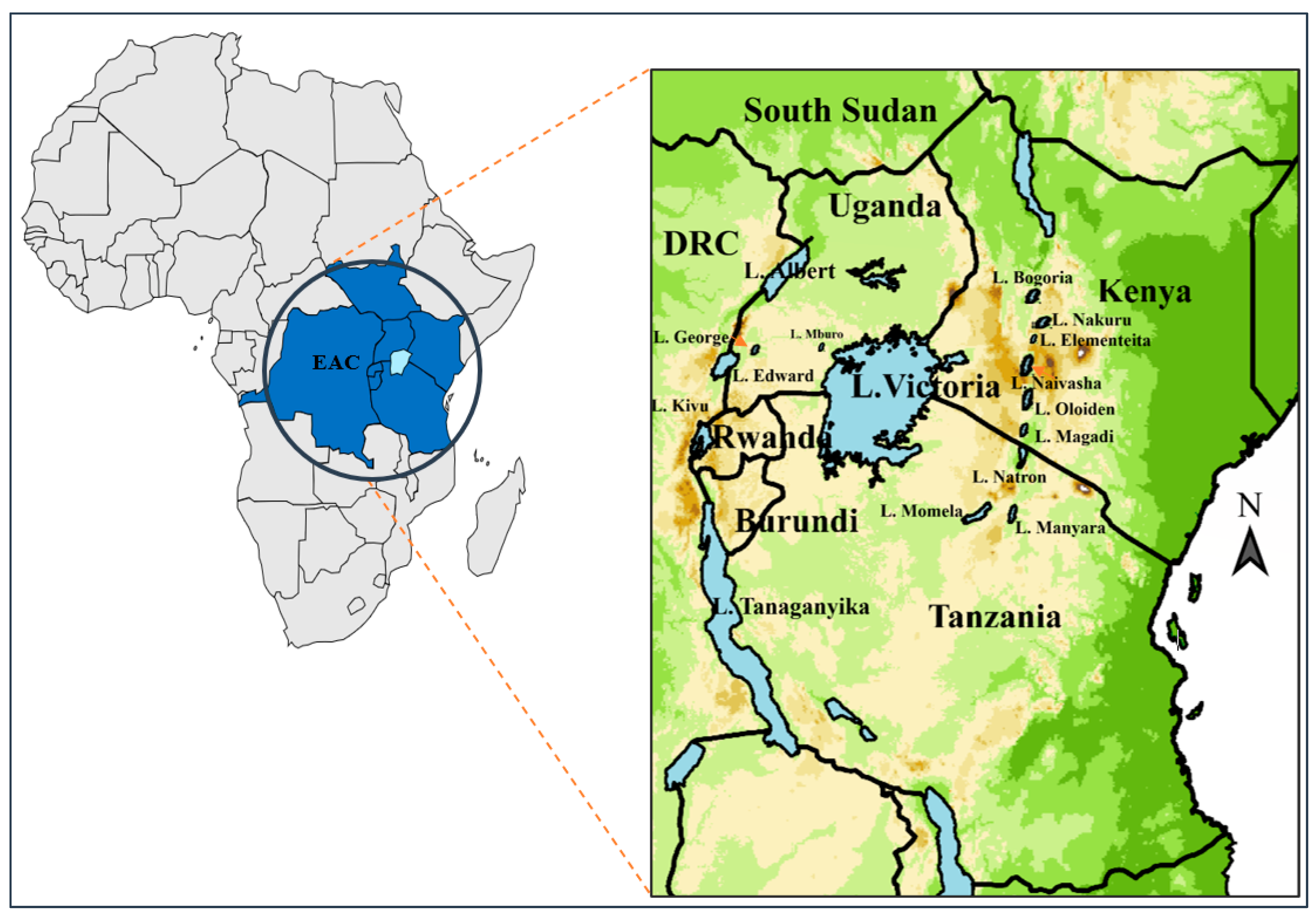
Figure 1. Map of East African Community (EAC) showing the partner states and location of lakes with reports of cyanobacteria, algal blooms, cyanotoxins, indiscriminate fish deaths and mass mortalities of lesser flamingos. Adapted from Ayugi et al. [18].
Phycology 03 00010 g001
Figure 2. Chemical structure of some of the phycotoxins reported in East African Community lakes: (a) ATX and HATX (R = CH3 and C2H5, respectively); (b) cylindrospermopsin; (c) microcystins: 1 to 7 are amino acid residues; congeners (variants) with different combinations of amino acids are shown; (d) nodularins.
Phycology 03 00010 g002
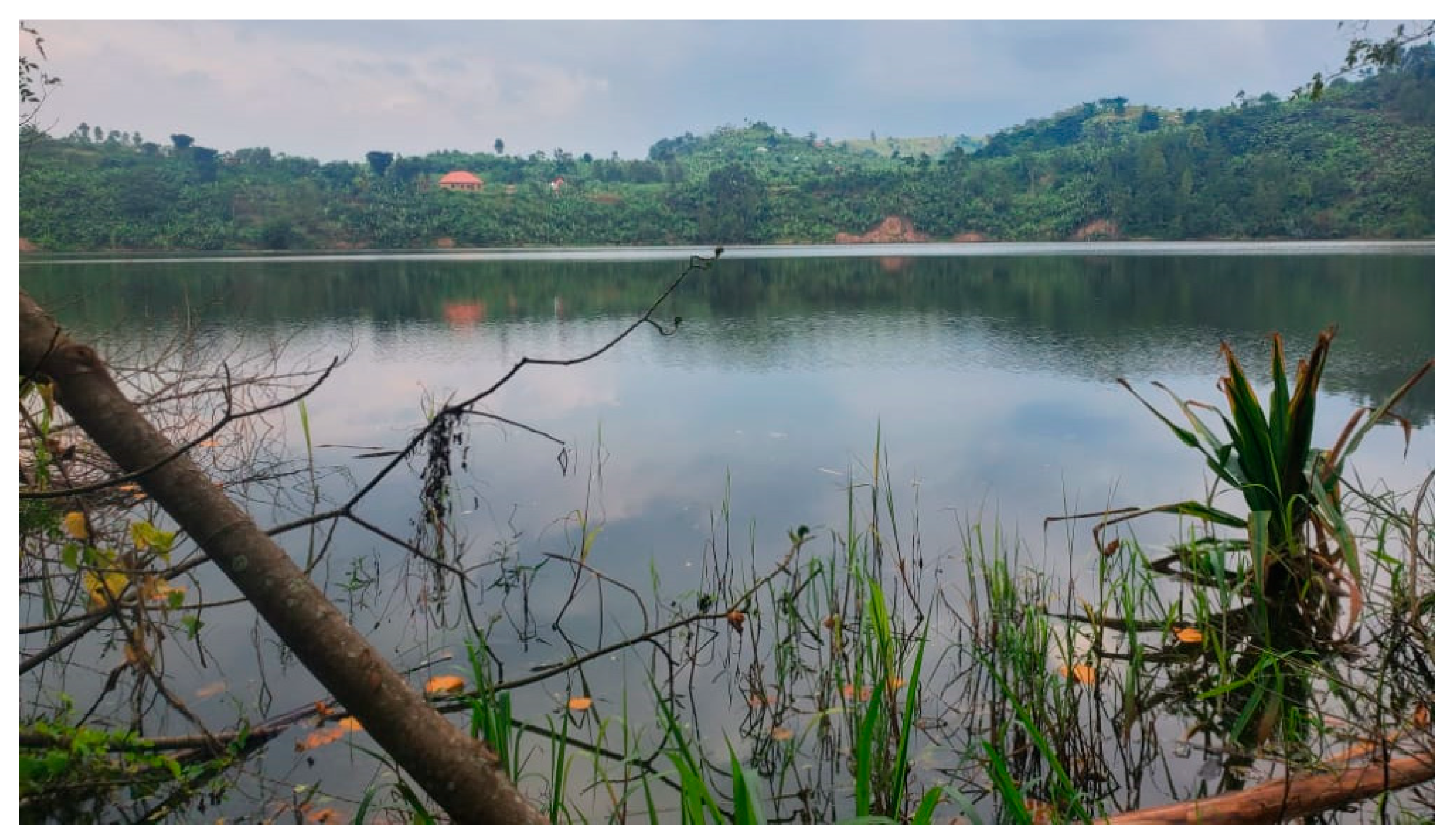
Figure 3. Brackish water in Lake Kamweru, a cyanobacteria-rich twin crater lake in Rubirizi district, Western Uganda. The lake and its twin (Lake Kyema) are the only co-joined lakes in Africa, which are filled by water from a heritage cave (photo credit: Ivan Kahwa, December 2022).
Phycology 03 00010 g003
Figure 4. Lesser flamingos at the Kenyan Lake Bogoria (a) a flock wading ashore (photo by Steve Garvie. Source: Riley [92]), and (b) massive die-off in July 2008 [93].
Phycology 03 00010 g004
Table 1. Occurrence and levels of microcystins in fish from some East African lakes.
LakeFish Species MCs Content (Average/Range, μg kg−1) 1References
L. Victoria (Murchison Bay)Clarias gariepinus23.9[64]
Haplochromis spc (filleted and whole)35.6 and 19.9
Lates niloticus13.5
Oreochromis leucostictus30.3
Oreochromis niloticus13.7
Protopterus aethiopicus4.1
Rastrineobola argentea (whole/dry from market)36.2–41.2
Synodontis afrofischeri28.8
Synodontis victoriae16.7
Tilapia zilli15.5
Oreochromis niloticus (gut, liver and muscle)1479.24, 48.07 and 9.65[65]
Lates niloticus (gut, liver and muscle)27.78, 3.74 and 1.86
L. Victoria (Napoleon Gulf)Astatoreochromis alluaudi6.2[64]
Bagrus docmac15.1
Brycinus sadleri24.6
Haplochromis spc (filleted and whole)13.0–17.1 and 15.3
Lates niloticus (filleted and gutted/beheaded)7.3 and 12.9
Mormyrus kannume21.1
Oreochromis leucostictus3.2–4.3
Oreochromis niloticus (filleted and gutted/beheaded)9.8 and 6.1
Oreochromis variabilis30.1
Protopterus aethiopicus2.8
Rastrineobola argentea (whole/dry from market)83.7
Synodontis afrofischeri31.0
Synodontis victoriae16.7
Tilapia zilli (filleted and gutted)8.4 and 3.4
L. Victoria (open lake at Rusinga channel and Nyanza Gulf)Rastrineobola argentea14 and 25–109[49]
Lake MburoBagrus docmac13.4[64]
Clarias gariepinus20.6
Haplochromis spc (filleted)2.5–5.6
Haplochromis spc (gutted/beheaded)5.4–11.8
Haplochromis spc (whole)12.1
Oreochromis esculentus17.9
Oreochromis leucostictus8.4
Oreochromis leucostictus (gutted/head removed)7.4
Protopterus aethiopicus2.5
Oreochromis niloticus (gut, liver and muscle)1312.08, 73.10 and 208.65[65]
Lake NkurubaPoecelia reticulata4.5 to 73.3[64]
Tilapia zilli (filleted and whole)11.7 and 42.5
Oreochromis leucostictus (filleted and gutted/beheaded)8.3 and 17.2
Lake GeorgeBagrus docmac9.1
Clarias gariepinus6.1
Oreochromis leucostictus21.2
Oreochromis niloticus10.2
Protopterus aethiopicus2.4
Oreochromis esculentus6.3
Haplochromis squamipinnis (filleted and gutted)6.7 and 11.8
Lake SakaAstatoreochromis alluaudi (filleted and gutted/beheaded)71.3 and 10.5[64]
Astatoreochromis alluaudi (whole)32.5
Barbus neumayerii (gutted/beheaded)9.5
Haplochromis spc (filleted)52.1
Haplochromis spc (gutted/beheaded and whole)23.2–1189.3 and 21.3–215.2
Lates niloticus16.4
Oreochromis niloticus17.0
Tilapia zilli (filleted and whole)4.9 and 898.7
Lake EdwardBagrus docmac6.2
Barbus bynni5.3
Clarias gariepinus8.6
Haplochromis spc10.0
Haplochromis squamipinnis8.6
Oreochromis leucostictus21.9
Oreochromis niloticus8.0
Protopterus aethiopicus5.3
Lake AlbertLates niloticus3.9–11.6
Tilapia zilli2.7–6.2
1 All mean values exceed the permissible MCs limit of 0.04 µg kg−1 in fish [65]; spc = species.
Table 2. Microcystin and anatoxin-a-related massive mortalities of lesser pink flamingos in alkaline-saline (soda-rich and schizohaline) lakes of East African Community in comparison with cyanotoxin-related bird mortality reports from other countries.
Waterbody (Country) Report (s)YearReference(s)
East Africa
Lake Nakuru (Kenya)0.00003 to 0.0004 µg MC-LR eq kg−1 and 0.00004–0.0058 µg kg−1 in liver, stomach/intestine2001–2003[94]
35,000 birds died2006[44]
Lake Bogoria and Lake Nakuru (Kenya)40,000 birds died1991[93]
More than 30,000 birds died; 0.00021 and 0.00093 µg MC-LR eq kg−1 fresh weight, ATX ranged between 0.00106 and 0.00582 µg kg−1 fresh weight1993[85,86,95,96] 1
50,000 birds died1995/1996[94]
Lake Bogoria30,000 birds died1999/2000[89,93]
0.00003–0.0009 µg MC-LR eq kg−1 and 0.00004–0.0002 µg kg−1 in liver, stomach/intestine2001–2003[94]
30,000 birds died2008[44,93]
2000 birds died2009[93]
Lake Natron and Empakai crater (Tanzania)43,800 birds died. Total MCs (MC-RR, -YR, -LR and -RY) were 0.1–4.5 μg mL−1July–August 2004[51,85,88] 2
Lake Big Momela (Tanzania)15 and 50 individuals per day for 2004 case; elevated levels (up to 150 million filaments L−1 of A. fusiformis were quantified in sampled scum; no MCs detected
Lake Manyara (Tanzania)521 deaths per month; bird livers contained 0.0003–0.0541 µg kg−1 wet weight of MCs. Corynebacteria species, Pasteurella multocida and Proteus species were found in visceral organs of all carcasses tested2004, August–October 2008[87,88,97] 3*
Other regions
The Salton Sea (USA)Over 20,000 deaths of Eared grebe (Podiceps nigricollis). Water contained up to 0.001 µg kg−1 DW and UDT to 0.00011 µg kg−1 DW in grebe liver tissues1990–2006[98,99]
Doñana National Park (Spain)579 Greater flamingos (Phoenicopterus roseus) died; MCs at concentrations of 0.44 μg kg−1 of liver wet weight and 0.625 μg kg−1 in crop contents2001[100]
Pond in Nishinomiya (Japan)20 spot-billed ducks died; MCs were detected in water (0.512 μg kg−1 cyanobacterial cell powder)1995[101]
Lake Knudsø (Denmark)3 ducks, 16 ducklings, 1 coot, coot chicks23rd and 26th June 1981[102] **
2 ducks and crows (unknown number)12th and 17th June 1988
2 grebes; 14–19 birds (black-necked and crested grebes, seagulls and a duck) and 2 grebes, respectively. ATX was recorded at 2.30 μg kg−110th June, 1st and 4th July 1993
Birds (unreported number), and 1 coot, 1 duck. ATX was recorded at 3.30 μg kg−1 while MCs occurred at 0.0001 to 0.0009 μg kg−128th June and 6th July 1994
1 duck9th July 1995
1,2,3 In part, Pseudomonas aeruginosa, Mycobacterium avium, Escherichia coli and heavy metals were claimed to be contributors to these flamingo die-offs [96,103,104]. * In 2004, over 43,800 flamingo die-off was experienced in this lake [105]. ** Several other animals (6 dogs, cows, crows and some fish) succumbed to cyanotoxins after swimming in this lake or drinking its water, which was rich in Anabaena species.
Disclaimer/Publisher’s Note: The statements, opinions and data contained in all publications are solely those of the individual author(s) and contributor(s) and not of MDPI and/or the editor(s). MDPI and/or the editor(s) disclaim responsibility for any injury to people or property resulting from any ideas, methods, instructions or products referred to in the content.