Communication

Seroprevalence of Anti-Neospora caninum and Anti-Toxoplasma gondii Antibodies in Cattle Intended for Human Consumption in the State of Paraíba, Brazil

by
Geraldo Moreira Silva Filho
1,
Jordania Oliveira Silva
2,
Audisio Alves Costa Filho
3,
Roberta Nunes Parentoni
4,
Arthur Willian Lima Brasil
3,4,
Thais Ferreira Feitosa
1,2 and
Vinícius Longo Ribeiro Vilela
1,2,*
1
Postgraduate Program in Science and Animal Health, Federal University of Campina Grande—UFCG, Patos 58708-110, Paraíba, Brazil
2
Department of Veterinary Medicine, Federal Institute of Paraíba—IFPB, Sousa 58814-000, Paraíba, Brazil
3
Postgraduate Program in Animal Science, Federal University of Paraíba—UFPB, Areia 58397-000, Paraíba, Brazil
4
Department of Morphology, Federal University of Paraíba—UFPB, João Pessoa 58059-900, Paraíba, Brazil
*
Author to whom correspondence should be addressed.
Ruminants 2025, 5(4), 48; https://doi.org/10.3390/ruminants5040048 (registering DOI)
Submission received: 6 September 2025 / Revised: 5 October 2025 / Accepted: 7 October 2025 / Published: 11 October 2025
(This article belongs to the Special Issue Parasitological Diagnosis and Alternative Control in Ruminants)

Abstract

Simple Summary

This study investigated the infection by two important parasites, Neospora caninum and Toxoplasma gondii, in cattle intended for human consumption in the state of Paraíba, Northeastern Brazil. Blood samples from 110 animals were analyzed using the Indirect Fluorescence Antibody Test. The results showed that 8.2% of the animals were infected with N. caninum and 18.2% with T. gondii. The analysis identified that the extensive farming system is a significant risk factor for both infections. For N. caninum infections, cows showed a higher risk, whereas for T. gondii infection, the frequent animal purchase without quarantine and the lack of separation between young and adult animals were associated with the infections. We conclude that these infections are present in herds and represent a risk to both animal health, by causing reproductive losses, and public health, in the case of T. gondii, which can be transmitted through the consumption of undercooked contaminated meat.

Abstract

We aimed to assess the seroprevalence of anti-Neospora caninum and anti-Toxoplasma gondii antibodies in cattle intended for human consumption in Paraíba, Brazil, and the associated risk factors. A total of 110 serum samples from slaughtered cattle were analyzed using the Indirect Fluorescence Antibody Test (IFAT), with cut-off points of 1:200 for N. caninum and 1:64 for T. gondii. Seroprevalence was 8.2% (9/110) for N. caninum (titers 1:200–1:6400) and 18.2% (20/110) for T. gondii (titers 1:64–1:512). Risk factor analysis revealed that the variable female sex (cows) and the extensive farming system were statistically significantly associated with seroprevalence for N. caninum. Whereas for T. gondii, extensive farming, frequent animal purchase, and the lack of separation between calves and adult cattle were statistically significant. These findings demonstrate the circulation of these parasites in herds, with implications for animal and public health, indicating a potential risk of transmission to definitive hosts and humans through the consumption of raw or undercooked infected meat.

Graphical Abstract

1. Introduction

Cattle are affected by coccidia, obligatory intracellular parasites of the phylum Apicomplexa. Among them, Neospora caninum, the causative agent of neosporosis, stands out as one of the main reproductive diseases affecting these animals [1], as well as Toxoplasma gondii, the causative agent of toxoplasmosis, a cosmopolitan zoonosis of great relevance that can be transmitted to humans through the ingestion of raw or undercooked meat [2].
Neosporosis in cattle is associated with conditions such as fetal mummification, stillbirths, the birth of weak calves, and abortions, and it is considered one of the main causes of infectious abortion in cattle herds worldwide [3]. Studies have reported seropositivity rates for N. caninum in cattle ranging from 9.1% to 91.2% in Brazil [4,5]. Economic losses caused by this disease, considering both dairy and beef herds, are estimated to exceed US$150 million in Brazil [6]. Although there is still no concrete evidence regarding its zoonotic risks to humans, several studies have reported the presence of antibodies against the parasite, and its DNA has already been detected in the umbilical cord of pregnant women [7,8], suggesting possible implications for human health.
Toxoplasmosis is a parasitic infection with high prevalence in both animals and humans [9]. T. gondii has the ability to infect most homeothermic species and form tissue cysts [10]. It is estimated that more than one-third of the human population is infected by this parasite [11]. In Brazil, infection rates of T. gondii in cattle can reach up to 89.1%, depending on the region studied and the diagnostic technique employed, which may be indirect, such as Indirect Fluorescence Antibody Test (IFAT), or Enzyme-Linked Immunosorbent Assay (ELISA), or direct, such as Polymerase Chain Reaction (PCR) [12]. The consumption of raw or undercooked meat is an important vehicle for the transmission of toxoplasmosis to humans, raising concerns from a public health perspective [13,14]. According to a worldwide survey by Dubey [15] on human toxoplasmosis outbreaks (1966–2020), Brazil stood out with the highest number of recorded events. Within the country, seven major outbreaks across different states totaled more than 1350 laboratory-confirmed cases. The clinical presentation of these cases was diverse, ranging from abortions to documented instances of congenital transmission.
Considering the reproductive losses associated with cattle neosporosis and the zoonotic potential of toxoplasmosis, which poses risks to human health through the consumption of contaminated meat, the objective of this study was to determine the seroprevalence of anti-N. caninum and anti-T. gondii antibodies using IFAT in cattle slaughtered for human consumption in the state of Paraíba, as well as to investigate the risk factors associated with these infections.

2. Materials and Methods

Study Area and Sampling

Serological samples were collected during bleeding of cattle from municipal slaughterhouses in the state of Paraíba. To determine the minimum sample size, a simple random sampling calculation was applied [16].
n = Z 2 × P ( 1 P ) d 2
where
n = number of sampled animals;
Z = value of the normal distribution for a 95% confidence level;
P = expected prevalence of 18.1% for anti-N. caninum [5] and 18% for anti-T. gondii [17];
d = absolute error of 10%.
The minimum number of samples to be evaluated, as determined by sample size calculation, was 57. However, for convenience, 110 samples were collected. The slaughterhouses used were located in the municipalities of Campina Grande, Itapororoca, Mamanguape, Patos, Santa Rita, and Sapé (Figure 1).
The samples were collected in pre-identified glass tubes without anticoagulant for serum extraction. For transportation, they were placed in a thermal box with ice, maintaining the temperature between 2 and 8 °C until arrival at the laboratory.
Serological analyses were performed using the IFAT, as described by Camargo [18]. For the detection of anti-T. gondii IgG antibodies, tachyzoites of the ME-49 strain fixed on fluorescence microscopy slides were used, adopting a cutoff point of 1:64 as the criterion for positivity [19]. For N. caninum, the protocol established by Gondim et al. [20] was followed, using cell culture-derived tachyzoites of the Nc-1 strain as antigen and a cutoff titer of 1:200 [21]. Complete peripheral fluorescence of the tachyzoites, observed under a fluorescence microscope, was considered the criterion for confirming seropositivity. Positive samples were subjected to titration through twofold serial dilutions until complete absence of fluorescence, at which point they were classified as negative [22].
A questionnaire was completed with the following information: breed (purebred or crossbred), age (≤24 months, >24 months), sex (male or female), contact with cats (yes or no), contact with dogs (yes or no), provision of treated water (yes or no), presence of maternity pens (yes or no), separation of young and adult animals (yes or no), frequent purchase of animals (yes or no), quarantine of newly acquired animals (yes or no), production system (extensive or semi-intensive), history of abortion in the last 12 months (yes or no), and regular veterinary assistance (yes or no).
Descriptive statistical analysis was initially employed to calculate the frequencies of seropositivity obtained through IFAT. To investigate the factors associated with the seroprevalence of anti-N. caninum and anti-T. gondii antibodies, data from the epidemiological questionnaires were subjected to a two-step analytical approach. First, a univariate analysis was performed, in which each independent variable was individually tested against the dependent variable using Fisher’s exact test. Variables with a p-value ≤ 0.05 were selected for the subsequent step. Next, a multivariate analysis was carried out through multiple logistic regression, adopting a significance level of p ≤ 0.05 to identify risk factors independently associated with the infections. All analyses were performed using GraphPad Prism software, version 8.0.1.

3. Results

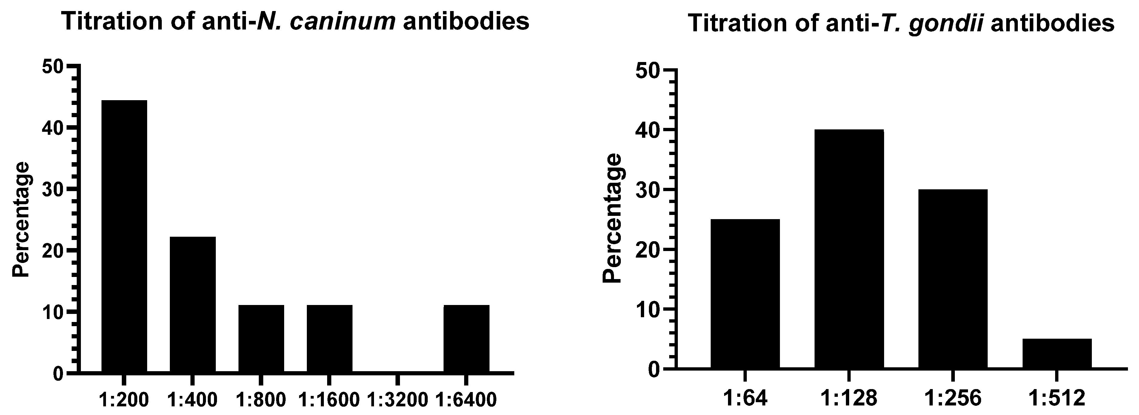
Among the 110 cattle serum samples analyzed, 8.2% (9/110) tested positive for anti-N. caninum antibodies by IFAT, with titers ranging from 1:200 to 1:6400 (Figure 2). Three animals showed high titers (≥1:800). For anti-T. gondii antibodies, the prevalence was 18.2% (20/110), with titers ranging from 1:64 to 1:512.
The variables obtained from the epidemiological questionnaires are presented in Table 1. A univariate analysis was performed, and those showing statistical significance (p ≤ 0.05) were considered for subsequent multivariate analysis. For N. caninum, the variables included were: sex, production system, and presence of a maternity pen. For T. gondii, the selected variables were: production system, regular acquisition of animals, and separation of young and adult animals.
The multivariate analysis (Table 2) identified the following risk factors: female sex and extensive farming system for N. caninum; and extensive farming system, frequent purchase of animals, and lack of separation between young and adult cattle for T. gondii.

4. Discussion

The results of the present study demonstrate significant circulation of N. caninum (8.2%) and T. gondii (18.2%) in cattle slaughter and intended for human consumption in the state of Paraíba, Brazil, corroborating the importance of these parasites for regional livestock and indicating a potential public health risk. The presence of animals with high titers suggests recent infections or reactivations, a scenario in which parasitic replication is more active and the tissue cyst load in meat may be elevated, increasing the zoonotic risk [12,23,24].
The seroprevalence of N. caninum found in this study (8.2%) falls within the wide range reported in Brazil, which can reach up to 90% depending on the region and production system [4]. Similar values were reported by Guerra et al. [25] in Paraná (9.2%), while studies within Paraíba, such as Maia et al. [5], reported a higher prevalence of 17.8%. This disparity can be attributed to the epidemiological particularities of each herd, including the density of definitive hosts and management practices [26]. The fact that our study focused specifically on slaughter animals, generally younger, rather than dairy herds, may partially explain the lower prevalence observed, a factor previously demonstrated in studies comparing seroprevalence across herds with different production purposes [27]. Furthermore, parasite circulation is corroborated by molecular evidence, such as the detection of N. caninum DNA by PCR in 14.3% (5/35) of bovine fetal brains from asymptomatic pregnant dams submitted to slaughter [28], demonstrating the occurrence of active infections and the maintenance of the parasite in the bovine population, even in the absence of overt clinical signs.
For T. gondii, the prevalence of 18.2% aligns with recent studies in the Northeast region, where 18% was reported in cows in Paraíba and 11.8% in Bahia [17,29]. However, it is considerably lower than the 89.1% reported by Gomes et al. [12]. This wide variability reinforces that the epidemiology of cattle toxoplasmosis is complex and hyperendemic in specific microregions, influenced by factors such as the presence of felines, management practices [30], and, critically, climatic conditions that affect oocyst sporulation and survival. In a molecular study analyzing 100 brains from beef cattle, T. gondii DNA was identified in 5% of the samples [31], providing direct evidence of the parasite’s circulation.
Silva et al. [32] demonstrated that conditions in the semi-arid Northeast, characterized by high temperatures and low relative humidity, are decisive for the rapid inactivation of oocysts, which can be destroyed within 36 h. This environment creates a natural barrier to environmental transmission. In contrast, the South and Southeast regions of Brazil, with temperate or more humid tropical climates and soils with greater water retention capacity, offer ideal conditions for prolonged oocyst survival [15]. Therefore, extreme climatic conditions represent a key environmental factor explaining, in part, the lower seroprevalences generally found in herds from hot and dry regions, as observed in our study, compared to the high rates reported in cooler and more humid regions.
Multivariate analysis identified critical risk factors for both infections. The extensive farming system emerged as a significant risk factor for N. caninum and T. gondii. This finding is consistent with the literature, as this system favors cattle contact with pastures and water sources potentially contaminated with oocysts excreted by dogs and felids [33,34]. The difficulty in implementing biosecurity measures and controlling synanthropic fauna in these systems amplifies exposure [35].
Specifically for N. caninum, female sex was an important risk factor, likely due to the longer lifespan of females in herds, whether for replacement, lactation, or rearing, extending their window of exposure to environmental oocysts compared to males, which are typically sent for early slaughter [36]. Additionally, vertical transmission is a primary route for maintaining N. caninum in herds, creating a cycle where infected females perpetuate the parasite in their offspring, which in turn remain in the herd [27]. However, our data did not find a significant association with abortion history, possibly due to sample size or because many abortions caused by neosporosis may go unreported.
For T. gondii, two management factors were particularly relevant: frequent purchase of animals and the lack of separation between young and adult cattle. Introducing animals without prior quarantine or testing is a classic pathway for the entry of infectious diseases into a closed herd [37]. The lack of separation between calves and adults can facilitate parasite transmission. First, studies demonstrate that raw milk can be a significant route of T. gondii transmission, especially during the acute phase of infection, and co-mingling allows calves to consume this milk [38,39]. Second, as it is common for adult animals to receive concentrate-based supplementation, feed residues can attract rodents. These, in turn, attract felids, which are the definitive hosts of the parasite and can contaminate the environment with oocysts [29]. T. gondii sporulated oocysts are extremely resistant in favorable environments, surviving for months in soil and water, thereby turning common grazing or living areas into permanent foci of infection [31].
The public health implications are notable, particularly for T. gondii. The detection of antibodies indicates that the tissues of these animals may harbor parasite cysts. Consumption of raw or undercooked beef from these animals represents a transmission route to humans [14,15]. Brazil, as highlighted by [16], experiences numerous foodborne toxoplasmosis outbreaks, making herd surveillance crucial. Although N. caninum transmission via meat is not confirmed in humans, the presence of the parasite in cattle tissues poses a risk to dogs, its definitive hosts, which, upon infection, complete the cycle and maintain the parasite in the environment [26].

5. Conclusions

There was demonstrated the significant presence of anti-N. caninum and anti-T. gondii antibodies in cattle slaughtered for human consumption in the state of Paraíba, Brazil, indicating exposure of the animals to these protozoa and highlighting the importance of the infections in local livestock. Therefore, measures such as controlling the presence of definitive hosts in the environment, restricting their contact with herds, adopting proper sanitary management practices, and implementing serological monitoring are essential to minimize the impact of these infections on animal and public health, thereby promoting food safety in livestock production.

Author Contributions

All authors contributed significantly to the conception of this study. The preparation of materials, sample collection, and sample processing were carried out by G.M.S.F., A.A.C.F., A.W.L.B., R.N.P., J.O.S., V.L.R.V. and T.F.F. The first draft of the manuscript was written by G.M.S.F. and V.L.R.V., and all authors provided comments on subsequent versions of the manuscript. All authors have read and agreed to the published version of the manuscript.

Funding

Paraíba State Research Foundation (FAPESQ) for funding the project under public notice 010/2021, grant term 3205/2021. The National Council for Scientific and Technological Development (CNPq/Brazil) provided scholarship to V.L.R.V. (grant number 304472/2021-2).

Institutional Review Board Statement

Ethics Committee on the Use of Animals of the Federal Institute of Paraíba—CEUA/IFPB, n◦ 275/2023, approved on 17 August 2023.

Informed Consent Statement

Not applicable.

Data Availability Statement

The datasets generated during and/or analyzed during the current study are available from the corresponding author on reasonable request.

Conflicts of Interest

The authors declare no conflicts of interest.

References

  1. Dubey, J.P.; Schares, G. Neosporosis in animals—The last five years. Vet. Parasitol. 2011, 180, 90–108. [Google Scholar] [CrossRef] [PubMed]
  2. Vilela, V.L.R.; Feitosa, T.F. Recent Advances in Toxoplasma gondii Infection and Toxoplasmosis. Trop. Med. Infect. Dis. 2024, 9, 160. [Google Scholar] [CrossRef]
  3. Reichel, M.P.; Wahl, L.C.; Ellis, J.T. Research into Neospora caninum—What Have We Learnt in the Last Thirty Years? Pathogens 2020, 9, 505. [Google Scholar] [CrossRef]
  4. Guedes, M.H.P.; Guimarães, A.M.; Rocha, C.M.B.M.; Hirsch, C. Frequência de anticorpos anti-Neospora caninum em vacas e fetos provenientes de municípios do sul de Minas Gerais. Rev. Bras. Parasitol. Vet. 2008, 17, 189–194. [Google Scholar] [PubMed]
  5. Maia, A.R.A.; de Melo, R.P.B.; Mota, R.A.M.; Clementino, I.J.; Alves, C.J.; de Santos, C.S.A.B.; Fernandes, L.G.; de Azevedo, S.S. Herd and animal level prevalences and risk factors for Neospora caninum infection in cattle in the state of Paraíba, northeastern Brazil. Vet. Parasitol. 2023, 40, 100866. [Google Scholar] [CrossRef]
  6. Reichel, M.P.; Ayanegui-Alcérreca, M.; Gondim, L.F.; Ellis, J.T. What is the global economic impact of Neospora caninum in cattle—The billion dollar question. Int. J. Parasitol. 2013, 43, 133–142. [Google Scholar] [CrossRef]
  7. Duarte, P.O.; Oshiro, L.M.; Zimmermann, N.P.; Csorda, B.G.; Dourado, D.M.; Barros, J.C.; Andreott, R. Serological and molecular detection of Neospora caninum and Toxoplasma gondii in human umbilical cord blood and placental tissue samples. Sci. Rep. 2020, 10, 9043. [Google Scholar] [CrossRef]
  8. Duarte, P.O.; Csordas, B.G.; Oshiro, L.M.; de Higa, L.O.S.; Zimmermann, N.P.; Martins, K.R.; Barros, J.C.; Andreotti, R. Serological evaluation of Neospora caninum in pregnant women treated at referral center for prenatal screening in Mato Grosso do Sul, Brazil. Rev. Bras. Parasitol. Vet. 2020, 29, 4. [Google Scholar] [CrossRef] [PubMed]
  9. Abbas, I.E.; Villena, I.; Dubey, J.P. A review on toxoplasmosis in humans and animals from Egypt. Parasitology 2020, 147, 135–159. [Google Scholar] [CrossRef] [PubMed]
  10. Dubey, J.P.; Jones, J.L. Toxoplasma gondii infection in humans and animals in the United States. Int. J. Parasitol. 2008, 38, 1257–1278. [Google Scholar] [CrossRef]
  11. World Organization for Animal Health—OIE. Terrestrial Manual: Toxoplasmosis. Available online: https://www.oie.int/fileadmin/Home/eng/Health_standards/tahm/2.09.09_TOXO.pdf (accessed on 6 October 2025).
  12. Gomes, D.F.C.; da Krawczak, F.S.; de Oliveira, C.H.S.; Ferreira Júnior, Á.; Fernandes, E.K.K.; Lopes, W.D.Z.; da Sevá, A.P.; Gennari, S.M. Toxoplasma gondii in cattle in Brazil: A review. Rev. Bras. Parasitol. Vet. 2020, 29, e015719. [Google Scholar] [CrossRef]
  13. Silva, J.O.; Batista, S.P.; Matos, T.S.; Bison, I.; Parentoni, R.N.; Santos, J.R.S.; Brasil, A.W.L.; Feitosa, T.F.; Vilela, V.L.R. Elevated anti-Toxoplasma gondii antibody levels among slaughterhouse workers in Paraíba, Brazil: Implications for occupational health. Acta Trop. 2025, 270, 107773. [Google Scholar] [CrossRef]
  14. Kean, B.H.; Kimball, A.C.; Christenson, W.N. An epidemic of acute toxoplasmosis. J. Am. Med. Assoc. 1969, 208, 1002–1004. [Google Scholar]
  15. Dubey, J.P. Outbreaks of clinical toxoplasmosis in humans: Five decades of personal experience, perspectives and lessons learned. Parasites Vectors 2021, 14, 263. [Google Scholar] [CrossRef] [PubMed]
  16. Thrusfield, M. Veterinary Epidemiology, 3rd ed.; Blackwell Science: Oxford, UK, 2007; pp. 1–610. [Google Scholar]
  17. Maia, A.R.A.; Bezerra, R.A.; Silva, S.S.; Álvares, F.B.V.; de Santos, C.S.A.B.; Alves, C.J.; Clementino, I.J.; Feitosa, T.F.; Vilela, V.L.R.; de Azevedo, S.S. Herd-level based seroprevalence and associated factors for Toxoplasma gondii in cows in the state of Paraíba, Northeastern Brazil. Braz. J. Vet. Parasitol. 2023, 32, e017222. [Google Scholar] [CrossRef]
  18. Camargo, M.E. Improvised technique of indirect immunofluorescence for serological diagnosis of toxoplasmosis. Rev. Inst. Med. Trop. 1964, 6, 117–118. [Google Scholar]
  19. Costa, F.R.T.; Nogueira, D.B.; Oliveira, M.A.G.; Silva, S.S.; Silva, R.F.; Sarmento, W.F.; Azevedo, S.S.; Gennari, S.M.; Pena, H.F.J.; Brasil, A.W.L.; et al. Vertical transmission of Toxoplasma gondii in naturally infected ewes in the semiarid region of Brazil. Comp. Immunol. Microbiol. Infect. Dis. 2021, 74, 101595. [Google Scholar] [CrossRef]
  20. Gondim, L.F.P.; Sartor, I.F.; Hasegawa, M.; Yamane, I. Seroprevalence of Neospora caninum in dairy cattle in Bahia, Brazil. Vet. Parasitol. 1999, 86, 71–75. [Google Scholar] [CrossRef] [PubMed]
  21. Amaral, R.L.G.; Silva, L.B.G.; Pinheiro Júnior, J.W.; Souza Neto, O.L.; Leal, C.A.S.; Porto, W.J.N.; Mota, R.A. Neospora caninum em bovinos em matadouros de Pernambuco e Alagoas. Pesq. Vet. Bras. 2012, 32, 963–966. [Google Scholar] [CrossRef]
  22. Batista, S.P.; Silva, J.O.; Ferraz, C.M.; de Assis, J.P.B.; Rossi, G.A.M.; Daleprani, L.G.; Tobias, F.L.; Feitosa, T.F.; Braga, F.R.; Vilela, V.L.R. Anti-Neospora caninum and anti-Toxoplasma gondii antibodies in cattle intended for human consumption in the State of Espírito Santo, Brazil: Prevalence and associated factors. Braz. J. Vet. Parasitol. 2025, 34, e001225. [Google Scholar]
  23. Holec-Gąsior, L.; Sołowińska, K. IgG Avidity Test as a Tool for Discrimination Between Recent and Distant Toxoplasma gondii Infection—Current Status of Studies. Antibodies 2022, 11, 52. [Google Scholar] [CrossRef]
  24. Webster, J. Toxoplasmosis: A review of the disease and its public health significance. J. Am. Vet. Med. Assoc. 2010, 236, 15–22. [Google Scholar]
  25. Guerra, J.L.; Okano, W.; Bogado, A.L.G.; Nino, B.S.L.; Martins, F.D.C.; Cardim, S.T.; de Barros, L.D.; Garcia, J.L. Anti-Neospora caninum antibodies in beef cattle from the northern region of Paraná state, Brazil. Cienc. Rural 2019, 49, e20180869. [Google Scholar] [CrossRef]
  26. Dubey, J.P.; Schares, G.; Ortega-Mora, L.M. Epidemiology and control of neosporosis and Neospora caninum. Clin. Microbiol. Rev. 2007, 20, 323–367. [Google Scholar] [CrossRef] [PubMed]
  27. Roman, I.J.; Tagarra, L.G.; Rodrigues, F.S.; Cargnelutti, J.F.; Sangioni, L.A.; Vogel, F.S.F. Sarcocystis neurona, Toxoplasma gondii and Neospora caninum infection in bovine fetuses from a slaughterhouse in southern Brazil. Pesq. Vet. Bras. 2025, 45, e07504. [Google Scholar]
  28. Sartor, I.F.; Garcia Filho, A.; Vianna, L.C.; Pituco, E.M.; Dal Pai, V.; Sartor, R. Occurrence of antibodies anti-Neospora caninum in dairy and beef cattle in the region of Presidente Prudente, SP, Brazil. Arq. Inst. Biol. 2005, 72, 4. [Google Scholar]
  29. Spagnol, R.; Paranhos, E.B.; Oliveira, L.L.S.; Medeiros, S.M.; Lopes, C.W.G.; Albuquerque, G.R. Prevalência de anticorpos anti-Toxoplasma gondii em bovinos abatidos em matadouros do estado da Bahia, Brasil. Rev. Bras. Parasitol. Vet. 2009, 18, 42–45. [Google Scholar]
  30. Tenter, A.M.; Heckeroth, A.R.; Weiss, L.M. Toxoplasma gondii: From animals to humans. Int. J. Parasitol. 2000, 30, 1217–1258. [Google Scholar] [CrossRef]
  31. Santos, S.L.; de Souza Costa, K.; Gondim, L.Q.; da Silva, M.S.A.; Uzêda, R.S.; Gondim, L.F.P. Investigation of Neospora caninum, Hammondia sp., and Toxoplasma gondii in tissues from slaughtered beef cattle in Bahia, Brazil. Parasitol. Res. 2010, 106, 457–461. [Google Scholar] [CrossRef]
  32. Silva, A.L.P.; Lima, B.A.; Formiga, V.H.A.S.; Lima, E.F.; Silva Filho, G.M.; Silva, W.I.; Silva, J.O.; Alvares, F.B.V.; Vilela, V.L.R.; Feitosa, T.F. Survival and viability of Toxoplasma gondii oocysts under natural dry season conditions in the Brazilian semi-arid region. Vet. Res. Commun. 2025, 49, 191. [Google Scholar] [CrossRef]
  33. Magalhães, F.J.R.; Ribeiro-Andrade, M.; Alcântara, A.M.; Pinheiro Júnior, J.W.; Sena, M.J.; Porto, W.J.N.; Vieira, R.F.D.C.; Mota, R.A. Risk factors for Toxoplasma gondii infection in sheep and cattle from Fernando de Noronha Island, Brazil. Braz. J. Vet. Parasitol. 2016, 25, 511–515. [Google Scholar] [CrossRef]
  34. Snak, A.; Garcia, F.G.; Lara, A.A.; Pena, H.F.J.; Osaki, S.C. Neospora caninum in properties in the west region of Paraná, Brazil: Prevalence and risk factors. Rev. Bras. Parasitol. Vet. 2018, 27, 52–60. [Google Scholar] [CrossRef] [PubMed]
  35. Dubey, J.P. Review of Neospora caninum and neosporosis in animals. Korean J. Parasitol. 2003, 41, 1–16. [Google Scholar] [CrossRef] [PubMed]
  36. Ribeiro, A.C.; McAllister, A.J.; de Queiroz, S.A. Efeito das taxas de descarte sobre medidas econômicas de vacas leiteiras em Kentucky. Rev. Bras. Zootec. 2003, 32, 1737–1746. [Google Scholar] [CrossRef]
  37. Hill, D.E.; Dubey, J.P. Toxoplasma gondii prevalence in farm animals in the United States. Int. J. Parasitol. 2013, 43, 107–113. [Google Scholar] [CrossRef]
  38. Da Fonseca, F.M.; Sato, A.P.; Becker, A.P.B.B.; da Pinto, G.O.P.A.; de Souza, G.S.; Perotta, J.H.; de Filho, I.R.B.; Rodriguez, M.C.; Locatelli-Dittrich, R. Detection of Toxoplasma gondii DNA in milk of dairy cows from southern Brazil. Parasitol. Int. 2023, 95, 102750. [Google Scholar] [CrossRef]
  39. Khan, S.; Rafiq, K.; Khabir, M.N.; Khan, M.B.; Khan, S.N.; Khattak, A.; Attaullah, S. Toxoplasma gondii in lactating animals: Potential risk to milk consuming population in Khyber Pakhtunkhwa. Braz. J. Biol. 2023, 83, e267369. [Google Scholar] [CrossRef]
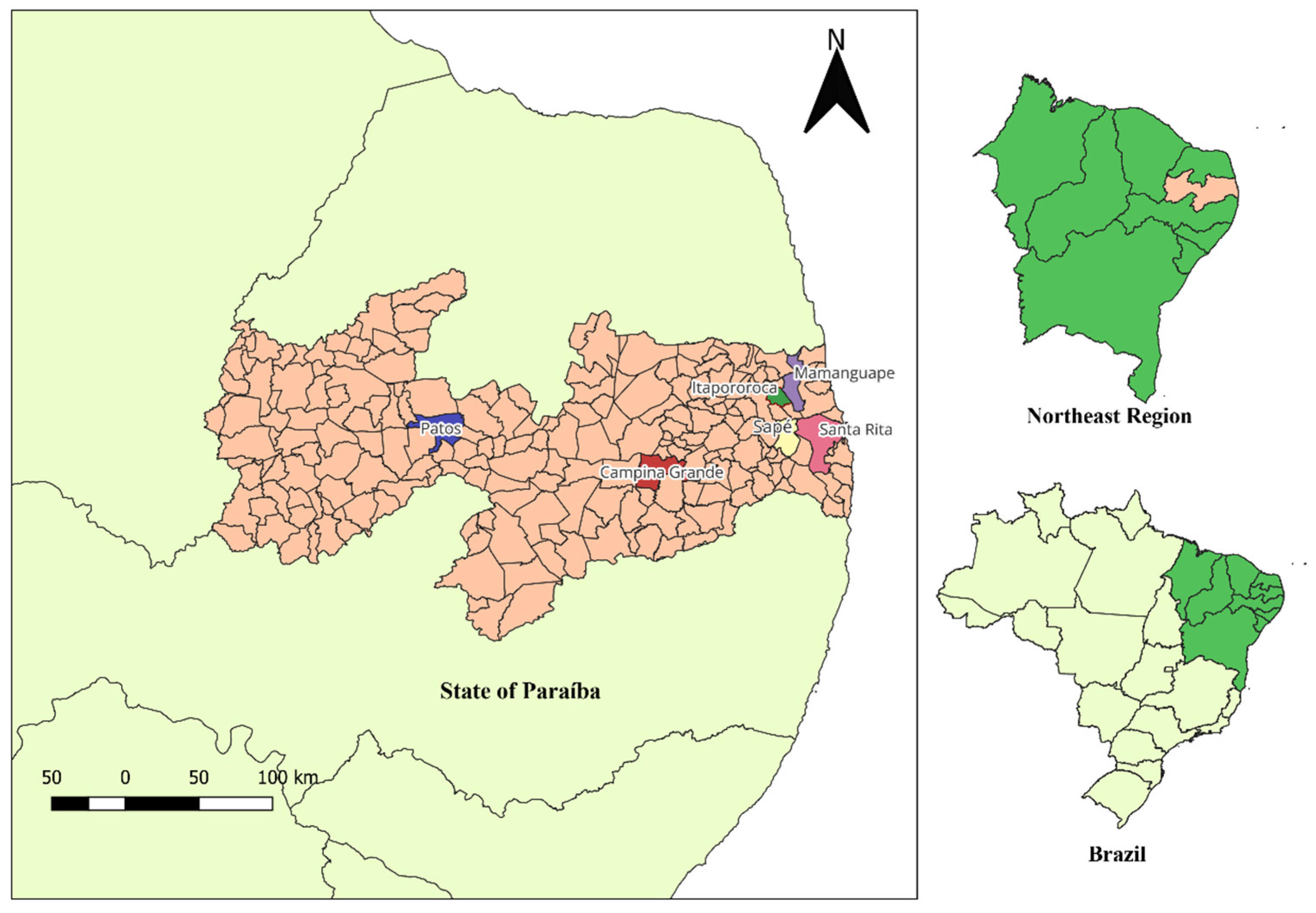
Figure 1. Geographical location of the municipalities in the State of Paraíba, Northeast region of Brazil (bottom right). The map highlights the municipalities included in the study (in salmon), where slaughterhouses sampled from cattle intended for human consumption are located. Specific municipalities are indicated in different colors: Patos (blue), Campina Grande (red), Santa Rita (pink), Sapé (yellow), Itapororoca (dark green), and Mamanguape (purple). Other areas of Northeast Brazil are shown in light green.
Ruminants 05 00048 g001
Figure 2. Percentage distribution of anti-N. caninum (n = 9) and anti-T. gondii (n = 20) antibody titers ob-tained by the Indirect Fluorescence Antibody Test (IFAT) in cattle intended for human con-sumption in the state of Paraíba, Brazil.
Ruminants 05 00048 g002
Table 1. Univariate analysis of factors associated with seropositivity for anti-N. caninum and anti-T. gondii antibodies in cattle intended for human consumption in the state of Paraíba, Brazil.
Variables N. caninumT. gondii
TotalPositive (%)pPositive (%)p
Sex
Male530 (0)0.003 *14 (26.4)0.046 *
Female579 (15.8)6 (10.5)
Age
≤24 months382 (5.3)0.71614 (10.5)0.193
>24 months727 (9.7)16 (22.2)
Breed
Pure191 (5.3)>0.9993 (15.8)>0.999
Crossbred918 (8.8)17 (18.7)
History of abortion in the last 12 months
Yes686 (8.8)>0.99916 (23.5)0.078
No423 (7.1)4 (9.5)
Contact with cats
Yes746 (8.3)>0.99914 (18.9)>0.999
No363 (8.1)6 (16.7)
Contact with dogs
Yes837 (8.4)>0.99913 (15.6)0.256
No272 (7.4)7 (25.9)
Provision of treated water
Yes344 (11.8)0.4545 (14.7)0.603
No765 (6.6)15 (19.7)
Frequent purchase of animals
Yes205 (10.9)0.48714 (30.4)0.006 *
No904 (6.2)6 (9.4)
Separation of young and adult animals
Yes 705 (7.1)0.7217 (10)0.005 *
No404 (10)13 (32.5)
Farming system
Extensive397 (18)0.009 *16 (41)<0.001 *
Semi-intensive712 (2.8)4 (5.6)
Veterinary assistance
Yes323 (9.4)0.7186 (18.7)>0.999
No786 (7.7)14 (18)
Presence of maternity pens
Yes592 (3.9)0.1725 (9.8)0.047 *
No517 (11.9)15 (25.4)
* Variables with a p-value ≤ 0.05 according to the chi-square or Fisher’s exact test.
Table 2. Multivariate analysis of factors associated with seropositivity for anti-N. caninum and anti-T. gondii antibodies in cattle intended for human consumption in the state of Paraíba.
VariablesPrevalence RatioIC 95%p
anti-N. caninum antibodies
Sex (Female)4.81.2–19.10.025 *
Farming system (Extensive)5.11.5–17.30.009 *
anti-T. gondii antibodies
Farming system (Extensive)4.31.4–13.20.011 *
Frequent purchase of animals (Yes)3.91.2–12.80.023 *
Separation of young/adult animals (No)3.11.1–8.70.032 *
* Variables with a p-value ≤ 0.05.
Disclaimer/Publisher’s Note: The statements, opinions and data contained in all publications are solely those of the individual author(s) and contributor(s) and not of MDPI and/or the editor(s). MDPI and/or the editor(s) disclaim responsibility for any injury to people or property resulting from any ideas, methods, instructions or products referred to in the content.