Article

Assessment of the Immunohistochemical Expression of Vitamin D Receptor, β-Catenin, and Ki-67 in Urothelial Carcinoma: A Cross-Sectional Study from Egypt


by
Marwa M. El-Mosely
1,
Abdulkarim Hasan
2,*,
Mohamed Tharwat
3,
Ahmed Abdellatief
3,
Reda Elhawary
2,
Mahmoud Salem
2,
Mostafa Fawzy
2,
Dina Sakr
4,
Salah-el-din Sayed O. Semary
5,
Sabah Mohamed Sharaf
6,
Heba Gamil
6,
Ahmed Abdulwahab Bawahab
7,
Dahlia Soleman A. Mirdad
7,
Mohammed S. Abdelwahed
7 and
Mohamed Mahmoud Abdellah
8,9
1
Pathology Department, Faculty of Medicine, Zagazig University, Zagazig 44511, Egypt
2
Pathology Department, Faculty of Medicine, Al-Azhar University, Cairo 11884, Egypt
3
Pathology Department, Faculty of Medicine, Al-Azhar University, Assiut 71524, Egypt
4
Histopathology Department, University Hospital Southampton NHS Foundation Trust, Hampshire SO16 6YD, UK
5
Pathology Department, Faculty of Medicine, Al-Azhar University, Damietta 34711, Egypt
6
Pathology Department, Faculty of Medicine for Girls, Al-Azhar University, Cairo 11754, Egypt
7
Department of Basic Medical Sciences, College of Medicine, University of Jeddah, Jeddah 21589, Saudi Arabia
8
Pathology Department, Faculty of Medicine, Fayoum University, Fayoum 63514, Egypt
9
Pathology Department, Faculty of Medicine, Galala University, Attaka, Suez 43727, Egypt
*
Author to whom correspondence should be addressed.
Int. J. Transl. Med. 2026, 6(2), 14; https://doi.org/10.3390/ijtm6020014
Submission received: 21 January 2026 / Revised: 11 March 2026 / Accepted: 15 March 2026 / Published: 30 March 2026

Abstract

Background: Conventional urothelial carcinoma (UC) requires accurate risk stratification, particularly differentiation between non-muscle-invasive (NMIBC) and muscle-invasive bladder cancer (MIBC) and between low- and high-grade tumors. This study evaluated immunohistochemical (IHC) expression of vitamin D receptor (VDR), β-catenin, and Ki-67 index in Egyptian patients with conventional UC. Methods: A cross-sectional study was conducted on 58 archived conventional UC cases diagnosed in 2023 at Al-Azhar University Hospitals. VDR positivity was defined as ≥10% cytoplasmic and/or nuclear tumor cell staining. Membranous β-catenin was considered preserved when >80% of tumor cell membranes were stained; otherwise, it was reduced. Nuclear β-catenin was considered positive when ≥5% of tumor nuclei were stained. Ki-67 was categorized as high using a ≥30% cutoff. Associations with grade, muscle invasion status, and lymphovascular invasion (LVI) were analyzed. Results: Mean age was 65.3 ± 9.3 years; 86.2% were males; 51.7% were MIBC. Compared with NMIBC, MIBC was significantly associated with high grade, non-papillary architecture, LVI, and high Ki-67. VDR positivity was detected in 82.7% of cases and showed no significant association with grade, muscle invasion, or LVI. Preserved membranous β-catenin was seen in 34.5% and was significantly associated with tumor grade but not with muscle invasion or LVI; nuclear β-catenin was absent. High Ki-67 (60.3%) was significantly associated with high grade and MIBC, with no association with age, sex, or LVI. Conclusions: In Egyptian conventional UC, Ki-67 was a significant marker for aggressive clinicopathologic features, while VDR lacked discriminatory associations and β-catenin findings were mainly grade-related.

1. Introduction

Urothelial carcinoma (UC) of the urinary bladder represents a major global cancer burden, with hundreds of thousands of new cases and deaths reported annually worldwide [1]. In several Middle Eastern countries, including Egypt, bladder cancer remains a prominent malignancy and reflects a distinctive epidemiologic context shaped by historical and evolving exposures [2]. For decades, urinary schistosomiasis contributed substantially to bladder cancer risk and to a higher proportion of squamous cell carcinoma; however, contemporary patterns increasingly resemble those seen in non-endemic regions, with urothelial carcinoma becoming the dominant histology as parasitic infection rates declined and lifestyle/occupational factors gained relative importance [2,3]. Importantly, large Egyptian registry-based analyses have documented a clear temporal shift in histopathology, showing rising frequencies of transitional/urothelial carcinoma alongside declining squamous carcinoma across successive decades and parallel observations from Upper Egypt also support a changing risk-factor landscape in which the burden of endemic schistosomiasis has markedly decreased, while other carcinogenic exposures (notably tobacco use and environmental/occupational factors) continue to contribute to disease risk and presentation [4,5,6]. This geographic heterogeneity underscores the need for regionally generated biomarker data, because tumor biology and clinicopathologic behavior may vary across populations and evolving exposure profiles. Moreover, as UC predominates in Egypt today, studies focused specifically on conventional UC are needed to provide locally relevant evidence and avoid confounding introduced by variant histologies that may follow different molecular pathways.
From a clinical standpoint, accurate separation of non-muscle-invasive bladder cancer (NMIBC) from muscle-invasive bladder cancer (MIBC) is essential. NMIBC constitutes the majority of newly diagnosed bladder cancers and is typically managed with transurethral resection and risk-adapted intravesical therapy, whereas MIBC is an aggressive disease that frequently requires radical treatment (radical cystectomy and/or multimodal chemoradiation) due to its markedly higher risk of metastasis and cancer-specific mortality [7]. In parallel, tumor grading (low-grade vs. high-grade) is a cornerstone of risk stratification: low-grade papillary UC is characterized by frequent recurrence but relatively low progression risk, while high-grade UC is strongly associated with lamina propria invasion, progression to muscle invasion, and worse outcomes, prompting intensified surveillance and therapy [8]. Therefore, biomarkers that help reflect or explain the biological differences between grade groups and invasion categories may improve prognostication and, potentially, therapeutic targeting.
In routine practice, “conventional” UC constitutes the dominant clinicopathologic pathway and serves as the principal basis for guideline-driven management [5,6]. However, even within conventional UC there is marked biological heterogeneity, and morphologic assessment alone may not fully capture the pathways that support invasion, epithelial–mesenchymal transition (EMT), and aggressive growth patterns. Regional data are particularly valuable because epidemiologic background and exposure profiles differ across the Middle East, and because potentially relevant host factors (including nutritional status) may modify the expression and clinical meaning of certain biomarkers [2,3,4]. This provides a rationale for evaluating mechanistically distinct markers—differentiation/inflammation signaling, adhesion/EMT, and proliferation—within a single well-defined conventional UC cohort.
Vitamin D receptor (VDR) is a ligand-activated nuclear receptor that mediates the genomic effects of vitamin D, including pathways related to cellular differentiation, proliferation control, immune modulation, and epithelial barrier integrity. In oncology, VDR expression has been explored across many tumor types, often with evidence that reduced VDR signaling accompanies aggressive behavior [9,10]. In the urinary bladder, VDR is detectable in normal urothelial epithelium, whereas several studies have reported decreased VDR expression in UC, particularly in advanced or metastatic disease, and an association between low VDR and unfavorable survival metrics [9]. Nevertheless, published results are not fully consistent across cohorts and methodologies, and variability in staining platforms, scoring systems, and population characteristics may contribute to discordant findings [11,12]. Accordingly, generating additional data from under-represented populations (including Egyptian patients) is relevant to clarify the range of VDR expression patterns in conventional UC and their clinicopathologic correlates.
β-catenin is a multifunctional protein with dual roles in (i) cell–cell adhesion through the E-cadherin–catenin complex at the membrane and (ii) canonical Wnt signaling, where cytoplasmic stabilization and nuclear translocation can promote transcriptional programs linked to proliferation, EMT, invasion, and metastasis. In normal urothelium, β-catenin is typically expressed predominantly along the cell membrane, consistent with preserved epithelial adhesion [13]. In UC, altered membranous expression and/or aberrant cytoplasmic/nuclear localization has been associated (depending on cohort and assay) with EMT phenotypes and more aggressive disease pathways, including patterns seen in MIBC biology [13,14]. Because β-catenin may reflect both adhesion integrity and Wnt pathway activation, its immunohistochemical (IHC) compartmentalization (membranous versus nuclear) can be informative.
Ki-67 is a nuclear proliferation marker widely used in surgical pathology and translational oncology. In bladder cancer, higher Ki-67 labeling indices have repeatedly correlated with adverse clinicopathologic features (including higher grade and invasion) and have been investigated as predictors of recurrence and progression in NMIBC as well as outcomes in invasive disease; however, studies vary in their proposed cutoffs (commonly in the mid-teens to ~20%) and in the technical approaches to scoring [15,16]. Despite this variability, Ki-67 remains a practical marker to capture proliferative activity and to complement morphology-based grading.
Accordingly, we hypothesized a priori that biomarkers reflecting distinct biologic axes—VDR signaling, β-catenin-linked adhesion/EMT biology, and proliferative activity (Ki-67)—would differ across clinically meaningful risk strata in conventional UC. The primary outcomes were the associations of (i) VDR positivity (≥10%), (ii) membranous β-catenin pattern (preserved >80% vs. reduced), and (iii) Ki-67 category (≥30% vs. <30%) with muscle invasion status (NMIBC vs. MIBC) and histologic grade (low vs. high). Secondary analyses examined relationships with lymphovascular invasion and predominant architecture (papillary vs. non-papillary).

2. Materials and Methods

This cross-sectional study included a consecutive archive-based series of urinary bladder carcinoma cases diagnosed during 2023 at Al-Azhar University Hospitals. Cases were retrieved from the pathology laboratory archive following the ethical approval from the bioethical committee of the faculty of medicine Al-Azhar University, Assiut, under ID: Research/AZAST./PAT005/7/245/6/2025, approval date 12 June 2025 and were included only when adequate formalin-fixed paraffin-embedded (FFPE) tumor material and representative hematoxylin and eosin (H&E) slides were available for immunohistochemistry and histologic reassessment.
Inclusion criteria were: (1) diagnosis of urothelial carcinoma of the urinary bladder; (2) availability of sufficient tumor tissue on FFPE blocks for ancillary immunohistochemistry; and (3) availability of representative H&E slides for re-review.
Exclusion criteria were: (1) unavailable FFPE blocks/material, including cases in which blocks could not be retrieved because of institutional policies allowing release of paraffin blocks to patients; (2) insufficient viable tumor or extensive necrosis/cautery artifact precluding reliable immunohistochemical interpretation; (3) prior therapy documented to potentially alter morphology/IHC interpretation (e.g., neoadjuvant therapy); (4) non-conventional urothelial carcinoma, including tumors showing variant histology or divergent differentiation on expert review (e.g., micropapillary, nested, plasmacytoid, sarcomatoid, squamous or glandular differentiation); and (5) lack of agreement of at least three author pathologists on the categorization of the tumor (onsite or remote examination).
Operational definition of conventional UC: “Conventional urothelial carcinoma” was defined as tumors fulfilling WHO diagnostic criteria for urothelial carcinoma without morphologic evidence of variant histology or divergent differentiation on review of all available H&E slides. Tumor grade (low-grade vs. high-grade) was assigned according to the WHO urothelial tumor grading framework [8]. Tumor architecture was recorded as papillary versus non-papillary based on the predominant growth pattern in the reviewed sections.
Handling of multifocal tumors: When multifocal disease was present, the index tumor was defined as the lesion with the highest pathologic stage and/or highest grade; when discrepant, the focus showing the deepest invasion was used for classification and immunohistochemical assessment.
Pathologic staging was recorded, and tumors were categorized as NMIBC versus MIBC based on the presence of muscularis propria (detrusor muscle) invasion [5]. Because assessment of muscle invasion in TURBT specimens can be challenging, particular attention was paid to distinguishing muscularis propria from muscularis mucosae and from cautery-distorted smooth muscle bundles. Muscle invasion was assigned only when true muscularis propria was present and demonstrably invaded on H&E review. Cases lacking sufficient evaluable muscularis propria for confident assignment were excluded under the predefined “insufficient/unavailable material” criteria.
Assessment of lymphovascular invasion: Lymphovascular invasion (LVI) was assessed on routine H&E sections and recorded only when there was unequivocal tumor within an endothelial-lined vascular/lymphatic space in the examined slides. Ancillary endothelial/lymphatic immunostains (e.g., CD31, D2-40) were not used for adjudication; therefore, in equivocal foci, conservative criteria were applied to minimize false-positive classification.

2.1. Immunohistochemistry

Formalin-fixed paraffin-embedded (FFPE) blocks were selected using predefined criteria to reduce selection bias. For each case, the block with adequate viable tumor, minimal cautery/necrosis, and the highest diagnostic value was prioritized. For MIBC, blocks containing the deepest invasive component and/or the invasive front were preferred when present; for NMIBC, blocks with the largest and best-preserved tumor area and representation of the diagnostic architecture were selected. When multiple eligible blocks were available, the block with the most extensive viable tumor and best tissue preservation was chosen.
Sections (approximately 4 μm) were cut on coated slides, deparaffinized in xylene, and rehydrated through graded alcohols to water. Heat-induced epitope retrieval was performed using citrate buffer (pH ~6.0), followed by endogenous peroxidase blocking. Slides were incubated with primary antibodies, and staining was visualized using an HRP-based detection system with 3,3′-diaminobenzidine (DAB) chromogen and hematoxylin counterstain.
Antibodies and technical parameters. For transparency and reproducibility, the following antibody details have been added and should match the laboratory records used in this study:
VDR:
Antibody: Anti-VDR clone D6 (mouse monoclonal). Dilution: 1:200. Manufacturer/catalog: Medaysis, Livermore, CA, USA, #MC0304RTU7. Positive control: renal tissue.
β-catenin:
Primary antibody: monoclonal mouse anti-human β-catenin (Dako, Carpinteria, CA, USA). Dilution: 1:100. Incubation: overnight at 4 °C. Antigen retrieval: citrate buffer pH 6.0, microwave boiling.
Ki-67:
Ki-67 primary antibody: MB67 (Neomarkers, Fremont, CA, USA), 1:100, 30 min. Control: tonsil
Slides were reviewed for technical adequacy (appropriate control staining, expected staining localization, and absence of diffuse non-specific background) prior to scoring.

2.2. Interpretation and Scoring

All immunostains were evaluated in tumor tissue only (normal urothelium was not scored). Each slide was first assessed by whole-slide scanning at low magnification to evaluate overall staining distribution, background, and heterogeneity, followed by higher-power evaluation for scoring. Areas with extensive necrosis or severe cautery/crush artifact were avoided; otherwise, scoring was performed on representative viable tumor and, when possible, across multiple tumor regions to capture intratumoral variability.
To reduce observer bias, immunostains were evaluated independently by two pathologists blinded to the key clinicopathologic groupings (grade and muscle invasion status) during initial assessment. Discrepancies were resolved by joint review to reach a consensus interpretation. In addition to categorical classification, the estimated percentage of immunoreactive tumor cells and the predominant localization pattern (membranous vs. cytoplasmic vs. nuclear/nuclear membrane) were recorded during scoring to retain continuous information, while dichotomous categories were used for the prespecified primary analyses.
Cutoffs and rationale: Thresholds were prespecified a priori based on commonly applied criteria in the urothelial carcinoma immunohistochemistry literature to facilitate comparability across studies and to avoid data-driven cutoff optimization in a modest cross-sectional cohort lacking outcome follow-up [10,11,12,13]. We acknowledge that collapsing continuous markers into binary categories may reduce information and power; therefore, percentages were documented during scoring, and future outcome-linked studies should explore continuous modeling and alternative thresholds.
VDR: VDR staining was assessed with attention to subcellular localization. Consistent with prior bladder cancer methodology in which nuclear membrane staining is considered positive when ≥10% of tumor cells show stained nuclear membranes [10], we applied a prespecified 10% proportion threshold to define VDR positivity in our cohort. Specifically, tumors were classified as VDR-positive when ≥10% of tumor cells showed convincing VDR immunoreactivity (cytoplasmic and/or nuclear membrane/nuclear), and VDR-negative when staining involved <10% of tumor cells. Unlike the reference methodology that additionally dichotomized cytoplasmic intensity using a ≥20% rule [10], our primary analysis was based on the proportion threshold to maintain consistency across cases and support clinicopathologic comparisons.
β-catenin (membranous and cytoplasmic/nuclear): Membranous β-catenin expression was classified as preserved/normal when >80% of tumor cell membranes were stained and as reduced otherwise [13,14]. Cytoplasmic and nuclear localization were also recorded; cytoplasmic and/or nuclear β-catenin was considered positive when >10% of tumor cells demonstrated unequivocal staining. For nuclear β-catenin, the entire slide was specifically screened for any convincing nuclear labeling; if present, the approximate percentage of positive nuclei was recorded. In the present cohort, no nuclear β-catenin staining meeting the predefined criteria was identified.
Ki-67 labeling index (hot-spot method): Ki-67 was recorded as the percentage of tumor nuclei showing unequivocal staining. To standardize assessment in heterogeneous tumors, the slide was scanned at low power to identify hot-spot areas (regions of highest labeling), avoiding necrosis/cautery artifact and dense inflammation. The labeling index was then calculated by counting tumor nuclei in 3–5 high-power fields (×400) within the hot-spot region(s), targeting a minimum of 500 tumor nuclei per case whenever feasible (and at least 300 nuclei in small or limited TURBT samples). The Ki-67 index was dichotomized using a 30% cutoff (≥30% vs. <30%) to define high versus low proliferative activity, consistent with published threshold optimization supporting 30% as a clinically useful discriminator in urothelial cancer settings [15].

2.3. Statistical Analysis

Data were analyzed using standard descriptive statistics. Categorical variables were compared using χ2 or Fisher’s exact test, as appropriate. Univariate associations with muscle invasion (MIBC vs. NMIBC) were explored using odds ratios (ORs) with 95% confidence intervals. Because several predictors produced sparse or zero-cell contingency tables, ORs and confidence intervals were interpreted cautiously. Statistical significance was defined as p < 0.05.

3. Results

3.1. Clinicopathologic Characteristics

The cohort included 58 patients with conventional urothelial carcinoma (mean age 65.3 ± 9.3 years, range 42–84). Males predominated (50/58, 86.2%), and 35/58 (60.3%) patients were older than 60 years. Most cases were derived as TURBT specimens (n = 53), while cystectomy specimens accounted for a minority (n = 5). Based on depth of invasion, 30 tumors (51.7%) were classified as muscle-invasive bladder cancer (MIBC) and 28 (48.3%) as non-muscle-invasive bladder cancer (NMIBC) (Figure 1).
Tumor size > 2 cm was observed in both groups without a statistically significant difference (Table 1). Likewise, age category (>60 vs. ≤60) and sex distribution did not differ significantly between NMIBC and MIBC, although the cohort overall showed marked male predominance (Table 1).
Compared with NMIBC, MIBC was significantly associated with adverse pathologic features, including high histologic grade (p < 0.001), non-papillary architecture (p < 0.001), lymphovascular invasion (LVI) (p = 0.011), and high Ki-67 index (≥30%) (p = 0.002) (Table 1). These relationships were also evident on univariate effect-size analysis. High grade showed a very strong association with MIBC (OR 74.2), and non-papillary architecture showed the largest magnitude of association (OR 178.6), while LVI was also strongly associated (OR 18.2). Importantly, the confidence intervals for grade, non-papillary architecture, and LVI were wide, reflecting sparse data and zero-cell counts in NMIBC (no non-papillary cases and no LVI in NMIBC) rather than biological uncertainty alone. High Ki-67 index was associated with increased odds of muscle invasion (OR 6.18, 95% CI 1.9–20.0) and demonstrated the most stable precision among the significant predictors.

3.2. Immunohistochemical Findings

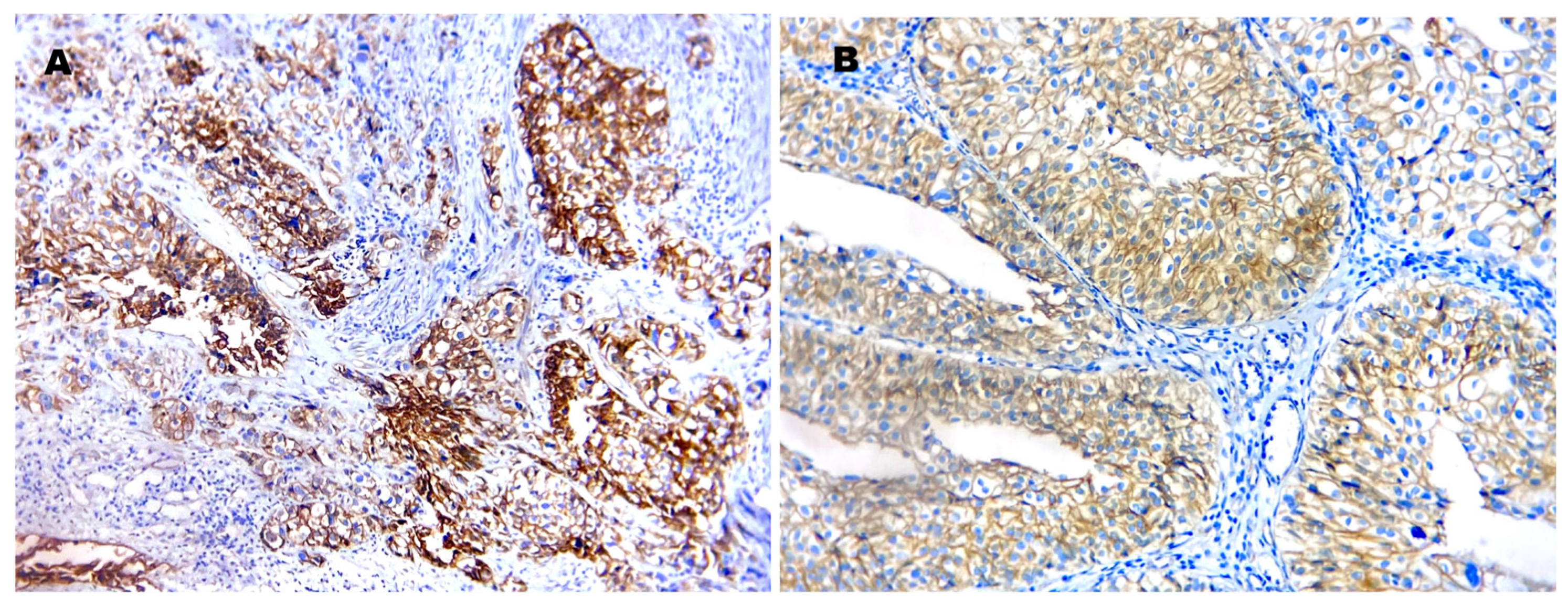
Vitamin D receptor (VDR): Using a cutoff of ≥10% cytoplasmic and/or nuclear staining, VDR positivity was detected in 48/58 cases (82.7%) (Figure 2). VDR expression showed no statistically significant association with tumor grade, muscle invasion status, or LVI (Table 2). In the univariate model, VDR positivity was not predictive of muscle invasion (OR 0.90; p = 0.419), indicating that VDR immunoreactivity, as scored here, did not contribute to invasive risk stratification beyond the established clinicopathologic variables.
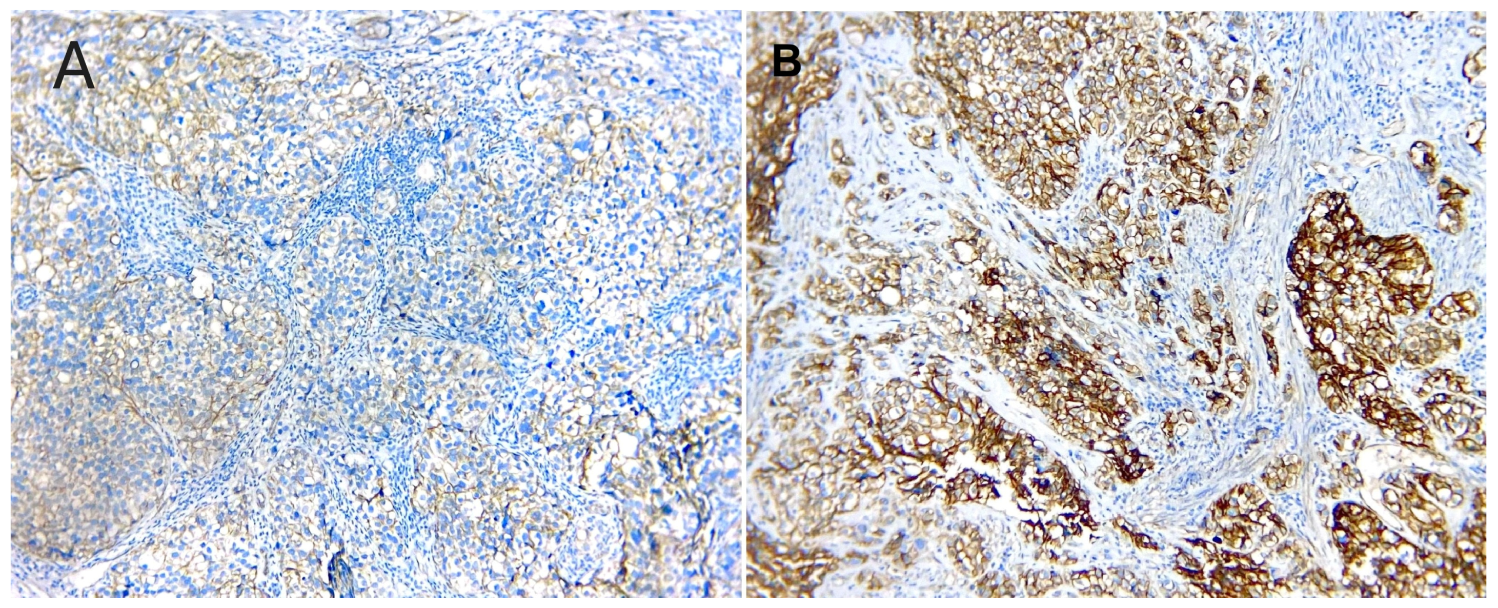
β-catenin: Preserved membranous β-catenin expression (>80% membranous staining) was observed in 20/58 cases (34.5%), while reduced membranous expression was present in 38/58 cases (65.5%) (Figure 3). Reduced membranous β-catenin was significantly associated with tumor grade (Table 3), supporting a relationship between loss of adhesion phenotype and high-grade morphology. In contrast, membranous β-catenin pattern showed no statistically significant association with muscle invasion or LVI (Table 3). On univariate effect-size analysis, reduced β-catenin demonstrated a non-significant trend toward association with muscle invasion (OR 2.80; p = 0.068), suggesting that the direction of association favored higher invasive risk but did not reach the significance threshold in this sample. Nuclear β-catenin positivity (≥5% nuclear staining) was not detected in any case (0/58, 0%), indicating absence of detectable nuclear translocation in this cohort under the applied scoring criteria.
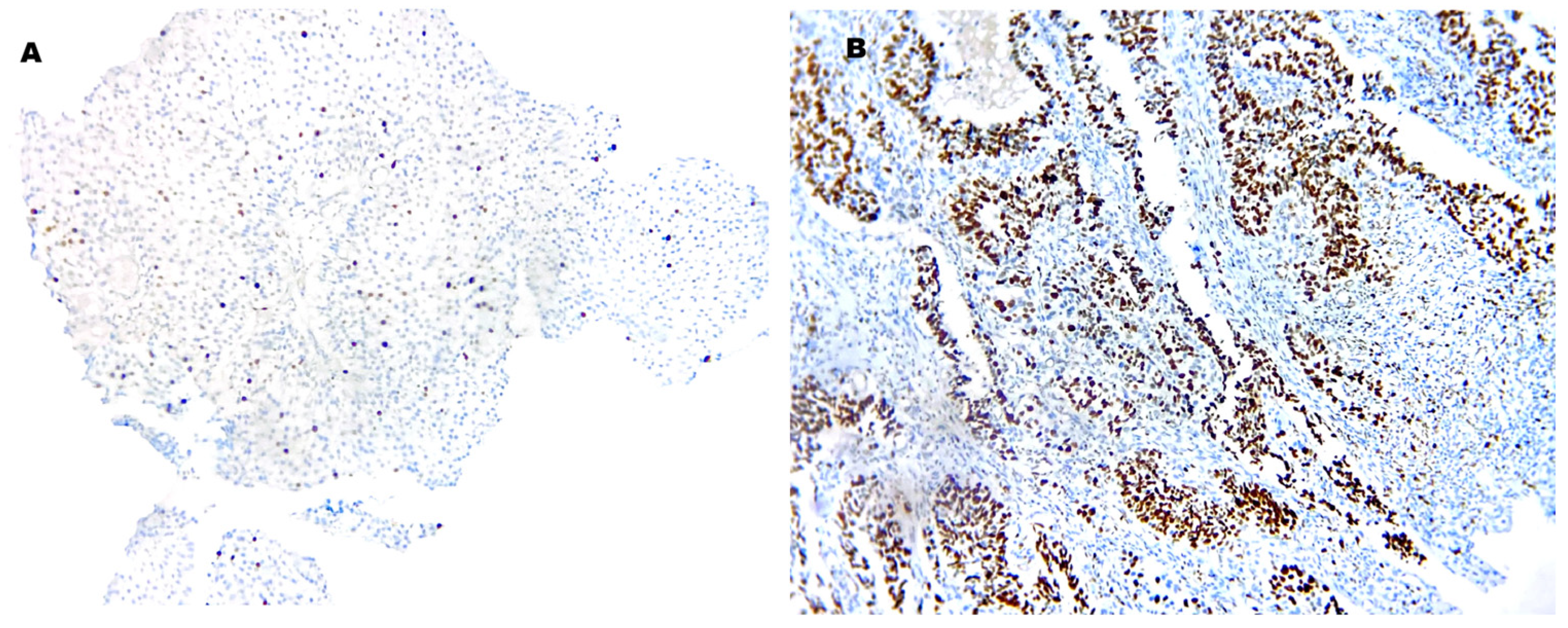
Ki-67 labeling index: A high Ki-67 index (≥30%) was present in 35/58 tumors (60.3%) (Figure 4). High Ki-67 index was significantly more frequent in high-grade than low-grade tumors (p = 0.005) and in MIBC than NMIBC (p = 0.002) (Table 4). In contrast, Ki-67 index showed no statistically significant association with LVI, age group, or sex (Table 4). In univariate analysis, a high Ki-67 index was associated with higher odds of muscle invasion (OR 6.18; 95% CI 1.91–19.97) (Table 5). Given the small sample size and the biological overlap among Ki-67, grade, and architecture, this finding should be interpreted cautiously as reflecting an aggressive phenotype rather than an independent effect.

4. Discussion

VDR, β-catenin, and Ki-67 represent complementary biologic axes in conventional UC: nuclear receptor signaling, adhesion/Wnt–epithelial–mesenchymal transition (EMT) biology, and proliferation. Evaluating them together is clinically relevant because conventional UC represents the dominant clinicopathologic pathway in bladder cancer, and because management decisions hinge primarily on the transition from NMIBC to MIBC and from low-grade to high-grade disease. In our cohort, muscle invasion clustered with high grade, non-papillary architecture, lymphovascular invasion (LVI), and high proliferative activity (Table 1), consistent with established UC behavior [7,8]. The univariate effect-size analysis (Table 5) further showed very large odds ratios for grade and architecture and a strong association for LVI; however, the corresponding confidence intervals were wide, which is expected when sparse/zero cells occur (e.g., no non-papillary tumors and no LVI in NMIBC), and this should be interpreted as imprecision rather than overstatement of biologic effect [17,18]. Clinically, these findings support that invasion in this cohort aligns with classic adverse histologic features, while emphasizing that the magnitude of some estimates cannot be precisely resolved in small strata.
VDR signaling has been implicated as a tumor-modulating pathway across malignancies through effects on differentiation, cell-cycle regulation, apoptosis, and inflammatory modulation [19,20]. In urothelial bladder cancer, VDR expression has been variably reported and inconsistently linked to aggressiveness and outcome. Jóźwicki et al. reported that higher VDR expression correlated with improved survival, supporting a protective or differentiation-associated role [11]. Conversely, Sharaf et al. (Egyptian cohort) described VDR expression in all tumors and found stronger VDR staining—particularly nuclear—in association with adverse pathologic parameters including higher stage and invasion [12]. In our series, VDR positivity was frequent (82.7%) yet showed no significant association with grade, muscle invasion, or LVI (Table 2), and VDR positivity did not predict muscle invasion (Table 5). Several methodological and biological factors can plausibly reconcile these inconsistencies. Methodologically, VDR immunohistochemistry is sensitive to pre-analytical and analytical variability (fixation duration, antigen retrieval, detection system), and to interpretive differences such as nuclear-only versus combined nuclear/cytoplasmic scoring, whether intensity is incorporated, and the selected cutoff defining positivity. These factors can shift borderline cases between categories and can materially affect correlations with grade or invasion across studies [11,12]. Cohort composition may also play a role: studies enriched for more advanced stage, treated/recurrent disease, or mixed histologic variants may yield different patterns than cohorts restricted to conventional UC. Biologically, IHC demonstrates receptor presence rather than pathway activity; ligand availability and host context (including inflammatory signaling and downstream co-regulators) may be crucial. Vitamin D deficiency is common in Middle East and North Africa populations [21], and meta-analytic evidence suggests that higher circulating 25-hydroxyvitamin D may be associated with reduced bladder cancer risk [22]. Accordingly, VDR positivity may not translate uniformly into anti-proliferative or pro-differentiation effects across populations, which could limit risk stratification by receptor expression alone. Taken together, our findings suggest that VDR immunoreactivity, as scored here, should be interpreted cautiously as a standalone clinicopathologic discriminator in Egyptian conventional UC; future investigations should integrate standardized compartment-specific scoring with clinical vitamin D measures and outcome endpoints to define prognostic and potentially predictive value.
β-catenin plays dual roles in epithelial adhesion (E-cadherin–catenin complex) and canonical Wnt signaling via cytoplasmic stabilization and potential nuclear translocation [23]. Wnt/β-catenin dysregulation has been implicated in bladder tumor biology, including epigenetic alterations affecting Wnt inhibitors and downstream transcriptional programs that promote EMT and invasive behavior [13,14,24]. In our cohort, reduced membranous β-catenin was common and significantly associated with higher grade (Table 3), supporting an EMT-linked relationship with aggressive morphology and reduced epithelial cohesion. Similar observations have been reported in NMIBC and Egyptian cohorts and in other cross-sectional studies of bladder neoplasms [25,26,27]. However, nuclear β-catenin was absent in our cases. This is consistent with the idea that true nuclear accumulation may characterize a molecular subset rather than a universal UC feature and may depend on specific Wnt-pathway genetic/epigenetic events or tumor subtype distribution [27,28]. Importantly, loss of membranous β-catenin can occur through multiple mechanisms (including disruption of adhesion complexes and EMT-associated remodeling) without necessarily producing prominent nuclear β-catenin detectable by routine IHC, particularly if canonical Wnt activation is heterogeneous or limited to subclones. This interpretation is compatible with broader evidence that bladder cancer is molecularly heterogeneous, with multiple pathways converging on high-grade and invasive phenotypes [29,30]. Mechanistic interactions between vitamin D/VDR signaling and Wnt/β-catenin pathways have been described in experimental cancer contexts [29]. Nevertheless, the present study did not assess vitamin D status, downstream pathway activity, or other molecular readouts, and nuclear β-catenin accumulation was not detected by IHC in this cohort. Accordingly, any inference that VDR expression in our cases reflects functional antagonism of canonical Wnt/β-catenin signaling would be speculative and is not supported directly by our data [31]. Instead, our findings support a morphologic association in which reduced membranous β-catenin correlates with higher grade (Table 3), while VDR immunoreactivity does not correlate with grade or invasion (Table 2). Future studies combining tissue markers with circulating vitamin D measures and molecular assays of Wnt pathway activation are needed to test whether VDR expression relates to functional pathway modulation or clinical outcome in Egyptian UC. Clinically, this supports reporting membranous β-catenin patterns in relation to grade, while avoiding over-interpretation of negative nuclear staining as evidence against any Wnt-pathway involvement.
Ki-67 is an established marker of proliferation, present in cycling cells and absent in quiescent cells [30,32]. Multiple studies and meta-analyses have shown that higher Ki-67 indices correlate with adverse clinicopathologic features and poorer outcomes in UC [16,33]. Using a 30% cutoff, we found that high Ki-67 was significantly associated with high grade and muscle invasion (Table 4) and with increased odds of muscle invasion (Table 5). This supports Ki-67 as the most clinically informative marker among the three in our dataset, particularly as a practical indicator of aggressive biology. Nevertheless, Ki-67 assessment remains challenged by heterogeneity in methodology (hot-spot vs. whole-section scoring) and variability in cutoffs across studies, which limits cross-study comparability. A pT1-focused cohort supported its prognostic utility but applied different thresholds and a different clinical context [34], and a recent NMIBC analysis proposed an optimal cutoff of around 18% for recurrence prediction [33]. These data emphasize that “optimal” cutoffs likely differ by disease setting (NMIBC vs. MIBC), treatment context, and scoring strategy. Accordingly, future work should evaluate multiple cutoffs and, where possible, model Ki-67 as a continuous variable to minimize information loss, while aligning reporting with established tumor-marker and observational-study reporting guidance [17,35].
Overall, in this study, VDR immunopositivity was not discriminatory for grade or invasion, reduced membranous β-catenin was aligned with high-grade biology, and a high Ki-67 index was associated with aggressive clinicopathologic behavior. These findings add region-specific evidence to a literature characterized by methodological variability and highlight priorities for standardization and outcome-linked validation in future cohorts [35,36].

Limitations and Future Directions

This study has limitations inherent to its single-center cross-sectional design. The sample size limits power for modest associations and contributes to sparse/zero-cell comparisons that can reduce the precision of effect estimates. Lack of follow-up prevents direct evaluation of recurrence, progression, and survival endpoints, which are necessary to validate true prognostic utility and to refine risk prediction models. In addition, immunohistochemical scoring may be influenced by pre-analytical variables (fixation duration, tissue processing), analytical factors (antibody performance, retrieval conditions), and interpretive variability, particularly for compartment-specific patterns (membranous vs. nuclear). Future multicenter studies in Egyptian and regional populations should include prospective outcome data, standardized scoring protocols, and—where feasible—integration of clinical variables (e.g., treatment, comorbidities, and laboratory measures such as vitamin D status). Larger cohorts would also enable multivariable modeling and subgroup analyses (NMIBC risk groups, MIBC stage strata) to better define the independent and combined roles of VDR, β-catenin, and Ki-67 in conventional UC.

5. Conclusions

In this cohort of Egyptian patients with conventional urothelial carcinoma, VDR immunopositivity was present but showed no significant association with tumor grade, muscle invasion, or lymphovascular invasion, suggesting limited clinicopathologic discriminatory value using the current scoring approach. Reduced membranous β-catenin was significantly associated with higher tumor grade, supporting a link between loss of epithelial adhesion phenotype and aggressive morphology, whereas nuclear β-catenin was not detected. Ki-67 (≥30%) was significantly associated with high grade and muscle invasion and demonstrated the strongest association with adverse histopathologic features in this cross-sectional analysis.
These findings provide population-specific data and underscore the need for standardized, compartment-specific immunohistochemical scoring and harmonized cutoffs before VDR and β-catenin can be considered for routine prognostic application. Larger, multicenter studies incorporating longitudinal outcomes and integrated clinical–molecular data are required to clarify the prognostic and potential therapeutic relevance of these markers in conventional urothelial carcinoma.

Author Contributions

Conceptualization, Methodology, Data collection: M.M.E.-M., M.T., A.A., R.E., M.S., M.F., D.S., S.-e.-d.S.O.S., S.M.S., H.G., A.H., M.S.A., D.S.A.M., A.A.B., M.M.A. Data analysis: A.H., M.T. Writing drafts: M.M.E.-M., M.T., A.A., R.E., M.S., A.H. Review & final revision: M.M.E.-M., M.T., A.A., R.E., M.S., M.F., D.S., S.-e.-d.S.O.S., S.M.S., H.G., A.H., M.S.A., A.A.B., D.S.A.M., M.M.A. All authors have read and agreed to the published version of the manuscript.

Funding

This research received no external funding.

Institutional Review Board Statement

The study was approved by the Bioethical Committee of the Faculty of Medicine, Al-Azhar University, Assiut, Egypt. (Code No. Research/AZAST./PAT005/7/245/6/2025; approved 12 June 2025).

Informed Consent Statement

The Ethics Committee of the Faculty of Medicine, Al-Azhar University, Assiut waived the requirement for informed consent because of the retrospective nature of the study.

Data Availability Statement

The original contributions presented in this study are included in the article. Further inquiries can be directed at the corresponding author.

Conflicts of Interest

The authors declare no conflicts of interest.

References

  1. Bray, F.; Laversanne, M.; Sung, H.; Ferlay, J.; Siegel, R.L.; Soerjomataram, I.; Jemal, A. Global cancer statistics 2022: GLOBOCAN estimates of incidence and mortality worldwide for 36 cancers in 185 countries. CA Cancer J. Clin. 2024, 74, 229–263. [Google Scholar] [CrossRef] [Scilit]
  2. Abbas, N.F.; Aoude, M.R.; Kourie, H.R.; Al-Shamsi, H.O. Uncovering the epidemiology of bladder cancer in the Arab world: A review of risk factors, molecular mechanisms, and clinical features. Asian J. Urol. 2024, 11, 406–422. [Google Scholar] [CrossRef] [Scilit] [PubMed]
  3. Amin, H.A.A.; Kobaisi, M.H.; Samir, R.M. Schistosomiasis and bladder cancer in Egypt: Truths and myths. Open Access Maced. J. Med. Sci. 2019, 7, 4023–4029. [Google Scholar] [CrossRef] [Scilit]
  4. Hasan, A.; Mohammed, Y.; Basiony, M.; Hanbazazh, M.; Samman, A.; Abdelaleem, M.F.; Nasr, M.; Abozeid, H.; Mohamed, H.I.; Faisal, M.; et al. Clinico-Pathological Features and Immunohistochemical Comparison of p16, p53, and Ki-67 Expression in Muscle-Invasive and Non-Muscle-Invasive Conventional Urothelial Bladder Carcinoma. Clin. Pract. 2023, 13, 806–819. [Google Scholar] [CrossRef] [Scilit] [PubMed]
  5. Felix, A.S.; Soliman, A.S.; Khaled, H.; Zaghloul, M.S.; Banerjee, M.; El-Baradie, M.; El-Kalawy, M.; Abd-Elsayed, A.A.; Ismail, K.; Hablas, A.; et al. The changing patterns of bladder cancer in Egypt over the past 26 years. Cancer Causes Control 2008, 19, 421–429. [Google Scholar] [CrossRef] [Scilit]
  6. Zarzour, A.H.; Selim, M.; Abd-Elsayed, A.A.; Hameed, D.A.; AbdelAziz, M.A. Muscle invasive bladder cancer in Upper Egypt: The shift in risk factors and tumor characteristics. BMC Cancer 2008, 8, 250. [Google Scholar] [CrossRef] [Scilit]
  7. European Association of Urology. EAU Guidelines: Non-Muscle-Invasive Bladder Cancer (TaT1 and CIS); 2024 Edition; European Association of Urology: Arnhem, The Netherlands, 2024. [Google Scholar]
  8. WHO Classification of Tumours Editorial Board. Urinary and Male Genital Tumours. In WHO Classification of Tumours, 5th ed.; International Agency for Research on Cancer: Lyon, France, 2022; Volume 8. [Google Scholar]
  9. Bebars, S.M.; Hasan, A.; Ghanam, W.; Al-Ghamdi, H.S.; Alghamdi, M.A.; Samman, A.; Noorwali, H.; Abdelwahed, M.S.; Mohammed, Y.; Salem, M.; et al. Assessment of Immunohistochemical Expression of Vitamin D Receptor in Surgically Excised Cutaneous Melanoma from Egyptian Patients. J. Microsc. Ultrastruct. 2026. [Google Scholar] [CrossRef] [Scilit]
  10. Huss, L.; Gulz-Haake, I.; Nilsson, E.; Tryggvadottir, H.; Nilsson, L.; Nodin, B.; Jirström, K.; Isaksson, K.; Jernström, H. The Vitamin D Receptor as a Prognostic Marker in Breast Cancer—A Cohort Study. Nutrients 2024, 16, 931. [Google Scholar] [CrossRef] [Scilit]
  11. Jóźwicki, W.; Brożyna, A.A.; Siekiera, J.; Slominski, A.T. Expression of vitamin D receptor (VDR) positively correlates with survival of urothelial bladder cancer patients. Int. J. Mol. Sci. 2015, 16, 24369–24386. [Google Scholar] [CrossRef] [Scilit]
  12. Sharaf, R.M.; Abdalla, B.M.M.; El Naggar, S.I.; Moustafa, W.I. Expression of vitamin D receptor (VDR) in urinary bladder carcinoma: Immunohistochemical and histopathological study. Asian Pac. J. Cancer Biol. 2022, 7, 245–253. [Google Scholar] [CrossRef] [Scilit]
  13. Baumgart, E.; Cohen, M.S.; Neto, B.S.; Jacobs, M.A.; Wotkowicz, C.; Rieger-Christ, K.M.; Biolo, A.; Zeheb, R.; Loda, M.; Libertino, J.A.; et al. Identification and prognostic significance of an epithelial-mesenchymal transition expression profile in human bladder tumors. Clin. Cancer Res. 2007, 13, 1685–1694. [Google Scholar] [CrossRef] [Scilit]
  14. Li, Y.; Zheng, Y.; Izumi, K.; Ishiguro, H.; Ye, B.; Li, F.; Miyamoto, H. Androgen activates β-catenin signaling in bladder cancer cells. Endocr. Relat. Cancer 2013, 20, 293–304. [Google Scholar] [CrossRef] [Scilit]
  15. Harsanyi, S.; Novakova, Z.V.; Neuschlova, L.; Galbavy, S.; Danisovic, L.; Ziaran, S.; Bevizova, K. Optimizing Ki-67 and E-Cadherin Thresholds for Improved Grade and Stage Classification in Urothelial Bladder Cancer. J. Clin. Med. 2026, 15, 338. [Google Scholar] [CrossRef] [Scilit] [PubMed]
  16. Tian, Y.; Ma, Z.; Chen, Z.; Li, M.; Wu, Z.; Hong, M.; Wang, H.; Svatek, R.; Rodriguez, R.; Wang, Z. Clinicopathological and prognostic value of Ki-67 expression in bladder cancer: A systematic review and meta-analysis. PLoS ONE 2016, 11, e0158891. [Google Scholar] [CrossRef] [Scilit] [PubMed]
  17. McShane, L.M.; Altman, D.G.; Sauerbrei, W.; Taube, S.E.; Gion, M.; Clark, G.M. Reporting recommendations for tumor marker prognostic studies (REMARK). J. Natl. Cancer Inst. 2005, 97, 1180–1184. [Google Scholar] [CrossRef] [Scilit]
  18. Amin, M.B.; Greene, F.L.; Edge, S.B.; Compton, C.C.; Gershenwald, J.E.; Brookland, R.K.; Meyer, L.; Gress, D.M.; Byrd, D.R.; Winchester, D.P. The Eighth Edition AJCC Cancer Staging Manual: Continuing to build a bridge from a population-based to a more “personalized” approach to cancer staging. CA Cancer J. Clin. 2017, 67, 93–99. [Google Scholar] [CrossRef] [Scilit]
  19. Kanwal, B.; Shah, S.S.; Shaheen, F.; Shiferaw, M.S.; Maurya, D.; Li, Y.; Pounraj, S.; Kovacevic, Z. The Effects of Vitamin D on the Breast Cancer Tumor Microenvironment. Cancers 2025, 17, 3751. [Google Scholar] [CrossRef] [Scilit]
  20. Deeb, K.K.; Trump, D.L.; Johnson, C.S. Vitamin D signalling pathways in cancer: Potential for anticancer therapeutics. Nat. Rev. Cancer 2007, 7, 684–700. [Google Scholar] [CrossRef] [Scilit]
  21. Machuron, F.; Dessalles, R.; Ribet, L.; Bentouhami, H.; Escutnaire, J.; Brusselaers, N.; Durand-Dubief, M. Vitamin D Nutritional Status in the Middle East and North Africa Region: A Systematic Review and Meta-analysis. Curr. Dev. Nutr. 2025, 9, 107577. [Google Scholar] [CrossRef] [Scilit] [PubMed]
  22. Liao, Y.; Huang, J.L.; Qiu, M.X.; Ma, Z.W. Impact of serum vitamin D level on risk of bladder cancer: A systemic review and meta-analysis. Tumor Biol. 2015, 36, 1567–1572. [Google Scholar] [CrossRef] [Scilit]
  23. Garg, M.; Maurya, N. WNT/β-catenin signaling in urothelial carcinoma of bladder. World J. Nephrol. 2019, 8, 83–94. [Google Scholar] [CrossRef] [Scilit]
  24. Urakami, S.; Shiina, H.; Enokida, H.; Kawakami, T.; Tokizane, T.; Ogishima, T.; Tanaka, Y.; Li, L.-C.; Ribeiro-Filho, L.A.; Terashima, M.; et al. Epigenetic inactivation of Wnt inhibitory factor-1 plays an important role in bladder cancer through aberrant canonical Wnt/β-catenin signaling pathway. Clin. Cancer Res. 2006, 12, 383–391. [Google Scholar] [CrossRef] [Scilit]
  25. Elsherif, E.; Abdel Elbaky, T.A.; Elserafy, F.; Elkady, N.; Dawood, M.; Gaber, M.A.; Badawy, A.; El Gharabawy, M. β-catenin and SKP2 proteins as predictors of grade and stage of non-muscle invasive urothelial bladder carcinoma. Chin. Clin. Oncol. 2016, 5, 6. [Google Scholar] [CrossRef] [Scilit] [PubMed]
  26. Elzamy, S.; Allah, A.; Kandeel, W. The prognostic significance of androgen receptor and β-catenin immunohistochemical expression in urothelial carcinoma with and without detrusor muscle invasion from an Egyptian institution. Pol. J. Pathol. 2018, 69, 234–242. [Google Scholar] [CrossRef] [Scilit]
  27. Yazdi, S.A.M.; Batmani, S.; Moradi Tabriz, H.; Nazar, E.; Pourebrahimi, E. The incidence and prognostic role of β-catenin and E-cadherin biomarkers in bladder neoplasms: A cross-sectional study. Int. J. Surg. Open 2023, 54, 100609. [Google Scholar] [CrossRef] [Scilit]
  28. Kastritis, E.; Murray, S.; Kyriakou, F.; Horti, M.; Tamvakis, N.; Kavantzas, N.; Patsouris, E.S.; Noni, A.; Legaki, S.; Dimopoulos, M.A.; et al. Somatic mutations of adenomatous polyposis coli gene and nuclear β-catenin accumulation have prognostic significance in invasive urothelial carcinomas: Evidence for Wnt pathway implication. Int. J. Cancer 2009, 124, 103–108. [Google Scholar] [CrossRef] [Scilit]
  29. Robertson, A.G.; Kim, J.; Al-Ahmadie, H.; Bellmunt, J.; Guo, G.; Cherniack, A.D.; Hinoue, T.; Laird, P.W.; Hoadley, K.A.; Akbani, R. Comprehensive molecular characterization of muscle-invasive bladder cancer. Cell 2017, 171, 540–556. [Google Scholar] [CrossRef] [Scilit]
  30. Kamoun, A.; de Reyniès, A.; Allory, Y.; Sjödahl, G.; Robertson, A.G.; Seiler, R.; Hoadley, K.A.; Groeneveld, C.S.; Al-Ahmadie, H.; Choi, W.; et al. A consensus molecular classification of muscle-invasive bladder cancer. Eur. Urol. 2020, 77, 420–433. [Google Scholar] [CrossRef] [Scilit]
  31. Larriba, M.J.; González-Sancho, J.M.; Barbáchano, A.; Niell, N.; Ferrer-Mayorga, G.; Muñoz, A. Vitamin D Is a Multilevel Repressor of Wnt/β-Catenin Signaling in Cancer Cells. Cancers 2013, 5, 1242–1260. [Google Scholar] [CrossRef] [Scilit]
  32. Scholzen, T.; Gerdes, J. The Ki-67 protein: From the known and the unknown. J. Cell. Physiol. 2000, 182, 311–322. [Google Scholar] [CrossRef] [Scilit]
  33. Ko, K.; Jeong, C.W.; Kwak, C.; Kim, H.H.; Ku, J.H. Significance of Ki-67 in non-muscle invasive bladder cancer patients: A systematic review and meta-analysis. Oncotarget 2017, 8, 100614–100630. [Google Scholar] [CrossRef] [Scilit] [PubMed]
  34. Culpan, M.; Turan, T.; Ozkanli, S.S.; Zenginkinet, T.; Kazan, O.; Ucar, T.; Atis, G.; Caskurlu, T.; Yildirim, A. Prognostic and clinicopathologic value of Ki-67 and profilin 1 immunohistochemical expression in primary pT1 urothelial bladder cancer. J. Cancer Res. Ther. 2021, 17, 434–442. [Google Scholar] [CrossRef] [Scilit]
  35. Buechel, D.; Sugiyama, N.; Rubinstein, N.; Saxena, M.; Kalathur, R.K.R.; Lüönd, F.; Vafaizadeh, V.; Valenta, T.; Hausmann, G.; Cantù, C.; et al. Parsing β-catenin’s cell adhesion and Wnt signaling functions in malignant mammary tumor progression. Proc. Natl. Acad. Sci. USA 2021, 118, e2020227118. [Google Scholar] [CrossRef] [Scilit]
  36. Peng, R.; Zhang, Y.; Jia, M.; Yi, X.; Yi, X.; Li, S.; Pi, J.; Meng, W. Predicting recurrence of non-muscle invasive bladder urothelial carcinoma: Predictive value of the optimal cut-off value of Ki67. Front. Oncol. 2025, 14, 1522009. [Google Scholar] [CrossRef] [Scilit] [PubMed]
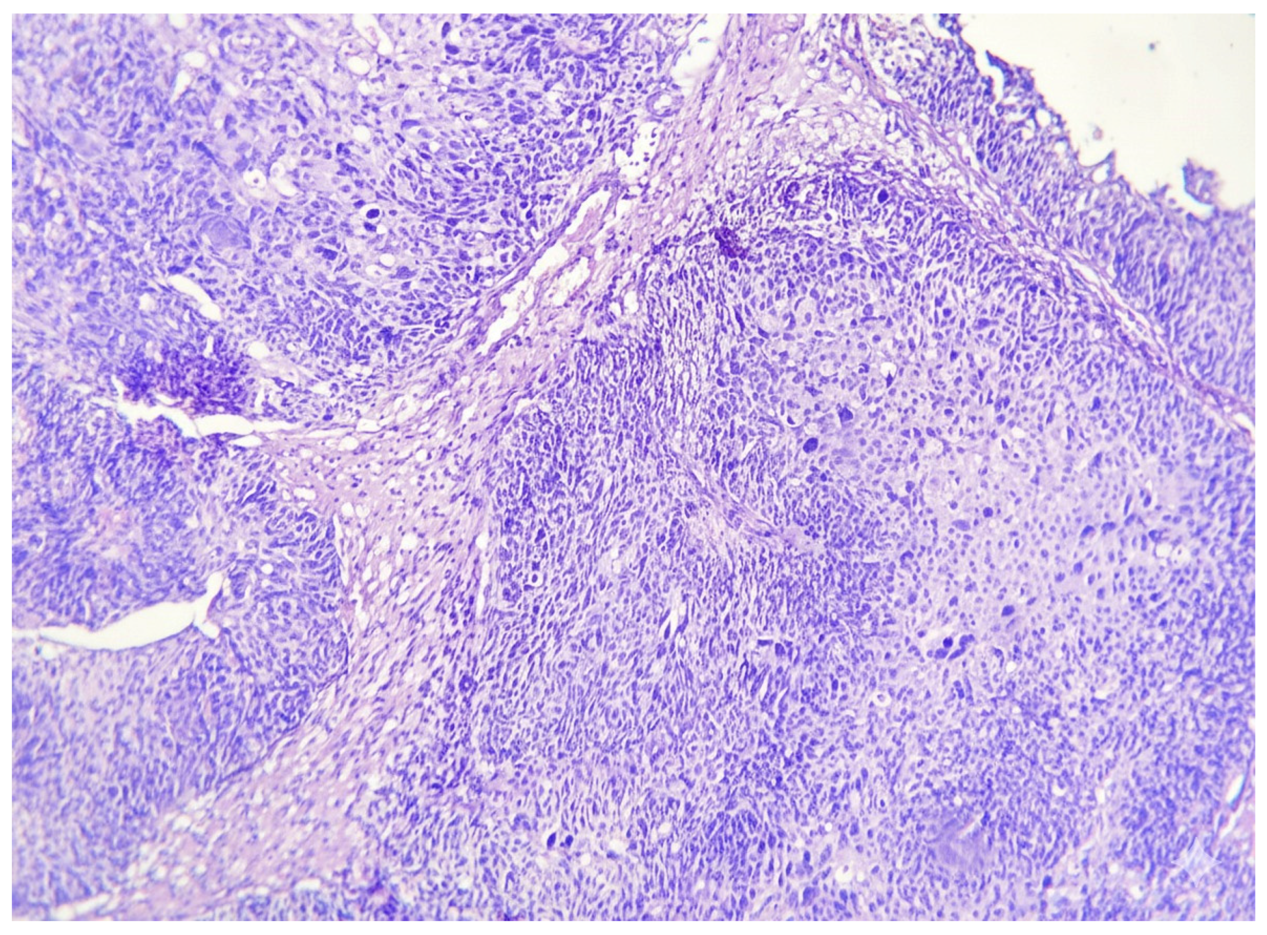
Figure 1. Conventional UC showing papillary architecture and high-grade cytologic features (H&E, 200× original magnification).
Ijtm 06 00014 g001
Figure 2. Vitamin D receptor (VDR) immunostaining in urothelial carcinoma. (A) Muscle-invasive urothelial carcinoma showing VDR immunoreactivity in tumor cells. (B) Non-muscle-invasive urothelial carcinoma showing VDR immunoreactivity.
Ijtm 06 00014 g002
Figure 3. Membranous β-catenin pattern in urothelial carcinoma (IHC). (A) Reduced membranous β-catenin expression. (B) Preserved membranous β-catenin expression.
Ijtm 06 00014 g003
Figure 4. Ki-67 immunostaining in urothelial carcinoma (IHC, DAB). (A) Low Ki-67 labeling index (<30%). (B) High Ki-67 labeling index (≥30%).
Ijtm 06 00014 g004
Table 1. Clinicopathologic characteristics according to muscle invasion status (n = 58).
VariableNMIBC (n = 28)MIBC (n = 30)p Value
Age (years), mean ± SD64.4 ± 9.866.2 ± 8.80.464
>60 years14 (50.0%)21 (70.0%)0.120
Male sex22 (78.6%)28 (93.3%)0.138
Tumor size > 2 cm13 (46.4%)18 (60.0%)0.300
Papillary architecture28 (100.0%)7 (23.3%)<0.001
High grade1 (3.6%)22 (73.3%)<0.001
Lymphovascular invasion (LVI)0 (0.0%)7 (23.3%)0.011
Ki-67 high index (≥30%)11 (39.3%)24 (80.0%)0.002
Data are presented as mean ± SD or n (%). NMIBC: non-muscle-invasive bladder cancer; MIBC: muscle-invasive bladder cancer.
Table 2. Association between VDR immunoexpression and clinicopathologic parameters (n = 58).
ParameterVDR Positive (n = 48)VDR Negative (n = 10)p Value
Low grade29 (60.4%)6 (60.0%)1.000
High grade19 (39.6%)4 (40.0%)
Papillary30 (62.5%)5 (50.0%)0.412
Non-papillary18 (37.5%)5 (50.0%)
Muscle invasion present26 (54.2%)4 (40.0%)0.419
Muscle invasion absent22 (45.8%)6 (60.0%)
LVI present6 (12.5%)1 (10.0%)0.734
LVI absent42 (87.5%)9 (90.0%)
Table 3. Association between membranous β-catenin expression and clinicopathologic parameters (n = 58).
ParameterPreserved (n = 20)Reduced (n = 38)p Value
Low grade16 (80.0%)19 (50.0%)0.025
High grade4 (20.0%)19 (50.0%)
Papillary13 (65.0%)22 (57.9%)0.064
Non-papillary7 (35.0%)16 (42.1%)
Muscle invasion present7 (35.0%)23 (60.5%)0.068
Muscle invasion absent13 (65.0%)15 (39.5%)
LVI present1 (5.0%)6 (15.8%)0.179
LVI absent19 (95.0%)32 (84.2%)
Table 4. Association of Ki-67 index category (≥30% vs. <30%) with clinicopathologic variables (n = 58).
VariableHigh Ki-67 (n = 35)Low Ki-67 (n = 23)p Value
High grade19 (54.3%)4 (17.4%)0.005
Low grade16 (45.7%)19 (82.6%)
MIBC24 (68.6%)6 (26.1%)0.002
NMIBC11 (31.4%)17 (73.9%)
LVI present5 (14.3%)2 (8.7%)0.692
LVI absent30 (85.7%)21 (91.3%)
>60 years22 (62.9%)13 (56.5%)0.629
<60 years13 (37.1%)10 (43.5%)
Table 5. Univariate predictors of muscle invasion (MIBC).
VariableOdds Ratio (OR)95% CIp Value
High grade (vs low grade)74.28.6–639.8<0.001
Non-papillary (vs papillary)178.69.7–3292.9<0.001
Lymphovascular invasion (yes/no)18.20.99–335.40.011
High Ki-67 index (high/low)6.21.9–19.90.002
Reduced β-catenin2.80.1–15.60.068
VDR positivity0.90.1–180.419
Disclaimer/Publisher’s Note: The statements, opinions and data contained in all publications are solely those of the individual author(s) and contributor(s) and not of MDPI and/or the editor(s). MDPI and/or the editor(s) disclaim responsibility for any injury to people or property resulting from any ideas, methods, instructions or products referred to in the content.