Article

Decoding the CD36-Centric Axis in Gastric Cancer: Insights into Lipid Metabolism, Obesity, and Hypercholesterolemia

by
Preyangsee Dutta
1,†,
Dwaipayan Saha
1,†,
Atanu Giri
2,
Aseem Rai Bhatnagar
3 and
Abhijit Chakraborty
4,*
1
Metabolic, Nutrition and Exercise Research (MiNER) Laboratory, The University of Texas at El Paso, El Paso, TX 79968, USA
2
Computational Science Program, University of Texas at El Paso, El Paso, TX 79968, USA
3
Department of Radiation Oncology, Henry Ford Cancer—Detroit, 2800 W Grand Blvd, Detroit, MI 48202, USA
4
Department of Investigational Cancer Therapeutics, The University of Texas MD Anderson Cancer Center, 1400 Holcombe Blvd., Houston, TX 77030, USA
*
Author to whom correspondence should be addressed.
These authors contributed equally to this work.
Int. J. Transl. Med. 2025, 5(3), 26; https://doi.org/10.3390/ijtm5030026
Submission received: 16 May 2025 / Revised: 8 June 2025 / Accepted: 18 June 2025 / Published: 23 June 2025

Abstract

Background: Gastric cancer is a leading cause of cancer-related mortality worldwide, with approximately one million new cases diagnosed annually. While Helicobacter pylori infection remains a primary etiological factor, mounting evidence implicates obesity and lipid metabolic dysregulation, particularly in hypercholesterolemia, as emerging drivers of gastric tumorigenesis. This study investigates the molecular intersections between gastric cancer, obesity, and hypercholesterolemia through a comprehensive multi-omics and systems biology approach. Methods: We conducted integrative transcriptomic analysis of gastric adenocarcinoma using The Cancer Genome Atlas (TCGA) RNA-sequencing dataset (n = 623, 8863 genes), matched with standardized clinical metadata (n = 413). Differential gene expression between survival groups was assessed using Welch’s t-test with Benjamini–Hochberg correction (FDR < 0.05, |log2FC| ≥ 1). High-confidence gene sets for obesity (n = 128) and hypercholesterolemia (n = 97) were curated from the OMIM, STRING (confidence ≥ 0.7), and KEGG databases using hierarchical evidence-based prioritization. Overlapping gene signatures were identified, followed by pathway enrichment via Enrichr (KEGG 2021 Human) and protein–protein interaction (PPI) analysis using STRING v11.5 and Cytoscape v3.9.0. CD36’s prognostic value was evaluated via Kaplan–Meier and log-rank testing alongside clinicopathological correlations. Results: We identified 36 genes shared between obesity and gastric cancer, and 31 genes shared between hypercholesterolemia and gastric cancer. CD36 emerged as the only gene intersecting all three conditions, marking it as a unique molecular integrator. Enrichment analyses implicated dysregulated fatty acid uptake, adipocytokine signaling, cholesterol metabolism, and NF-κB-mediated inflammation as key pathways. Elevated CD36 expression was significantly correlated with higher tumor stage (p = 0.016), reduced overall survival (p = 0.001), and race-specific expression differences (p = 0.007). No sex-based differences in CD36 expression or survival were observed. Conclusions: CD36 is a central metabolic–oncogenic node linking obesity, hypercholesterolemia, and gastric cancer. It functions as both a mechanistic driver of tumor progression and a clinically actionable biomarker, particularly in metabolically comorbid patients. These findings provide a rationale for targeting CD36-driven pathways as part of a precision oncology strategy and highlight the need to incorporate metabolic profiling into gastric cancer risk assessment and treatment paradigms.

1. Introduction

Gastric cancer represents the fifth most common malignancy and the third leading cause of cancer-related mortality globally, with approximately 1.1 million new cases diagnosed annually and nearly 660,000 deaths reported in 2024 alone [1,2]. Despite advances in early detection, surgical techniques, and targeted therapies, the 5-year survival rate remains poor, particularly for patients diagnosed at advanced stages, underscoring the critical need for the improved understanding of risk factors, molecular mechanisms, and potential prevention strategies.
The contemporary clinical management of gastric cancer is increasingly guided by tumor staging, anatomical localization, and molecular profiling, enabling stratified therapeutic decisions. Early-stage cases often respond well to organ-preserving interventions such as endoscopic submucosal dissection and laparoscopic gastrectomy, particularly in East Asian regions with robust screening programs [3]. In contrast, advanced gastric adenocarcinomas demand multimodal treatment regimens incorporating surgical resection, perioperative chemotherapy (e.g., FLOT regimen), radiotherapy, and precision-targeted agents [4]. Molecular stratification has driven significant advances, with HER2-positive tumors benefiting from trastuzumab-based therapies, and immunotherapeutic agents like nivolumab offering efficacy in PD-L1-positive or MSI-high tumors [5,6]. Further innovations include ramucirumab, FGFR2 inhibitors, and claudin 18.2-targeting therapies such as zolbetuximab [7]. Yet, therapeutic resistance and molecular heterogeneity persist as clinical challenges, especially among patients with metabolic comorbidities. In complex scenarios like perforated gastric cancer, urgent surgical intervention is vital but remains controversial due to the lack of standardized guidelines, requiring the individualized assessment of metastases and comorbidities [8,9]. For unresectable or palliative cases, chemoradiation offers symptom relief and improved quality of life [10,11]. Additionally, nutritional management has emerged as a crucial factor in optimizing outcomes for patients with advanced disease [12], emphasizing the need for a multidisciplinary, patient-tailored approach that integrates evolving therapeutic modalities across global healthcare contexts [13,14,15,16].
The etiology of gastric cancer is multifactorial, with established risk factors including Helicobacter pylori infection, tobacco use, high salt intake, and genetic predisposition [1,17]. However, these traditional risk factors do not fully explain the observed epidemiological patterns, particularly the rising incidence of certain gastric cancer subtypes in regions where H. pylori prevalence is declining [18]. In recent decades, the global prevalence of obesity has increased dramatically, with more than 650 million adults classified as obese worldwide [19]. Concurrently, metabolic disorders including hypercholesterolemia have become increasingly prevalent. Obesity has been firmly established as a risk factor for several cancer types, including esophageal, colorectal, pancreatic, and breast cancers [20], with emerging evidence suggesting potential associations with gastric cancer.
Lipid metabolism, particularly cholesterol homeostasis, plays a critical role in cellular membrane integrity, energy homeostasis, and signal transduction. Dysregulation of these pathways, often observed in obesity and hypercholesterolemia, may contribute to carcinogenesis through multiple mechanisms, including altered cell membrane composition, dysregulated signaling pathways, chronic inflammation, and oxidative stress [21]. While previous studies have investigated the associations between obesity, lipid disorders, and gastric cancer individually, a comprehensive analysis of the molecular intersections between these conditions has been lacking.
This study aims to identify common genetic signatures between obesity, hypercholesterolemia, and gastric cancer through a comprehensive analysis of The Cancer Genome Atlas (TCGA) gastric cancer dataset; elucidate key molecular pathways and mechanistic links connecting these metabolic conditions with gastric carcinogenesis; identify potential therapeutic targets based on shared molecular pathways; and develop a predictive framework for risk stratification in gastric cancer patients with metabolic comorbidities. By integrating bioinformatic analyses with molecular pathway mapping, this study provides a comprehensive framework for understanding the complex interplay between metabolic dysfunction and gastric cancer, with significant implications for clinical practice and future research directions.

2. Materials and Methods

2.1. Data Sources and Acquisition

2.1.1. Gene Expression and Clinical Metadata Acquisition

Publicly available gene expression and clinical metadata were obtained from two independent sources. The gene expression matrix, containing normalized expression values for 8863 genes across 623 patient samples, was downloaded from a curated dataset on Kaggle (Kaggle, Inc., San Francisco, CA, USA) by Mahdieh Hajian et al. [22]. Clinical metadata, including demographic, pathological, and survival information for patients with stomach adenocarcinoma (STAD), was retrieved from The Cancer Genome Atlas (TCGA) via the Genomic Data Commons (GDC) portal (Supplementary File S1). Molecular subtype classifications (CIN, EBV, GS, MSI) were obtained separately from the TCGA Pan-Cancer Atlas database and merged using sample identifiers.
After preprocessing, gene expression values were merged with clinical metadata using sample IDs as the linking key so that only samples common to the expression and clinical datasets were retained. A total of 413 sample IDs were common between the gene expression matrix and GDC clinical metadata, and 410 sample IDs were common between the gene expression matrix and the TCGA Pan-Cancer Atlas subtype annotations. For analyses involving individual genes, such as CD36, the expression vector was extracted and appended as a column to the clinical Data Frame. The resulting merged Data Frame was used for all downstream analyses. The overall analysis pattern was illustrated in Supplementary Figure S1.

2.1.2. Gene Sets for Obesity and Hypercholesterolemia

Comprehensive gene sets related to obesity and hypercholesterolemia were curated from multiple databases, including the Online Mendelian Inheritance in Man (OMIM) database for genetic associations (McKusick–Nathans Institute of Genetic Medicine, Johns Hopkins University School of Medicine, Baltimore, MD, USA), Search Tool for the Retrieval of Interacting Genes/Proteins (STRING) database for protein–protein interaction networks (v12, European Molecular Biology Laboratory, Heidelberg, Germany), and Kyoto Encyclopedia of Genes and Genomes (KEGG) for pathway annotations (Kanehisa Laboratories, Institute for Chemical Research, Kyoto University, Kyoto, Japan). For hypercholesterolemia, we included genes associated with both familial hypercholesterolemia (all four established groups) and atherosclerotic-driven hypercholesterolemia. The obesity gene set included genes implicated in both monogenic and polygenic forms of obesity.
Only genes from human studies supported by genome-wide associations, functional validation, or clear clinical relevance were included. For OMIM-derived genes, confirmed genetic associations with well-defined phenotypes were required. STRING-derived interactions were limited to direct protein–protein interactions demonstrating confidence scores of 0.7 or greater. KEGG pathway selections only included core components with well-established functional roles. When confronted with conflicting evidence across data sources, we implemented a hierarchical prioritization framework based on consistency across independent studies, statistical association strength, functional relevance evidence, and validation in human clinical samples. This comprehensive, evidence-based approach yielded high-confidence gene sets comprising 128 obesity-associated genes and 97 hypercholesterolemia-associated genes for subsequent comparative analysis with differentially expressed genes identified in gastric cancer.

2.2. Bioinformatic Analysis Pipeline

2.2.1. Differential Gene Expression Analysis and Volcano Plot with FDR Correction

To identify genes differentially expressed between surviving and deceased patients, a Welch’s t-test was performed for each of the 8863 genes. For each gene, the log_2_fold change (mean expression in deceased minus alive) and corresponding p-value were computed. To correct for multiple hypothesis testing, false discovery rate (FDR) adjustment was applied using the Benjamini–Hochberg procedure. Genes with FDR < 0.05 and |log_2_fold change| ≥ 1 were considered statistically significant.
A volcano plot was generated to visualize the differential expression landscape, displaying all genes with log_2_fold change on the x-axis and −log_10_(p-value) on the y-axis. Genes passing the FDR threshold were highlighted and labeled.

2.2.2. Identification of Common Genetic Signatures

To identify molecular intersections, the TCGA-derived gastric cancer gene signature was compared with curated gene sets for obesity and hypercholesterolemia. Shared genes between gastric cancer and each metabolic condition were identified, and genes common to all three conditions were designated as key molecular nodes.

2.2.3. Pathway Analysis and Functional Annotation

Common gene sets from the above comparisons were analyzed using Enrichr to identify enriched biological pathways and functional annotations. Pathway libraries queried included Gene Ontology (GO), Reactome, and KEGG. Visualizations were generated using the KEGG 2021 Human database through the Enrichr Appyter tool, ranking pathways by −log10(p-value) for intuitive interpretation. This enabled the identification of key molecular mechanisms linking metabolic disorders to gastric cancer pathogenesis.

2.2.4. Protein–Protein Interaction Network Analysis

STRING (v12) was used to construct protein–protein interaction networks for the following: (1) genes shared between obesity and gastric cancer, (2) genes shared between hypercholesterolemia and gastric cancer, and (3) the central node gene CD36 and its direct interactors. Networks were visualized in Cytoscape (v3.9.0), and topological parameters were calculated to identify hub proteins.

2.3. CD36 Expression Analysis and Clinicopathological Correlations in Gastric Cancer

2.3.1. Survival Analysis

Survival analysis was performed to assess the prognostic significance of gene expression levels. For Kaplan–Meier (KM) survival curves, patients were dichotomized into “high” and “low” expression groups using the median gene expression as the cutoff. The overall survival time (in months) and status (alive, deceased) were obtained from the clinical dataset. In addition, Cox proportional hazards models were used to estimate the effect of gene expression on survival, adjusting for relevant covariates (e.g., age, tumor stage, sex). Hazard ratios (HRs) and 95% confidence intervals were reported.

2.3.2. Clinical Parameter Analysis

CD36 expression was compared across clinical and metabolic variables. For demographics, expression was analyzed by race (one-way ANOVA) and sex (Welch’s t-test). Tumor-related variables included AJCC stage and Lauren classification (ANOVA with post hoc Tukey’s tests). Metabolic variables included BMI category and lipid profile status (ANOVA or t-test). Sex-specific survival durations were also compared using boxplots and Welch’s t-test to explore potential associations with CD36 expression.

2.3.3. Statistical Analysis

All statistical analyses were performed in Python (v3.8, Python Software Foundation, CWI (Centrum Wiskunde & Informatica), Amsterdam, The Netherlands) using the following packages: pandas (v1.3.3, Pandas development team, NumFOCUS organization, USA) for data manipulation, NumPy (v1.21.2, NumFOCUS organization, USA) for numerical operations, SciPy (v1.7.1, NumFOCUS organization, USA) for statistical testing, and lifelines (v0.26.0, Cameron Davidson-Pilon and contributors, Canada) for survival analysis. A p-value < 0.05 was considered statistically significant for all tests. For multiple comparisons, p-values were adjusted using the Benjamini–Hochberg procedure to control the false discovery rate.

2.3.4. Data Visualization

Figures were generated using Matplotlib (v3.4.3, Matplotlib Development Team, NumFOCUS organization, USA) and seaborn (v0.11.2, Python, Michael Waskom and contributors, USA). Boxplots included jittered data points to show the distribution. Kaplan–Meier curves included 95% confidence intervals. A consistent color scheme was used throughout for interpretability.

3. Results

3.1. CD36 Identified as a Molecular Intersection Linking Gastric Cancer with Obesity and Hypercholesterolemia

A detailed summary of patient demographics, clinical characteristics, and molecular subtypes is provided in Supplementary Table S1. Transcriptomic profiling of gastric adenocarcinoma patients (n = 623) from The Cancer Genome Atlas (TCGA) revealed 2157 differentially expressed genes (DEGs) meeting stringent statistical criteria (FDR < 0.05, |log2 fold change| ≥ 1). Integrative analysis with curated, high-confidence gene sets for obesity (n = 128 genes) and hypercholesterolemia (n = 97 genes) identified substantial overlaps with the gastric cancer DEG dataset. The networks generated from the STRING, KEGG, and OMIM analyses are provided in Supplementary File S2. Specifically, 36 genes were shared between gastric cancer- and obesity-related profiles, while 31 overlapped with hypercholesterolemia-associated genes. Notably, the scavenger receptor and fatty acid transporter CD36 was the only gene common to all three datasets (Figure 1A). This intersection points to CD36 as a unique molecular integrator of metabolic dysregulation in gastric carcinogenesis. Although CD36 ranked 3870th in inter-sample variance (variance = 2.3162), it was prioritized due to its biological relevance and cross-disease associations documented in the literature.
Functional enrichment analysis of the obesity–gastric cancer overlapping genes uncovered key signaling and metabolic pathways. The most significantly enriched pathway was “Maturity onset diabetes of the young” (FDR = 2.04 × 10⁻¹⁹), reflecting strong associations with β-cell function and insulin regulation. Additional enrichment was observed in “Neuroactive ligand-receptor interaction” (FDR = 1.33 × 10⁻¹⁴) and the “Adipocytokine signaling pathway” (FDR = 3.7 × 10⁻¹³), indicating the influence of neuroendocrine and hormonal signaling networks on the tumor microenvironment (Figure 1B).
Similarly, the hypercholesterolemia–gastric cancer gene set was enriched in cholesterol handling and inflammatory signaling pathways, with “Cholesterol metabolism” emerging as the top hit (FDR = 3.44 × 10⁻¹⁷), followed by “Fat digestion and absorption” and “Lipid and atherosclerosis” pathways (Figure 1C). Additional immune-modulatory pathways, including “NF-κB signaling,” “Epstein–Barr virus infection,” and “Viral myocarditis,” were significantly enriched, supporting the notion that systemic dyslipidemia may exacerbate oncogenesis through inflammation and altered immune surveillance. These results collectively highlight CD36 as a putative hub gene linking lipid metabolism, inflammation, and gastric cancer pathogenesis.

3.2. CD36 as a Central Mediator of Metabolic Reprogramming, Inflammation, and Tumor Progression in Gastric Cancer

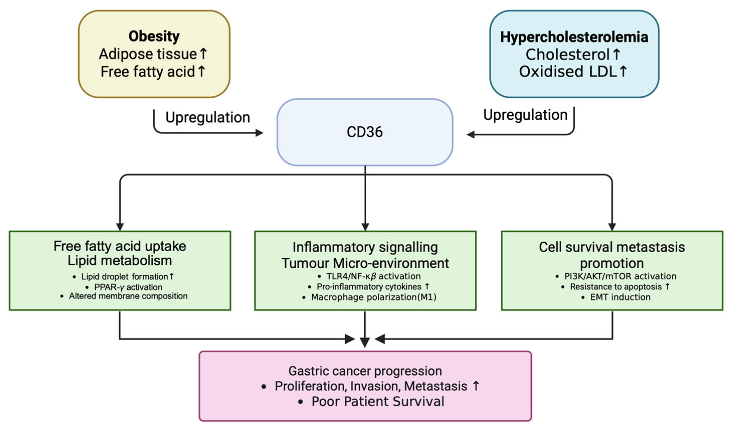
Mechanistic mapping of CD36 functions based on the literature evidence and protein interaction networks identified three core oncogenic pathways by which CD36 contributes to gastric cancer progression (Figure 2). First, CD36 facilitates the cellular uptake of long-chain fatty acids, resulting in lipid droplet accumulation and the activation of PPAR-γ signaling, which supports lipid metabolism and energy storage. Second, CD36 serves as a pattern recognition receptor for oxidized LDL, activating TLR4/NF-κB signaling and promoting a pro-inflammatory tumor microenvironment through cytokine production and M1 macrophage polarization. Third, CD36 enhances PI3K/AKT/mTOR signaling and interacts with thrombospondin-1, thereby promoting epithelial–mesenchymal transition (EMT), apoptosis resistance, and metastatic potential. These multifaceted roles support CD36 as a metabolic inflammatory–tumor progression hub in gastric cancer.
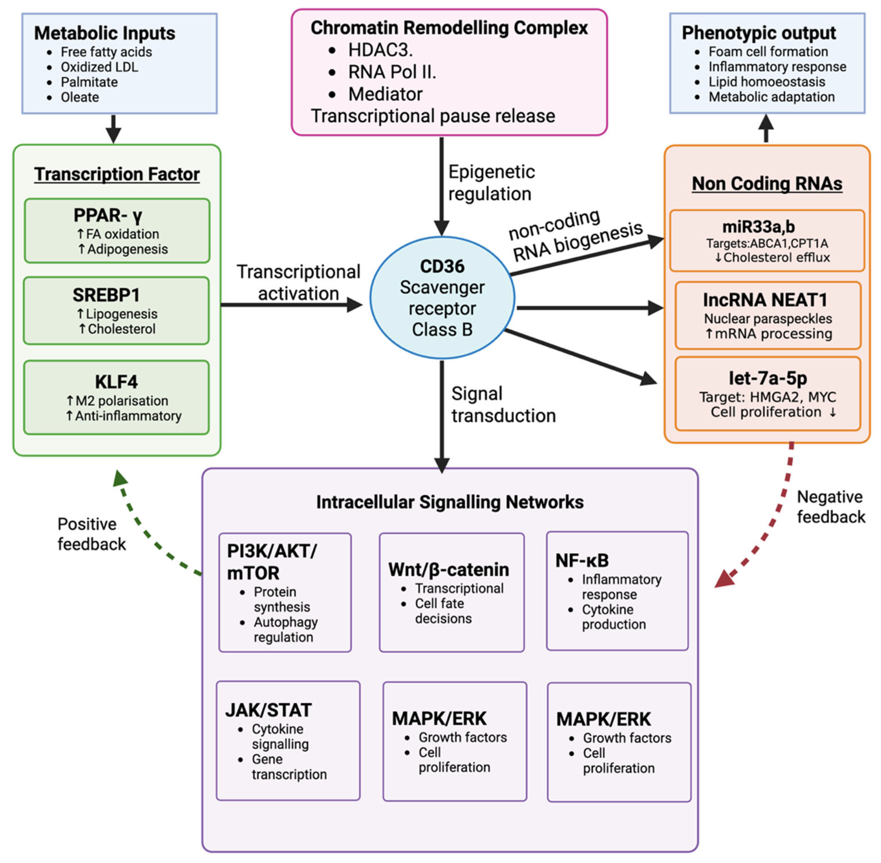
In addition to our systems-level analysis, experimental evidence from animal models substantiates the mechanistic role of CD36 in gastric cancer. Notably, Jiang et al. (2019) demonstrated that the fatty acid-induced O-GlcNAcylation of CD36 enhances its expression and promotes peritoneal and hepatic metastases in nude mice xenografted with human gastric cancer cells (MKN45 and AGS) [23]. Knockdown of CD36 or its antibody-mediated blockade significantly suppressed tumor dissemination, accompanied by reduced lipid uptake and the downregulation of key oncogenic pathways, including PPAR-γ and PI3K/AKT signaling. These in vivo findings directly support our mechanistic model, which posits that CD36 overexpression exacerbated by obesity and hypercholesterolemia drives metabolic reprogramming via PPAR-γ activation, fosters chronic inflammation through TLR4/NF-κB signaling, and promotes EMT and metastatic potential via the PI3K/AKT/mTOR axis [24]. While some downstream interactions remain computationally inferred, the convergence of transcriptomic insights with animal model validation highlights CD36 as a central node linking metabolic dysfunction to gastric tumor progression and underscores its translational potential as a therapeutic target in metabolically comorbid patients. A systems-level schematic of CD36-mediated transcriptional and post-transcriptional regulation is presented in Figure 3.

3.3. CD36 Expression Correlates with Clinical Stage, Race, and Prognosis in Gastric Cancer

We analyzed CD36 expression across various demographic and clinical variables using data from the TCGA gastric cancer cohort. CD36 expression significantly varied across gastric cancer subtypes (ANOVA, F = 8.975, p = 6.242 × 10⁻⁷), with the highest levels observed in genomically stable (GS) and the lowest in the MSI subtype (Figure 4A).
Assessment of CD36 expression across AJCC cancer stages I–IV revealed a significant stage-dependent increase (ANOVA, F = 3.477, p = 0.016), with stage IV tumors showing the highest expression (Figure 4B), suggesting that CD36 upregulation may reflect an aggressive tumor biology and potential for metastasis.
Racial differences in CD36 expression were also observed (ANOVA: F = 4.998, p = 0.007), with White patients showing the highest median CD36 expression, followed by Asian, and then Black or African American patients (Figure 4C). This racial disparity suggests that population-level genomic or epigenetic differences may influence CD36 expression, potentially contributing to outcome disparities.
To assess prognostic significance, CD36 expression was compared between deceased and surviving patients. The deceased cohort exhibited significantly higher CD36 expression (Welch’s t-test, t = 2.743, p = 0.006), suggesting a direct association between elevated CD36 levels and increased mortality risk (Figure 4D, left). Furthermore, Kaplan–Meier survival analysis demonstrated that patients with high CD36 expression had significantly lower overall survival compared to those with low expression (median: 20.9 vs. 55.4 months; HR = 1.69, 95% CI: 1.23–2.31; log-rank p = 0.001) (Figure 4D, right). These findings underscore the prognostic relevance of CD36 and support its utility as a stratification biomarker for outcome prediction.
To determine whether sex influences CD36 expression or its prognostic value, we conducted stratified analyses by sex. CD36 expression was comparable between male and female patients (t = 0.035, p = 0.972), with no significant difference in expression distributions (Figure 4E, left), indicating no sex-dependent transcriptional regulation. Additionally, Kaplan–Meier analysis revealed no statistically significant difference in survival between males and females (HR = 0.81, 95% CI: 0.58–1.14; log-rank p = 0.225), despite females with high CD36 expression showing a numerically longer median survival than males (34.3 vs. 26.3 months) (Figure 4E, right).

3.4. Transcriptomic Profiling Highlights Mortality-Associated Differential Expression Patterns

To investigate gene expression signatures associated with survival, we performed differential expression analysis between deceased and surviving patients. A volcano plot visualization revealed four genes ADIPOQ, C7, CHRDL1, and ADH1B to be significantly upregulated in deceased individuals (FDR < 0.05, |log2FC| ≥ 1) (Figure 5). While CD36 expression showed an upward trend in deceased patients, it did not meet the stringent threshold, ranking 3870th in overall variance, suggesting moderate yet biologically meaningful expression variation.

4. Discussion

The findings of our study emphasize the pivotal role of CD36 as a molecular intersection linking obesity, hypercholesterolemia, and gastric cancer pathogenesis. Through integrative transcriptomic analysis, we identified CD36 as a gene consistently associated with gastric cancer, obesity, and hypercholesterolemia across diverse datasets. The identification of CD36 underscores its potential as an integrator of metabolic dysregulation and cancer development, a concept supported by the existing literature [23,25]. Our results revealed significant functional enrichments in pathways related to insulin regulation, hormonal signaling, cholesterol metabolism, and inflammation, all of which are implicated in cancer progression.
Notably, this study is among the first to characterize CD36 expression in relation to both clinical outcomes and racial variability in gastric cancer. These results highlight CD36’s potential utility as a multifactorial prognostic biomarker and therapeutic target across diverse patient populations.

4.1. Mechanistic Insights from Genetic Intersections

The identification of 36 genes shared between obesity and gastric cancer underscores the pivotal role of metabolic and hormonal dysregulation in gastric carcinogenesis. These genes are enriched in insulin signaling, glucose metabolism, and adipokine regulatory pathways, indicating that the metabolic alterations associated with obesity may directly influence tumor development. The presence of neuroendocrine regulators, including NPY and POMC, further suggests that neuro-hormonal pathways may link energy balance to oncogenic signaling in gastric tissues.
Similarly, the 31 genes common to hypercholesterolemia and gastric cancer are primarily involved in lipid metabolism, immune activation, and inflammatory signaling. These include genes regulating lipoprotein transport and cholesterol homeostasis (e.g., APOE, PCSK9, ABCG8), as well as immune mediators (e.g., VCAM1, IL18). The convergence of these pathways suggests that cholesterol dysregulation and chronic inflammation synergize to support tumor progression.
Of particular significance, CD36 emerged as the only gene common to all three conditions of obesity, hypercholesterolemia, and gastric cancer, highlighting its central role in metabolic–oncogenic cross-talk. CD36 functions as a scavenger receptor and fatty acid translocase and is regulated by transcription factors such as PPARγ, LXR, and SREBP1c, all of which respond to nutrient overload and oxidative stress [26]. These regulatory interactions reinforce the role of CD36 as a master node in metabolic reprogramming during gastric carcinogenesis.

4.2. CD36-Centered Mechanistic Pathway

Transcriptomic data and pathway modeling suggest that CD36 connects metabolic stress to gastric cancer progression. Early metabolic disturbances from obesity and hypercholesterolemia elevate free fatty acids and oxidized LDL, upregulating CD36 in gastric epithelial cells and macrophages. CD36-driven lipid uptake induces lipid droplet accumulation, membrane remodeling, and PPAR-γ activation, promoting a pro-adipogenic state, oxidative stress, and DNA damage. This reprogramming enhances tumor cell proliferation and survival [27].
In parallel, CD36 serves as a pattern recognition receptor that binds oxidized LDL, activating TLR4/NF-κB signaling. This cascade leads to increased production of IL-6 and TNF-α, M1 macrophage polarization, and the establishment of a pro-inflammatory tumor-promoting microenvironment [28]. In the later stages, CD36 enhances gastric cancer cell invasiveness and metastatic potential via PI3K/AKT/mTOR activation and interactions with extracellular matrix proteins such as thrombospondin-1. It also facilitates EMT through TGF-β pathway activation. CD36-expressing cells acquire stem-like features, with heightened metastatic capacity and survival in lipid-rich microenvironments. This model explains the observed correlation between CD36 expression and poor clinical outcomes in gastric cancer. Our results support the notion that CD36 is a key driver of systemic inflammation in gastric cancer, integrating metabolic dysfunction with innate immune activation to promote chronic low-grade inflammation, a hallmark of cancer progression.
CD36’s interaction with immune cells, especially tumor-associated macrophages and endothelial CD36, promotes immune evasion and metastatic niche formation. In metastasis, CD36-high cells show enhanced survival via fatty acid utilization, extravasation through endothelial CD36, and adaptation to lipid-rich environments [29]. These mechanisms reflect clinical observations linking CD36 expression with obesity, hypercholesterolemia, and adverse gastric cancer prognosis, and highlight CD36-driven lipid uptake and downstream signaling as potential therapeutic targets.
CD36’s function as a fatty acid translocase positions it at the intersection of lipid metabolism, inflammation, and EMT. It facilitates long-chain fatty acid uptake, directly influencing cellular energy homeostasis and metabolic health, particularly in obesity and metabolic disorders [30]. In cancer, its expression is shaped by interactions within the tumor microenvironment and supports the metabolic phenotype of malignant cells [31].
Its role in inflammation is underscored by its ability to recognize oxidized lipids and apoptotic cells, thereby activating macrophages and stimulating pro-inflammatory cytokine release [32]. This inflammatory signaling, often driven by fatty acid uptake, contributes to EMT induction in various cancer types [33].
EMT itself is a critical mechanism for metastasis, involving the transformation of epithelial cells into more mobile, invasive mesenchymal cells. CD36 expression can be induced by TGF-β signaling, which plays a central role in EMT [34]. Furthermore, fatty acid uptake through CD36 enhances EMT traits in cancers such as hepatocellular carcinoma and gastric cancer [33,35]. In chronic myeloid leukemia, CD36 is linked to cancer stemness and therapy resistance, further illustrating its influence on cellular plasticity and tumor progression [36].
Together, these findings position CD36 as a key regulator of cancer cell metabolism, immune interactions, and EMT integrating metabolic and inflammatory cues in ways that facilitate tumor growth, metastasis, and resistance, making it an attractive target for therapeutic intervention (Figure 6).

4.3. Additional Molecular Connections Beyond CD36

4.3.1. Hypercholesterolemia–Gastric Cancer Pathway

In the hypercholesterolemia-associated gene set, multiple regulators beyond CD36 contribute significantly to tumor biology. For instance, APOE, PCSK9, and ABCG8 regulate lipoprotein metabolism and cholesterol transport. Dysregulation of these genes can lead to intracellular cholesterol accumulation, influencing membrane fluidity, lipid raft composition, and signal transduction relevant to tumor growth. Inflammatory mediators such as IL18, CRP, VCAM1, and ICAM1, and complement components like CFI and C2 orchestrate immune cell recruitment and chronic low-grade inflammation, a shared hallmark of both metabolic syndrome and cancer [37,38]. SYK and BID, involved in immune signaling and apoptosis, also modulate cancer progression through cross-talk with NF-κB and mitochondrial stress pathways [39].
Genes like CAV1 and CAV3 regulate caveolae-dependent signaling and are linked to PI3K/AKT and MAPK pathway activation [40], while FBLN5, DAAM2, and RSPO1 influence extracellular matrix remodeling and Wnt signaling, both of which are critical for tumor invasion and metastasis [41,42]. ATF4, a stress-responsive transcription factor, integrates endoplasmic reticulum stress with lipid dysregulation and cancer cell survival under nutrient-deprived conditions [43].

4.3.2. Obesity–Gastric Cancer Pathway

Among obesity-linked genes, LEP, ADIPOQ, and LEPR form the core of adipokine signaling dysregulation. An elevated LEP/ADIPOQ ratio enhances PI3K/AKT and JAK2/STAT3 activity, while suppressing anti-inflammatory AMPK signaling, favoring a pro-tumorigenic environment. Adipokine signaling is central: obesity-induced LEP upregulation activates JAK2/STAT3, PI3K/AKT, and MAPK pathways, promoting proliferation and invasion [44], while reduced ADIPOQ weakens AMPK activation and anti-inflammatory effects. A high LEP/ADIPOQ ratio correlates with advanced tumor stage and poor prognosis [45]. Inflammatory cytokines such as TNF, IL6, and IL1B produced by adipose tissue and tumor-associated immune cells activate NF-κB and STAT3, supporting cell proliferation and immune evasion [46]. CCL2 recruits tumor-associated macrophages, reinforcing inflammation. Obese patients’ tumors show higher macrophage infiltration and PD-L1 expression, indicating immune evasion [47].
Metabolic reprogramming via GLUT1, LDHA, PDK1, FASN, and SCD drives the Warburg effect and lipogenesis [48]. Obesity-induced pseudohypoxia stabilizes HIF1A, enhancing glycolysis and VEGFA-mediated angiogenesis. The AMPK–mTOR axis is disrupted by reduced AMPK activity, allowing unchecked mTOR-driven growth [49]. Insulin resistance impairs IRS1/2, leading to hyperinsulinemia and the activation of IGF1R/INSR, stimulating PI3K/AKT signaling to promote survival and inhibit autophagy [50]. FOXO1 phosphorylation through this pathway inactivates its tumor-suppressive functions, further contributing to carcinogenesis. Epigenetic regulators like EZH2, DNMT1, and SIRT1 are also frequently dysregulated in obesity and cancer, linking nutrient excess to oncogenic chromatin remodeling. For example, EZH2 promotes the silencing of tumor suppressor genes via H3K27 methylation, while SIRT1 modulates metabolic adaptation and inflammation under stress [2,51].
The multifaceted interactions among these 36 genes demonstrate how obesity creates a perfect storm of conditions that promote gastric carcinogenesis through the interconnected mechanisms of inflammation, metabolic dysfunction, hormonal imbalance, and epigenetic dysregulation. This integrated pathway analysis provides a comprehensive framework for understanding the obesity–gastric cancer relationship and identifying high-value therapeutic targets.

4.4. Clinical Implications

4.4.1. Therapeutic Targeting of CD36 and Its Axis

Our findings have several important clinical implications for gastric cancer prevention, risk stratification, and treatment. For risk assessment and screening, combining obesity, lipid profile abnormalities, and CD36 expression into stratification models could improve the early detection of metabolically driven gastric cancer subtypes. CD36 expression might also serve as a non-invasive biomarker, as it can be detected in circulating monocytes or plasma-derived exosomes, potentially enabling real-time monitoring.
Therapeutically, CD36 represents a promising target. A variety of CD36-targeted interventions, including sulfosuccinimidyl oleate (SSO), hexarelin, and anti-CD36 monoclonal antibodies, as well as downstream pathway inhibitors such as AMPK activators and mTOR inhibitors, have shown potential across preclinical and clinical settings (Table 1). Additionally, TLR4 inhibitors, PPAR-γ modulators, and FASN inhibitors may block key CD36 downstream pathways. This opens avenues for combination regimens integrating CD36-targeted agents with chemotherapy, immunotherapy, or anti-angiogenic agents, particularly in patients with metabolic syndrome.
Notably, CD36’s downstream activation of NF-κB links it directly to systemic inflammation, a hallmark of gastric cancer progression. Chronic low-grade inflammation driven by dysregulated lipid signaling may sustain an immunosuppressive tumor microenvironment. Therefore, anti-inflammatory agents or cytokine inhibitors may also be viable strategies in CD36-high patients [92].
In addition, recent evidence highlights the regulatory role of angiogenesis-related microRNAs (miRNAs) in gastric cancer progression. These miRNAs modulate vascular remodeling, immune evasion, and metastatic potential, complementing the lipid-inflammatory axis described in our study. For example, a recent study [93] identified several miRNAs influencing VEGF and other angiogenic factors, providing further insight into the complexity of the tumor microenvironment and its regulatory layers.

4.4.2. Immunometabolic Targeting and Biomarker Applications

The immunometabolic role of CD36 positions it as a promising biomarker for precision oncology. Its association with immune checkpoint expression and macrophage polarization suggests that CD36 status may predict responsiveness to immunotherapy [94]. In patients with high CD36 expression, combination strategies targeting both immune checkpoints and metabolic pathways may be necessary to achieve optimal therapeutic efficacy, an approach already under investigation in melanoma and lung cancer [95,96].
From a biomarker standpoint, CD36 expression on circulating immune cells provides a potential non-invasive proxy for tumor metabolic activity [69]. Notably, elevated CD36 levels on peripheral blood mononuclear cells have been linked to metabolic syndrome and cardiovascular disease [57], highlighting its utility as a systemic metabolic biomarker with translational relevance for cancer monitoring.

4.4.3. PPAR-γ Modulation and Metabolic Reprogramming

Targeting PPAR-γ offers a complementary strategy to counteract CD36-driven lipid accumulation and inflammation. As a key regulator of lipid metabolism and inflammation, PPAR-γ modulation may reverse the metabolic reprogramming associated with CD36 overexpression [97]. Thiazolidinediones, PPAR-γ agonists used clinically in type 2 diabetes, have demonstrated anti-tumor effects in gastric cancer cell lines through the induction of apoptosis and restoration of metabolic homeostasis [98]. The dual role of PPAR-γ as a metabolic regulator and tumor suppressor underscores its therapeutic potential in metabolically driven malignancies, including gastric cancer [99].

4.5. Limitations and Future Directions

This study is limited by the absence of detailed clinical variables such as body mass index (BMI), lipid profiles (e.g., total cholesterol, LDL, HDL), and Helicobacter pylori infection status in the TCGA cohort, which restricts precise metabolic stratification and weakens the direct correlation between CD36 expression and metabolic phenotypes. While our multi-omics and gene set overlap approach provides strong inferential links between CD36 and metabolic dysfunction, future validation in prospective, metabolically annotated cohorts is essential to establish causality. Studies incorporating longitudinal sampling, comprehensive metabolic panels, and H. pylori virulence profiling would further elucidate how these variables modulate CD36 expression and gastric tumor biology. The cross-sectional nature of TCGA data also precludes determining whether CD36 upregulation precedes tumor initiation or arises as a metabolic adaptation during progression. Emerging single-cell and spatial transcriptomics techniques will be valuable for resolving CD36 dynamics across tumor compartments and disease stages.
Several confounding variables were addressed through multivariate analyses. CD36 expression showed a positive correlation with tumor stage but retained independent prognostic value in survival models. It was also elevated in intestinal-type cancers and remained a poor prognostic marker across Lauren subtypes. Subgroup analyses suggest a potential link between CD36 and chemotherapy resistance, although larger studies are needed for confirmation. Lastly, preliminary findings indicate increased CD36 expression in the chromosomal instability (CIN) subtype of gastric cancer, highlighting the need for validation in subtype-specific, well-annotated clinical cohorts.

5. Conclusions

This study established the role of CD36 as a critical mediator linking obesity, hypercholesterolemia, and gastric cancer, suggesting a significant metabolic–oncogenic interface. Our integrative analysis reveals that CD36 is integral to a complex oncogenic network characterized by altered lipid metabolism, chronic inflammation, and immune modulation factors pivotal to cancer progression in metabolically challenged individuals.
Crucially, CD36 emerges as an active promoter of gastric tumorigenesis, evidenced by its interaction with key signaling pathways such as PPAR-γ, TLR4/NF-κB, and PI3K/AKT/mTOR. Elevated CD36 expression correlates with advanced disease stages and reduced patient survival, highlighting its independent prognostic potential and reinforcing its consideration as a therapeutic target. Translationally, these findings support the development of CD36-focused interventions, including fatty acid transport inhibitors and inflammatory signaling modulators, particularly for patients with associated metabolic disorders. Incorporating CD36 metrics with clinical and metabolic data could enhance risk stratification and tailor treatment strategies.
This research emphasizes the necessity of incorporating metabolic factors into gastric cancer diagnosis and prognosis, extending beyond conventional histopathological and genetic paradigms. By establishing CD36 as a pivotal element in lipid-induced carcinogenesis, we propose innovative avenues for diagnostics and metabolic-targeted therapies. Future investigations should aim to authenticate CD36’s prognostic and therapeutic significance in clinical cohorts, further explore its functions in preclinical models, and assess the efficacy of CD36 inhibitors in conjunction with immunotherapeutic approaches. In summary, CD36 emerges as a central nexus between metabolic dysfunction and gastric cancer, offering promising translational potential and redefining the landscape of metabolically informed oncology.

Supplementary Materials

The following supporting information can be downloaded at: https://www.mdpi.com/article/10.3390/ijtm5030026/s1, Figure S1: Graphical representation of analyzed data; Table S1: Summary of clinical and molecular characteristics of the gastric cancer cohort (n = 413). Subtype data from TCGA Pan-Cancer Atlas was available for 410 patients; File S1: TCGA dataset; File S2: OMIM analyses.

Author Contributions

P.D.: Writing—review and editing, Writing—original draft, Methodology, Prepared figures, Conceptualization. D.S.: Writing—review and editing, Writing—original draft, Methodology, Prepared figures, Conceptualization. A.G.: Methodology, Formal analysis, Prepared figures. A.R.B.: Writing—review and editing, Writing—original draft, Conceptualization. A.C.: Writing—review and editing, Writing—original draft, Supervision, Resources, Methodology, Formal analysis, Conceptualization. All authors have read and agreed to the published version of the manuscript.

Funding

This research received no external funding.

Institutional Review Board Statement

Not applicable.

Informed Consent Statement

Not applicable.

Data Availability Statement

All code and analyses for this manuscript are available at: https://github.com/atanugiri/gastric-cancer (accessed on 27 April 2025). The original contributions presented in this study are included in the article/Supplementary Materials; further inquiries can be directed to the corresponding author.

Conflicts of Interest

The authors declare no competing interests.

References

  1. Morgan, E.; Arnold, M.; Camargo, M.C.; Gini, A.; Kunzmann, A.T.; Matsuda, T.; Meheus, F.; Verhoeven, R.H.A.; Vignat, J.; Laversanne, M.; et al. The current and future incidence and mortality of gastric cancer in 185 countries, 2020–40: A population-based modelling study. EClinicalMedicine 2022, 47, 101404. [Google Scholar] [CrossRef] [PubMed]
  2. Gao, W.; Liu, J.L.; Lu, X.; Yang, Q. Epigenetic regulation of energy metabolism in obesity. J. Mol. Cell Biol. 2021, 13, 480–499. [Google Scholar] [CrossRef] [PubMed]
  3. Conti, C.B.; Agnesi, S.; Scaravaglio, M.; Masseria, P.; Dinelli, M.E.; Oldani, M.; Uggeri, F. Early Gastric Cancer: Update on Prevention, Diagnosis and Treatment. Int. J. Environ. Res. Public Health 2023, 20, 2149. [Google Scholar] [CrossRef] [PubMed]
  4. Sisic, L.; Crnovrsanin, N.; Nienhueser, H.; Jung, J.O.; Schiefer, S.; Haag, G.M.; Bruckner, T.; Schneider, M.; Muller-Stich, B.P.; Buchler, M.W.; et al. Perioperative chemotherapy with 5-FU, leucovorin, oxaliplatin, and docetaxel (FLOT) for esophagogastric adenocarcinoma: Ten years real-life experience from a surgical perspective. Langenbecks Arch. Surg. 2023, 408, 81. [Google Scholar] [CrossRef]
  5. Gajria, D.; Chandarlapaty, S. HER2-amplified breast cancer: Mechanisms of trastuzumab resistance and novel targeted therapies. Expert. Rev. Anticancer. Ther. 2011, 11, 263–275. [Google Scholar] [CrossRef]
  6. Riedl, J.M.; Moik, F.; Esterl, T.; Kostmann, S.M.; Gerger, A.; Jost, P.J. Molecular diagnostics tailoring personalized cancer therapy-an oncologist’s view. Virchows Arch. 2024, 484, 169–179. [Google Scholar] [CrossRef]
  7. Wadhwa, R.; Taketa, T.; Sudo, K.; Blum-Murphy, M.; Ajani, J.A. Ramucirumab: A novel antiangiogenic agent. Future Oncol. 2013, 9, 789–795. [Google Scholar] [CrossRef]
  8. Di Carlo, S.; Franceschilli, M.; Rossi, P.; Cavallaro, G.; Cardi, M.; Vinci, D.; Sibio, S. Perforated gastric cancer: A critical appraisal. Discov. Oncol. 2021, 12, 15. [Google Scholar] [CrossRef]
  9. Bozkurt, E.; Omeroglu, S.; Capkinoglu, E.; Guven, O.; Mihmanli, M. Management of Gastric Tumor Perforation with Conservative Treatment during Neoadjuvant Chemotherapy. JCPSP-J. Coll. Physicians Surg. Pak. 2022, 32, 117–118. [Google Scholar]
  10. Anthony, M.; Goyal, U. Definitive chemoradiation with dose escalation for locally advanced gastric cancer: Case studies. Cureus 2020, 12, e11040. [Google Scholar] [CrossRef]
  11. Song, Z.; Wu, Y.; Yang, J.; Yang, D.; Fang, X. Progress in the treatment of advanced gastric cancer. Tumor Biol. 2017, 39, 1010428317714626. [Google Scholar] [CrossRef] [PubMed]
  12. Mizukami, T.; Piao, Y. Role of nutritional care and general guidance for patients with advanced or metastatic gastric cancer. Future Oncol. 2021, 17, 3101–3109. [Google Scholar] [CrossRef] [PubMed]
  13. Izuishi, K.; Mori, H. Recent Strategies for Treating Stage IV Gastric Cancer: Roles of Palliative Gastrectomy, Chemotherapy, and Radiotherapy. J. Gastrointest. Liver Dis. 2016, 25, 87–94. [Google Scholar] [CrossRef]
  14. Katano, A.; Yamashita, H. Long-Term Survival After Repeated Salvage Chemoradiation Therapy for Metastatic Lymph Node Recurrence in Advanced Gastric Cancer: A Case Report. Cureus 2023, 15, e36649. [Google Scholar] [CrossRef] [PubMed]
  15. Nguyen, T.Q.; Nguyen, H.N.M.; Ngo, D.-H.; Phan, P.-H.; Vo, T.S. The Suppressive Activity of Water Mimosa Extract on Human Gastric Cancer Cells. Appl. Sci. 2022, 12, 6817. [Google Scholar] [CrossRef]
  16. Yang, T.-W.; Wang, C.-C.; Hung, W.-C.; Liu, Y.-H.; Sung, W.-W.; Tsai, M.-C. Improvement in the mortality-to-incidence ratios for gastric cancer in developed countries with high health expenditures. Front. Public Health 2021, 9, 713895. [Google Scholar] [CrossRef]
  17. Mamun, T.I.; Younus, S.; Rahman, M.H. Gastric cancer-Epidemiology, modifiable and non-modifiable risk factors, challenges and opportunities: An updated review. Cancer Treat. Res. Commun. 2024, 41, 100845. [Google Scholar] [CrossRef]
  18. Yang, L.; Ying, X.; Liu, S.; Lyu, G.; Xu, Z.; Zhang, X.; Li, H.; Li, Q.; Wang, N.; Ji, J. Gastric cancer: Epidemiology, risk factors and prevention strategies. Chin. J. Cancer Res. 2020, 32, 695–704. [Google Scholar] [CrossRef]
  19. Collaborators, G.B.D.R.F. Global burden and strength of evidence for 88 risk factors in 204 countries and 811 subnational locations, 1990–2021: A systematic analysis for the Global Burden of Disease Study 2021. Lancet 2024, 403, 2162–2203. [Google Scholar]
  20. Pati, S.; Irfan, W.; Jameel, A.; Ahmed, S.; Shahid, R.K. Obesity and Cancer: A Current Overview of Epidemiology, Pathogenesis, Outcomes, and Management. Cancers 2023, 15, 485. [Google Scholar] [CrossRef]
  21. Fu, Y.; Zou, T.; Shen, X.; Nelson, P.J.; Li, J.; Wu, C.; Yang, J.; Zheng, Y.; Bruns, C.; Zhao, Y.; et al. Lipid metabolism in cancer progression and therapeutic strategies. MedComm 2021, 2, 27–59. [Google Scholar] [CrossRef] [PubMed]
  22. Hajian, M. Kaggle. Gene Expression in Gastric Cancer. 2022. Available online: https://www.kaggle.com/datasets/mahdiehhajian/gene-expression-in-gastric-cancer (accessed on 27 April 2025).
  23. Jiang, M.; Wu, N.; Xu, B.; Chu, Y.; Li, X.; Su, S.; Chen, D.; Li, W.; Shi, Y.; Gao, X.; et al. Fatty acid-induced CD36 expression via O-GlcNAcylation drives gastric cancer metastasis. Theranostics 2019, 9, 5359–5373. [Google Scholar] [CrossRef]
  24. Luo, X.; Zheng, E.; Wei, L.; Zeng, H.; Qin, H.; Zhang, X.; Liao, M.; Chen, L.; Zhao, L.; Ruan, X.Z.; et al. The fatty acid receptor CD36 promotes HCC progression through activating Src/PI3K/AKT axis-dependent aerobic glycolysis. Cell Death Dis. 2021, 12, 328. [Google Scholar] [CrossRef]
  25. Zhou, X.; Su, M.; Lu, J.; Li, D.; Niu, X.; Wang, Y. CD36: The Bridge between Lipids and Tumors. Molecules 2024, 29, 531. [Google Scholar] [CrossRef] [PubMed]
  26. Feng, Y.; Sun, W.; Sun, F.; Yin, G.; Liang, P.; Chen, S.; Liu, X.; Jiang, T.; Zhang, F. Biological Mechanisms and Related Natural Inhibitors of CD36 in Nonalcoholic Fatty Liver. Drug Des. Devel Ther. 2022, 16, 3829–3845. [Google Scholar] [CrossRef]
  27. Li, S.; Yuan, H.; Li, L.; Li, Q.; Lin, P.; Li, K. Oxidative Stress and Reprogramming of Lipid Metabolism in Cancers. Antioxidants 2025, 14, 201. [Google Scholar] [CrossRef] [PubMed]
  28. Chen, Y.; Zhang, J.; Cui, W.; Silverstein, R.L. CD36, a signaling receptor and fatty acid transporter that regulates immune cell metabolism and fate. J. Exp. Med. 2022, 219, e20211314. [Google Scholar] [CrossRef]
  29. Guerrero-Rodriguez, S.L.; Mata-Cruz, C.; Perez-Tapia, S.M.; Velasco-Velazquez, M.A. Role of CD36 in cancer progression, stemness, and targeting. Front. Cell Dev. Biol. 2022, 10, 1079076. [Google Scholar] [CrossRef]
  30. Silverstein, R.L.; Febbraio, M. CD36, a scavenger receptor involved in immunity, metabolism, angiogenesis, and behavior. Sci. Signal. 2009, 2, re3. [Google Scholar] [CrossRef]
  31. Liao, X.; Yan, S.; Li, J.; Jiang, C.; Huang, S.; Liu, S.; Zou, X.; Zhang, G.; Zou, J.; Liu, Q. CD36 and its role in regulating the tumor microenvironment. Curr. Oncol. 2022, 29, 8133–8145. [Google Scholar] [CrossRef]
  32. Ferracini, M.; Rios, F.J.; Pecenin, M.; Jancar, S. Clearance of apoptotic cells by macrophages induces regulatory phenotype and involves stimulation of CD36 and platelet-activating factor receptor. Mediat. Inflamm. 2013, 2013, 950273. [Google Scholar] [CrossRef] [PubMed]
  33. Nath, A.; Li, I.; Roberts, L.R.; Chan, C. Elevated free fatty acid uptake via CD36 promotes epithelial-mesenchymal transition in hepatocellular carcinoma. Sci. Rep. 2015, 5, 14752. [Google Scholar] [CrossRef] [PubMed]
  34. Deng, M.; Cai, X.; Long, L.; Xie, L.; Ma, H.; Zhou, Y.; Liu, S.; Zeng, C. CD36 promotes the epithelial–mesenchymal transition and metastasis in cervical cancer by interacting with TGF-β. J. Transl. Med. 2019, 17, 352. [Google Scholar] [CrossRef] [PubMed]
  35. Wang, C.; Yang, Z.; Xu, E.; Shen, X.; Wang, X.; Li, Z.; Yu, H.; Chen, K.; Hu, Q.; Xia, X. Apolipoprotein C-II induces EMT to promote gastric cancer peritoneal metastasis via PI3K/AKT/mTOR pathway. Clin. Transl. Med. 2021, 11, e522. [Google Scholar] [CrossRef]
  36. Zhang, T.; Yang, J.; Vaikari, V.P.; Beckford, J.S.; Wu, S.; Akhtari, M.; Alachkar, H. Apolipoprotein C2-CD36 promotes leukemia growth and presents a targetable axis in acute myeloid leukemia. Blood Cancer Discov. 2020, 1, 198–213. [Google Scholar] [CrossRef]
  37. Brishti, M.A.; Raghavan, S.; Lamar, K.; Singh, U.P.; Collier, D.M.; Leo, M.D. Diabetic endothelial cell glycogen synthase kinase 3β activation induces VCAM1 ectodomain shedding. Int. J. Mol. Sci. 2023, 24, 14105. [Google Scholar] [CrossRef]
  38. Li, S.; He, R.-C.; Wu, S.-G.; Song, Y.; Zhang, K.-L.; Tang, M.-L.; Bei, Y.-R.; Zhang, T.; Lu, J.-B.; Ma, X. LncRNA PSMB8-AS1 instigates vascular inflammation to aggravate atherosclerosis. Circ. Res. 2024, 134, 60–80. [Google Scholar] [CrossRef]
  39. Carneiro, B.A.; El-Deiry, W.S. Targeting apoptosis in cancer therapy. Nat. Rev. Clin. Oncol. 2020, 17, 395–417. [Google Scholar] [CrossRef]
  40. Simon, L.; Campos, A.; Leyton, L.; Quest, A.F.G. Caveolin-1 function at the plasma membrane and in intracellular compartments in cancer. Cancer Metastasis Rev. 2020, 39, 435–453. [Google Scholar] [CrossRef]
  41. Gore, A.V.; Swift, M.R.; Cha, Y.R.; Lo, B.; McKinney, M.C.; Li, W.; Castranova, D.; Davis, A.; Mukouyama, Y.S.; Weinstein, B.M. Rspo1/Wnt signaling promotes angiogenesis via Vegfc/Vegfr3. Development 2011, 138, 4875–4886. [Google Scholar] [CrossRef]
  42. Lee, H.K.; Chaboub, L.S.; Zhu, W.; Zollinger, D.; Rasband, M.N.; Fancy, S.P.; Deneen, B. Daam2-PIP5K is a regulatory pathway for Wnt signaling and therapeutic target for remyelination in the CNS. Neuron 2015, 85, 1227–1243. [Google Scholar] [CrossRef] [PubMed]
  43. Wu, D.; Liang, J. Activating transcription factor 4: A regulator of stress response in human cancers. Front. Cell Dev. Biol. 2024, 12, 1370012. [Google Scholar] [CrossRef]
  44. Garcia-Miranda, A.; Garcia-Hernandez, A.; Castaneda-Saucedo, E.; Navarro-Tito, N.; Maycotte, P. Adipokines as Regulators of Autophagy in Obesity-Linked Cancer. Cells 2022, 11, 3230. [Google Scholar] [CrossRef]
  45. Kim, J.W.; Kim, J.H.; Lee, Y.J. The Role of Adipokines in Tumor Progression and Its Association with Obesity. Biomedicines 2024, 12, 97. [Google Scholar] [CrossRef]
  46. Kern, L.; Mittenbuhler, M.J.; Vesting, A.J.; Ostermann, A.L.; Wunderlich, C.M.; Wunderlich, F.T. Obesity-Induced TNFalpha and IL-6 Signaling: The Missing Link between Obesity and Inflammation-Driven Liver and Colorectal Cancers. Cancers 2018, 11, 24. [Google Scholar] [CrossRef] [PubMed]
  47. Naik, A.; Monjazeb, A.M.; Decock, J. The Obesity Paradox in Cancer, Tumor Immunology, and Immunotherapy: Potential Therapeutic Implications in Triple Negative Breast Cancer. Front. Immunol. 2019, 10, 1940. [Google Scholar] [CrossRef] [PubMed]
  48. Schiliro, C.; Firestein, B.L. Mechanisms of Metabolic Reprogramming in Cancer Cells Supporting Enhanced Growth and Proliferation. Cells 2021, 10, 1056. [Google Scholar] [CrossRef]
  49. Marino, A.; Hausenloy, D.J.; Andreadou, I.; Horman, S.; Bertrand, L.; Beauloye, C. AMP-activated protein kinase: A remarkable contributor to preserve a healthy heart against ROS injury. Free Radic. Biol. Med. 2021, 166, 238–254. [Google Scholar] [CrossRef]
  50. Makhijani, P.; Basso, P.J.; Chan, Y.T.; Chen, N.; Baechle, J.; Khan, S.; Furman, D.; Tsai, S.; Winer, D.A. Regulation of the immune system by the insulin receptor in health and disease. Front. Endocrinol. 2023, 14, 1128622. [Google Scholar] [CrossRef]
  51. Wang, A.J.; Wang, S.; Wang, B.J.; Xiao, M.; Guo, Y.; Tang, Y.; Zhang, J.; Gu, J. Epigenetic Regulation Associated With Sirtuin 1 in Complications of Diabetes Mellitus. Front. Endocrinol. 2020, 11, 598012. [Google Scholar] [CrossRef]
  52. Kuda, O.; Pietka, T.A.; Demianova, Z.; Kudova, E.; Cvacka, J.; Kopecky, J.; Abumrad, N.A. Sulfo-N-succinimidyl oleate (SSO) inhibits fatty acid uptake and signaling for intracellular calcium via binding CD36 lysine 164: SSO also inhibits oxidized low density lipoprotein uptake by macrophages. J. Biol. Chem. 2013, 288, 15547–15555. [Google Scholar] [CrossRef]
  53. Mansor, L.S.; Sousa Fialho, M.D.L.; Yea, G.; Coumans, W.A.; West, J.A.; Kerr, M.; Carr, C.A.; Luiken, J.; Glatz, J.F.C.; Evans, R.D.; et al. Inhibition of sarcolemmal FAT/CD36 by sulfo-N-succinimidyl oleate rapidly corrects metabolism and restores function in the diabetic heart following hypoxia/reoxygenation. Cardiovasc. Res. 2017, 113, 737–748. [Google Scholar] [CrossRef] [PubMed]
  54. Ma, Q.; Wen, L.; Tian, Y.; Ma, L.; Wen, Z.; Kun, Y.; Xu, M.; Liu, X. Sulfosuccinimidyl oleate ameliorates the high-fat diet-induced obesity syndrome by reducing intestinal and hepatic absorption. Front. Pharmacol. 2023, 14, 1193006. [Google Scholar] [CrossRef] [PubMed]
  55. Bodart, V.; Febbraio, M.; Demers, A.; McNicoll, N.; Pohankova, P.; Perreault, A.; Sejlitz, T.; Escher, E.; Silverstein, R.L.; Lamontagne, D.; et al. CD36 mediates the cardiovascular action of growth hormone-releasing peptides in the heart. Circ. Res. 2002, 90, 844–849. [Google Scholar] [CrossRef] [PubMed]
  56. Mao, Y.; Tokudome, T.; Kishimoto, I. The cardiovascular action of hexarelin. J. Geriatr. Cardiol. 2014, 11, 253–258. [Google Scholar]
  57. Febbraio, M.; Silverstein, R.L. CD36: Implications in cardiovascular disease. Int. J. Biochem. Cell Biol. 2007, 39, 2012–2030. [Google Scholar] [CrossRef]
  58. Park, Y.M.; Febbraio, M.; Silverstein, R.L. CD36 modulates migration of mouse and human macrophages in response to oxidized LDL and may contribute to macrophage trapping in the arterial intima. J. Clin. Investig. 2009, 119, 136–145. [Google Scholar] [CrossRef]
  59. Rena, G.; Hardie, D.G.; Pearson, E.R. The mechanisms of action of metformin. Diabetologia 2017, 60, 1577–1585. [Google Scholar] [CrossRef]
  60. Foretz, M.; Guigas, B.; Bertrand, L.; Pollak, M.; Viollet, B. Metformin: From mechanisms of action to therapies. Cell Metab. 2014, 20, 953–966. [Google Scholar] [CrossRef]
  61. Saxton, R.A.; Sabatini, D.M. mTOR Signaling in Growth, Metabolism, and Disease. Cell 2017, 168, 960–976. [Google Scholar] [CrossRef]
  62. Szwed, A.; Kim, E.; Jacinto, E. Regulation and metabolic functions of mTORC1 and mTORC2. Physiol. Rev. 2021, 101, 1371–1426. [Google Scholar] [CrossRef] [PubMed]
  63. Grygiel-Gorniak, B. Peroxisome proliferator-activated receptors and their ligands: Nutritional and clinical implications--a review. Nutr. J. 2014, 13, 17. [Google Scholar] [CrossRef]
  64. Monsalve, F.A.; Pyarasani, R.D.; Delgado-Lopez, F.; Moore-Carrasco, R. Peroxisome proliferator-activated receptor targets for the treatment of metabolic diseases. Mediat. Inflamm. 2013, 2013, 549627. [Google Scholar] [CrossRef] [PubMed]
  65. Parsons, S.J.; Parsons, J.T. Src family kinases, key regulators of signal transduction. Oncogene 2004, 23, 7906–7909. [Google Scholar] [CrossRef] [PubMed]
  66. Wheeler, D.L.; Iida, M.; Dunn, E.F. The role of Src in solid tumors. Oncologist 2009, 14, 667–678. [Google Scholar] [CrossRef]
  67. Handberg, A.; Norberg, M.; Stenlund, H.; Hallmans, G.; Attermann, J.; Eriksson, J.W. Soluble CD36 (sCD36) clusters with markers of insulin resistance, and high sCD36 is associated with increased type 2 diabetes risk. J. Clin. Endocrinol. Metab. 2010, 95, 1939–1946. [Google Scholar] [CrossRef]
  68. Handberg, A.; Hojlund, K.; Gastaldelli, A.; Flyvbjerg, A.; Dekker, J.M.; Petrie, J.; Piatti, P.; Beck-Nielsen, H.; Investigators, R. Plasma sCD36 is associated with markers of atherosclerosis, insulin resistance and fatty liver in a nondiabetic healthy population. J. Intern. Med. 2012, 271, 294–304. [Google Scholar] [CrossRef]
  69. Puchalowicz, K.; Rac, M.E. The Multifunctionality of CD36 in Diabetes Mellitus and Its Complications-Update in Pathogenesis, Treatment and Monitoring. Cells 2020, 9, 1877. [Google Scholar] [CrossRef]
  70. Febbraio, M.; Abumrad, N.A.; Hajjar, D.P.; Sharma, K.; Cheng, W.; Pearce, S.F.; Silverstein, R.L. A null mutation in murine CD36 reveals an important role in fatty acid and lipoprotein metabolism. J. Biol. Chem. 1999, 274, 19055–19062. [Google Scholar] [CrossRef]
  71. Koch, M.; Hussein, F.; Woeste, A.; Grundker, C.; Frontzek, K.; Emons, G.; Hawighorst, T. CD36-mediated activation of endothelial cell apoptosis by an N-terminal recombinant fragment of thrombospondin-2 inhibits breast cancer growth and metastasis in vivo. Breast Cancer Res. Treat. 2011, 128, 337–346. [Google Scholar] [CrossRef]
  72. Podrez, E.A.; Byzova, T.V.; Febbraio, M.; Salomon, R.G.; Ma, Y.; Valiyaveettil, M.; Poliakov, E.; Sun, M.; Finton, P.J.; Curtis, B.R.; et al. Platelet CD36 links hyperlipidemia, oxidant stress and a prothrombotic phenotype. Nat. Med. 2007, 13, 1086–1095. [Google Scholar] [CrossRef]
  73. Vorchheimer, D.A.; Becker, R. Platelets in atherothrombosis. Mayo Clin. Proc. 2006, 81, 59–68. [Google Scholar] [CrossRef] [PubMed]
  74. Rahaman, S.O.; Lennon, D.J.; Febbraio, M.; Podrez, E.A.; Hazen, S.L.; Silverstein, R.L. A CD36-dependent signaling cascade is necessary for macrophage foam cell formation. Cell Metab. 2006, 4, 211–221. [Google Scholar] [CrossRef] [PubMed]
  75. Stewart, C.R.; Stuart, L.M.; Wilkinson, K.; van Gils, J.M.; Deng, J.; Halle, A.; Rayner, K.J.; Boyer, L.; Zhong, R.; Frazier, W.A.; et al. CD36 ligands promote sterile inflammation through assembly of a Toll-like receptor 4 and 6 heterodimer. Nat. Immunol. 2010, 11, 155–161. [Google Scholar] [CrossRef]
  76. Xu, S.; Jay, A.; Brunaldi, K.; Huang, N.; Hamilton, J.A. CD36 enhances fatty acid uptake by increasing the rate of intracellular esterification but not transport across the plasma membrane. Biochemistry 2013, 52, 7254–7261. [Google Scholar] [CrossRef]
  77. Ma, X.; Xiao, L.; Liu, L.; Ye, L.; Su, P.; Bi, E.; Wang, Q.; Yang, M.; Qian, J.; Yi, Q. CD36-mediated ferroptosis dampens intratumoral CD8(+) T cell effector function and impairs their antitumor ability. Cell Metab. 2021, 33, 1001–1012.E5. [Google Scholar] [CrossRef] [PubMed]
  78. Hayasaka, T.; Fuda, H.; Hui, S.P.; Chiba, H. Imaging Mass Spectrometry Reveals a Decrease of Cardiolipin in the Kidney of NASH Model Mice. Anal. Sci. 2016, 32, 473–476. [Google Scholar] [CrossRef]
  79. Su, P.; Wang, Q.; Bi, E.; Ma, X.; Liu, L.; Yang, M.; Qian, J.; Yi, Q. Enhanced Lipid Accumulation and Metabolism Are Required for the Differentiation and Activation of Tumor-Associated Macrophages. Cancer Res. 2020, 80, 1438–1450. [Google Scholar] [CrossRef]
  80. Zhang, Y.; Kurupati, R.; Liu, L.; Zhou, X.Y.; Zhang, G.; Hudaihed, A.; Filisio, F.; Giles-Davis, W.; Xu, X.; Karakousis, G.C.; et al. Enhancing CD8(+) T Cell Fatty Acid Catabolism within a Metabolically Challenging Tumor Microenvironment Increases the Efficacy of Melanoma Immunotherapy. Cancer Cell 2017, 32, 377–391 e379. [Google Scholar] [CrossRef]
  81. Wang, H.; Franco, F.; Tsui, Y.C.; Xie, X.; Trefny, M.P.; Zappasodi, R.; Mohmood, S.R.; Fernandez-Garcia, J.; Tsai, C.H.; Schulze, I.; et al. CD36-mediated metabolic adaptation supports regulatory T cell survival and function in tumors. Nat. Immunol. 2020, 21, 298–308. [Google Scholar] [CrossRef]
  82. Pascual, G.; Avgustinova, A.; Mejetta, S.; Martin, M.; Castellanos, A.; Attolini, C.S.; Berenguer, A.; Prats, N.; Toll, A.; Hueto, J.A.; et al. Targeting metastasis-initiating cells through the fatty acid receptor CD36. Nature 2017, 541, 41–45. [Google Scholar] [CrossRef] [PubMed]
  83. Sag, D.; Carling, D.; Stout, R.D.; Suttles, J. Adenosine 5′-monophosphate-activated protein kinase promotes macrophage polarization to an anti-inflammatory functional phenotype. J. Immunol. 2008, 181, 8633–8641. [Google Scholar] [CrossRef]
  84. Bonen, A.; Parolin, M.L.; Steinberg, G.R.; Calles-Escandon, J.; Tandon, N.N.; Glatz, J.F.; Luiken, J.J.; Heigenhauser, G.J.; Dyck, D.J. Triacylglycerol accumulation in human obesity and type 2 diabetes is associated with increased rates of skeletal muscle fatty acid transport and increased sarcolemmal FAT/CD36. FASEB J. 2004, 18, 1144–1146. [Google Scholar] [CrossRef]
  85. Glatz, J.F.; Luiken, J.J.; Bonen, A. Membrane fatty acid transporters as regulators of lipid metabolism: Implications for metabolic disease. Physiol. Rev. 2010, 90, 367–417. [Google Scholar] [CrossRef] [PubMed]
  86. Aguer, C.; Mercier, J.; Man, C.Y.; Metz, L.; Bordenave, S.; Lambert, K.; Jean, E.; Lantier, L.; Bounoua, L.; Brun, J.F.; et al. Intramyocellular lipid accumulation is associated with permanent relocation ex vivo and in vitro of fatty acid translocase (FAT)/CD36 in obese patients. Diabetologia 2010, 53, 1151–1163. [Google Scholar] [CrossRef]
  87. Holloway, G.P.; Bezaire, V.; Heigenhauser, G.J.; Tandon, N.N.; Glatz, J.F.; Luiken, J.J.; Bonen, A.; Spriet, L.L. Mitochondrial long chain fatty acid oxidation, fatty acid translocase/CD36 content and carnitine palmitoyltransferase I activity in human skeletal muscle during aerobic exercise. J. Physiol. 2006, 571, 201–210. [Google Scholar] [CrossRef] [PubMed]
  88. Nickerson, J.G.; Alkhateeb, H.; Benton, C.R.; Lally, J.; Nickerson, J.; Han, X.X.; Wilson, M.H.; Jain, S.S.; Snook, L.A.; Glatz, J.F.C.; et al. Greater transport efficiencies of the membrane fatty acid transporters FAT/CD36 and FATP4 compared with FABPpm and FATP1 and differential effects on fatty acid esterification and oxidation in rat skeletal muscle. J. Biol. Chem. 2009, 284, 16522–16530. [Google Scholar] [CrossRef]
  89. Greco, D.; Kotronen, A.; Westerbacka, J.; Puig, O.; Arkkila, P.; Kiviluoto, T.; Laitinen, S.; Kolak, M.; Fisher, R.M.; Hamsten, A.; et al. Gene expression in human NAFLD. Am. J. Physiol. Gastrointest. Liver Physiol. 2008, 294, G1281–G1287. [Google Scholar] [CrossRef]
  90. Zhou, J.; Febbraio, M.; Wada, T.; Zhai, Y.; Kuruba, R.; He, J.; Lee, J.H.; Khadem, S.; Ren, S.; Li, S.; et al. Hepatic fatty acid transporter Cd36 is a common target of LXR, PXR, and PPARgamma in promoting steatosis. Gastroenterology 2008, 134, 556–567. [Google Scholar] [CrossRef]
  91. Miquilena-Colina, M.E.; Lima-Cabello, E.; Sanchez-Campos, S.; Garcia-Mediavilla, M.V.; Fernandez-Bermejo, M.; Lozano-Rodriguez, T.; Vargas-Castrillon, J.; Buque, X.; Ochoa, B.; Aspichueta, P.; et al. Hepatic fatty acid translocase CD36 upregulation is associated with insulin resistance, hyperinsulinaemia and increased steatosis in non-alcoholic steatohepatitis and chronic hepatitis C. Gut 2011, 60, 1394–1402. [Google Scholar] [CrossRef]
  92. Feng, W.W.; Zuppe, H.T.; Kurokawa, M. The Role of CD36 in Cancer Progression and Its Value as a Therapeutic Target. Cells 2023, 12, 1605. [Google Scholar] [CrossRef] [PubMed]
  93. Giuppi, M.; La Salvia, A.; Evangelista, J.; Ghidini, M. The Role and Expression of Angiogenesis-Related miRNAs in Gastric Cancer. Biology 2021, 10, 146. [Google Scholar] [CrossRef] [PubMed]
  94. Chen, Y.J.; Liao, W.X.; Huang, S.Z.; Yu, Y.F.; Wen, J.Y.; Chen, J.; Lin, D.G.; Wu, X.Y.; Jiang, N.; Li, X. Prognostic and immunological role of CD36: A pan-cancer analysis. J. Cancer 2021, 12, 4762–4773. [Google Scholar] [CrossRef] [PubMed]
  95. Tang, Y.; Chen, Z.; Zuo, Q.; Kang, Y. Regulation of CD8+ T cells by lipid metabolism in cancer progression. Cell Mol. Immunol. 2024, 21, 1215–1230. [Google Scholar] [CrossRef]
  96. Suri, C.; Pande, B.; Suhasini Sahithi, L.; Swarnkar, S.; Khelkar, T.; Verma, H.K. Metabolic crossroads: Unravelling immune cell dynamics in gastrointestinal cancer drug resistance. Cancer Drug Resist. 2025, 8, 7. [Google Scholar] [CrossRef]
  97. Christofides, A.; Konstantinidou, E.; Jani, C.; Boussiotis, V.A. The role of peroxisome proliferator-activated receptors (PPAR) in immune responses. Metabolism 2021, 114, 154338. [Google Scholar] [CrossRef]
  98. Blanquicett, C.; Roman, J.; Hart, C.M. Thiazolidinediones as anti-cancer agents. Cancer Ther. 2008, 6, 25–34. [Google Scholar]
  99. Chi, T.; Wang, M.; Wang, X.; Yang, K.; Xie, F.; Liao, Z.; Wei, P. PPAR-gamma Modulators as Current and Potential Cancer Treatments. Front. Oncol. 2021, 11, 737776. [Google Scholar] [CrossRef]
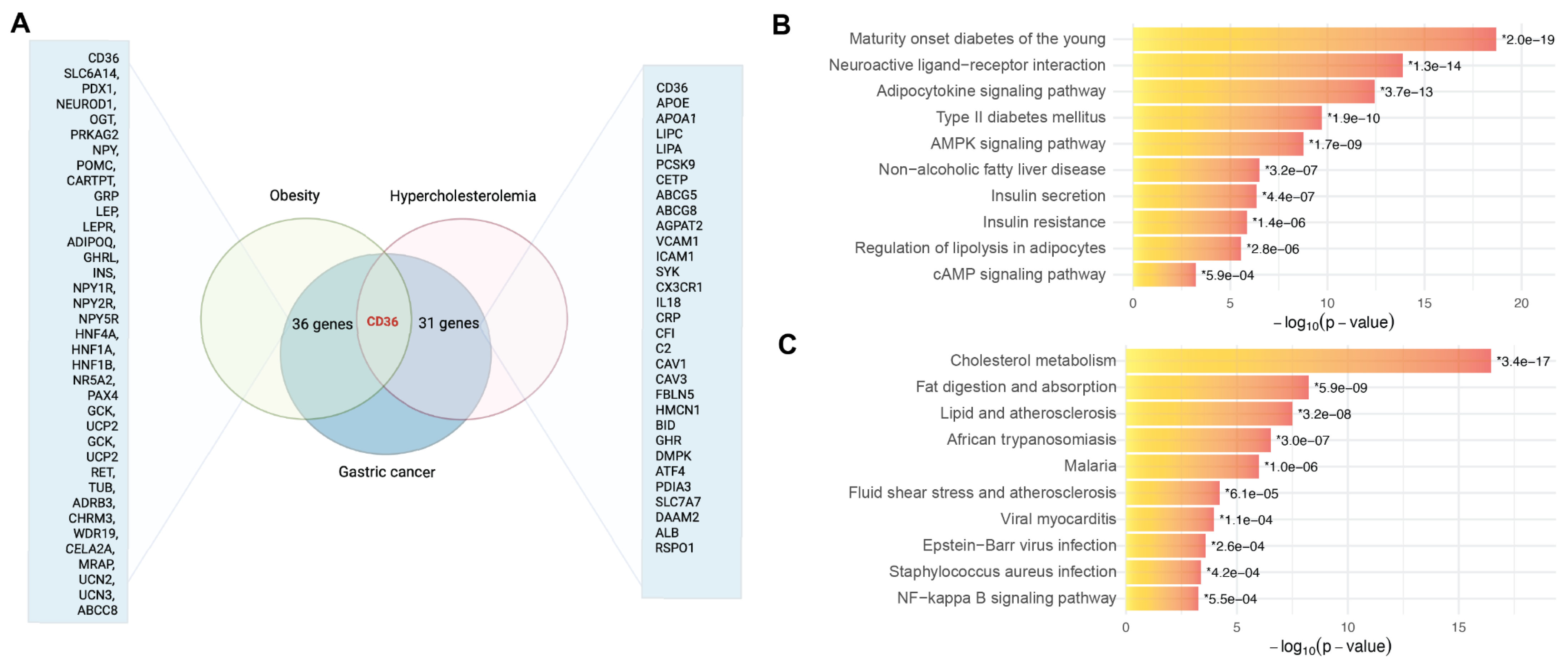
Figure 1. Integrative transcriptomic analysis identifies CD36 as a central molecular node linking gastric cancer, obesity, and hypercholesterolemia. (A) Venn diagram illustrating the overlap between gastric cancer differentially expressed genes (DEGs) and curated gene sets for obesity (36 shared genes) and hypercholesterolemia (31 shared genes), with CD36 being the only gene common to all three conditions. (“Created with BioRender.com”, accessed date 8 June 2025). (B) KEGG pathway enrichment analysis of obesity–gastric cancer overlapping genes shows strong associations with insulin signaling, adipocytokine regulation, and diabetes-related pathways. (“Created with Enrichr Appyter visualization tool”.) (C) KEGG enrichment of hypercholesterolemia–gastric cancer overlapping genes highlights the significant involvement of cholesterol metabolism, lipid transport, and immune-related inflammatory signaling. (“Created with Enrichr Appyter visualization tool”.) The asterisk (*) next to a p-value indicates the term also has a significant adjusted p-value (<0.05).
Ijtm 05 00026 g001
Figure 2. Mechanistic model depicting CD36-driven molecular pathways in gastric cancer progression. In the context of obesity and hypercholesterolemia, the upregulation of CD36 leads to the following: (i) increased fatty acid uptake and altered lipid metabolism via PPAR-γ activation, (ii) induction of pro-inflammatory signaling within the tumor microenvironment via TLR4/NF-κB activation, and (iii) enhanced survival and metastatic potential through PI3K/AKT/mTOR signaling and epithelial–mesenchymal transition (EMT). Collectively, these pathways promote tumor proliferation, invasion, and poor patient prognosis. (“Created with BioRender.com”, accessed date 8 June 2025).
Ijtm 05 00026 g002
Figure 3. CD36-mediated transcriptional and post-transcriptional regulatory network in gastric cancer. The schematic illustrates how CD36 serves as a central integrator of lipid metabolic inputs, transcription factor activation, epigenetic regulation, and intracellular signaling pathways. CD36 modulates downstream outputs including PI3K/AKT/mTOR, MAPK/ERK, Wnt/β-catenin, and NF-κB signaling, contributing to inflammation, proliferation, and metabolic adaptation. It also participates in chromatin remodeling and non-coding RNA biogenesis, impacting foam cell formation, cytokine production, and tumor progression. Positive feedback loops and negative regulatory mechanisms via non-coding RNAs (e.g., miR-33a/b, let-7a-5p) further modulate CD36-driven signaling complexity. (“Created with BioRender.com”, accessed date 8 June 2025).
Ijtm 05 00026 g003
Figure 4. CD36 expression correlates with gastric cancer subtype, clinical stage, race, and survival outcomes, but is independent of sex. (A) CD36 expression varies significantly among gastric cancer molecular subtypes (ANOVA, F = 8.975, p = 6.2 × 10⁻⁷), with the highest expression in the GS subtype, and lowest in MSI tumors. (B) CD36 expression increases with advancing AJCC stage, with stage IV tumors showing the highest expression (ANOVA: F = 3.477, p = 0.016). (C) CD36 expression is highest in White patients compared to Asian and Black or African American groups (ANOVA: F = 4.998, p = 0.007). (D) Left: Deceased patients show significantly higher CD36 expression than survivors (Welch’s t-test, t = 2.743, p = 0.006). Right: We examined overall survival stratified by CD36 expression level. The median overall survival was 55.4 months in the CD36-low group and 20.9 months in the CD36-high group. Cox proportional hazards regression indicated a significantly higher risk of death in the CD36-high group compared to the CD36-low group (HR = 1.69, 95% CI: 1.23–2.31, p = 0.001), suggesting that elevated CD36 expression is associated with worse prognosis. (E) Left: No difference in CD36 expression between male and female patients (t-test: t = 0.035, p = 0.972). Right: A Kaplan–Meier survival analysis was performed to evaluate overall survival differences between sexes. The median overall survival was 34.3 months for females and 26.3 months for males. A Cox proportional hazards model estimated a hazard ratio (HR) of 1.23 for males compared to females (95% CI: 0.88–1.72, p = 0.225), indicating the sex-independent prognostic relevance of CD36.
Ijtm 05 00026 g004
Figure 5. Global transcriptomic profiling identifies key mortality-associated genes in gastric cancer. Volcano plot displaying differentially expressed genes between deceased and surviving patients. Four genes ADIPOQ, C7, CHRDL1, and ADH1B are significantly upregulated in deceased individuals (FDR < 0.05, |log2FC| ≥ 1). Although CD36 shows a trend toward higher expression in deceased patients, it does not meet the defined threshold for statistical significance.
Ijtm 05 00026 g005
Figure 6. CD36 as a central integrator of lipid metabolism, inflammation, and epithelial–mesenchymal transition (EMT) in cancer. CD36 acts as a multifunctional fatty acid translocase that facilitates the uptake of long-chain fatty acids, thereby influencing cellular energy homeostasis and contributing to metabolic disorders such as obesity. It supports the metabolic phenotype of cancer cells and links lipid uptake with immune signaling by binding oxidized lipids and apoptotic cells, which activates macrophages and promotes pro-inflammatory cytokine production. These inflammatory signals, including TNF-α and TGF-β, are implicated in the regulation of EMT, a process enhanced by CD36-mediated fatty acid uptake that promotes invasion, metastasis, and therapy resistance in chronic myeloid leukemia. Together, these roles underscore CD36 as a key regulator at the intersection of metabolism, inflammation, and cancer progression [30,31,32,33,34,35,36]. (“Created with BioRender.com”, accessed date 8 June 2025).
Ijtm 05 00026 g006
Table 1. CD36-targeted therapeutic interventions and clinical biomarker applications.
CategoryIntervention/
Application
Mechanism of ActionTarget PathwayClinical StatusEvidence LevelPotential ApplicationsLimitations/Challenges
CD36 Direct Antagonists Sulfosuccinimidyl oleate (SSO)
[52,53,54]
Irreversible CD36 inhibitionFatty acid uptake blockadePreclinicalIn vitro/animal studiesMetabolic disorders, cancer metabolismLimited bioavailability, specificity concerns
Hexarelin
[55,56]
CD36 receptor antagonismScavenger receptor functionPhase I/II trialsClinical evidenceCardiovascular disease, atherosclerosisPotential off-target effects
Anti-CD36 monoclonal antibodies
[57,58]
Direct receptor blockadeMultiple CD36 functionsPreclinicalExperimentalCancer immunotherapy, metabolic syndromeAntibody delivery challenges
Downstream Pathway InhibitorsAMPK activators (Metformin)
[59,60]
Metabolic reprogrammingAMPK-mTOR axisFDA-approvedExtensive clinical dataType 2 diabetes, cancer preventionVariable response rates
mTOR inhibitors (Rapamycin analogs)
[61,62]
Protein synthesis inhibitionmTOR signaling cascadeFDA-approvedPhase III trialsCancer therapy, metabolic disordersImmunosuppressive effects
PPAR modulators
[63,64]
Transcriptional regulationNuclear receptor signalingFDA-approvedClinical evidenceMetabolic syndrome, NASHPotential cardiovascular risks
Src kinase inhibitors
[65,66]
Signal transduction blockadeCD36-Src pathwayPhase II/III trialsClinical developmentCancer, inflammatory diseasesBroad kinase inhibition
Non-invasive BiomarkersPlasma-soluble CD36 (sCD36) [67,68,69]Circulating receptor fragmentMembrane sheddingClinical validation ongoingObservational studiesCardiovascular risk assessmentStandardization needed
Serum CD36+ extracellular vesicles
[70,71]
Vesicle-associated CD36Cellular communicationResearch phaseProof-of-conceptCancer progression monitoringTechnical complexity
Platelet CD36 expression
[72,73]
Flow cytometry analysisThrombotic functionClinical researchCase–control studiesAtherothrombosis riskPlatelet activation variability
Monocyte CD36 levels
[74,75]
Cell surface expressionImmune cell phenotypingResearch applicationObservational dataInflammatory disease monitoringInter-individual variation
Immunotherapy PredictorsCD36 tumor expression
[76,77,78]
Immunohistochemistry/RNA-seqT cell dysfunction pathwayBiomarker developmentRetrospective analysesAnti-PD-1/PD-L1 response predictionTumor heterogeneity
CD36+ tumor-associated macrophages
[79,80,81]
Flow cytometry/imagingM2 polarization statusResearch phasePreclinical evidenceCombination immunotherapySampling accessibility
Circulating CD36+ immune cells
[82,83]
Peripheral blood analysisSystemic immune suppressionEarly developmentPilot studiesTreatment stratificationNeed for validation cohorts
Metabolic Drug PredictorsAdipose CD36 expression
[84,85,86]
Tissue biopsy analysisFatty acid metabolismResearch applicationMetabolic studiesInsulin sensitizer responseInvasive sampling required
Muscle CD36 localization
[87,88]
ImmunofluorescenceSubstrate utilizationResearch phaseExercise physiologyMetabolic flexibility assessmentBiopsy limitations
Hepatic CD36 levels
[89,90,91]
Imaging/biopsyLipid accumulationClinical researchNAFLD studiesSteatosis treatment responseSampling challenges
Disclaimer/Publisher’s Note: The statements, opinions and data contained in all publications are solely those of the individual author(s) and contributor(s) and not of MDPI and/or the editor(s). MDPI and/or the editor(s) disclaim responsibility for any injury to people or property resulting from any ideas, methods, instructions or products referred to in the content.