Article

Effects of Commercially Available Plastics on Estuarine Sediment Dweller Polychaeta Hediste diversicolor

1
Centro de Estudos do Ambiente e do Mar (CESAM), Universidade de Aveiro, Campus de Santiago, 3810-193 Aveiro, Portugal
2
Departamento de Biologia, Universidade de Aveiro, Campus de Santiago, 3810-193 Aveiro, Portugal
3
Departamento de Química, Universidade de Aveiro, Campus de Santiago, 3810-193 Aveiro, Portugal
4
Departamento de Engenharia de Materiais e Cerâmica, CICECO, Universidade de Aveiro, Campus de Santiago, 3810-193 Aveiro, Portugal
*
Author to whom correspondence should be addressed.
Microplastics 2025, 4(3), 46; https://doi.org/10.3390/microplastics4030046
Submission received: 31 October 2024 / Revised: 13 May 2025 / Accepted: 7 June 2025 / Published: 30 July 2025

Abstract

Microplastics (MPs) are a major contaminant in aquatic environments. Due to their size, they are likely to cause deleterious effects. In this study, we assessed the effects of MPs obtained from two commercially available plastics (PP and PET) in the polychaeta Hediste diversicolor after different periods (4 and 28 days). Toxic effects were assessed by measuring burrowing and spontaneous activities, phase I (CYP1A1, 1A2, and 3A4) activities), conjugation metabolism (GSTs), and antioxidant defense (CAT). Behavioral traits and phase I activities were nonresponsive to the presence of both plastics and for the two durations of exposure, indicating that these organisms are not affected by exposure to MPs and do not metabolize them. Conjugation metabolism was inhibited, which may be explained by the MPs’ capability of inhibiting certain enzymes. CAT activity was increased in animals acutely exposed to PP and decreased in animals chronically exposed to PET. This study shows that PP- and PET-MPs do not cause adverse effects on H. diversicolor.

1. Introduction

Plastics are an essential asset of human society. Due to their wide range of properties, namely, their capability to acquire tailor-made properties, chemical stability, and resistance, plastics have been used in several areas, such as packaging, construction, transportation, vehicle manufacturing, furniture, clothing, medicine, health care, electronics, and toys [1,2]. The extensive use and inadequate treatment of plastics contribute to an ever-increasing amount of plastic waste being dumped into the environment [1]. This waste is classified into primary and secondary plastic waste; primary plastic waste is discarded into the environment in its original form (plastic bottles, cigarettes butts, microbeads, and resin pellets, among others). Secondary plastic waste, on the other hand, results from the degradation of primary waste into smaller pieces [3] due to weathering, chemical degradation, and physical abrasion [4].
Plastics may be of several types, but the most produced and used in everyday activities include low-density polyethylene (LDPE), polyvinyl chloride (PVC), high density polyethylene (HDPE), polystyrene (PS), linear low-density polyethylene (LLDPE), polypropylene (PP), and poly(ethylene terephthalate) (PET) [5]. Among the most used plastic polymers, one may find PP and PET, which are highly used and account for approximately 21% and 10% of the total global plastic demand, respectively [6]. Due to their resistance to degradation processes, waste resulting from these plastics tends to accumulate in the environment, especially in their smaller forms. Small-sized plastics are divided into microplastics (MPs) and nanoplastics [7]. MPs have subsequently been defined by the Joint Group of Experts on the Scientific Aspects of Marine Environmental Protection (GESAMP) as plastic particles < 5 mm in size, which include particles in the nano-size range (1 nm) [8,9].
The environmental occurrence of MPs of varied chemical nature in the wild is nowadays an undisputable reality, and this occurrence includes PP and PET. In fact, there are several studies reporting on the presence of PP- and PET-MPs in the environment, as reviewed by [10,11]. Specifically, PET-MPs have been found in lake surface sediments (depth of 0–5 cm) in amounts varying from 2.43 to 10.62 mg/kg [12] and up to 130 mg/kg in marine and freshwater sediments (depth of 0–4 cm) [13].
Once in the environment, these MPs can be ingested by living organisms and accumulate in their organs. These organisms may then be consumed by predators, facilitating the transfer of MPs through the trophic web [14,15]. In addition to MPs, plastic additives of different chemical nature may also be bioaccumulated by aquatic organisms, which are pivotal in their transference to higher trophic levels [16,17]. Ultimately, this can lead to human exposure, potentially posing risks to human health through bioaccumulation and associated toxic effects [18,19,20]. The occurrence of MPs in the aquatic compartment may also justify their presence in tap water, augmenting human exposure and effects [21,22].
In general, MPs may exert toxicity at distinct levels (including biochemical changes), and these include an increase in energy consumption in Mytilus edulis and Arenicola marina [23], among others. MPs are usually metabolized by cytochrome P450 enzymes (CYP450) in both invertebrates and vertebrate species [24,25,26], which can lead to a possible oxidative stress scenario [27] by an interaction of the resulting products of phase I metabolism with other biomolecules [28,29]. In addition, compounds resulting from MPs’ metabolism can be excreted through the conjugation route with free glutathione (GSH), with the involvement of glutathione S-transferases, which is the most significant pathway for the detoxification of microplastic metabolites [30].
PET- and PP-MPs have also been reported to cause adverse effects in different species, namely in animals [31] and plants [32]. Such effects include growth inhibition in the aquatic plant Spirodela polyrhiza [32] as well as a reduction in the photosynthetic pigment content in species from two algae genera, namely, Chlorella and Scenedesmus [33]. Further, behavioral alterations in animals, such as alterations in the activity of light and dark cycles in zebrafish [34], burrowing activity of polychaetes [35], and reduction in the filtration efficacy in bivalves (namely, Mytilus edulis and Ostrea edulis; [36]), have been observed. Other reported effects caused by both polymers include an increase in the expression of the cyp1a1 gene and changes in oxidative stress parameters in the marine fish Dicentrarchus labrax [37]. This set of documented effects suggests that PP and PET are likely to impact aquatic organisms from different taxa, but their overall adverse effects are not yet sufficiently characterized.
Estuarine areas are usually in close contact with human activities and are often contaminated with MPs [38]. This makes it important to assess the effects of these contaminants in the local aquatic biota, which generates biological data on the effects of such contaminants on non-target organisms at the mechanistic, individual, and population levels. This information can be later used by policymakers and consumers to make well-informed decisions to regulate and use products [39]. The polychaeta species Hediste diversicolor, commonly known as the ragworm, belongs to the Nereididae family and inhabits marine and estuarine coastal environments [40]. This species is of high ecological relevance as a sediment mixer and as a nutrient source for fishes and birds, thereby playing a key role in the carbon cycle [41]. It is also of economic interest since it is used as bait by recreational fishermen [42]. Furthermore, it is considered a sentinel species, with a broad geographic distribution [42], being abundant all year round and easily captured and maintained in laboratory conditions, with a significant sensitivity to contaminants [43,44]. Thus, the polychaeta species H. diversicolor may be a suitable candidate for assessing the adverse effects caused by these contaminants and has been largely used in ecotoxicological studies [45,46,47].
In this study, we analyzed both the short- and long-term effects of two secondary waste MPs (commercially available PP and PET, obtained from commercial sources that operate in the production of packaging for the food and beverage industries) in individuals of the estuarine species H. diversicolor. The selected endpoints were related to behavioral traits (spontaneous activity and burrowing behavior), phase I metabolism (through the quantification of the activities of the isoenzymes CYP1A1, CYP1A2, and CYP3A4), conjugation metabolism (through GST activity), and antioxidant defense (catalase activity, i.e., CAT). This work will help us to better understand the effects of plastics on aquatic life and provide information to policymakers to regulate the presence of MPs in the environment.

2. Materials and Methods

2.1. Plastics

Samples of both PP and PET were kindly donated by two industrial manufacturers of plastics. The samples were processed using the method described by Daniel and colleagues [48].

2.2. Animal Collection

Test organisms were collected during the low-tide period at Vila Nova de Gaia, Portugal (41°8′9.01″ N–8°39′47.07″ W). Due to the conservation status of this location, which is a natural reserve, this place is also characterized by a low anthropogenic impact, specifically regarding PAHs and metals [49]. After capture, the animals were immediately brought to the laboratory, where they underwent an acclimation/quarantine period with conditions described by [50].

2.3. Acute and Chronic Exposure

At the end of the acclimation/quarantine period of 15 days, a total of 60 animals were picked and individually exposed to previously obtained MP samples. Each treatment consisted of 10 animals exposed to the MP densities or the control group. Selected concentrations of 5 and 50 mg/L were based on levels previously determined in environmental matrices, specifically in top sediments (up to 5 cm) of salt and freshwater ecosystems [12,13], and on concentrations that had already been reported to cause measurable effects on non-target organisms [51,52,53]. Each animal was placed in a 1.5 L container with sediment and artificial seawater in a proportion of 3:7. Two exposure periods were chosen to assess the acute (4 days) and chronic (28 days) effects of each MP. During the exposure period, water media were replaced every other day and MP density was re-established. At the end of each exposure period, the animals were removed from the sediment, euthanized on ice, and dissected using a scalpel. Tissue samples were then stored at −80 °C for subsequent enzymatic activity determination.

2.4. Behavioral Endpoints

One day before the end of the exposure (3rd day for the acute tests and 27th day for the chronic tests), behavioral traits were assessed. These behavioral measurements included spontaneous and burrowing activities. The methodology used for this was described by [47]. Briefly, for the spontaneous activity, pre-exposed animals were placed in a submerged 30 cm long silicone tube, and the distance traveled was recorded for 1 min. For burrowing behavior, pre-exposed animals were placed at the surface of the sediment with clean sea water media, and the time each worm needed to totally burrow itself was registered.

2.5. Phase I Biomarkers

2.5.1. Preparation of Microsomal Fractions

A Branson 250 sonicator was used to homogenize previously weighed samples in buffer (Tris HCl 50 mM, KCl 0.15 M, and pH = 7.4). Following a 10,000× g centrifugation at 4 °C for 15 min (Eppendorf 5810R centrifuge), the homogenates were diluted using a solution containing 12.5 mM sucrose and 8 mM CaCl2. The microsomal fraction pellet was then resuspended in phosphate buffer (0.1 M, pH = 7.4, 1.0 mM EDTA, 20% v/v glycerol, 0.5% w/v sodium cholate, and 0.4% w/v Triton X-100) for the quantification of each CYP isoenzyme.

2.5.2. Enzymatic Activity Determination

The activity of the three enzymes was measured following procedures adapted from [51,52]. Previously obtained microsomal samples were incubated with the respective substrate. Subsequently, a NADPH solution was added to start the reaction. Resorufin formation was quantified using a Hitachi F-7000 fluorescence spectrometer (Hitachi Co., Ltd., Tokyo, Japan) (EX: 530 nm; EM: 550 nm) for a total of 15 min. The results of the activity of each enzyme were expressed as pmol of resorufin produced per minute (U) per milligram of protein.

2.6. Conjugation and Antioxidant Parameters

For the quantification of the activity of glutathione S-transferases (GSTs) and of catalase (CAT), samples were homogenized in phosphate buffer using a Branson 250 sonicator. After this step, homogenates were centrifuged (15,000× g, 10 min, and 4 °C; Eppendorf 5810R centrifuge), and the resulting supernatants were used for the quantification of enzymatic activities.
The GST activity assay followed [53] by monitoring the increment of absorbance of the reaction media at 340 nm (Thermo Scientific Multiskan (SkanIt Software 2.4.4 RE for Multiskan Spectrum) Waltham, MA, USA). The results were expressed as units (U)/mg of protein.
Catalase activity determination was performed according to [54] by following the degradation rate of hydrogen peroxide (H2O2) by CAT at 240 nm (Thermo Scientific Multiskan (SkanIt Software 2.4.4 RE for Multiskan Spectrum) Waltham, MA, USA). CAT activity was expressed as U/mg protein.

2.7. Quantification of Soluble Protein

Protein (soluble fraction) was quantified following Bradford’s methodology [55], using γ-globulin (1 mg/mL) as the standard.

2.8. Statistical Analysis

The statistical analysis was preceded by tests of the Brown–Forsythe and Shapiro–Wilk tests for the ANOVA assumptions of homogeneity of variance and normal distribution, respectively. Next, using the software SigmaPlot v.14, a one-way analysis of variance (ANOVA) or non-parametric equivalent was carried out. A Dunnet or Dunn’s post-hoc test was conducted with a significance level of α = 0.05.

3. Results

Because sieves were used to separate the different size classes, morphological analysis using SEM (Figure 1) revealed fragments of relatively uniform size. As anticipated, these materials’ surfaces also showed obvious markings and “fault lines” from the mechanical grinding process.
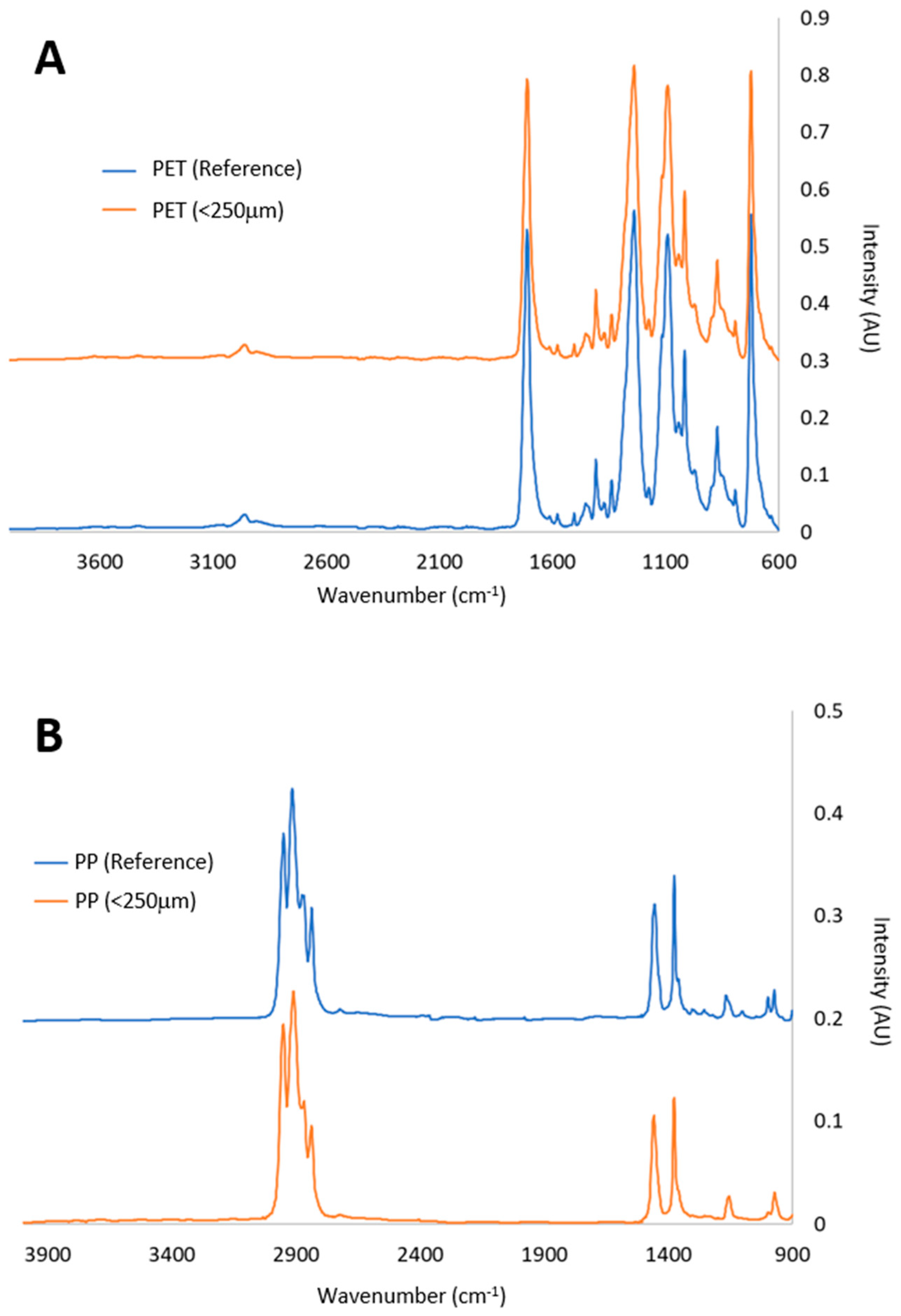
The nature of both PET and PP polymers was confirmed by chemical identification analyses. Figure 2 displays the distinctive PP peaks at wave numbers 2950, 2918, and 2836 cm−1, which correspond to CH stretching, as well as peaks at 1456 and 1376 cm−1, which correspond to CH2 and CH3 symmetric deformation, respectively. The exact nature of the polymer is also confirmed by other less noticeable peaks, such as those at 974 cm−1 (CC stretching) and 1166 cm−1 (CH3 rocking). Similarly, for PET, the commonly described peaks of 731 cm−1 (CH bending), 1245 and 1100 cm−1 (CCO stretching), and 1721 cm−1 (CO stretching) were discovered. The previously mentioned peaks for PET and PP are consistent with the findings and represent the native bonds found in the polymer [56].
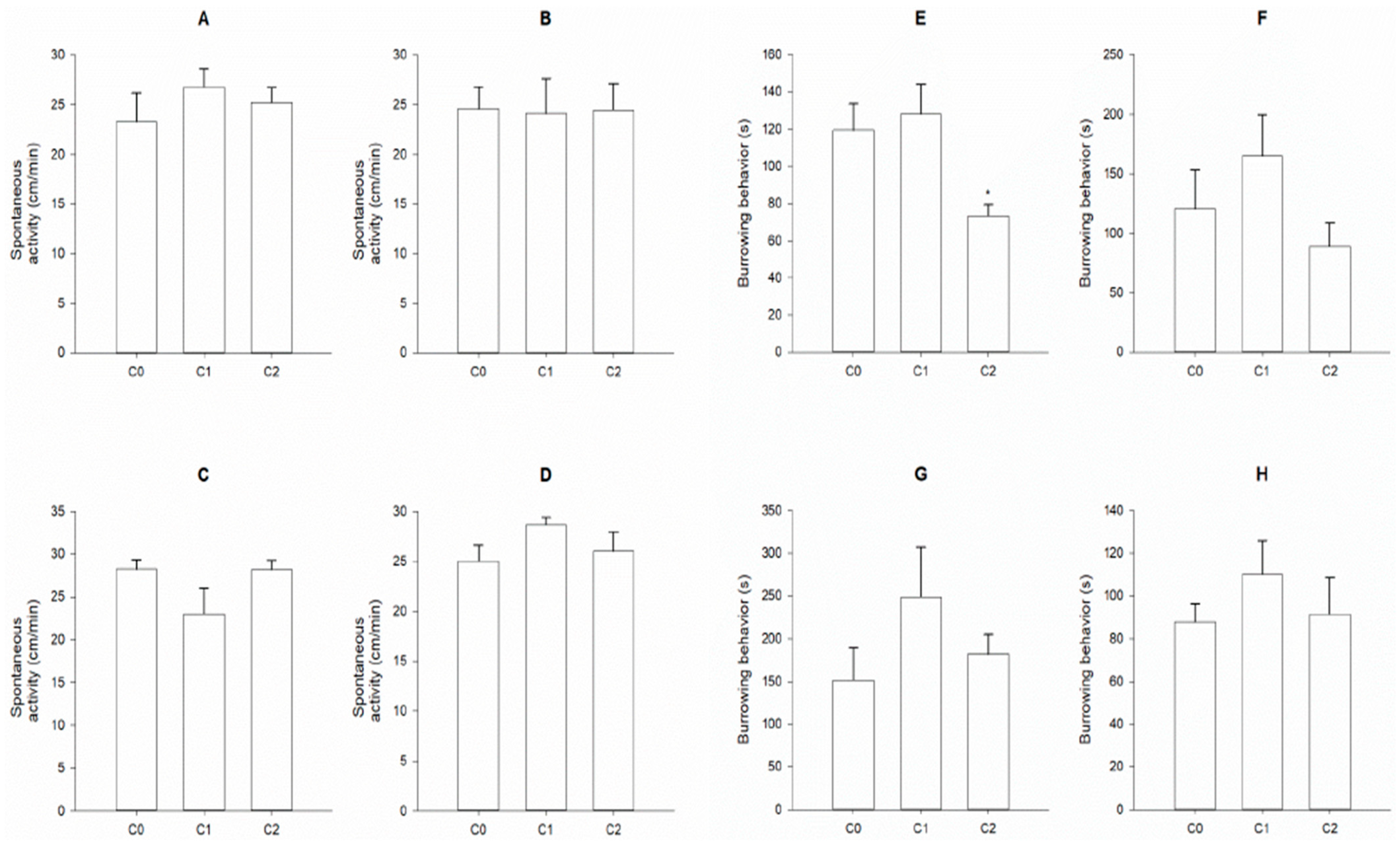
In terms of the assessed behavioral parameters, none of the polymers or exposure periods caused any measurable alterations in the exposed individuals of H. diversicolor (Figure 3).
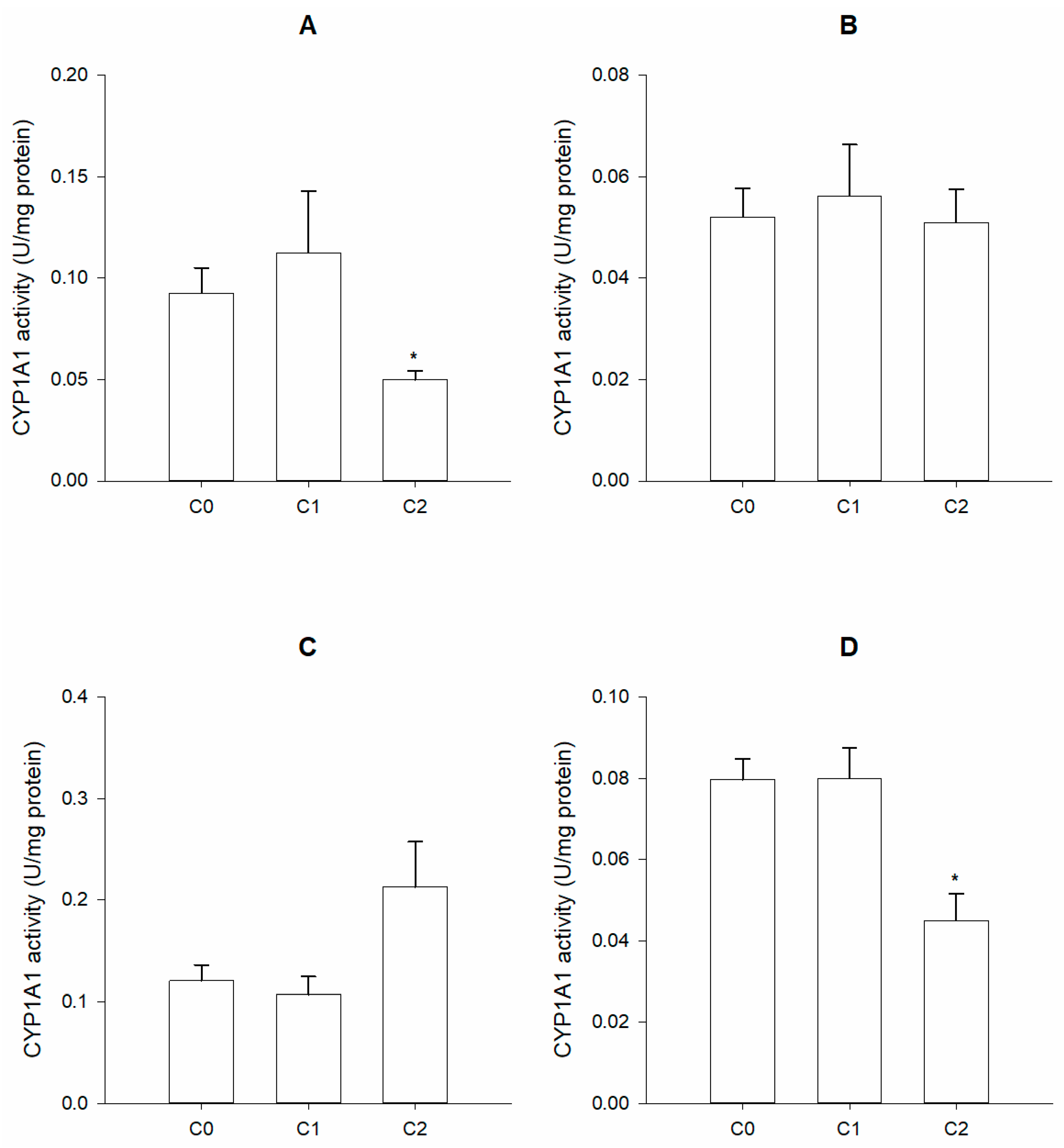
Data from phase I metabolism biomarkers showed divergent responses. CYP1A1 activity showed no straightforward pattern across all exposure periods and for the two polymers. Nevertheless, it was possible to observe a reduction in this enzyme’s activity in animals acutely exposed to the highest density of PP (Figure 4A) and in organisms chronically subjected to the highest PET density (Figure 4D).
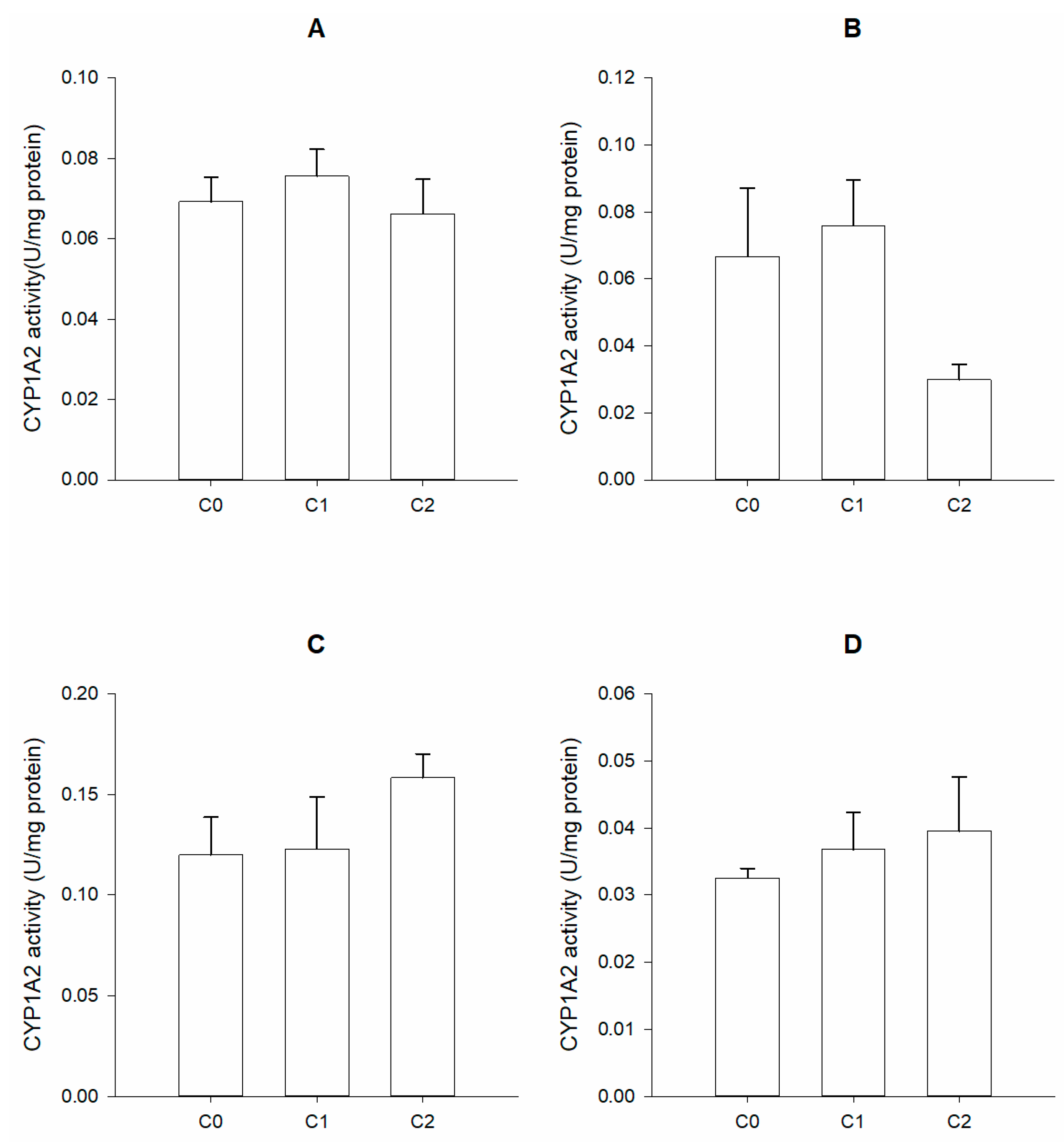
No statistically significant alterations were observed in the activity of CYP1A2 (Figure 5), in animals exposed to both polymers, and for the two exposure periods.
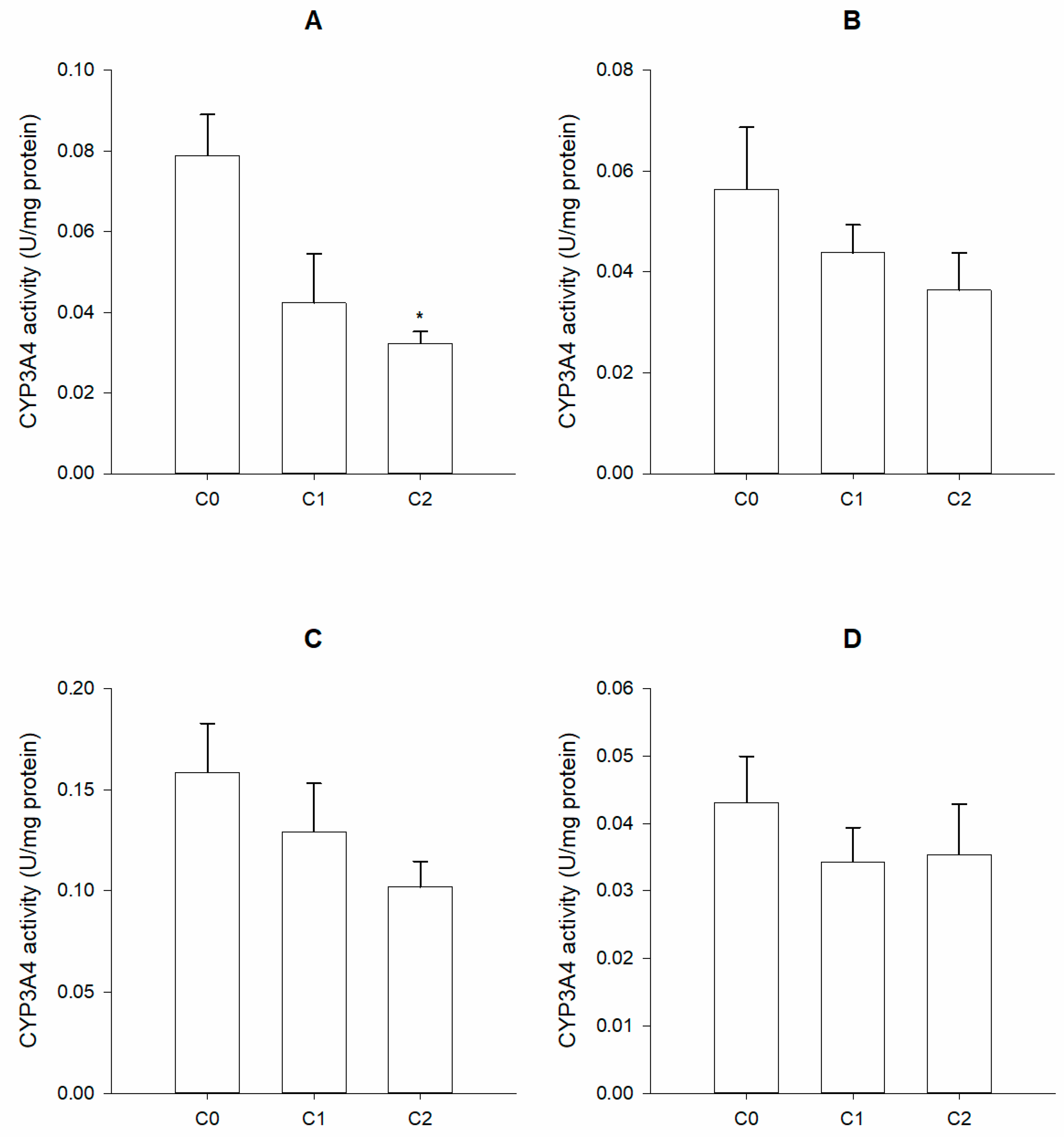
Finally, CYP3A4 activity showed a pattern of impaired activity caused by both polymers on acutely exposed animals (Figure 6A,C). Regarding chronically exposed animals, no significant alterations were observed (Figure 6B,D).
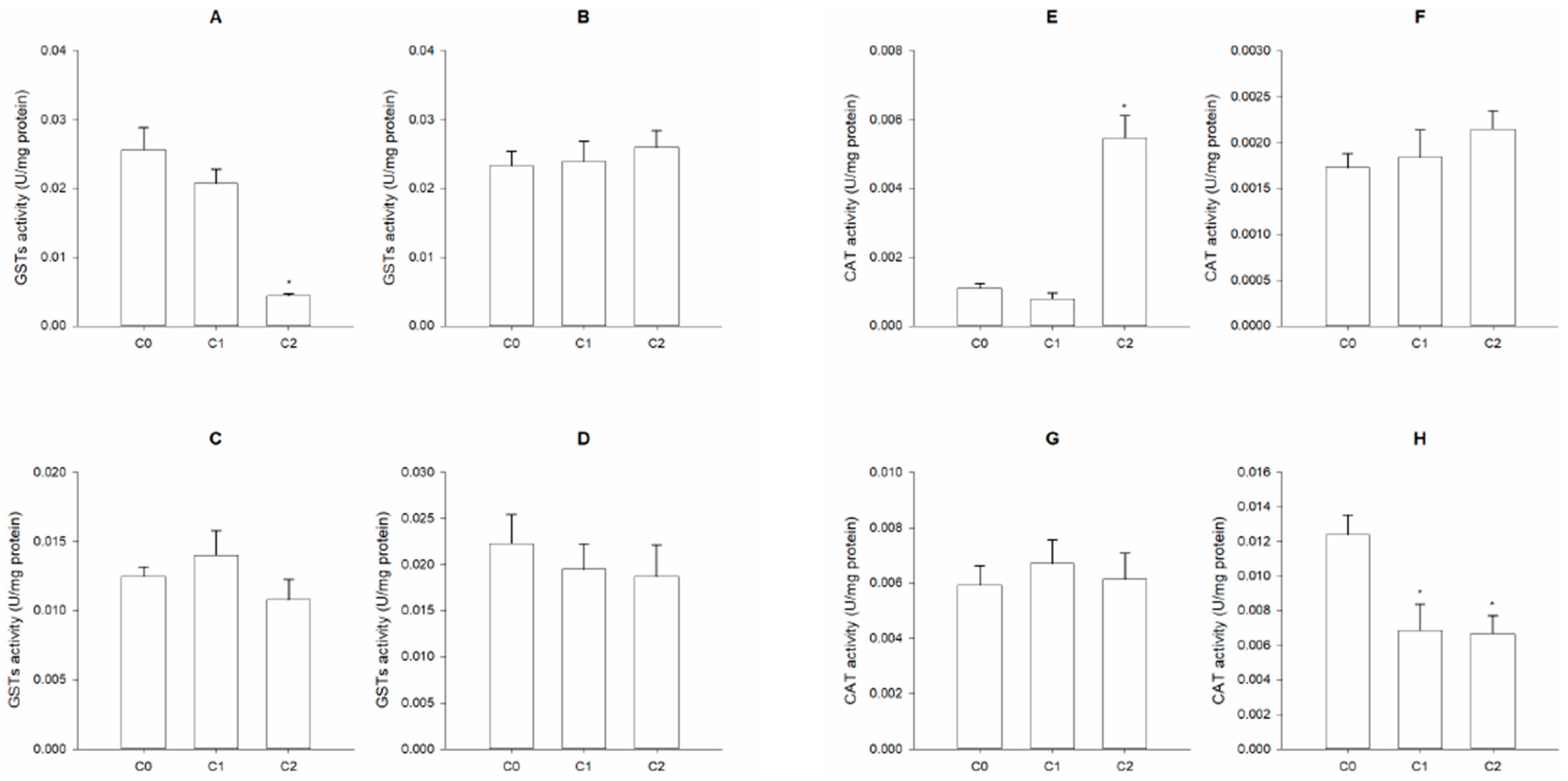
Conjugation metabolism, involving GST activity, showed a significant impairment in individuals of H. diversicolor acutely exposed to PP (Figure 7A). Nevertheless, no effects were observed for chronically exposed animals, and worms exposed to the other polymer (PET) did not show any straightforward pattern in the response of these isoenzymes (Figure 7B–D).
In terms of the antioxidant enzyme, CAT activity was significantly increased in animals acutely exposed to the highest density of PP (Figure 7E). However, no statistically significant differences were inferred, despite an increase in CAT activity that was observed in animals exposed to PP (Figure 7F). In terms of animals exposed to PET, a reduction in this biomarker’s activity was observed for organisms chronically exposed to both PET treatments (Figure 7H).

4. Discussion

In the context of marine contamination, plastics are nowadays one of the major contributors to this issue, and MPs are a current focus of research. Consequently, it is important to study environmentally realistic effects caused by the amounts of MPs that actually exist in the aquatic environment. The present study focused on this exact approach by exposing test organisms to amounts of MPs that have been reported in the aquatic environment. The most common way of expressing the levels of MPs found to occur in the wild is the number of particles per unit volume, which sometimes makes it difficult to interpret the levels of MPs in the aquatic compartment, especially under the scope of ecotoxicological assays, which refer to levels of MPs in terms of mass per unit volume. However, after converting the data obtained in monitoring studies, we assume that the concentrations tested in this study are similar to those found in environment [57,58,59]. Although we tested environmentally realistic concentrations, no significant alterations were observed both in terms of spontaneous activity and burrowing capacity, shown by individuals of H. diversicolor. These endpoints are of special interest since they can directly inform about these animals’ capacity to hide from predators and/or perform their ecological role. Several types of MPs are known to cause behavioral alterations in several animals, although without any clearly established mechanism, and most authors attribute these behavioral alterations to cholinesterase inhibition [60]. It has been observed that PS nanoplastics can cause behavioral alterations in H. diversicolor, namely, an increase in the time animals take to bury themselves [35]. However, these authors only observed such alterations in animals exposed to the lower density (µg/L range) of the tested polymer; these were not observed in individuals exposed to MPs in the mg/L range. The absence of alterations in exposed animals reported here may be explained by a possible mechanism of selective feeding [42,60,61], i.e., by selecting particles of different sizes and densities, such as plastics [62]. H. diversicolor is more prone to ingest microfibers by feeding from the filtration of the water column than from deposit feeding [60]. This may have occurred in our study since the contamination was performed via water, and the animals are present in the sediment. Furthermore, Ref. [3] suggested that polychaeta worms are able to ingest and expel plastic microspheres without any apparent detrimental behavioral effects.
Regarding phase I enzymes, which include cytochrome P450 superfamily enzymes, these enzymatic forms are known to catalyze the oxidation of a vast number of lipophilic xenobiotic compounds, allowing their ulterior excretion [63]. One class of those lipophilic xenobiotics, which are of particular environmental interest, is that of additives of MPs [64], such as phthalates, acrylonitrile, polychlorinated biphenyls, and dioxins [65,66]. It is known that these chemicals may be metabolized with the contribution of cytochrome P450 (CYP450) enzymes [67]. Several authors observed that exposure to some MPs, namely, PS, caused both increases and decreases in the activity of several CYP450 isoenzymes in distinct experimental models, specifically that of CYP1A1 in the fish Oreochromis niloticus [24,34], and that of CYP2E1 in human hepatocytes [68]. Although some plastics may not exert direct toxicity, most constituents of the plastic matrix may leach and cause deleterious effects [69]. Some of such constituents (namely, phthalates, acrylonitrile, polychlorinated biphenyls, and dioxins; Refs. [65,66]) are present in the MPs tested in this study. Although some PET and PP components, such as aromatic hydrocarbons, can increase the activity of CYP1A1 [65], this enzyme’s activity did not show any straightforward pattern of alteration following exposure to both plastics and for the two different exposure periods. On the other hand, phthalates, also known to be present in the MPs studied here, have the potential to inhibit CYP1A1 activity [70]. This makes it difficult to interprete the responses of organisms exposed to MPs if one is analyzing the levels of cytochrome p450 isoenzymes.
Similar to CYP1A1, the activity of CYP1A2 showed no straightforward pattern following exposure to both plastics and for the two different exposure periods. Although several authors established a causative relationship between some plastic additives and the induction of CYP1A2 activity [71,72,73], our results showed no significant alteration in this enzyme’s activity. Although other studies have not reported alterations in CYP1A2 activity, some have established a relation between MP exposure and an increase in the expression of the Cyp1a2 gene, which can further reflect on increases in enzymatic activity following longer exposure [74,75,76]. We can therefore infer that this enzyme was not the preferential short-term pathway for the metabolism of the tested MPs by the polychaeta species H. diversicolor.
Finally, CYP3A4 activity was generally inhibited in animals exposed to the highest density of both MPs and for both exposure durations. CYP3A4 is not known to directly metabolize plastics (nor most constituents of plastics); however, Ref. [77] linked the presence of PS nanoparticles to a reduction in CYP3A4 activity in insect cells and liver microsomes. Further, other authors have observed that MPs can reduce the gene expression of Cyp3a4, which can result in fewer enzymes being transcribed, which results in a reduction in its activity [78,79]. Furthermore, reductions in available energy can also limit enzyme activity [80]. Ref. [81] showed that Arenicola marina, when exposed to PVC-MPs for one month, showed a significant decrease in feeding activity and a significant increase, of approximately 1.5-fold, in the gut passage time of sediments. Additionally, mussels that ingested MPs have altered digestive enzymes, which can affect the effectiveness of digestion and, consequently, may limit their energy pool [82]. Therefore, we can infer that not only a lower quantity of food may be ingested but also that animals ingesting MPs can have their digestive tract clogged, preventing further absorption of nutrients [83,84]. In such scenarios, it is possible to have reductions in the energy metabolism activity in polychaeta species, as suggested by [81].
GSTs are responsible for the conjugation of glutathione with a large array of chemicals, including some plastic constituents, such as dioxins, furans, and PCBs [85], being also important for the elimination of ROS through the conversion of peroxides into less toxic hydroxyl compounds [86]. Alterations in these isoenzymes’ activities have been related to exposure to MPs [87,88]. The reported decrease in GST activity in animals acutely exposed to PP can be related to the capacity of MPs to cause a reduction in the levels of free glutathione (GSH) as a consequence of their interaction with ROS generated by exposure to MPs. This effect has been previously described in individuals of the marine rotifer Brachionus koreanus exposed to PS microbeads [27] but without any indication of a significant decrease in GST activity. Also, it is known that GSH is the cofactor required for the conjugation role played by GSTs, and it is critical that this enzyme functions properly [89]. We can thus infer that a reduction in intracellular available GSH by an interaction with ROS generated in phase I metabolism may impact the activity of enzymatic forms that require this cofactor, such as GSTs. Furthermore, Ref. [90] showed a decrease in GSH content in mice’s brains exposed to PS-MPs, showing the potential of MPs to reduce the levels of this cofactor by interacting with ROS. Other authors have also suggested that an inhibition of GST activity may be related to the oxidation of the thiol groups of the enzymes by ROS resulting from MPs’ interaction with endogenous biomolecules, which causes a denaturation and, consequently, a loss of activity of the measured enzymatic forms [91], as already suggested by [92]. These results agree with those obtained by [93], who found that individuals of the marine clam Ruditapes philippinarum that were exposed for 7 days to PET-MPs (0.125 or 12.5 μg/mL) had their GST levels unchanged. These authors did not register any alterations in GST activity in both the gills and the digestive gland, which may be due to the density of PET itself. Usually, PET plastics’ density is around 1.38 g/cm3, which makes them denser than seawater, whose density is around 1.04 g/cm3 [94]. Due to this difference in density, PET-MPs may deposit at the bottom of the test recipient and not be in suspension, which is the fraction of MPs that is most likely ingested by polychaetes, as shown by [60]. Nevertheless, individuals of this species exhibited selective feeding behavior [42,61] by actively avoiding the ingestion of MPs when searching for nutritional particles in the sediment; this is a common behavior in deposit feeders, which have the capacity to select particles of lower specific gravities [62], such as MPs. This corresponds to a defensive behavior, which can prevent damages from the ingestion of toxic sediment particles, including MPs [60].
It is known that MPs can disrupt key physiological processes, causing an oxidative stress scenario [92], either by disrupting lysosomes and mitochondria membranes [95] or through the resulting products of phase I metabolism, which include ROS [28,29]. One example of these antioxidant defenses is catalase (CAT), which is responsible for the degradation of the oxidant metabolite oxygen peroxide (H2O2) into water and oxygen [96]. Several authors observed alterations in this enzyme activity when exposing zebrafish to MPs [97]. A similar trend was also observed in individuals of the microcrustacean Daphnia magna [98] and also in mussels of the genus Mytilus [92,99]. The data obtained here showed a significant increase in CAT activity after exposing H. diversicolor to PP for 96h. This increase in CAT levels may be explained by the assumption that the metabolism of MPs can induce the formation of ROS through CYP450 enzymatic induction [28,29] or by the disruption of mitochondria and lysosome membranes and the consequent release of their content, which leads to an increase in ROS formation [95]. Nevertheless, this same pattern of response (induction of CAT activity) was not observed after a 28-day exposure. This difference in responses at distinct exposure periods can be related to the organisms’ capacity to adapt over the exposure period, as suggested by [35], after exposing the same species (H. diversicolor) to PS-MPs. In terms of animals exposed to PET-MPs, a decrease in CAT activity was observed after chronic exposure, but this trend was not reported after the acute test. This tendency was also observed by [99] in mussels that were exposed to PS-MPs. These authors suggested that a decrease in CAT activity may be related to a possible activation of the same enzyme within the first days of exposure, which was followed by a decrease in the gene expression of CAT afterwards. This effect can be due to the activation of other enzymes also responsible for H2O2 degradation, such as GPx.

5. Conclusions

In conclusion, secondary PET- and PP-MPs do not seem to significantly alter the evaluated behavioral traits. Exposed worms were also generally nonresponsive to exposure to both plastics, suggesting that these organisms may cope with the potential deleterious effects caused by MPs, probably by selective feeding behaviors or by reducing their feeding activity. Furthermore, the evaluated toxicological parameters, i.e., cytochrome P450 isoenzymes (CYP1A1, CYP1A2, and CYP3A4), also showed no straightforward pattern of responses to exposure to these MPs. This indicates that MP components are probably not metabolized by the evaluated phase I metabolic enzymes. Regarding the conjugation metabolism via GSTs, only a short period of exposure to PP was able to induce an inhibitory effect of these enzymes’ activities. This effect can be explained by MPs’ capability of inhibiting certain enzymes due to the cofactors’ exhaustion caused by inadequate food uptake. In terms of antioxidant enzymatic activity, CAT showed significant alterations when exposed to PP and PET. However, there was no straightforward response, with an increase in this enzyme’s activity being observed when animals were acutely exposed to PP, along with a decrease in organisms chronically exposed to PET. The observed transient increase in CAT activity may be attributed to the capacity of MPs to induce ROS production, which may be compensated for at longer exposure periods by triggering an adaption from exposed animals as a consequence of the activation of other antioxidant metabolic pathways (e.g., GPx). Based on the results of this study, we can conclude that PP- and PET-MPs do not seem to adversely affect individuals of the estuarine polychaeta Hediste diversicolor under the tested conditions and in terms of the analyzed endpoints. Nevertheless, further tests are needed to fully evaluate the effects of these MPs on other endpoints, including neurotoxicity markers, energetic reserves, alternative detoxification pathways, as well as the determination of ingestion and egestion rates.

Author Contributions

Conceptualization, B.N. and D.D.; methodology, D.D. and A.V.G.; validation, B.N. and D.D.; formal analysis, D.D.; investigation, A.V.G. and D.D.; data curation, B.N.; writing—original draft preparation, D.D.; writing—review and editing, D.D., J.P.d.C. and B.N.; supervision, B.N.; and funding acquisition, B.N. All authors have read and agreed to the published version of the manuscript.

Funding

This work was financed by the project BETTER PLASTICS—PLASTICS IN A CIRCULAR ECONOMY (POCI-01-0247-FEDER-046091, co-financed by Agência Nacional de Inovação, S.A, by COMPETE 2020). This research was financially supported by CESAM (UIDP/50017/2020+UIDB/50017/2020+LA/P/0094/2020). BN and JPC also thank the following for their financial support: the Portuguese Foundation for Science and Technology (FCT), under contracts 2020.03531.CEECIND and 2021.00909.CEECIND, and BioPlasMar (PTDC/CTA-AMB/0934/2021) from the FCT.

Institutional Review Board Statement

Not applicable.

Data Availability Statement

The raw data supporting the conclusions of this article will be made available by the authors on request.

Conflicts of Interest

The authors declare no conflicts of interest.

References

  1. Koshti, R.; Mehta, L.B.; Samarth, N. Biological Recycling of Polyethylene Terephthalate: A Mini-Review. J. Polym. Environ. 2018, 26, 3520–3529. [Google Scholar] [CrossRef]
  2. Maurya, A.; Bhattacharya, A.; Khare, S.K. Enzymatic Remediation of Polyethylene Terephthalate (PET)–Based Polymers for Effective Management of Plastic Wastes: An Overview. Front. Bioeng. Biotechnol. 2020, 8, 602325. [Google Scholar] [CrossRef] [PubMed]
  3. Cole, M.; Lindeque, P.; Halsband, C.; Galloway, T.S. Microplastics as contaminants in the marine environment: A review. Mar. Pollut. Bull. 2011, 62, 2588–2597. [Google Scholar] [CrossRef] [PubMed]
  4. Wayman, C.; Niemann, H. The fate of plastic in the ocean environment—A minireview. Environ. Sci. Process. Impacts 2021, 23, 198–212. [Google Scholar] [CrossRef] [PubMed]
  5. Li, P.; Wang, X.; Su, M.; Zou, X.; Duan, L.; Zhang, H. Characteristics of Plastic Pollution in the Environment: A Review. Bull. Environ. Contam. Toxicol. 2021, 107, 577–584. [Google Scholar] [CrossRef]
  6. Ali, S.S.; Elsamahy, T.; Al-Tohamy, R.; Zhu, D.; Mahmoud, Y.A.; Koutra, E.; Metwally, M.A.; Kornaros, M.; Sun, J. Plastic wastes biodegradation: Mechanisms, challenges and future prospects. Sci. Total Environ. 2021, 780, 146590. [Google Scholar] [CrossRef]
  7. Frias, J.P.L.; Nash, R. Microplastics: Finding a consensus on the definition. Mar. Pollut. Bull. 2019, 138, 145–147. [Google Scholar] [CrossRef]
  8. GESAMP. Sources, Fate and Effects of Microplastics in the Marine Environment (Part 1)|GESAMP. 2015. Available online: http://www.gesamp.org/publications/reports-and-studies-no-90 (accessed on 18 February 2022).
  9. GESAMP. Sources, Fate and Effects of Microplastics in the Marine Environment (Part 2)|GESAMP. 2016. Available online: http://www.gesamp.org/publications/microplastics-in-the-marine-environment-part-2 (accessed on 18 February 2022).
  10. Alimi, O.S.; Farner Budarz, J.; Hernandez, L.M.; Tufenkji, N. Microplastics and Nanoplastics in Aquatic Environments: Aggregation, Deposition, and Enhanced Contaminant Transport. Environ. Sci. Technol. 2018, 52, 1704–1724. [Google Scholar] [CrossRef]
  11. Picó, Y.; Barceló, D. Analysis and Prevention of Microplastics Pollution in Water: Current Perspectives and Future Directions. ACS Omega 2019, 4, 6709–6719. [Google Scholar] [CrossRef]
  12. Yan, M.; Yang, J.; Sun, H.; Liu, C.; Wang, L. Occurrence and distribution of microplastics in sediments of a man-made lake receiving reclaimed water. Sci. Total Environ. 2022, 813, 152430. [Google Scholar] [CrossRef]
  13. Zhang, J.; Wang, L.; Kannan, K. Quantitative analysis of polyethylene terephthalate and polycarbonate microplastics in sediment collected from South Korea, Japan and the USA. Chemosphere 2021, 279, 130551. [Google Scholar] [CrossRef]
  14. Huang, W.; Song, B.; Liang, J.; Niu, Q.; Zeng, G.; Shen, M.; Deng, J.; Luo, Y.; Wen, X.; Zhang, Y. Microplastics and associated contaminants in the aquatic environment: A review on their ecotoxicological effects, trophic transfer, and potential impacts to human health. J. Hazard. Mater. 2021, 405, 124187. [Google Scholar] [CrossRef] [PubMed]
  15. Hasegawa, T.; Nakaoka, M. Trophic transfer of microplastics from mysids to fish greatly exceeds direct ingestion from the water column. Environ. Pollut. 2021, 273, 116468. [Google Scholar] [CrossRef] [PubMed]
  16. Li, X.; Pu, Q.; Xu, Y.; Yang, H.; Wu, Y.; Wang, W.; Li, Y. The masking phenomenon of microplastics additives on oxidative stress responses in freshwater food chains. Sci. Total Environ. 2024, 927, 172156. [Google Scholar] [CrossRef] [PubMed]
  17. Hasegawa, T.; Mizukawa, K.; Yeo, B.G.; Sekioka, T.; Takada, H.; Nakaoka, M. The significance of trophic transfer of microplastics in the accumulation of plastic additives in fish: An experimental study using brominated flame retardants and UV stabilizers. Mar. Pollut. Bull. 2022, 185, 114343. [Google Scholar] [CrossRef]
  18. Smith, M.; Love, D.C.; Rochman, C.M.; Neff, R.A. Microplastics in Seafood and the Implications for Human Health. Curr. Environ. Health Rep. 2018, 5, 375–386. [Google Scholar] [CrossRef]
  19. Nair, H.T.; Perumal, S. Trophic Transfer and Accumulation of Microplastics in Freshwater Ecosystem: Risk to Food Security and Human Health. Int. J. Ecol. 2022, 2022, 1234078. [Google Scholar] [CrossRef]
  20. Carbery, M.; O’COnnor, W.; Palanisami, T. Trophic transfer of microplastics and mixed contaminants in the marine food web and implications for human health. Environ. Int. 2018, 115, 400–409. [Google Scholar] [CrossRef]
  21. Gambino, I.; Bagordo, F.; Grassi, T.; Panico, A.; De Donno, A. Occurrence of Microplastics in Tap and Bottled Water: Current Knowledge. Int. J. Environ. Res. Public Health 2022, 19, 5283. [Google Scholar] [CrossRef]
  22. Tong, H.; Jiang, Q.; Hu, X.; Zhong, X. Occurrence and identification of microplastics in tap water from China. Chemosphere 2020, 252, 126493. [Google Scholar] [CrossRef]
  23. Van Cauwenberghe, L.; Claessens, M.; Vandegehuchte, M.B.; Janssen, C.R. Microplastics are taken up by mussels (Mytilus edulis) and lugworms (Arenicola marina) living in natural habitats. Environ. Pollut. 2015, 199, 10–17. [Google Scholar] [CrossRef] [PubMed]
  24. Ding, J.; Zhang, S.; Razanajatovo, R.M.; Zou, H.; Zhu, W. Accumulation, tissue distribution, and biochemical effects of polystyrene microplastics in the freshwater fish red tilapia (Oreochromis niloticus). Environ. Pollut. 2018, 238, 1–9. [Google Scholar] [CrossRef] [PubMed]
  25. Solomando, A.; Capó, X.; Alomar, C.; Álvarez, E.; Compa, M.; Valencia, J.M.; Pinya, S.; Deudero, S.; Sureda, A. Long-term exposure to microplastics induces oxidative stress and a pro-inflammatory response in the gut of Sparus aurata Linnaeus, 1758. Environ. Pollut. 2020, 266, 115295. [Google Scholar] [CrossRef] [PubMed]
  26. Gardon, T.; Morvan, L.; Huvet, A.; Quillien, V.; Soyez, C.; Le Moullac, G.; Le Luyer, J. Microplastics induce dose-specific transcriptomic disruptions in energy metabolism and immunity of the pearl oyster Pinctada margaritifera. Environ. Pollut. 2020, 266, 115180. [Google Scholar] [CrossRef]
  27. Jeong, C.B.; Won, E.J.; Kang, H.M.; Lee, M.C.; Hwang, D.S.; Hwang, U.K.; Zhou, B.; Souissi, S.; Lee, S.J.; Lee, J.S. Microplastic Size-Dependent Toxicity, Oxidative Stress Induction, and p-JNK and p-p38 Activation in the Monogonont Rotifer (Brachionus koreanus). Environ. Sci. Technol. 2016, 50, 8849–8857. [Google Scholar] [CrossRef]
  28. Hu, M.; Palić, D. Micro- and nano-plastics activation of oxidative and inflammatory adverse outcome pathways. Redox Biol. 2020, 37, 101620. [Google Scholar] [CrossRef]
  29. Sharifian, S.; Homaei, A.; Kamrani, E.; Etzerodt, T.; Patel, S. New insights on the marine cytochrome P450 enzymes and their biotechnological importance. Int. J. Biol. Macromol. 2020, 142, 811–821. [Google Scholar] [CrossRef]
  30. Ribeiro, F.; Garcia, A.R.; Pereira, B.P.; Fonseca, M.; Mestre, N.C.; Fonseca, T.G.; Ilharco, L.M.; Bebianno, M.J. Microplastics effects in Scrobicularia plana. Mar. Pollut. Bull. 2017, 122, 379–391. [Google Scholar] [CrossRef]
  31. de Sá, L.C.; Oliveira, M.; Ribeiro, F.; Rocha, T.L.; Futter, M.N. Studies of the effects of microplastics on aquatic organisms: What do we know and where should we focus our efforts in the future? Sci. Total Environ. 2018, 645, 1029–1039. [Google Scholar] [CrossRef]
  32. Dovidat, L.C.; Brinkmann, B.W.; Vijver, M.G.; Bosker, T. Plastic particles adsorb to the roots of freshwater vascular plant Spirodela polyrhiza but do not impair growth. Limnol. Oceanogr. Lett. 2020, 5, 37–45. [Google Scholar] [CrossRef]
  33. Bhattacharya, P.; Lin, S.; Turner, J.P.; Ke, P.C. Physical adsorption of charged plastic nanoparticles affects algal photosynthesis. J. Phys. Chem. C 2010, 114, 16556–16561. [Google Scholar] [CrossRef]
  34. Limonta, G.; Mancia, A.; Benkhalqui, A.; Bertolucci, C.; Abelli, L.; Fossi, M.C.; Panti, C. Microplastics induce transcriptional changes, immune response and behavioral alterations in adult zebrafish. Sci. Rep. 2019, 9, 15775. [Google Scholar] [CrossRef] [PubMed]
  35. Martins, M.; Valente, P.; Figueira, E.; Oliveira, M.; Pires, A.; Silva, M. Behavior and biochemical responses of the polychaeta Hediste diversicolor to polystyrene nanoplastics. Sci. Total Environ. 2020, 707, 134434. [Google Scholar] [CrossRef]
  36. Green, D.S.; Boots, B.; O’cOnnor, N.E.; Thompson, R. Microplastics Affect the Ecological Functioning of an Important Biogenic Habitat. Environ. Sci. Technol. 2017, 51, 68–77. [Google Scholar] [CrossRef]
  37. Mazurais, D.; Ernande, B.; Quazuguel, P.; Severe, A.; Huelvan, C.; Madec, L.; Mouchel, O.; Soudant, P.; Robbens, J.; Huvet, A.; et al. Evaluation of the impact of polyethylene microbeads ingestion in European sea bass (Dicentrarchus labrax) larvae. Mar. Environ. Res. 2015, 112, 78–85. [Google Scholar] [CrossRef]
  38. Hitchcock, J.N.; Mitrovic, S.M. Microplastic pollution in estuaries across a gradient of human impact. Environ. Pollut. 2019, 247, 457–466. [Google Scholar] [CrossRef]
  39. Rudén, C.; Adams, J.; Ågerstrand, M.; Brock, T.C.; Poulsen, V.; Schlekat, C.E.; Wheeler, J.R.; Henry, T.R. Assessing the relevance of ecotoxicological studies for regulatory decision making. Integr. Environ. Assess. Manag. 2017, 13, 652–663. [Google Scholar] [CrossRef]
  40. Fonseca, L.C.; e Costa, P.F.; Gil, J.; Passos, A.M.; Pereira, P.; Melo, P.; Batista, F. The market features of imported non-indigenous polychaetes in Portugal and consequent ecological concerns. Sci. Mar. 2006, 70 (Suppl. 3), 287–292. [Google Scholar] [CrossRef]
  41. Thit, A.; Dybowska, A.; Købler, C.; Kennaway, G.; Selck, H. Influence of copper oxide nanoparticle shape on bioaccumulation, cellular internalization and effects in the estuarine sediment-dwelling polychaete, Nereis diversicolor. Mar. Environ. Res. 2015, 111, 89–98. [Google Scholar] [CrossRef]
  42. Scaps, P. A review of the biology, ecology and potential use of the common ragworm Hediste diversicolor (O.F. Müller) (Annelida: Polychaeta). Hydrobiologia 2002, 470, 203–218. [Google Scholar] [CrossRef]
  43. Nogueira, A.F.; Nunes, B. Effects of paracetamol on the polychaete Hediste diversicolor: Occurrence of oxidative stress, cyclooxygenase inhibition and behavioural alterations. Environ. Sci. Pollut. Res. 2021, 28, 26772–26783. [Google Scholar] [CrossRef]
  44. Nunes, B.; Daniel, D.; Correia, A.T. Toxic effects of environmentally realistic concentrations of diclofenac in organisms from two distinct trophic levels, Hediste diversicolor and Solea senegalensis. Comp. Biochem. Physiol. Part C Toxicol. Pharmacol. 2020, 231, 108722. [Google Scholar] [CrossRef] [PubMed]
  45. da Fonseca, T.G.; Abessa, D.M.; Bebianno, M.J. Effects of mixtures of anticancer drugs in the benthic polychaete Nereis diversicolor. Environ. Pollut. 2019, 252, 1180–1192. [Google Scholar] [CrossRef] [PubMed]
  46. Fonseca, T.; Morais, M.; Rocha, T.; Abessa, D.; Aureliano, M.; Bebianno, M. Ecotoxicological assessment of the anticancer drug cisplatin in the polychaete Nereis diversicolor. Sci. Total Environ. 2017, 575, 162–172. [Google Scholar] [CrossRef] [PubMed]
  47. Nogueira, A.F.; Nunes, B. Effects of low levels of the antibiotic ciprofloxacin on the polychaete Hediste diversicolor: Biochemical and behavioural effects. Environ. Toxicol. Pharmacol. 2020, 80, 103505. [Google Scholar] [CrossRef]
  48. Daniel, D.; Barros, L.; da Costa, J.P.; Girão, A.V.; Nunes, B. Using marine mussels to assess the potential ecotoxicological effects of two different commercial microplastics. Mar. Pollut. Bull. 2024, 203, 116441. [Google Scholar] [CrossRef]
  49. Ghribi, R.; Correia, A.T.; Elleuch, B.; Nunes, B. Toxicity Assessment of Impacted Sediments from Southeast Coast of Tunisia Using a Biomarker Approach with the Polychaete Hediste diversicolor. Arch. Environ. Contam. Toxicol. 2019, 76, 678–691. [Google Scholar] [CrossRef]
  50. Daniel, D.; Nunes, B.; Pinto, E.; Ferreira, I.M.P.L.V.O.; Correia, A.T. Assessment of Paracetamol Toxic Effects under Varying Seawater pH Conditions on the Marine Polychaete Hediste diversicolor Using Biochemical Endpoints. Biology 2022, 11, 581. [Google Scholar] [CrossRef]
  51. Burke, M.D.; Mayer, R.T. Ethoxyresorufin: Direct fluorimetric assay of a microsomal o-dealkylation which is preferentially inducible by 3-methylcholanthrene. Drug Metab. Dispos. 1974, 2, 583–588. [Google Scholar] [CrossRef]
  52. MattSiebert, M.N.; Mattos, J.J.; Piazza, C.E.; de Lima, D.; Gomes, C.H.A.; de Melo, C.M.; Bainy, A.C. Characterization of ethoxyresorufin O-deethylase activity (EROD) in oyster Crassostrea brasiliana. Comp. Biochem. Physiol. Part B Biochem. Mol. Biol. 2017, 203, 115–121. [Google Scholar] [CrossRef]
  53. Habig, W.H.; Pabst, M.J.; Jakoby, W.B. Glutathione S-transferases. The first enzymatic step in mercapturic acid formation. J. Biol. Chem. 1974, 249, 7130–7139. [Google Scholar] [CrossRef]
  54. Aebi, H. Catalase in vitro. Methods Enzymol. 1984, 105, 121–126. [Google Scholar] [CrossRef]
  55. Bradford, M.M. A rapid and sensitive method for the quantitation of microgram quantities of protein utilizing the principle of protein-dye binding. Anal. Biochem. 1976, 72, 248–254. [Google Scholar] [CrossRef]
  56. Kuptsov, A.; Zhizhin, G. Handbook of Fourier Transform Raman and Infrared Spectra of Polymers; Elsevier Science: Amsterdam, The Netherlands, 1998; 581p. [Google Scholar]
  57. Beiras, R.; Schönemann, A.M. Currently monitored microplastics pose negligible ecological risk to the global ocean. Sci. Rep. 2020, 10, 22281. [Google Scholar] [CrossRef] [PubMed]
  58. Mehinto, A.C.; Coffin, S.; Koelmans, A.A.; Brander, S.M.; Wagner, M.; Thornton Hampton, L.M.; Burton, A.G.; Miller, E.; Gouin, T.; Weisberg, S.B.; et al. Risk-based management framework for microplastics in aquatic ecosystems. Microplast. Nanoplast. 2022, 2, 17. [Google Scholar] [CrossRef]
  59. Leusch, F.D.; Ziajahromi, S. Converting mg/L to Particles/L: Reconciling the Occurrence and Toxicity Literature on Microplastics. Environ. Sci. Technol. 2021, 55, 11470–11472. [Google Scholar] [CrossRef]
  60. Porter, A.; Barber, D.; Hobbs, C.; Love, J.; Power, A.L.; Bakir, A.; Galloway, T.S.; Lewis, C. Uptake of microplastics by marine worms depends on feeding mode and particle shape but not exposure time. Sci. Total Environ. 2023, 857, 159287. [Google Scholar] [CrossRef] [PubMed]
  61. Vecchi, S.; Bianchi, J.; Scalici, M.; Fabroni, F.; Tomassetti, P. Field evidence for microplastic interactions in marine benthic invertebrates. Sci. Rep. 2021, 11, 20900. [Google Scholar] [CrossRef]
  62. Taghon, G.L. Optimal foraging by deposit-feeding invertebrates: Roles of particle size and organic coating. Oecologia 1982, 52, 295–304. [Google Scholar] [CrossRef]
  63. Esteves, F.; Rueff, J.; Kranendonk, M. The Central Role of Cytochrome P450 in Xenobiotic Metabolism—A Brief Review on a Fascinating Enzyme Family. J. Xenobiotics 2021, 11, 94–114. [Google Scholar] [CrossRef]
  64. O’Donovan, S.; Mestre, N.C.; Abel, S.; Fonseca, T.G.; Carteny, C.C.; Cormier, B.; Keiter, S.H.; Bebianno, M.J. Ecotoxicological effects of chemical contaminants adsorbed to microplastics in the Clam Scrobicularia plana. Front. Mar. Sci. 2018, 5, 143. [Google Scholar] [CrossRef]
  65. Żwierełło, W.; Maruszewska, A.; Skórka-Majewicz, M.; Goschorska, M.; Baranowska-Bosiacka, I.; Dec, K.; Styburski, D.; Nowakowska, A.; Gutowska, I. The influence of polyphenols on metabolic disorders caused by compounds released from plastics—Review. Chemosphere 2020, 240, 124901. [Google Scholar] [CrossRef]
  66. Cooper, B.W.; Cho, T.M.; Thompson, P.M.; Wallace, A.D. Phthalate induction of CYP3A4 is dependent on glucocorticoid regulation of PXR expression. Toxicol. Sci. 2008, 103, 268–277. [Google Scholar] [CrossRef]
  67. Anbumani, S.; Kakkar, P. Ecotoxicological effects of microplastics on biota: A review. Environ. Sci. Pollut. Res. 2018, 25, 14373–14396. [Google Scholar] [CrossRef]
  68. Cheng, W.; Li, X.; Zhou, Y.; Yu, H.; Xie, Y.; Guo, H.; Wang, H.; Li, Y.; Feng, Y.; Wang, Y. Polystyrene microplastics induce hepatotoxicity and disrupt lipid metabolism in the liver organoids. Sci. Total Environ. 2022, 806, 150328. [Google Scholar] [CrossRef]
  69. Zimmermann, L.; Bartosova, Z.; Braun, K.; Oehlmann, J.; Völker, C.; Wagner, M. Plastic Products Leach Chemicals That Induce In Vitro Toxicity under Realistic Use Conditions. Environ. Sci. Technol. 2021, 55, 11814–11823. [Google Scholar] [CrossRef]
  70. Goh, J.J.; Behn, J.; Chong, C.S.; Zhong, G.; Maurer-Stroh, S.; Fan, H.; Loo, L.H. Structure-based virtual screening of CYP1A1 inhibitors: Towards rapid tier-one assessment of potential developmental toxicants. Arch. Toxicol. 2021, 95, 3031–3048. [Google Scholar] [CrossRef]
  71. Devito, M.; Ma, X.; Babish, J.; Menache, M.; Birnbaum, L. Dose-Response Relationships in Mice Following Subchronic Exposure to 2,3,7,8-Tetrachlorodibenzo-p-dioxin: CYP1A1, CYP1A2, Estrogen Receptor, and Protein Tyrosine Phosphorylation. Toxicol. Appl. Pharmacol. 1994, 124, 82–90. [Google Scholar] [CrossRef] [PubMed]
  72. Lambert, G.H.; Needham, L.L.; Turner, W.; Lai, T.J.; Patterson, D.G.; Guo, Y.L. Induced CYP1A2 Activity as a Phenotypic Biomarker in Humans Highly Exposed to Certain PCBs/PCDFs. Environ. Sci. Technol. 2006, 40, 6176–6180. [Google Scholar] [CrossRef] [PubMed]
  73. Xu, L.; Li, A.P.; Kaminski, D.L.; Ruh, M.F. 2,3,7,8 Tetrachlorodibenzo-p-dioxin induction of cytochrome P4501A in cultured rat and human hepatocytes. Chem. Interact. 2000, 124, 173–189. [Google Scholar] [CrossRef] [PubMed]
  74. Hirt, N.; Body-Malapel, M. Immunotoxicity and intestinal effects of nano- and microplastics: A review of the literature. Part. Fibre Toxicol. 2020, 17, 57. [Google Scholar] [CrossRef]
  75. Santos, L.H.; Rodríguez-Mozaz, S.; Barceló, D. Microplastics as vectors of pharmaceuticals in aquatic organisms—An overview of their environmental implications. Case Stud. Chem. Environ. Eng. 2021, 3, 100079. [Google Scholar] [CrossRef]
  76. Yang, X.; Zhang, X.; Shu, X.; Gong, J.; Yang, J.; Li, B.; Lin, J.; Chai, Y.; Liu, J. The effects of polyethylene microplastics on the growth, reproduction, metabolic enzymes, and metabolomics of earthworms Eisenia fetida. Ecotoxicol. Environ. Saf. 2023, 263, 115390. [Google Scholar] [CrossRef] [PubMed]
  77. Fröhlich, E.; Kueznik, T.; Samberger, C.; Roblegg, E.; Wrighton, C.; Pieber, T.R. Size-dependent effects of nanoparticles on the activity of cytochrome P450 isoenzymes. Toxicol. Appl. Pharmacol. 2010, 242, 326–332. [Google Scholar] [CrossRef] [PubMed]
  78. Rosellini, M.; Omer, E.A.; Schulze, A.; Ali, N.T.; Boulos, J.C.; Marini, F.; Küpper, J.H.; Efferth, T. Impact of plastic-related compounds on the gene expression signature of HepG2 cells transfected with CYP3A4. Arch. Toxicol. 2024, 98, 525–536. [Google Scholar] [CrossRef] [PubMed]
  79. Kılıç, Ö.; Belivermiş, M.; Sıkdokur, E.; Sezer, N.; Aksüt, Y.; Pekmez, M.; Kösesakal, T.; Gerçek, Y.C. The combined effects of polyethylene microplastics and benzoanthracene on Manila clam Ruditapes philippinarum. Chemosphere 2023, 329, 138664. [Google Scholar] [CrossRef]
  80. Ward, J.E.; Rosa, M.; Shumway, S.E. Capture, ingestion, and egestion of microplastics by suspension-feeding bivalves: A 40-year history. Anthr. Coasts 2019, 2, 39–49. [Google Scholar] [CrossRef]
  81. Wright, S.L.; Rowe, D.; Thompson, R.C.; Galloway, T.S. Microplastic ingestion decreases energy reserves in marine worms. Curr. Biol. 2013, 23, R1031–R1033. [Google Scholar] [CrossRef]
  82. Trestrail, C.; Walpitagama, M.; Miranda, A.; Nugegoda, D.; Shimeta, J. Microplastics alter digestive enzyme activities in the marine bivalve, Mytilus galloprovincialis. Sci. Total Environ. 2021, 779, 146418. [Google Scholar] [CrossRef]
  83. Weis, J.S. Aquatic Microplastic Research—A Critique and Suggestions for the Future. Water 2020, 12, 1475. [Google Scholar] [CrossRef]
  84. Eltemsah, Y.S.; Bøhn, T. Acute and chronic effects of polystyrene microplastics on juvenile and adult Daphnia magna. Environ. Pollut. 2019, 254, 112919. [Google Scholar] [CrossRef]
  85. Shore, L.J.; Mogilevsky, W.S.; Smith, P.B.; Fenselau, C.; Odell, G.B. In vitro formation of glutathione conjugates of the dimethylester of bilirubin. Biochem. Pharmacol. 1991, 42, 1969–1976. [Google Scholar] [CrossRef] [PubMed]
  86. Testa, B.; Clement, B. Biotransformation Reactions and Their Enzymes. In The Practice of Medicinal Chemistry, 4th ed.; Springer: Berlin/Heidelberg, Germany, 2015; pp. 561–584. [Google Scholar] [CrossRef]
  87. Zitouni, N.; Bousserrhine, N.; Missawi, O.; Boughattas, I.; Chèvre, N.; Santos, R.; Belbekhouche, S.; Alphonse, V.; Tisserand, F.; Balmassiere, L.; et al. Uptake, tissue distribution and toxicological effects of environmental microplastics in early juvenile fish Dicentrarchus labrax. J. Hazard. Mater. 2021, 403, 124055. [Google Scholar] [CrossRef] [PubMed]
  88. Yu, P.; Liu, Z.; Wu, D.; Chen, M.; Lv, W.; Zhao, Y. Accumulation of polystyrene microplastics in juvenile Eriocheir sinensis and oxidative stress effects in the liver. Aquat. Toxicol. 2018, 200, 28–36. [Google Scholar] [CrossRef] [PubMed]
  89. Salinas, A.E.; Wong, M.G. Glutathione S-Transferases—A Review. Curr. Med. Chem. 1999, 6, 279–309. [Google Scholar] [CrossRef]
  90. Wang, Y.; Han, Q.; Wei, Z.; Wang, S.; Xie, J.; Chen, M. Polystyrene microplastics affect learning and memory in mice by inducing oxidative stress and decreasing the level of acetylcholine. Food Chem. Toxicol. 2022, 162, 112904. [Google Scholar] [CrossRef]
  91. Letelier, M.; Martínez, M.; González-Lira, V.; Faúndez, M.; Aracena-Parks, P. Inhibition of cytosolic glutathione S-transferase activity from rat liver by copper. Chem. Interact. 2006, 164, 39–48. [Google Scholar] [CrossRef]
  92. Revel, M.; Lagarde, F.; Perrein-Ettajani, H.; Bruneau, M.; Akcha, F.; Sussarellu, R.; Rouxel, J.; Costil, K.; Decottignies, P.; Cognie, B.; et al. Tissue-specific biomarker responses in the blue mussel Mytilus spp. exposed to a mixture of microplastics at environmentally relevant concentrations. Front. Environ. Sci. 2019, 7, 33. [Google Scholar] [CrossRef]
  93. Parolini, M.; De Felice, B.; Gazzotti, S.; Annunziata, L.; Sugni, M.; Bacchetta, R.; Ortenzi, M.A. Oxidative stress-related effects induced by micronized polyethylene terephthalate microparticles in the Manila clam. J. Toxicol. Environ. Health Part A 2020, 83, 168–179. [Google Scholar] [CrossRef]
  94. Telford, W.M.; Geldart, L.P.; Sheriff, R.E. Applied Geophysics, 2nd ed.; Cambridge University Press: Cambridge, UK, 1990. [Google Scholar]
  95. Wang, F.; Salvati, A.; Boya, P. Lysosome-dependent cell death and deregulated autophagy induced by amine-modified polystyrene nanoparticles. Open Biol. 2018, 8, 170271. [Google Scholar] [CrossRef]
  96. Nandi, A.; Yan, L.-J.; Jana, C.K.; Das, N. Role of Catalase in Oxidative Stress- and Age-Associated Degenerative Diseases. Oxidative Med. Cell. Longev. 2019, 2019, 9613090. [Google Scholar] [CrossRef]
  97. Lu, Y.; Zhang, Y.; Deng, Y.; Jiang, W.; Zhao, Y.; Geng, J.; Ding, L.; Ren, H. Uptake and Accumulation of Polystyrene Microplastics in Zebrafish (Danio rerio) and Toxic Effects in Liver. Environ. Sci. Technol. 2016, 50, 4054–4060. [Google Scholar] [CrossRef]
  98. Zhang, P.; Yan, Z.; Lu, G.; Ji, Y. Single and combined effects of microplastics and roxithromycin on Daphnia magna. Environ. Sci. Pollut. Res. 2019, 26, 17010–17020. [Google Scholar] [CrossRef]
  99. Paul-Pont, I.; Lacroix, C.; Fernández, C.G.; Hégaret, H.; Lambert, C.; Le Goïc, N.; Frère, L.; Cassone, A.L.; Sussarellu, R.; Fabioux, C.; et al. Exposure of marine mussels Mytilus spp. to polystyrene microplastics: Toxicity and influence on fluoranthene bioaccumulation. Environ. Pollut. 2016, 216, 724–737. [Google Scholar] [CrossRef]
Figure 1. SEM micrographs of the PET (A) and PP (B) microplastic particles used in this study. Both images highlight the morphological variation of the particles used as well as their relative homogeneity in terms of size, owing to the separation (sieving) technique used.
Microplastics 04 00046 g001
Figure 2. FTIR spectra of the tested polymers, PET (A) and PP (B). Reference spectra for the materials prior to mechanical treatment (grinding and sieving) are also shown.
Microplastics 04 00046 g002
Figure 3. Acute (A) and chronic (B) effects of polypropylene (PP) and acute (C) and chronic (D) effects of polyethylene terephthalate (PET) on the spontaneous activity of the polychaeta H. diversicolor (n = 10). Acute (E) and chronic (F) effects of polypropylene (PP) and acute (G) and chronic (H) effects of polyethylene terephthalate (PET) on the burrowing behavior of the polychaeta H. diversicolor (n = 10). The x-axis represents the treatments (C0—0 mg/L; C1—5 mg/L; and C2—50 mg/L). Each bar represents the mean ± se. * stands for statistically significant differences (p < 0.05) between treatments and the control group (C0).
Microplastics 04 00046 g003
Figure 4. Acute (A) and chronic (B) effects of polypropylene (PP) and acute (C) and chronic (D) effects of polyethylene terephthalate (PET) on CYP1A1 activity of the polychaeta H. diversicolor (n = 10). The x-axis represents the treatments (C0—0 mg/L; C1—5 mg/L; and C2—50 mg/L). Each bar represents the mean ± se. * stands for statistically significant differences (p < 0.05) between treatments and the control group (C0).
Microplastics 04 00046 g004
Figure 5. Acute (A) and chronic (B) effects of polypropylene (PP) and acute (C) and chronic (D) effects of polyethylene terephthalate (PET) on CYP1A2 activity of the polychaeta H. diversicolor (n = 10). The x-axis represents the treatments (C0—0 mg/L; C1—5 mg/L; and C2—50 mg/L). Each bar represents the mean ± se.
Microplastics 04 00046 g005
Figure 6. Acute (A) and chronic (B) effects of polypropylene (PP) and acute (C) and chronic (D) effects of polyethylene terephthalate (PET) on CYP3A4 activity of the polychaeta H. diversicolor (n = 10). The x-axis represents the treatments (C0—0 mg/L; C1—5 mg/L; and C2—50 mg/L). Each bar represents the mean ± se. * stands for statistically significant differences (p < 0.05) between treatments and the control group (C0).
Microplastics 04 00046 g006
Figure 7. Acute (A) and chronic (B) effects of polypropylene (PP) and acute (C) and chronic (D) effects of polyethylene terephthalate (PET) on glutathione S-transferase (GST) activity of the polychaeta H. diversicolor (n = 10). Acute (E) and chronic (F) effects of polypropylene (PP) and acute (G) and chronic (H) effects of polyethylene terephthalate (PET) on catalase (CAT) activity of the polychaeta H. diversicolor (n = 10). The x-axis represents the treatments (C0—0 mg/L; C1—5 mg/L; and C2—50 mg/L). Each bar represents the mean ± se. * stands for statistically significant differences (p < 0.05) between treatments and the control group (C0).
Microplastics 04 00046 g007
Disclaimer/Publisher’s Note: The statements, opinions and data contained in all publications are solely those of the individual author(s) and contributor(s) and not of MDPI and/or the editor(s). MDPI and/or the editor(s) disclaim responsibility for any injury to people or property resulting from any ideas, methods, instructions or products referred to in the content.