Review

The Individual and Combined Effects of Microplastics and Heavy Metals on Marine Organisms

1
Shibaura Institute of Technology, 3 Chome-7-5 Toyosu, Tokyo 135-8548, Japan
2
Department of Mechanical and Manufacturing Engineering, Faculty of Engineering, University of Ruhuna, Hapugala, Galle 80000, Sri Lanka
3
Graduate School of Science and Engineering, Saitama University, Saitama-shi 338-8570, Japan
4
Innovative Global Program, College of Engineering, Shibaura Institute of Technology, 3-7-5 Toyosu, Tokyo 135-8548, Japan
*
Authors to whom correspondence should be addressed.
Microplastics 2025, 4(3), 38; https://doi.org/10.3390/microplastics4030038
Submission received: 4 April 2025 / Revised: 9 May 2025 / Accepted: 30 May 2025 / Published: 1 July 2025

Abstract

Microplastics (MP) have recently become an emerging problem with the advent of bountiful and widespread pollutants in the aquatic environment. Owing to their large surface areas, microplastics act as an effective carrier of heavy metals and tend to form complex contamination. This combined pollution created by them poses a new threat to the world. This review summarizes the effects of microplastics and heavy metals on the aquatic fauna, along with their combined adverse effects and potential threats to human health. Furthermore, the adsorption kinetics adopted by microplastics to adsorb the heavy metal is also explained and some future research directions in this field are suggested.

1. Introduction

Plastic (in Greek, “plastikos” mean moldable) is one of the most widely used synthetic or semisynthetic materials due to its durability, lightness, ductility, and cost-effectiveness [1]. Plastics are classified dimensionally into four categories based on their size: macroplastics (>25 mm), mesoplastics (5–25 mm), microplastics (<5 mm), and nanoplastics (<1 μm) [2]. Moreover, plastics can be chemically classified according to their polymeric composition, with common types including polyethylene (PE), polypropylene (PP), polystyrene (PS), polyvinyl chloride (PVC), and polyethylene terephthalate (PET) [3]. Each type of plastic exhibits different physical and chemical properties that influence its environmental behavior, degradation rates, and capacity to adsorb pollutants.
In 1950, the world’s total plastic production was only 2 million tons per year. Since then, annual production has increased to 381 million tons in 2015, which is roughly a 200-fold increase over 65 years. Only 21% of the produced plastic is recycled, while the remainder is weathered and broken down into tiny particles known as microplastics (MPs) with a size less than 5 mm [4]. However, the minimum size of MPs has not been specified yet. The MPs are classified into two types: primary and secondary. Primary MPs are intensively manufactured for commercial usage, while secondary MPs are disintegrated from larger pieces of plastic waste through various environmental processes such as biodegradation, UV radiation, mechanical abrasion, aging, and weathering. MPs are widely used in various fields, including medicinal applications, construction, packaging, municipal applications, facial cleansers, automobile and aerospace industries, energy generation, and many others [5]. “Plastics have made it possible for us to push the limits and go further, faster, and safer than we have dared to go before.”(Plastics Europe, “Plastics—the Facts 2015”) [1].
The accumulation of MPs is increasing in the environment by leaps and bounds because of their inert nature. Owing to their ubiquitous presence, the accumulation of MPs in the aquatic environment has risen alarmingly. Studies show that around 3% of the plastic waste, in various shapes and sizes, ends up in the coastal regions [6]. These MPs not only disturb the balance of the ecological system but also pose a significant threat to human health and other living organisms. Many researchers have reported the adverse effects of MPs on the health of microorganisms, including natural feeding, reproductive impairment, growth, innate immunity, and antioxidant defense [7]. Once released into aquatic environments, microplastics are ingested by a wide range of organisms, from plankton and small invertebrates to fish and seabirds, often due to their resemblance to natural food sources. Because MPs are similar in size to plankton, they are readily consumed by filter feeders and primary consumers at the base of the food web. This ingestion facilitates the transfer of MPs up the trophic levels, leading to bioaccumulation and potential biomagnification in top predators, including species consumed by humans. Figure 1 illustrates this trophic transfer of MPs, highlighting the risk of long-term ecological and human health impacts [8].
Considering the current scenario of global climate changes, our heavy reliance on conventional sources of energy and other anthropological activities like mining, urbanization, industrialization, burning of fossil fuels, excessive use of agricultural chemicals, dumping of e-wastes, etc., has considerably increased the quantity of heavy metals in nature, which upon entering the food chain cause fatal diseases like itai-itai, Minamata, and many others, like the recently diagnosed disease caused by nickel in India [9,10]. Microorganisms such as phytoplankton, zooplankton, etc., form the basis of the aquatic ecological pyramid and are more susceptible to the invasion of heavy metals, which upon entering, make their way through successive trophic levels of the food chain leading to bioaccumulation and, eventually, biomagnification. The presence of heavy metals, particularly lead (Pb), cadmium (Cd), arsenic (As), and mercury (Hg), causes adverse effects on their growth and development. Heavy metals initiate the formation of reactive oxygen species (ROS) and free radicals that trigger uncontrolled oxidation and radical chain reactions that damage cellular biomolecules like nucleic acids, lipids, and proteins [11,12]. Mercury, a toxic heavy metal, is regarded as one of the “ten leading chemicals of concern” by WHO (2017) and the United Nations Environment Program (UNEP, 2018). In 2015, the emission of Hg from anthropogenic activities was about 2220 tons globally [13].
In order to explain the source-sink and destination of MPs in the environment, a lot of research has been conducted on analyzing the types, shapes, and sizes of MPs. It was found that MPs carry a wide range of heavy metals on their surface, indicating that MPs act as unavoidable carriers of toxic metals. When these MPs are exposed to aquatic systems, their interaction is further modulated by the microbes, making them hazardous to aquatic organisms. Until now, there has been a paucity of review articles on the interaction of heavy metals and MPs and their effects on marine organisms and human health. Here, in this review, we aim to compile the research carried out across the globe in this field, highlighting possible future research areas in this direction, along with potential hazards to human health.

2. Effects of Microplastics on Marine Organisms

Many studies support the claims that microorganisms easily ingest MPs that often contain hazardous substances. In aquatic organisms, microplastics have been found to cause many physical toxic effects, such as false food satiation, damage to the liver, kidney, and other organs, reduction in predatory performance, efficiency, and swimming activity, and many other adverse effects, potentially leading to death [14,15,16,17,18,19].
Multiple studies have demonstrated that microplastics (MPs) of various shapes, sizes, and compositions are readily ingested by a wide range of aquatic organisms, from unicellular protozoa to higher trophic organisms such as fish and crustaceans. Notably, the uptake and internalization of MPs are influenced by particle size and organism morphology. For instance, Bulannga and Schmidt et al. investigated the ingestion of plain and fluorescently labeled polystyrene microspheres (2, 5, and 10 µm) by two freshwater ciliates, Paramecium and Tetrahymena. Paramecium ingested all three particle sizes, while Tetrahymena only ingested the smaller 2 and 5 µm particles due to size [20]. These findings suggest a size-selective uptake behavior at the cellular level, which may influence bioavailability and retention.
In another study, polyethylene flakes were exposed to freshwater snakes (Hydra attenuata), which showed non-lethal morphological changes, but no significant effect on reproduction was observed [21]. Polystyrene microplastic beads of different diameters were exposed to holoplankton, meroplankton, and microzooplankton [22]. The researchers demonstrated that in the absence of natural food, almost all plankton can uptake microplastics; they also studied the effect of MPs on feeding, swimming, and reproduction. In a study by Setälä et al., various marine zooplankton taxa—including rotifers, copepods, mysid shrimps, polychaete larvae, and cladocerans—were shown to ingest MPs, providing the first evidence of trophic transfer of microplastics from lower to higher trophic levels in the marine food web [23]. This highlights the potential for bioaccumulation and biomagnification of MPs within aquatic ecosystems, which poses risks for long-term exposure and chronic physiological impacts. Wang et al. demonstrated a clear dose–response relationship in mysid shrimps exposed to fluorescent polystyrene beads at concentrations ranging from 50 to 1000 μg/L. While no mortality was observed at lower concentrations (≤500 μg/L), the highest dose (1000 μg/L) led to 30% mortality within a short exposure window, indicating acute toxic effects at high levels [24].
Many studies confirm that almost all kinds of MPs are readily ingested by the aquatic organisms regardless of morphology, plane, or whether they are fluorescently labeled. Fluorescently labeled MPs have played a key role in enabling researchers to visualize uptake pathways and trace particle movement within organisms. These studies underline how exposure duration and dose are critical in differentiating acute versus chronic effects. This advancement has enabled researchers to trace and observe the interaction of these particles in aquatic environments and within the bodies of organisms. Several studies have utilized this technique to investigate the internalization, bioaccumulation, and potential toxicological effects of microplastics, offering new insights into their behavior at cellular and sub-cellular levels. For example, experiments on Paramecium aurelia using fluorescent polystyrene beads revealed that MP ingestion rates are both time- and concentration-dependent, influencing food vacuole formation and clearance rates [25]. Sun et al. and Vroom et al. further confirmed the ingestion of various microplastic forms (pellets, fragments, fibers, beads) by zooplankton such as chaetognaths, copepods, jellyfish, fish larvae, and shrimp [26,27]. Chronic exposure to MPs in these organisms has been linked to inhibited growth, impaired feeding, and reduced reproductive success. Morphological and developmental impacts have also been reported. Lo and Chan showed that Crepidula onyx larvae fed micro-polystyrene exhibited slower growth rates but faster settlement [28], while exposure to polystyrene beads caused skeletal deformities in sea urchin (Paracentrotus lividus) embryos, including reduced arm and body length [29]. Moreover, behavioral toxicity was evident in studies involving Cyprinodon variegatus (sheepshead minnows), where both spherical and irregular MPs induced oxidative stress (via reactive oxygen species generation), and negatively impacted swimming behavior and maximum velocity, suggesting neuromuscular interference [30].
As discussed above, most researchers have used fluorescently labelled polystyrene microplastics (Figure 2), as these microplastic particles are easily detected even when they are inside the body. Overall, these findings confirm that MPs are not only widely ingested across taxa, but also capable of inducing both acute effects (e.g., behavioral impairment, mortality) and chronic outcomes (e.g., developmental disruption, oxidative stress, reduced reproductive capacity). The ecological implications of such interactions are significant, particularly considering the potential for trophic transfer and long-term accumulation of MPs in aquatic food webs.
Figure 1 illustrates fluorescent microplastic inside various microorganisms.
In addition to that, the ingestion of microplastics has been studied in various marine species, including Arctic seabirds [32], harbor seals (phoca vitulina) [33], huge fish (cetaceans) [34,35], eastern Atlantic fish (boops boops) [36], mollusks [37], echinoderms [38], 2009), Fulmarus glacialis [39], zooplankton [31], and scleractinian corals [40], and several detrimental effects have been reported of ingestion of microplastics across many taxa ranging from reduction in feeding behavior to changes in development [41], growth and reproduction [15,29,42], as well as physical injury [19] with the knock on effects on lifespan [43]. A summary of various microplastics, their sizes, affected species, and biological effects is presented in Table 1.

3. Effects of Heavy Metals on Marine Organisms

It has been investigated that some metals, such as cobalt, copper, iron, chromium, magnesium, manganese, and zinc, are essential micronutrients for an organism to perform various biochemical and physiological activities. However, inadequate amounts of these nutrients are considered detrimental and cause various deficiencies, diseases, and syndromes [49]. Many researchers have reported that Cd readily accumulates in gills, kidneys, and liver and causes kidney disease, while lead directly attacks the nervous system and damages brain cells and tissues; some heavy metals even block the transport of essential nutrients and produce reactive oxygen species. The toxicity of heavy metals depends on their valence state. Different valence states of heavy metals show different levels of toxicity. For example, the effect of hexavalent chromium is more adverse than that of trivalent chromium [50]. Also, exposure to heavy metals causes a variety of toxic effects in aquatic organisms, which can be classified as either acute (short-term, high-concentration exposures) or chronic (long-term, lower-concentration exposures).
Selenium is a micronutrient essential for organisms in very low concentrations, but excessive amounts can cause severe problems, such as damage to the liver and kidney tissues. In fish, high levels of selenium can also affect the circulatory system [49]. Tian et al. conducted acute exposure experiments on Perinereis aibuhitensis and revealed a strong positive correlation between lead (Pb2+) concentration and mortality. During a 96 h acute toxicity test, no mortality was observed at 125 mg/L, whereas exposure to the highest tested concentration of 1250 mg/L resulted in 100% mortality, indicating a clear dose-dependent lethal effect [47]. In contrast, under chronic exposure conditions (10 days), P. aibuhitensis was subjected to lower Pb2+ concentrations (3.13–50.00 mg/L). While no visible morphological changes were observed, significant alterations occurred in antioxidative enzyme activities (e.g., CAT, POD, SOD, GSH-PX) and biochemical content (MDA, TSP), suggesting sub-lethal physiological stress [51]. An acute sub-lethal exposure study on Carassius auratus (goldfish) demonstrated that manganese (Mn2+) can trigger significant physiological stress responses even within a 96 h period [49]. Fish were exposed to Mn2+ at concentrations of 3.88 ± 0.193 mg/L and 7.52 ± 0.234 mg/L, which led to a marked increase in antioxidant enzyme activities (SOD, CAT, GST), elevated plasma glucose and cortisol levels, and a significant reduction in total protein. The immune response was also notably altered, as indicated by increased monocytes and neutrophils and decreased lymphocytes in peripheral blood [52].
Metals are typically discharged from pollution sources as mixtures rather than as individual metals. Metal toxicology studies often focus on single metal species, while aquatic organisms are exposed to binary mixtures of metals. In this context, Palaniappan and Karthikeyan studied the effects of individual and binary mixtures of nickel and chromium on freshwater fish (Cirrhinus mrigala) and demonstrated that nickel accumulated more in the kidney than chromium [53]. They also found that the accumulation from the binary mixture was higher than from the individual metals, although no further serious toxic effects were observed. Similarly, chronic exposure to Cu and Pb over 14 days resulted in significant accumulation of these metals in the liver and gills of Tilapia zillii, leading to a dose-dependent inhibition of branchial Na+, K+-ATPase activity, which is critical for osmoregulation [54]. The liver, kidney, and gills have been identified as the most vulnerable organs in Oreochromis niloticus (Nile tilapia) when exposed to cadmium (Cd) and zinc (Zn), both individually and in combination. Sub-lethal chronic exposure to these metals over 7- and 28-day periods led to significant biochemical alterations, including elevated serum alanine aminotransferase (ALT) and aspartate aminotransferase (AST) activities, as well as increased glucose and cortisol levels. A concurrent decrease in serum cholesterol was also observed, collectively indicating hepatic stress, oxidative imbalance, and broader metabolic disturbances in the exposed fish [55].
When fish and other organisms are exposed to heavy metals or toxic substances, reactive oxygen species (ROS) such as hydrogen peroxide (H2O2), hydroxyl radicals, and oxygen radicals are generated, leading to oxidative stress. In response, the biological system activates antioxidant enzymes, including superoxide dismutase (SOD), catalase (CAT), glutathione peroxidase (GPx), glutathione S-transferase (GST), and ATPases, to counteract the effects of ROS. Heavy metal exposure and ROS production often make these enzymes useful as biomarkers of stress. Kong et al. investigated the impact of varying copper concentrations (0 to 1.0 mg/L) on different growth stages of goldfish (Carassius auratus), measuring CAT, SOD, and malondialdehyde (MDA) levels [56]. Their results indicated that SOD and CAT activities were inhibited in larvae exposed to higher copper concentrations, while lower concentrations increased SOD activity without significantly affecting CAT. MDA levels increased with copper concentration in both embryos and larvae. In another study, freshwater fish (Oreochromis niloticus) were exposed to Cd2+, Ag2+, and Cr6+, and ATPase activity in the kidney, gill, and muscle was assessed. While Cd2+ and Cr6+ did not cause mortality, all fish exposed to Ag2+ were dead within 12 days. Cd2+ and Ag2+ were found to be more toxic to ATPase activity than Cr6+ [57]. Additionally, chronic dietary exposure of juvenile Korean rockfish (Sebastes schlegelii) to hexavalent chromium (Cr6+) at concentrations of 0, 30, 60, 120, and 200 mg/kg over four weeks led to significant alterations in antioxidant enzyme activity, particularly superoxide dismutase (SOD) and glutathione S-transferase (GST). Neurotoxicity was also indicated by marked inhibition of acetylcholinesterase (AChE) activity in both brain and muscle tissues. These effects were observed across both low and high Cr6+ concentrations, suggesting a dose-independent physiological stress response [58]. In a separate study, the same species was exposed to arsenic (As3+) via waterborne exposure for 20 days. Arsenic accumulation occurred in nearly all tissues, with the highest levels detected in the liver and kidney, and relatively low accumulation in muscle [59]. The exposure also induced significant increases in antioxidant enzymes, including SOD and GST, indicating an oxidative stress response to chronic sub-lethal arsenic exposure [60]. Garcia-Santos et al. conducted a chronic exposure study in which Oreochromis niloticus were intraperitoneally injected with cadmium (Cd) at a dose of 2.5 mg/kg over a 7-day period. The results indicated that the kidney and gills were the most affected organs, showing marked histopathological changes and metal accumulation [61]. In contrast, Vieira et al. investigated the acute effects of mercury (HgCl2) and copper (CuSO4) on estuarine fish (Pomatoschistus microps). Short-term exposure led to a significant reduction in swimming velocity, which in turn impaired the fishes’ ability to capture prey, indicating a rapid behavioral disruption in response to metal toxicity [50]. They also noted that the effects of copper and mercury were concentration- and time-dependent, with negative correlations observed between enzyme activities such as lipid peroxidation (LPO), antioxidant enzymes, and lactate dehydrogenase (LDH). Further studies on the toxic effects of different heavy metals like Cu, Cd, and Hg on freshwater fish (Zacco barbata and Varicorhinus barbatus) revealed that for V. barbatus, the order of toxicity was Hg > Cu > Cd. Surprisingly, for Z. barbata, Cu was found to be the most toxic, showing a sequence of Cu > Hg > Cd [49]. As presented in Table 2, exposure to heavy metals such as copper oxide nanoparticles, selenium, and cadmium leads to a wide range of biological responses in aquatic organisms, including growth inhibition, oxidative stress, and impaired reproduction.
Studies have confirmed that both micro- and nano-sized plastics exhibit toxic effects when ingested by microalgae such as Chlorella and Scenedesmus. These plastics cause adverse effects on photosynthesis and lead to elevated levels of toxic reactive oxygen species (ROS) [62]. Additionally, research on the impact of nano- and microplastics on zebrafish larvae found that while microplastics alone did not significantly affect swimming activity, their combination with nanoplastics considerably reduced locomotion and body length, and also impaired acetylcholinesterase (AChE) activity [63]. Similarly, the swimming ability of ciliates Paramecia caudatum and Euglena gracilis was significantly reduced by exposure to heavy metals such as Fe and Zn [64,65,66].
Table 2. Effects of heavy metals on aquatic organisms.
Heavy MetalConcentrationSpecies NameEffectsRef.
Copper oxide nanoparticles30, 60, 90 and 120 mg/LParamecium sp.Inhibition in the growth of paramecium, especially for 120 mg/L concentration[67]
Selenium nanoparticles0.5 mg/kg, 1 mg/kg, 2 mg/kgCyprinus carpio1 mg/kg concentration of Se can improve fish growth and antioxidant defense system.[68]
Titanium dioxide0.1, 0.5, 1, 5, 10, 50 and 100 mg/LDaphnia magnaAbnormal food intake, which considerably affected growth and reproduction[69]
Cadmium2.5 mg/kgOreochromis niloticusKidney, gill, and other organs were badly affected[61]
Manganese4 mg/L, 8 mg/LCarassius auratusEnhancement in antioxidant enzyme activity, modification in differential blood cell count.[52]
Copper0.84 μM, 0.34 ΜmCarassius auratusIncrement in kidney activities at high concentration[70]
Copper sulfate and zinc chloride1, 2, 5 and 10 mg/LParamecium aureliaThe average swimming speed dropped to almost half in both media[71]
Copper oxide nanoparticles0, 30, 60, 90 and 120 mg/LParamecium sp.Dose-dependent inhibition on cell growth[67]
Chromium0, 30, 60, 120, 200 mg/LSebastes schlegeliiSignificant alteration in antioxidant enzymes[58]
Copper100 mg/LTetrahyrnenaIntense stimulation found in food vacuoles[72]

4. Combined Effect of Microplastic and Heavy Metals on Microorganisms

Microplastics (MPs) and heavy metals (HMs) are widely distributed pollutants in aquatic environments and are frequently found coexisting in water bodies, sediments, and biological tissues. These pollutants not only individually impact marine flora and fauna but can also interact to produce enhanced toxic effects that surpass those caused by each pollutant alone [73]. Numerous studies have shown that various aquatic organisms, including plankton, algae, marine mammals, benthic invertebrates, fish, and bivalves, can readily ingest microplastics [73,74]. Once ingested, MPs can obstruct food passages, reduce energy intake, and cause false satiety, leading to impaired nutrient absorption and, in severe cases, mortality [75]. Importantly, MPs can act as vectors for toxicants like heavy metals due to their high surface-area-to-volume ratio, hydrophobicity, and capacity for long-term environmental persistence. This property enhances their ability to adsorb and transport HMs such as cadmium (Cd), lead (Pb), chromium (Cr), and mercury (Hg) through aquatic food chains [76]. Recent research has confirmed that plastic pellets can absorb trace metals, with some findings revealing that the concentration of certain metals on these pellets can exceed levels found in local sediments. For example, microplastics collected from Southeast European beaches contained higher concentrations of Pb, Zn, Mn, and Cd than the local sediments [77]. Similarly, another study observed that the concentrations of Ni, Pb, Zn, Cr, Fe, and Cd on plastic pellets from beaches in Southwest England were higher than those in the surrounding environment [78]. This raises concerns about the ecological and physiological consequences of MP-metal complexes, especially in benthic feeders and filter feeders.
Experimental studies further confirm that combined exposure to MPs and HMs affects key physiological systems in aquatic organisms. For example, marine mussels and fish exposed to MPs bound with toxicants exhibit altered immune responses, oxidative stress, liver inflammation, and other pathologies [79]. Additionally, exposure to microplastics has been shown to activate antioxidant enzymes in Nile tilapia (Oreochromis niloticus) [80]. In common carp (Cyprinus carpio), the combination of cadmium (Cd) and microplastics resulted in significant alterations to blood biochemical and immunological parameters. Specifically, Cd and microplastics reduced total protein content while increasing glucose, cholesterol, and triglyceride levels, severely disrupting the immune system and increasing susceptibility to death [81]. Similarly, in zebrafish (Danio rerio), combined exposure to Cd and polyethylene MPs induced reactive oxygen species (ROS) production, oxidative stress, and apoptosis during early developmental stages [82].
Several studies also highlight the greater toxicity of HMs relative to MPs, especially when both are present. In yellow seahorses (Hippocampus kuda), combined Cd-MP exposure increased mortality and oxidative damage, with effects primarily attributed to the heavy metal component [83]. Likewise, in marine medaka (Oryzias melastigma), combinations of Pb, Cd, and Zn with MPs disrupted gonadal development and intestinal microbiota more severely than MPs alone [84]. The interaction effects, whether synergistic, additive, or antagonistic—depend heavily on factors such as exposure time, MP particle size, and metal concentration. In northern snakehead (Channa argus), smaller MPs (80 nm) were found to adsorb more Cd than larger MPs (0.5 µm), resulting in increased oxidative damage and immune system disruption [85]. Additionally, some studies suggest that the severity of the adverse effects of heavy metals and MPs is directly related to exposure time and inversely related to the size of microplastics. For example, microplastics sized at 80 nm showed a higher capacity to accumulate cadmium compared to 0.5 μm particles when exposed to northern snakehead (Channa argus). This combination led to both antagonistic and synergistic effects, severely impacting the antioxidant system and causing oxidative stress [85]. Behavioral and developmental impairments are also well-documented. In European seabass (Dicentrarchus labrax), exposure to MPs and Hg reduced swimming performance and predator evasion [86]. Moreover, conversely, in discus fish (Symphysodon aequifasciatus), MPs appeared to reduce Cd bioaccumulation by decreasing metallothionein expression, suggesting a potential antagonistic effect [87].
Recent research also points to the aging of microplastics as a factor that intensifies their toxicity. Aged MPs, when combined with heavy metals, induced stronger inhibitory effects on microalgae (Chlorella vulgaris) growth compared to virgin MPs, due to enhanced surface reactivity and oxidative stress potential [88]. Similarly, polystyrene beads and Cd co-exposure in zebrafish led to significantly higher Cd accumulation in the gills, gut, and liver, compared to Cd exposure alone [89].
The combined effects of MPs and HMs in aquatic organisms are complex and influenced by a variety of factors including exposure duration, MP size and age, and metal concentration. The combined toxic effects of microplastics and heavy metals on various aquatic organisms are summarized in Table 3. Both acute and chronic exposures can result in oxidative stress, inflammation, reproductive disruption, and behavioral impairment. These findings underscore the importance of studying pollutant interactions in environmentally realistic conditions and considering them in ecological risk assessments.

5. Adsorption Mechanism of Heavy Metals on Microplastics

MPs and polymer substances have the ability to absorb heavy metals from the surrounding environment. The absorption of heavy metals on the surface of MPs can be described by the mechanism of various existing adsorption models, including Elovich kinetic model, pseudo-first order kinetic model (PFO), pseudo-second-order kinetic model (PSO), Bangham channel diffusion, and Weber Morris model [94]. The absorption pathways of heavy metals by MPs can be understood by the kinetic study and fitting of these models, allowing further analysis of the absorption process. Among these models, the pseudo-first-order kinetic model is the best fit with the absorption mechanism adopted by the MPs to absorb heavy metals [95,96]. However, some researchers claim that the PSO kinetics model can yield a better fit [97,98]. Over the last two decades, most studies have used kinetic PFO and PSO.
The PSO model assumes that the changes in heavy metal absorption by the MPs directly correlate with the difference in saturation concentration and the number of heavy metals absorbed with time, which is applicable only for the initial stage. While in the second-order model, the uptake of heavy metal on MP takes place mainly through the chemisorption mechanism, which involves the exchange or transfer of electron pairs rather than by material transport [99]. Further adsorption isotherms can be described by the distribution of pollutants (heavy metals and MPs) between solid and solid in an equilibrium state. Langmuir and Freundlich isotherms are also the most commonly used models. According to Langmuir isotherms model, heavy metals are absorbed by the monolayer of the MPs surface, and when the surface is covered completely, maximum adsorption occurs [100]. While Freundlich assumes the multiple adsorption layers on heterogeneous surfaces with different affinities and in the presence of high-energy adsorption sites, the pollutant molecules will occupy them, then diffuse to lower energy adsorption sites [101]. The adsorption mechanism adopted by the MPs and heavy metals can be understood by the interaction between the π-π and van der wall. It has been seen that MPs carry a negative charge and when positively charged metal ions come in contact with MPs, absorption occurs. Guo et al. studied the adsorption of cadmium by the MPs such as polyethylene (PE), polypropylene (PE), polystyrene (PS), and polyvinyl chloride (PVC) and found the adsorption order to be PE < PP < PS < PVC [102]. And further investigations confirmed that in PP and PE, van der wall forces could be the dominant sorption mechanism due to the presence of a specific functional group. Still, in PS, it could be a π-π interaction. Similarly, the adsorption of Pb and Cu by PP, PE, PES, and PVC and adsorption processes followed the Freundlich model. It is revealed that the first-order kinetic model and a pseudo-second-order kinetic model were suitable for describing adsorption from tri-n-butyl phosphate (TnBP) and tris (2-chloroethyl) phosphate (TCEP) on PE and PVC microplastic, respectively. Freundlich isotherm model and Langmuir isotherm model were best fitted to explain the equilibrium isotherm, and an inverse relation was found between the adsorption efficiency and particle size [103].
In practical studies, Freundlich isotherms and pseudo-second-order kinetics have frequently been reported as best fitting real-world data, especially for weathered or irregular MPs found in natural aquatic systems.
The Table 4 below summarizes the key models used to describe adsorption kinetics and equilibrium.

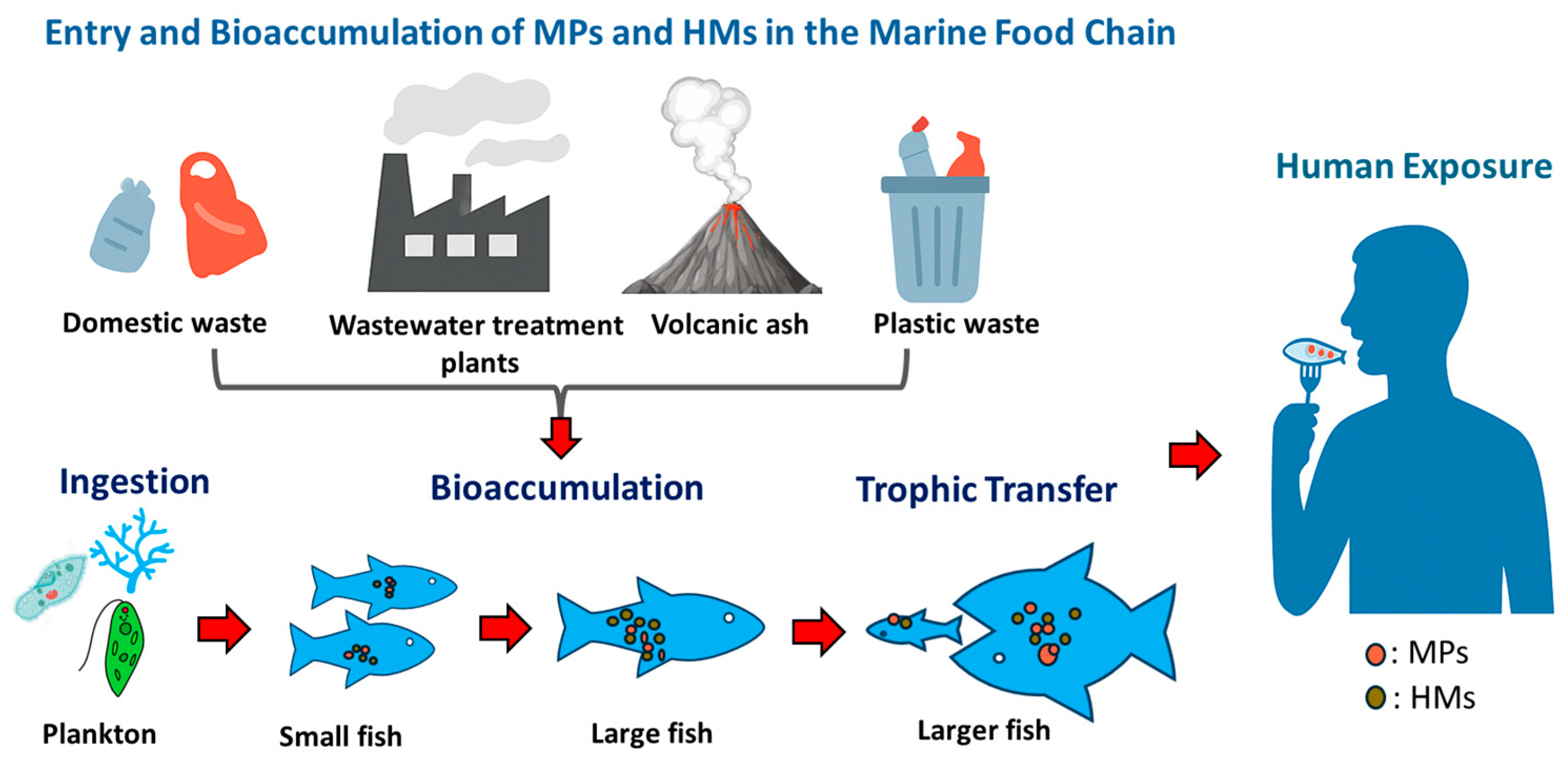
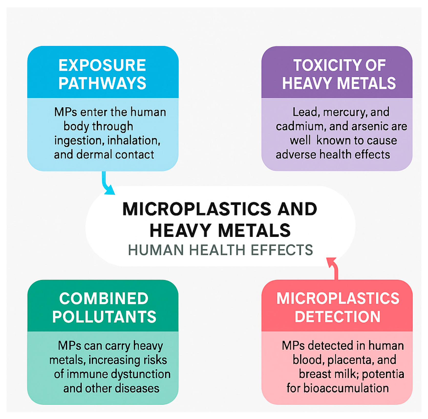
6. Potential Risk to Human Health

The potential impacts of microplastic (MP) exposure on human health remain an area of active investigation. The World Health Organization (WHO) and Science Advice for Policy by European Academies (SAPEA) have stated that, based on currently available evidence, there is insufficient data to conclusively determine whether the ingestion of MPs poses a direct risk to human health. However, the absence of definitive evidence does not equate to safety. Given the persistence and pervasiveness of MPs in the environment, global research efforts are intensifying to explore their possible toxicological effects using in vitro models, animal studies, and biomonitoring approaches.
On the other hand, sufficient data and research work are available for heavy metals, which successfully claim exposure to heavy metals is toxic to human health as shown in Table 5. The “Environmental Protection Agency” (EPA), in cooperation with the “Agency for Toxic Substance and Disease Registry (ATSDR)”, Georgia, reported a list called “priority list for 2001”, including the most toxic hazardous substance to human health, with As, Pb, and Hg ranked first, second, and third, respectively, while Cd is ranked seventh. Many researchers have confirmed the adverse effects of heavy metals on humans. Anthropogenic activities have increased mercury levels be in the atmosphere to roughly three times what they used to, and every year they are growing by 1.5 percent. It was found that 16 out of 70 fish on the southwest coast of India were polyethylene-contaminated. They also found that polyethylene and polypropylene are dominant pollutants in the Indian marine environment [108]. Tayebi and Sobhanardakani randomly collected 27 samples of tilapia fish from nine study areas and found Cd and Pb levels higher than the maximum permissible level (MPL) set by WHO [109]. The other group published similar results, showing that the mercury level in bluefin tuna fish was higher than the upper limit established by the European Commission [110].
The increase in the level of MPs in fish is supposed to increase the concentration of heavy metals, which threaten human health via the food chain. Many studies claim the vast presence of MPs and heavy metals in aquatic animals [111,112,113,114]. The MPs affect not only aquatic organisms but also plants. It is confirmed that polyethylene MPs can significantly reduce the internal activity of lentil seeds [4,115]. Microplastics have been detected in human stool, blood, placenta, breast milk, and urine, suggesting pervasive exposure through food, water, air, and dermal contact. MPs of 50 to 500 µm in size in human stool confirm that MPs enter the human body via various pathways such as skin, food, and water intake, and indeed affect human health after accumulation [116]. A recent study by Dr. Dick vethaak and his team found MPs in human blood for the first time. MPs were found in the blood of 80% of tested individuals, indicating potential for systemic transport and bioaccumulation. The deleterious effect on the human body is yet to be confirmed (12). MPs are found in 26 out of 34 women’s breast milk. Most MPs were irregular fragments of orange/yellow and blue colors, ranging from 2 µm to 12 µm. Reported MPs in breast milk samples further suggest potential neonatal exposure during breastfeeding. Ragusa detected the plastic in 4 out of 6 human placentae with dimensions ranging from 5 to 10 μm. The MPs were found spherical and irregular in shape [2,117]. These studies raise serious concerns about fetal exposure during pregnancy, when one has to be the safest. These findings support the hypothesis that MPs can cross biological barriers, especially those <10 µm. Furthermore, MPs were detected in the ovarian follicular fluid of 14 out of 18 women undergoing in vitro fertilization treatments, suggesting possible implications for female fertility [118]. Male reproductive health may also be at risk, as MPs have been identified in human testicular tissues and the penis, with animal models showing an association between MP exposure and reduced testicular weight [119]. A 2024 study identified MPs in the bone marrow of all 16 examined human samples, marking the first report of such a finding [120]. MPs have also been consistently found in the kidney and liver tissues, with increasing concentrations over time, suggesting systemic bioaccumulation through dietary and environmental exposure [121]. A groundbreaking study conducted between 2016 and 2024 found MPs in all 52 examined human brain samples, with a notable increase in particle concentration in the more recent specimens. Individuals diagnosed with dementia were found to have significantly higher levels of MPs, suggesting a potential link between MP accumulation and neurodegenerative diseases [122]. Similarly, MPs have been detected in the olfactory bulbs of eight out of fifteen cadavers, indicating that inhaled particles can bypass the blood–brain barrier and accumulate directly in the central nervous system [122]. However, their toxicokinetics, how they are absorbed, distributed, metabolized, and excreted, remain poorly understood. Early in vitro studies suggest that MPs can induce inflammation, oxidative stress, and genotoxicity, but human-scale evidence is still lacking.
The environment is spoiled by the industries that dispose of large amounts of hazardous chemicals, including heavy metals, which is why heavy metals in drinking water have exceeded the recommended dose. Heavy metals such as Cd and Hg are known to be very toxic to human health. Japan experienced fatal diseases caused by cd in 1915, known as as “Itai Itai”, and by methyl Hg in Minimata in 1956, which affected many people [123]. It has been found that lipid peroxidation significantly increases in some brain areas, such as the cortex and cerebellum, following cadmium exposure. Cd also has toxic effects on the enzymes that promote redox reactions. Consequently, lipid peroxidation is induced, which in turn causes microvasculature damage [124]. Worldwide, more than 140 million people are exposed to arsenic-contaminated potable water with a concentration exceeding the maximum limit (10 ppb) set by the World Health Organization. In Bangladesh and West Bengal, 40 to 80 million people drink arsenic-contaminated water ranging from 10 ppb to 4 ppm, resulting in one casualty out of every five people [125], as it affects the liver [126], kidney [127], lungs [128], skin [129,130], and leads to bladder cancer [131]. MPs can form a complex contamination bond with heavy metals, affecting the immune system even more severely and causing various diseases in humans and animals. The combined exposure to MPs and heavy metals is of particular concern. These co-contaminants may synergistically compromise the immune system, disrupt metabolic pathways, and enhance the risk of chronic diseases, including neurological disorders and cancer. As research progresses, there is growing consensus that the complex interactions between MPs and heavy metals could amplify toxicity beyond the effects of individual contaminants.
Table 5. Summary of the main sources of heavy metal pollution and their toxic effects on human health.
MetalsMain SourcesToxic EffectsRef.
ArsenicArsenical pesticides, natural mineral deposits, ground water and soil, improperly disposed arsenical chemicals, sewage fertilizers, and miningWeakness and anemia, skin and lung cancer, diabetic problems, neuropathy, gastrointestinal problems, cardiovascular failure, hematopoietic effects, and sometimes acute illness leading to death[129]
LeadStorage batteries; petrol additives, paint, cable sheathing and cosmetics, mining, pottery, and ceramic dishesAnemia, high risk of kidney damage, B.P problem, fertility and reproduction problems, nervous/brain system damage.[132]
MercuryMedical waste, toothpaste, skin cream, vaccines, batteries, volcanoes and oceans, and contaminated fishMuscle weakness, blindness, mental retardation, impairment of hearing, speech or/and walking, swollen gums, kidneys, and liver, immune system damage, and loss of memory and concentration[133]
CadmiumIndustrial waste, nickel–cadmium batteries, plastic and paint industries, cadmium alloy, welding, and smeltingBone fracture, diarrhea, nausea, problems in reproduction, stomach aches, shortness of breath, severe kidney, liver, and lung disease[134]
CopperMining, smelting, water pipes, copper wires, combustion of fossil fuels and copper sheet metalsNausea, headache, damage to red blood results, anemia, liver and kidney injury, and sometimes death[135]

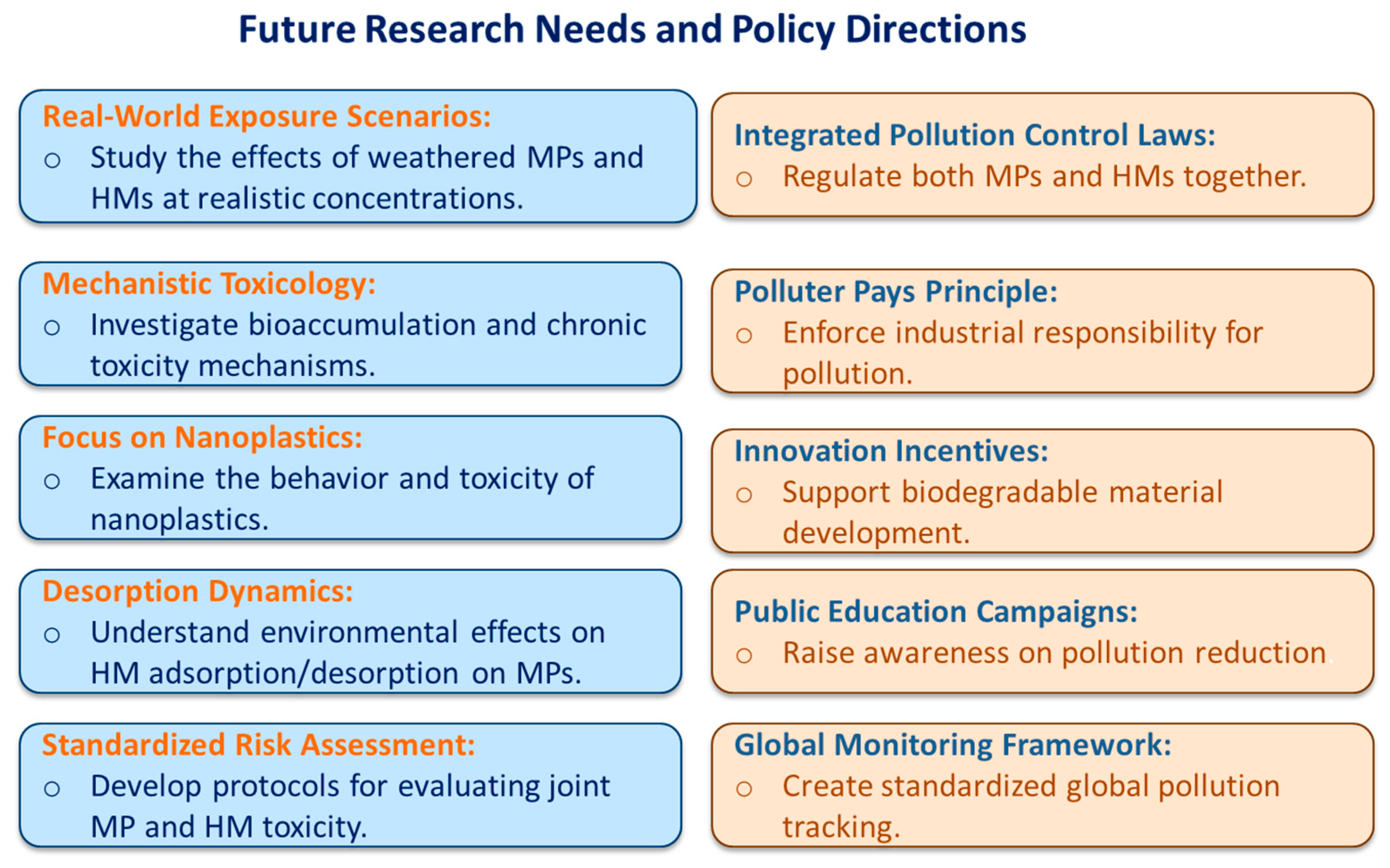
Critical Insights and Research Needs

While the presence of MPs in human tissues is now beyond dispute, causality between exposure and disease outcomes remains to be clearly established. Key knowledge gaps include the following: (1) The threshold dose and particle size required to induce toxic effects in humans. (2) Chronic exposure outcomes across different age groups, especially infants and pregnant women. (3) The potential for transgenerational effects via placental and breastfeeding routes.
Moreover, the risk assessment paradigm should move beyond single-substance analysis to mixture toxicity, particularly in real-world scenarios where MPs often co-exist with metals, organic pollutants, or pathogens. The possible exposure routes and associated health impacts of microplastics and heavy metals in humans are illustrated in Figure 3.

7. Conclusions and Future Perspective

The spread of MPs and heavy metals will continue to increase with an increase in human activities, since countries do not have adequate waste management measures. It is challenging to eliminate MPs and heavy metals from the environment due to their resistance to degradation and transformation. In order to control the pollution by heavy metals and MPs, strict policies need to be developed and implemented in a time-bound fashion. To conclude the research presented here, we can say that microplastics are harmful to aquatic organisms/microorganisms. Almost all types of microplastics, regardless of their shape, size, color, age, or virginity, can be consumed by aquatic organisms/microorganisms and transferred to higher trophic levels. Heavy metals can obstruct nutrient uptake within organisms, resulting in death. MPs and heavy metals can interact in several ways. This study discussed several potential relationships between MPs and heavy metals and concluded that MPs become more toxic when combined with heavy metals. Pollution caused by MPs and heavy metals is not only detrimental to aquatic microorganisms but also to humans. In recent years, many studies have revealed that microplastics are also present inside human beings, which will be a severe concern in the upcoming decades.
Although a tremendous amount of research has been conducted in this field, substantial gaps are yet to be filled; below are the few areas that must be addressed to fill these gaps.
Toxicity Studies: The toxic ecological effects of microplastic at environmental concentrations must be investigated. Toxicology tests are generally conducted using new and pristine MPs with defined sizes and high MP concentrations. Biotic or abiotic factors can also degrade MPs; however, the concentrations and toxicity of these minor MPs, including nanosized ones, are poorly understood, making it difficult to assess the risk. The effects of nanoplastics may be even greater.
Adsorption Mechanisms: To understand the adsorption/desorption mechanism of heavy metals on MPs, a variety of heavy metals and MP types should be studied.
Human Health Impacts: It has been confirmed that MPs can enter human bodies, but their toxic effects are yet to be proved. More research is needed to determine the harmful effects of MPs on humans and the mechanisms by which MPs and HMs influence human physiological systems, especially regarding bioaccumulation and chronic toxicity.
Policy and Mitigation: From a policy standpoint, comprehensive legislation is required to synergistically manage plastic and heavy metal pollution. Efforts must extend beyond banning single-use plastics to include the following:
  • Stricter control of industrial effluents.
  • Incentives for biodegradable material innovation.
  • Improved waste management and recycling systems.
  • Public awareness campaigns promoting responsible plastic use.
International cooperation is essential to standardize monitoring methods, set pollutant thresholds, and ensure compliance. Without decisive action, the environmental and health impacts of MPs and HMs are likely to escalate in the coming decades Figure 4, briefly highlights the future research needs and policy directions

Author Contributions

Conceptualization, A. and A.D.; methodology, A.D., Y.S.K.D.S., L.T. and A.; resources, A., A.D. and L.T.; data curation, A.D., Y.S.K.D.S., L.T. and A.; writing—original draft preparation, A.D., Y.S.K.D.S., L.T. and A.; writing—review and editing, A.D., Y.S.K.D.S., L.T. and A.; project administration, A. and A.D. All authors have read and agreed to the published version of the manuscript.

Funding

This research received no external funding.

Institutional Review Board Statement

Not applicable.

Informed Consent Statement

Not applicable.

Data Availability Statement

Not applicable.

Conflicts of Interest

The authors declare no conflicts of interest.

References

  1. Chae, Y.; An, Y.-J. Current research trends on plastic pollution and ecological impacts on the soil ecosystem: A review. Environ. Pollut. 2018, 240, 387–395. [Google Scholar] [CrossRef] [PubMed]
  2. Enyoh, C.E.; Devi, A.; Kadono, H.; Wang, Q.; Rabin, M.H. The plastic within: Microplastics invading human organs and bodily fluids systems. Environments 2023, 10, 194. [Google Scholar] [CrossRef]
  3. Enyoh, C.E.; Devi, A.; Maduka, T.O.; Tyagi, L.; Rana, S.; Akuwudike, I.S.; Wang, Q. A Review of Materials for the Removal of Micro- and Nanoplastics from Different Environments. Micro 2025, 5, 17. [Google Scholar] [CrossRef]
  4. Law, K.L. Plastics in the marine environment. Annu. Rev. Mar. Sci. 2017, 9, 205–229. [Google Scholar] [CrossRef] [PubMed]
  5. De Silva, Y.; Rajagopalan, U.; Kadono, H. Microplastics on the growth of plants and seed germination in aquatic and terrestrial ecosystems. Glob. J. Environ. Sci. Manag. 2021, 7, 347–368. [Google Scholar]
  6. do Sul, J.A.I.; Costa, M.F. The present and future of microplastic pollution in the marine environment. Environ. Pollut. 2014, 185, 352–364. [Google Scholar] [CrossRef]
  7. Crawford, C.B.; Quinn, B. Microplastic Pollutants; Elsevier Limited: Amsterdam, The Netherlands, 2016. [Google Scholar]
  8. Kim, H.J.; Shin, S.R.; Park, J.J.; Lee, J.S. Feeding, excretion, survival, and histological alterations in zebrafish Danio rerio from single and combined exposure to microplastics and copper. Korean J. Environ. Biol. 2024, 42, 1–14. [Google Scholar] [CrossRef]
  9. Mance, G. Pollution Threat of Heavy Metals in Aquatic Environments; Springer Science & Business Media: Berlin/Heidelberg, Germany, 2012. [Google Scholar]
  10. Bhupander, K.; Mukherjee, D. Assessment of human health risk for arsenic, copper, nickel, mercury and zinc in fish collected from tropical wetlands in India. Adv. Life Sci. Technol. 2011, 2, 13–24. [Google Scholar]
  11. Łuczyńska, J.; Paszczyk, B.; Łuczyński, M.J. Fish as a bioindicator of heavy metals pollution in aquatic ecosystem of Pluszne Lake, Poland, and risk assessment for consumer’s health. Ecotoxicol. Environ. Saf. 2018, 153, 60–67. [Google Scholar] [CrossRef]
  12. Al Naggar, Y.; Khalil, M.S.; Ghorab, M.A. Environmental pollution by heavy metals in the aquatic ecosystems of Egypt. Open Acc. J. Toxicol 2018, 3, 555603. [Google Scholar]
  13. Hylander, L.D.; Goodsite, M.E. Environmental costs of mercury pollution. Sci. Total Environ. 2006, 368, 352–370. [Google Scholar] [CrossRef]
  14. Besseling, E.; Wegner, A.; Foekema, E.M.; Van Den Heuvel-Greve, M.J.; Koelmans, A.A. Effects of microplastic on fitness and PCB bioaccumulation by the lugworm Arenicola marina (L.). Environ. Sci. Technol. 2013, 47, 593–600. [Google Scholar] [CrossRef]
  15. Lee, K.-W.; Shim, W.J.; Kwon, O.Y.; Kang, J.-H. Size-dependent effects of micro polystyrene particles in the marine copepod Tigriopus japonicus. Environ. Sci. Technol. 2013, 47, 11278–11283. [Google Scholar] [CrossRef] [PubMed]
  16. Von Moos, N.; Burkhardt-Holm, P.; Köhler, A. Uptake and effects of microplastics on cells and tissue of the blue mussel Mytilus edulis L. after an experimental exposure. Environ. Sci. Technol. 2012, 46, 11327–11335. [Google Scholar] [CrossRef] [PubMed]
  17. Oliveira, M.; Ribeiro, A.; Hylland, K.; Guilhermino, L. Single and combined effects of microplastics and pyrene on juveniles (0+ group) of the common goby Pomatoschistus microps (Teleostei, Gobiidae). Ecol. Indic. 2013, 34, 641–647. [Google Scholar] [CrossRef]
  18. de Sá, L.C.; Luís, L.G.; Guilhermino, L. Effects of microplastics on juveniles of the common goby (Pomatoschistus microps): Confusion with prey, reduction of the predatory performance and efficiency, and possible influence of developmental conditions. Environ. Pollut. 2015, 196, 359–362. [Google Scholar] [CrossRef]
  19. Wright, S.L.; Thompson, R.C.; Galloway, T.S. The physical impacts of microplastics on marine organisms: A review. Environ. Pollut. 2013, 178, 483–492. [Google Scholar] [CrossRef]
  20. Bulannga, R.B.; Schmidt, S. Uptake and accumulation of microplastic particles by two freshwater ciliates isolated from a local river in South Africa. Environ. Res. 2022, 204, 112123. [Google Scholar] [CrossRef]
  21. Murphy, F.; Quinn, B. The effects of microplastic on freshwater Hydra attenuata feeding, morphology & reproduction. Environ. Pollut. 2018, 234, 487–494. [Google Scholar]
  22. Cole, M.; Lindeque, P.; Fileman, E.; Halsband, C.; Goodhead, R.; Moger, J.; Galloway, T.S. Microplastic ingestion by zooplankton. Environ. Sci. Technol. 2013, 47, 6646–6655. [Google Scholar] [CrossRef]
  23. Setälä, O.; Fleming-Lehtinen, V.; Lehtiniemi, M. Ingestion and transfer of microplastics in the planktonic food web. Environ. Pollut. 2014, 185, 77–83. [Google Scholar] [CrossRef] [PubMed]
  24. Wang, M.; Wang, X.; Luo, X.; Zheng, H. Short-term toxicity of polystryrene microplastics on mysid shrimps Neomysis japonica. IOP Conf. Ser. Earth Environ. Sci. 2017, 61, 012136. [Google Scholar] [CrossRef]
  25. Nugroho, F.A.; Fyda, J. Uptake of plastic microbeads by ciliate Paramecium aurelia. Sci. Technol. Innov. 2020, 9, 1–9. [Google Scholar] [CrossRef]
  26. Sun, X.; Li, Q.; Zhu, M.; Liang, J.; Zheng, S.; Zhao, Y. Ingestion of microplastics by natural zooplankton groups in the northern South China Sea. Mar. Pollut. Bull. 2017, 115, 217–224. [Google Scholar] [CrossRef]
  27. Vroom, R.J.E.; Koelmans, A.A.; Besseling, E.; Halsband, C. Aging of microplastics promotes their ingestion by marine zooplankton. Env. Pollut 2017, 231, 987–996. [Google Scholar] [CrossRef]
  28. Lo, H.K.A.; Chan, K.Y.K. Negative effects of microplastic exposure on growth and development of Crepidula onyx. Environ. Pollut. 2018, 233, 588–595. [Google Scholar] [CrossRef]
  29. Messinetti, S.; Mercurio, S.; Parolini, M.; Sugni, M.; Pennati, R. Effects of polystyrene microplastics on early stages of two marine invertebrates with different feeding strategies. Environ. Pollut. 2018, 237, 1080–1087. [Google Scholar] [CrossRef]
  30. Choi, J.S.; Jung, Y.-J.; Hong, N.-H.; Hong, S.H.; Park, J.-W. Toxicological effects of irregularly shaped and spherical microplastics in a marine teleost, the sheepshead minnow (Cyprinodon variegatus). Mar. Pollut. Bull. 2018, 129, 231–240. [Google Scholar] [CrossRef]
  31. Desforges, J.-P.W.; Galbraith, M.; Ross, P.S. Ingestion of microplastics by zooplankton in the Northeast Pacific Ocean. Arch. Environ. Contam. Toxicol. 2015, 69, 320–330. [Google Scholar] [CrossRef]
  32. Amélineau, F.; Bonnet, D.; Heitz, O.; Mortreux, V.; Harding, A.M.; Karnovsky, N.; Walkusz, W.; Fort, J.; Grémillet, D. Microplastic pollution in the Greenland Sea: Background levels and selective contamination of planktivorous diving seabirds. Environ. Pollut. 2016, 219, 1131–1139. [Google Scholar] [CrossRef]
  33. Rebolledo, E.L.B.; Van Franeker, J.A.; Jansen, O.E.; Brasseur, S.M. Plastic ingestion by harbour seals (Phoca vitulina) in The Netherlands. Mar. Pollut. Bull. 2013, 67, 200–202. [Google Scholar] [CrossRef] [PubMed]
  34. Besseling, E.; Foekema, E.; Van Franeker, J.; Leopold, M.; Kühn, S.; Rebolledo, E.B.; Heße, E.; Mielke, L.; IJzer, J.; Kamminga, P. Microplastic in a macro filter feeder: Humpback whale Megaptera novaeangliae. Mar. Pollut. Bull. 2015, 95, 248–252. [Google Scholar] [CrossRef]
  35. Lusher, A.L.; Hernandez-Milian, G.; O’Brien, J.; Berrow, S.; O’Connor, I.; Officer, R. Microplastic and macroplastic ingestion by a deep diving, oceanic cetacean: The True’s beaked whale Mesoplodon mirus. Environ. Pollut. 2015, 199, 185–191. [Google Scholar] [CrossRef]
  36. Nadal, M.; Alomar, C.; Deudero, S. High levels of microplastic ingestion by the semipelagic fish bogue Boops boops (L.) around the Balearic Islands. Environ. Pollut. 2016, 214, 517–523. [Google Scholar] [CrossRef] [PubMed]
  37. Browne, M.A.; Dissanayake, A.; Galloway, T.S.; Lowe, D.M.; Thompson, R.C. Ingested microscopic plastic translocates to the circulatory system of the mussel, Mytilus edulis (L.). Environ. Sci. Technol. 2008, 42, 5026–5031. [Google Scholar] [CrossRef]
  38. Graham, E.R.; Thompson, J.T. Deposit-and suspension-feeding sea cucumbers (Echinodermata) ingest plastic fragments. J. Exp. Mar. Biol. Ecol. 2009, 368, 22–29. [Google Scholar] [CrossRef]
  39. Kühn, S.; van Franeker, J.A. Plastic ingestion by the northern fulmar (Fulmarus glacialis) in Iceland. Mar. Pollut. Bull. 2012, 64, 1252–1254. [Google Scholar] [CrossRef] [PubMed]
  40. Hall, N.; Berry, K.; Rintoul, L.; Hoogenboom, M. Microplastic ingestion by scleractinian corals. Mar. Biol. 2015, 162, 725–732. [Google Scholar] [CrossRef]
  41. Cole, M.; Lindeque, P.; Fileman, E.; Halsband, C.; Galloway, T.S. The impact of polystyrene microplastics on feeding, function and fecundity in the marine copepod Calanus helgolandicus. Environ. Sci. Technol. 2015, 49, 1130–1137. [Google Scholar] [CrossRef]
  42. Nobre, C.; Santana, M.; Maluf, A.; Cortez, F.; Cesar, A.; Pereira, C.; Turra, A. Assessment of microplastic toxicity to embryonic development of the sea urchin Lytechinus variegatus (Echinodermata: Echinoidea). Mar. Pollut. Bull. 2015, 92, 99–104. [Google Scholar] [CrossRef]
  43. Kaposi, K.L.; Mos, B.; Kelaher, B.P.; Dworjanyn, S.A. Ingestion of microplastic has limited impact on a marine larva. Environ. Sci. Technol. 2014, 48, 1638–1645. [Google Scholar] [CrossRef] [PubMed]
  44. Cole, M. Microplastic Swallowing Zooplankton Environ. Sci. Technol. 2013, 47, 6646–6655. [Google Scholar] [CrossRef]
  45. Farrell, P.; Nelson, K. Trophic level transfer of microplastic: Mytilus edulis (L.) to Carcinus maenas (L.). Environ. Pollut. 2013, 177, 1–3. [Google Scholar] [CrossRef] [PubMed]
  46. Costa, E.; Piazza, V.; Lavorano, S.; Faimali, M.; Garaventa, F.; Gambardella, C. Trophic transfer of microplastics from copepods to jellyfish in the marine environment. Front. Environ. Sci. 2020, 8, 571732. [Google Scholar] [CrossRef]
  47. Lu, Y.; Zhang, Y.; Deng, Y.; Jiang, W.; Zhao, Y.; Geng, J.; Ding, L.; Ren, H. Uptake and accumulation of polystyrene microplastics in zebrafish (Danio rerio) and toxic effects in liver. Environ. Sci. Technol. 2016, 50, 4054–4060. [Google Scholar] [CrossRef]
  48. Watts, A.J.; Urbina, M.A.; Goodhead, R.; Moger, J.; Lewis, C.; Galloway, T.S. Effect of microplastic on the gills of the shore crab Carcinus maenas. Environ. Sci. Technol. 2016, 50, 5364–5369. [Google Scholar] [CrossRef]
  49. Pandey, G.; Madhuri, S. Heavy metals causing toxicity in animals and fishes. Res. J. Anim. Vet. Fish. Sci. 2014, 2, 17–23. [Google Scholar]
  50. Vieira, L.R.; Gravato, C.; Soares, A.; Morgado, F.; Guilhermino, L. Acute effects of copper and mercury on the estuarine fish Pomatoschistus microps: Linking biomarkers to behaviour. Chemosphere 2009, 76, 1416–1427. [Google Scholar] [CrossRef]
  51. Tian, Y.; Liu, H.; Wang, Q.; Zhou, J.; Tang, X. Acute and chronic toxic effects of Pb2+ on polychaete Perinereis aibuhitensis: Morphological changes and responses of the antioxidant system. J. Environ. Sci. 2014, 26, 1681–1688. [Google Scholar] [CrossRef]
  52. Aliko, V.; Qirjo, M.; Sula, E.; Morina, V.; Faggio, C. Antioxidant defense system, immune response and erythron profile modulation in gold fish, Carassius auratus, after acute manganese treatment. Fish Shellfish Immunol. 2018, 76, 101–109. [Google Scholar] [CrossRef]
  53. Palaniappan, P.R.; Karthikeyan, S. Bioaccumulation and depuration of chromium in the selected organs and whole body tissues of freshwater fish Cirrhinus mrigala individually and in binary solutions with nickel. J. Environ. Sci. 2009, 21, 229–236. [Google Scholar] [CrossRef]
  54. Ay, Ö.; Kalay, M.; Tamer, L.; Canli, M. Copper and lead accumulation in tissues of a freshwater fish Tilapia zillii and its effects on the branchial Na, K-ATPase activity. Bull. Environ. Contam. Toxicol. 1999, 62, 160–168. [Google Scholar] [CrossRef]
  55. Firat, Ö.; Kargın, F. Biochemical alterations induced by Zn and Cd individually or in combination in the serum of Oreochromis niloticus. Fish Physiol. Biochem. 2010, 36, 647–653. [Google Scholar] [CrossRef]
  56. Kong, X.; Jiang, H.; Wang, S.; Wu, X.; Fei, W.; Li, L.; Nie, G.; Li, X. Effects of copper exposure on the hatching status and antioxidant defense at different developmental stages of embryos and larvae of goldfish Carassius auratus. Chemosphere 2013, 92, 1458–1464. [Google Scholar] [CrossRef]
  57. Atli, G.; Canli, M. Metals (Ag+, Cd2+, Cr6+) affect ATPase activity in the gill, kidney, and muscle of freshwater fish Oreochromis niloticus following acute and chronic exposures. Environ. Toxicol. 2013, 28, 707–717. [Google Scholar] [CrossRef]
  58. Kim, J.-H.; Kang, J.-C. Oxidative stress, neurotoxicity, and metallothionein (MT) gene expression in juvenile rock fish Sebastes schlegelii under the different levels of dietary chromium (Cr6+) exposure. Ecotoxicol. Environ. Saf. 2016, 125, 78–84. [Google Scholar] [CrossRef]
  59. Aaryashree; Kumar Choudhary, A.; Devi, A.; Tyagi, L.; Maheshawari Rajagopalan, U.; Kadono, H. Non-invasive detection of arsenic-induced changes in lentil seed activity using biospeckle optical coherence tomography. Opt. Contin. 2025, 4, 968–984. [Google Scholar] [CrossRef]
  60. Kim, J.-H.; Kang, J.-C. The arsenic accumulation and its effect on oxidative stress responses in juvenile rockfish, Sebastes schlegelii, exposed to waterborne arsenic (As3+). Environ. Toxicol. Pharmacol. 2015, 39, 668–676. [Google Scholar] [CrossRef]
  61. Garcia-Santos, S.; Monteiro, S.; Malakpour-Kolbadinezhad, S.; Fontaínhas-Fernandes, A.; Wilson, J. Effects of Cd injection on osmoregulation and stress indicators in freshwater Nile tilapia. Comp. Biochem. Physiol. Part C Toxicol. Pharmacol. 2015, 167, 81–89. [Google Scholar] [CrossRef]
  62. Bhattacharya, P.; Samal, A.C.; Majumdar, J.; Santra, S.C. Uptake of arsenic in rice plant varieties cultivated with arsenic rich groundwater. Environ. Asia 2010, 3, 34–37. [Google Scholar]
  63. Chen, Q.; Gundlach, M.; Yang, S.; Jiang, J.; Velki, M.; Yin, D.; Hollert, H. Quantitative investigation of the mechanisms of microplastics and nanoplastics toward zebrafish larvae locomotor activity. Sci. Total Environ. 2017, 584, 1022–1031. [Google Scholar] [CrossRef]
  64. Devi, A.; Kadono, H.; Rajagopalan, U.M. Fast Assessment of Quality of Water Containing Inorganic Pollutants Using Laser Biospeckles in Microbioassay. Appl. Sci. 2024, 14, 5558. [Google Scholar] [CrossRef]
  65. Devi, A.; Kadono, H.; Rajagopalan, U.M. Laser biospeckle method for a fast and reliable microbioassay. Appl. Opt. 2024, 63, 5721–5727. [Google Scholar] [CrossRef]
  66. Devi, A.; Kadono, H.; Rajagopalan, U.M. Fast and reliable micro bioassay techniques based on biospeckle for swift water assessment using plankton. Biophotonics Point-Care III 2024, 13008, 1300804. [Google Scholar]
  67. Bouraiou, C.; Berrebbah, H.; Djekoun, M.; Bouarroudj, T.; Yahyaoui, A.; Khene, L.; Kahli, H.; Djebar, M.R. Growth inhibition and oxidative stress in the freshwater Ciliate paramecium sp. exposed to copper oxide nanoparticles. Stud. Univ. “ Vasile Goldis” Arad. Ser. Stiintele Vietii (Life Sci. Ser.) 2019, 29, 82–90. [Google Scholar]
  68. Ashouri, S.; Keyvanshokooh, S.; Salati, A.P.; Johari, S.A.; Pasha-Zanoosi, H. Effects of different levels of dietary selenium nanoparticles on growth performance, muscle composition, blood biochemical profiles and antioxidant status of common carp (Cyprinus carpio). Aquaculture 2015, 446, 25–29. [Google Scholar] [CrossRef]
  69. Zhu, X.; Chang, Y.; Chen, Y. Toxicity and bioaccumulation of TiO2 nanoparticle aggregates in Daphnia magna. Chemosphere 2010, 78, 209–215. [Google Scholar] [CrossRef]
  70. Moyson, S.; Liew, H.J.; Fazio, A.; Van Dooren, N.; Delcroix, A.; Faggio, C.; Blust, R.; De Boeck, G. Kidney activity increases in copper exposed goldfish (Carassius auratus auratus). Comp. Biochem. Physiol. Part C Toxicol. Pharmacol. 2016, 190, 32–37. [Google Scholar] [CrossRef]
  71. Shunmugam, A.P.; Subramanian, G.; Fernandez, J.G. Measurements of the swimming speeds of motile microorganisms using object tracking and their correlation with water pollution and rheology levels. Sci. Rep. 2021, 11, 11821. [Google Scholar] [CrossRef]
  72. Nilsson, J.R. Effects of copper on phagocytosis inTetrahymena. Protoplasma 1981, 109, 359–370. [Google Scholar] [CrossRef]
  73. Steer, M.; Cole, M.; Thompson, R.C.; Lindeque, P.K. Microplastic ingestion in fish larvae in the western English Channel. Environ. Pollut. 2017, 226, 250–259. [Google Scholar] [CrossRef]
  74. McGoran, A.R.; Clark, P.F.; Morritt, D. Presence of microplastic in the digestive tracts of European flounder, Platichthys flesus, and European smelt, Osmerus eperlanus, from the River Thames. Environ. Pollut. 2017, 220, 744–751. [Google Scholar] [CrossRef]
  75. Andrady, A.L. Microplastics in the marine environment. Mar. Pollut. Bull. 2011, 62, 1596–1605. [Google Scholar] [CrossRef]
  76. Shen, M.; Zhang, Y.; Zhu, Y.; Song, B.; Zeng, G.; Hu, D.; Wen, X.; Ren, X. Recent advances in toxicological research of nanoplastics in the environment: A review. Environ. Pollut. 2019, 252, 511–521. [Google Scholar] [CrossRef]
  77. Maršić-Lučić, J.; Lušić, J.; Tutman, P.; Varezić, D.B.; Šiljić, J.; Pribudić, J. Levels of trace metals on microplastic particles in beach sediments of the island of Vis, Adriatic Sea, Croatia. Mar. Pollut. Bull. 2018, 137, 231–236. [Google Scholar] [CrossRef]
  78. Holmes, L.A.; Turner, A.; Thompson, R.C. Interactions between trace metals and plastic production pellets under estuarine conditions. Mar. Chem. 2014, 167, 25–32. [Google Scholar] [CrossRef]
  79. Rochman, C.M.; Hoh, E.; Kurobe, T.; Teh, S.J. Ingested plastic transfers hazardous chemicals to fish and induces hepatic stress. Sci. Rep. 2013, 3, 3263. [Google Scholar] [CrossRef]
  80. Hamed, M.; Soliman, H.A.; Osman, A.G.; Sayed, A.E.-D.H. Antioxidants and molecular damage in Nile Tilapia (Oreochromis niloticus) after exposure to microplastics. Environ. Sci. Pollut. Res. 2020, 27, 14581–14588. [Google Scholar] [CrossRef]
  81. Banaee, M.; Soltanian, S.; Sureda, A.; Gholamhosseini, A.; Haghi, B.N.; Akhlaghi, M.; Derikvandy, A. Evaluation of single and combined effects of cadmium and micro-plastic particles on biochemical and immunological parameters of common carp (Cyprinus carpio). Chemosphere 2019, 236, 124335. [Google Scholar] [CrossRef]
  82. Chen, X.; Peng, L.-B.; Wang, D.; Zhu, Q.-L.; Zheng, J.-L. Combined effects of polystyrene microplastics and cadmium on oxidative stress, apoptosis, and GH/IGF axis in zebrafish early life stages. Sci. Total Environ. 2022, 813, 152514. [Google Scholar] [CrossRef]
  83. Jinhui, S.; Sudong, X.; Yan, N.; Xia, P.; Jiahao, Q.; Yongjian, X. Effects of microplastics and attached heavy metals on growth, immunity, and heavy metal accumulation in the yellow seahorse, Hippocampus kuda Bleeker. Mar. Pollut. Bull. 2019, 149, 110510. [Google Scholar] [CrossRef]
  84. Yan, W.; Hamid, N.; Deng, S.; Jia, P.-P.; Pei, D.-S. Individual and combined toxicogenetic effects of microplastics and heavy metals (Cd, Pb, and Zn) perturb gut microbiota homeostasis and gonadal development in marine medaka (Oryzias melastigma). J. Hazard. Mater. 2020, 397, 122795. [Google Scholar] [CrossRef]
  85. Davarpanah, E.; Guilhermino, L. Single and combined effects of microplastics and copper on the population growth of the marine microalgae Tetraselmis chuii. Estuar. Coast. Shelf Sci. 2015, 167, 269–275. [Google Scholar] [CrossRef]
  86. Barboza, L.G.A.; Vieira, L.R.; Guilhermino, L. Single and combined effects of microplastics and mercury on juveniles of the European seabass (Dicentrarchus labrax): Changes in behavioural responses and reduction of swimming velocity and resistance time. Environ. Pollut. 2018, 236, 1014–1019. [Google Scholar] [CrossRef]
  87. Wen, B.; Jin, S.-R.; Chen, Z.-Z.; Gao, J.-Z.; Liu, Y.-N.; Liu, J.-H.; Feng, X.-S. Single and combined effects of microplastics and cadmium on the cadmium accumulation, antioxidant defence and innate immunity of the discus fish (Symphysodon aequifasciatus). Environ. Pollut. 2018, 243, 462–471. [Google Scholar] [CrossRef]
  88. Wang, Z.; Fu, D.; Gao, L.; Qi, H.; Su, Y.; Peng, L. Aged microplastics decrease the bioavailability of coexisting heavy metals to microalga Chlorella vulgaris. Ecotoxicol. Environ. Saf. 2021, 217, 112199. [Google Scholar] [CrossRef]
  89. Lu, K.; Qiao, R.; An, H.; Zhang, Y. Influence of microplastics on the accumulation and chronic toxic effects of cadmium in zebrafish (Danio rerio). Chemosphere 2018, 202, 514–520. [Google Scholar] [CrossRef]
  90. Yuan, W.; Zhou, Y.; Chen, Y.; Liu, X.; Wang, J. Toxicological effects of microplastics and heavy metals on the Daphnia magna. Sci. Total Environ. 2020, 746, 141254. [Google Scholar] [CrossRef]
  91. Wang, Z.; Dong, H.; Wang, Y.; Ren, R.; Qin, X.; Wang, S. Effects of microplastics and their adsorption of cadmium as vectors on the cladoceran Moina monogolica Daday: Implications for plastic-ingesting organisms. J. Hazard. Mater. 2020, 400, 123239. [Google Scholar] [CrossRef]
  92. Zhou, Y.; Liu, X.; Wang, J. Ecotoxicological effects of microplastics and cadmium on the earthworm Eisenia foetida. J. Hazard. Mater. 2020, 392, 122273. [Google Scholar] [CrossRef]
  93. Santos, D.; Félix, L.; Luzio, A.; Parra, S.; Bellas, J.; Monteiro, S.M. Single and combined acute and subchronic toxic effects of microplastics and copper in zebrafish (Danio rerio) early life stages. Chemosphere 2021, 277, 130262. [Google Scholar] [CrossRef]
  94. Liu, S.; Shi, J.; Wang, J.; Dai, Y.; Li, H.; Li, J.; Liu, X.; Chen, X.; Wang, Z.; Zhang, P. Interactions between microplastics and heavy metals in aquatic environments: A review. Front. Microbiol. 2021, 12, 730. [Google Scholar] [CrossRef]
  95. Zhang, X.; Zheng, M.; Wang, L.; Lou, Y.; Shi, L.; Jiang, S. Sorption of three synthetic musks by microplastics. Mar. Pollut. Bull. 2018, 126, 606–609. [Google Scholar] [CrossRef]
  96. Zon, N.F.; Iskendar, A.; Azman, S.; Sarijan, S.; Ismail, R. Sorptive behaviour of chromium on polyethylene microbeads in artificial seawater. MATEC Web Conf. 2018, 250, 06001. [Google Scholar] [CrossRef]
  97. Nethaji, S.; Sivasamy, A.; Mandal, A. Adsorption isotherms, kinetics and mechanism for the adsorption of cationic and anionic dyes onto carbonaceous particles prepared from Juglans regia shell biomass. Int. J. Environ. Sci. Technol. 2013, 10, 231–242. [Google Scholar] [CrossRef]
  98. Öz, N.; Kadizade, G.; Yurtsever, M. Investigation of heavy metal adsorption on microplastics. Appl. Ecol. Environ. Res. 2019, 17, 7301–7310. [Google Scholar] [CrossRef]
  99. Sahoo, T.R.; Prelot, B. Adsorption processes for the removal of contaminants from wastewater: The perspective role of nanomaterials and nanotechnology. In Nanomaterials for the Detection and Removal of Wastewater Pollutants; Elsevier: Amsterdam, The Netherlands, 2020; pp. 161–222. [Google Scholar]
  100. Ford, R.G.; Sparks, D.L. Frontiers in metal sorption/precipitation mechanisms on soil mineral surfaces. Adv. Agron. 2001, 74, 41–62. [Google Scholar]
  101. Abdurahman, A.; Cui, K.; Wu, J.; Li, S.; Gao, R.; Dai, J.; Liang, W.; Zeng, F. Adsorption of dissolved organic matter (DOM) on polystyrene microplastics in aquatic environments: Kinetic, isotherm and site energy distribution analysis. Ecotoxicol. Environ. Saf. 2020, 198, 110658. [Google Scholar] [CrossRef]
  102. Guo, X.; Hu, G.; Fan, X.; Jia, H. Sorption properties of cadmium on microplastics: The common practice experiment and a two-dimensional correlation spectroscopic study. Ecotoxicol. Environ. Saf. 2020, 190, 110118. [Google Scholar] [CrossRef] [PubMed]
  103. Chen, S.; Tan, Z.; Qi, Y.; Ouyang, C. Sorption of tri-n-butyl phosphate and tris (2-chloroethyl) phosphate on polyethylene and polyvinyl chloride microplastics in seawater. Mar. Pollut. Bull. 2019, 149, 110490. [Google Scholar] [CrossRef] [PubMed]
  104. Ezzati, R.; Ezzati, S.; Azizi, M. Exact solution of the Langmuir rate equation: New Insights into pseudo-first-order and pseudo-second-order kinetics models for adsorption. Vacuum 2024, 220, 112790. [Google Scholar] [CrossRef]
  105. Satyam, S.; Patra, S. Innovations and challenges in adsorption-based wastewater remediation: A comprehensive review. Heliyon 2024, 10, e29573. [Google Scholar] [CrossRef]
  106. Kanagalakshmi, M.; Devi, S.G.; Ananthi, P.; Pius, A. Adsorption isotherms and kinetic models. In Carbon Nanomaterials and Their Composites as Adsorbents; Springer: Berlin/Heidelberg, Germany, 2024; pp. 135–154. [Google Scholar]
  107. Tenea, A.-G.; Dinu, C.; Rus, P.A.; Ionescu, I.A.; Gheorghe, S.; Iancu, V.I.; Vasile, G.G.; Pascu, L.F.; Chiriac, F.L. Exploring adsorption dynamics of heavy metals onto varied commercial microplastic substrates: Isothermal models and kinetics analysis. Heliyon 2024, 10, e35364. [Google Scholar] [CrossRef]
  108. Robin, R.; Karthik, R.; Purvaja, R.; Ganguly, D.; Anandavelu, I.; Mugilarasan, M.; Ramesh, R. Holistic assessment of microplastics in various coastal environmental matrices, southwest coast of India. Sci. Total Environ. 2020, 703, 134947. [Google Scholar] [CrossRef]
  109. Tayebi, L.; Sobhanardakani, S. Analysis of heavy metal contents and non-carcinogenic health risk assessment through consumption of Tilapia Fish (Oreochromis niloticus). Pollution 2020, 6, 59–67. [Google Scholar]
  110. Milatou, N.; Dassenakis, M.; Megalofonou, P. Mercury concentrations in reared Atlantic bluefin tuna and risk assessment for the consumers: To eat or not to eat? Food Chem. 2020, 331, 127267. [Google Scholar] [CrossRef]
  111. Karami, A.; Golieskardi, A.; Ho, Y.B.; Larat, V.; Salamatinia, B. Microplastics in eviscerated flesh and excised organs of dried fish. Sci. Rep. 2017, 7, 5473. [Google Scholar] [CrossRef]
  112. Simionov, I.-A.; Cristea, V.; Petrea, Ş.-M.; Coadă, M.T.; Cristea, D.S. The presence of heavy metals in fish meat from Danube River: An overview. Aquac. Aquar. Conserv. Legis. 2016, 9, 1388–1399. [Google Scholar]
  113. Ture, M.; Kilic, M.B.; Altinok, I. Relationship between heavy metal accumulation in fish muscle and heavy metal resistance genes in bacteria isolated from fish. Biol. Trace Elem. Res. 2021, 199, 1595–1603. [Google Scholar] [CrossRef] [PubMed]
  114. Wootton, N.; Reis-Santos, P.; Gillanders, B.M. Microplastic in fish–A global synthesis. Rev. Fish Biol. Fish. 2021, 31, 753–771. [Google Scholar] [CrossRef]
  115. De Silva, Y.S.K.; Rajagopalan, U.M.; Kadono, H.; Li, D. Positive and negative phenotyping of increasing Zn concentrations by Biospeckle Optical Coherence Tomography in speedy monitoring on lentil (Lens culinaris) seed germination and seedling growth. Plant Stress 2021, 2, 100041. [Google Scholar] [CrossRef]
  116. Schwabl, P.; Köppel, S.; Königshofer, P.; Bucsics, T.; Trauner, M.; Reiberger, T.; Liebmann, B. Detection of various microplastics in human stool: A prospective case series. Ann. Intern. Med. 2019, 171, 453–457. [Google Scholar] [CrossRef]
  117. Ragusa, A.; Notarstefano, V.; Svelato, A.; Belloni, A.; Gioacchini, G.; Blondeel, C.; Zucchelli, E.; De Luca, C.; D’Avino, S.; Gulotta, A. Raman Microspectroscopy Detection and Characterisation of Microplastics in Human Breastmilk. Polymers 2022, 14, 2700. [Google Scholar] [CrossRef]
  118. Montano, L.; Raimondo, S.; Piscopo, M.; Ricciardi, M.; Guglielmino, A.; Chamayou, S.; Gentile, R.; Gentile, M.; Rapisarda, P.; Conti, G.O. First evidence of microplastics in human ovarian follicular fluid: An emerging threat to female fertility. Ecotoxicol. Environ. Saf. 2025, 291, 117868. [Google Scholar] [CrossRef]
  119. Zhao, Q.; Zhu, L.; Weng, J.; Jin, Z.; Cao, Y.; Jiang, H.; Zhang, Z. Detection and characterization of microplastics in the human testis and semen. Sci. Total Environ. 2023, 877, 162713. [Google Scholar] [CrossRef]
  120. Guo, X.; Wang, L.; Wang, X.; Li, D.; Wang, H.; Xu, H.; Liu, Y.; Kang, R.; Chen, Q.; Zheng, L. Discovery and analysis of microplastics in human bone marrow. J. Hazard. Mater. 2024, 477, 135266. [Google Scholar] [CrossRef]
  121. Sánchez, A.; Rodríguez-Viso, P.; Domene, A.; Orozco, H.; Vélez, D.; Devesa, V. Dietary microplastics: Occurrence, exposure and health implications. Environ. Res. 2022, 212, 113150. [Google Scholar] [CrossRef]
  122. Amato-Lourenço, L.F.; Dantas, K.C.; Júnior, G.R.; Paes, V.R.; Ando, R.A.; de Oliveira Freitas, R.; da Costa, O.M.M.M.; Rabelo, R.S.; Bispo, K.C.S.; Carvalho-Oliveira, R. Microplastics in the olfactory bulb of the human brain. JAMA Netw. Open 2024, 7, e2440018. [Google Scholar] [CrossRef] [PubMed]
  123. Kaji, M. Role of experts and public participation in pollution control: The case of Itai-itai disease in Japan1. Ethics Sci. Environ. Politics 2012, 12, 99–111. [Google Scholar] [CrossRef]
  124. Shukla, A.; Shukla, G.S.; Srimal, R. Cadmium-induced alterations in blood-brain barrier permeability and its possible correlation with decreased microvessel antioxidant potential in rat. Hum. Exp. Toxicol. 1996, 15, 400–405. [Google Scholar] [CrossRef] [PubMed]
  125. Fu, Z.; Xi, S. The effects of heavy metals on human metabolism. Toxicol. Mech. Methods 2020, 30, 167–176. [Google Scholar] [CrossRef] [PubMed]
  126. Liu, J.; Waalkes, M.P. Liver is a target of arsenic carcinogenesis. Toxicol. Sci. 2008, 105, 24–32. [Google Scholar] [CrossRef] [PubMed]
  127. Yuan, Y.; Marshall, G.; Ferreccio, C.; Steinmaus, C.; Liaw, J.; Bates, M.; Smith, A.H. Kidney cancer mortality: Fifty-year latency patterns related to arsenic exposure. Epidemiology 2010, 21, 103–108. [Google Scholar] [CrossRef]
  128. Smith, A.H.; Marshall, G.; Yuan, Y.; Ferreccio, C.; Liaw, J.; Von Ehrenstein, O.; Steinmaus, C.; Bates, M.N.; Selvin, S. Increased mortality from lung cancer and bronchiectasis in young adults after exposure to arsenic in utero and in early childhood. Environ. Health Perspect. 2006, 114, 1293–1296. [Google Scholar] [CrossRef]
  129. Mazumder, D.G. Chronic arsenic toxicity & human health. Indian J. Med. Res. 2008, 128, 436–447. [Google Scholar]
  130. Yu, H.-S.; Liao, W.-T.; Chai, C.-Y. Arsenic carcinogenesis in the skin. J. Biomed. Sci. 2006, 13, 657–666. [Google Scholar] [CrossRef]
  131. Marshall, G.; Ferreccio, C.; Yuan, Y.; Bates, M.N.; Steinmaus, C.; Selvin, S.; Liaw, J.; Smith, A.H. Fifty-year study of lung and bladder cancer mortality in Chile related to arsenic in drinking water. J. Natl. Cancer Inst. 2007, 99, 920–928. [Google Scholar] [CrossRef] [PubMed]
  132. Bellinger, D.C. Lead contamination in Flint—An abject failure to protect public health. N. Eng. J. Med. 2016, 374, 1101–1103. [Google Scholar] [CrossRef]
  133. Rice, K.M.; Walker Jr, E.M.; Wu, M.; Gillette, C.; Blough, E.R. Environmental mercury and its toxic effects. J. Prev. Med. Public Health 2014, 47, 74. [Google Scholar] [CrossRef]
  134. Thompson, J.; Bannigan, J. Cadmium: Toxic effects on the reproductive system and the embryo. Reprod. Toxicol. 2008, 25, 304–315. [Google Scholar] [CrossRef]
  135. Mahurpawar, M. Effects of heavy metals on human health. Int. J. Reseacrh-Granthaalayah 2015, 530, 2394–3629. [Google Scholar]
Figure 1. Entry and bioaccumulation of MPs and HMs in the marine food chain.
Microplastics 04 00038 g001
Figure 2. (A) 0.8 mm microplastic engulfed by E. Pacifica [31], (B,G) Copepod Centropages typicus and Calanus helgolandicus containing 7.3 and 20.6 μm polystyrene MP, respectively [22], (C) 2 μm polystyrene microplastic readily ingested by the zooplankton paramecium aurelia [22], (D,E) Tetraselmis suecica and Tigriopus japonicus containing 6 μm polystyrene microplastic [15], (F) 10 mm fluorescently labelled polystyrene microspheres eaten by zooplankton intinnopsis lobiancoi [23].
Microplastics 04 00038 g002
Figure 3. Pathways and health effects of microplastic and heavy metal exposure in humans.
Microplastics 04 00038 g003
Figure 4. Schematic summary of future research needs and policy directions.
Microplastics 04 00038 g004
Table 1. Effect of microplastic on marine organisms.
Name of MPs/ShapeSizeSpeciesEffectReference
Polyethylene (Beads)7.3–30.3 μmHolo, mero, microplanktonZooplanktons easily ingest MPs[44]
Polystyrene (Sphere)2 µmMytilus edulis,Ingested microscopic plastic translocates to the circulatory system[37]
Polystyrene (Sphere)0.5 μmMytilus edulis prey, Carcinus maenas predatorTrophic level transfer of microplastic from Mytilus edulis to Carcinus maenas[45]
Polystyrene (Beads)2, 5, and 10 μmParamecium sp. strain RB1 and tetrahymena sp. strain RB2Paramecium sp. strain RB1 ingested all three sizes of microspheres, while tetrahymena sp. strain RB2 only ingested 2 and 5 μm[20]
Polyethylene (Beads)1–5 μmTigriopus fulvus, and Aurelia sp.Trophic level transfer of MPs from Tigriopus fulvus as prey to Aurelia sp. as a predator[46]
Polystyrene (Beads)2 μmParamecium AureliaIngestion and accumulation of microbeads in P. Aurelia[25]
Polyethylene (Flakes)<400 μmHydra attenuataNon-lethal morphological changes[21]
Polystyrene (Beads)2–5 μmCrepidula onyxRelatively slower growth rate and faster settlement[28]
Polystyrene (Beads)70 nm, 5 and 20 μmDanio rerioInflammation and lipid accumulation in the fish liver caused by 5 μm and 70 nm[47]
Polystyrene (sphere)8 μmC. maenasSignificant dose-dependent effect on oxygen consumption[48]
Table 3. Summary of the combined adverse effects of microplastics and heavy metals in aquatic Organisms.
OrganismsHeavy Metals/ConcentrationMicroplastic/ConcentrationExposure TimeToxic EffectReference
Zebrafish
(Danio rerio)
Cadmium
(100 mg/L)
Polystyrene beads
(20, 200 µg/L)
3 weeksThe toxicity of Cd enhanced by the MPs caused oxidative damage and tissue inflammation[89]
Daphnia magnaPb (0.836 mg/L), Cu (0.085 mg/L), Cd (0.108 mg/L),
Ni (1.846 mg/L)
Polystyrene (50 mg/L)72 Hrs.Size-dependent toxicity observed[90]
Cladoceran (Moina monogolica Daday)Cd (5, 10 μg/L)Polyethylene (300 μg/L)21-daysImpaired development, fecundity, and reproductive output across treatment groups lead to parental mortality and poor nutritional status in progeny[91]
Earthworm (Eisenia foetida)Cd (8 mg/kg)PP (300, 3000, 6000 and 9000 mg/kg soil)14, 28, and 42 daysCombined exposure to MPs and Cd posed higher adverse effects[92]
Zebrafish (Danio rerio)Cu (60 and 125 μg/L)MPs (2 mg/L),14-daysInduced oxidative stress and inhibited antioxidant enzymes [93]
Table 4. Summary of adsorption kinetics and isotherm models describing the interaction between microplastics and heavy metals in aquatic environments.
ModelTypeMain AssumptionEnvironmental RelevanceRef.
Pseudo-First Order (PFO)KineticAdsorption rate depends on unoccupied sites (initial stages)Limited to early-stage adsorption[104]
Pseudo-Second Order (PSO)KineticAdsorption occurs via chemisorption (electron sharing/exchange)Frequently fits experimental data; widely used[105]
ElovichKineticAdsorption rate decreases exponentially with timeUseful for heterogeneous surfaces[106]
LangmuirIsothermMonolayer adsorption on homogeneous surfaceBest for uniform materials, less realistic[100]
FreundlichIsothermMultilayer adsorption on heterogeneous surfacesMost applicable in environmental studies[107]
Disclaimer/Publisher’s Note: The statements, opinions and data contained in all publications are solely those of the individual author(s) and contributor(s) and not of MDPI and/or the editor(s). MDPI and/or the editor(s) disclaim responsibility for any injury to people or property resulting from any ideas, methods, instructions or products referred to in the content.

