Review

Captain Tardigrade and Its Shield to Protect DNA

Department of Medical, Surgical and Neurological Sciences, University of Siena, 53100 Siena, Italy
*
Author to whom correspondence should be addressed.
Submission received: 27 March 2025 / Revised: 29 April 2025 / Accepted: 4 May 2025 / Published: 3 June 2025

Abstract

:
Tardigrades, also known as “water bears”, are microscopic invertebrates capable of surviving extreme conditions, including extreme temperatures, intense radiation, and the vacuum of space. Recent studies have unveiled a novel nucleosome-binding protein in the tardigrade Ramazzottius varieornatus, known as the damage suppressor protein (Dsup). This protein has proven essential for enabling tardigrades to thrive in the most challenging environmental conditions, highlighting its pivotal role in their remarkable survival capabilities. Dsup is a highly disordered protein with DNA-binding abilities that reduces DNA damage and enhances cell survival and viability caused by several stresses such as oxidative stress, UV exposure, and X-ray and ionizing radiation. In this review, we summarized articles describing the protective role of Dsup upon different stressors across diverse organisms, including bacteria, yeast, plants, and animals (cell lines and organisms). The multifaceted properties of Dsup open avenues for biotechnological applications, such as developing stress-resistant crops and innovative biomaterials for DNA manipulation. Furthermore, investigations into its potential in space exploration, particularly in protecting organisms from space radiation, underscore its relevance in extreme environments.

1. The Biology of Tardigrades

Tardigrades, commonly known as water bears or moss piglets, are microscopic metazoans belonging to the phylum Tardigrada discovered in 1773 by the German pastor and zoologist J.A.E. Goeze [1]. The name Tardigrada, meaning “slow walker”, was coined by the Italian scientist Spallanzani in 1776, referring to their slow movements [1]. These organisms have fascinated scientists for over 200 years due to their extraordinary resistance to dehydration and other physical and chemical factors. Tardigrades have a bilaterally symmetrical body, composed of five segments: a cephalic segment and four trunk segments, each with a pair of unsegmented legs ending (in most cases) in claws. There are around 1300 species of tardigrades in the world. They are classified as aquatic because they need to keep a layer of water around them to prevent drying out. However, the species is also known for its ability to enter an anhydrobiotic state, which allows it to resist complete dehydration and extreme temperatures. This ability to enter an anhydrobiotic state, known as cryptobiosis, is a key survival mechanism for tardigrades. During this state, they can replace most of their body water with a sugar called trehalose, preventing cellular damage and allowing them to survive in harsh conditions for years [2]. Researchers have found tardigrades in diverse habitats ranging from the deep sea to mountain tops and even Antarctic ice. They are found in mosses, lichens, leaf litter, soil, and even deep layers of ocean sediments. Their adaptability has led researchers to investigate their potential role in understanding the limits of life on Earth. Ramazzottius varieornatus is a species of tardigrade known for its high radiation tolerance and ability to survive in extreme environments, even surviving in conditions like those on Mars [2]. In 2016, the sequencing of the Ramazzottius varieornatus tardigrade genome [3] led to a groundbreaking discovery: the identification of a novel DNA-associated protein known as Damage suppressor protein (Dsup). Scientists hypothesize that Dsup could be the key factor behind the extraordinary resilience of tardigrades.

2. Dsup Protein

Dsup is a 445 amino acid protein composed of a central helical region flanked by flexible, disordered tails rich in positively charged amino acids (Figure 1) [4]. Within its structure, in the C-terminal region, a Nuclear Localisation Signal (383–404 AA) and a conserved region that shares sequence similarity with the nucleosome-binding domain of vertebrate High Mobility Group N (HMGN) proteins (363–370 AA) have been found [4,5]. A comparative analysis of R. varieornatus genome with another tardigrade species, Hypsibius exemplaris, found a 328-residue protein with 24.5% homology to Dsup, termed Dsup-like protein. Functionally, it did not show the same protective effect when tested in human cells [4]. Alignment between the two proteins allowed authors to identify, also in the Dsup-like protein, a putative Nuclear Localization Signal and a sequence segment similar to the conserved core sequence required for HMGN proteins to bind to nucleosomes. The alignment also revealed two short motifs strictly conserved between the two proteins localized, in Dsup, from 90–100 and 262–275 amino acids and flanking the central helical region [4] (Figure 1). The genome of other tardigrades like Milnesium tardigradum and Hypsibius dujardini have been sequenced but Dsup homologs were not found in those. Comparative transcriptomics across Hypsibius exemplaris, Acutuncus antarcticus, Paramacrobiotus fairbanksi, and Milnesium tardigradum revealed that P. fairbanksi and M. tardigradum had no Dsup protein while the two members of Hypsibiodea (H. exemplaris and A. anatarcticus) showed a Dsup-homolog protein, which allowed protein levels to remain stable under stress conditions with ionized radiation and bleomycin, indicating that it is not involved with protection in these species [5,6].

3. Dsup’s Mechanism(s) of Binding to DNA

The mechanism by which Dsup binds to DNA has been investigated using computational, biochemical, and biophysical methods. Dsup, is an intrinsically disordered protein (IDP) [4,7] and thus it lacks a stable and well-defined three-dimensional structure under physiological conditions. This absence of structure allows IDP to adopt a variety of conformations and confers the ability to interact with multiple partners. Specifically, Dsup directly binds to chromatin, preferentially to nucleosomes rather than free DNA [7]. Mínguez Toral et al. [4] demonstrated that the C-terminal domain contains both a nuclear localization signal (NLS) and a nucleosome-binding domain, facilitating Dsup’s positioning within the nucleus and its interaction with chromatin. Mutation or deletion of the C-terminal region almost completely abolished nucleosome binding [4]. When researchers analyzed Dsup sequence, they found that Dsup holds a region similar to High Mobility Group N (HMGN) proteins, known for their binding to nucleosomes. This sequence similarity suggests a conserved binding mechanism between Dsup and HMGN proteins [4]. Electrostatic interactions also play a crucial role enabling Dsup binding to DNA through charge-driven mechanisms [4]. Dsup is rich in positively charged residues, while DNA carries a high density of negative charges due to its phosphate groups. In the Dsup C-terminal region, several clusters of positively charged residues can be found, and one of them (260–283 AA) also includes the second conserved sequence motif [4]. Charge complementarity between Dsup and DNA molecules creates a strong electrostatic attraction, facilitating the formation of molecular complexes between the two biomolecules. Additionally, the presence of numerous opposite charges generates intense electric fields (~109 V/m), which can influence interactions even at significant intermolecular distances. Proximity to DNA modulates the disordered motion of Dsup’s flexible regions, particularly in the C-terminal domain, which conforms structurally to DNA to optimize binding. Minguez Toral et al. also speculate that the two conserved sequence motifs could have a role for structural conformation of Dsup central helical region, acting as switches upon binding to DNA. Interaction between Dsup and DNA results in electrostatic shielding, where Dsup coats and protects DNA from external factors, acting independently of the DNA’s specific conformation [4]. Since electrostatic attraction is non-specific, the binding between Dsup and DNA occurs regardless of nucleotide or amino acid sequences, granting the protein remarkable versatility. More recently, Zarubin et al. [8] indicate that the Dsup protein contains disordered regions where potential phosphorylation sites are located. According to Zarubin [8], the in silico analysis of the Dsup estimated potential phosphorylation sites for serine, threonine, and tyrosine residues. These sites are predominantly located in the N-terminal and C-terminal regions of the protein, while they are almost absent in the central region. Phosphorylation is crucial for the structural organization of IDPs and regulates protein functions, including protein-DNA binding. Dsup exists as a dynamic ensemble of conformations, adapting its structure based on environmental conditions [8]. In solution, it predominantly adopts a random coil structure, as indicated by a Flory exponent (ν) close to 0.5 under native conditions. However, its conformational state can shift between compact and expanded forms depending on the solvent. In PBS, Dsup assumes a more compact conformation with a lower radius of gyration (Rg ~ 53 Å), while in H2O or in the presence of urea, it expands significantly, with Rg increasing to 63 Å and 80 Å, respectively. This flexibility is further supported by EOM (Ensemble Optimization Method) analysis, which reveals a bimodal distribution of Rg values, indicating the coexistence of both compact (~50 Å) and extended (~100 Å) conformations. Small Angle X-ray Scattering (SAXS) measurements confirm this high flexibility, with an Rflex value of 89.2% in PBS and 88.3% in H2O, demonstrating Dsup’s ability to transition between different structural states [9]. These characteristics are summarized in Table 1. Additionally, despite its disordered nature, Dsup can form transient secondary structures such as α-helices and β-sheets in specific conditions, such as in the presence of trifluoroethanol (TFE). During DNA interaction, Dsup forms a “fuzzy” complex, maintaining a high level of disorder without adopting a stable, ordered structure.
In summary, Dsup’s ability to protect DNA hinges on its flexible, intrinsically disordered structure, its electrostatic interactions, its ability to form dynamic, fuzzy complexes with DNA, and its ability to bind to chromatin.

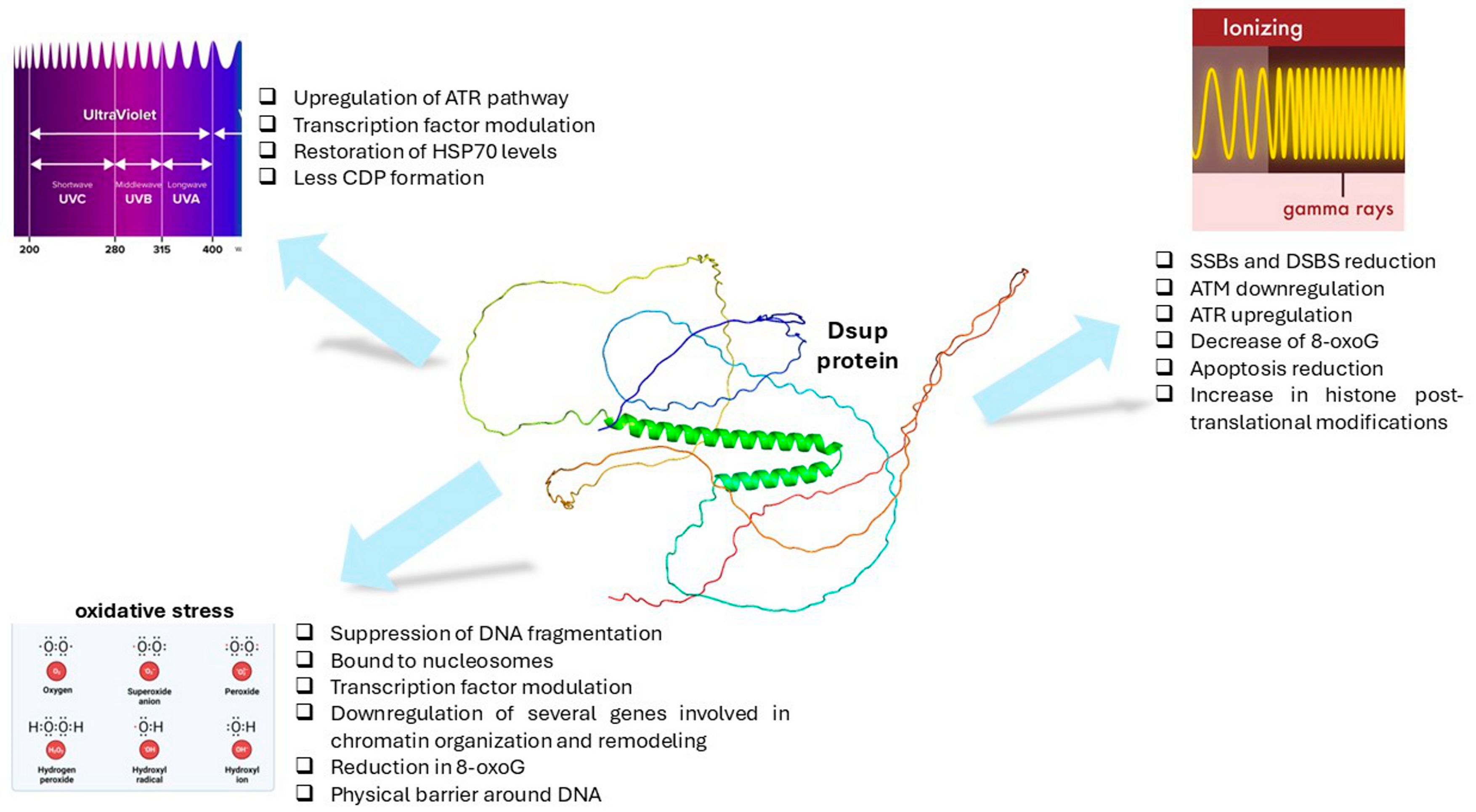
4. DSUP and DNA Protection

Given its ability to bind chromatin, Dsup has been shown to protect DNA from a range of insults, including oxidative stress, UV radiation, ionizing radiation, and chemical agents, thereby protecting cells from damage and death. Various cell models and transgenic organisms have been employed to investigate the protective effects of Dsup and to clarify the mechanisms underlying its action.

4.1. Oxidative Stress

Oxidative DNA damage, which is caused by exposure to reactive oxygen species (ROS), is a constant challenge to the genome. ROS are generated from a variety of endogenous and exogenous sources. Endogenous ROS are produced primarily by leakage from the mitochondrial electron transport chain [10]. Other sources include cytosolic enzyme systems such as NADPH oxidases and peroxisomal by-products. Oxidative damage by intracellular ROS leads to DNA base modifications, single- and double-strand breaks, and apurinic and apyrimidic lesions, many of which are toxic and/or mutagenic [11].
The most prominent ROS species are the superoxide radical (O2•−), hydrogen peroxide (H2O2), and the hydroxyl radical (•OH) [12]. Among these ROS species, the •OH radical is the most reactive. It is formed as a by-product of the Fenton reaction of H2O2 with Fe2+ and is capable of damaging DNA, proteins, and lipids [13,14]. The •OH radical can induce the opening of the imidazole ring in guanine and adenine, resulting in a fragmented purine structure called formamidopyrimidine [15,16]. Moreover, the •OH can led to the formation of 8-oxo-7,8-dihydroguanine (8-oxoG) from guanine residues. Since guanine has the lowest ionization potential of the DNA bases [17], it is the most easily oxidized base. The presence of 8-oxo-dG is considered a biomarker of oxidative stress. During replication of DNA containing 8-oxo-dG, adenine is most often incorporated opposite the lesion. After replication, the repair process removes the 8-oxo-dG and incorporates a thymine in its place. For this reason, 8-oxo-dG mutations typically lead to a G-to-T transversion [18,19,20,21]. As well as affecting DNA bases, ROS radicals can also damage the DNA backbone, causing single strand breaks in the cell [22]. Cells counteract ROS damage in a variety of ways. While the base excision repair (BER) pathway corrects oxidized bases, the single strand break repair (SSBR) or double strand break repair (DSBR) pathways are involved in repairing breaks in the DNA backbone [23].

4.1.1. DSUP Role in Response to Oxidative Stress

The first study on the properties of the Dsup protein was carried out by Hashimoto et al. [3]. They investigated the role of this protein in DNA protection and resistance to various insults in human cultured cells. The authors established a HEK293 cell line stably expressing Dsup and confirmed co-localization of Dsup protein with nuclear DNA by immunocytochemistry. Among the stresses examined, they exposed the cells to hydrogen peroxide (H2O2) and then evaluated the effect of Dsup protein on ROS-generated DNA SSBs using the comet assay. Exposure to hydrogen peroxide induced severe DNA fragmentation (71% of total DNA in the tail) in control HEK293 cells, whereas DNA fragmentation in Dsup-expressing cells was significantly suppressed to only 18% of total DNA. Pretreatment with N-acetyl-L-cysteine (NAC) suppressed peroxide-induced SSBs. Combining NAC and Dsup resulted in greater suppression, though less than each agent’s individual effect, suggesting that NAC and Dsup share a suppression mechanism.
The mechanism of DNA protection mediated by Dsup was investigated by Chavez et al. [7]. The authors examined how the purified Dsup protein may affect hydroxyl radical-mediated DNA cleavage in a purified and controlled biochemical system. Hydroxyl radical-mediated DNA cleavage reactions were carried out in the absence or presence of Dsup. The DNA of the 3.3 kb plasmid pGIE-0 (commonly used in chromatin assembly experiments) was mostly degraded to DNA fragments ranging from 100 to 1000 nt in the absence of Dsup. Since the amount of free plasmid DNA cleavage was almost equal to the amount of chromatin cleavage, nucleosomes provided only a small amount of protection to DNA from hydroxyl radicals. Upon addition of Dsup, free DNA as well as chromatin were substantially protected from hydroxyl radicals. Dsup-mediated protection was more evident with chromatin than with free DNA, probably because of its ability to bind to nucleosomes and form a resistant structure. In the presence of four Dsup molecules per nucleosome, most of the full-length linear DNA was unaffected after cleavage by hydroxyl radicals, demonstrating that Dsup can protect chromatin from oxidative stress. Since hydroxyl radicals react primarily with hydrogen atoms exposed in the minor groove of DNA [24], it is likely that Dsup blocks access to this region of DNA. The authors also observed that Dsup and histone H1 can simultaneously bind to nucleosomes without significantly altering or disrupting chromatin structure. This is in line with the observation that Dsup does not have a deleterious effect on human cells that contain histone H1 [3]. The authors also showed that the HMGN-like sequence in Dsup has an important function because of its ability to bind to nucleosomes and to protect the chromatin from the action of hydroxyl radicals. Notably, HMGN proteins are only found in vertebrates [25], so the origin of the HMGN-like sequences in the tardigrade Dsup proteins is not clear.
The cellular pathways involved in Dsup-mediated protection against external insults in cells expressing the Dsup protein were further investigated in subsequent studies. A first study looked at the modulation of transcription factors and gene pathways associated with damage response and repair in untransfected and Dsup-expressing HEK293 cells [26]. When exposed to H2O2, untransfected cells reacted to damage by activating pCREB and AP-1 family transcription factors. It is known that, when upregulated, AP-1 accumulates in the nucleus to activate gene expression [27]. ATM mediated CREB activation depends on the extent of DNA damage [28] and is triggered by ROS [26]. In addition, CREB signaling protects neuronal cells from oxidative DNA damage by increasing the expression of genes responsible for DNA repair [29]. The up-regulation of these factors was not observed in the Dsup-expressing cells. CREB was downregulated, suggesting that all these mechanisms are unlikely to be activated. In Dsup-expressing cells the authors also observed an upregulation of c-Myc, which is normally negatively regulated by CREB [30]. The induction of c-Myc can promote the G1/S phase transition and the progression of the cell cycle. At the level of gene expression, among the apoptotic genes, in Dsup-expressing cells only, Bcl2 expression increased after treatment with H2O2. Conversely, a constant increase in the expression of PARP1, hTERT, and SOD1 was observed over time. Notably, PARP1 detects DNA breaks and is responsible for the early activation of factors that promote DSB repair [31], including BRCA1 [32], whose expression was upregulated in Dsup-expressing cells. hTERT is the catalytic subunit of the telomerase holoenzyme. In addition to its role in telomere maintenance, hTERT has antioxidant activity and can reduce basal cellular levels of ROS, as well as inhibit endogenous ROS production [33]. SOD1 is a highly conserved, ubiquitously expressed enzyme that is considered a key regulator of the antioxidant response. On the other hand, the expression of ATM and ATR kinases, two key factors in the oxidative stress-induced DNA damage response, did not show significant changes in Dsup-expressing cells. Taken together, these data suggest that H2O2 treatment of Dsup-expressing cells has only a minor impact on DNA repair pathways and that the extent of DNA damage may be limited. Thus, in agreement with the report of Chavez [7], it can be hypothesized that while DNA is “physically” protected from damage by Dsup, detoxification mechanisms are activated to remove ROS and limit oxidative stress, allowing cells to survive and proliferate.
Zarubin et al. used Dsup-expressing lines of Drosophila melanogaster to study the contribution to ROS damage prevention and other biological processes at the transcriptome and organism level [34]. The tardigrade Dsup protein could protect D. melanogaster from ROS-related stress, with an increase in the survival rate, median lifespan, maximum lifespan, and age of 90% mortality after hydrogen peroxide treatment. The efficiency of the protection mediated by Dsup seemed to depend on the level of Dsup expression. This level had to exceed a certain point, at which the amount of Dsup protein produced is sufficient to provide effective physical protection of DNA. Indeed, since only Dsup homozygous flies were protected against oxidative stress, while heterozygous flies had a survival rate very close to the control, this suggests that in heterozygotes the amount of Dsup protein is too low to provide significant protection. The author also evaluated the impact of Dsup protein on other important physiological parameters, such as locomotor activity. It was reduced in the Dsup-expressing D. melanogaster lines, indicating a certain degree of stress at the level of the organism. Furthermore, the median lifespan was increased in Dsup-expressing females, which may be related to the modulation of the expression of some sex-limited genes mediated by Dsup. At the transcriptome level, most differentially expressed genes were down-regulated in Dsup-expressing D. melanogaster lines compared to the control. The down-regulated genes could be grouped into five groups: pathways related to DNA transcription, neural system function, nucleosome-chromatin-chromosome assembly/disassembly processes, cellular metabolic processes, Drosophila development, and morphogenesis. Competition for binding to DNA between the Dsup protein and host proteins involved in chromatin organization/remodeling may explain these observations. Similarly, genes related to DNA transcription and regulation, including transcription factors, were downregulated, consistent with what was observed in HEK293 cells [26]. The authors hypothesized that proteins involved in DNA transcription compete directly with Dsup for binding sites on DNA and that Dsup-nucleosome contacts could reduce the accessibility of DNA to transcription factors, RNA polymerase II, etc., leading to an overall reduction in gene expression. In addition, they showed that Dsup can bind RNA, strengthening the hypothesis that Dsup may not only act as a DNA protector, but also as an RNA-binding protein and a non-specific transcriptional repressor, which could disturb several biological processes in D. melanogaster. However, further experiments are needed to clarify whether there is competition for binding sites between Dsup and other proteins, and whether the Dsup protein can be considered a non-specific transcriptional repressor.
Further insights into the mechanisms mediated by Dsup against oxidative damage in vivo have come from experiments in budding yeast [35]. The authors observed a ~25-fold increase in survival in Dsup-expressing yeast compared to yeast lacking Dsup in the presence of serial dilutions of H2O2 and measured a significant reduction in 8-oxoG in the presence of Dsup, confirming a protective activity against ROS damage. In addition, Dsup significantly increased the replicative lifespan of cells lacking superoxide dismutase, suggesting enhanced survival and longevity against chronic oxidative damage. On the other hand, Dsup expression had no effect on the redox state of the yeast nucleus, suggesting that it uses mechanisms other than ROS scavenging to protect the genome from oxidative damage. Dsup used two C-terminal regions to protect DNA for oxidative stress: HMGN-like domain was responsible for the interaction with nucleosomes, while the distal C-terminal sequences interacted with DNA. Like human HMGN2, Dsup bound nucleosomes at the H2A/H2B acidic patch, with this interaction lost upon mutation of all three arginine in its HMGN-like domain (3R/3E; R363E/R364E/R367E). Furthermore, the Dsup HMGN-like domain interacted with histone tails. Mutations within the H2A/H2B acidic patch or deletion of the histone tails were each sufficient to remove the interaction between Dsup and nucleosomes, suggesting that its HMGN-like domain may bind these regions in a co-operative manner [35].
To better understand the effect of Dsup expression at the organism level, Ni et al. [36] combined molecular dynamics (MD) simulations and fluorescence lifetime imaging microscopy (FLIM)-Foerster resonance energy transfer (FRET) to investigate Dsup-DNA interactions and showed that Dsup binds to DNA in living mammalian cells. They also confirmed that Dsup expression increased cell survival in the presence of H2O2 in both HEK293T cells and yeast, and that Dsup enables both mammalian and yeast cells to better tolerate oxidative stress. The authors performed MD simulations of the binding between DNA and the C-terminal disordered region of Dsup to obtain a mechanistic understanding of the protective effects of Dsup. The simulations showed that Dsup chains wrap around DNA. Arg and Lys interact with the phosphates and bases of the DNA backbone via salt bridges and hydrogen bonds, respectively. As a result of these interactions, Dsup may act as a physical barrier to prevent ROS-induced double-strand breaks and damage to the DNA base pairs. In addition, simulations at 500 K showed that Dsup slows down the melting of DNA, suggesting that Dsup may prevent DNA from undergoing structural changes that make it susceptible to damage. Notably, the resulting decrease in disrupted base pairs appeared to be qualitatively similar to the decrease in cell death. The authors developed an assay based on FRET, a well-known spectroscopic tool for monitoring molecular interactions in living cells, to directly probe the interactions between Dsup and DNA in living mammalian cells. In combination with FLIM, FRET is a reliable method for studying biomolecular interactions. In this case, FLIM-FRET studies have been successfully used to study interactions between DNA and DNA-binding proteins in live cells. This approach provided clear evidence of Dsup-DNA interactions in the nuclei of live mammalian cells, confirming what had previously been observed in the in vitro studies.
The results of all these studies are summarized in Table 2. Taken together, they show that Dsup expression can protect different cells/organisms from ROS-induced damage, suggesting that the function of Dsup may be universal across different species, modulating common mechanisms of defense and response to oxidative stress.

4.2. UV Radiation

UV radiation is divided into three classes based on wavelength: UV-C (190–290 nm), UV-B (290–320 nm), and UV-A (320–400 nm). The maximum absorption of UV radiation by DNA is at 260 nm, beyond which the photo-absorption decreases dramatically. Sunlight consists of 5.1% UV-A, 0.3% UV-B, 62.7% visible light, and 31.9% infrared. The dangerous UV-C is largely filtered out by the ozone layer [37]. However, UV-C is widely used in laboratory studies since it has the highest absorption by DNA and generates more photoproducts than UV-A and UV-B radiation. UV-C produces two classes of DNA lesions: cyclobutane pyrimidine dimers (CPDs) and 6-4 photoproducts (6-4 PPs), the relative frequency of which depends on the wavelength and dose of light [37,38,39]. In CPDs, two adjacent pyrimidines are covalently linked by a cyclobutane ring, while in 6-4 PPs, the C6 position of one pyrimidine is covalently linked to the C4 position of an adjacent pyrimidine. Both lesions cause a distortion of the DNA structure, interfering with transcription and replication. It is the relatively flexible regions of the DNA double helix that are most susceptible to damage. In mammalian cells, UV-B leads to the formation of pyrimidine dimers, although less efficiently than UV-C [16,40,41], whereas UV-A radiation causes DNA strand breaks [16,42]. CPD and 6-4 PP are both repaired by a process called nucleotide excision repair (NER). In eukaryotes, this complex process requires the products of about 30 genes, which act in four steps: detection of the damage, excision of the section of DNA containing and surrounding the lesion, filling of the resulting gap by DNA polymerase, and ligation of the newly synthesized and older DNA [43].

4.2.1. DSUP Role in Response to UV Exposure

The first study of the Dsup potential in response to UV exposure was carried out in plants [44]. Although UV light from the sun, detected by flavonoid pigments, may be an important developmental signal for plants, UV radiation is a continuous stressor that can alter DNA, affect DNA replication and transcription, and may interfere with plant development and growth [45,46]. The potential protection provided by Dsup could therefore be of great importance. The study provided evidence that Dsup expression in tobacco plants affects endogenous genes involved in DNA damage signaling and repair and confers protection against UV-induced DNA damage stress. After exposure to UV-C, the comet assay showed less DNA damage in the Dsup-expressing plant than in the Dsup-deficient plant, suggesting greater genome protection. Gene expression analysis revealed different transcriptional patterns of some of the endogenous genes studied. While the Ataxia Telangiectasia Mutated (ATM) gene expression was unvaried, the ATR (Ataxia Telangiectasia and Rad-3-related) gene, which is activated by SSBs in DNA [47], was up-regulated in Dsup-expressing plants. Increased ATR expression could enhance DNA damage sensing and DNA repair pathways directly involving ATR. This active DNA repair process could enable Dsup-expressing plants to mitigate the harmful effects of DNA damage. Although the exact mechanisms by which Dsup works in the cell are only partially understood, the response of Dsup plants to radiation treatment supports the idea that the tardigrade protein Dsup protects DNA from fragmentation and lesions.
The modification of gene expression pathways after UV-C exposure was also studied in the Dsup-expressing HEK293 cell line [26]. The results confirmed what had been observed in tobacco plants. Dsup-expressing cells showed a decrease in cell death and an increase in cell growth after UV-C exposure; in addition, no CDP formation was observed. There was a significant up-regulation of ATR kinase expression and increased expression of other genes related to the ATR rescue pathway: BRCA1, which is involved in gap repair at the level of photoproduct-stalled replication forks [48]; XRCC6, which is involved in the UV-G2 checkpoint [49]; and ERCC6, whose gene is a potential target for inactivation by UV light and which appears to act as a “dosimeter” of DNA damage [50]. On the contrary, the expression of ATM, which is mainly activated by oxidative stress and double-stranded DNA breaks, was similar or lower than in non-transfected cells. All these data agree with what was reported in tobacco plants [44]. Transcription factor analysis revealed that, after UV-C exposure, non-transfected cells showed an activation of FosB, Fra-1, JunD, and p-c-JUN, whereas p-c-Jun and STAT1alfa were activated in Dsup-expressing cells. In mammalian cells, STAT1 participates in type I interferon (IFN) pathways, and is actively involved in the response to UV damage [51,52]. These data seemed to suggest that, unlike H2O2 treatment, Dsup cannot physically protect DNA after UV-C irradiation. Instead, it activates damage repair mechanisms (STAT1, c-Myc, p-c-Jun; ATR, BRCA1) that may help cells remove CPDs more quickly, reducing the impact on cell survival.
The HEK293 cell line was also used for a functional proteomics approach to investigate the molecular mechanisms and pathways modulated by Dsup under UV-C stress [53]. The study showed that even under basal conditions, the Dsup protein affects cells by modulating several proteins associated with protein folding, telomere maintenance, and metabolic pathways. After UV-C exposure, Dsup mediated cell protection against damage by activating more efficiently the mechanisms of DNA damage repair, telomere elongation and maintenance, mRNA stability, cytoplasmic stress granule, and unfolded protein responses, in addition to metabolic modulation. Notable proteins include HSP90, HSP70, ERK1/2, TIF1B, and UBA1, which are associated with DNA repair mechanisms and protein degradation. For example, it is known that HSPs are induced by UV exposure as a physiological response to activate repair mechanisms and limit cellular damage [54]. The activation of ERK1/2 is triggered by UV exposure [55] and may modulate nucleotide excision repair (NER) pathways [56]. It has been reported that TIF1B protein species are involved in DNA damage repair mechanisms [57,58]. The network analysis also showed that TIF1B transcriptionally regulates UBA1 [59], whose four proteoforms were overexpressed in Dsup-expressing cells after UV-C exposure. Interestingly, UBA1 has been reported to be potentially involved in the repair of UV-induced DNA damage [60], and the ubiquitination pathway plays a positive regulatory role for efficient NER machinery. All these data suggest that Dsup can modulate different pathways into cells, which together help to increase recovery from UV-induced DNA damage and promote cell survival.
Given the impact that UV radiation can have on crops, particularly those of commercial interest, it is not surprising that subsequent studies have been carried out on plants. Ye et al. focused on rice, a staple food for many people and a source of income for farmers, especially in Asia [61]. Dsup was introduced into transgenic rice lines by Agrobacterium-mediated transformation. Overexpression of Dsup enhanced DNA damage resistance at the seed and seedling stages, with a more pronounced impact of Dsup under severe UV radiation conditions. In addition, Dsup increased grain size and altered starch granule structure and cell size, suggesting that it could potentially modulate normal plant growth. RNA-seq analysis revealed increased expression of radiation- and abiotic stress-related genes in Dsup rice compared to wild type. Furthermore, IP-LC-MS identified 21 proteins that preferentially interact with Dsup, indicating that Dsup may bind to transcription- and translation-associated proteins to regulate the balance between DNA protection and plant development. Interestingly, like what was observed in HEK293 cells [53], DNA damage and repair genes were also highly expressed in Dsup-expressing plants under normal conditions, partially explaining the phenotypic results of the study and suggesting that DNA repair pathways are also activated prior to damage.
Del Casino et al. [62] investigated the effect of Dsup expression on pollen, the male gametophyte of seed plants, known to be extremely sensitive to UV light, which can prevent fertilization [63]. In the study, several parameters were measured, including pollen viability, rate of germination, length of pollen tube, male germ unit position, development of callose plug, marker protein content, and antioxidant capacity. UV-B stress had a significant negative effect on both pollen grain and pollen tube growth. Expression of Dsup increased the levels of antioxidants and reversed some of the changes induced by UV-B radiation, restoring pollen tube length, callose plug formation, and tubulin and HSP70 levels (as observed in HEK293 [53]). In addition, the protective effect of Dsup was reflected in an increased level of antioxidants, polyphenols, and flavonoids, all of which have protective properties. All these data show that Dsup expression enhances pollen resistance to UV-B stress and suggests its potential for improving plant tolerance to environmental radiation.
The results of all these studies are summarized in Table 3.

4.3. Ionizing Radiation

Ionizing radiation (IR) consists of alpha, beta, gamma, neutrons, and X-rays. It is abundant in the environment and comes from a variety of sources, including rocks, soil, radon, cosmic rays, and medical devices. IR can damage DNA either directly or indirectly, for example by radiolysis of the surrounding water to form hydroxyl radicals (•OH) [16,64]. In addition, the damage can be amplified by the presence of oxygen and other reactive species in the environment [65]. Indirect DNA damage caused by (•OH) radicals accounts for about 65% of the DNA damage caused by radiation [66]. For this reason, IR produces base lesions similar to those induced by ROS species, including 8-oxo-guanine and formamidopyrimidines. Furthermore, ionizing radiation also produces single strand breaks with peculiar features, in which the DNA breaks have 3′-phosphate or 3′-phosphoglycolate ends rather than 3′-OH ends. In addition, IR can lead to the formation of fragmented sugar derivatives and loss of terminal base residues, which generate clustered damage or single stranded gaps [67,68,69]. The double strand break, which consists of multiple damaged sites close to each other on both DNA strands, is another important radiation-induced lesion [70,71]. Although toxic, these breaks can be repaired by the homologous recombination (HR) pathway [72].

4.3.1. DSUP Role in Response to Ionizing Radiation

The first research into the possibility of Dsup conferring resistance to ionizing radiation was carried out in the landmark study by Hashimoto et al. [3]. The HEK293 cells engineered to express Dsup protein were exposed to X-rays and DNA fragmentation was measured. Dsup-expressing cells showed about half the DNA fragmentation of control cells (16% vs. 33%) when exposed to an alkaline condition to denature damaged DNA, dissociate single-stranded DNA fragments, and reveal SSBs. Since X-rays can induce DNA damage by direct absorption of X-ray energy into DNA (direct effects) and by ROS generation from molecules of water activated by X-ray energy (indirect effects), the authors also investigated the effect of Dsup protein on ROS-induced DNA SSB generation, as discussed in the Section 4.1. The authors then explored the effect of Dsup protein on DSBs using a neutral comet assay, which analyses DNA fragmentation without dissociating DNA fragments. In this case, the proportion of fragmented DNA was reduced by approximately 40% in Dsup-expressing cells compared with untransfected cells, suggesting that Dsup protein suppresses both x-rays induced DNA DSBs and SSBs. The DSBs were also detected by measuring gamma-H2AX, the phosphorylated form of histone H2AX that is found around DSBs. Dsup-expressing cells had much lower levels of the DNA break marker γ-H2AX, which normally accumulates immediately after irradiation and persists for several hours, even after DNA break repair has been completed [73]. Furthermore, after irradiation with a sub-lethal dose of X-rays, which causes mammalian cells to lose their ability to proliferate [74], many Dsup-expressing cells had a normal morphology and continued to increase their number over time, suggesting that these irradiated cells retained their ability to proliferate.
Similarly to what was observed with UV-C, after X-ray irradiation, tobacco plants expressing Dsup had a greater number of intact nuclei with significantly shorter tails than control plants, indicating less DNA damage and greater protection of the genome [44]. Again, several endogenous genes involved in DNA damage responses differed between Dsup-expressing and control plants. In particular, the ATM gene, which is involved in DSB signaling [47], was downregulated in Dsup-expressing plants compared to the control. This may indicate that there were fewer DSBs in Dsup-expressing plants and therefore less response from the DSB signaling pathways. On the other hand, the ATR gene, which is activated by SSBs in DNA, was up-regulated, probably reflecting an association of Dsup expression with increased SSB signaling in response to genomically damaging stress. However, neither SSBs nor DSBs were measured directly.
Westover et al. continued the research in HEK293 cells, achieving stable integration by inserting a human codon-optimized CMV promoter Dsup via lentiviral transduction [75]. They tested the presence of the molecular marker of oxidative stress, 8-hydroxy-2-deoxoguanosine (8-oxoG), and showed that it was decreased 1 h after irradiation, and its levels were still significantly reduced at 24 h after irradiation. The authors also examined whether the higher viability previously reported in irradiated Dsup-expressing cells [3] could be also related to a reduction in programmed cell death. They examined caspase-3 activity and binding of Anexin V to phosphatidylserine, both markers of apoptosis, at different time-points, and reported a general significant reduction in both parameters. They therefore used several omics approaches to study the effect of Dsup: RNA-seq to analyze gene expression, Omni-ATAC-seq to assess chromatin accessibility, and Cut&Run to examine the presence of histone modifications and Dsup binding. Dsup expression resulted in an active transcriptional profile, like that observed with HMGN1 overexpression [76]. ATAC-seq analysis showed that Dsup-expressing cells had a selective differential opening and closing of the chromatin landscape, similar to HMGN1 binding to nucleosomes, which promotes a less transcriptionally repressed chromatin state. In addition, there was a global increase in histone post-translational modifications, suggestive of open chromatin, with Dsup binding preferentially to promoter regions marked by H3K27ac and H3K4me3, which are histone modifications associated with active gene expression. In addition, the authors reported a greater enrichment of nucleosome occupancy at CpG island promoters in HEK293 expressing Dsup. In light of these data, the authors propose that, contrary to the predominant hypothesis that Dsup binds to nucleosomes to create a shield against damaging stresses, it promotes a more open and permissive chromatin state, creating less steric hindrance for the recruitment of repair machinery to sites of damage. It is important to note, though, that these two mechanisms are not incompatible, and that the latter hypothesis needs to be further investigated.
Zarubin et al. [34] have shown that Dsup-expressing lines of Drosophila melanogaster are protected from exposure to G-rays, as has been observed for ROS-related stress. Again, Dsup improved survival, median life, maximum life, and 90% mortality after irradiation. Transcriptome profiling of lines exposed to G-rays (1000 Gy) showed a reduction in the expression level of 99.3% of the total differentially expressed genes in Dsup-expressing lines compared to the control. These genes were enriched in the categories of biological processes that are related to DNA repair, neurogenesis, and proteostasis. However, comparison between irradiated and non-irradiated groups with and without Dsup revealed a reduced response to irradiation in Dsup-expressing lines. These data raise the question of what causes the reduced response in Dsup-expressing lines: a reduction in DNA damage due to Dsup shielding, or a different modulation of transcription by Dsup, or a combination of both.
Finally, the properties of the Dsup protein have attracted considerable interest as a potential approach to mitigating the risks associated with this type of research, since ionizing radiation is one of the most serious hazards in space exploration. To address this issue, the E. coli strain K-12 MG1655 was transformed with the Dsup gene and then exposed twice to ionizing radiation, with the surviving fraction being studied after each exposure [77]. The Dsup-transformed strain showed an increase in survival of more than two orders of magnitude compared to the wild-type strain at the 3000 Gy dose. This increase was progressive over the course of the exposures. When exposed to X-rays up to 500 Gy, equivalent to ~2500 years of exposure on the ISS Exposure Facility, colonies of the Dsup strain showed almost complete survival for this dose in one of the colonies and a general increase in survival across the different ionizing radiation exposures. In addition, increasing the time it takes to reach the same dose could have a direct positive effect on cell survival, allowing DNA repair mechanisms to work more effectively. These data suggest that the tolerance of tardigrades to the extreme conditions of space [2] may be linked, at least in part, to the Dsup protein.
The results of all these studies are summarized in Table 4.

5. Discussion

Tardigrades, also known as water bears, are animals able to survive in extreme conditions. The sequencing of the genome of the tardigrade Ramazzottius varieornatus has revealed that the tardigrade’s extraordinary ability to survive extreme stress is due to a unique nucleosome-binding protein, the damage suppressor (Dsup) [3].
Since its identification, the Dsup protein has attracted a great deal of interest and numerous studies have been carried out to understand how it performs this function. Studies in organisms as diverse as bacteria [77], yeast [35], mammalian cells [3,26,53,75], plants [44,61,62], and animal models such as D. melanogaster [34] have shown that Dsup can protect cells from many types of abiotic damage. In all studies, Dsup protein increased cell survival and viability following oxidative stress, UV exposure, and ionizing radiation. In general, there was also an increase in the cellular systems that protect the cell and repair DNA damage. These included increased expression of the ATR gene in both plants and mammalian cells [26,44]. ATR is a key regulator of the DNA damage response, which responds to a wide range of DNA damages, including those that disrupt DNA replication, and plays a critical role in genome stabilization and cell survival [78]. The cells also showed increased expression of proteins involved in the response to external stress, such as HSPs, especially HSP70 and HSP90 [53,62]. These are known to have chaperoning functions and help protect cells from the adverse effects of physiological stress [58].
It is interesting to note that Dsup was also able to improve the response to various stresses in complex organisms. In these models, it is also possible to evaluate the overall effects of Dsup in the whole organism and assess potential adverse outcomes. In tobacco plants, expression of the Dsup gene did not alter growth or plant characteristics under basal conditions and increased the tolerance of the plants to genomutagenic stress [44]. In rice, overexpression of Dsup was associated with a reduced number of primary branches compared to the control. The total panicle size was also smaller in Dsup lines. On the other hand, Dsup not only improved DNA damage resistance at the seed and seedling stages but also increased grain size and altered starch granule structure and cell size. This suggests that Dsup may not only be involved in chromatin protection under critical conditions such as radiation or abiotic stress, but also in the regulation of plant growth under normal conditions [61]. In Drosophila melanogaster, Dsup increased the survival after γ-ray irradiation and hydrogen peroxide treatment. However, it reduced the level of locomotor activity, indicating some stress at the organism level [34]. This may be related to the observation that among the differential expressed genes in Dsup-expressing D. melanogaster lines, there is a significant number of genes encoding transcription factors that are expressed in the nervous system. The reduction in expression of these genes could lead to a decrease in the activity of a large group of genes involved in synapse organization, axon guidance, and visual perception. Interestingly, overexpression of other genes encoding DNA-binding intrinsically disordered proteins, such as HMGN, FUS, and TDP43, is associated with several neurodegenerative diseases [79,80,81]. Thus, overexpression of Dsup may be responsible for an imbalance in neuronal and neuromuscular communication that could lead to a deterioration in locomotor activity in Dsup-expressing D. melanogaster lines. This observation is consistent with the results of a study of cortical neurons cultured from rat embryos carried out by Escacerga et al. [82], who investigated the potential protective role of Dsup in this model. Surprisingly, they found that Dsup, which localizes to the nucleus, promotes neurotoxicity causing neurodegeneration. Expression of Dsup did indeed lead to DNA DSBs in cultured neurons and promoted chromatin condensation. As it has been hypothesized that the functions of Dsup and HMGN proteins may at least partially overlap [7,83], it is hypothesized that Dsup may act in neuronal cells in a similar way to HMGN proteins. Interestingly, it has been reported that overexpression of HMGN4 also alters transcription and increases levels of the DNA DSB marker γH2AX [84]. It should also be considered that, as neurons are post-mitotic and often last a lifetime, the mechanisms for maintaining the integrity of their genome may be more complex than in other cell types [85]. This suggests that the effect of Dsup protein may vary depending on the cell type, its differentiation, and characteristics.
Considering these data, it is clear that, in addition to studying the mechanisms of action of Dsup in response to external stress, it is important to understand what changes Dsup expression can cause in cells under basal conditions. Research has shown that cells expressing Dsup undergo certain changes even in the absence of external stimuli. Studies have shown that DNA damage and repair genes are highly expressed under normal conditions in Dsup-expressing plants as well as in HEK293 cells, suggesting that DNA repair pathways are also activated prior to damage [54,61]. It remains to be seen whether these changes are an advantage for the cell, which is prepared for possible damage, or whether they are themselves a cellular response to a stressful condition related to the presence of a heterologous protein. This will be an important point to clarify in future investigations.
Lastly, a topic worthy of further investigation is the mechanism by which Dsup binds to DNA. Computational, biochemical, and biophysical methods have shown that Dsup binds directly to chromatin and preferentially to nucleosomes rather than to free DNA [7]. Binding is mediated by the C-terminal domain, which contains a nucleosome-binding domain that directs its interaction with chromatin [4]. Since Dsup is positively charged, it interacts with negatively charged DNA. The result of this interaction is an electrostatic shield in which Dsup coats and protects DNA from external factors, acting independently of the DNA-specific conformation [4]. The hypothesis is that, because electrostatic attraction is non-specific, the binding between Dsup and DNA is independent of nucleotide or amino acid sequence. In other words, it is aspecific and global protection. On the other hand, in vitro studies in HEK293 cells have shown that the Dsup protein may act as a chromatin-architecture protein and that, in addition to its effect as a nucleosome shield, it may confer resistance through chromatin modulation [75]. Therefore, it appears that the binding of Dsup to DNA is not completely aspecific, but rather that Dsup binds preferentially to specific promoter regions, as evidenced by a general increase in histone post-translational modifications, suggestive of open chromatin, and an overall decrease in repressive marks. This would explain the modulation of gene expression in the presence of Dsup after exposure to external stress, observed at the level of transcription, protein, and transcription factor expression [26,34,44,53,62,63,75]. A detailed analysis of the promoter sequences of these deregulated genes, in the presence or absence of external stresses, could provide important insights into the mechanisms of protection mediated by Dsup. However, the two hypotheses proposed (aspecific vs. specific protection of DNA) are not incompatible. Recently, experiments using circular dichroism (CD) spectroscopy have shown that in the complex between Dsup and DNA, there are no transitions from disordered to ordered conformations in Dsup protein [8]. These structures may be classified as a fuzzy complex, a typical feature of many DNA-binding proteins that contain intrinsically disordered regions [86,87]. Post-translational modifications and subsequent secondary structure transitions can regulate the binding affinity of these complexes [88,89]. Notably, Dsup putative phosphorylation-prone sites are mostly located in intrinsically disordered regions and are depleted in the central helical region. It is possible that the binding affinity in the Dsup-DNA complex may be regulated by phosphorylation of the Dsup protein [88,90]. The mechanisms by which Dsup exerts its protective role are depicted in Figure 2.
Several areas of application could benefit from a better understanding of the mechanism of action and the molecular mechanisms modulated by Dsup. Given the protective effects of Dsup on radiotolerance and resistance to oxidative stress in human cells, it has been suggested that the protein could be used as a therapeutic agent to reduce the negative effects of radiation and chemotherapy, which are known to induce oxidative stress and DNA damage beyond the target cancer cells [91]. One of the potential applications suggested was the development of protective agents for neuronal tissue in the treatment of brain tumors, which could help to minimize collateral damage to healthy cells, thereby increasing the efficacy and safety of these treatments. However, in the light of data from neurons in culture in rats, where Dsup causes neurotoxicity leading to DNA DSBs and chromatin rearrangements, this prospect is still a long way off [82]. These data suggest that the use of Dsup as a protective agent may not be possible in all cell types. How this happens is yet to be clarified. The binding of Dsup to DNA can stall DNA polymerase and block transcription, and the subsequent action of endonucleases to remove this stall can damage DNA. Alternatively, disruption of DNA repair complexes in the nucleus can lead to DNA damage, chromatin condensation, and apoptosis. It is also possible that a combination of different mechanisms may lead to neurotoxicity. Further studies in other cell types, both mitotic and post-mitotic, are needed to better understand whether Dsup can be practically used to recapitulate DNA protective mechanisms.
More recently, Kirtane et al. [92] have shown that transient local expression of Dsup protein in mice, achieved by nanoparticle-delivered mRNA, was able to reduce DNA damage induced by radiation in oral and rectal epithelial tissues. These tissues are frequently affected by radiotherapy for head and neck cancer and prostate cancer, respectively. In addition, in mice with oral cancer, radioprotection of normal tissue did not affect the effectiveness of radiotherapy. This initial research paves the way for a more general strategy for protecting healthy tissue from DNA-damaging agents. In parallel with this approach, the study of Dsup at the level of mammalian cell models could reveal the molecular pathways involved and identify potential cellular targets whose modulation could reduce oxidative stress- or radiation-induced damage. Instead of using Dsup directly, it may be possible to interfere with these targets.
In terms of botanical applications, the benefits of obtaining crops that are more resistant to environmental stresses, even extreme ones, would be clearly positive. Of course, research is still in its infancy and the safety of such crops has yet to be established.
The properties of Dsup could be very useful in the development of new materials. Recently, DNA non-adsorbing polyethylene terephthalate track membranes were functionalized with the Dsup protein by covalent bonding with glutaraldehyde. Filtration experiments demonstrated the ability of these membranes to adsorb cell-free DNA. The track-membraned biomaterial could be used to filter and separate DNA molecules from solutions and environmental samples, and to store and manipulate them [93].
Lastly, the Dsup protein has aroused great interest in the field of space exploration. Space is perhaps the most dangerous and precarious environment that mankind has explored, where exposure to ionizing radiation is inevitably high, with potentially severe effects on human health. The Minerva mission, a space exploration mission, aims to engineer Caenorhabditis elegans with the Dsup gene and assess whether Dsup protein can protect engineered organisms from DNA damage caused by space radiation [94].

6. Conclusions

In conclusion, although the study of Dsup protein properties is still in its early stages, the discovery of which properties confer extreme tolerance to various abiotic stresses and the exploration of the mechanisms behind this tolerance provide an exciting ground for future research and applications. Further research is needed to investigate how Dsup interacts with cell DNA damage sensing and repair systems to provide protection against environmental stress. However, the lesson we can learn from studying tardigrade, and its shield to protect DNA, is that understanding the mechanisms by which it provides this protection can increase our tolerance to adverse environmental conditions and prepare us for future challenges such as climate change, human space exploration, and the fight against many diseases.

Funding

This research received no external funding.

Institutional Review Board Statement

Not applicable.

Informed Consent Statement

Not applicable.

Data Availability Statement

No new data were created or analyzed in this study.

Conflicts of Interest

The authors declare no conflicts of interest.

References

  1. Schill, R.O. (Ed.) Water Bears: The Biology of Tardigrades; Zoological Monographs; Springer International Publishing: Cham, Switzerland, 2018; Volume 2. [Google Scholar] [CrossRef]
  2. Sadowska-Bartosz, I.; Bartosz, G. Antioxidant Defense in the Toughest Animals on the Earth: Its Contribution to the Extreme Resistance of Tardigrades. Int. J. Mol. Sci. 2024, 25, 8393. [Google Scholar] [CrossRef]
  3. Hashimoto, T.; Horikawa, D.D.; Saito, Y.; Kuwahara, H.; Kozuka-Hata, H.; Shin-I, T.; Minakuchi, Y.; Ohishi, K.; Motoyama, A.; Aizu, T.; et al. Extremotolerant Tardigrade Genome and Improved Radiotolerance of Human Cultured Cells by Tardigrade-Unique Protein. Nat. Commun. 2016, 7, 12808. [Google Scholar] [CrossRef]
  4. Mínguez-Toral, M.; Cuevas-Zuviría, B.; Garrido-Arandia, M.; Pacios, L.F. A Computational Structural Study on the DNA-Protecting Role of the Tardigrade-Unique Dsup Protein. Sci. Rep. 2020, 10, 13424. [Google Scholar] [CrossRef] [PubMed]
  5. Bemm, F.; Burleigh, L.; Förster, F.; Schmucki, R.; Ebeling, M.; Janzen, C.J.; Dandekar, T.; Schill, R.O.; Certa, U.; Schultz, J. Draft genome of the Eutardigrade Milnesium tardigradum sheds light on ecdysozoan evolution. bioRxiv 2017, bioRxiv: 122309. [Google Scholar] [CrossRef]
  6. Anoud, M.; Delagoutte, E.; Helleu, Q.; Brion, A.; Duvernois-Berthet, E.; As, M.; Marques, X.; Lamribet, K.; Senamaud-Beaufort, C.; Jourdren, L.; et al. Comparative transcriptomics reveal a novel tardigrade-specific DNA-binding protein induced in response to ionizing radiation. eLife 2024, 13, RP92621. [Google Scholar] [CrossRef] [PubMed]
  7. Chavez, C.; Cruz-Becerra, G.; Fei, J.; Kassavetis, G.A.; Kadonaga, J.T. The Tardigrade Damage Suppressor Protein Binds to Nucleosomes and Protects DNA from Hydroxyl Radicals. eLife 2019, 8, e47682. [Google Scholar] [CrossRef] [PubMed]
  8. Zarubin, M.; Murugova, T.; Ryzhykau, Y.; Ivankov, O.; Uversky, V.N.; Kravchenko, E. Structural Study of the Intrinsically Disordered Tardigrade Damage Suppressor Protein (Dsup) and Its Complex with DNA. Sci. Rep. 2024, 14, 22910. [Google Scholar] [CrossRef]
  9. Receveur-Brechot, V.; Durand, D. How random are intrinsically disordered proteins? A small angle scattering perspective. Curr. Protein Pept. Sci. 2012, 13, 55–75. [Google Scholar] [CrossRef]
  10. Finkel, T.; Holbrook, N.J. Oxidants, Oxidative Stress and the Biology of Ageing. Nature 2000, 408, 239–247. [Google Scholar] [CrossRef]
  11. Girard, P.M.; Boiteux, S. Repair of Oxidized DNA Bases in the Yeast Saccharomyces Cerevisiae. Biochimie 1997, 79, 559–566. [Google Scholar] [CrossRef]
  12. Chatterjee, N.; Walker, G.C. Mechanisms of DNA Damage, Repair, and Mutagenesis. Environ. Mol. Mutagen. 2017, 58, 235–263. [Google Scholar] [CrossRef] [PubMed]
  13. Imlay, J.A.; Chin, S.M.; Linn, S. Toxic DNA Damage by Hydrogen Peroxide through the Fenton Reaction in Vivo and in Vitro. Science 1988, 240, 640–642. [Google Scholar] [CrossRef]
  14. Dizdaroglu, M.; Rao, G.; Halliwell, B.; Gajewski, E. Damage to the DNA Bases in Mammalian Chromatin by Hydrogen Peroxide in the Presence of Ferric and Cupric Ions. Arch. Biochem. Biophys. 1991, 285, 317–324. [Google Scholar] [CrossRef] [PubMed]
  15. Chetsanga, C.J.; Lozon, M.; Makaroff, C.; Savage, L. Purification and Characterization of Escherichia Coli Formamidopyrimidine-DNA Glycosylase That Excises Damaged 7-Methylguanine from Deoxyribonucleic Acid. Biochemistry 1981, 20, 5201–5207. [Google Scholar] [CrossRef] [PubMed]
  16. Friedberg, E.C.; Walker, G.C.; Siede, W.; Wood, R.D.; Schultz, R.A.; Ellenberger, T. DNA Repair and Mutagenesis; ASM Press: Washington, DC, USA, 2005. [Google Scholar] [CrossRef]
  17. Steenken, S.; Jovanovic, S.V. How Easily Oxidizable Is DNA? One-Electron Reduction Potentials of Adenosine and Guanosine Radicals in Aqueous Solution. J. Am. Chem. Soc. 1997, 119, 617–618. [Google Scholar] [CrossRef]
  18. Kasai, H.; Nishimura, S. Hydroxylation of Deoxyguanosine at the C-8 Position by Ascorbic Acid and Other Reducing Agents. Nucleic Acids Res. 1984, 12, 2137–2145. [Google Scholar] [CrossRef]
  19. Cheng, K.C.; Cahill, D.S.; Kasai, H.; Nishimura, S.; Loeb, L.A. 8-Hydroxyguanine, an Abundant Form of Oxidative DNA Damage, Causes G-T and A-C Substitutions. J. Biol. Chem. 1992, 267, 166–172. [Google Scholar] [CrossRef]
  20. Cadet, J.; Delatour, T.; Douki, T.; Gasparutto, D.; Pouget, J.P.; Ravanat, J.L.; Sauvaigo, S. Hydroxyl Radicals and DNA Base Damage. Mutat. Res./Fundam. Mol. Mech. Mutagen. 1999, 424, 9–21. [Google Scholar] [CrossRef]
  21. Cadet, J.; Douki, T.; Ravanat, J.-L. Oxidatively Generated Base Damage to Cellular DNA. Free Radic. Biol. Med. 2010, 49, 9–21. [Google Scholar] [CrossRef]
  22. Juan, C.A.; Pérez de la Lastra, J.M.; Plou, F.J.; Pérez-Lebeña, E. The Chemistry of Reactive Oxygen Species (ROS) Revisited: Outlining Their Role in Biological Macromolecules (DNA, Lipids and Proteins) and Induced Pathologies. Int. J. Mol. Sci. 2021, 22, 4642. [Google Scholar] [CrossRef]
  23. Demple, B.; Harrison, L. Repair of Oxidative Damage to DNA: Enzymology and Biology. Annu. Rev. Biochem. 1994, 63, 915–948. [Google Scholar] [CrossRef] [PubMed]
  24. Balasubramanian, B.; Pogozelski, W.K.; Tullius, T.D. DNA Strand Breaking by the Hydroxyl Radical Is Governed by the Accessible Surface Areas of the Hydrogen Atoms of the DNA Backbone. Proc. Natl. Acad. Sci. USA 1998, 95, 9738–9743. [Google Scholar] [CrossRef] [PubMed]
  25. González-Romero, R.; Eirín-López, J.M.; Ausió, J. Evolution of High Mobility Group Nucleosome-Binding Proteins and Its Implications for Vertebrate Chromatin Specialization. Mol. Biol. Evol. 2015, 32, 121–131. [Google Scholar] [CrossRef]
  26. Ricci, C.; Riolo, G.; Marzocchi, C.; Brunetti, J.; Pini, A.; Cantara, S. The Tardigrade Damage Suppressor Protein Modulates Transcription Factor and DNA Repair Genes in Human Cells Treated with Hydroxyl Radicals and UV-C. Biology 2021, 10, 970. [Google Scholar] [CrossRef] [PubMed]
  27. Marinho, H.S.; Real, C.; Cyrne, L.; Soares, H.; Antunes, F. Hydrogen Peroxide Sensing, Signaling and Regulation of Transcription Factors. Redox Biol. 2014, 2, 535–562. [Google Scholar] [CrossRef]
  28. Shi, Y.; Venkataraman, S.L.; Dodson, G.E.; Mabb, A.M.; LeBlanc, S.; Tibbetts, R.S. Direct Regulation of CREB Transcriptional Activity by ATM in Response to Genotoxic Stress. Proc. Natl. Acad. Sci. USA 2004, 101, 5898–5903. [Google Scholar] [CrossRef]
  29. Pregi, N.; Belluscio, L.M.; Berardino, B.G.; Castillo, D.S.; Cánepa, E.T. Oxidative Stress-Induced CREB Upregulation Promotes DNA Damage Repair Prior to Neuronal Cell Death Protection. Mol. Cell. Biochem. 2017, 425, 9–24. [Google Scholar] [CrossRef]
  30. Rajabi, H.N.; Baluchamy, S.; Kolli, S.; Nag, A.; Srinivas, R.; Raychaudhuri, P.; Thimmapaya, B. Effects of Depletion of CREB-Binding Protein on c-Myc Regulation and Cell Cycle G1-S Transition. J. Biol. Chem. 2005, 280, 361–374. [Google Scholar] [CrossRef]
  31. Ali, A.A.E.; Timinszky, G.; Arribas-Bosacoma, R.; Kozlowski, M.; Hassa, P.O.; Hassler, M.; Ladurner, A.G.; Pearl, L.H.; Oliver, A.W. The Zinc-Finger Domains of PARP1 Cooperate to Recognize DNA Strand Breaks. Nat. Struct. Mol. Biol. 2012, 19, 685–692. [Google Scholar] [CrossRef]
  32. Li, M.; Yu, X. Function of BRCA1 in the DNA Damage Response Is Mediated by ADP-Ribosylation. Cancer Cell 2013, 23, 693–704. [Google Scholar] [CrossRef]
  33. Indran, I.R.; Hande, M.P.; Pervaiz, S. hTERT Overexpression Alleviates Intracellular ROS Production, Improves Mitochondrial Function, and Inhibits ROS-Mediated Apoptosis in Cancer Cells. Cancer Res. 2011, 71, 266–276. [Google Scholar] [CrossRef] [PubMed]
  34. Zarubin, M.; Azorskaya, T.; Kuldoshina, O.; Alekseev, S.; Mitrofanov, S.; Kravchenko, E. The Tardigrade Dsup Protein Enhances Radioresistance in Drosophila Melanogaster and Acts as an Unspecific Repressor of Transcription. iScience 2023, 26, 106998. [Google Scholar] [CrossRef]
  35. Tyler, J.; Aguilar, R.; Arslanovic, N.; Birmingham, K.; Kaliwal, K.; Chakraborty, U.; Postnikoff, S.; Hickman, A.; Kahn, L.; Watson, R.; et al. Multivalent Binding of the Tardigrade Dsup Protein to Chromatin Promotes Yeast Survival and Longevity upon Exposure to Oxidative Damage. Res. Sq. 2023, rs-3, rs-3182883. [Google Scholar] [CrossRef]
  36. Ni, G.S.; Su, H.; Zhu, Y.; Dhiman, A.; Zhou, H.-X.; Lin, W.; Hao, N. Tardigrade Dsup: Interactions with DNA and Protection of Cells from Oxidative Stress. bioRxiv 2024, bioRxiv:11.06.622393. [Google Scholar] [CrossRef]
  37. Davies, R.J. Royal Irish Academy Medal Lecture. Ultraviolet Radiation Damage in DNA. Biochem. Soc. Trans. 1995, 23, 407–418. [Google Scholar] [CrossRef]
  38. Varghese, A.J. Photochemistry of Nucleic Acids and Their Constituents. Photophysiology 1972, 7, 207–274. [Google Scholar]
  39. Mitchell, D.L.; Nairn, R.S. The Biology of the (6-4) Photoproduct. Photochem. Photobiol. 1989, 49, 805–819. [Google Scholar] [CrossRef] [PubMed]
  40. You, Y.H.; Szabó, P.E.; Pfeifer, G.P. Cyclobutane Pyrimidine Dimers Form Preferentially at the Major P53 Mutational Hotspot in UVB-Induced Mouse Skin Tumors. Carcinogenesis 2000, 21, 2113–2117. [Google Scholar] [CrossRef]
  41. Rastogi, R.P.; Richa; Kumar, A.; Tyagi, M.B.; Sinha, R.P. Molecular Mechanisms of Ultraviolet Radiation-Induced DNA Damage and Repair. J. Nucleic Acids 2010, 2010, 592980. [Google Scholar] [CrossRef]
  42. Peak, M.J.; Peak, J.G. DNA-to-Protein Crosslinks and Backbone Breaks Caused by Far- and near-Ultraviolet, and Visible Light Radiations in Mammalian Cells. Basic Life Sci. 1986, 38, 193–202. [Google Scholar] [CrossRef]
  43. Sinha, R.P.; Häder, D.P. UV-Induced DNA Damage and Repair: A Review. Photochem. Photobiol. Sci. 2002, 1, 225–236. [Google Scholar] [CrossRef] [PubMed]
  44. Kirke, J.; Jin, X.-L.; Zhang, X.-H. Expression of a Tardigrade Dsup Gene Enhances Genome Protection in Plants. Mol. Biotechnol. 2020, 62, 563–571. [Google Scholar] [CrossRef]
  45. Oztas, O.; Selby, C.P.; Sancar, A.; Adebali, O. Genome-Wide Excision Repair in Arabidopsis Is Coupled to Transcription and Reflects Circadian Gene Expression Patterns. Nat. Commun. 2018, 9, 1503. [Google Scholar] [CrossRef] [PubMed]
  46. Britt, A.B. DNA Damage and Repair in Plants. Annu. Rev. Plant Biol. 1996, 47, 75–100. [Google Scholar] [CrossRef] [PubMed]
  47. Manova, V.; Gruszka, D. DNA Damage and Repair in Plants—From Models to Crops. Front. Plant Sci. 2015, 6, 885. [Google Scholar] [CrossRef]
  48. Pathania, S.; Nguyen, J.; Hill, S.J.; Scully, R.; Adelmant, G.O.; Marto, J.A.; Feunteun, J.; Livingston, D.M. BRCA1 Is Required for Postreplication Repair after UV-Induced DNA Damage. Mol. Cell 2011, 44, 235–251. [Google Scholar] [CrossRef]
  49. Pavey, S.; Pinder, A.; Fernando, W.; D’Arcy, N.; Matigian, N.; Skalamera, D.; Lê Cao, K.-A.; Loo-Oey, D.; Hill, M.M.; Stark, M.; et al. Multiple Interaction Nodes Define the Postreplication Repair Response to UV-Induced DNA Damage That Is Defective in Melanomas and Correlated with UV Signature Mutation Load. Mol. Oncol. 2020, 14, 22–41. [Google Scholar] [CrossRef]
  50. Lefkofsky, H.B.; Veloso, A.; Ljungman, M. Transcriptional and Post-Transcriptional Regulation of Nucleotide Excision Repair Genes in Human Cells. Mutat. Res. Mol. Mech. Mutagen. 2015, 776, 9–15. [Google Scholar] [CrossRef]
  51. Marais, T.L.D.; Kluz, T.; Xu, D.; Zhang, X.; Gesumaria, L.; Matsui, M.S.; Costa, M.; Sun, H. Transcription Factors and Stress Response Gene Alterations in Human Keratinocytes Following Solar Simulated Ultra Violet Radiation. Sci. Rep. 2017, 7, 13622. [Google Scholar] [CrossRef]
  52. Ismail, A.; Yusuf, N. Type I Interferons: Key Players in Normal Skin and Select Cutaneous Malignancies. Dermatol. Res. Pract. 2014, 2014, 847545. [Google Scholar] [CrossRef]
  53. Shaba, E.; Landi, C.; Marzocchi, C.; Vantaggiato, L.; Bini, L.; Ricci, C.; Cantara, S. Proteomics Reveals How the Tardigrade Damage Suppressor Protein Teaches Transfected Human Cells to Survive UV-C Stress. Int. J. Mol. Sci. 2023, 24, 11463. [Google Scholar] [CrossRef] [PubMed]
  54. Roh, B.H.; Kim, D.H.; Cho, M.K.; Park, Y.L.; Whang, K.U. Expression of Heat Shock Protein 70 in Human Skin Cells as a Photoprotective Function after UV Exposure. Ann. Dermatol. 2008, 20, 184–189. [Google Scholar] [CrossRef] [PubMed]
  55. López-Camarillo, C.; Aréchaga Ocampo, E.; López Casamichana, M.; Pérez-Plasencia, C.; Álvarez-Sánchez, E.; Marchat, L.A. Protein Kinases and Transcription Factors Activation in Response to UV-Radiation of Skin: Implications for Carcinogenesis. Int. J. Mol. Sci. 2011, 13, 142–172. [Google Scholar] [CrossRef]
  56. Andrieux, L.O.; Fautrel, A.; Bessard, A.; Guillouzo, A.; Baffet, G.; Langouët, S. GATA-1 Is Essential in EGF-Mediated Induction of Nucleotide Excision Repair Activity and ERCC1 Expression through ERK2 in Human Hepatoma Cells. Cancer Res. 2007, 67, 2114–2123. [Google Scholar] [CrossRef]
  57. McAvera, R.M.; Crawford, L.J. TIF1 Proteins in Genome Stability and Cancer. Cancers 2020, 12, 2094. [Google Scholar] [CrossRef]
  58. Hu, C.; Yang, J.; Qi, Z.; Wu, H.; Wang, B.; Zou, F.; Mei, H.; Liu, J.; Wang, W.; Liu, Q. Heat Shock Proteins: Biological Functions, Pathological Roles, and Therapeutic Opportunities. MedComm 2022, 3, e161. [Google Scholar] [CrossRef] [PubMed]
  59. Moudry, P.; Lukas, C.; Macurek, L.; Hanzlikova, H.; Hodny, Z.; Lukas, J.; Bartek, J. Ubiquitin-Activating Enzyme UBA1 Is Required for Cellular Response to DNA Damage. Cell Cycle 2012, 11, 1573–1582. [Google Scholar] [CrossRef]
  60. Wang, Q.; Wani, M.A.; Chen, J.; Zhu, Q.; Wani, G.; El-Mahdy, M.A.; Wani, A.A. Cellular Ubiquitination and Proteasomal Functions Positively Modulate Mammalian Nucleotide Excision Repair. Mol. Carcinog. 2005, 42, 53–64. [Google Scholar] [CrossRef]
  61. Ye, C.; Guo, J.; Zhou, X.-Q.; Chen, D.-G.; Liu, J.; Peng, X.; Jaremko, M.; Jaremko, Ł.; Guo, T.; Liu, C.-G.; et al. The Dsup Coordinates Grain Development and Abiotic Stress in Rice. Plant Physiol. Biochem. PPB 2023, 205, 108184. [Google Scholar] [CrossRef]
  62. Del Casino, C.; Conti, V.; Licata, S.; Cai, G.; Cantore, A.; Ricci, C.; Cantara, S. Mitigation of UV-B Radiation Stress in Tobacco Pollen by Expression of the Tardigrade Damage Suppressor Protein (Dsup). Cells 2024, 13, 840. [Google Scholar] [CrossRef]
  63. Torabinejad, J.; Caldwell, M.M.; Flint, S.D.; Durham, S. Susceptibility of Pollen to UV-B Radiation: An Assay of 34 Taxa. Am. J. Bot. 1998, 85, 360–369. [Google Scholar] [CrossRef]
  64. Desouky, O.; Ding, N.; Zhou, G. Targeted and Non-Targeted Effects of Ionizing Radiation. J. Radiat. Res. Appl. Sci. 2015, 8, 247–254. [Google Scholar] [CrossRef]
  65. Wardman, P. The Importance of Radiation Chemistry to Radiation and Free Radical Biology (The 2008 Silvanus Thompson Memorial Lecture). Br. J. Radiol. 2009, 82, 89–104. [Google Scholar] [CrossRef] [PubMed]
  66. Vignard, J.; Mirey, G.; Salles, B. Ionizing-Radiation Induced DNA Double-Strand Breaks: A Direct and Indirect Lighting Up. Radiother. Oncol. 2013, 108, 362–369. [Google Scholar] [CrossRef] [PubMed]
  67. Henner, W.D.; Grunberg, S.M.; Haseltine, W.A. Sites and Structure of Gamma Radiation-Induced DNA Strand Breaks. J. Biol. Chem. 1982, 257, 11750–11754. [Google Scholar] [CrossRef] [PubMed]
  68. Henner, W.D.; Rodriguez, L.O.; Hecht, S.M.; Haseltine, W.A. Gamma Ray Induced Deoxyribonucleic Acid Strand Breaks. 3′ Glycolate Termini. J. Biol. Chem. 1983, 258, 711–713. [Google Scholar] [CrossRef]
  69. Obe, G.; Johannes, C.; Schulte-Frohlinde, D. DNA Double-Strand Breaks Induced by Sparsely Ionizing Radiation and Endonucleases as Critical Lesions for Cell Death, Chromosomal Aberrations, Mutations and Oncogenic Transformation. Mutagenesis 1992, 7, 3–12. [Google Scholar] [CrossRef]
  70. Hutchinson, F. Chemical Changes Induced in DNA by Ionizing Radiation. Prog. Nucleic Acid Res. Mol. Biol. 1985, 32, 115–154. [Google Scholar] [CrossRef]
  71. Iliakis, G. The Role of DNA Double Strand Breaks in Ionizing Radiation-Induced Killing of Eukaryotic Cells. BioEssays 1991, 13, 641–648. [Google Scholar] [CrossRef]
  72. Lomax, M.E.; Folkes, L.K.; O’Neill, P. Biological Consequences of Radiation-Induced DNA Damage: Relevance to Radiotherapy. Clin. Oncol. 2013, 25, 578–585. [Google Scholar] [CrossRef]
  73. Andrievski, A.; Wilkins, R.C. The Response of Gamma-H2AX in Human Lymphocytes and Lymphocytes Subsets Measured in Whole Blood Cultures. Int. J. Radiat. Biol. 2009, 85, 369–376. [Google Scholar] [CrossRef]
  74. Puck, T.T.; Marcus, P.I. Action of X-Rays on Mammalian Cells. J. Exp. Med. 1956, 103, 653–666. [Google Scholar] [CrossRef] [PubMed]
  75. Westover, C.; Najjar, D.; Meydan, C.; Grigorev, K.; Veling, M.T.; Chang, R.L.; Chin, C.; Butler, D.; Afshin, E.E.; Silver, P.A.; et al. Multi-Omics Analysis of Dsup Expressing Human Cells Reveals Open Chromatin Architectural Dynamics Underyling Radioprotection. bioRxiv 2020, bioRxiv:10.373571. [Google Scholar] [CrossRef]
  76. Mowery, C.T.; Reyes, J.M.; Cabal-Hierro, L.; Higby, K.J.; Karlin, K.L.; Wang, J.H.; Kimmerling, R.J.; Cejas, P.; Lim, K.; Li, H.; et al. Trisomy of a Down Syndrome Critical Region Globally Amplifies Transcription via HMGN1 Overexpression. Cell Rep. 2018, 25, 1898–1911.e5. [Google Scholar] [CrossRef] [PubMed]
  77. Puig, J.; Knödlseder, N.; Quera, J.; Algara, M.; Güell, M. DNA Damage Protection for Enhanced Bacterial Survival Under Simulated Low Earth Orbit Environmental Conditions in Escherichia Coli. Front. Microbiol. 2021, 12, 789668. [Google Scholar] [CrossRef]
  78. Shiotani, B.; Zou, L. ATR Signaling at a Glance. J. Cell Sci. 2009, 122, 301–304. [Google Scholar] [CrossRef]
  79. Santamaria, N.; Alhothali, M.; Alfonso, M.H.; Breydo, L.; Uversky, V.N. Intrinsic Disorder in Proteins Involved in Amyotrophic Lateral Sclerosis. Cell. Mol. Life Sci. CMLS 2017, 74, 1297–1318. [Google Scholar] [CrossRef]
  80. Hazelett, D.J.; Chang, J.-C.; Lakeland, D.L.; Morton, D.B. Comparison of Parallel High-Throughput RNA Sequencing Between Knockout of TDP-43 and Its Overexpression Reveals Primarily Nonreciprocal and Nonoverlapping Gene Expression Changes in the Central Nervous System of Drosophila. G3 Genes|Genomes|Genet. 2012, 2, 789–802. [Google Scholar] [CrossRef]
  81. Nagao, M.; Lanjakornsiripan, D.; Itoh, Y.; Kishi, Y.; Ogata, T.; Gotoh, Y. High Mobility Group Nucleosome-Binding Family Proteins Promote Astrocyte Differentiation of Neural Precursor Cells. Stem Cells 2014, 32, 2983–2997. [Google Scholar] [CrossRef]
  82. Escarcega, R.D.; Patil, A.A.; Meyer, M.D.; Moruno-Manchon, J.F.; Silvagnoli, A.D.; McCullough, L.D.; Tsvetkov, A.S. The Tardigrade Damage Suppressor Protein Dsup Promotes DNA Damage in Neurons. Mol. Cell. Neurosci. 2023, 125, 103826. [Google Scholar] [CrossRef]
  83. Nanduri, R.; Furusawa, T.; Bustin, M. Biological Functions of HMGN Chromosomal Proteins. Int. J. Mol. Sci. 2020, 21, 449. [Google Scholar] [CrossRef] [PubMed]
  84. Kugler, J.; Postnikov, Y.V.; Furusawa, T.; Kimura, S.; Bustin, M. Elevated HMGN4 Expression Potentiates Thyroid Tumorigenesis. Carcinogenesis 2017, 38, 391–401. [Google Scholar] [CrossRef]
  85. Iyama, T.; Wilson, D.M. DNA Repair Mechanisms in Dividing and Non-Dividing Cells. DNA Repair 2013, 12, 620–636. [Google Scholar] [CrossRef]
  86. Musselman, C.A.; Kutateladze, T.G. Characterization of Functional Disordered Regions within Chromatin-Associated Proteins. iScience 2021, 24, 102070. [Google Scholar] [CrossRef]
  87. Miskei, M.; Horvath, A.; Vendruscolo, M.; Fuxreiter, M. Sequence-Based Prediction of Fuzzy Protein Interactions. J. Mol. Biol. 2020, 432, 2289–2303. [Google Scholar] [CrossRef] [PubMed]
  88. Darling, A.L.; Uversky, V.N. Intrinsic Disorder and Posttranslational Modifications: The Darker Side of the Biological Dark Matter. Front. Genet. 2018, 9, 158. [Google Scholar] [CrossRef]
  89. van der Lee, R.; Buljan, M.; Lang, B.; Weatheritt, R.J.; Daughdrill, G.W.; Dunker, A.K.; Fuxreiter, M.; Gough, J.; Gsponer, J.; Jones, D.T.; et al. Classification of Intrinsically Disordered Regions and Proteins. Chem. Rev. 2014, 114, 6589–6631. [Google Scholar] [CrossRef] [PubMed]
  90. Iakoucheva, L.M.; Radivojac, P.; Brown, C.J.; O’Connor, T.R.; Sikes, J.G.; Obradovic, Z.; Dunker, A.K. The Importance of Intrinsic Disorder for Protein Phosphorylation. Nucleic Acids Res. 2004, 32, 1037–1049. [Google Scholar] [CrossRef]
  91. Kasianchuk, N.; Rzymski, P.; Kaczmarek, Ł. The Biomedical Potential of Tardigrade Proteins: A Review. Biomed. Pharmacother. 2023, 158, 114063. [Google Scholar] [CrossRef]
  92. Kirtane, A.R.; Bi, J.; Rajesh, N.U.; Tang, C.; Jimenez, M.; Witt, E.; McGovern, M.K.; Cafi, A.B.; Hatfield, S.J.; Rosenstock, L.; et al. Radioprotection of Healthy Tissue via Nanoparticle-Delivered mRNA Encoding for a Damage-Suppressor Protein Found in Tardigrades. Nat. Biomed. Eng. 2025, 1–14. [Google Scholar] [CrossRef]
  93. Zarubin, M.; Andreev, E.; Kravchenko, E.; Pinaeva, U.; Nechaev, A.; Apel, P. Developing Tardigrade-inspired Material: Track Membranes Functionalized with Dsup Protein for Cell-free DNA Isolation. Biotechnol. Prog. 2024, 40, e3478. [Google Scholar] [CrossRef] [PubMed]
  94. Klomchitcharoen, S.; Tangwattanasirikun, T.; Gallup, S.; Smerwong, N.; Arunwiriyakit, P.; Tachavises, P.; Tangkijngamwong, J.; Phatthanaanukun, P.; Jirapanyalerd, B.; Chattanupakorn, S.; et al. MINERVA: A CubeSat for Demonstrating DNA Damage Mitigation against Space Radiation in C. Elegans by Using Genetic Modification. Heliyon 2022, 8, e10267. [Google Scholar] [CrossRef] [PubMed]
Figure 1. Schematic representation of Dsup protein. (a) Software predicted structure of Dsup protein, a central helical region is flanked by two disordered tails. (b) Main regions currently identified along the Dsup protein; the amino acidic position of each region is reported at the top of the figure, while its sequence is shown in the lower part.
Dna 05 00027 g001
Figure 2. Diagram summarizing the mechanisms of Dsup activity to various types of environmental stress.
Dna 05 00027 g002
Table 1. Changing in Dsup parameters in different solvents according to SAXS experiments.
ParametersPBSH2OH2O + Urea
Radius of gyration (Rg) (Å) *536380
Flory exponent (ν) **0.5 0.58
Maximum dimension (Dm)More compact state Expanded conformation
Shape and 3D model200 × 80 Å270 × 80 Å
Flexibility (Rflex value) (%)89.288.3
* Radius of gyration (Rg) represents a measure of the overall size and compactness of a molecule. ** Flory exponent (ν) describes how a IDP scales with its size in a specific solvent reflecting the relationship between the Rg and the number of amino acids in the chain.
Table 2. Studies exploring Dsup protection against H2O2.
ModelExperimental ConditionsMethodsResultsRef.
HEK293 cells100 H2O2 at 4 °C for 30 min.Comet AssayDNA fragmentation was significantly suppressed in Dsup-expressing cells compared to control[3]
Purified Dsup0.3% (v/v) H2O2Gel mobility shift analyses with mononucleosomes;
ACF-mediated assembly of periodic nucleosome arrays;
Hydroxyl radical-mediated cleavage of nucleosomal DNA
Dsup protected free DNA and chromatin from hydroxyl radicals;
Dsup bound specifically to nucleosomes through a conserved region at C-terminal with sequence similarity to vertebrate HMGN proteins.
[7]
HEK293 cells250, 500 and 1000 μM H2O2 for 4 h or O/NMTT metabolic assay (viability);
ELISA (transcription factors);
RT-qPCR (gene expression)
Increase survival;
Modulation of transcription factors;
Minor impact on DNA repair pathways;
Increase of antioxidant mechanisms
[26]
Drosophila melanogaster9% (v/v) H2O2 added to mediumMeasurement of survival and physiological parameters;
Comet assay;
Microarray (gene expression);
gel mobility shift analysis,
Dsup increased the survival rate, but reduced the level of their locomotor activity;
Downregulation of several genes involved in chromatin organisation and remodeling, and DNA transcription and regulation;
Dsup could bind RNA.
[34]
Yeast4, 6, or 8 mM H2O2, 90 min at 30 °C for acute exposure, 3 days for chronic exposureColony counting
Cytometry (Redox assay)
ELISA (8-oxoG assay)
Cleavage Under Targets & Release Using Nuclease (CUT&RUN) assay
dCypher binding assays
Increase in survival in Dsup-expressing yeast compared to controls;
Reduction in 8-oxoG in the presence of Dsup;
No effect on the redox state of the yeast nucleus;
HMGN-like domain was responsible for the interaction with nucleosomes, while the distal C-terminal sequences bound to DNA
[35]
HEK293 cells and yeast0.125, 0.25, 0.5, 1, 2, 4 mM H2O2
Viability assays: CCK-8 colorimetric assay (HEK293) and stainingwith Propidium Iodide (Yeast)
MD simulations
FLIM-FRET imaging
Molecular dynamics (MD) simulations and fluorescence lifetime imaging microscopy (FLIM)-Förster resonance energy transfer (FRET) in living cellsDsup chains wrap around DNA and slows down the melting of DNA, acting as a physical barrier;
Evidence of Dsup-DNA interactions in the nuclei of live mammalian cells
[36]
Table 3. Studies exploring Dsup protection against UV.
ModelExperimental ConditionsMethodsResultsRef
Tobacco plantsUV-C lamp at a distance of 20 cm for 30 min (estimated exposure dosage was 3 kJ/m2)Comet assay
RT-qPCR (gene expression)
Nuclei from the Dsup-expressing plants were more protected from UV than the control plants.
Gene expression analysis revealed the upregulation of genes involved in active DNA repair process in Dsup-expressing plants
[44]
HEK293 cells5 sec or 15 sec exposure to UV-C (source 8 W lamp, 4 mJ/cm2)MTT assay (viability)
T4 Endonuclease V enzyme assay (CDP evaluation)
RT-qPCR (gene expression)
ELISA (transcription factors)
Dsup-expressing cells showed a reduction in cell death and an increase in cell growth after UV-C exposure
No CDP formation was observed
Up-regulation of ATR-rescue pathway
Modulation of transcription factors
[26]
HEK293 cells5 sec or 15 sec exposure to UV-C (source 8 W lamp, 4 mJ/cm2)Proteomic Analysis
MALDI-ToF Mass Spectrometry
Dsup activated mechanisms of DNA damage repair, telomere elongation and maintenance, mRNA stability, cytoplasmic stress granule and unfolded protein responses, in addition to metabolic modulation[53]
Rice plants400 mW/m2, for continuously treatment over 8 helectron microscopy
RNA-seqencing
IP-LC-MS
Dsup enhanced DNA damage resistance at the seed and seedling stages;
Dsup increased grain size and altered starch granule structure and cell size;
Increased expression of radiation- and abiotic stress-related genes (both after UV exposure and under normal conditions)
[61]
Tobacco PollenTL20W/12 lamps (UV-B wavelengths), 25 KJ m−2 d−1Evaluation of Pollen Tube Length, Position of Callose Plugs and Nuclei Fluorochrome reaction (FCR) test with fluorescein diacetate (FDA) dye (cell viability)
ferric reducing antioxidant power (FRAP) assay (total antioxidant power)
Folin–Ciocâlteu assay (total polyphenols content)
aluminum chloride assay (total flavonoids content)
Dsup expression increased levels of antioxidants and restored the proper distance between the tip and the last callose plug formed, pollen tube length, tubulin, and HSP70 levels.[62]
Table 4. Studies exploring Dsup protection in response to ionizing radiation.
ModelExperimental ConditionsMethodsResultsRef
HEK293 cells10 and 5 Gy for alkaline or neutral conditions, respectively (comet assay)
1 Gy of X-ray (gamma H2AX foci detection)
Comet Assay
confocal microscopy (gamma H2AX)
PrestoBlue Cell Viability Reagent (viability assay)
incorporation of 5-bromo-2-deoxyuridine (BrdU) and flow cytometry (cell cycle analysis)
DNA fragmentation in Dsup-expressing cells was significantly suppressed compared to control
Both SSBs and DSBs were reduced
Dsup-expressing cells were more viable, able to proliferate and showed a normal morphology after irradiation
[3]
Tobacco plants80 kilovolts and 44 microamps at a distance of 188 mm for 2 minComet assay
RT-qPCR (gene expression)
Nuclei from the Dsup-expressing plants were more protected from X-ray than the control plants;
Gene expression analysis revealed the downregulation of ATM gene, involved in DSB signalling, and the upregulation of ATR gene, activated by SSBs, in Dsup-expressing plants.
[44]
HEK293 cells0.5 Gy, 2 Gy, and 4 GyBrdU Cell Proliferation ELISA Kit
ELISA (8-oxoG assay)
Capsase-3 and Annexin V binding assay (apoptosis)
RNA-seq (gene expression)
ATAC-seq (chromatin accessibility)
Cleavage Under Targets & Release Using Nuclease (CUT&RUN) assay (histone modifications and Dsup binding)
Decrease of 8-oxoG;
Increase of viability;
Reduction of apoptosis;
Transcriptomic profile similar to that observed with HMGN1 overexpression;
Selective differential opening and closing of the chromatin;
Global increase in histone post-translational modifications, indicative of open chromatin.
[75]
Drosophila melanogaster300 mGy/sec; obtained absorbed dose = 500, 1000 and 1500 GyMeasurement of survival and physiological parameters
Microarray (gene expression)
Dsup increased the survival rate, but reduced the level of their locomotor activity;
Downregulation of several genes involved in DNA repair, neurogenesis and proteostasis, but also of genes related to response to irradiation.
[34]
E. coli0, 500, 1500 and 3000 Gy, at a dose rate of 12.6 Gy/min, up to two timesColony-Forming Units (CFU)Increase in survival of more than two orders of magnitude The in Dsup-transformed strain at the 3000 Gy dose, increasing over the course of the exposures;
Almost complete survival when exposed up to 500 Gy.
[77]
Disclaimer/Publisher’s Note: The statements, opinions and data contained in all publications are solely those of the individual author(s) and contributor(s) and not of MDPI and/or the editor(s). MDPI and/or the editor(s) disclaim responsibility for any injury to people or property resulting from any ideas, methods, instructions or products referred to in the content.

