Article

Production of Anthocyanin-Rich Red Rose Petal Extract by Enzymatic Maceration

by
Bernardo Dias Ribeiro
1,*,
Rachel de Moraes Ferreira
1,
Liliana Areia Bastos Coelho
2 and
Daniel Weingart Barreto
2
1
Biochemical Engineering Department, School of Chemistry, Federal University of Rio de Janeiro—UFRJ, Rio de Janeiro 21941-853, Brazil
2
Organic Processes Department, School of Chemistry, Federal University of Rio de Janeiro—UFRJ, Rio de Janeiro 21941-853, Brazil
*
Author to whom correspondence should be addressed.
Biomass 2024, 4(2), 429-441; https://doi.org/10.3390/biomass4020021
Submission received: 4 March 2024 / Revised: 14 April 2024 / Accepted: 7 May 2024 / Published: 10 May 2024

Abstract

:
The use of enzymes to hydrolyze the plant cell matrix is a method known for extracting bioactive substances. The current work used this strategy to produce a rose petal extract rich in anthocyanins that is stable in the presence of marine polysaccharides and has a high antioxidant activity. The process evaluation was carried out sequentially, initially comparing water, ethanol, and their mixtures to anthocyanins extracted in the presence or absence of enzymes. Then, a multi-objective desirability function optimized experimental conditions such as solvent and enzyme concentrations. This study is the first report describing the use of a statistical tool, the central composite rotatable design (CCRD), to optimize anthocyanin extraction from rose petals. This method obtained a maximum extraction of 9.99 mg/g of phenols. The stability of the rose petal extract when using marine polysaccharides retained 60% of the anthocyanins over 28 days without deterioration when protected from sunlight but was practically degraded upon exposure to sunlight. The rose petal extract demonstrated a very high antioxidant capacity of 3.19 μg/mL, close to the literature data for citrus compounds, known to be high in antioxidant compounds for cosmetic food purposes.

1. Introduction

From the diversity of cut flowers which are planted in the open air, roses are some of the most important, being popular and traded worldwide in various colors and varieties [1,2]. The rose is one of the most commercial ornamental flowers, and these kinds of roses are part of the huge quantities of flowers that are discarded as waste at temples, mosques, churches, dargahs, gurudwaras, hotels, banquets, and houses [3].
Currently, with urgent proposals for the sustainability of planetary consumption, the use of waste is imperative. Every year, tons of flowers are produced around the world and consequently discarded [4]. Flowers need more time for their commercialization, as well as adequate transport and climate conditions [5]. These factors mean that a portion of the flowers that are not sold is discarded in large distribution centers. The search for ecological alternatives for reusing and consequently valuing these kinds of waste has become essential [6].
As an alternative, components from the plant biomass of roses are extracted for industrial use. Structurally, roses have compounds responsible for the huge diversity in rose dyeing the flavonoids, especially the anthocyanins peonin and cyanin (Figure 1) [7,8].
Anthocyanins, found in red-to-purple fruits and vegetables [8,9], have an intense red color and a strong color-fixing capacity [10] and are known for hypoglycemic, anti-inflammatory, and antioxidant properties, increasing their consumption [11,12]. These properties are beneficial for preventing neural cancer, diabetes, and inflammatory and cardiovascular diseases [13,14]. Recent studies indicate that the antioxidant capacity of anthocyanins is even higher than that of vitamins C and D [15,16].
However, these properties give it a highly unstable structure, making extracting them efficiently and ecologically difficult [17]. Recently, many extraction methods have been investigated using non-thermal energies (microwave and ultrasound) and neoteric solvents (supercritical fluids, for example), which can obtain a high amount of extract in a short time [13,14,15]. However, these methods often require expensive equipment or the use of large amounts of solvents such as methanol and ethanol for extraction, which, in turn, require the addition of small amounts of hydrochloric or formic acid to prevent the degradation of non-acylated anthocyanins [18]. Thus, new, gentler processes for extracting these biomolecules from roses are of great interest to the cosmetic and food industries.
Regarding other alternatives, the enzymes for extraction processes are more effective since their interaction with the compound of interest occurs more smoothly, reducing the probability of the degradation of more unstable structures [19], such as anthocyanins.
Enzymes have catalytic properties that bind to the cell matrix, promoting hydrolysis of the cell wall and a rupture which releases metabolites into the external environment. This process allows for better solvent absorption into the cell wall, facilitating the extraction process of the bioactive compounds of interest [20]. This method, as it is gentler, helps conserve the chemical structures of bioactive compounds and provides a better performance [21].
The potential applicability of enzymes in extractions of a variety of products for industrial application has been verified for various biological matrices such as carotenoids from marigold flower [22], grape seed oil [23], vanillin from green pods from vanilla [24], and polyphenols from Geranium sibiricu Linne [25]. Based on the literature, the extraction of bioactive enzymes from other plant sources is very promising and commercially attractive [26,27,28].
Enzymes have also shown promising results in the extraction of anthocyanins from different plant sources, such as saffron tepals [29], the skin of Babeasca neagra grapes [30], mulberry wine residues [31], the leaf of monguba [32], blueberries [31], roselle samples [33], and raspberry wine residues [34].
This work proposed a clean and gentle process of the enzymatic maceration of rose petals to obtain a non-degraded extract rich in anthocyanins. This process’ evaluation was carried out sequentially, initially comparing water, ethanol, and their mixtures to anthocyanins extracted in the presence or absence of enzymes. Then, experimental conditions such as solvents and enzyme concentrations were optimized using a multi-objective desirability function [35]. This is the first report describing the use of this statistical tool to optimize anthocyanin extraction from rose petals.

2. Methods and Materials

2.1. Materials

Roses (Rosaceae) were obtained from a local flower market. The enzymes viscozyme® and ultrazyme® from Sigma-Aldrich (St. Louis, MO, USA). We also obtained ethanol 95.6% of DPPH (2,2′-diphenylpicrylhydrazyl) from Sigma-Aldrich, PA, commercial Pectin from Adicel, commercial marine polysaccharides, and Folin-Denis reactive from Êxodo científica.

2.2. Experimental

The first set of experiments consisted of a comparison of different conditions for the extraction of anthocyanins: only water (aqueous system), alcoholic system, hydroalcoholic system (1, 5, and 10% v/v ethanol), and in the presence of 1% w/v of each carbohydrase product (aqueous system). The processes were carried out at constant petal/solvent ratio (1 g/3 mL), temperature (50 °C), and agitation speed (200 rpm) for one hour.
Additional screening experiments were also performed, combining two enzymes, viscozyme® and ultrazyme® (separated or as a mixture of them, 0.5% of each enzyme, in this case), with the best solvent detected in the first set of experiments. All the experiments were carried out in triplicate, and the mean and standard deviation were calculated for each test in this work.
After the initial tests, a central composite rotatable design (CCRD) (Table 1) was established using the software Statistica 6.0, in which the factors were the petal/solvent ratio (g/ mL), the ethanol content in the solvent (% ethanol), and the enzyme concentration (% enzyme, in % w/v), aiming to obtain optimized operational conditions. The response variables were the concentration of phenolic compounds (g/L), the concentration of reducing sugars (g/L), and the total solids’ contents (% w/w).
With the operational conditions defined, stability tests were carried out by adding citric pectin (5% w/v) and commercial algae extract, rich in marine polysaccharides, supplied by the Assessa company, in dilutions of 2, 4, and 8 times the original concentration before light sun exposure and shelter for 7, 14, and 28 days.

2.3. Analytical Methods

The total reducing sugars and phenolic compounds were quantified according to Somogyi’s [36] and Folin-Denis’s [37] methods. The total solids’ content (% w/w) was determined according to the AOAC method [38]. The antioxidant capacity of the anthocyanin extracts was evaluated through the kinetics of decompositions of DPPH (2,2′-diphenylpicrylhydrazyl) from the total phenolics’ content in the rose petal extracts. This determination was used for the calculation of the half-maximum inhibitory concentration (inhibitory concentration) IC50 (the sample concentration which decomposes 50% of the DPPH initial content) [39].
The variation in the anthocyanin quantity was measured in a UV–Visible spectrophotometer model 2800V (Shimadzu, Kyoto, Japan) and calculated based on the absorbance shift between 520 and 700 nm of the samples and a blank (rose hydrolysate without stabilizers before incubation), as shown in Equation (1). This protocol adapted the differential pH method described by Wrolstad et al. [40].
% A n t h o c y a n i n s = 100 × 1 ( A b s 520 n m A b s 700 n m ) B l a n k ( A b s 520 n m A b s 700 n m ) S a m p l e ( A b s 520 n m A b s 700 n m ) B l a n k

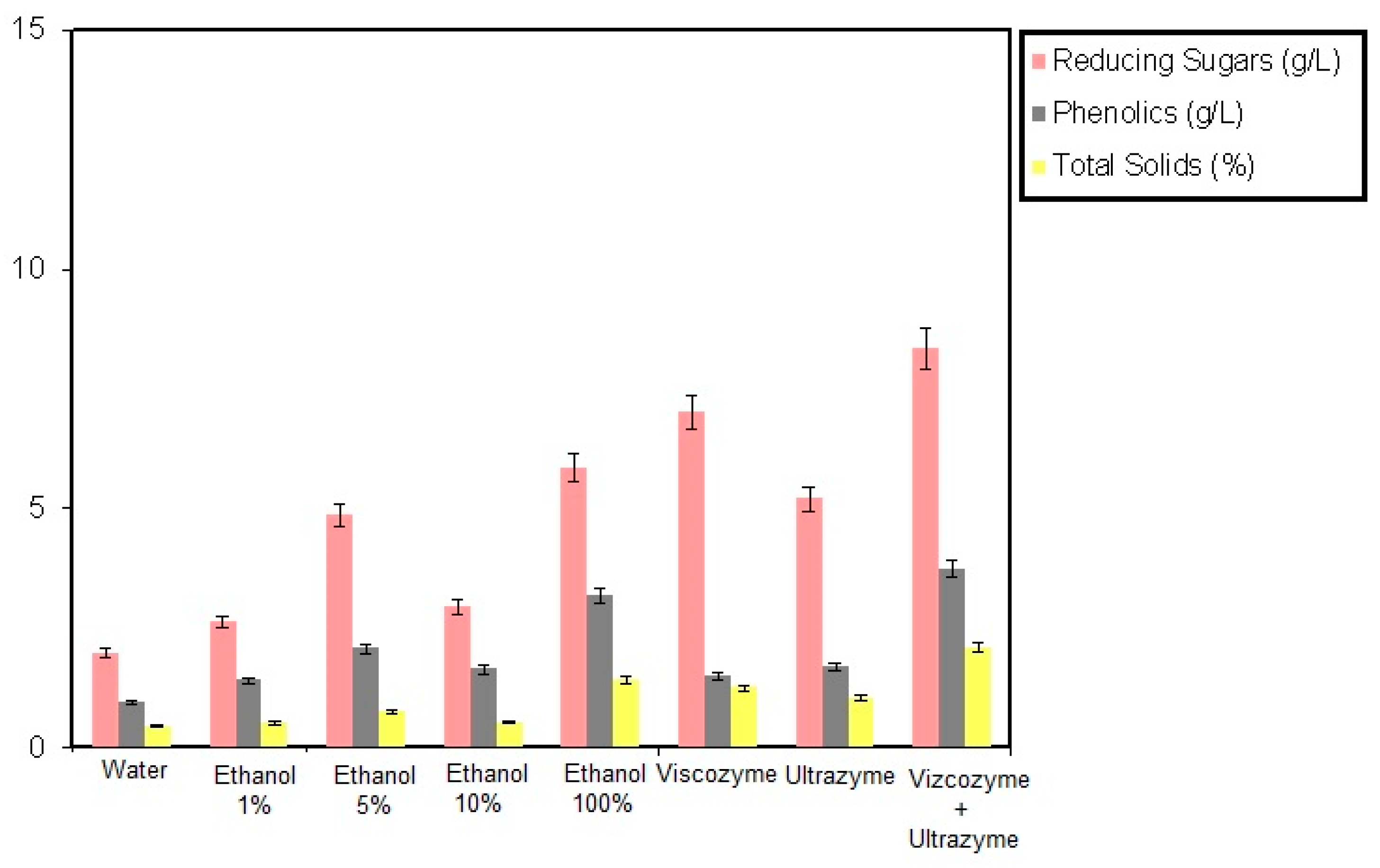
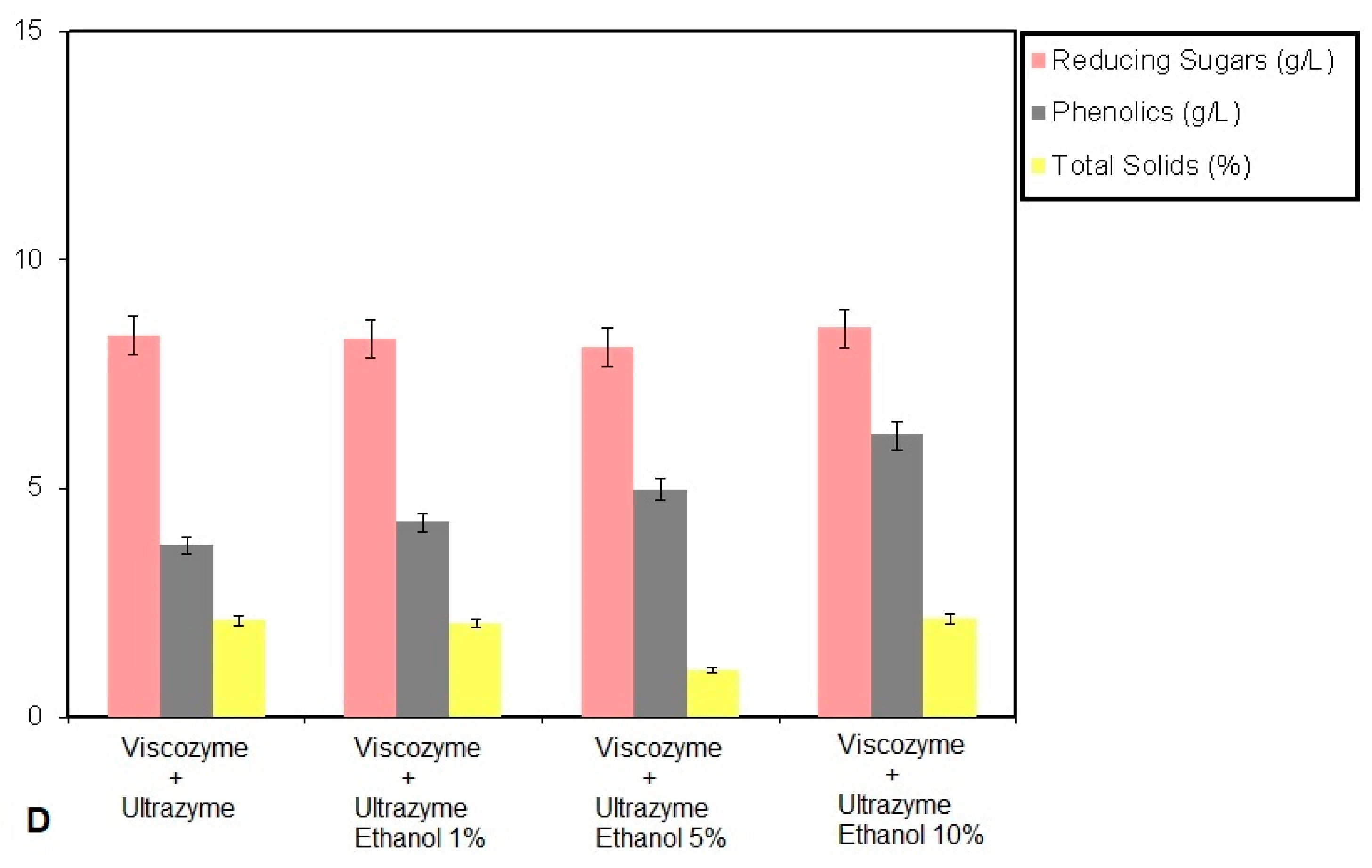
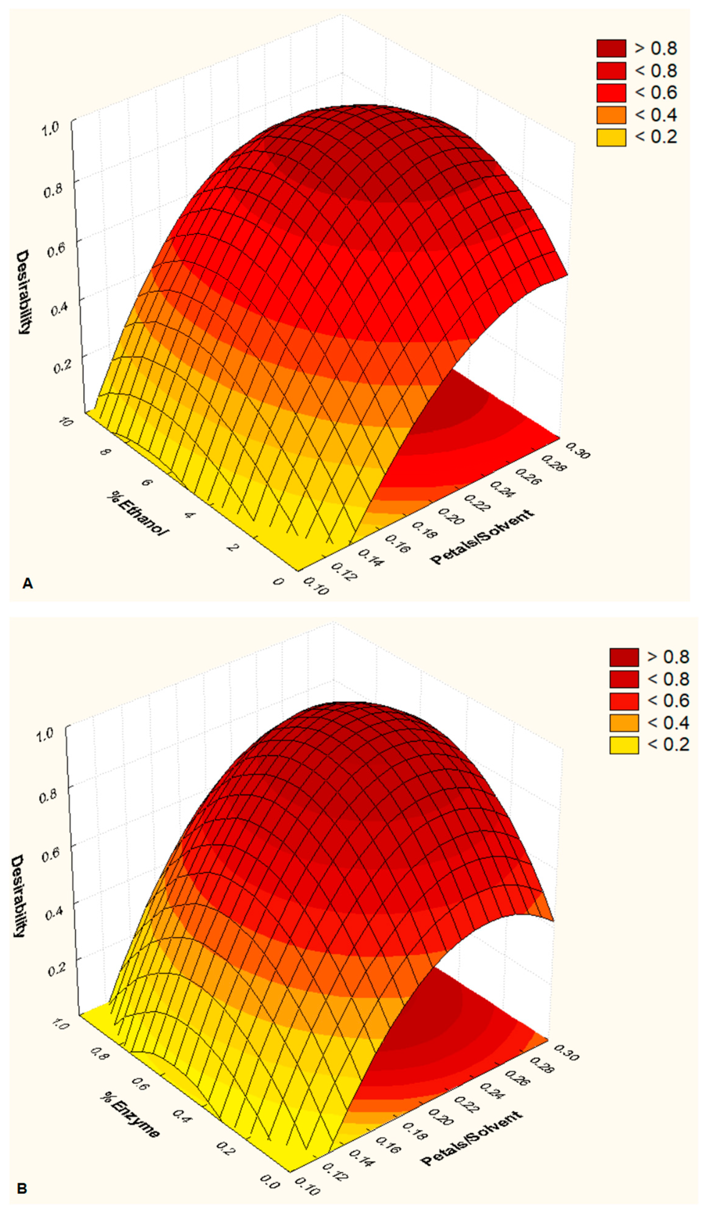
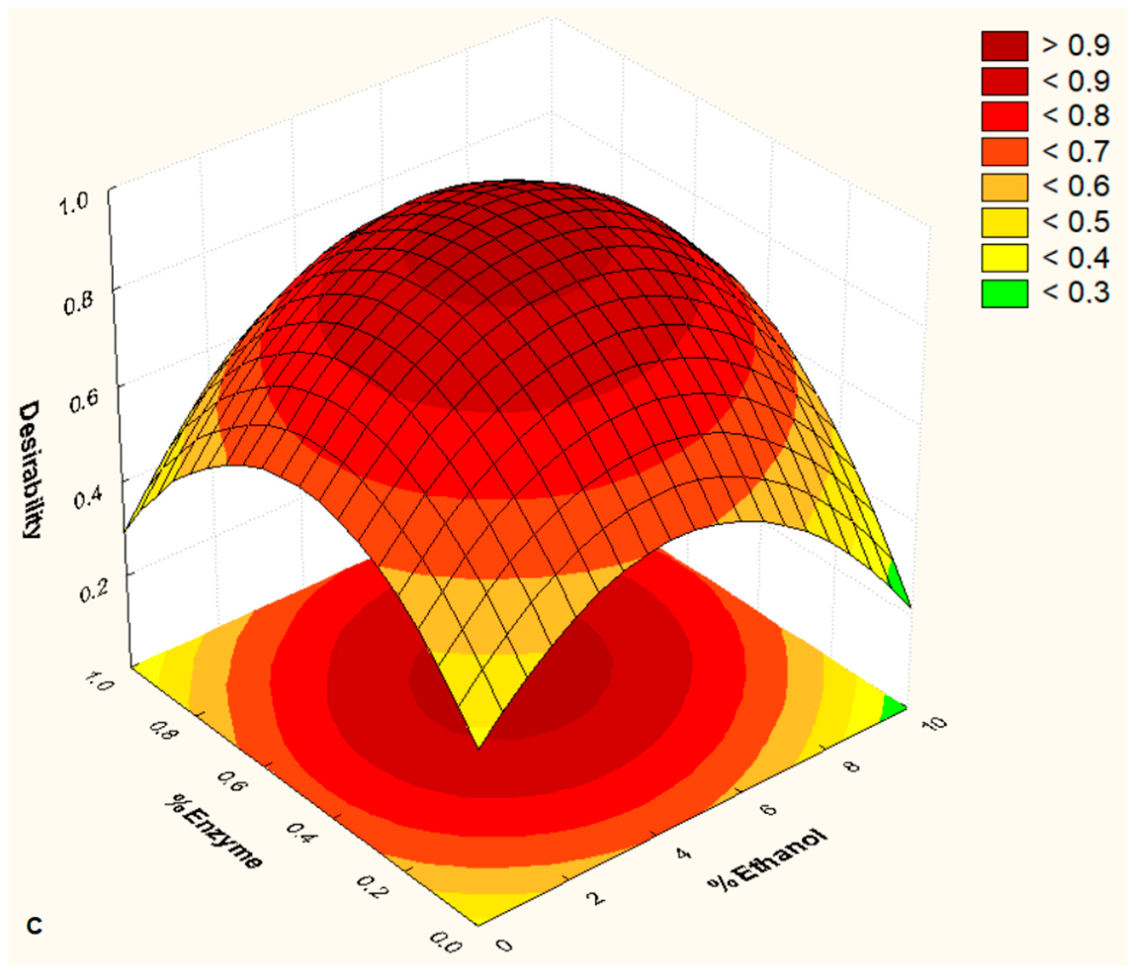
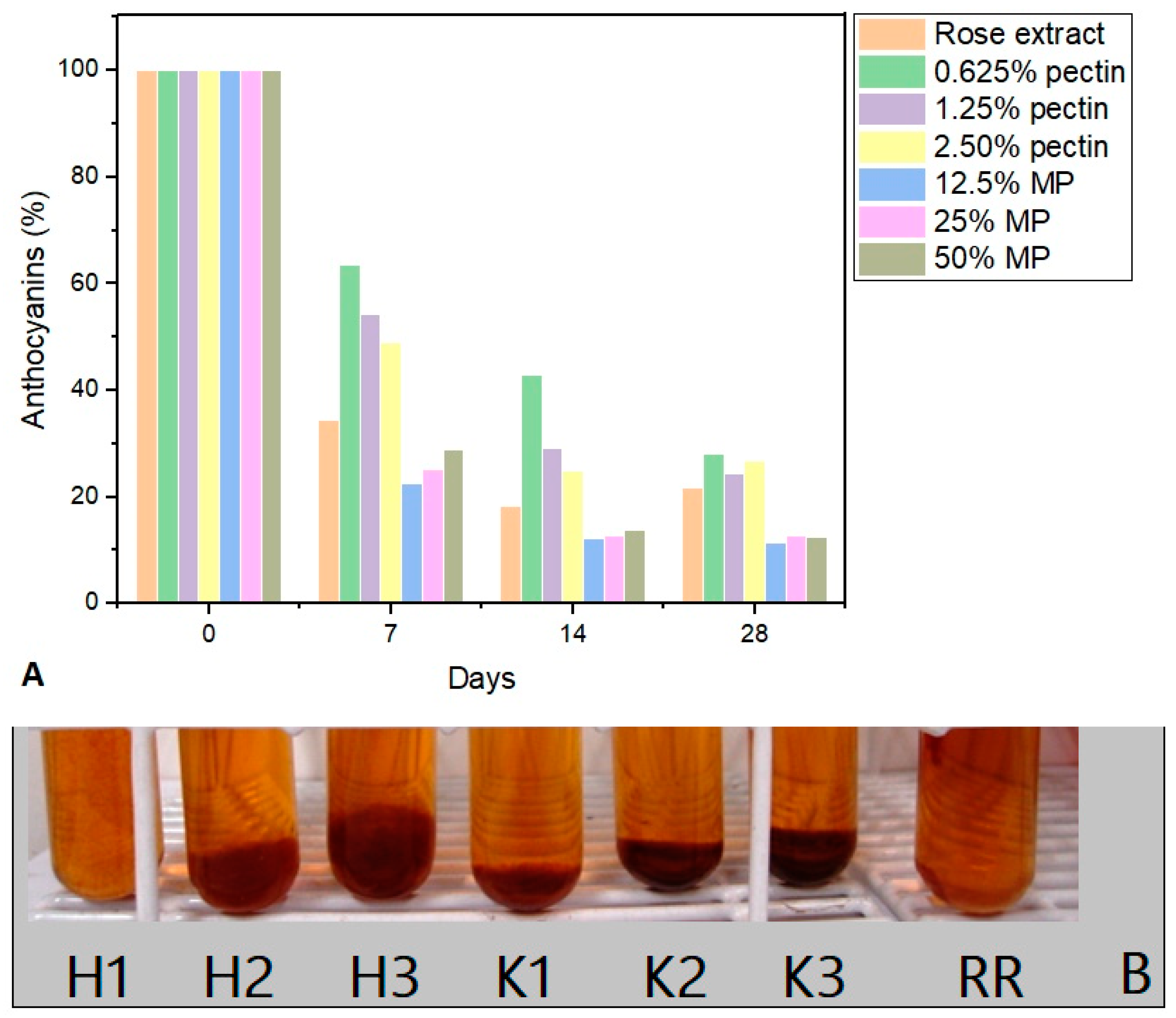
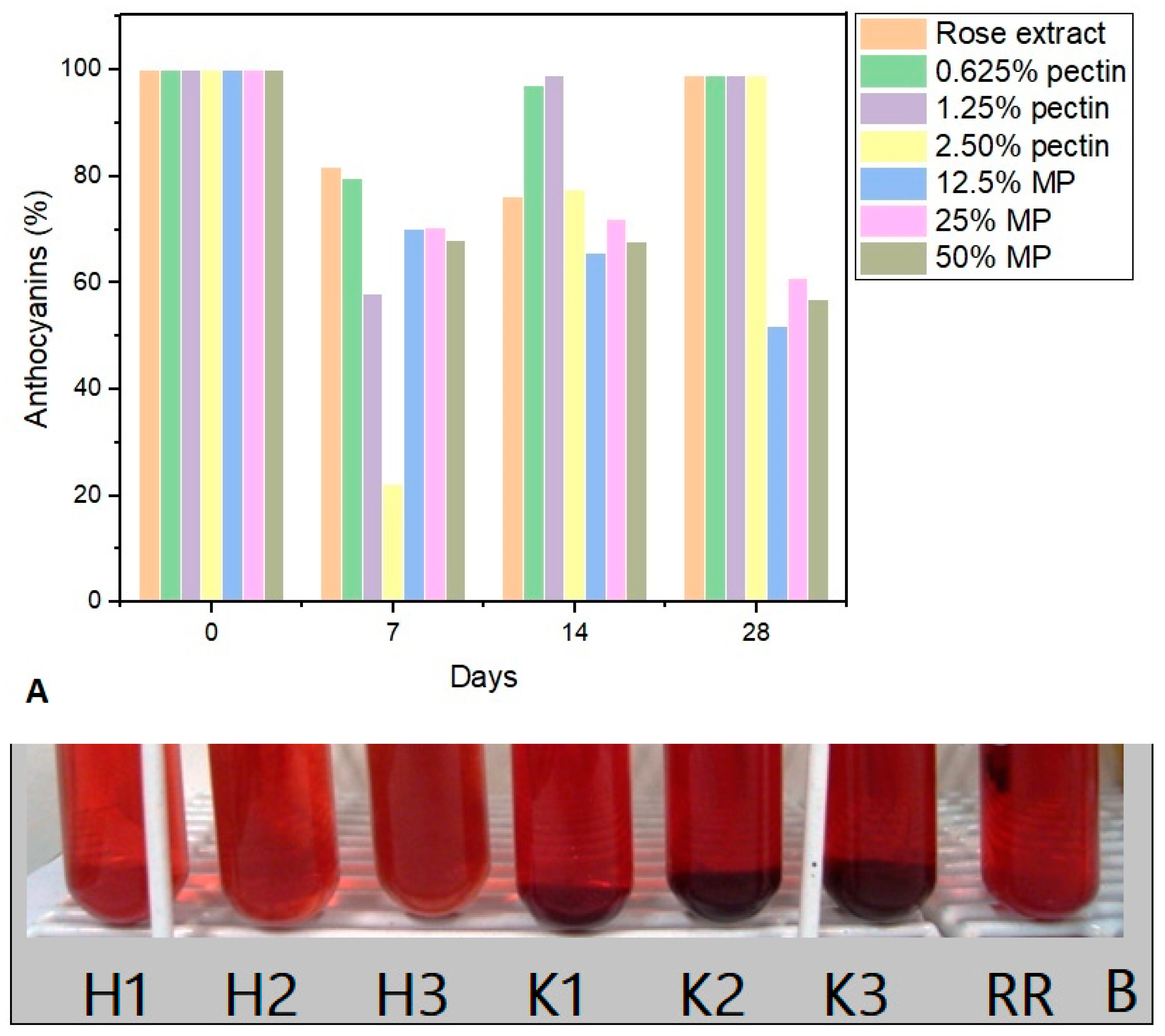
3. Results and Discussion

Enzyme extraction is an unconventional technique that has grown recently due to its clean nature and simple application. The mechanism is based on the hydrolysis or weakening of cell wall polysaccharides, such as cellulose, hemicellulose, and pectin, through an enzyme. After this rupture or weakening of the structure, the bioactive compounds are released and incorporated into the external environment, where extraction is facilitated by dragging these components through solvents [24,26,29,32,33].
According to the results shown in Figure 1, the method that obtained the best extraction of anthocyanins was the combination of the two enzymes with a maximum extraction of 3.75 g/L (11.25 mg/g rose petals) of total phenolics, followed by extraction with ethanol, which presented approximately 3.18 g/L (3.18 mg/g) of total phenolics. According to the results shown in Figure 2, it can be observed that the combination of enzymes allows an optimization of the process, during which the two combined enzymes obtain an extraction of total phenolics which is almost double compared to that obtained by the separate enzymes, around 1.6 and 1.8 g/L for enzymes A and B, respectively. These data are compatible with those reported in the literature for red rose petals, as in the study of the ultrasound-assisted extraction of red rose petals using ethanol as a solvent and an extraction temperature of 30 °C, during which 3.20 mg/g of anthocyanins rose petals was obtained [41].
Anthocyanins are polar molecules with hydroxyl, methoxyl, and carboxyl substituent groups and glycosyls linked to their aromatic rings that provide greater solubility in polar solvents [42]. In Figure 3A, it is possible to observe the visual comparison of the extraction of anthocyanin from rose petals using combinations of solvents—(i) water, (ii) water and pectinase, (iii) water and hemicellulase, and (iv) ethanol—with ethanol showing better results than water. Based on the literature, ethanol is a solvent already used successfully to extract anthocyanins [18], and it acts as a better solvent than water in the extraction of anthocyanins. In the following tests, 1, 5, and 10% ethanol concentrations were tested in water during enzymatic maceration to verify the best ethanol/water ratio for extracting more anthocyanins and their stability. In Figure 3B–D, it can be observed that ethanol positively influences the extraction of anthocyanins. However, the higher the ethanol concentration, the slower the maceration becomes, as indicated by the decrease in sugar and total solids’ concentrations.
Regarding the enzymes, the results show that the combination of the two enzymes obtained better results. Pectinase and hemicellulase, which are accessory enzymes, are known for converting lignocellulosic materials into monomeric sugars [43]. The combination of both would act on different parts of a biopolymer, favoring the extraction of anthocyanins.
According to the results obtained by the enzymatic maceration of rose petals in the presence of ethanol, it can be observed that the presence of this solvent is important for the process. Ethanol is included as a parameter to be studied during the experimental design, in addition to the need for a minimum concentration of enzymes and the proportion of petals/extractant liquids to reduce the costs. The results of this planning are shown in Table 2; in bold are the settings in which the maximum amount of phenols was obtained.
The optimization of the data can be seen in Figure 3, in which the three response variables are combined into a new variable, called desirability, and the 3-D graphs use this new variable and two factors: % ethanol and petals/liquid (Figure 4A); % enzyme and petals/liquid (Figure 4B); and % ethanol and % enzymes (Figure 4C). According to the graphs’ analysis, optimal conditions were generated: petal/extractor liquid ratio, 0.25; ethanol concentration, 6.5%; and enzyme concentration, 0.7%. These parameters of optimal conditions agree with the best result in the extraction of anthocyanins obtained by the central composite design, in which a maximum concentration of phenols of 9.99 mg/g was obtained.
With the operating conditions already established, the stability test of the anthocyanins in the rose petal extract was carried out. Two stabilizers were used, pectin and marine polysaccharides (MP), which can act either by modifying the viscosity of the medium, preventing the anthocyanins from self-oxidizing, or by ion exchange, since these polysaccharides have functional groups such as carboxyl and sulfates which can interact with the anthocyanins [25,44,45]. It was observed that the samples exposed to sunlight for 28 days showed a marked decline in their anthocyanin content (Figure 5A), both for the samples with polysaccharides and for the sample without the addition of a stabilizer, with a sharp drop during the first seven days. The sample that maintained the greatest stability was the one with 0.625% pectin, which, at the end of the 28 days, maintained an anthocyanin content of 30%. All the samples in marine polysaccharide showed the lowest stability, with a final anthocyanin content of around 10%. A yellowish-orange color, visible in Figure 5B, became noticeable after exposure, confirming anthocyanin residues in all the samples.
Hubbermann et al. [46] carried out similar tests with exposure to sunlight, still using currant and elderberry concentrates in an acetic acid buffer solution (0.2 mol/L, pH 3.9), which established a color retention around 60% after 21 and 35 days, respectively.
Li et al. [17] investigated the stability of acylated anthocyanins extracted from rose petals by an eutectic solvent—choline chloride/lactic acid—and purified, which were modified through acylation by the enzymatic catalysis method to improve the stability of anthocyanins. According to the tests, after 12 h of exposure to sunlight, around 50% stability was observed; in contrast, the acylated anthocyanins showed a decay of around 10%. According to the data obtained in this work, the enzymatic extraction process can provide excellent stability to the structure of anthocyanins.
The stability of the anthocyanins was checked for 28 days, protected from light, as shown in Figure 6A, for the samples with pectin and marine polysaccharide stabilizers; in these, it was possible to verify that the samples containing pectin and no stabilizers showed an increase in absorbance from the seventh day, resulting in a higher concentration of anthocyanins. A hypothesis is that this result could have been due to microbial contamination, which possibly hydrolyzed the anthocyanins (3,5-glycosylated) into anthocyanidins (aglycones), which have a higher molar absorptivity coefficient and, therefore, emit a greater absorbance in the visible light spectrum. A similar effect can be observed when there is acid hydrolysis of anthocyanin [40,47]. This hypothesis can be supported by the samples with marine polysaccharide stabilizers that did not develop microorganisms and maintained anthocyanin levels around 55–60% after 28 days.
At the end of the 28 days of being protected from the light, in Figure 6B, it is possible to observe that the samples still present an intense color tending towards red, corroborating the quantification results which detected the presence of anthocyanins with a final concentration of around 60% of the initial concentration. Color intensity is important when considering its applicability as a dye [10].
In the study of the degradation kinetics and antioxidant capacity of aqueous extracts based on purple carrot anthocyanins in comparison with synthetic and natural food dyes [10], the behavior of anthocyanins was similar to that obtained by the samples exposed to sunlight in this study, with a sharp drop during the first seven days and an almost total degradation after 28 days.
Anthocyanins have an excellent antioxidant efficacy, as verified in several studies [48,49,50], and it has been demonstrated that they can be widely used as eco-friendly natural pigments for various applications, such as food, pharmaceutical products, and cosmetics [51].
The antioxidant capacity was calculated for the rose petal extracts at different intervals—initial time, 60, and 120 days—obtaining IC50 results of 3.19, 5.41, and 5.59 μg/mL, respectively. These results were close to the IC50 of 5.06 μg/mL found for tannic acid and that of 2.59 μg/mL for epigallocatechin gallate, used as standards. In the study by Li et al. [17] on the antioxidant activity of anthocyanins and anthocyanin acylated with a DPPH ethanolic solution, the IC50 of the anthocyanin was 22.917 μg/mL and that of the acylated anthocyanin was 4.451 μg/mL.

4. Limitations and Future Studies

This study has limitations in the characterization of anthocyanins obtained by the enzymatic maceration of rose petals, so the identification of the structures obtained by the proposed method could be carried out in future studies. Then, there are other studies about stability tests, such as pH and temperature variation. Obtaining these structures will also help us understand the high antioxidant capacity of the anthocyanins in this study, which may make it possible to contribute to the literature in terms of understanding the extraction process with a combination of viscozyme® and ultrazym® enzymes.

5. Conclusions

Enzymatic technology is an alternative to avoid the degradation of anthocyanins and use a cleaner extraction method. Enzymes are proteins that participate in various biochemical reactions, accelerate thermodynamically favored reactions, and have stereospecific characteristics. Typically, enzymatic processes have a fast action, lack toxicity, and do not generate environmental problems. In addition, they occur at mild temperatures and pHs and act on a specific substrate with a low concentration of enzyme preparations. Therefore, the enzymatic maceration of rose petals has proven to be a promising alternative in the extraction of anthocyanins, as it is a clean and green process, which uses solvents such as water and ethanol. According to the multi-objective desirability function statistical tool, a maximum extraction of 9.99 mg/g of phenols was obtained. The stability of the rose petal extract using marine polysaccharides as stabilizers retained 60% of anthocyanins over 28 days without degradation and maintained color intensity when protected from sunlight. The rose petal extract demonstrated a high antioxidant capacity, close to the literature data for citrus compounds, and is known to be high in antioxidant compounds for cosmetic food purposes. These results indicate that anthocyanins extracted by the proposed method are a potential antioxidant dye for the pharmaceutical, food, and cosmetic industries.

Author Contributions

Conceptualization, D.W.B. and B.D.R.; methodology, B.D.R.; validation, B.D.R. and L.A.B.C. formal analysis, B.D.R.; investigation, R.d.M.F. and B.D.R.; resources, D.W.B. and B.D.R.; da-ta curation, B.D.R.; writing—original draft preparation, B.D.R. and R.d.M.F.; writing—review and editing, B.D.R., L.A.B.C. and R.d.M.F.; visualization, D.W.B., L.A.B.C. and B.D.R.; super-vision, D.W.B. and B.D.R.; project administration, B.D.R.; funding acquisition D.W.B. All authors have read and agreed to the published version of the manuscript.

Funding

This research received no external funding.

Institutional Review Board Statement

Not applicable.

Informed Consent Statement

Not applicable.

Data Availability Statement

Data are contained within the article.

Conflicts of Interest

The authors declare no conflicts of interest.

References

  1. Erdogan Eliuz, E.A.; Yabalak, E. Chicken feather hydrochar incorporated with phenolic extract of Rosa damascena Mill. to enlarge the antibacterial performance against Acinobacter baumannii and Staphylococcus aureus. J. Environ. Chem. Eng. 2022, 10, 108289. [Google Scholar] [CrossRef]
  2. Nikolova, G.; Karamalakova, Y.; Kovacheva, N.; Stanev, S.; Zheleva, A.; Gadjeva, V. Protective effect of two essential oils isolated from Rosa damascena Mill. and Lavandula angustifolia Mill, and two classic antioxidants against L-dopa oxidative toxicity induced in healthy mice. Regul. Toxicol. Pharmacol. 2016, 81, 1–7. [Google Scholar] [CrossRef]
  3. Gupta, S.; Krishna Tewari, S.; Pathak, S. Chapter 16—Temple floral waste for various bio-products in India. In Recent Trends in Solid Waste Management; Ravindran, B., Gupta, S.K., Bhat, S.A., Chauhan, P.S., Tyagi, N., Eds.; Advances in Pollution Research; Elsevier: Amsterdam, The Netherlands, 2023; pp. 293–307. ISBN 978-0-443-15206-1. [Google Scholar]
  4. Falla, N.M.; Contu, S.; Demasi, S.; Caser, M.; Scariot, V. Environmental Impact of Edible Flower Production: A Case Study. Agronomy 2020, 10, 579. [Google Scholar] [CrossRef]
  5. Karpun, O. Conceptual model of floriculture supply chain management. Intellectualization Logist. Supply Chain. Manag. 2020, 1, 41–52. [Google Scholar] [CrossRef] [PubMed]
  6. Athanasiadis, V.; Chatzimitakos, T.; Kotsou, K.; Kalompatsios, D.; Bozinou, E.; Lalas, S.I. Utilization of Blackthorn Plums (Prunus spinosa) and Sweet Cherry (Prunus avium) Kernel Oil: Assessment of Chemical Composition, Antioxidant Activity, and Oxidative Stability. Biomass 2024, 4, 49–64. [Google Scholar] [CrossRef]
  7. Harborne, J. The anthocyanins of roses. Occurrence of peonin. Experientia 1961, 17, 72–73. [Google Scholar] [CrossRef]
  8. Trujillo-Reyes, Á.; Cubero-Cardoso, J.; Rodríguez-Gutiérrez, G.; García-Martín, J.F.; Rodríguez-Galán, M.; Borja, R.; Serrano, A.; Fermoso, F.G. Extraction of phenolic compounds and production of biomethane from strawberry and raspberry extrudates. Biochem. Eng. J. 2019, 147, 11–19. [Google Scholar] [CrossRef]
  9. Kendrick, A. 7-Coloring Aqueous Food Types. In Handbook on Natural Pigments in Food and Beverages; Carle, R., Schweiggert, R.M., Eds.; Woodhead Publishing Series in Food Science, Technology and Nutrition; Woodhead Publishing: Cambridge, UK, 2016; pp. 163–177. ISBN 978-0-08-100371-8. [Google Scholar]
  10. Perez, M.B.; Da Peña Hamparsomian, M.J.; Gonzalez, R.E.; Denoya, G.I.; Dominguez, D.L.E.; Barboza, K.; Iorizzo, M.; Simon, P.W.; Vaudagna, S.R.; Cavagnaro, P.F. Physicochemical properties, degradation kinetics, and antioxidant capacity of aqueous anthocyanin-based extracts from purple carrots compared to synthetic and natural food colorants. Food Chem. 2022, 387, 132893. [Google Scholar] [CrossRef]
  11. Cho, E.; Chung, E.Y.; Jang, H.-Y.; Hong, O.-Y.; Chae, H.S.; Jeong, Y.-J.; Kim, S.-Y.; Kim, B.-S.; Yoo, D.J.; Kim, J.-S.; et al. Anti-cancer Effect of Cyanidin-3-glucoside from Mulberry via Caspase-3 Cleavage and DNA Fragmentation in vitro and in vivo. Anticancer Agents Med. Chem. 2017, 17, 1519–1525. [Google Scholar] [CrossRef]
  12. Herrera-Balandrano, D.D.; Chai, Z.; Hutabarat, R.P.; Beta, T.; Feng, J.; Ma, K.; Li, D.; Huang, W. Hypoglycemic and hypolipidemic effects of blueberry anthocyanins by AMPK activation: In vitro and in vivo studies. Redox Biol. 2021, 46, 102100. [Google Scholar] [CrossRef]
  13. Andrade, T.A.; Hamerski, F.; López Fetzer, D.E.; Roda-Serrat, M.C.; Corazza, M.L.; Norddahl, B.; Errico, M. Ultrasound-assisted pressurized liquid extraction of anthocyanins from Aronia melanocarpa pomace. Sep. Purif. Technol. 2021, 276, 119290. [Google Scholar] [CrossRef]
  14. Sidor, A.; Drożdżyńska, A.; Gramza-Michałowska, A. Black chokeberry (Aronia melanocarpa) and its products as potential health-promoting factors—An overview. Trends Food Sci. Technol. 2019, 89, 45–60. [Google Scholar] [CrossRef]
  15. Song, J.; Yu, Y.; Chen, M.; Ren, Z.; Chen, L.; Fu, C.; Li, Z. Advancement of protein-and polysaccharide-based biopolymers for anthocyanin encapsulation. Front. Nutr. 2022, 9, 938829. [Google Scholar] [CrossRef] [PubMed]
  16. Tang, R.; He, Y.; Fan, K. Recent advances in stability improvement of anthocyanins by efficient methods and its application in food intelligent packaging: A review. Food Biosci. 2023, 56, 103164. [Google Scholar] [CrossRef]
  17. Li, J.; Guo, X.; Wang, R.; Geng, Z.; Jia, J.; Pang, S.; Du, Y.; Jia, S.; Cui, J. Ultrasonic assisted extraction of anthocyanins from rose flower petal in DES system and enzymatic acylation. LWT 2023, 180, 114693. [Google Scholar] [CrossRef]
  18. Lapornik, B.; Prošek, M.; Wondra, A.G. Comparison of extracts prepared from plant by-products using different solvents and extraction time. J. Food Eng. 2005, 71, 214–222. [Google Scholar] [CrossRef]
  19. Didion, Y.P.; Tjalsma, T.G.; Su, Z.; Malankowska, M.; Pinelo, M. What is next? the greener future of solid liquid extraction of biobased compounds: Novel techniques and solvents overpower traditional ones. Sep. Purif. Technol. 2023, 320, 124147. [Google Scholar] [CrossRef]
  20. Das, S.; Nadar, S.S.; Rathod, V.K. Integrated strategies for enzyme assisted extraction of bioactive molecules: A review. Int. J. Biol. Macromol. 2021, 191, 899–917. [Google Scholar] [CrossRef]
  21. Wijesinghe, W.; Jeon, Y.-J. Enzyme-assistant extraction (EAE) of bioactive components: A useful approach for recovery of industrially important metabolites from seaweeds: A review. Fitoterapia 2012, 83, 6–12. [Google Scholar] [CrossRef]
  22. Barzana, E.; Rubio, D.; Santamaria, R.; Garcia-Correa, O.; Garcia, F.; Ridaura Sanz, V.; López-Munguía, A. Enzyme-mediated solvent extraction of carotenoids from marigold flower (Tagetes erecta). J. Agric. Food Chem. 2002, 50, 4491–4496. [Google Scholar] [CrossRef]
  23. Passos, C.P.; Yilmaz, S.; Silva, C.M.; Coimbra, M.A. Enhancement of grape seed oil extraction using a cell wall degrading enzyme cocktail. Food Chem. 2009, 115, 48–53. [Google Scholar] [CrossRef]
  24. Ruiz-Terán, F.; Perez-Amador, I.; López-Munguia, A. Enzymatic extraction and transformation of glucovanillin to vanillin from vanilla green pods. J. Agric. Food Chem. 2001, 49, 5207–5209. [Google Scholar] [CrossRef] [PubMed]
  25. Yang, Y.-C.; Li, J.; Zu, Y.-G.; Fu, Y.-J.; Luo, M.; Wu, N.; Liu, X.-L. Optimisation of microwave-assisted enzymatic extraction of corilagin and geraniin from Geranium sibiricum Linne and evaluation of antioxidant activity. Food Chem. 2010, 122, 373–380. [Google Scholar] [CrossRef]
  26. Puri, M.; Sharma, D.; Barrow, C.J. Enzyme-assisted extraction of bioactives from plants. Trends Biotechnol. 2012, 30, 37–44. [Google Scholar] [CrossRef] [PubMed]
  27. Yazdanian, E.; Golkar, P.; Vahabi, M.R.; Taghizadeh, M. Elicitation Effects on Some Secondary Metabolites and Antioxidant Activity in Callus Cultures of Allium jesdianum Boiss. & Buhse.: Methyl Jasmonate and Putrescine. Appl. Biochem. Biotechnol. 2022, 194, 601–619. [Google Scholar] [CrossRef] [PubMed]
  28. Sankaralingam, B.; Balan, L.; Chandrasekaran, S.; Muthu Selvam, A. Anthocyanin: A Natural Dye Extracted from Hibiscus Sabdariffa (L.) for Textile and Dye Industries. Appl. Biochem. Biotechnol. 2023, 195, 2648–2663. [Google Scholar] [CrossRef] [PubMed]
  29. Lotfi, L.; Kalbasi-Ashtari, A.; Hamedi, M.; Ghorbani, F. Effects of enzymatic extraction on anthocyanins yield of saffron tepals (Crocos sativus) along with its color properties and structural stability. J. Food Drug Anal. 2015, 23, 210–218. [Google Scholar] [CrossRef]
  30. Serea, D.; Râpeanu, G.; Constantin, O.E.; Bahrim, G.E.; Stănciuc, N.; Croitoru, C. Ultrasound and enzymatic assisted extractions of bioactive compounds found in red grape skins băbească neagră (vitis vinifera) variety. Ann. Univ. Dunarea Jos Galati Fascicle VI-Food Technol. 2021, 45, 9–25. [Google Scholar] [CrossRef]
  31. Li, X.; Zhu, F.; Zeng, Z. Effects of different extraction methods on antioxidant properties of blueberry anthocyanins. Open Chem. 2021, 19, 138–148. [Google Scholar] [CrossRef]
  32. Rezende, Y.R.R.S.; Nogueira, J.P.; Silva, T.O.M.; Barros, R.G.C.; de Oliveira, C.S.; Cunha, G.C.; Gualberto, N.C.; Rajan, M.; Narain, N. Enzymatic and ultrasonic-assisted pretreatment in the extraction of bioactive compounds from Monguba (Pachira aquatic Aubl) leaf, bark and seed. Food Res. Int. 2021, 140, 109869. [Google Scholar] [CrossRef]
  33. Oancea, S.; Perju, M. Influence of enzymatic and ultrasonic extraction on phenolics content and antioxidant activity of Hibiscus Sabdariffa, L. flowers. Bulg. Chem. Commun. 2020, 52, 25–29. [Google Scholar]
  34. Xue, H.; Tan, J.; Li, Q.; Tang, J.; Cai, X. Ultrasound-assisted enzymatic extraction of anthocyanins from raspberry wine residues: Process optimization, isolation, purification, and bioactivity determination. Food Anal. Methods 2021, 14, 1369–1386. [Google Scholar] [CrossRef]
  35. Talbi, E.-G. Metaheuristics: From Design to Implementation; John Wiley & Sons: New York, NY, USA, 2009; ISBN 0-470-49690-8. [Google Scholar]
  36. Somogyi, M. Notes on sugar determination. J. Biol. Chem. 1952, 195, 19–23. [Google Scholar] [CrossRef]
  37. Folin, O.; Denis, W. On phosphotungstic-phosphomolybdic compounds as color reagents. J. Biol. Chem. 1912, 12, 239–243. [Google Scholar] [CrossRef]
  38. AOAC. Official Methods of Analysis of the AOAC International; Oxford University Press: Oxford, UK, 1996. [Google Scholar]
  39. da Silva, C.; Herdeiro, R.; Mathias, C.; Panek, A.; Silveira, C.; Rodrigues, V.; Rennó, M.; Falcão, D.; Cerqueira, D.; Minto, A. Evaluation of antioxidant activity of Brazilian plants. Pharmacol. Res. 2005, 52, 229–233. [Google Scholar] [CrossRef] [PubMed]
  40. Wrolstad, R.E.; Durst, R.W.; Lee, J. Tracking color and pigment changes in anthocyanin products. Trends Food Sci. Technol. 2005, 16, 423–428. [Google Scholar] [CrossRef]
  41. Özgür, M.Ü.; Çimen, E. Ultrasound-Assisted Extraction of Anthocyanins from Red Rose Petals and New Spectrophotometric Methods for the Determination of Total Monomeric Anthocyanins. J. AOAC Int. 2018, 101, 967–980. [Google Scholar] [CrossRef] [PubMed]
  42. Lopes, T.; Xavier, M.; Quadri, M.G.; Quadri, M. Antocianinas: Uma breve revisão das características estruturais e da estabilidade. Curr. Agric. Sci. Technol. 2007, 13, 112861433. [Google Scholar]
  43. García-Aparicio, M.P.; Ballesteros, M.; Manzanares, P.; Ballesteros, I.; González, A.; Negro, M.J. Xylanase Contribution to the Efficiency of Cellulose Enzymatic Hydrolysis of Barley Straw; Springer: Berlin/Heidelberg, Germany, 2007; pp. 353–365. [Google Scholar]
  44. Huang, H.-L.; Tsai, I.-L.; Lin, C.; Hang, Y.-H.; Ho, Y.-C.; Tsai, M.-L.; Mi, F.-L. Intelligent films of marine polysaccharides and purple cauliflower extract for food packaging and spoilage monitoring. Carbohydr. Polym. 2023, 299, 120133. [Google Scholar] [CrossRef] [PubMed]
  45. Ko, A.; Lee, J.; Sop Nam, H.; Gyu Lee, H. Stabilization of black soybean anthocyanin by chitosan nanoencapsulation and copigmentation. J. Food Biochem. 2017, 41, e12316. [Google Scholar] [CrossRef]
  46. Hubbermann, E.M.; Heins, A.; Stöckmann, H.; Schwarz, K. Influence of acids, salt, sugars and hydrocolloids on the colour stability of anthocyanin rich black currant and elderberry concentrates. Eur. Food Res. Technol. 2006, 223, 83–90. [Google Scholar] [CrossRef]
  47. Giusti, M.M.; Wrolstad, R.E. Characterization and measurement of anthocyanins by UV-visible spectroscopy. Curr. Protoc. Food Anal. Chem. 2001, 1, F1.2.1–F1.2.13. [Google Scholar] [CrossRef]
  48. Kumar, M.; Dahuja, A.; Sachdev, A.; Kaur, C.; Varghese, E.; Saha, S.; Sairam, K.V.S.S. Evaluation of enzyme and microwave-assisted conditions on extraction of anthocyanins and total phenolics from black soybean (Glycine max L.) seed coat. Int. J. Biol. Macromol. 2019, 135, 1070–1081. [Google Scholar] [CrossRef] [PubMed]
  49. Vila, M.M.D.C.; Chaud, M.V.; Balcão, V.M. Chapter 19—Microencapsulation of Natural Anti-Oxidant Pigments. In Microencapsulation and Microspheres for Food Applications; Sagis, L.M.C., Ed.; Academic Press: San Diego, CA, USA, 2015; pp. 369–389. ISBN 978-0-12-800350-3. [Google Scholar]
  50. Ryu, D.; Koh, E. Application of response surface methodology to acidified water extraction of black soybeans for improving anthocyanin content, total phenols content and antioxidant activity. Food Chem. 2018, 261, 260–266. [Google Scholar] [CrossRef]
  51. Sun, H.; Zhang, P.; Zhu, Y.; Lou, Q.; He, S. Antioxidant and prebiotic activity of five peonidin-based anthocyanins extracted from purple sweet potato (Ipomoea batatas (L.) Lam.). Sci. Rep. 2018, 8, 5018. [Google Scholar] [CrossRef]
Figure 1. Chemical structures of anthocyanins and their radicals, peonine or cyanin.
Biomass 04 00021 g001
Figure 2. Comparison of red rose petal anthocyanin extraction methods with different types of solvents and enzymes.
Biomass 04 00021 g002
Figure 3. (A) Visual comparison of rose petal anthocyanin extraction methods: 1—water; 2—water + viscozyme; 3—water + ultrazym; and 4—100% ethanol. (B) Enzymatic maceration of rose petals in the presence of ethanol with viscozyme. (C) Enzymatic maceration of rose petals in the presence of ethanol with ultrazym. (D) Enzymatic maceration of rose petals in the presence of ethanol with a combination of enzymes.
Biomass 04 00021 g003aBiomass 04 00021 g003b
Figure 4. Optimization graphs for the enzymatic maceration of rose petals: (A) desirability, % ethanol, and petals/liquid; (B) desirability, % enzyme, and petals/liquid; and (C) desirability, % ethanol, and % enzymes.
Biomass 04 00021 g004aBiomass 04 00021 g004b
Figure 5. Stability of rose petal extract when exposed to sunlight. (A) % of anthocyanins as a function of time. (B) Samples after 28 days: H1, marine polysaccharides 12.5%; H2, marine polysaccharides 25%; H3, marine polysaccharides 50%; K1, pectin 0.625%; K2, pectin 1.25%; K3, pectin 2.5%; and RR, red rose petal extract.
Biomass 04 00021 g005
Figure 6. Stability of rose petal extract protected from sunlight. (A) % of anthocyanins as a function of time. (B) Samples after 28 days: H1, marine polysaccharide 12.5%; H2, marine polysaccharide 25%; H3, marine polysaccharide 50%; K1, pectin 0.625%; K2, pectin 1.25%; K3, pectin 2.5%; and RR, red rose petal extract.
Biomass 04 00021 g006
Table 1. Central composite rotatable design (CCRD) of the enzymatic maceration of rose petals.
Factors−1.68−10+1+1.68
Rose petals/solvent0.100.140.20.260.30
%Ethanol (v/v)025810
%Enzyme00.20.50.81.0
Table 2. Results of enzymatic maceration of rose petals using central composite planning.
AssaysPetals/Liq.% Ethanol%EnzymeSugars (g/L)Phenols (g/L)Total Solids (%)
10.142.00.206.035.331.08
20.142.00.806.134.361.25
30.148.00.206.776.501.78
40.148.00.807.406.691.44
50.262.00.209.078.762.43
60.262.00.809.207.262.37
70.268.00.208.768.822.27
80.268.00.808.509.992.71
90.105.00.505.926.071.19
100.305.00.509.066.442.56
110.200.00.508.396.951.80
120.2010.00.508.295.802.06
130.205.00.006.797.901.51
140.205.01.008.877.661.57
150.205.00.509.168.132.68
160.205.00.509.078.142.65
170.205.00.509.108.112.70
Disclaimer/Publisher’s Note: The statements, opinions and data contained in all publications are solely those of the individual author(s) and contributor(s) and not of MDPI and/or the editor(s). MDPI and/or the editor(s) disclaim responsibility for any injury to people or property resulting from any ideas, methods, instructions or products referred to in the content.

