Review

Effects of Innovative Processing Methods on Microalgae Cell Wall: Prospects towards Digestibility of Protein-Rich Biomass

by
Luís Machado
1,2,
Gonçalo Carvalho
1,2 and
Ricardo N. Pereira
1,2,*
1
CEB—Centre of Biological Engineering, University of Minho, 4710-057 Braga, Portugal
2
LABBELS—Associate Laboratory, 4710-057 Braga, Portugal
*
Author to whom correspondence should be addressed.
Biomass 2022, 2(2), 80-102; https://doi.org/10.3390/biomass2020006
Submission received: 28 February 2022 / Revised: 6 April 2022 / Accepted: 7 April 2022 / Published: 14 April 2022

Abstract

:
Microalgae are known to have higher photosynthetic efficiencies when compared to land-based plants. The use of microalgae biomass as a protein source is attracting attention due to its interesting protein composition and sustainable character when compared to conventional animal and plant protein-based sources. Nonetheless, the existence of a rigid cell wall is typical for most microalgae species, and this presents a serious obstacle to a higher bioaccessibility of their valuable protein fractions. Depending on the cell wall composition, the gastrointestinal digestion process itself can result in different pathways of protein absorption. It is then important to understand how microalgae cell wall structure can be affected during traditional and industrial production of its biomass once these questions are often overlooked. This review intends to fulfill this gap by addressing the major impacts of innovative sustainable processing of microalgae biomass, giving particular attention to drying operations and cellular disruption methods based on electric field application—such as pulsed electric fields (PEF) and moderate electric fields (MEF). Using microalgae biomass as food supplements at its full potential depends on its protein digestibility patterns, and subsequently their bioaccessibility and bioavailability. The importance of using in vitro gastrointestinal systems to understand the impact of innovative downstream processing of microalgae biomass will be addressed.

Graphical Abstract

1. Introduction

1.1. Historical Overview

Microalgae are aquatic unicellular organisms with photosynthetic capabilities and are capable of producing organic compounds in a more efficient way than land-based plants [1,2]. Their existence is dated to millions of years ago, as they were one of the first organisms that appeared on Earth, being a very diverse group of microorganisms with thousands of different species [3,4]. Aztecs and Indian tribes consumed microalgae for centuries [5,6,7] and the modern utilization of microalgae began in the 1950s, as a food supplement [3]. A few years later in Japan, the first large-scale microalgae cultivation with a commercial purpose was developed [8]. The first microalgae to be commercially sold as food products in Japan, Taiwan and Mexico were Chlorella and Arthrospira between the 1960s and 1970s [9,10]. Although the consumption of microalgae dates back to ancient times, it was only in the last century that microalgae biomass started to gain interest from a biotechnological point of view [5,11].

1.2. Sustainable Production and Industrial Applications

The growth of the world population and subsequent increase in food demand and pressure on natural resources [12] are arousing the interest of the food industry towards microalgae as a promising and sustainable source of nutrients [13,14]. Microalgae has easy and sustainable growth conditions that discard the need for arable land. Further, its biomass has a high nutritional content, which makes them a valuable food source for the population, [4,5,13]. Microalgae are considered to be one of the best renewable sources of biomass due to their ability to double their initial biomass after 2 to 5 days of growth, and to fix CO2 for photosynthesis more efficiently than commonly used plant crops [15]. As a promising source of several nutrients and biocompounds, they can also have different industrial applications (Figure 1), such as the ones for the development of pharmaceuticals and health products, food and feed ingredients, and the production of biofuels [8]. Generally, the biocompounds found on microalgae can be classified into two main categories: primary metabolites and secondary metabolites. The primary metabolites are essential for microalgae metabolism and to ensure their growth and development (e.g., proteins, lipids, carbohydrates). The secondary metabolites (e.g., carotenoids, vitamins, and other biologically active compounds) can present some vital functions, but they are not needed for normal microalgae development; for example, they can also enhance defense mechanisms against other living organisms [1,16].

1.3. A Promising Protein Source

Microalgae synthesize proteins in their primary metabolism, which makes them an alternative protein source for the food industry. Some reports have shown that microalgae can present similar amounts of proteins, which includes the presence in its composition of essential amino acids (EAA), when compared to other more used food crops, such as milk, soybean, or meat [4,17,18]. Nonetheless, in some cases, microalgae still need to be evaluated and classified as food safe for human consumption. For microalgae to be used as a food product, they must be first recognized by the Food and Drug Administration (FDA) as: Generally Recognized as Safe (GRAS). This regulation is valid in the U.S. jurisdiction, while in Europe this is the responsibility of the European Food Safety Authority (EFSA). In the European Union, some of the accepted microalgae species for human consumption are: Arthrospira platensis, some species of Chlorella, such as Chlorella vulgaris and Chlorella pyrenoidosa. While in the US, there are more microalgae species with GRAS recognition, for example: Arthrospira platensis, Chlamydomonas reinhardtii, Chlorella vulgaris, and Euglena gracilis [7,12].
Chlorella vulgaris and Arthrospira platensis are already being classified as non-novel food due to being widely used over the last years, both being used as food supplements in their pure form [16]. In the case of Chlorella vulgaris, it is widely used as a commercial food product in quite different forms such as powder, capsules, tablets, and extracts [17]. On the other hand, Arthrospira (commercial known as Spirulina) is also commonly used for human nutrition due to its high protein content and nutritional value. These microalgae also have some health effects associated, which include the suppression of hypertension and protection against renal failure, among others [15]. In addition to these two well-known microalgae, Dunaliella salina can also be a valuable food source and thus be consumed as a dietary supplement in the human diet in different forms, similar to the ones used with Chlorella (e.g., pills and capsules) [16].
C. vulgaris is described to have around 51–58% of crude proteins per dry weight, while other green species can have between 39% and 71% of proteins per dry weight [4,19]. The green microalgae are the ones with the higher amounts of proteins produced in terms of the percentage of dry biomass. Besides the non-novel microalgae for human consumption, there are some others that are becoming of great interest recently, such as Haematococcus pluvialis, Tetraselmis chuii, and Euglena gracilis. There are also some promising microalgae that are still under evaluation to be used for human nutrition, such as Galdiera sulphuraria [12]. The Galdiera sulphuraria, for example, can have a protein content of 33% in autotrophic growth, but this value can change if the growth is performed under heterotrophic conditions [20]. On the other hand, Tetraselmis chuii can have a protein content of around 36% [21]. Another great advantage of using microalgae as a non-conventional protein source is their ability to synthesize some of the EAA, meeting the minimum requirements imposed by the Food and Agriculture Organization (FAO) [12], which cannot be achieved by specific vegetable crops or by humans and animals during their normal metabolism [3,22]. It is also important to highlight that the group of green microalgae is attracting attention for human and animal nutrition since besides their high protein content they also contain some valuable pigments. Chlorophylls, as the main pigment present in green microalgae, can present some anti-inflammatory effects when used in pharmaceutical industries. Other pigments can also be found in microalgae, for example, carotenoids, astaxanthin and phycobiliproteins, with this last one being more common in cyanobacteria presenting interesting bioactive properties [3].

1.4. Main Challenge

A myriad of microalgae species is being characterized, and cultivation strategies are being optimized toward the production of food and feed ingredients. Currently, one of the major challenges of microalgae biomass is placed on the fundamental understanding of the role of their main nutrients in human health. Better elucidation of digestion dynamics and its relationship with processing methods is needed. Innovative processing with a promise of less energy input can be used to obtain protein-enriched fractions and overcome limitations imposed by the resistance of microalgae cell structure upon gastrointestinal digestion. Microalgae biomass is seen as an alternative and sustainable protein source, but it is of crucial importance to develop a better understanding of how to design and integrate innovative processing towards efficiency, safety, and quality envisaging beneficial health outcomes. This review intends to recognize microalgae as a promising protein source, but critically emphasize the importance that downstream processing may have on structural aspects of microalgae cells, something that is often overlooked. This is proposed to be achieved by addressing three main topics: (i) microalgae as a protein source that encompasses an interesting nutritional composition (Section 2); (ii) the importance of efficient and innovative and “green” disruption methods (based on electric field technology) to allow efficient disruption of a cell wall structure as a way to ease human digestion or extraction strategies for valorization (Section 3); (iii) and to establish a relationship between downstream processing and biomass digestibility by addressing the importance of integrating gastrointestinal digestion assessment strategies to evaluate quality and functionality of microalgae derived products (Section 4).

2. Microalgae Protein Content

Proteins are one of the most important algal metabolites and their quality can be measured by certain parameters such as protein efficiency ratio (PER) and biological value (BV). PER indicates the weight gain by the test subject for unit of protein consumed, while BV estimates the retention of nitrogen used for growth or maintenance. The quality of the protein can also be accessed by the digestibility coefficient (DC) and the net protein utilization (NPU) which is the digestibility of the protein and the biological value of the amino acids present in the food. DC of microalgae is often below 80%, while casein and eggs are nearly 95%. This lower DC is explained by the presence of a cellulosic cell wall, whose composition and resistance to breakdown is very dependent on species and downstream processing methods; air-dried Chlorella presents a DC of 59%, while the drum-dried can go up to 89% [13,22]. These parameters are affected by the type of microalgae itself, and their type of cell wall, and are very dependent on the chosen cellular disruption method applied during the extraction process, to obtain protein-rich fractions [13].
Table 1 resumes several microalgae with a high protein content that can range from 26% to 71% (per dry weight). For example, Arthrospira sp. presents exceptional amounts of protein, but is a cyanobacterium, which is a prokaryotic microorganism. These cyanobacteria are known to have a Gram-negative cell wall, composed of peptidoglycan with an absence of cellulosic compounds in their structure, thus presenting some structural differences from other microalgae such as Chlorella sp. [19,23]. A fully developed Chlorella vulgaris cell can present about 42% to 58% of total proteins in their biomass dry weight [17,19,22]. More than 50% of proteins can be found inside the cell of C. vulgaris, where about 20% can be part of the microalgae cell wall, and some other proteins can be migrating from the inside and outside of the cell (around 30%) [17]. With the majority of the protein fraction placed inside the cell, its bioavailability is dependent on the way they become accessible when applying different treatments that can affect the permeabilization of the cells’ structure. Another important limiting factor for the effective use of microalgae proteins in the food industry is their nutritional quality. A good alternative to animal protein source should contain all the EAA for the human diet, thus guaranteeing an adequate protein synthesis [18,24].
Regarding the protein composition of plants, they not only present some deficiencies in EAA such as lysine, leucine, and methionine [24] but also present some digestibility issues, for example, soybeans have trypsin inhibitors that can block the digestive enzymes, such as trypsin [25]. Plant proteins have some significant structural differences when compared to animal proteins, presenting different polypeptidic chains and different secondary and tertiary structures. Another great difference between these two protein sources is that plant contains storage proteins and usually possesses larger and more compact structures when compared to animal proteins [26]. Microalgae biomass appears here as another alternative, as a rich source of some of EAA [3,22]. For these reasons, microalgae proteins are gathering interest as a sustainable alternative to meet the current global protein demand [27]. Besides the high nutritional properties of microalgae proteins, these are also described as having low allergenicity potential when compared to other conventional proteins sources, such as soy and milk [28].
Table 1. Microalgae and macroalgae with high protein content for human consumption and comparison to some conventional and non-conventional sources.
Protein SourcesProtein Content (% per Dry Weight)Reference
Microalgae
Chlorella vulgaris51–58[4,19]
Arthrospira platensis55.8/46–63[19,22]
Arthrospira maxima60–71[3,22]
Euglena gracilis30–47[7,22]
Dunaliela salina57[2,4]
Porphyridium cruentum28–39[2,29]
Tetraselmis chuii35–40[21,30]
Galdieria sulphuraria26–32[20]
Macroalgae
Ulva lactuca12–20[12,31]
Palmaria palmata9.8–18.8[12,32]
Insects
Crickets (Acheta domesticus)60–75[33]
Flies (Musca domestica)55–70
Conventional sources
Soy37[2]
Meat42
Egg47
Milk26
Rice8
Microalgae proteins can be classified into different categories based on their nutraceutical and functional properties. Regarding the nutraceutical properties, it is important to focus on their EAA content, digestibility, and bioactivity [28]. Simultaneously, the functional properties of the proteins can contribute to improving structural food properties such as emulsifying, gelation, and foaming abilities [28,34]. These different properties are important for the development of novel food formulations [28]. Recent research has been developed towards the use of microalgae to produce three-dimensional gel structures obtained by thermal processing in combination with pH and ionic strength variations. These more complex structures could facilitate the incorporation of microalgae proteins into food products, while also providing the opportunity of producing innovative and functional food products [35,36].
Protein products from microalgae can be used as a whole-cell protein, hydrolysates, lysates, and bioactive peptides. These different ways of consuming microalgae proteins are related to the degree of refinement and purity of those products. When the proteins are used in the form of whole-cell, the presence of a complex structure of the microalgae cell wall could affect the protein’s digestibility and bioaccessibility. To overcome this limitation, microalgae proteins can be extracted from the whole cell, isolated, and even concentrated. Extraction of protein fractions can thus be a very important step as it can contribute to assuring its digestibility during gastrointestinal (GI) digestion. The protein hydrolysates are smaller structures with improved biological value and bioactivity and thus be regarded as a valuable approach to improving digestibility [28,37]. Bioactive peptides are the smaller structures derived from microalgae proteins. They can be classified as the purest structures derived from proteins and are often associated with several biological functions such as antioxidant, anti-cancer, and anti-hypertensive [28,38], while are also used for the development of nutraceutical solutions.

3. Processing of Microalgae Biomass

Processing of microalgae is then important and brings different outcomes regarding quality and functionality. Commercialization of microalgae biomass implies processing methods, and the most common ones involve the use of heat. These methods may affect the structure of microalgae cell walls, facilitating access to their intracellular products as aforementioned [23,39]. Different microalgae species present variations in the composition of their cell wall, this can affect directly the cell integrity and the release of intracellular products upon certain processing methods [40].

3.1. Integrity of the Cell Wall and Membrane

The presence of the cell wall is crucial, and it seriously affects the extraction of proteins from microalgae, presenting a major barrier to protein extraction [40,41]. So, it is important to consider an efficient cell wall disruption method as an important step to assure the protein´s solubility in the extraction solvent [41]. Some microalgae cells will present some strong components in their cell wall, one of those is algaenan. This is a resistant outer layer compound present in certain species, and it is a non-hydrolyzable biopolymer, also known as sporopollenin. This macromolecule makes microalgae have a strong resistance to digestion and disruption techniques [42]. Microalgae may also present a less rigid cell wall made essentially of exopolysaccharides, which can lead to a higher recovery yield after extraction treatments [40,43]. Table 2 resumes the chemical composition of cell walls belonging to different microalgae genera as well as an empirical classification of the cell wall resistance to disruption according to published literature.
The microalgae cell wall can vary between species, this is used as a taxonomy marker [42]. Microalgae, such as Chlorella, have a complex cell wall and membrane which will show better resistance to disruption treatments, resulting in lower recovery yields. Usually, microalgae with cell walls composed of cellulose and/or hemicellulose are more difficult to damage. The cell walls of these genera are also composed of some glycoprotein structures and some carbohydrates such as glucose, xylose, rhamnose, and galactose. Some species of Chlorella also have algaenan in their constitution [42], such as the case of Chlorella vulgaris [40,49]. Chlorella vulgaris is considered a promising protein source for the food industry due to its high protein content—more than 50% of proteins in their composition in terms of the percentage of its dry matter [4,19,22]. Despite its interesting protein content, their rigid cell wall is a problem that leads to low protein bioaccessibility during human gastrointestinal digestion [50]. Several authors have addressed this problem, proposing different methodologies to overcome it. Doucha and Lívanský tested the efficiency of beads mills homogenizers to disrupt Chlorella vulgaris, achieving 67% of disintegration with a single 30 min passage through the homogenizer [51]. It is also important to understand that the cell wall integrity and rigidity can vary accordingly to the development stage of the cell and the growth conditions [52,53]. It has been hypothesized that during the onset growth stage, the amount of extracted proteins from Chlorella vulgaris is high due to the presence of a thinner cell wall [50]. It would be important to proceed with a detailed structural and biochemical characterization of microalgae cells during the different growth stages for a fine-tune adjustment of the extraction procedures.
The Nannochloropsis genera usually has algaenan in their cell wall composition, with an inner cellulose layer followed by the presence of hemicellulose [42,43]. This genus is described to have almost 80% of carbohydrates in its cell wall and around 6% of proteins [42,46] and is also well known for its high lipid content, which makes them a good source for biofuel production [46]. For example, Nannochloropsis oculata cells, due to their small size (around 3 µm in diameter) and the presence of algaenan in the cell wall, present high robustness [54]. Haematococcus pluvialis is also a well-known microalga for having a rigid and thick cell wall composed of cellulose and sporopollenin [43,55,56].
Opposed to these microalgae, there are some examples of microalgae with less rigid cell walls, such as the case of the Porphyridium genera, which is usually composed of glucose, galactose, xylose, glucuronic acid, and methylglucuronic acid [42,57]. Porphyridium cruentum is a good example of these cells. These microalgae are involved in a layer of sulfurized polysaccharides, which makes them very fragile cells during disruption treatments [40,58]. The Porphyridium genera, although having species in which cell disruption is easy to be attained (e.g., Porphyridium cruentum), also presents some species such as Porphyridium purpureum which has a lower digestibility by gastrointestinal enzymes [23,43].
Arthrospira platensis is another example of microalgae presenting a fragile cell wall, composed essentially of peptidoglycan [23,40]. Arthrospira platensis do not have in their constitution a thick cell wall like other microalgae [23], thus being easily broken [6] and presenting a high potential for enhanced digestibility and bioaccessibility of its components.
Euglena gracilis is another microalgae specie that can easily grow in different conditions—i.e., photoautotrophic, photomixotrophic, and heterotrophic [59]. It has many potential applications in the food industry, and it is recognized by its unique cell wall mainly composed of proteins, known as a pellicle. This makes Euglena gracilis a very nutritive and easy to digest microalgae when compared to other species [60,61].
Dunaliella is another genus of microalgae with interesting characteristics to be used in the food industry. One of those characteristics is its fragile cell wall and low toxicity. For example, Dunaliella salina has been tested in mice and rats as a supplement in their diet and it did not show any toxicological effects during these trials. This could hint at the possibility of using this species for human food and animal feed [62,63].
Studies with the mentioned microalgae species have been performed in order to understand some of the effects of cell disruption methods. Safi and co-workers [43] have studied the effects of high-pressure cell disruption, ultrasonication, manual grinding, and chemical treatment on different microalgae species, such as Arthrospira platensis, Chlorella vulgaris, Nannochloropsis oculata, Haematococcus pluvialis, and Porphyridium cruentum. All these species were submitted to the same disruption conditions and the yield of protein recovery was evaluated. This was performed to understand the effects that the disruption caused on the cell wall and on the subsequent release of intracellular proteins. They were able to conclude that species with a thinner cell wall, such as Arthrospira platensis and Porphyridium cruentum can release higher amounts of protein—i.e., 19.0% ± 0.1 and 24.8% ± 0.3, respectively—by natural occurrence due to the transport of proteins through the cell membrane. The other three species, with stronger cell wall constitution, concomitantly presented lower amounts of natural protein release. In terms of cellular disruption treatment, the procedure with higher amounts of protein release (for all of the species) was the high-pressure homogenization (HPH), while manual grinding resulted in less release of the protein fraction. With HPH, the microalgae species with thinner cell walls were also the ones with higher amounts of protein released after the treatment [43]. These results can be easily justified by the fact that Porphyridium cruentum is composed of a layer of sulfurized polysaccharides instead of a rigid cell wall composed of cellulose and hemicellulose like the other species [40]. Usually, the microalgae that present a cell wall with carbohydrates and glycoproteins are the most resistant to mechanical and chemical treatment. While the cells without these biomolecules in their cell wall composition can be easily damaged [64], and this can occur during industrial processing steps. It is known that microalgae cell wall and membrane constitution will determine different outcomes when disruption methods are applied, with this affecting GI digestion as well [23,43].

3.2. Relationship between the Processing Method and Quality of the Intracellular Products

The processing of microalgae has two important routes that can be explored. The first one is related to the drying of microalgae biomass for further applications (e.g., food and feed), while the second one with the extraction of the high-value compounds [63] which can be more onerous due to the cost of downstream processing and refinement. Table 3 resumes some of the most important methods of drying and extraction, including their associated effects.
Harvesting plays an important role in the concentration of microalgae biomass. Besides that, a drying step is crucial for food applications, by assuring a longer shelf-life by reducing water activity to desirable levels [69]. Some of the most highly-efficient processes of drying include: freeze-drying, drum-drying, spray-drying, sun-drying, heat-pump drying, and superheated steam drying [63,69,79]. Among all of these methods, one of the most common drying methods is spray-drying, due to its versatility and easiness, allowing to control the product´s quality throughout the entire process [67]. Nonetheless, spray-drying has the disadvantages of being a high energy demanding method and potentially leading to the deterioration of some bioactive compounds, such as pigments [63,79]. During the drying process, the use of hot air can cause some unwanted effects on the food products. The inlet temperature of spray-drying can range between 120 °C and 220 °C [80]. This could lead to thermal degradation of some products, changes in structural aspects, as well as physical and chemical changes [69,81]. In recent years, there has been an increasing interest in using some novel drying approaches. A very interesting one is based on the application of electric fields for biomass drying, such as electrohydrodynamic drying (EHD) [69]. This process can be useful for more heat-sensitive compounds, since it is a non-thermal approach, and it can maintain the quality of proteins and other intracellular compounds. This method consists of the application of a high voltage difference (in the kV range), which generates an airflow between an emitter electrode and a plate where the sample is placed. There are several important parameters to consider when applying EHD, such as voltage, electric field, air velocity, and energy consumption. The EHD usually takes advantage of direct current, but in some cases, the alternating current can also be used. The electric fields in EHD are usually between 1 kV/cm and 10 kV/cm [82]. The air velocity plays an important part in EHD since it is the main responsible parameter for the drying process. The air molecules will induce an ionic wind through the system, commonly called “corona wind”. The documented speed for the air flow in EHD is between 10−1 and 10 m/s [69,71]. The last parameter to consider in EHD is energy consumption. The energy required for EHD is usually low when compared with convective or freeze-drying; the power for the corona discharge is between 1 and 10 W [69,71]. The advantages of using EHD is to reduce the drying time and product shrinkage, but it can also improve the rehydration capacity, and the texture and nutritional value of the food product [70,82].
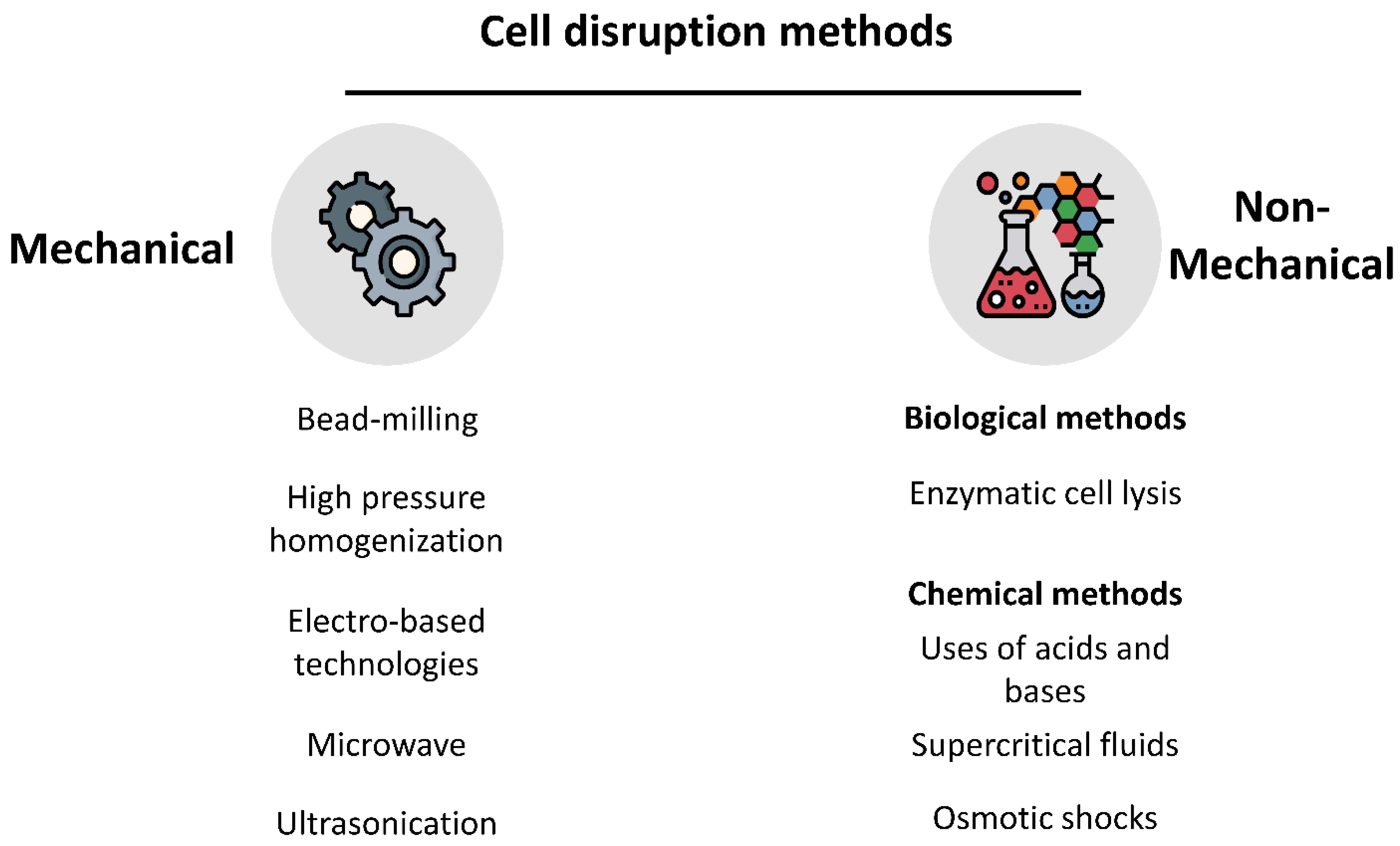
After the process of drying the microalgae biomass, the cell wall is still an important barrier limiting the access to intracellular compounds [40]. Several cell disruption methods are currently available (Figure 2), and they can be divided into different categories. Some of them can be described as mechanical or physical methods, involving techniques such as bead-mills, high-pressure homogenization, microwave, ultrasonication, electric field-based technologies, and others. There are also the non-mechanical methods, which can also be subdivided into biological and chemical methods. In biological classification, the most common method is enzymatic cell lysis, while the use of acids, bases, supercritical fluids, or osmotic shocks are classified as chemical methods [63,73,83].
After assuring an effective cell wall disintegration, it is important to efficiently design a process to extract the proteins. This will allow increasing the protein bioaccessibility and potentially its bioavailability during GI digestion and absorption, or leverage the development of novel functional food systems. The protein extraction may require the use of some organic base solvents. The choice of the most appropriate extraction solvent is constrained by several factors: biochemical characteristics of the cells, the final application of the product, the cost of the operation, and the expected extraction yield and time [63].
Protein solubilization could be a problem due to the protein´s hydrophobic nature and the presence of disulfide bonds. This can be surpassed by using alkali conditions [84]. Then, it is possible to separate the soluble proteins from the carbohydrates matrix by performing acidic precipitation at low pH [17]. The efficiency of this method can vary with the presence of neutral or ionic polysaccharides [29]. With strong alkali solutions, protein solubilization can be improved, leading to a more efficient extraction process. However, these stronger alkali solutions can also damage proteins or other valuable compounds [85,86]. Protein extraction using alkali solutions as a strategy to integrate microalgae biomass into a biorefinery process has already been described. The goal of this type of strategy is to extract proteins from microalgae biomass and with the resulting deproteinized biomass, extract lipids for biodiesel production. For example, the use of strong alkali solutions (2 mol/L NaOH) allowed to obtain up to 10% of solubilized protein from the total biomass used [87]. The utilization of aqueous and acidic methods for the extraction of microalgae proteins is also commonly used [83].
Some studies reported the effects of well-known microalgae disruption methods on protein solubilization. Ursu and co-workers (2014), have studied the effects of certain processes, such as high pressure and alkaline treatment with a few drops of 2 mol/L NaOH in the recovery of protein fraction from microalgae without damaging the other intracellular compounds [84]. It has been observed that Chlorella vulgaris treated only with chemical treatment resulted in a very low extraction yield (1.5 ± 0.1% at pH 9 and 2.3 ± 0.2% at pH 12) when compared to the total content of proteins in Chlorella vulgaris (around 42% to 58%) [17,22]. However, when the same cells were treated in the high-pressure cell disruptor at 270 MPa, the efficiency of protein solubilization increased. The best results were obtained with the combination of chemical treatment and high-pressure treatment; this condition achieved the solubilization of 98% at pH 12 and 71% at pH 7, of the total protein in Chlorella vulgaris [84], thus pointing out that pH and its combination with physical methods can bring different outcomes regarding extraction efficiency.
Taking advantage of protein´s solubility at different pH is an easy way to isolate and recover proteins from the intact microalgae cells or after the cell wall disruption process. It has been hypothesized that with a pH-shift process it would be possible to precipitate the microalgae proteins and then recover them from the cellulosic cell wall [88]. This process uses the pH variation and isoelectric point of proteins to solubilize and precipitate proteins that are not soluble [19]. This can be achieved by lowering the solution´s pH towards the protein’s isoelectric point, thus neutralizing their charges and making the proteins insoluble in water, leading to precipitation [88,89]. After applying this pH-shift process to proteins, it is then possible to recover the different fractions (soluble and non-soluble) by centrifugation procedures [19]. The production of protein isolates is dependent on proper protein solubilization after their release during the cell wall disruption [90].

3.3. Impact of Electric Fields on the Cell Integrity

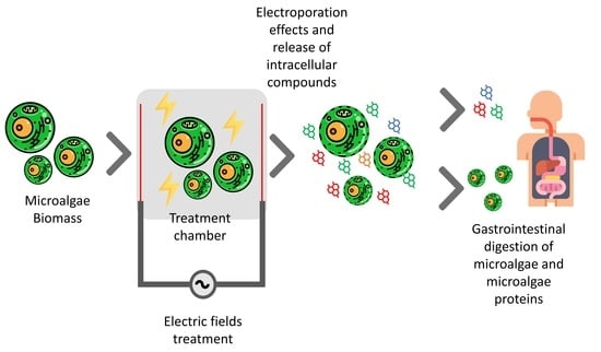
In the last few years, the use of electric fields (EF) has gained attention in several biotechnological areas due to their efficiency, versatility, easy scale-up, as well as environment-friendly character (Table 4) [77,78,91]. EF processing can contribute to increasing the microalgae cell wall permeability allowing an easy release of the intracellular compounds such as proteins and lipids [92]. Considering these features, EF-based technologies are more advantageous when compared to other pretreatment methods, such as mechanical (i.e., high-pressure homogenization), microwave and ultrasounds, [93]. Processing of microalgae biomass can be carried out through different EF technologies such as pulsed electric fields (PEF), high voltage electric discharge (HVED), moderate electric fields (MEF) that can be combined with ohmic heating (OH) effect, direct current (DC) and subsequent electrolysis, also known as electrochemical cell lysis [78,92]. These methods offer the opportunity of a fine-tune process control and specificity, resembling enzymatic ones [94], resulting in total disruption or mild permeabilization of the cell wall as illustrated by Figure 3. Table 5 includes some examples of PEF, HVED, and MEF applications and their reported effects on microalgae cells.
When applying EF, an important factor to consider is the effect of electroporation (Figure 4), which is greatly dependent on the time of exposure and intensity of the applied EF and the medium composition [77,108]. When the EF voltage exceeds the transmembrane threshold, between 0.2 and 1.0 V, it can cause the electroporation effect [78]. This type of electroporation allows the cell to stay on a high permeability stage, allowing the introduction and/or extraction of molecules from/to the cell. Depending on the intensity of the applied EF the cell can or not return to its original state. If the EF strength is too high for the cell to handle, it will impact an irreversible electroporation and the cell will be disrupted [78,108].

3.3.1. PEF

The use of PEF technology is already discussed in the literature as a promising and energy-efficient alternative to the current extraction methods employed nowadays [109]. PEF processing is considered a non-thermal technology that applies high-intensity electric pulses to a certain sample—i.e., between 20 and 100 kV/cm for microbial inactivation and between 0.5 and 10 kV/cm for tissue softening. The treatment duration with this technology is frequently established between 0.01 µs and 2.4 ms [98]. The PEF treatment has already shown promising results in the inactivation of some microorganisms in the food industry. The basic principle of PEF technology consists of inducing an electroporation effect on the microorganism membrane, causing an increase in the cell´s permeability and even cellular disruption [78,110]. This process of membrane permeabilization makes the access of solvents easier for the extraction of valuable compounds [78]. Several works with the application of electrotechnologies have already been conducted, using Chlorella vulgaris and Nannochloropsis salina, showing some leakage of proteins after the treatments. The authors also claim that it is possible to achieve an efficient protein release from different microalgae genera by adjusting the electrical parameters during the treatments [91]. Buchmann and co-workers achieved a total protein extraction from Chlorella vulgaris cells of 96% using PEF technology [50]. They also observed that depending on the microalgae growth stage, the protein extraction efficiency can vary. These conclusions are in agreement with a report made by Canelli and co-workers where it is claimed that different cell phases show different cell wall stability, thickness, and composition [50,52]. Buchmann and co-workers also compared the efficacy of PEF with a well-known method, the HPH. They found that PEF treatment resulted in more intact cells after the treatment, while the HPH led to a total disintegration of the cell structure. Another advantage of using PEF treatments is the possibility of a full recovery of the microalgae cells after the treatments due to a reversible electroporation effect. Their results show that depending on the intensity of the electric fields and the yield of proteins extracted from the cells, the recovery and cell viability level can also vary. As expected, higher electric fields and extraction rates led to less recovery of the microalgae cells after treatment [50]. During treatments with PEF, Joule heating effects may also happen which could contribute to damage to the target compounds if not properly controlled [73,78,91].
However, the successful use of PEF is still somehow controversial. There are some works, where the authors describe the use of PEF on microalgae, but with less promising results [77,106]. Lam and co-workers have compared the effects of using PEF and bead-milling on two strains of microalgae—i.e., Chlorella vulgaris and Neochloris oleoabundans. They were able to induce an electroporation effect on both strains because there was a release of high amounts of ions after PEF treatment from both species, but not proteins. Regarding the protein release, this was around 13% which is lower when compared to the bead-milling method which had a release of proteins between 30% and 50% [106]. These values of protein release with PEF technology corroborate previous studies with other species of microalgae [95,111]. Further fundamental understanding of the effects of PEF on cell wall permeabilization is still needed.

3.3.2. HVED

The use of HVED can also be used in microalgae processing. In this process, there is a direct release of high energy into the medium. This process can cause an electric breakdown of the water and create a wave of side effects such as high-pressure shock waves and cavitation. These phenomena will cause electroporation effects on the cells due to the presence of the strong EF. This will fragment the cell tissue and facilitate the release of intracellular compounds [78,93]. The HVED presents certain advantages when compared to more conventional treatments: higher rate of extraction, reduction in processing time and temperature, which will lead to less degradation of thermosensitive compounds and also presents less environmental impact [92,93]. The HVED is described to have less efficiency in extracting high molecular weight components from the cells, such as proteins and pigments. Another problem with this approach is the low sensitivity of this technology, making it difficult to extract specific compounds from the cells [78]. Zhang and co-workers theorize that HVED promotes the release of water-soluble proteins of small molecular weight [92,112].

3.3.3. MEF

Another processing method that takes advantage of using EF is the moderate electric fields (MEF), which is a non-pulsed approach characterized by the presence of an alternating current without treatment time restrictions [78]. MEF processing is also characterized by the presence of electric fields below 1 kV/cm and a square or sinusoidal wave with electrical frequencies ranging from 50 Hz to 25 kHz. The MEF processing has been described to have several effects such as permeabilization of the cellular membrane, microbial inactivation, and cells destruction [78,113]. When using MEF processing, there is an important parameter to consider, the EF frequency which can promote electrochemical reactions, particularly when working at lower frequencies (typically at 50 Hz) [114]. The use of low frequencies is described to lead to the release of some metal ions which can react with other molecules. These reactions are undesirable and must be avoided because this can lead to the degradation of nutrients from the sample and could lead to the formation of radical species, as well as the corrosion and erosion of the electrodes [77,115]. When MEF is applied at high frequencies, these electrochemical reactions are minimized. However, using low frequencies can contribute to the permeabilization of biological membranes as shown in some bacteria [113]. The effects of the applied electrical frequency on microalgae cells’ structure are still undisclosed.

3.3.4. OH

The use of MEF brings also heat generation through the sample, this effect is called Joule heating or commercially known as ohmic heating (OH) [98]. OH depends on the electrical conductivity of the product to be treated, the intensity of the electric field applied, and treatment time. The OH effect can be used for pasteurization and sterilization purposes, but in a more efficient way than conventional heating processes [102]. The use of OH in the food industry has already been recognized as advantageous because it can improve food quality [116] and opens an opportunity for more sustainable processing, once it reduces water consumption and increases process efficiency. Through this technology, the heating effect can be properly controlled, allowing a faster and more homogeneous extraction of intracellular compounds with high energetic efficiency [101,117]. When OH is applied to a certain microorganism, such as microalgae, the heat generated will act in synergy together with the electric fields which can enhance the permeabilization of the cell tissues [12].

3.3.5. DC Methods

Another way of using EF on microalgae is by applying DC methods. This takes advantage of the unidirectional flow of the electric fields from oppositely charged electrodes. This will induce an electrophoretic movement of the charged particles to the oppositely charged electrode [78]. This approach is mainly used for harvesting operations allowing the separation of the biomass from the culture medium through eletroflocculation or electrocoagulation–flotation methods [103,105,118].

4. Gastrointestinal Digestion of Microalgae Biomass

The quality of microalgae biomass as a food ingredient will be dependent on its nutritional composition and the bioaccessibility of their nutrients for absorption in the human gut. It is important to address and understand how this biomass and valuable fractions will behave during the GI digestion. It is clear that different microalgae species have different cell wall compositions, which will be a key factor during GI digestion [23,119]. During the last years, the development of in vitro GI systems had gained some interest, and today it is already possible to conduct several studies through in vitro simulations, either in batch or dynamic systems. These systems allow having a more comprehensive understanding of the digestibility of those nutrients and their bioaccessibility [120,121,122]. The application of in vitro GI methods has several advantages against using human trials, as they can be used for preliminary screenings and the development of new study hypotheses, as well as the fact that they are rapid, less laborious, and do not include ethical restrictions [121].
Some authors have already studied the influence of some processing methods on microalgae digestibility, but more body of knowledge about the behavior of this emergent food biomass is needed. Janczyk and co-workers (2005), studied the influence of the digestibility of Chlorella vulgaris in rats, where their cells were processed using three different methods: spray-drying, electroporation, and ultrasonication. Their results have shown that when C. vulgaris cells are treated with ultrasounds, they became more accessible to GI enzymes, thus increasing the nutritional value of Chlorella vulgaris. Their results have also shown that ultrasounds have more effects on destroying microalgae cells walls than electroporation, which can explain a higher protein availability [119].
Niccolai and co-authors (2019) have studied the digestibility patterns of some microalgae species, including the effects on their chemical composition. Their findings addressed that in terms of digestibility of biomass dry matter, Arthrospira platensis is the most digestible of all the tested microalgae. They explain this result, taking into consideration the existence of a Gram-negative cell wall, mainly composed of peptidoglycan. In the case of other cyanobacteria characterized by a higher presence of polysaccharides in their cell wall, a lower digestibility was also observed [23]. Mišurcová and co-workers (2010) compared the digestibility of blue-green (Arthrospira platensis), green (i.e., Chlorella pyrenoidosa), red (i.e., Palmaria palmata), and brown (i.e., Laminaria japonica) algae. Results show that for all of the studied groups, Arthrospira platensis, presented the highest digestibility among all of the species tested, corroborating previous studies. The group of algae with lower digestibility was the brown algae, it is hypothesized that the high presence of dietary fibers in the composition of these types of microalgae may have contributed to this outcome [123].
There seems to be a strong relationship between the cell wall composition and the digestibility patterns of some of the species. For example, the green algae cell wall is rich in cellulose and hemicellulose, and this can turn them more resistant to digestion [23,45]. As mentioned before, Chlorella is also known for having a layer of a very resistant polymer called algaenan, which will also hamper the action of enzymes [45]. Other species (e.g., Nannochloropsis oceanica) with algaenan in their cell wall composition also show some reduced digestibility [23,46].

5. Conclusions and Future Perspectives

Microalgae biomass has become an interesting and sustainable food source for human consumption, but there are still some gaps in knowledge regarding how their cellular structure could affect their nutrient bioaccessibility. The microalgae cell wall presents an important and protective barrier of the cell against environmental factors and outside threats. This also has some implications in the ways that downstream processing of microalgae biomass affects its GI digestion. It seems generally accepted that microalgae with stronger cell walls, such as Chlorella vulgaris, will be more difficult to be digested than other photosynthetic species such as Arthrospira sp. For this reason, the importance of understanding the cell wall composition and adapting the processing methods and conditions to each specific microalgae will be of extreme importance. A fundamental understanding of how processing affects the cell wall integrity, protein digestibility, and subsequently the bioaccessibility of their intracellular compounds is needed. The in vitro GI systems can appear here as an accurate and valuable tool for a comprehensive assessment of the digestibility of microalgae proteins supporting also previous adjustments in upstream and downstream strategies to produce microalgae biomass.
The electro-based technologies such as PEF and MEF are attracting attention in the last years, appearing as a promising way of processing microalgae biomass. A huge advantage of using this type of methodologies is the possibility to minimize deleterious effects of heat during the treatment (such as PEF) or combine electrical and thermal effects for a tailored cellular disruption (such as MEF and the possibility of ohmic heating). The heat can affect the overall quality of the intracellular products, reducing their biological value, thus its use needs to be properly balanced throughout the process. There is still a lot of research to be conducted to make these emergent methods more efficient and standardized. Despite being energy demanding, the conventional approaches for cellular disruption, such as bead-milling and high-speed homogenization, still present higher efficiencies when compared to some electro-based technologies. For this reason, it is important to focus on the optimization of these electric-based approaches, aiming at a fine-tune permeabilization of the cell wall barrier and to establish fundamental relationships between processing parameters and the biological value of microalgae biomass. In this sense integration of electric-based processing effects with in vitro gastrointestinal digestion assessment will be of crucial importance. This will offer the opportunity to increase the bioaccessibility (and eventually bioavailability) of protein fractions from different species of microalgae, anticipating approval of a greater number of species for human consumption in the next forthcoming years.

Author Contributions

Conceptualization, L.M. and R.N.P.; writing—original draft preparation, L.M.; writing—review and editing, G.C. and R.N.P.; supervision, R.N.P. All authors have read and agreed to the published version of the manuscript.

Funding

This study was supported by the Portuguese Foundation for Science and Technology (FCT) under the scope of the strategic funding of UIDB/04469/2020 unit and by ALGAVALOR—Lisboa-01-0247-FEDER-035234, supported by the Operational Programme for Competitiveness and Internationalization (COMPETE2020), Lisbon Portugal Regional Operational Programme (Lisboa 2020) and Algarve Regional Operational Programme (Algarve 2020) under the Portugal 2020 Partnership Agreement, through the European Regional Development Fund+ (ERDF).

Acknowledgments

Luís Machado acknowledges FCT for its fellowship supported by doctoral advanced training (SFRH/BD/07475/2020). Ricardo N. Pereira acknowledges FCT for its Assistant Research program under the scope of Scientific Stimulus Employment with reference CEECIND/02903/2017.

Conflicts of Interest

The authors declare no conflict of interest.

References

  1. Barsanti, L.; Gualtieri, P. Is Exploitation of Microalgae Economically and Energetically Sustainable? Algal Res. 2018, 31, 107–115. [Google Scholar] [CrossRef]
  2. Chacón-Lee, T.L.; González-Mariño, G.E. Microalgae for “Healthy” Foods-Possibilities and Challenges. Compr. Rev. Food Sci. Food Saf. 2010, 9, 655–675. [Google Scholar] [CrossRef] [PubMed]
  3. Christaki, E.; Florou-Paneri, P.; Bonos, E. Microalgae: A Novel Ingredient in Nutrition. Int. J. Food Sci. Nutr. 2011, 62, 794–799. [Google Scholar] [CrossRef] [PubMed]
  4. Koyande, A.K.; Chew, K.W.; Rambabu, K.; Tao, Y.; Chu, D.-T.; Show, P.-L. Microalgae: A Potential Alternative to Health Supplementation for Humans. Food Sci. Hum. Wellness 2019, 8, 16–24. [Google Scholar] [CrossRef]
  5. Caporgno, M.P.; Mathys, A. Trends in Microalgae Incorporation into Innovative Food Products with Potential Health Benefits. Front. Nutr. 2018, 5, 58. [Google Scholar] [CrossRef]
  6. Lupatini, A.L.; Colla, L.M.; Canan, C.; Colla, E. Potential Application of Microalga Spirulina platensis as a Protein Source. J. Sci. Food Agric. 2017, 97, 724–732. [Google Scholar] [CrossRef]
  7. Torres-Tiji, Y.; Fields, F.J.; Mayfield, S.P. Microalgae as a Future Food Source. Biotechnol. Adv. 2020, 41, 107536. [Google Scholar] [CrossRef]
  8. Khan, M.I.; Shin, J.; Kim, J.-D. The Promising Future of Microalgae: Current Status, Challenges, and Optimization of a Sustainable and Renewable Industry for Biofuels, Feed, and Other Products. Microb. Cell Fact. 2018, 17, 36. [Google Scholar] [CrossRef]
  9. Cuellar-Bermudez, S.P.; Aguilar-Hernandez, I.; Cardenas-Chavez, D.L.; Ornelas-Soto, N.; Romero-Ogawa, M.A.; Parra-Saldivar, R. Extraction and Purification of High-value Metabolites from Microalgae: Essential Lipids, Astaxanthin and Phycobiliproteins. Microb. Biotechnol. 2015, 8, 190–209. [Google Scholar] [CrossRef]
  10. Spolaore, P.; Joannis-Cassan, C.; Duran, E.; Isambert, A. Commercial Applications of Microalgae. J. Biosci. Bioeng. 2006, 101, 87–96. [Google Scholar] [CrossRef] [Green Version]
  11. Gantar, M.; Svirčev, Z. Microalgae and Cyanobacteria: Food for Thought. J. Phycol. 2008, 44, 260–268. [Google Scholar] [CrossRef] [PubMed]
  12. Geada, P.; Moreira, C.; Silva, M.; Nunes, R.; Madureira, L.; Rocha, C.M.R.; Pereira, R.N.; Vicente, A.A.; Teixeira, J.A. Algal Proteins: Production Strategies and Nutritional and Functional Properties. Bioresour. Technol. 2021, 332, 125125. [Google Scholar] [CrossRef] [PubMed]
  13. Matos, Â.P. The Impact of Microalgae in Food Science and Technology. J. Am. Oil Chem. Soc. 2017, 94, 1333–1350. [Google Scholar] [CrossRef]
  14. Gonzalez, J.; Olivieri, G.; de Vree, J.; Bosma, R.; Willems, P.; Reith, H.; Eppink, M.; Kleinegris, D.; Wijffels, R.; Barbosa, M.J. Towards Industrial Products from Microalgae. Energy Environ. Sci. 2016, 9, 3036–3043. [Google Scholar] [CrossRef] [Green Version]
  15. da Silva Vaz, B.; Moreira, J.B.; de Morais, M.G.; Costa, J.A.V. Microalgae as a New Source of Bioactive Compounds in Food Supplements. Curr. Opin. Food Sci. 2016, 7, 73–77. [Google Scholar] [CrossRef]
  16. Kabera, J.N.; Semana, E.; Mussa, A.R.; He, X. Plant Secondary Metabolites: Biosynthesis, Classification, Function and Pharmacological Properties. J. Pharm. Pharmacol. 2014, 2, 377–392. [Google Scholar]
  17. Safi, C.; Zebib, B.; Merah, O.; Pontalier, P.-Y.; Vaca-Garcia, C. Morphology, Composition, Production, Processing and Applications of Chlorella vulgaris: A Review. Renew. Sustain. Energy Rev. 2014, 35, 265–278. [Google Scholar] [CrossRef] [Green Version]
  18. Bleakley, S.; Hayes, M. Algal Proteins: Extraction, Application, and Challenges Concerning Production. Foods 2017, 6, 33. [Google Scholar] [CrossRef] [Green Version]
  19. Grossmann, L.; Hinrichs, J.; Weiss, J. Cultivation and Downstream Processing of Microalgae and Cyanobacteria to Generate Protein-Based Technofunctional Food Ingredients. Crit. Rev. Food Sci. Nutr. 2020, 60, 2961–2989. [Google Scholar] [CrossRef]
  20. Graziani, G.; Schiavo, S.; Nicolai, M.A.; Buono, S.; Fogliano, V.; Pinto, G.; Pollio, A. Microalgae as Human Food: Chemical and Nutritional Characteristics of the Thermo-Acidophilic Microalga Galdieria Sulphuraria. Food Funct. 2013, 4, 144–152. [Google Scholar] [CrossRef]
  21. Schwenzfeier, A.; Wierenga, P.A.; Gruppen, H. Isolation and Characterization of Soluble Protein from the Green Microalgae Tetraselmis sp. Bioresour. Technol. 2011, 102, 9121–9127. [Google Scholar] [CrossRef] [PubMed]
  22. Becker, E.W. Micro-Algae as a Source of Protein. Biotechnol. Adv. 2007, 25, 207–210. [Google Scholar] [CrossRef] [PubMed]
  23. Niccolai, A.; Chini Zittelli, G.; Rodolfi, L.; Biondi, N.; Tredici, M.R. Microalgae of Interest as Food Source: Biochemical Composition and Digestibility. Algal Res. 2019, 42, 101617. [Google Scholar] [CrossRef]
  24. Gorissen, S.H.M.; Crombag, J.J.R.; Senden, J.M.G.; Waterval, W.A.H.; Bierau, J.; Verdijk, L.B.; van Loon, L.J.C. Protein Content and Amino Acid Composition of Commercially Available Plant-Based Protein Isolates. Amino Acids 2018, 50, 1685–1695. [Google Scholar] [CrossRef] [PubMed] [Green Version]
  25. Sathe, S.K. Chemistry and Implications of Antinutritional Factors in Dry Beans and Pulses. In Dry Beans and Pulses Production, Processing and Nutrition; Wiley: Hoboken, NJ, USA, 2012; pp. 359–377. [Google Scholar]
  26. Day, L.; Cakebread, J.A.; Loveday, S.M. Food Proteins from Animals and Plants: Differences in the Nutritional and Functional Properties. Trends Food Sci. Technol. 2022, 119, 428–442. [Google Scholar] [CrossRef]
  27. Bertsch, P.; Böcker, L.; Mathys, A.; Fischer, P. Proteins from Microalgae for the Stabilization of Fluid Interfaces, Emulsions, and Foams. Trends Food Sci. Technol. 2021, 108, 326–342. [Google Scholar] [CrossRef]
  28. Soto-Sierra, L.; Stoykova, P.; Nikolov, Z.L. Extraction and Fractionation of Microalgae-Based Protein Products. Algal Res. 2018, 36, 175–192. [Google Scholar] [CrossRef]
  29. Chronakis, I.S.; Madsen, M. Algal Proteins. In Handbook of Food Proteins; Elsevier: Amsterdam, The Netherlands, 2011; pp. 353–394. [Google Scholar]
  30. Mantecón, L.; Moyano, R.; Cameán, A.M.; Jos, A. Safety Assessment of a Lyophilized Biomass of Tetraselmis Chuii (TetraSOD®) in a 90 Day Feeding Study. Food Chem. Toxicol. 2019, 133, 110810. [Google Scholar] [CrossRef]
  31. Mansour, A.T.; Ashour, M.; Alprol, A.E.; Alsaqufi, A.S. Aquatic Plants and Aquatic Animals in the Context of Sustainability: Cultivation Techniques, Integration, and Blue Revolution. Sustainability 2022, 14, 3257. [Google Scholar] [CrossRef]
  32. Pimentel, F.B.; Alves, R.C.; Harnedy, P.A.; FitzGerald, R.J.; Oliveira, M.B.P.P. Macroalgal-Derived Protein Hydrolysates and Bioactive Peptides: Enzymatic Release and Potential Health Enhancing Properties. Trends Food Sci. Technol. 2019, 93, 106–124. [Google Scholar] [CrossRef]
  33. Roos, N. Insects and Human Nutrition. In Edible Insects in Sustainable Food Systems; Springer International Publishing: Cham, Switzerland, 2018; pp. 83–91. [Google Scholar]
  34. Kinsella, J.E.; Melachouris, N. Functional Properties of Proteins in Foods: A Survey. Crit. Rev. Food Sci. Nutr. 1976, 7, 219–280. [Google Scholar] [CrossRef]
  35. Chronakis, I.S. Gelation of Edible Blue-Green Algae Protein Isolate (Spirulina platensis Strain Pacifica):  Thermal Transitions, Rheological Properties, and Molecular Forces Involved. J. Agric. Food Chem. 2001, 49, 888–898. [Google Scholar] [CrossRef] [PubMed]
  36. Grossmann, L.; Hinrichs, J.; Goff, H.D.; Weiss, J. Heat-Induced Gel Formation of a Protein-Rich Extract from the Microalga Chlorella sorokiniana. Innov. Food Sci. Emerg. Technol. 2019, 56, 102176. [Google Scholar] [CrossRef]
  37. Morris, H.J.; Almarales, A.; Carrillo, O.; Bermúdez, R.C. Utilisation of Chlorella vulgaris Cell Biomass for the Production of Enzymatic Protein Hydrolysates. Bioresour. Technol. 2008, 99, 7723–7729. [Google Scholar] [CrossRef]
  38. Sheih, I.-C.; Wu, T.-K.; Fang, T.J. Antioxidant Properties of a New Antioxidative Peptide from Algae Protein Waste Hydrolysate in Different Oxidation Systems. Bioresour. Technol. 2009, 100, 3419–3425. [Google Scholar] [CrossRef]
  39. Vanthoor-Koopmans, M.; Wijffels, R.H.; Barbosa, M.J.; Eppink, M.H.M. Biorefinery of Microalgae for Food and Fuel. Bioresour. Technol. 2013, 135, 142–149. [Google Scholar] [CrossRef]
  40. Safi, C.; Charton, M.; Pignolet, O.; Silvestre, F.; Vaca-Garcia, C.; Pontalier, P.-Y. Influence of Microalgae Cell Wall Characteristics on Protein Extractability and Determination of Nitrogen-to-Protein Conversion Factors. J. Appl. Phycol. 2013, 25, 523–529. [Google Scholar] [CrossRef] [Green Version]
  41. Phong, W.N.; Show, P.L.; Le, C.F.; Tao, Y.; Chang, J.-S.; Ling, T.C. Improving Cell Disruption Efficiency to Facilitate Protein Release from Microalgae Using Chemical and Mechanical Integrated Method. Biochem. Eng. J. 2018, 135, 83–90. [Google Scholar] [CrossRef]
  42. Alhattab, M.; Kermanshahi-Pour, A.; Brooks, M.S.-L. Microalgae Disruption Techniques for Product Recovery: Influence of Cell Wall Composition. J. Appl. Phycol. 2019, 31, 61–88. [Google Scholar] [CrossRef]
  43. Safi, C.; Ursu, A.V.; Laroche, C.; Zebib, B.; Merah, O.; Pontalier, P.-Y.; Vaca-Garcia, C. Aqueous Extraction of Proteins from Microalgae: Effect of Different Cell Disruption Methods. Algal Res. 2014, 3, 61–65. [Google Scholar] [CrossRef] [Green Version]
  44. Corre, G.; Templier, J.; Largeau, C.; Rousseau, B.; Berkaloff, C. Influence of Cell Wall Composition on the Resistance of Two Chlorella Species (Chlorophyta) to Detergents. J. Phycol. 1996, 32, 584–590. [Google Scholar] [CrossRef]
  45. Domozych, D.S.; Ciancia, M.; Fangel, J.U.; Mikkelsen, M.D.; Ulvskov, P.; Willats, W.G.T. The Cell Walls of Green Algae: A Journey through Evolution and Diversity. Front. Plant Sci. 2012, 3, 82. [Google Scholar] [CrossRef] [PubMed] [Green Version]
  46. Scholz, M.J.; Weiss, T.L.; Jinkerson, R.E.; Jing, J.; Roth, R.; Goodenough, U.; Posewitz, M.C.; Gerken, H.G. Ultrastructure and Composition of the Nannochloropsis gaditana Cell Wall. Eukaryot. Cell 2014, 13, 1450–1464. [Google Scholar] [CrossRef] [PubMed] [Green Version]
  47. Voigt, J.; Stolarczyk, A.; Zych, M.; Malec, P.; Burczyk, J. The Cell-Wall Glycoproteins of the Green Alga Scenedesmus obliquus. The Predominant Cell-Wall Polypeptide of Scenedesmus obliquus Is Related to the Cell-Wall Glycoprotein Gp3 of Chlamydomonas reinhardtii. Plant Sci. 2014, 215–216, 39–47. [Google Scholar] [CrossRef] [PubMed]
  48. Lu, H.-K.; Hsieh, C.-C.; Hsu, J.-J.; Yang, Y.-K.; Chou, H.-N. Preventive Effects of Spirulina Platensis on Skeletal Muscle Damage under Exercise-Induced Oxidative Stress. Eur. J. Appl. Physiol. 2006, 98, 220–226. [Google Scholar] [CrossRef] [Green Version]
  49. Safi, C.; Frances, C.; Ursu, A.V.; Laroche, C.; Pouzet, C.; Vaca-Garcia, C.; Pontalier, P.-Y. Understanding the Effect of Cell Disruption Methods on the Diffusion of Chlorella vulgaris Proteins and Pigments in the Aqueous Phase. Algal Res. 2015, 8, 61–68. [Google Scholar] [CrossRef] [Green Version]
  50. Buchmann, L.; Brändle, I.; Haberkorn, I.; Hiestand, M.; Mathys, A. Pulsed Electric Field Based Cyclic Protein Extraction of Microalgae towards Closed-Loop Biorefinery Concepts. Bioresour. Technol. 2019, 291, 121870. [Google Scholar] [CrossRef]
  51. Doucha, J.; Lívanský, K. Influence of Processing Parameters on Disintegration of Chlorella Cells in Various Types of Homogenizers. Appl. Microbiol. Biotechnol. 2008, 81, 431–440. [Google Scholar] [CrossRef]
  52. Canelli, G.; Neutsch, L.; Carpine, R.; Tevere, S.; Giuffrida, F.; Rohfritsch, Z.; Dionisi, F.; Bolten, C.J.; Mathys, A. Chlorella vulgaris in a Heterotrophic Bioprocess: Study of the Lipid Bioaccessibility and Oxidative Stability. Algal Res. 2020, 45, 101754. [Google Scholar] [CrossRef]
  53. Ratledge, C.; Streekstra, H.; Cohen, Z.; Fichtali, J. Downstream Processing, Extraction, and Purification of Single Cell Oils. In Single Cell Oils; Elsevier: Amsterdam, The Netherlands, 2010; pp. 179–197. [Google Scholar]
  54. Montalescot, V.; Rinaldi, T.; Touchard, R.; Jubeau, S.; Frappart, M.; Jaouen, P.; Bourseau, P.; Marchal, L. Optimization of Bead Milling Parameters for the Cell Disruption of Microalgae: Process Modeling and Application to Porphyridium Cruentum and Nannochloropsis oculata. Bioresour. Technol. 2015, 196, 339–346. [Google Scholar] [CrossRef]
  55. Aflalo, C.; Meshulam, Y.; Zarka, A.; Boussiba, S. On the Relative Efficiency of Two- vs. One-Stage Production of Astaxanthin by the Green Alga Haematococcus pluvialis. Biotechnol. Bioeng. 2007, 98, 300–305. [Google Scholar] [CrossRef] [PubMed]
  56. Mendes-Pinto, M.M.; Raposo, M.F.J.; Bowen, J.; Young, A.J.; Morais, R. Evaluation of Different Cell Disruption Processes on Encysted Cells of Haematococcus pluvialis: Effects on Astaxanthin Recovery and Implications for Bio-Availability. J. Appl. Phycol. 2001, 13, 19–24. [Google Scholar] [CrossRef]
  57. Morineau-Thomas, O.; Jaouen, P.; Legentilhomme, P. The Role of Exopolysaccharides in Fouling Phenomenon during Ultrafiltration of Microalgae (Chlorella sp. and Porphyridium purpureum): Advantage of a Swirling Decaying Flow. Bioprocess Biosyst. Eng. 2002, 25, 35–42. [Google Scholar] [CrossRef] [PubMed]
  58. Geresh, S.; Mamontov, A.; Weinstein, J. Sulfation of Extracellular Polysaccharides of Red Microalgae: Preparation, Characterization and Properties. J. Biochem. Biophys. Methods 2002, 50, 179–187. [Google Scholar] [CrossRef]
  59. Shibata, S.; Arimura, S.; Ishikawa, T.; Awai, K. Alterations of Membrane Lipid Content Correlated with Chloroplast and Mitochondria Development in Euglena gracilis. Front. Plant Sci. 2018, 9, 370. [Google Scholar] [CrossRef] [Green Version]
  60. Barsanti, L.; Gualtieri, P. Anatomy of Euglena gracilis. In Handbook of Algal Science, Technology and Medicine; Elsevier: Amsterdam, The Netherlands, 2020; pp. 61–70. [Google Scholar]
  61. Wang, Y.; Seppänen-Laakso, T.; Rischer, H.; Wiebe, M.G. Euglena gracilis Growth and Cell Composition under Different Temperature, Light and Trophic Conditions. PLoS ONE 2018, 13, e0195329. [Google Scholar] [CrossRef] [Green Version]
  62. El-Baz, F.K.; Aly, H.F.; Salama, A.A.A. Toxicity Assessment of the Green Dunaliella salina Microalgae. Toxicol. Rep. 2019, 6, 850–861. [Google Scholar] [CrossRef]
  63. Morais Junior, W.G.; Gorgich, M.; Corrêa, P.S.; Martins, A.A.; Mata, T.M.; Caetano, N.S. Microalgae for Biotechnological Applications: Cultivation, Harvesting and Biomass Processing. Aquaculture 2020, 528, 735562. [Google Scholar] [CrossRef]
  64. Lari, Z.; Ahmadzadeh, H.; Hosseini, M. Cell Wall Disruption: A Critical Upstream Process for Biofuel Production. In Advances in Feedstock Conversion Technologies for Alternative Fuels and Bioproducts; Elsevier: Amsterdam, The Netherlands, 2019; pp. 21–35. [Google Scholar]
  65. de Carvalho, J.C.; Magalhães, A.I.; de Melo Pereira, G.V.; Medeiros, A.B.P.; Sydney, E.B.; Rodrigues, C.; Aulestia, D.T.M.; de Souza Vandenberghe, L.P.; Soccol, V.T.; Soccol, C.R. Microalgal Biomass Pretreatment for Integrated Processing into Biofuels, Food, and Feed. Bioresour. Technol. 2020, 300, 122719. [Google Scholar] [CrossRef]
  66. Lafarga, T. Cultured Microalgae and Compounds Derived Thereof for Food Applications: Strain Selection and Cultivation, Drying, and Processing Strategies. Food Rev. Int. 2020, 36, 559–583. [Google Scholar] [CrossRef]
  67. Chen, C.-L.; Chang, J.-S.; Lee, D.-J. Dewatering and Drying Methods for Microalgae. Dry. Technol. 2015, 33, 443–454. [Google Scholar] [CrossRef]
  68. Min, K.H.; Kim, D.H.; Ki, M.-R.; Pack, S.P. Recent Progress in Flocculation, Dewatering, and Drying Technologies for Microalgae Utilization: Scalable and Low-Cost Harvesting Process Development. Bioresour. Technol. 2022, 344, 126404. [Google Scholar] [CrossRef] [PubMed]
  69. Zhang, M.; Chen, H.; Mujumdar, A.S.; Tang, J.; Miao, S.; Wang, Y. Recent Developments in High-Quality Drying of Vegetables, Fruits, and Aquatic Products. Crit. Rev. Food Sci. Nutr. 2017, 57, 1239–1255. [Google Scholar] [CrossRef] [PubMed]
  70. Defraeye, T.; Martynenko, A. Electrohydrodynamic Drying of Multiple Food Products: Evaluating the Potential of Emitter-Collector Electrode Configurations for Upscaling. J. Food Eng. 2019, 240, 38–42. [Google Scholar] [CrossRef]
  71. Defraeye, T.; Martynenko, A. Electrohydrodynamic Drying of Food: New Insights from Conjugate Modeling. J. Clean. Prod. 2018, 198, 269–284. [Google Scholar] [CrossRef]
  72. Amorim, M.L.; Soares, J.; dos Reis Coimbra, J.S.; de Oliveira Leite, M.; Albino, L.F.T.; Martins, M.A. Microalgae Proteins: Production, Separation, Isolation, Quantification, and Application in Food and Feed. Crit. Rev. Food Sci. Nutr. 2021, 61, 1976–2002. [Google Scholar] [CrossRef]
  73. Günerken, E.; D’Hondt, E.; Eppink, M.H.M.; Garcia-Gonzalez, L.; Elst, K.; Wijffels, R.H. Cell Disruption for Microalgae Biorefineries. Biotechnol. Adv. 2015, 33, 243–260. [Google Scholar] [CrossRef]
  74. Postma, P.R.; Miron, T.L.; Olivieri, G.; Barbosa, M.J.; Wijffels, R.H.; Eppink, M.H.M. Mild Disintegration of the Green Microalgae Chlorella vulgaris Using Bead Milling. Bioresour. Technol. 2015, 184, 297–304. [Google Scholar] [CrossRef]
  75. de Carvalho, J.C.; Medeiros, A.B.P.; Letti, L.A.J.; Kirnev, P.C.S.; Soccol, C.R. Cell Disruption and Isolation of Intracellular Products. In Current Developments in Biotechnology and Bioengineering; Elsevier: Amsterdam, The Netherlands, 2017; pp. 807–822. [Google Scholar]
  76. Zheng, H.; Yin, J.; Gao, Z.; Huang, H.; Ji, X.; Dou, C. Disruption of Chlorella vulgaris Cells for the Release of Biodiesel-Producing Lipids: A Comparison of Grinding, Ultrasonication, Bead Milling, Enzymatic Lysis, and Microwaves. Appl. Biochem. Biotechnol. 2011, 164, 1215–1224. [Google Scholar] [CrossRef]
  77. Rocha, C.M.R.; Genisheva, Z.; Ferreira-Santos, P.; Rodrigues, R.; Vicente, A.A.; Teixeira, J.A.; Pereira, R.N. Electric Field-Based Technologies for Valorization of Bioresources. Bioresour. Technol. 2018, 254, 325–339. [Google Scholar] [CrossRef] [Green Version]
  78. Geada, P.; Rodrigues, R.; Loureiro, L.; Pereira, R.; Fernandes, B.; Teixeira, J.A.; Vasconcelos, V.; Vicente, A.A. Electrotechnologies Applied to Microalgal Biotechnology—Applications, Techniques and Future Trends. Renew. Sustain. Energy Rev. 2018, 94, 656–668. [Google Scholar] [CrossRef] [Green Version]
  79. Molina Grima, E.; Belarbi, E.-H.; Acién Fernández, F.; Robles Medina, A.; Chisti, Y. Recovery of Microalgal Biomass and Metabolites: Process Options and Economics. Biotechnol. Adv. 2003, 20, 491–515. [Google Scholar] [CrossRef]
  80. Zhang, H.; Gong, T.; Li, J.; Pan, B.; Hu, Q.; Duan, M.; Zhang, X. Study on the Effect of Spray Drying Process on the Quality of Microalgal Biomass: A Comprehensive Biocomposition Analysis of Spray-Dried S. acuminatus Biomass. BioEnergy Res. 2022, 15, 320–333. [Google Scholar] [CrossRef]
  81. Miranda, M.; Maureira, H.; Rodríguez, K.; Vega-Gálvez, A. Influence of Temperature on the Drying Kinetics, Physicochemical Properties, and Antioxidant Capacity of Aloe Vera (Aloe Barbadensis Miller) Gel. J. Food Eng. 2009, 91, 297–304. [Google Scholar] [CrossRef]
  82. Defraeye, T.; Martynenko, A. Future Perspectives for Electrohydrodynamic Drying of Biomaterials. Dry. Technol. 2018, 36, 1–10. [Google Scholar] [CrossRef]
  83. Khanra, S.; Mondal, M.; Halder, G.; Tiwari, O.N.; Gayen, K.; Bhowmick, T.K. Downstream Processing of Microalgae for Pigments, Protein and Carbohydrate in Industrial Application: A Review. Food Bioprod. Process. 2018, 110, 60–84. [Google Scholar] [CrossRef]
  84. Ursu, A.-V.; Marcati, A.; Sayd, T.; Sante-Lhoutellier, V.; Djelveh, G.; Michaud, P. Extraction, Fractionation and Functional Properties of Proteins from the Microalgae Chlorella vulgaris. Bioresour. Technol. 2014, 157, 134–139. [Google Scholar] [CrossRef]
  85. Chronakis, I.S. Biosolar Proteins from Aquatic Algae. In Developments in Food Science; Elsevier: Amsterdam, The Netherlands, 2000; pp. 39–75. [Google Scholar]
  86. Hedenskog, G.; Hofsten, A.V. The Ultrastructure of Spirulina platensis—A New Source of Microbial Protein. Physiol. Plant. 1970, 23, 209–216. [Google Scholar] [CrossRef]
  87. Muñoz, R.; Navia, R.; Ciudad, G.; Tessini, C.; Jeison, D.; Mella, R.; Rabert, C.; Azócar, L. Preliminary Biorefinery Process Proposal for Protein and Biofuels Recovery from Microalgae. Fuel 2015, 150, 425–433. [Google Scholar] [CrossRef]
  88. Cavonius, L.R.; Albers, E.; Undeland, I. PH-Shift Processing of Nannochloropsis Oculata Microalgal Biomass to Obtain a Protein-Enriched Food or Feed Ingredient. Algal Res. 2015, 11, 95–102. [Google Scholar] [CrossRef] [Green Version]
  89. Nolsøe, H.; Undeland, I. The Acid and Alkaline Solubilization Process for the Isolation of Muscle Proteins: State of the Art. Food Bioprocess Technol. 2009, 2, 1–27. [Google Scholar] [CrossRef]
  90. Gerde, J.A.; Wang, T.; Yao, L.; Jung, S.; Johnson, L.A.; Lamsal, B. Optimizing Protein Isolation from Defatted and Non-Defatted Nannochloropsis Microalgae Biomass. Algal Res. 2013, 2, 145–153. [Google Scholar] [CrossRef]
  91. Coustets, M.; Al-Karablieh, N.; Thomsen, C.; Teissié, J. Flow Process for Electroextraction of Total Proteins from Microalgae. J. Membr. Biol. 2013, 246, 751–760. [Google Scholar] [CrossRef] [PubMed]
  92. bin Azmi, A.A.; Sankaran, R.; Show, P.L.; Ling, T.C.; Tao, Y.; Munawaroh, H.S.H.; Kong, P.S.; Lee, D.J.; Chang, J.S. Current Application of Electrical Pre-Treatment for Enhanced Microalgal Biomolecules Extraction. Bioresour. Technol. 2020, 302, 122874. [Google Scholar] [CrossRef] [PubMed]
  93. Puértolas, E.; Barba, F.J. Electrotechnologies Applied to Valorization of By-Products from Food Industry: Main Findings, Energy and Economic Cost of Their Industrialization. Food Bioprod. Processing 2016, 100, 172–184. [Google Scholar] [CrossRef]
  94. Canelli, G.; Murciano Martínez, P.; Maude Hauser, B.; Kuster, I.; Rohfritsch, Z.; Dionisi, F.; Bolten, C.J.; Neutsch, L.; Mathys, A. Tailored Enzymatic Treatment of Chlorella vulgaris Cell Wall Leads to Effective Disruption while Preserving Oxidative Stability. LWT 2021, 143, 111157. [Google Scholar] [CrossRef]
  95. Goettel, M.; Eing, C.; Gusbeth, C.; Straessner, R.; Frey, W. Pulsed Electric Field Assisted Extraction of Intracellular Valuables from Microalgae. Algal Res. 2013, 2, 401–408. [Google Scholar] [CrossRef]
  96. Grimi, N.; Dubois, A.; Marchal, L.; Jubeau, S.; Lebovka, N.I.; Vorobiev, E. Selective Extraction from Microalgae Nannochloropsis sp. Using Different Methods of Cell Disruption. Bioresour. Technol. 2014, 153, 254–259. [Google Scholar] [CrossRef]
  97. Luengo, E.; Martínez, J.M.; Bordetas, A.; Álvarez, I.; Raso, J. Influence of the Treatment Medium Temperature on Lutein Extraction Assisted by Pulsed Electric Fields from Chlorella vulgaris. Innov. Food Sci. Emerg. Technol. 2015, 29, 15–22. [Google Scholar] [CrossRef]
  98. Rodrigues, R.M.; Avelar, Z.; Machado, L.; Pereira, R.N. Electric Field Effects on Proteins—Novel Perspectives on Food and Potential Health Implications. Food Res. Int. 2020, 137, 109709. [Google Scholar] [CrossRef]
  99. Vorobiev, E.; Lebovka, N. Enhanced Extraction from Solid Foods and Biosuspensions by Pulsed Electrical Energy. Food Eng. Rev. 2010, 2, 95–108. [Google Scholar] [CrossRef]
  100. Xi, J.; He, L.; Yan, L. Continuous Extraction of Phenolic Compounds from Pomegranate Peel Using High Voltage Electrical Discharge. Food Chem. 2017, 230, 354–361. [Google Scholar] [CrossRef] [PubMed]
  101. Ferreira-Santos, P.; Nunes, R.; De Biasio, F.; Spigno, G.; Gorgoglione, D.; Teixeira, J.A.; Rocha, C.M.R. Influence of Thermal and Electrical Effects of Ohmic Heating on C-Phycocyanin Properties and Biocompounds Recovery from Spirulina Platensis. LWT 2020, 128, 109491. [Google Scholar] [CrossRef]
  102. Indiarto, R.; Rezaharsamto, B. A Review on Ohmic Heating and Its Use in Food. Int. J. Sci. Technol. Res. 2020, 9, 485–490. [Google Scholar]
  103. Pandey, A.; Shah, R.; Yadav, P.; Verma, R.; Srivastava, S. Harvesting of Freshwater Microalgae Scenedesmus sp. by Electro–Coagulation–Flocculation for Biofuel Production: Effects on Spent Medium Recycling and Lipid Extraction. Environ. Sci. Pollut. Res. 2020, 27, 3497–3507. [Google Scholar] [CrossRef]
  104. Pearsall, R.; Connelly, R.; Fountain, M.; Hearn, C.; Werst, M.; Hebner, R.; Kelley, E. Electrically Dewatering Microalgae. IEEE Trans. Dielectr. Electr. Insul. 2011, 18, 1578–1583. [Google Scholar] [CrossRef] [Green Version]
  105. Shuman, T.R.; Mason, G.; Reeve, D.; Schacht, A.; Goodrich, A.; Napan, K.; Quinn, J. Low-Energy Input Continuous Flow Rapid Pre-Concentration of Microalgae through Electro-Coagulation–Flocculation. Chem. Eng. J. 2016, 297, 97–105. [Google Scholar] [CrossRef]
  106. ’t Lam, G.P.; Postma, P.R.; Fernandes, D.A.; Timmermans, R.A.H.; Vermuë, M.H.; Barbosa, M.J.; Eppink, M.H.M.; Wijffels, R.H.; Olivieri, G. Pulsed Electric Field for Protein Release of the Microalgae Chlorella vulgaris and Neochloris oleoabundans. Algal Res. 2017, 24, 181–187. [Google Scholar] [CrossRef]
  107. Pagels, F.; Pereira, R.N.; Amaro, H.M.; Vasconcelos, V.; Guedes, A.C.; Vicente, A.A. Continuous Pressurized Extraction versus Electric Fields-Assisted Extraction of Cyanobacterial Pigments. J. Biotechnol. 2021, 334, 35–42. [Google Scholar] [CrossRef]
  108. Mahnič-Kalamiza, S.; Vorobiev, E.; Miklavčič, D. Electroporation in Food Processing and Biorefinery. J. Membr. Biol. 2014, 247, 1279–1304. [Google Scholar] [CrossRef]
  109. Eing, C.; Goettel, M.; Straessner, R.; Gusbeth, C.; Frey, W. Pulsed Electric Field Treatment of Microalgae—Benefits for Microalgae Biomass Processing. IEEE Trans. Plasma Sci. 2013, 41, 2901–2907. [Google Scholar] [CrossRef]
  110. Toepfl, S.; Heinz, V.; Knorr, D. Applications of Pulsed Electric Fields Technology for the Food Industry. In Pulsed Electric Fields Technology for the Food Industry; Springer: Boston, MA, USA, 2006; pp. 197–221. [Google Scholar]
  111. Parniakov, O.; Barba, F.J.; Grimi, N.; Marchal, L.; Jubeau, S.; Lebovka, N.; Vorobiev, E. Pulsed Electric Field Assisted Extraction of Nutritionally Valuable Compounds from Microalgae Nannochloropsis spp. Using the Binary Mixture of Organic Solvents and Water. Innov. Food Sci. Emerg. Technol. 2015, 27, 79–85. [Google Scholar] [CrossRef]
  112. Zhang, R.; Grimi, N.; Marchal, L.; Vorobiev, E. Application of High-Voltage Electrical Discharges and High-Pressure Homogenization for Recovery of Intracellular Compounds from Microalgae Parachlorella kessleri. Bioprocess Biosyst. Eng. 2019, 42, 29–36. [Google Scholar] [CrossRef] [PubMed]
  113. Machado, L.F.L.F.; Pereira, R.N.; Martins, R.C.R.C.; Teixeira, J.A.J.A.; Vicente, A.A.A. Moderate Electric Fields Can Inactivate Escherichia Coli at Room Temperature. J. Food Eng. 2010, 96, 520–527. [Google Scholar] [CrossRef] [Green Version]
  114. Ferreira, S.; Machado, L.; Pereira, R.N.; Vicente, A.A.; Rodrigues, R.M. Unraveling the Nature of Ohmic Heating Effects in Structural Aspects of Whey Proteins—The Impact of Electrical and Electrochemical Effects. Innov. Food Sci. Emerg. Technol. 2021, 74, 102831. [Google Scholar] [CrossRef]
  115. Pataro, G.; Barca, G.M.J.; Pereira, R.N.; Vicente, A.A.; Teixeira, J.A.; Ferrari, G. Quantification of Metal Release from Stainless Steel Electrodes during Conventional and Pulsed Ohmic Heating. Innov. Food Sci. Emerg. Technol. 2014, 21, 66–73. [Google Scholar] [CrossRef] [Green Version]
  116. Varghese, K.S.; Pandey, M.C.; Radhakrishna, K. Technology, Applications and Modelling of Ohmic Heating: A Review. J. Food Sci. Technol. 2014, 51, 2304–2317. [Google Scholar] [CrossRef] [Green Version]
  117. Ferreira-Santos, P.; Genisheva, Z.; Pereira, R.N.; Teixeira, J.A.; Rocha, C.M.R. Moderate Electric Fields as a Potential Tool for Sustainable Recovery of Phenolic Compounds from Pinus pinaster Bark. ACS Sustain. Chem. Eng. 2019, 7, 8816–8826. [Google Scholar] [CrossRef] [Green Version]
  118. Visigalli, S.; Barberis, M.G.; Turolla, A.; Canziani, R.; Berden Zrimec, M.; Reinhardt, R.; Ficara, E. Electrocoagulation–Flotation (ECF) for Microalgae Harvesting—A Review. Sep. Purif. Technol. 2021, 271, 118684. [Google Scholar] [CrossRef]
  119. Janczyk, P.; Wolf, C.; Souffrant, W.B. Evaluation of Nutritional Value and Safety of the Green Microalgae Chlorella vulgaris Treated with Novel Processing Methods. Arch Zootech 2005, 8, 132–147. [Google Scholar]
  120. Mantovani, R.A.; Pinheiro, A.C.; Vicente, A.A.; Cunha, R.L. In Vitro Digestion of Oil-in-Water Emulsions Stabilized by Whey Protein Nanofibrils. Food Res. Int. 2017, 99, 790–798. [Google Scholar] [CrossRef] [PubMed] [Green Version]
  121. Minekus, M.; Alminger, M.; Alvito, P.; Ballance, S.; Bohn, T.; Bourlieu, C.; Carrière, F.; Boutrou, R.; Corredig, M.; Dupont, D.; et al. A Standardised Static in vitro Digestion Method Suitable for Food—An International Consensus. Food Funct. 2014, 5, 1113–1124. [Google Scholar] [CrossRef] [PubMed] [Green Version]
  122. Pinheiro, A.C.; Coimbra, M.A.; Vicente, A.A. In Vitro Behaviour of Curcumin Nanoemulsions Stabilized by Biopolymer Emulsifiers—Effect of Interfacial Composition. Food Hydrocoll. 2016, 52, 460–467. [Google Scholar] [CrossRef] [Green Version]
  123. Mišurcová, L.; Kráčmar, S.; Klejdus, B.; Vacek, J. Nitrogen Content, Dietary Fiber, and Digestibility in Algal Food Products. Czech J. Food Sci. 2010, 28, 27–35. [Google Scholar] [CrossRef] [Green Version]
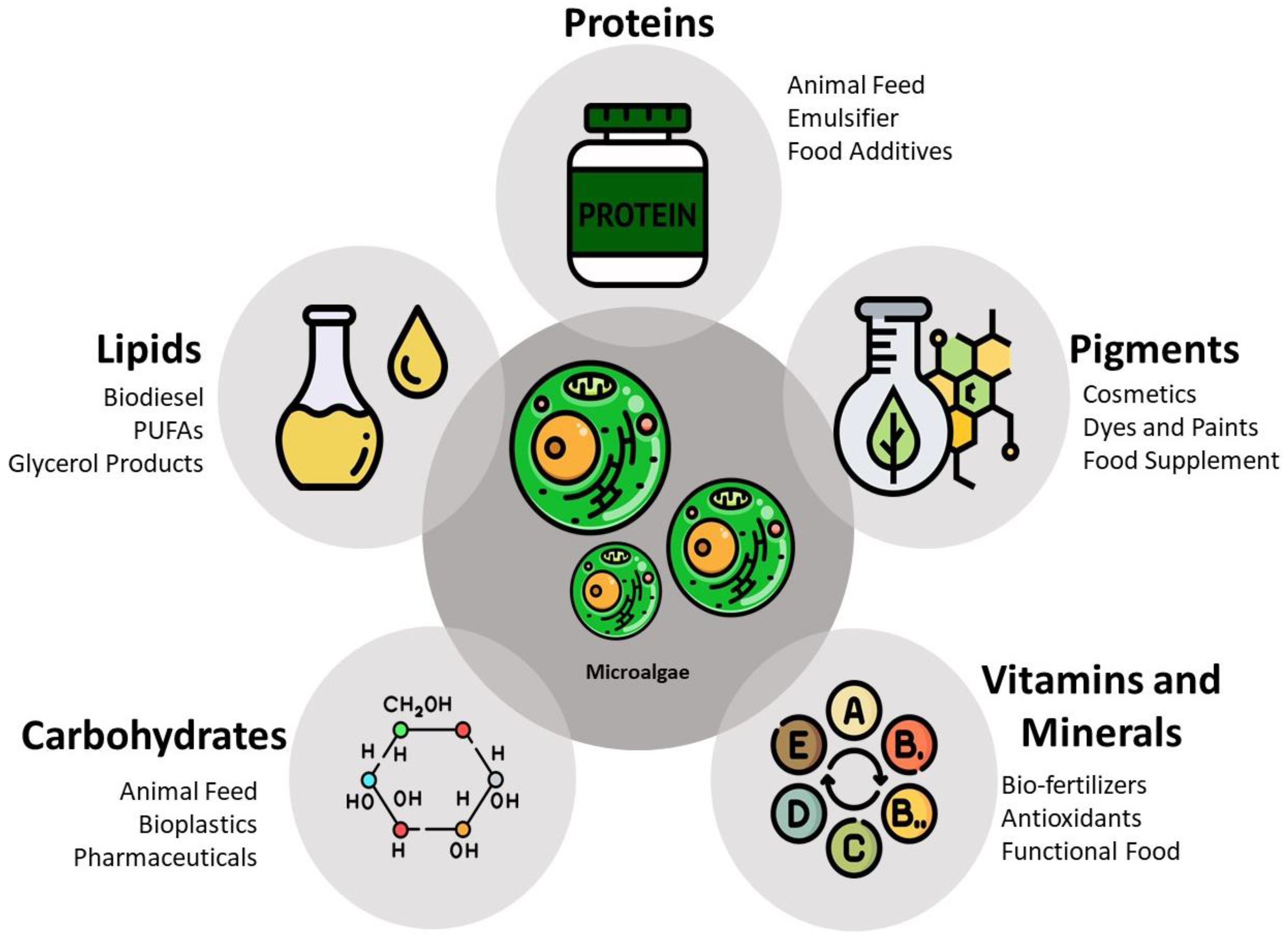
Figure 1. Application of the different products produced by microalgae. Adapted from: [17]. Icons by Freepik from flaticon.com, accessed on 1 April 2022.
Biomass 02 00006 g001
Figure 2. Commonly used cell disruption methods and their respective classifications in different categories. Icons by Freepik from flaticon.com, accessed on 1 April 2022.
Biomass 02 00006 g002
Figure 3. Different processing routes and possible pathways imposed by the application of EF technologies. Adapted from [94].
Biomass 02 00006 g003
Figure 4. Schematic representation of electroporation using alternating current.
Biomass 02 00006 g004
Table 2. Chemical composition of the cell wall of some relevant microalgae genus.
Microalgae GenusChemical Composition of the Cell WallResistanceReference
Chlorella/Chloroidium
  • Extracellular polysaccharides (rhamnose, galactose, xylose, cellulose, and hemicellulose);
  • Algaenan;
  • Some proteins can be found in the cellular membrane.
+[17,42,44,45]
Tetraselmis
  • Extracellular polysaccharides.
[42]
Nannochloropsis
  • Algaenan;
  • Polysaccharides (cellulose, glucose, rhamnose, galactose, ribose, fucose and xylose);
  • Some amino acids can be found in the cell wall.
++[42,46]
Scenedesmus/Tetradesmus
  • Glycoproteins;
  • An inner layer made by cellulose and an outer layer made by algaenan;
  • Glucosamine biopolymers.
+[42,47]
Arthrospira
  • Gram-negative cell wall composed of peptidoglycan, proteic, and lipopolysaccharidic outer membrane;
  • Some presence of murein;
  • No cellulose in their cell wall composition.
[23,43,48]
Symbols meaning: (−) means less resistant cell; (+) resistant cell wall; (++) very resistant cell wall.
Table 3. Examples of methods for microalgae drying and cellular disruption.
Technique AppliedPrinciple of the TechniqueEffects on the Biomass TreatedReference
Drying process
SolarDirect solar energy for biomass drying.
  • Some degradation during the slow drying;
  • Overheating of microalgae biomass;
  • Risk of fermentation and spoilage after long times of exposure.
[65,66]
Microwave DryingUses microwave heating for biomass drying.
  • Some degradation of the treated biomass.
[65]
Spray-dryingWarm liquid and air carry the water.
  • Used for high-valuable biocompounds;
  • Cause deterioration of some intracellular compounds, such as pigments;
  • Can retain more nutrients than convective drying.
[63,65,67]
Convective DryingConvective hot air to remove the water, such as an oven.
  • Led to some degradation at higher temperatures.
[65,67]
Lyophilization (Freeze-drying)Heat frozen biomass, leading to water removal through sublimation.
  • It keeps most of the proteins in the dried biomass, when compared to other drying methods;
  • Maintain cell viability;
  • Minor changes in the flavor, color, chemical composition, and texture;
  • High operation costs when applied at large scale.
[65,67,68,69]
Electrohydrodynamic Drying (EHD)Application of high voltage difference, this leads to the generation of airflow between the electrode and the plate where the sample is placed. It does not need any type of heat for the drying process.
  • Ideal for heat-sensitive products, due to the fact that it operates without any heat generation;
  • Reduced drying time and shrinkage;
  • Enhance rehydration capacity;
  • Preserve color and flavor.
[69,70,71]
Cell Disruption Methods
Bead MillShear stress between the beads and the cells in the sample.
  • Lack of selectively while treating the sample;
  • When applied to microalgae could achieve total disintegration of the cells;
  • Heat generation in the system.
[51,72,73,74]
High-pressure Homogenization (HPH)The cells are submitted to intense shear stress, turbulence, and cavitation, resulting in damage to the cell wall and membrane. The pressure transforms into steep velocity. It also has the advantage of being used without the drying step.
  • Heat generation by the system;
  • Nonselective treatment for the intracellular compounds, requiring some form of separation after the disruption;
  • Not suitable for processes that aim at the extraction of more fragile compounds.
[12,66,73,75]
Enzymatic HydrolysisTakes advantage of using enzymes that can affect structural components of the cell wall and membrane by weakening or even dissolving them.
  • Selective process which affects only certain components of the cell wall;
  • Due to the low energy requires it does not produce unwanted heat;
  • Some sugars present in the cells could suffer fermentation.
[72,73,75,76]
UltrasonicationUltrasonic waves propagating through a certain sample, produce some microbubbles which when expanded create violent shockwaves, thus damaging the cells.
  • Production of heat during the treatment, affecting some photosynthetic pigments;
  • Can show minor effects on disruption of stronger cell walls (e.g., Chlorella vulgaris).
[12,43,73,75]
MicrowaveThe cell wall disruption is caused by the evaporation of the cell water.
  • Protein denaturation, due to the heat production during the treatment;
  • Beside cellular disruption, it can also be used for biomass drying;
  • It causes an increase in pressure inside the cell, facilitating the extraction of intracellular compounds.
[12,72,73]
Pulsed Electric Fields (PEF)The application of an electric field will affect the transmembrane potential of the cell, causing an electroporation or electropermeabilization effect. This will lead to the release of the intracellular compounds.
  • Induce pore formation across the cellular membrane (electroporation);
  • Incapable of recovery of chloroplast proteins;
  • It is a non-heating technology, ideal for more heat-sensitive compounds;
  • If the electroporation effects are reversible, it could allow the microalgae cell walls to recover after the treatment, allowing the integration of PEF into a biorefinery process.
[50,72,73,77,78]
Table 4. Electric-based technologies for application on microalgae biomass.
Electric-Based TechnologyElectric Field StrengthTimeFrequencies UsedOperation TemperatureMain EffectsReference
Pulsed Electric Fields (PEF)
  • 20 to 100 kV/cm
  • 0.5 to 10 kV/cm
0.01 μs to 2.4 ms1 Hz to 2000 Hz10 °C to 60 °C
  • Microbial inactivation;
  • Tissue softening;
  • Electroporation effects on the cells, reversible and irreversible permeabilization;
  • Permeabilization of intracellular membranes (e.g., chloroplasts);
  • Extraction of intracellular ionic and water-soluble compounds;
  • Could be ineffective in the extraction of photosynthetic pigments;
  • Changes in protein structure due to the presence of electric fields.
[78,95,96,97,98]
High Voltage Electric Discharge (HVED)10 kV/cm to 100 kV/cm0.01 µs to 10 µsUp to 1000 Hz20 °C to 60 °C
  • Electrical breakdown in water leading to strong shockwaves of high amplitude and cavitation phenomena;
  • Electroporation effects, due to the presence of strong EF;
  • Damage to the cell, leading to their destruction;
  • It is proposed to be used for microorganisms inactivation.
[77,78,93,99,100]
Moderate Electric Fields (MEF)<1000 V/cmNo limit0.06 kHz to 25 kHz
  • Typically <60 °C
  • No upper limit for OH
  • Heat generation due to OH effects;
  • Changes in protein structure and conformation;
  • Effects on functional properties of globular proteins;
  • Capable of achieving a homogenous and fast extraction, with high energy efficiency;
  • Electrochemical reactions between the sample and the electrodes, when working at lower frequencies;
  • Permeabilization effects on microbial cell wall;
  • Can be used for pasteurization and sterilization.
[77,78,98,101,102]
Direct Current (DC)
  • 6.30 V/cm to 31.5 V/cm;
  • Voltage can range between 10 V and 300 V
No limit0 HzTemperature not relevant
  • Causes electrophoretic movement between two electrodes with opposite charges;
  • Has the possibility to concentrate microalgae biomass and separate them from the growth medium;
  • Harvest microalgae biomass using electro-flocculation.
[78,103,104,105]
Table 5. Examples of EF technologies for microalgae cellular disruption.
Microalgae SpeciesElectric-Based TreatmentEffects Caused in the MicroalgaeReference
Auxenochlorella protothecoidesPEF
  • Leakage of ionic substances and consequently increase in the conductivity;
  • Cell membrane permeabilization after PEF treatment;
  • The cell permeabilization led to the release of intracellular products;
  • Cell disintegration was achieved.
[95]
Nannochloropsis sp.PEF
  • After PEF treatment, the medium conductivity increased, indicating the release of ionic components from the cells;
  • Ineffective in the extraction of pigments.
[96]
Nannochloropsis sp.HVED
  • Extraction of high amount of ionic components from the cells;
  • The treated cells showed agglomeration;
  • Ineffective in extracting pigments.
[96]
Chlorella vulgarisPEF
  • Reversible and irreversible permeabilization of the cells after the PEF treatment;
  • Increase of electric conductivity after the treatment due to the release of small molecules such as ions;
  • Release of small amounts of intracellular proteins from the cells;
  • PEF treatment can improve the extraction of lutein from C. vulgaris biomass (being a consequence of the permeabilization of the cellular membrane);
  • With high EF, the chloroplasts were also permeabilized, leading to the availability of pigments, such as lutein;
  • Electric field strength, treatment time, and temperature are critical parameters that can affect the electroporation of C. vulgaris cells.
[97,106]
Arthrospira platensisMEF (with OH effects)
  • OH presents better extraction yields when compared to conventional (indirect) heating;
  • Stability of the extracted compounds was maintained during the OH treatment.
[101]
Neochloris oleoabundansPEF
  • Electroporation effects on the cell wall and significant release of small molecules, such as ions, causing an increase in the conductivity;
  • Release of a small amount of proteins from the cells.
[106]
Cyanobium sp.MEF (with OH effects)
  • Co-extraction of carotenoids and phycobiliproteins;
  • OH enhanced the antioxidant abilities of extracted pigments;
  • OH performed better than the conventional extraction method used as reference.
[107]
Publisher’s Note: MDPI stays neutral with regard to jurisdictional claims in published maps and institutional affiliations.