Article

Microstructural and Magnetic Properties of Polyamide-Based Recycled Composites with Iron Oxide Nanoparticles

by
Lucas G. Dos Santos
1,
Daina D. A. Buelvas
2,*,
Daniel F. Valezi
3,
Bruno L. S. Vicentin
4,
Christian M. M. Rocha
5,6,
Eduardo Di Mauro
1 and
Felipe de A. La Porta
2,7,*
1
Department of Physics, State University of Londrina, Paraná- Rodovia Celso Garcia Cid, PR-445, Km 380, Londrina 86057-970, Brazil
2
Nanotechnology and Computational Chemistry Laboratory, Federal University of Technology—Paraná, Paraná- Av. João Miguel Caram, 731—Jardim Morumbi, Londrina 86036-700, Brazil
3
Department of Physics and Mathematics, Institute of Chemistry, São Paulo State University UNESP, Rua Prof. Francisco Degni, 55—Quitandinha, Araraquara 14800-060, Brazil
4
Academic Department of Physics, Federal University of Technology—Paraná, Paraná-Rua Doutor Washington Subtil Chueire, 330, Ponta Grossa 84017-220, Brazil
5
Department of Energy, Universidad de la Costa CUC, Calle 58#55-66, Barranquilla 080003, Colombia
6
Department of Electronics and Computing, University of Santiago de Compostela, Rúa Lope Gómez de Marzoa, 15770 Santiago de Compostela, Spain
7
Post-Graduation Program in Chemistry, State University of Londrina, Paraná-Rodovia Celso Garcia Cid, PR-445, Km 380, Londrina 86057-970, Brazil
*
Authors to whom correspondence should be addressed.
Magnetism 2025, 5(1), 5; https://doi.org/10.3390/magnetism5010005
Submission received: 11 November 2024 / Revised: 11 February 2025 / Accepted: 18 February 2025 / Published: 25 February 2025
(This article belongs to the Special Issue Magnetism and Correlations in Nanomaterials)

Abstract

:
This study explores a sustainable approach to developing magnetic nanocomposites by synthesizing a mixed-phase iron oxide (IO) and recycled polyamide (RPA) composite from textile waste. The RPA/IO nanocomposite’s microstructural and magnetic properties were characterized using X-ray diffraction (XRD) with Rietveld refinement, scanning, transmission electron microscopy (SEM, TEM), and vibrating sample magnetometry (VSM). The proportions of the Fe3O4 and γ-Fe2O3 phases were found to be 23.2 wt% and 76.8 wt%, respectively. SEM and TEM showed a porous, agglomerated IO surface morphology with an average particle size of 14 nm. Magnetic analysis revealed ferrimagnetic and superparamagnetic behavior, with VSM showing saturation magnetization values of 21.81 emu g−1 at 5 K and 18.84 emu g−1 at 300 K. Anisotropy constants were estimated at 4.28 × 10⁵ and 1.53 × 10⁵, respectively, for IO and the composite, with a blocking temperature of approximately 178 K at 300 K. These results contribute to understanding the magnetic behavior of IO and their nanocomposites, which is crucial for their potential applications in emerging technologies.

1. Introduction

The textile and clothing industry’s reliance on polyamides (PAs), or nylon, has created a significant environmental challenge due to the large volumes of solid waste generated annually [1,2]. While these synthetic fibers offer high elasticity, durability, and resistance to various environmental factors, their non-biodegradable nature results in long-term pollution, as a significant portion ends up in landfill [3,4,5,6,7].
Although ongoing efforts to reduce and recycle PAs (RPAs) face obstacles, the urgency for sustainable solutions in the textile industry is undeniable. PAs are versatile, as semi-crystalline thermoplastics derived from petroleum [8,9], and possess excellent mechanical and chemical properties, making them indispensable across various industries [10,11,12,13].
However, the RPAs, particularly from post-consumer textile waste, are complex due to contamination and degradation during processing [3,4]. This underscores the importance of developing advanced recycling methods and incorporating RPAs into high-performance materials, which can transform the textile industry, reducing its environmental burden and ensuring a more sustainable future.
RPAs, despite their potential for sustainability, often fall short in their performance compared to virgin materials [14]. Various filters (organic and inorganic) are commonly incorporated into PAs to create composites, improving their properties and reducing costs to address these limitations. Recently, PA nanocomposites based on a wide range of nanometer-scale fillers, such as clay particles [15], titanium [16], cellulose nanofibers [17], graphene [18], and iron oxide (IO) [19,20], have been widely studied. Among these nanomaterials, IO polymorphs are an up-and-coming candidate for various emerging technologies owing to their abundance, cost-effectiveness, and diverse functionalities [21]. In addition, Fe3O4 (also known as magnetite) is a significant iron oxide with a mixed-valence IO structure, containing both Fe2⁺ and Fe3+ ions within its crystal lattice, giving its characteristic ferrimagnetic properties and distinguishing it from solely Fe3+—containing polymorphs [22]. The magnetite phase has an inverse cubic spinel structure (a = 8.396 Å) with Fe3+ ions distributed in tetrahedral and octahedral sites and Fe2⁺ ions found exclusively in octahedral sites [23]. Under certain conditions, Fe2⁺ ions within magnetite are susceptible to oxidation, which can result in their transformation to maghemite (γ-Fe2O3), another IO polymorph that only contains Fe3+ ions [24,25,26]. Both magnetite and maghemite have been extensively studied for their valuable magnetic properties [26,27,28,29,30,31,32,33,34,35].
Combining IO nanoparticles with polymeric matrices leads to functional materials with enhanced magnetic, mechanical, thermal, and biological properties [19,36,37,38]. One key advantage is their ability to form a well-distributed network within the polymer matrix, facilitating nanoscale interactions that directly influence the material’s overall properties [39,40]. This maximizes the contact between the IO nanoparticles and the RPA matrix, improving functionality and consistency. The versatility and economic benefits of IO nanoparticles make them an attractive option for enhancing the performance of RPA composites.
This study focuses on the microstructure and magnetic properties of the RPA/IO nanocomposites derived from textile waste. The knowledge gained from this research aims to contribute to developing sustainable and functional materials, tackling the environmental issues associated with textile waste, and promoting resource conservation within the industry.

2. Materials and Methods

2.1. Chemicals

Here, the RPA was prepared according to a patent filed by the State University of Londrina, Brazil (No. BR102013032153A2). Analytical-grade chemicals were used, including ferrous sulfate heptahydrate (FeSO4·7H2O—99%, Sigma-Aldrich, St. Louis, MO, USA), ferric chloride hexahydrate (FeCl3·6H2O—102%, Vetec, Darmstadt, Germany), and ammonium hydroxide solution (NH4OH— 30–33%, Sigma-Aldrich, St. Louis, MO, USA). All chemicals were used as received without further treatment. Solutions were prepared using ultrapure water with a resistivity exceeding 18.00 MΩ·cm, obtained from a water purification system (model USF CE; Elga LabWater, High Wycombe, United Kingdom). The pH was measured using the SevenEasy pH meter (Mettler Toledo, Columbus, OH, USA).

2.2. Synthesis of IO Nanoparticles

IO nanoparticles were synthesized by the co-precipitation method utilizing salts of Fe2+ (FeSO4·7H2O) and Fe3+ (FeCl3·6H2O) ions in an ammonia solution [41]. About 2.78 g of FeSO4·7H2O and 5.4 g of FeCl3·6H2O with a molar ratio of 1:2 were dissolved in 100 mL of distilled water with a final concentration of 0.3 mol L−1 iron ions. The mixed iron solution was maintained under constant mechanical stirring for 10 min to ensure thorough mixing of the ions. Next, 75 mL of NH4OH was slowly added to the solution at 25 °C under vigorous stirring until a pH of 10 was reached. Then, the solution was heated to 80 °C for 30 min, followed by filtration and washing with distilled water. Finally, the precipitate was dried and stored in a vacuum oven at 60 °C.

2.3. Synthesis of RPA/IO Nanocomposites

RPA/IO nanocomposites were synthesized following the same procedure as above, adding RPA alongside the iron salts. After dissolving the iron salts in 100 mL of water, 4 g of RPA was added. The remaining steps, including the NH4OH addition, heating, filtration, washing, and drying, were performed as described for the IO nanoparticle synthesis.

3. Materials Characterization

X-ray diffraction (XRD) patterns were acquired using a Panalytical Xpert PRO MPD diffractometer (Almelo, The Netherlands) with CuKα radiation (λ = 0.154 nm). A 2θ scanning range of 20° to 70° was employed with a step size of 0.05°s−1. Rietveld refinement analysis [42] was performed on the XRD patterns using the High Score Plus software (version 3.0). The average crystallite size (S) was determined using the well-known Debye–Scherrer formula, as presented in Equation (1) [43]:
S = k λ β   c o s θ
where S is the average size of the crystallite (nm), k (0.89) is the Scherrer constant, λ is the X-ray wavelength (0.15406 nm), β is the half-peak width of the diffraction peak (in radians), and θ is the Bragg diffraction angle (in degrees). The degree of crystallinity (%DOC) was calculated using the area ratio method, where the crystalline and amorphous contributions were determined from the XRD pattern, as described in previous studies [44,45,46,47]:
% D O C = 100 × A c A T
where Ac is the area under crystalline peaks, and AT is the total area (the area of crystalline and amorphous peaks).
Scanning electron microscopy (SEM) was conducted on a JEOL-FEG JSM-7100F electron microscope (Nieuw-vennep, The Netherlands) at 5 kV, utilizing secondary electrons for imaging. Samples were mounted on stubs with double-sided carbon tape and sputter-coated with a 20 nm gold layer (Bal-Tec SCD 050, Balzers, Liechtenstein).
Transmission electron microscopy (TEM) was performed on a JEOL 2100F microscope (Akishima, Japan) equipped with a Thermo SEVEN detector for energy-dispersive X-ray spectroscopy (EDXS) operating at 200 kV. Samples were prepared by dispersing the powder in isopropyl alcohol using an ultrasound for 20 min. A drop in the suspension was then deposited onto a copper grid with a carbon film, followed by solvent evaporation.
Magnetic properties were assessed using a SQUID-VSM spectrometer (Quantum Design MPSM XL; San Diego, CA, USA) at 5 and 300 K, applying a magnetic field ranging from −20 to 20 kOe.

4. Results and Discussion

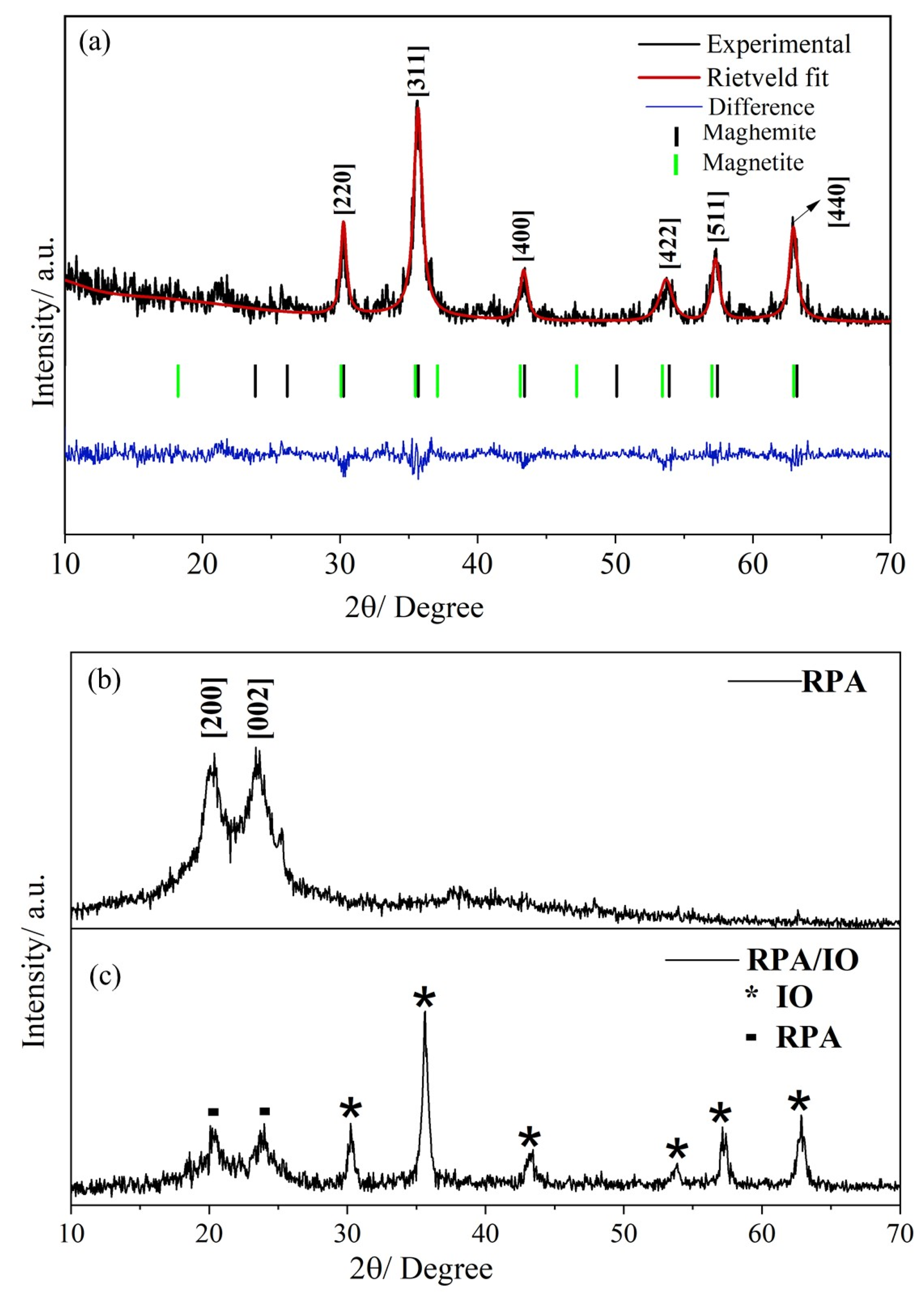
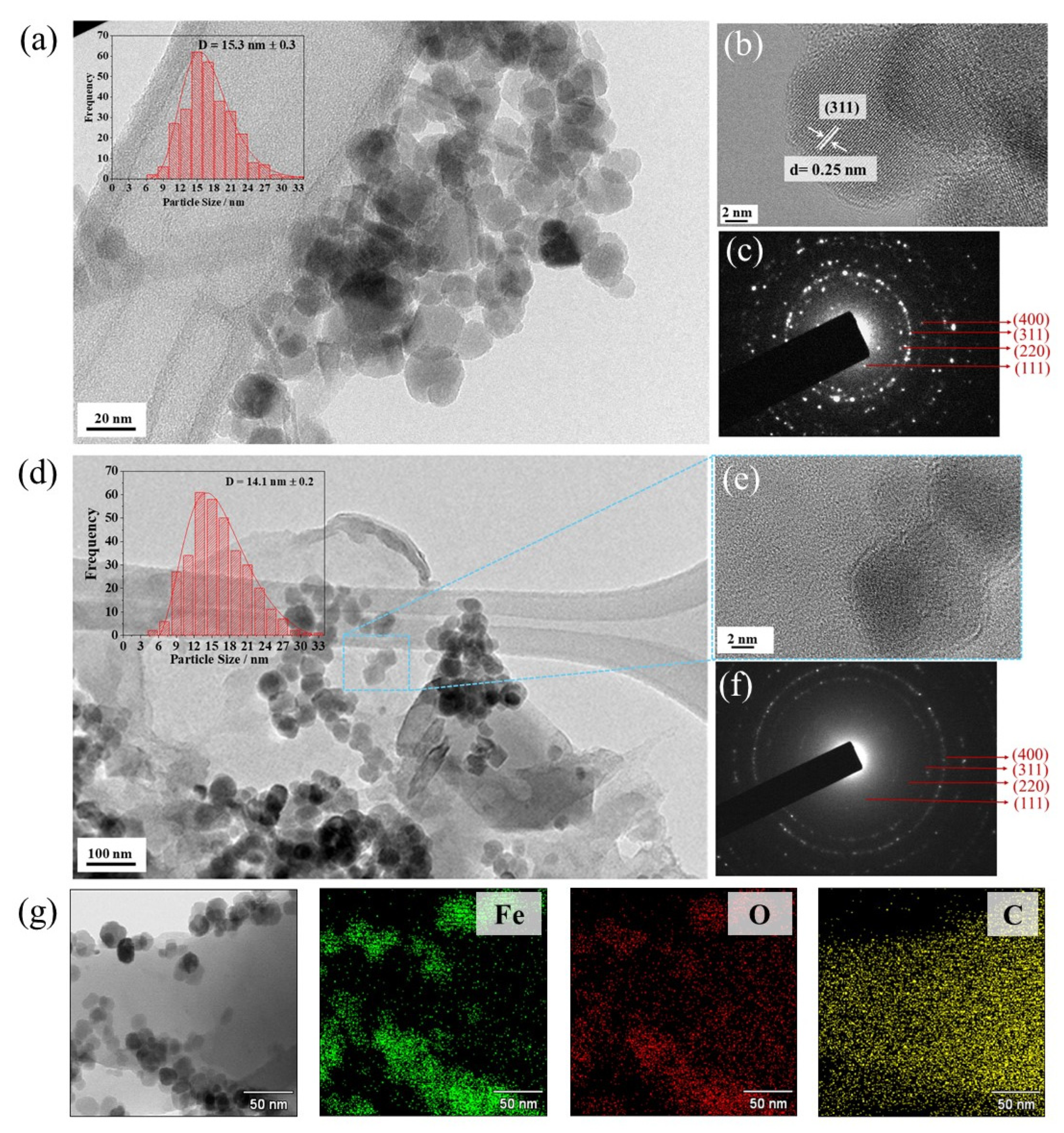
4.1. Microstructural Properties

Figure 1a–c present the XRD patterns and Rietveld refinement of the samples under investigation. The peak positions (2θ) and β values were extracted from these XRD patterns to characterize the microstructure (Table 1). As shown in Figure 1a, the diffraction peaks at the 2θ values of 30.3° (220), 35.6° (311), 37.4° (222), 43.3° (400), 54.1° (422), 57.3° (511), and 63.3° (440) correspond to the cubic system with both Fe3O4 (space group F d 3 ¯ m , No. 227) and γ-Fe2O3 (space group P4332, No. 212) phases, aligning with the Joint Committee on Powder Diffraction Standards (JCPDS) card no. 01-088-0315 for magnetite [48] and card no. 01-084-2783 for maghemite [49], respectively. Similar findings have been reported in the literature [26,49,50]. The peak with the most intensity at 2θ = 35.5° (311) in the IO sample was chosen to calculate the S value, which is about 10.0 nm, and the %DOC was determined at 69.2%. Additionally, the Rietveld refinement method was used to analyze the purity and confirm the long-range structure of the mixed-phase IO nanostructure (with experimental lattice values of a = 8.398 Å for Fe3O4 and a = 8.332 Å for γ-Fe2O3) of this sample. The proportions of the Fe3O4 and γ-Fe2O3 phases were found to be 23.2 wt% and 76.8 wt%, respectively. Herein, GOF = 1.54 indicates a good quality of fit. During synthesis, iron ions (Fe2⁺ and Fe3+) precipitate in an alkaline medium, forming Fe3O4, which has an inverse spinel structure belonging to the space group F d 3 ¯ m (No. 227). However, exposure to oxygen in the air oxidizes the Fe2⁺ ions on the surface of magnetite, converting it to γ-Fe2O3, which also has a spinel structure but with all iron ions in the Fe3+ state.
Figure 1b reveals an amorphous halo ranging from 2θ = 18° to 26°, indicative of the amorphous nature of the RPA material. Two distinct crystalline peaks at 20.1° and 23.6° are superimposed on this halo, corresponding to the (200) and (002) planes, respectively. These peaks are characteristic of the α-form crystal structure commonly observed in PA materials [51,52], suggesting the presence of some crystalline regions within the predominantly amorphous RPA structure [9,53]. For the RPA, the calculated S values for the (200) and (002) planes were found to be 3.2 and 2.9 nm, respectively, while the %DOC of RPA was measured at 35.8% (Table 1). These findings are consistent with the literature on various PAs. For example, Qianhui et al. [54] used CaCl2/ethanol/water-based solvents to analyze waste from PAs and RPAs. Both materials showed crystalline peaks at 20.2° and 23.5°, indicating the α crystal form. The %DOC was 52.0% for waste PAs and 35.9% for RPAs. Similarly, Colucci et al. [55] studied the effect of mechanical recycling on the microstructure and properties of PA composites reinforced with carbon fibers. They identified the α crystal form in PAs with 2θ peaks near 20° and 24°, respectively. The %DOC was calculated at 26.2% for PA66 and 24.6% for RPA. These studies suggest that recycling, whether through solvents or mechanical processes, may decrease the crystallinity of PAs. This is likely due to partial degradation or structural changes during recycling.
Regarding RPA/IO nanocomposites, the XRD patterns revealed the presence of characteristic RPA peaks alongside multiple crystalline peaks attributable to the mixed-phase IO nanostructure (Figure 1c). The Bragg intensities were characterized at 2θ angles of 20.3° (200), 23.9° (002), 30.2° (220), 35.6° (331), 43.4° (400), 54.4° (422), 57.3° (511), and 63.5° (440). A prominent IO crystalline peak was observed at 2θ = 35.6, corresponding to the (311) plane. The impact of incorporating IO into the RPA/IO nanocomposites was analyzed by calculating the S values and %DOC from the peaks at 20.3° and 23.9°, which were found to be 4.4 nm and 4.6 nm, respectively, with a %DOC of 11.5% (Table 1). These results indicated a slight increase in the S value compared to the bare RPA sample. Conversely, the %DOC decreased by approximately 24%, indicating the significant influence of IO on the crystalline structure of the resulting nanocomposite.
Detailed morphological data obtained through SEM on the different samples are illustrated in Figure 2. In Figure 2a, the SEM micrographs reveal that the RPA sample exhibits an irregular morphology characterized by a rough surface and several pores in the micrometer range. High-porosity surfaces are desired for multifunctional nanocomposites because they facilitate molecule entry, making them a promising material for adsorbing emerging pollutants due to their porous nature [56,57]. Additionally, RPA’s surface is abundant in oxygen and nitrogen atoms, which enables interactions with positively charged ions, further enhancing its adsorption capabilities [58,59].
The morphology and dispersion state of the IO material on the RPA surface can be seen in Figure 2b. It can be seen that the IO material is nearly spherical with the formation of agglomerates. On the other hand, it is observed that the IO nanoparticles adhere well to the RPA surface, which may be related to the porous nature of the RPA surface, which provides a large surface area for the IO nanostructures to adhere to, enhancing the interaction between the RPA matrix and the IO. This results in stronger interactions, increasing the overall stability of the RPA/IO nanocomposite. The irregular dispersion and formation of IO aggregates can also create localized regions with high magnetic activity, which can significantly improve the performance of the nanocomposite in applications that rely on magnetic properties, such as magnetic separation, catalysis, or sensors [19,60,61].
Figure 3 depicts the TEM images, particle size distributions, and selected area electron diffraction (SAED) patterns for both IO and the RPA/IO nanocomposite. The RPA/IO nanocomposite exhibited a quasi-spherical morphology, with a mean particle size of around 15.3 ± 0.3 nm (Figure 3a). The SAED pattern confirms the polycrystalline nature of the inverse spinel IO structure (Figure 3c), as indicated by the rings corresponding to the (111), (220), (311), and (400) planes [62]. The SAED results calculated the lattice parameter to be 8.369 Å, which is consistent with the Rietveld refinement of the XRD data. Figure 3b displays a well-ordered (311) lattice plane, which is attributed to the principal crystalline peak of the IO phase. Hence, the agglomerated IO can be observed in Figure 3d, which is consistent with the SEM results. The HRTEM image reveals the crystalline nature of IO (Figure 3e), while SAED and XRD patterns confirm the IO phase (Figure 3f). The bright field TEM images of the RPA/IO nanocomposite, presented in Figure 3g, indicate the nanoparticle nature of the synthesized IO powder, which has an average particle size of approximately 14.1 ± 0.2 nm and an irregular quasi-spherical morphology. Additionally, the EDX mapping indicates that the RPA/IO nanocomposite is rich in Fe and O, with the C content associated with both IO and RPA phases and no other contaminating elements. Hence, the presence of the IO in the nanocomposite suggests that this functional material likely exhibits magnetic properties.

4.2. Magnetic Properties

To study the magnetic behavior of the IO and RPA/IO nanocomposites, magnetization measurements were performed as a function of the magnetic field. In the VSM measurements, the saturation magnetization (Ms), remnant magnetization (Mr), and coercivity (Hc) were extracted from the hysteresis curves at 5 K and 300 K (see Table 2 and Figure 4). The mixed-phase IO nanostructure exhibited an MS of 48.83 ± 0.59 emu g−1 at 5 K, which is lower than that of the bulk magnetite [50,63,64], likely due to the presence of both the Fe3O4 and γ-Fe2O3 phases, as identified by the Rietveld refinement of XRD data. This presence of multiple phases could decrease the overall Ms value observed for the IO, as the magnetic properties of the mixed phases can result in a lower effective magnetization than pure bulk Fe3O4 [52,65,66]. At 300 K, the Ms, Mr, and Hc values were calculated as follows: Ms = 43.42 ± 0.12 emu g−1, Mr = 0.75 ± 0.02 emu g−1, and Hc = 1.9 × 10⁻⁴ ± 5.7 × 10⁻⁵ kOe, respectively. Hu et al. [64] suggested that the magnetic properties of these materials could be significantly influenced by their crystallinity and the method used for their synthesis. As observed in Figure 4a, the curve for the IO at 300 K does not show any hysteresis loop and exhibits low Hc (1.9 × 10⁻⁴ ± 5.7 × 10⁻⁵ kOe) and Mr (0.75 ± 0.02 emu g−1) values. At 300 K, the absence of a hysteresis loop and significantly reduced Mr and Hc suggest a transition to superparamagnetic behavior [67]. Conversely, a hysteresis loop at 5 K suggests ferrimagnetic behavior, as the reduced thermal energy allows the magnetic moments to align and remain aligned. These findings are consistent with previous studies in the literature [67,68,69]. Chirita et al. [68] synthesized magnetite nanoparticles and studied magnetic properties, particularly the effect of temperature. They recorded magnetization curves within the 5–300 K range, observing a decrease in the hysteresis loop as the temperature increased. Above 150 K, the cubic inverse spinel phase exhibited a nearly hysteresis-free magnetization cycle. Based on these findings, they concluded that the IO demonstrates superparamagnetic-like behavior.
For the RPA/IO nanocomposites, the Ms values at 5 K and 300 K were measured at 21.81 ± 0.05 and 18.84 ± 0.01 emu g−1, respectively, which are 50% lower compared to those of the pure IO. Additionally, the RPA/IO nanocomposites exhibited hysteresis loops at both temperatures, with the loops being less pronounced at 300 K. This reduction in magnetic properties relative to pure IO is likely due to the interaction between the polymer matrix and the IO. RPAs may disrupt the Fe-O-Fe interactions by altering the IO nanostructure’s local structure or surrounding environment. We hypothesize that RPAs could interfere with the formation or stability of the oxide layer on the IO, potentially modifying the interactions between iron atoms and, consequently, impacting magnetization.
As shown in Table 2, the magnetic moment ratio (Mr/Ms) was below 0.3 for all samples. At the elevated temperature of 300 K, these Mr/Ms ratios further decreased to 0.017 ± 0.001 for IO and 0.044 ± 0.001 for the RPA/IO nanocomposite. This reduction is likely associated with the material’s magnetic anisotropy [70], which can diminish at higher temperatures, leading to a more disordered state [71]. This disorder results in a lower Mr relative to the Ms. Additionally, the distribution and density of IO on the RPA surface significantly influences the magnetization behavior of the nanocomposites [70]. SEM and TEM analysis confirmed the aggregation of IO nanostructure, which can influence magnetic anisotropy and exchange interactions, thereby contributing to the observed variations in Ms values in the RPA/IO nanocomposites.
In our study, magnetic IO and the RPA/IO nanocomposite exhibited superparamagnetic behavior at 300 K, which is a characteristic of systems with single-domain particles [72]. In such systems, the absence of domain walls means that the magnetization reversal mechanism is primarily driven by magnetic anisotropy [73]. This anisotropy arises from a combination of magnetocrystalline, shape, strain, and surface anisotropies, with its magnitude represented by the magnetic anisotropy constant (K), which quantifies anisotropy energy per unit volume. For spherical magnetic nanoparticles, the dominant contribution typically comes from magnetocrystalline anisotropy, expressed by the magnetocrystalline anisotropy constant (K1) [74,75]. As the SEM micrographs and TEM images illustrate that the IO in our samples was almost spherical, we could reasonably approximate K as K1 in this study, making the magnetocrystalline anisotropy the primary focus for understanding the anisotropic properties of these nanoparticles.
To investigate magnetic anisotropy, the initial magnetization curves of the samples were fitted using the law of approach to saturation (LAS), which facilitates the estimation of the constant, K1. This approach was utilized to analyze magnetic behavior in the saturation region, illustrating the relationship between magnetization and the applied magnetic field when H ≫ Hc. The magnetization near saturation can be expressed as follows [76]:
M = M s 1 b H 2   + χ H
The term b/H2 represents the rotation of magnetization against magnetocrystalline anisotropy energy, where b = 8/105 × ( K 1 2 / μ 0 2 M s 2 ) (8/105 is a coefficient related to the cubic anisotropy of random polycrystalline samples; K1 is the cubic anisotropy constant; MS is the saturation magnetization; and μ0 is the permeability of free space). H is the applied magnetic field, and χH is the forced magnetization, which behaves similarly to a paramagnetic component, resulting from a linear increase in spontaneous magnetization with the applied magnetic field. Forced magnetization becomes significant under elevated temperatures and very high magnetic fields, necessitating its inclusion in such analyses. The observed magnetization data for the applied magnetic field above 2 kOe at 300 K were fitted using Equation (3). The values of Ms and b were then used to estimate the K1 constant, as shown in Equation (4):
k 1 = μ 0 M s 105 8 b
Figure 4c,d show the typical LAS fitting curves at 300 K for the IO and RPA/IO nanocomposites. Table 2 presents the K1 values of the samples at 300 and 5 K. The K1 values for the IO and RPA/IO nanocomposite were estimated to be 5.44 × 10⁵ ± 0.05 and 4.28 × 10⁵ ± 0.01 at 5 K, respectively, whereas at 300 K, the values were 3.34 × 10⁵ ± 0.01 and 1.53 × 10⁵ ± 0.02, respectively. Notably, in both cases, the K1 constant increased as the temperature decreased. According to Paswan et al. [74], magnetic anisotropy in spinel ferrite is caused by spin–orbit interactions and unquenched orbital magnetic moments. At 5 K, the higher K1 values suggest restricted moment orientations owing to stronger interactions and reduced thermal energy, resulting in stable magnetization. In contrast, the lower K1 values at 300 K indicate weaker spin–orbit interactions and suppressed orbital magnetic moments. In the superparamagnetic state, particles easily switch magnetization directions, which is consistent with the lower anisotropy constant observed at 300 K. Therefore, the relationship between K1, temperature, and magnetic behavior illustrates the transition from a stable ferrimagnetic state at low temperatures to a dynamic superparamagnetic state at higher temperatures.
In the study by Mamiya et al. [77], the magnetic anisotropy constant K1 ranged from 1.0 × 10⁵–2.0 × 10⁵ erg cm–3, corresponding to pure magnetite nanoparticles. In contrast, our IO sample consisted of magnetite and maghemite phases, potentially altering the K1 value due to the crystal structure and cation distribution differences. Magnetic anisotropy in magnetite results from a combination of factors, including magnetocrystalline anisotropy, shape anisotropy, surface anisotropy, and mechanical stress [77]. Specifically, magnetocrystalline anisotropy is influenced by strong super-exchange interactions between tetrahedral and octahedral sites. The presence of maghemite may affect these interactions, leading to variations in the magnetic anisotropy constant compared to pure magnetite nanoparticles. Moreover, for nanoparticles of volume V, the magnetic anisotropy constant can be related to the blocking temperature (TB) by the following Equation (5) [78]:
T B = K V 25 k B
Here, K is the magnetic anisotropy constant, V is the volume of the IO, and kB is the Boltzmann constant. This equation demonstrates that the blocking temperature is directly proportional to the magnetic anisotropy constant K1 and the volume V of the nanoparticle. This blocking temperature reflects the point at which thermal energy overcomes the energy barrier KV, allowing magnetization to fluctuate. Below TB, magnetization remains “blocked” in a stable direction, while above this temperature, it transitions to superparamagnetic behavior, allowing magnetization to reorient quickly due to thermal fluctuations. To determine TB for the samples, K1 at 300 K was used to investigate temperature-dependent magnetic characteristics (see Table 2). The volume of the nanoparticles was calculated from the diameter obtained through TEM analysis (Figure 3a,d). The TB for the IO and RPA/IO nanocomposites was 180 and 178 K, respectively. The IO exhibited ferrimagnetism at TB in the blocked state, transitioning to superparamagnetic behavior above this temperature. The TB can vary with particle size, synthesis methods, and interactions [79]. For instance, spherical magnetic nanoparticles with a size of approximately 5.7 nm exhibited a TB of roughly 28 K under an applied field of 100 Oe [80]. In another study, magnetite nanoparticles with a mean size of 20 nm embedded in polyvinyl alcohol exhibited a TB of approximately 300 K [81]. Under more specific conditions, including the field and particle size, similar blocking temperatures between 100 and 200 K were reported for nanoparticles in the 11–18 nm [82,83]. Understanding the temperature dependence of superparamagnetic behavior is crucial for applications requiring particle stability across various temperatures.

5. Conclusions

In conclusion, this study successfully demonstrated the fabrication of a novel RPA/IO nanocomposite using recycled textile PAs. The comprehensive XRD, SEM, TEM, and VSM characterization confirmed this nanocomposite’s successful synthesis, microstructure, and magnetic properties. XRD analysis revealed a semi-crystalline structure with an S value of 10.0 nm and a degree of crystallinity of 11.5%. At the same time, SEM and TEM showed a nanometric scale and porous morphology, with IO well dispersed on the nanocomposite’s surface. The observed average particle size of 14 nm aligns with expectations for such materials. Magnetic measurements demonstrate temperature-dependent behavior.
This study demonstrates the potential of recycled PAs as a valuable resource for developing cost-effective and functional nanocomposites, contributing to both material science and sustainable efforts. The simple preparation method and the promising properties of the RPA/IO nanocomposite suggest its suitability for various applications, including catalysis and wastewater treatment through the adsorptive removal of contaminants, magnetic field sensors, and chemical sensors. Future research will focus on scaling up the production process and evaluating the nanocomposite’s performance in specific applications, such as those requiring magnetic properties or enhanced mechanical strength.

Author Contributions

L.G.D.S., D.F.V., E.D.M., and D.D.A.B. Conceptualization, conceived, writing—review and editing, B.L.S.V., F.d.A.L.P., and C.M.M.R. validation, methodology and investigation. All authors discussed the results and contributed to the final manuscript. All authors have read and agreed to the published version of the manuscript.

Funding

This research was funded by the CNPq project 172349/2023-0.

Institutional Review Board Statement

Not applicable.

Informed Consent Statement

Not applicable.

Data Availability Statement

The data are contained within the article.

Acknowledgments

The authors gratefully acknowledge the National Council for Scientific and Technological Development (CNPq, Process no. 172349/2023-0) for the postdoctoral fellowship, and CAPES, CNPq, and Fundação Araucária for financial support through scholarships. The authors also extend they’re thanks to the Gleb Wataghin Institute of Physics (IFGW) at the University of Campinas (UNICAMP); Campinas, SP, Brazil, for providing facilities for magnetic measurements; and the Department of Chemical and Materials Engineering at PUC-Rio, Rio de Janeiro, Brazil, for their assistance with microscopic measurements.

Conflicts of Interest

The authors declare no conflicts of interest.

References

  1. Koszewska, M. Circular Economy—Challenges for the Textile and Clothing Industry. Autex Res. J. 2018, 18, 337–347. [Google Scholar] [CrossRef]
  2. Roy Choudhury, A.K. Environmental Impacts of the Textile Industry and Its Assessment Through Life Cycle Assessment. In Roadmap to Sustainable Textiles and Clothing: Environmental and Social Aspects of Textiles and Clothing Supply Chain; Springer: Singapore, 2014; pp. 1–39. [Google Scholar] [CrossRef]
  3. Juanga-Labayen, J.P.; Labayen, I.V.; Yuan, Q. A Review on Textile Recycling Practices and Challenges. Textiles 2022, 2, 174–188. [Google Scholar] [CrossRef]
  4. Wang, Y. Fiber and textile waste Utilization. Waste Biomass Valorization 2010, 1, 135–143. [Google Scholar] [CrossRef]
  5. Tang, K.H.D. State of the Art in Textile Waste Management: A Review. Textiles 2023, 3, 454–467. [Google Scholar] [CrossRef]
  6. Patwary, S. Clothing and Textile Sustainability: Current State of Environmental Challenges and the Ways Forward. Text. Leather Rev. 2020, 3, 158–173. [Google Scholar] [CrossRef]
  7. Markovičová, L.; Zatkalíková, V.; Kojnoková, T. Environmental impact on the life of a polymeric composite with polyamide matrix and glass fibres. IOP Conf. Ser. Mater. Sci. Eng. 2021, 1178, 012043. [Google Scholar] [CrossRef]
  8. Hirschberg, V.; Rodrigue, D. Recycling of polyamides: Processes and conditions. J. Polym. Sci. 2023, 61, 1937–1958. [Google Scholar] [CrossRef]
  9. Al-Shawabkeh, A.F. Optoelectronic investigation and spectroscopic characteristics of polyamide-66 polymer. E-Polymers 2022, 22, 858–869. [Google Scholar] [CrossRef]
  10. Hashemi Sanatgar, R.; Campagne, C.; Nierstrasz, V. Investigation of the adhesion properties of direct 3D printing of polymers and nanocomposites on textiles: Effect of FDM printing process parameters. Appl. Surf. Sci. 2017, 403, 551–563. [Google Scholar] [CrossRef]
  11. Baeg, K.-J.; Lee, J.; Baeg, J.K.; Lee, J. Flexible Electronic Systems on Plastic Substrates and Textiles for Smart Wearable Technologies. Adv. Mater. Technol. 2020, 5, 2000071. [Google Scholar] [CrossRef]
  12. Koronis, G.; Silva, A.; Fontul, M. Green composites: A review of adequate materials for automotive applications. Compos. Part B Eng. 2013, 44, 120–127. [Google Scholar] [CrossRef]
  13. Mahalik, N.P.; Nambiar, A.N. Trends in food packaging and manufacturing systems and technology. Trends Food Sci. Technol. 2010, 21, 117–128. [Google Scholar] [CrossRef]
  14. Singh, R.; Kumar, R.; Ranjan, N.; Penna, R.; Fraternali, F. On the recyclability of polyamide for sustainable composite structures in civil engineering. Compos. Struct. 2018, 184, 704–713. [Google Scholar] [CrossRef]
  15. Yao, W.H. The preparation of modified polyamide clay nanocomposite/recycled maleic anhydride polyamide 6 and blending with low density polyethylene for film blowing application. Polym. Polym. Compos. 2021, 29, S631–S643. [Google Scholar] [CrossRef]
  16. Medeiros, D.G.; Jardim, P.M.; De Tatagiba, M.K.V.; D’Almeida, J.R.M. Composites of recycled nylon 11 and titanium based nanofillers. Polym. Test. 2015, 42, 108–114. [Google Scholar] [CrossRef]
  17. Benaducci, D.; de Oliveira, V.; Yin Tze, W.T.; Hafez, I.; Branciforti, M.C. Nanocomposites of recycled and of virgin polyamide 6.6 with cellulose nanofibers. Hybrid Adv. 2024, 6, 100261. [Google Scholar] [CrossRef]
  18. Korkees, F.; Aldrees, A.; Barsoum, I.; Alshammari, D. Functionalised graphene effect on the mechanical and thermal properties of recycled PA6/PA6,6 blends. J. Compos. Mater. 2021, 55, 2211–2224. [Google Scholar] [CrossRef]
  19. Bassyouni, D.; Mohamed, M.; El-Ashtoukhy, E.S.; El-Latif, M.A.; Zaatout, A.; Hamad, H. Fabrication and characterization of electrospun Fe3O4/o-MWCNTs/polyamide 6 hybrid nanofibrous membrane composite as an efficient and recoverable adsorbent for removal of Pb (II). Microchem. J. 2019, 149, 103998. [Google Scholar] [CrossRef]
  20. Liang, Y.; Xia, X.; Luo, Y.; Jia, Z. Synthesis and performances of Fe2O3/PA-6 nanocomposite fiber. Mater. Lett. 2007, 61, 3269–3272. [Google Scholar] [CrossRef]
  21. Cornell, R.M.; Schwertmann, U. The Iron Oxides: Structure, Properties, Reactions, Occurrences, and Uses; De Gruyter: Berlin, Germany, 2003. [Google Scholar]
  22. Gutierrez, F.V.; De Falco, A.; Yokoyama, E.; Mendoza, L.A.F.; Luz-Lima, C.; Perez, G.; Loreto, R.P.; Pottker, W.E.; La Porta, F.A.; Solorzano, G.; et al. Magnetic Characterization by Scanning Microscopy of Functionalized Iron Oxide Nanoparticles. Nanomaterials 2021, 11, 2197. [Google Scholar] [CrossRef]
  23. Mohapatra, M.; Anand, S. Synthesis and applications of nano-structured iron oxides/hydroxides—A review. Int. J. Eng. Sci. Technol. 2010, 2, 127–146. [Google Scholar] [CrossRef]
  24. Hamed, M.H.; Mueller, D.N.; Müller, M. Thermal phase design of ultrathin magnetic iron oxide films: From Fe3O4 to γ-Fe2O3 and FeO. J. Mater. Chem. C 2020, 8, 1335–1343. [Google Scholar] [CrossRef]
  25. Li, Z.; Chanéac, C.; Berger, G.; Delaunay, S.; Graff, A.; Lefèvre, G. Mechanism and kinetics of magnetite oxidation under hydrothermal conditions. RSC Adv. 2019, 9, 33633–33642. [Google Scholar] [CrossRef] [PubMed]
  26. Winsett, J.; Moilanen, A.; Paudel, K.; Kamali, S.; Ding, K.; Cribb, W.; Seifu, D.; Neupane, S. Quantitative determination of magnetite and maghemite in iron oxide nanoparticles using Mössbauer spectroscopy. SN Appl. Sci. 2019, 1, 1636. [Google Scholar] [CrossRef]
  27. Buelvas, D.D.A.; Camargo, L.P.; Valezi, D.F.; Tupan, L.F.S.; Dall’Antonia, L.H.; Rocha, C.M.M.; Lopez, D.A.S.; Urbano, A.; Vicentin, B.L.S. Impact of Varying Magnetite Nanoparticle Concentrations on the Structural, Electrical, and Magnetic Properties of Polyaniline-Based Magnetic Nanocomposites. Synth. Met. 2024, 307, 117703. [Google Scholar] [CrossRef]
  28. Bragg, W.H. The Structure of Magnetite and the Spinels. Nature 1915, 95, 561. [Google Scholar] [CrossRef]
  29. Wareppam, B.; Kuzmann, E.; Garg, V.K.; Singh, L.H. Mössbauer spectroscopic investigations on iron oxides and modified nanostructures: A review. J. Mater. Res. 2022, 38, 937–957. [Google Scholar] [CrossRef] [PubMed]
  30. Petrychuk, M.; Kovalenko, V.; Pud, A.; Ogurtsov, N.; Gubin, A. Ternary magnetic nanocomposites based on core–shell Fe3O4/polyaniline nanoparticles distributed in PVDF matrix. Phys. Status Solidi 2010, 207, 442–447. [Google Scholar] [CrossRef]
  31. Buelvas, D.D.A.; Camargo, L.P.; Salgado, I.K.I.; Vicentin, B.L.S.; Valezi, D.F.; Dall’Antonia, L.H.; Tarley, C.R.T.; Mauro, E. Di: Study and optimization of the adsorption process of methylene blue dye in reusable polyaniline-magnetite composites. Synth. Met. 2023, 292, 117232. [Google Scholar] [CrossRef]
  32. Itoh, H.; Sugimoto, T. Systematic control of size, shape, structure, and magnetic properties of uniform magnetite and maghemite particles. J. Colloid Interface Sci. 2003, 265, 283–295. [Google Scholar] [CrossRef] [PubMed]
  33. Velásquez, A.A.; Urquijo, J.P. Synthesis and characterization of magnetite-maghemite nanoparticles in presence of polyethylene glycol obtained by mechanical milling. Mater. Sci. Eng. B 2021, 263, 114873. [Google Scholar] [CrossRef]
  34. Andronenko, S.I.; Nikolaev, A.M.; Suharzhevsky, S.M.; Sinelnikov, A.A.; Kovalenko, A.S.; Ivanova, A.G.; Shilova, O.A. Phase Composition and Magnetic Properties of Nanoparticles with Magnetite–Maghemite Structure. Ceramics 2023, 6, 1623–1631. [Google Scholar] [CrossRef]
  35. Meftah, S.; Ngo, A.T.; Bouteiller, L.; Russier, V.; Hrabovsky, D.; Konaté, A.; Kondo, D.; Bedoui, F.; Lisiecki, I. Synthesis and Magnetic Properties of Spherical Maghemite Nanoparticles with Tunable Size and Surface Chemistry. Langmuir 2024, 40, 22673–22683. [Google Scholar] [CrossRef] [PubMed]
  36. Gupta, R.; Pancholi, K.; De Sa, R.; Murray, D.; Huo, D.; Droubi, G.; White, M.; Njuguna, J. Effect of Oleic Acid Coating of Iron Oxide Nanoparticles on Properties of Magnetic Polyamide-6 Nanocomposite. JOM 2019, 71, 3119–3128. [Google Scholar] [CrossRef]
  37. Muhazeli, N.S.; Nordin, N.A.; Mazlan, S.A.; Abdul Aziz, S.A.; Ubaidillah Nazmi, N. Mini review: An insight on the fabrication methods of smart magnetic polymer foam. J. Magn. Magn. Mater. 2021, 534, 168038. [Google Scholar] [CrossRef]
  38. Nordin, A.H.; Ahmad, Z.; Husna, S.M.N.; Ilyas, R.A.; Azemi, A.K.; Ismail, N.; Nordin, M.L.; Ngadi, N.; Siti, N.H.; Nabgan, W.; et al. The State of the Art of Natural Polymer Functionalized Fe3O4 Magnetic Nanoparticle Composites for Drug Delivery Applications: A Review. Gels 2023, 9, 121. [Google Scholar] [CrossRef] [PubMed]
  39. Li, X.; Li, Z.; Shen, J.; Zheng, Z.; Liu, J. Role of a nanoparticle network in polymer mechanical reinforcement: Insights from molecular dynamics simulations. Phys. Chem. Chem. Phys. 2021, 23, 21797–21807. [Google Scholar] [CrossRef] [PubMed]
  40. Li, X.; Huang, B.; Liu, J.; Hu, X.; Zheng, Z.J. Revealing the reinforcing effect of a nanorod network on a polymer matrix through molecular dynamics simulations. Phys. Chem. Chem. Phys. 2023, 25, 18757–18765. [Google Scholar] [CrossRef] [PubMed]
  41. Wenlei, X.; Ning, M. Immobilized lipase on Fe3O4 nanoparticles as biocatalyst for biodiesel production. Energy Fuels 2009, 23, 1347–1353. [Google Scholar] [CrossRef]
  42. Rietveld, H.M. Line profiles of neutron powder-diffraction peaks for structure refinement. Acta Crystallogr. 1967, 22, 151–152. [Google Scholar] [CrossRef]
  43. Guinier, A. X-Ray Diffraction: In Crystals, Imperfect Crystals, and Amorphous Bodies; W. H. Freeman & Company: San Francisco, CA, USA, 1963. [Google Scholar]
  44. Buelvas, D.D.A.; Vicentin, B.L.S.; Urbano, A.; Besegato, J.F.; Hoeppner, M.G.; Galvão, T.D.; Parreira, P.S.; Di Mauro, E. Crystalline properties and morphology of bulk-fill dental resin composites as function of light-cure protocol and composition. Polym. Bull. 2023, 80, 2349–2366. [Google Scholar] [CrossRef]
  45. Segal, L.; Creely, J.J.; Martin, A.E.; Conrad, C.M. An Empirical Method for Estimating the Degree of Crystallinity of Native Cellulose Using the X-Ray Diffractometer. Text. Res. J. 1959, 29, 786–794. [Google Scholar] [CrossRef]
  46. Nica, S.L.; Nica, V.; Grigoras, V.C.; Varganici, C.D.; Popovici, D.; Hulubei, C.; Ioan, S. Influence of two structural phases of Fe3O4 and γ-Fe2O3 on the properties of polyimide/iron oxide composites. Polym. Int. 2015, 64, 1172–1181. [Google Scholar] [CrossRef]
  47. Stern, P.; Polymer, E.S. On the structure of polypropylene fibres. Polymer 1968, 9, 471–477. [Google Scholar] [CrossRef]
  48. Tsukimura, K.; Sasaki, S.; Kimizuka, N. Cation distributions in nickel ferrites. Jpn. J. Appl. Phys. Part 1 Regul. Pap. Short Notes Rev. Pap. 1997, 36, 3609–3612. [Google Scholar] [CrossRef]
  49. Solano, E.; Frontera, C.; Puig, T.; Obradors, X.; Ricart, S.; Ros, J. Neutron and X-ray diffraction study of ferrite nanocrystals obtained by microwave-assisted growth. A structural comparison with the thermal synthetic route. J. Appl. Crystallogr. 2014, 47, 414–420. [Google Scholar] [CrossRef]
  50. Rebodos, R.L.; Vikesland, P.J. Effects of oxidation on the magnetization of nanoparticulate magnetite. Langmuir 2010, 26, 16745–16753. [Google Scholar] [CrossRef] [PubMed]
  51. Xia, H.; Zhao, X.; Yang, G. Double in situ synthesis of Fe3O4/polyamide 6 magnetic nanocomposite. Mater. Lett. 2013, 98, 90–93. [Google Scholar] [CrossRef]
  52. Zhang, R.; Zhang, B.; Dou, W.; Wu, Y.; Luo, L. Preparation of Nano-Fe3O4/Nylon Composite Fabric with Magnetic Properties by Post Finishing Method. Fibers Polym. 2019, 20, 1396–1403. [Google Scholar] [CrossRef]
  53. Ranganathan, P.; Mutharani, B.; Tseng, C.-H.; Tsai, P.-S. The Isothermal and Nonisothermal Crystallization Kinetics and Morphology of Solvent-Precipitated Nylon 66. Polymers 2022, 14, 442. [Google Scholar] [CrossRef]
  54. Qianhui, X.; Hongmei, H.; Ruishu, Z.; Lina, S.; Jianyong, Y.; Xueli, W. A non-destructive, environment-friendly method for separating and recycling polyamide 6 from waste and scrap polyamide 6 blended textiles. Text. Res. J. 2023, 93, 3327–3340. [Google Scholar] [CrossRef]
  55. Colucci, G.; Ostrovskaya, O.; Frache, A.; Martorana, B.; Badini, C. The effect of mechanical recycling on the microstructure and properties of PA66 composites reinforced with carbon fibers. J. Appl. Polym. Sci. 2015, 132, 42275. [Google Scholar] [CrossRef]
  56. Morales-Luckie, R.A.; Sánchez-Mendieta, V.; Olea-Mejia, O.; Vilchis-Nestor, A.R.; López-Téllez, G.; Varela-Guerrero, V.; Huerta, L.; Arenas-Alatorre, J. Facile Solventless Synthesis of a Nylon-6,6/Silver Nanoparticles Composite and Its XPS Study. Int. J. Polym. Sci. 2013, 2013, 235850. [Google Scholar] [CrossRef]
  57. Sadeghi-Kiakhani, M.; Safapour, S.; Mirnezhad, S. Thermodynamic and kinetic studies of the adsorption behaviour of the natural dye cochineal on polyamide 66. Color. Technol. 2018, 134, 308–314. [Google Scholar] [CrossRef]
  58. Cheval, N.; Gindy, N.; Flowkes, C.; Fahmi, A. Polyamide 66 microspheres metallised with in situ synthesised gold nanoparticles for a catalytic application. Nanoscale Res. Lett. 2012, 7, 182. [Google Scholar] [CrossRef]
  59. Zakaria, Z.; Izzah, Z.; Tarmizi, A.; Jawaid, M.; Hassan, A. Effect of degree of deacetylation of chitosan on thermal stability and compatibility of chitosan-polyamide blend. BioResources 2012, 7, 5568–5580. [Google Scholar] [CrossRef]
  60. Abdel-Rahman, M.A.; El-Said, W.A.; Sayed, E.M.; Abdel-Wahab, A.M.A. Synthesis, Characterization of Some Conductive Aromatic Polyamides/Fe3O4 NPs/ITO, and Their Utilization for Methotrexate Sensing. Surfaces 2023, 6, 83–96. [Google Scholar] [CrossRef]
  61. Yu, D.; Ni, H.; Wang, L.; Wu, M.; Yang, X. Nanoscale-confined precursor of CuFe2O4 mediated by hyperbranched polyamide as an unusual heterogeneous Fenton catalyst for efficient dye degradation. J. Clean. Prod. 2018, 186, 146–154. [Google Scholar] [CrossRef]
  62. Mohan, A.; Singhal, R.; Ramanan, S.R. A study on the effect of the collector properties on the fabrication of magnetic polystyrene nanocomposite fibers using the electrospinning technique. J. Appl. Polym. Sci. 2023, 140, e53461. [Google Scholar] [CrossRef]
  63. O’Handley, R.C. Modern Magnetic Materials: Principles and Applications; IEEE Electrical Insulation Magazine: Hoboken, NJ, USA, 1999; Volume 13, p. 768. [Google Scholar]
  64. Hu, P.; Zhang, S.; Wang, H.; Pan, D.; Tian, J.; Tang, Z.; Volinsky, A.A. Heat treatment effects on Fe3O4 nanoparticles structure and magnetic properties prepared by carbothermal reduction. J. Alloys Compd. 2011, 509, 2316–2319. [Google Scholar] [CrossRef]
  65. Schwaminger, S.P.; Bauer, D.; Fraga-García, P.; Wagner, F.E.; Berensmeier, S. Oxidation of magnetite nanoparticles: Impact on surface and crystal properties. CrystEngComm. 2017, 19, 246–255. [Google Scholar] [CrossRef]
  66. Faaliyan, K.; Abdoos, H.; Borhani, E.; Afghahi, S.S.S. Magnetite-silica nanoparticles with core-shell structure: Single-step synthesis, characterization and magnetic behavior. J. Sol-Gel Sci. Technol. 2018, 88, 609–617. [Google Scholar] [CrossRef]
  67. Goya, G.F.; Berquó, T.S.; Fonseca, F.C.; Morales, M.P. Static and dynamic magnetic properties of spherical magnetite nanoparticles. J. Appl. Phys. 2003, 94, 3520–3528. [Google Scholar] [CrossRef]
  68. Chirita, M.; Bezergheanu, A.; Bazil Cizmas, C.; Ercuta, A. Superparamagnetic-like Micrometric Single Crystalline Magnetite for Biomedical Application Synthesis and Characterization. Magnetochemistry 2022, 9, 5. [Google Scholar] [CrossRef]
  69. Zhang, H.; Zhu, G. One-step hydrothermal synthesis of magnetic Fe3O4 nanoparticles immobilized on polyamide fabric. Appl. Surf. Sci. 2012, 258, 4952–4959. [Google Scholar] [CrossRef]
  70. Gupta, R.; Pancholi, P.V.; Yu, X.; Gupta, L.; Stenning, G.B.G.; Bucknall, D.; Flynn, D.; Pancholi, K. Role of interface in optimisation of polyamide-6/Fe3O4 nanocomposite properties suitable for induction heating. Nano-Struct. Nano-Objects 2023, 34, 100973. [Google Scholar] [CrossRef]
  71. Yoon, S. Determination of the Temperature Dependence of the Magnetic Anisotropy Constant in Magnetite Nanoparticles. J. Korean Phys. Soc. 2011, 59, 3069–3073. [Google Scholar] [CrossRef]
  72. Lee, J.S.; Cha, J.M.; Yoon, H.Y.; Lee, J.K.; Kim, Y.K. Magnetic multi-granule nanoclusters: A model system that exhibits universal size effect of magnetic coercivity. Sci. Rep. 2015, 5, 12135. [Google Scholar] [CrossRef]
  73. Lima, E.; Brandl, A.L.; Arelaro, A.D.; Goya, G.F. Spin disorder and magnetic anisotropy in Fe3O4 nanoparticles. J. Appl. Phys. 2006, 99, 083908. [Google Scholar] [CrossRef]
  74. Paswan, S.K.; Kumari, S.; Kar, M.; Singh, A.; Pathak, H.; Borah, J.P.; Kumar, L. Optimization of structure-property relationships in nickel ferrite nanoparticles annealed at different temperature. J. Phys. Chem. Solids. 2021, 151, 109928. [Google Scholar] [CrossRef]
  75. Rondinone, A.J.; Samia, A.C.S.; Zhang, Z.J. Characterizing the magnetic anisotropy constant of spinel cobalt ferrite nanoparticles. Appl. Phys. Lett. 2000, 76, 3624–3626. [Google Scholar] [CrossRef]
  76. Chikazumi, S. Physics of Ferromagnetism; Oxford University Press: Oxford, UK, 1997. [Google Scholar]
  77. Muscas, G.; Peddis, D.; Cobianchi, M.; Lascialfari, A.; Cannas, C.; Musinu, A.; Omelyanchik, A.; Rodionova, V.; Fiorani, D.; Mameli, V. Magnetic Interactions Versus Magnetic Anisotropy in Spinel Ferrite Nanoparticles. IEEE Magn. Lett. 2019, 10, 6110305. [Google Scholar] [CrossRef]
  78. Knobel, M.; Nunes, W.C.; Socolovsky, L.M.; De Biasi, E.; Vargas, J.M.; Denardin, J.C. Superparamagnetism and Other Magnetic Features in Granular Materials: A Review on Ideal and Real Systems. J. Nanosci. Nanotechnol. 2008, 8, 2836–2857. [Google Scholar] [CrossRef] [PubMed]
  79. Rumpf, K.; Granitzer, P.; Morales, P.M.; Poelt, P.; Reissner, M. Variable blocking temperature of a porous silicon/Fe3O4 composite due to different interactions of the magnetic nanoparticles. Nanoscale Res. Lett. 2012, 7, 445. [Google Scholar] [CrossRef] [PubMed]
  80. Mercante, L.A.; Melo, W.W.M.; Granada, M.; Troiani, H.E.; MacEdo, W.A.A.; Ardison, J.D.; Vaz, M.G.F.; Novak, M.A. Magnetic properties of nanoscale crystalline maghemite obtained by a new synthetic route. J. Magn. Magn. Mater. 2012, 324, 3029–3033. [Google Scholar] [CrossRef]
  81. Gogoi, B.; Das, U. Enhanced Study of Magnetic Properties of Polyvinyl Alcohol-Coated Superparamagnetic Iron Oxide Nanoparticles Below Blocking Temperatures. Powder Metall. Met. Ceram. 2023, 62, 41–57. [Google Scholar] [CrossRef]
  82. Correa, J.R.; Bordallo, E.; Canetti, D.; León, V.; Otero-Díaz, L.C.; Negro, C.; Gómez, A.; Sáez-Puche, R. Structure and superparamagnetic behaviour of magnetite nanoparticles in cellulose beads. Mater. Res. Bull. 2010, 45, 946–953. [Google Scholar] [CrossRef]
  83. Zheng, R.K.; Gu, H.; Xu, B.; Zhang, X.X. The origin of the non-monotonic field dependence of the blocking temperature in magneticnanoparticles. J. Phys. Condens. Matter. 2006, 18, 5905. [Google Scholar] [CrossRef]
Figure 1. XRD patterns of IO with the Rietveld plot for the IO nanostructure (a), RPA (b), and RPA/IO nanocomposite (c).
Magnetism 05 00005 g001
Figure 2. SEM micrographs with a zoom region for RPA (a) and the RPA/IO nanocomposite (b).
Magnetism 05 00005 g002
Figure 3. Low-magnification and high-magnification TEM images with SAED patterns of IO (ac) and RPA/IO nanocomposite (df). EDX mapping of the RPA/IO nanocomposite showing Fe, O, and C elements (g).
Magnetism 05 00005 g003
Figure 4. M–H loops at 5 K and 300 K (a,b), and the law of approach to saturation for IO (c,d) at 300 K.
Magnetism 05 00005 g004
Table 1. Size parameters for calculating the average crystallite size (S) and the degree of crystallinity percentage (%DOC).
SamplePlanes/h k l2θ°* β** S/nm*** %DOC
IO31135.60.834110.069.2
RPA20020.12.64083.235.8
00223.63.04032.9
RPA/IO20020.31.93834.411.5
00223.91.83304.6
* β is the half-peak width of the diffraction peak. ** S is the average crystallite size. *** %DOC is the degree of crystallinity percentage.
Table 2. The measured magnetic parameters of the IO and RPA/IO nanocomposite at 5 K and 300 K.
SampleTemperature/KMs/emu g–1Mr/emu g–1Mr/MsHc/kOeK1/erg cm–3
IO548.83 ± 0.5913.21 ± 0.010.271 ± 0.0030.4 × 10−3 ± 0.1 × 10−35.44 × 105 ± 0.05
30043.42 ± 0.120.75 ± 0.020.017 ± 0.0011.9 × 10−4 ± 5.7 × 10−53.34 × 105 ± 0.01
RPA/IO521.81 ± 0.055.55 ± 0.010.255 ± 0.0010.22 ± 0.014.28 × 105 ± 0.01
30018.84 ± 0.010.83 ± 0.010.044 ± 0.0010.02 ± 0.0011.53 × 105 ± 0.02
Disclaimer/Publisher’s Note: The statements, opinions and data contained in all publications are solely those of the individual author(s) and contributor(s) and not of MDPI and/or the editor(s). MDPI and/or the editor(s) disclaim responsibility for any injury to people or property resulting from any ideas, methods, instructions or products referred to in the content.

