Review

Perioperative Anesthetic Strategies in Emergent Neurosurgery During Severe Traumatic Brain Injury


1
College of Human Medicine, Michigan State University, Grand Rapids, MI 49503, USA
2
School of Medicine, Noorda College of Osteopathic Medicine, Provo, UT 84606, USA
3
Department of Neurosurgery, Taubman Health Care Center, University of Michigan, Ann Arbor, MI 48109, USA
4
Department of Neurosurgery, Jamaica Hospital Medical Center, New York City, NY 11418, USA
*
Authors to whom correspondence should be addressed.
Trauma Care 2026, 6(1), 5; https://doi.org/10.3390/traumacare6010005
Submission received: 27 December 2025 / Revised: 2 March 2026 / Accepted: 6 March 2026 / Published: 9 March 2026

Abstract

Introduction: Severe traumatic brain injury (sTBI) frequently coexists with polytrauma and often necessitates damage control neurosurgery (DCNS), where rapid decompression and temporary stabilization take precedence over definitive reconstruction. Within this context, anesthetic management must balance cerebral protection with ongoing resuscitation, yet high-quality DCNS-specific evidence remains limited. Materials and Methods: A comprehensive search of PubMed, Scopus, and Google Scholar (2015–2025) was conducted using MeSH terms and keywords related to neurotrauma, anesthesia, intracranial pressure, and perioperative management. Studies were included if they examined anesthetic or hemodynamic strategies in severe TBI or DCNS and reported relevant clinical or physiologic outcomes. Results: Nineteen articles addressing perioperative strategies for optimizing DCNS outcomes were analyzed. Discussion: Preoperative care emphasizes hemodynamic stabilization and permissive hypertension, damage control resuscitation including massive transfusion protocols, optimization of cerebral perfusion pressure (CPP) and neuromonitoring, and the use of hyperosmolar therapy. Transexamic acid can be used in sTBI safely but with unclear improvement in outcomes. Intraoperatively, propofol-based total intravenous anesthesia is generally preferred over volatile agents due to favorable effects on intracranial pressure (ICP), cerebral blood flow (CBF), autoregulation, and emergence. While historically contraindicated, ketamine and etomidate are now increasingly used as hemodynamically protective induction agents. Analgesic and sedative strategies prioritize dexmedetomidine and carefully titrated opioids to minimize respiratory depression and reduce postoperative complications. CPP and ICP-directed management relies on individualized blood pressure targets, vasopressor selection, lung-protective ventilation, and strict temperature control. Conclusions: Emerging evidence has suggested the benefit of DCNS for patient survival. Overall, perioperative care is guided largely by physiology and extrapolation, highlighting the need for standardized protocols.

1. Introduction

Severe traumatic brain injury (sTBI) remains a leading cause of death worldwide that frequently accompanies polytrauma and complicates resuscitation [1]. Despite its prevalence, perioperative management of severe TBI varies substantially across trauma centers, reflecting a lack of standardized anesthetic and resuscitative protocols. In patients with rapid neurological decline from mass lesions, cerebral edema, or intracranial hypertension, damage control neurosurgery (DCNS) conceptualizes polytrauma stabilization, urgent craniectomy, rapid hematoma evacuation and hemostasis, cerebral decompression, and temporary stabilization over definitive reconstruction to allow for multidisciplinary care [2]. Within this framework, the main aim of anesthetic management is to limit secondary brain injury by controlling intracranial pressure (ICP), maintaining adequate cerebral perfusion pressure (CPP), and integrating damage control resuscitation with neuroprotection. Primary brain injury occurs at impact and includes contusions, hematomas, axonal injury, and vascular disruption; these structural lesions are only modestly modifiable once established. Additionally, secondary brain injury evolves over hours to days and arises from insults such as hypotension, hypoxia, hypercarbia, coagulopathy, hyperglycemia, seizures, fever, or sustained neuroinflammatory response [3,4]. TBI also initiates excitotoxicity and blood–brain barrier (BBB) disruption that amplifies neuroinflammation and changes the pharmacokinetics and pharmacodynamics of many anesthetic agents [5]. Limited evidence surrounds the clinical management of sTBI, especially regarding unified perioperative strategies for DCNS candidates. This complex physiology requires that intraoperative anesthesia for DCNS maintains hemodynamic stability, optimizes CPP, limits ICP crises, and facilitates postoperative neurological assessment when appropriate [6].
Despite the publication of the Brain Trauma Foundation guidelines and extensive research on the anesthetic considerations during neurosurgical procedures, substantial variability persists in surgical timing, anesthetic technique, and sedation strategies, often without clear outcome-based justification. Much of the available evidence derives from heterogeneous populations of sTBI or neurocritical care patients, limiting guiding evidence for polytrauma patients and the role of DCNS. There is a pressing need to synthesize emerging evidence to clarify how anesthetic care can best optimize outcomes in patients undergoing DCNS for severe neurotrauma.

2. Methodology

A structured narrative literature search was conducted to identify relevant English-language publications from January 2015 to January 2025, allowing us to focus on contemporary anesthetic practices, modern neuromonitoring technologies, updated Brain Trauma Foundation recommendations, and evolving damage control paradigms. PubMed, Scopus, and Google Scholar were searched for randomized clinical trials (RCTs), retrospective clinical trials, systematic reviews, meta-analyses, cohort studies, and case series addressing perioperative anesthetic strategies in neurosurgical trauma using combinations of key words including the following: “traumatic brain injury,” “damage control neurosurgery,” “anesthesia,” “perioperative management,” “neuroprotection,” “intracranial pressure,” “cerebral perfusion pressure,” “ketamine,” “volatile anesthetics,” “vasopressors,” “blood pressure management,” “damage control resuscitation,” and “neurosurgical trauma.” Studies were included if they evaluated anesthetic management, hemodynamic strategies, or neuroprotective interventions in the context of severe traumatic brain injury or emergent damage control neurosurgery and reported relevant clinical or physiologic outcomes (see Table 1). Conference abstracts, non-peer-reviewed journals, and studies unrelated to the established goals were excluded. Titles and abstracts were screened by three authors independently; full texts of potentially eligible articles were reviewed and selected by consensus.
Overall, this narrative review therefore synthesizes some contemporary landmark trials, cohort, and randomized studies to characterize the evolving preoperative anesthetic strategies in DCNS.

3. Discussion

3.1. Preoperative and Resuscitative Considerations

3.1.1. Hemodynamic Stabilization Prior to Craniotomy/Decompression

Preoperative hemodynamic stabilization is a central determinant of neurosurgical safety, particularly before craniotomy in patients with elevated ICP, mass lesions, or hemorrhage. One key goal is the avoidance of excessive BP fluctuations that may exacerbate venous congestion or ongoing bleeding. In line with these principles, the Brain Trauma Foundation (BTF) guidelines emphasize the avoidance of hypotension and recommend maintaining systolic BP ≥ 100–110 mm Hg (age-dependent) in patients with severe brain injury to support adequate cerebral perfusion [3]. Randomized and prospective anesthetic studies further support perioperative strategies that minimize cerebral edema while tightly regulating BP to balance perfusion–hemorrhagic risk [12,22].
Insights from ICH trials reinforce the importance of controlled BP reduction. INTERACT showed that moderate systolic BP reduction limits hematoma growth without increasing ischemic risk, illustrating how carefully targeted BP management can protect vulnerable brain tissue [16]. Based on these data, maintaining CPP generally between 60–70 mm Hg is supported by available evidence during preoperative stabilization, with higher CPP thresholds considered in elderly patients or those with impaired autoregulation [3].
When autoregulation monitoring (e.g., PRx-guided CPPopt, where CPPopt is the individualized CPP at which cerebrovascular autoregulation is most intact, identified by the lowest correlation between MAP and ICP) is available, targeting CPP near CPPopt may further reduce secondary brain injury [23]. Autoregulation strongly influences the safe CPP range in TBI, as impaired cerebrovascular reactivity shifts tolerance to hypo- and hyperperfusion. PRx studies demonstrate that autoregulatory failure predicts mortality and that patients exhibit individualized CPPopt levels associated with optimal vascular reactivity [23]. Time spent below the lower limit of autoregulation is consistently linked to worse outcomes, whereas modest CPP elevations above CPPopt appear less harmful [8,9]. CPP values near CPPopt also maximize brain tissue oxygenation, with age-stratified analyses showing higher CPPopt targets in older adults and similar PRx–outcome relationships in pediatric cohorts [7]. The multicenter BOOST-3 trial, currently evaluating the interplay between ICP and brain tissue partial pressure (PbtO2) management, defines CPP targets based on autoregulation and oxygenation rather than fixed thresholds; however, these goals are primarily applicable to prolonged ICU management and are less directly transferable to the perioperative period, where dynamic surgical conditions and hemorrhagic risk constrain CPP manipulation [24]. Overall, current evidence supports targeting CPP values of 60–70 mm Hg in most adults undergoing DCNS, with adjustment toward individualized CPPopt when feasible.

3.1.2. Hyperosmolar Therapy Prior to DCNS

Hyperosmolar therapy is a cornerstone of preoperative and resuscitative management in patients undergoing DCNS, especially when rapid ICP reduction is required to stabilize the patient before craniotomy or decompressive craniectomy. Both mannitol and 3% hypertonic saline (HTS) have been first-line agents. In an RCT of patients undergoing supratentorial brain tumor resection, 3% HTS and mannitol achieved similar relaxation, although HTS was slightly greater, and mannitol caused greater diuresis and intravascular depletion [25]. Other studies supporting administering hyperosmolar boluses before dural opening similarly found that both mannitol and HTS improve surgical conditions, but HTS often yields equal or better relaxation with more stable hemodynamics [23,24,25]. Palazon et al. likewise showed equivalent relaxation but less central venous pressure drop and fluid loss with HTS [26]. Additional trials reported improved brain relaxation or lower fluid requirements with HTS in patients with elevated ICP [27,28]. Subsequent systematic reviews corroborate these findings, demonstrating comparable or superior brain relaxation with HTS, reduced diuresis, and improved hemodynamic stability compared with equiosmolar mannitol [29]. Additionally, meta-analyses report no mortality difference between agents but favor HTS for improved intraoperative conditions and a lower risk of hypotension and intravascular volume depletion [30].
Data on ICP and systemic hemodynamics also favor HTS [19]. In their prospective RCT using equiosmolar dosing, Sokhal et al. demonstrated that 3% HTS achieved intracranial pressure reduction comparable to mannitol while providing superior preservation of MAP and more stable cardiac indices during the critical early period following osmotherapy administration [19]. HTS was associated with a transient increase in cardiac output and cardiac index, minimal need for anesthetic dose reduction, and less diuresis, thereby avoiding the relative intravascular volume depletion observed with mannitol [29]. Additionally, HTS produced predictable, transient elevations in serum sodium without clinically significant complications, whereas mannitol was associated with greater urine output and higher lactate levels, suggesting relative hypovolemia. Although conducted in an intraoperative neurosurgical cohort, these physiologic effects are highly relevant to the resuscitative management of sTBI as the preservation of CPP depends on stable hemodynamics [19].
More concentrated hypertonic saline formulations have been increasingly studied as rescue therapy for refractory intracranial hypertension in TBI [31]. Observational studies report that 23.4% HTS boluses produce rapid ICP reduction without consistent renal injury or severe electrolyte complications when appropriately monitored [31]. However, comparative studies have not shown clear superiority in long-term neurological outcomes, and much of the evidence remains physiologic or retrospective [30].
Overall, the literature supports strong consideration of HTS during both early resuscitation and the immediate preoperative period before DCNS. Mannitol remains effective, but HTS consistently demonstrates advantages in hemodynamic stability, volume preservation, more durable ICP reduction, and the ability for electrolyte monitoring. Accordingly, a typical preoperative regimen includes 3% HTS 2–5 mL/kg as a bolus over 10–20 min for rapid ICP reduction [25,32]. Repeat boluses or continuous infusion (0.1–1.0 mL/kg/h) may be used when prolonged therapy is required. If mannitol is selected, 0.25–1 g/kg IV over 10–30 min is recommended, with 0.5–1 g/kg commonly used in severe elevations of ICP, ensuring careful monitoring of serum osmolality, sodium, and intravascular volume [32]. These collective findings support a strategic hyperosmolar approach that prioritizes physiologic optimization before DCNS to reduce the risk of secondary brain injury.

3.1.3. Coagulation Optimization

Recent trauma research identifies tranexamic acid (TXA) as an important adjunct in early hemostatic optimization, particularly for patients who may require urgent neurosurgical intervention [33,34]. Its benefit is greatest when administered within 1–2 h after injury, aligning well with preoperative stabilization goals. The CRASH-3 trial remains the most robust modern evaluation of TXA in TBI, demonstrating that administration within three hours reduced head injury-related mortality in mild-to-moderate TBI without increasing thromboembolic events [10]. More recent work highlights the feasibility and safety of even earlier administration: a prehospital randomized trial found that TXA given within two hours in moderate-to-severe TBI was safe, though it did not improve 6-month neurologic outcomes [33]. Importantly, early field dosing did not raise thrombotic risk, reinforcing TXA’s safety profile. A subgroup analysis of this work in moderate and sTBI supported the role of 2gm TXA administration within 2 h of injury to improve mortality and neurological outcomes in patients with intra- and extra-axial hemorrhage [34]. Furthermore, a 2024 systematic review and meta-analysis found that prehospital TXA reduces 24 h mortality in bleeding trauma patients without increasing venous thromboembolism [35]. Therefore, preoperative stabilization for DCNS appears advantageous based on extrapolated data of the CRASH-3 regimen of 1 g IV over 10 min followed by 1 g over 8 h when clinically indicated [10].
For patients taking anticoagulants or antiplatelet agents, rapid reversal to functional hemostasis is essential. Warfarin should be reversed with IV vitamin K and four-factor PCC (prothrombin complex concentrate) to rapidly normalize INR [36]. Four-factor PCC is typically dosed at 25–50 IU/kg based on presenting INR, with most neurotrauma guidelines recommending 50 IU/kg for life-threatening bleeding or when urgent neurosurgical intervention is required [36]. For DOACs, idarucizumab (5 g IV) is recommended for dabigatran reversal, while PCC is considered for factor-Xa inhibitors as the most recent Andexanet Alfa has been removed from the market due to concern for hypercoagulability, though protocols vary and evidence in neurotrauma remains limited [36]. For antiplatelet agents such as aspirin or P2Y12 inhibitors, routine platelet transfusion is not supported by randomized evidence and may worsen outcomes when no neurosurgical procedure is planned, as demonstrated in the PATCH trial of spontaneous ICH [37]. Although PATCH was conducted in spontaneous ICH rather than TBI, its findings highlight the potential harm of empiric platelet transfusion in patients not undergoing surgery. While routine platelet transfusion is not supported when no surgery is planned, platelet transfusion may be considered in the setting of urgent DCNS to support operative hemostasis, based on expert consensus guidance [38]. In such cases, one apheresis unit of platelets or desmopressin (0.3 µg/kg IV) may be given to safely restore platelet function before craniotomy or decompression, particularly in patients on aspirin or P2Y12 inhibitors [37].

3.2. Intraoperative Anesthetic Management

3.2.1. Analgesics

Analgesic therapy should combine pain control with the preservation of neurologic evaluation and ventilatory drive. Opioids remain effective for acute neurosurgical pain but risk oversedation, respiratory depression, hypercapnia, and resultant ICP rise, all of which are detrimental when frequent neurologic checks are needed [39]. Accordingly, opioid use is generally limited to the acute intraoperative period, and analgesic strategies in DCNS favor short-acting, titratable agents. Extrapolation of elective surgery enhanced recovery after surgery (ERAS)-based pathways are not widely applicable in emergent trauma settings.
Among opioids, remifentanil facilitates precise titration and suppression of airway reflexes during general anesthesia in mechanically ventilated patients, but it should not be continued into recovery due to the risk of respiratory depression and secondary insult [13]. Fentanyl also remains widely used due to its cardiovascular stability and rapid onset. Typical dosing includes 1–2 µg/kg IV for induction blunting and 25–100 µg boluses intraoperatively titrated to analgesic need [40]. In the ICU following neurosurgical intervention, fentanyl is commonly administered as a continuous infusion at approximately 0.5–2 µg/kg/h (equivalent to about 0.01–0.03 µg/kg/min), allowing for steady analgesia with minimal hemodynamic disturbance while avoiding excessive sedation [40]. However, like other opioids, fentanyl must be used judiciously to avoid prolonged sedation and CO2 retention that could worsen ICP.
Dexmedetomidine provides sedation and analgesia without respiratory depression. This allows for smoother emergence with reduced hypertension, tachycardia, and coughing which is key in patients with elevated ICP [13]. For semi-awake neurosurgical phases, combinations of propofol, remifentanil, and dexmedetomidine are used frequently, with dexmedetomidine having shorter arousal times [39]. Dexmedetomidine is typically infused at 0.2–0.7 µg/kg/h, with or without a loading dose depending on hemodynamics [41,42]. In damage control neurosurgery, these attributes make dexmedetomidine particularly valuable for ICP stability while allowing for rapid postoperative neurologic assessment.

3.2.2. Intravenous vs. Volatile Agents

Among available agents, propofol consistently demonstrates the most favorable cerebral profile by lowering CBF, ICP, and cerebral metabolic rate while also preserving autoregulation and CO2 responsiveness [40,43]. Its rapid onset, short duration, and predictable emergence make it valuable in trauma settings needing frequent neurologic reassessment [40,43]. Clinical data further support propofol’s ability to diminish ICP elevation and coughing during airway manipulation (such as endotracheal suctioning), which improves long-term neurologic prognosis in severely injured patients [28]. Meta-analyses have indicated that propofol provides the highest probability of achieving satisfactory brain relaxation compared with sevoflurane, isoflurane, or desflurane, especially in patients with elevated ICP or a space-occupying lesion [44]. Propofol-based total intravenous anesthesia (TIVA) also preserves autoregulation better than volatile anesthetics, has lower intraoperative ICP, and has higher CPP [40]. Propofol is typically administered as an induction bolus of 1–2 mg/kg, followed by a maintenance infusion of approximately 75–150 µg/kg/min depending on the depth of anesthesia required [43].
In contrast, volatile anesthetics exhibit dose-dependent cerebral vasodilation, increasing cerebral blood flow (CBF) and potentially worsening ICP, which is concerning in patients with traumatic intracranial hypertension [40]. Though volatile agents decrease the cerebral metabolic rate of oxygen (CMRO2), their vasodilatory effects predominate at a mean minimum alveolar concentration (MAC) > 1.0, impairing autoregulation and raising ICP [40,44]. Clinical studies have shown that sevoflurane and desflurane allow for rapid emergence (which can be beneficial for postoperative exam) but both are inferior to propofol for the maintenance of optimal intracranial conditions [40]. Isoflurane, previously favored in neuroanesthesia for its hemodynamic stability, has been linked to neurotoxicity such as mitochondrial dysfunction and therefore carries risk for vulnerable brain tissue [40]. Nitrous oxide is generally avoided entirely in neurotrauma because it increases CBF/ICP and may exacerbate intracranial hypertension [45].
The role of ketamine has shifted substantially. Previously avoided for fear of ICP elevation, newer studies show that ketamine does not increase ICP in TBI and does not worsen mortality [46]. Some research indicates that combining ketamine with propofol may even reduce hypotension compared to propofol alone, which is relevant in trauma resuscitation where maintaining CPP is essential [46]. Ketamine is commonly dosed at 1–2 mg/kg IV for induction and 0.5–1 mg/kg/h for maintenance when used as part of a TIVA strategy [46]. Etomidate also significantly maintains hemodynamic stability and reduces CBF/CMRO2, making it valuable when hypotension would be catastrophic [47]. A typical etomidate induction dose is 0.2–0.3 mg/kg IV [42]. However, concerns about adrenal suppression and potential pediatric neurotoxicity necessitate case-by-case management [43,47]. Overall, modern evidence supports ketamine and etomidate as hemodynamically protective induction agents when blood pressure preservation is a main perioperative goal.

3.3. Neuroprotective Strategies

Neuroprotective strategies in damage control neurosurgery aim to limit secondary brain injury by combining pharmacology, intraoperative monitoring, and resuscitation principles. Increasing evidence supports the neuroprotective potential of ketamine in severe TBI as it is anti-inflammatory, anticonvulsant, anti-spreading depolarization, and reduces glutamate toxicity [48]. Dexmedetomidine similarly conveys neuroprotection through its stable hemodynamics, decreased agitation, and absence of respiratory depression, all which aid ventilated TBI patients [46]. Furthermore, xenon shows promise based on its ability to lessen excitotoxicity and reperfusion injury [13].
Continuous neuromonitoring alongside pharmacy allows for neuroprotection by detecting cerebral hypoxia. In bispectral index (BIS)-guided anesthesia, semi-quantitative monitoring of EEG signals can be effective in monitoring anesthesia depth, which can be helpful for minimization of hypotension-inducing anesthetic agents, and improving earlier postoperative emergence, cognitive function, and neuroexam monitoring. However, it is important to note that BIS accuracy may be reduced in patients with neurologic impairment [49]. Near-infrared spectroscopy (NIRS) offers another noninvasive monitoring method for oximetry and can detect desaturation events associated with ischemia, but its use in TBI is limited by frequent signal inaccuracy due to intracranial injury or surgery [50,51]. Despite these limitations, monitoring using BIS and selective NIRS can help avoid over sedation and guide neuroprotection during acute neurosurgical stabilization.

3.4. Hemodynamic and ICP Control

Early hemodynamic and ICP management in damage control neurosurgery centers can maintain adequate CPP while avoiding intracranial hypertension [3,21]. Brain Trauma Foundation guidelines recommend treating ICP once it exceeds 22 mmHg, as values above this threshold are associated with increased mortality in sTBI [3]. Systolic blood pressure should be maintained at ≥100 mmHg for patients aged 50 to 69 years and ≥110 mmHg for those 15 to 49 or >70 years to reduce secondary ischemic injury [3]. These blood pressure goals are translated into CPP targets of 60 to 70 mmHg, though the optimal lower limit varies with each patient’s cerebrovascular autoregulation [3,9]. Studies suggest that personalized CPP targets outperform standard thresholds, with even small deviations below an individual’s optimal CPP associated with worse outcomes [9]. ICP monitoring is thus strongly considered in severe (GCS 3–8) and moderate (GCS 9–12) TBI, both to guide treatment of ICP crises and reduce mortality [52].

3.4.1. Vasopressors

When fluids alone cannot sustain CPP, vasopressors can augment MAP, with norepinephrine and phenylephrine being the most commonly used agents in TBI [21,53]. Phenylephrine, a pure α1-agonist, increases MAP via arterial vasoconstriction but induces reflex bradycardia and can reduce cardiac output, whereas norepinephrine has both α-mediated vasoconstriction and β effects to better preserve cardiac output [21,53]. In a single-center cohort of severe TBI, phenylephrine produced the largest early rise in MAP and CPP compared with norepinephrine or dopamine and was associated with lower mortality [53]. Vasopressor effects on CPP may also increase ICP and promote secondary cerebral edema and induced hypertension that is linked to complications such as acute respiratory distress syndrome (ARDS) and pulmonary edema [53]. Contrastingly, multicenter TRACK-TBI data found no significant differences in outcomes between patients initially treated with norepinephrine vs. phenylephrine, showing that current evidence does not clearly favor one agent over another [21]. As a result, preoperative vasopressor selection in DCNS should be individualized: phenylephrine may be advantageous in patients with tachycardia or ischemic heart disease because of its lesser chronotropic effect, whereas norepinephrine may be preferred when maintaining cardiac output is critical, with both CPP and ICP closely monitored [21,53].

3.4.2. Controlled Ventilation

Controlled ventilation is another major determinant of cerebral hemodynamics because PaCO2 strongly modulates cerebrovascular tone. Hypocapnia causes cerebral vasoconstriction and reduces CBF, while hypercapnia causes vasodilation [54]. Hyperventilation is used to lower ICP or “relax” a tight brain intraoperatively, but sustained or prophylactic hyperventilation can critically reduce CBF, increasing the risk of cerebral ischemia [54,55]. For this reason, guidelines discourage routine prophylactic hyperventilation to PaCO2 ≤ 25 mmHg (especially within the first 24 h after severe TBI) and favor maintaining PaCO2 in a near-normal range of 35 to 45 mmHg during stable phases of care [52,53]. Mild hyperventilation (PaCO2 31–35 mmHg) is best reserved as a temporary rescue in refractory intracranial hypertension or impending herniation during neuromonitoring to minimize ischemic risk [54,55]. Because many TBI patients also have lung contusions, aspiration, or pneumonia, achieving hyperventilation through high tidal volumes can precipitate ventilator induced lung injury, worsen V/Q mismatch, and delay pulmonary recovery. This supports a lung protective ventilation strategy of only brief, targeted hyperventilation when necessary [55].

3.4.3. Temperature Management

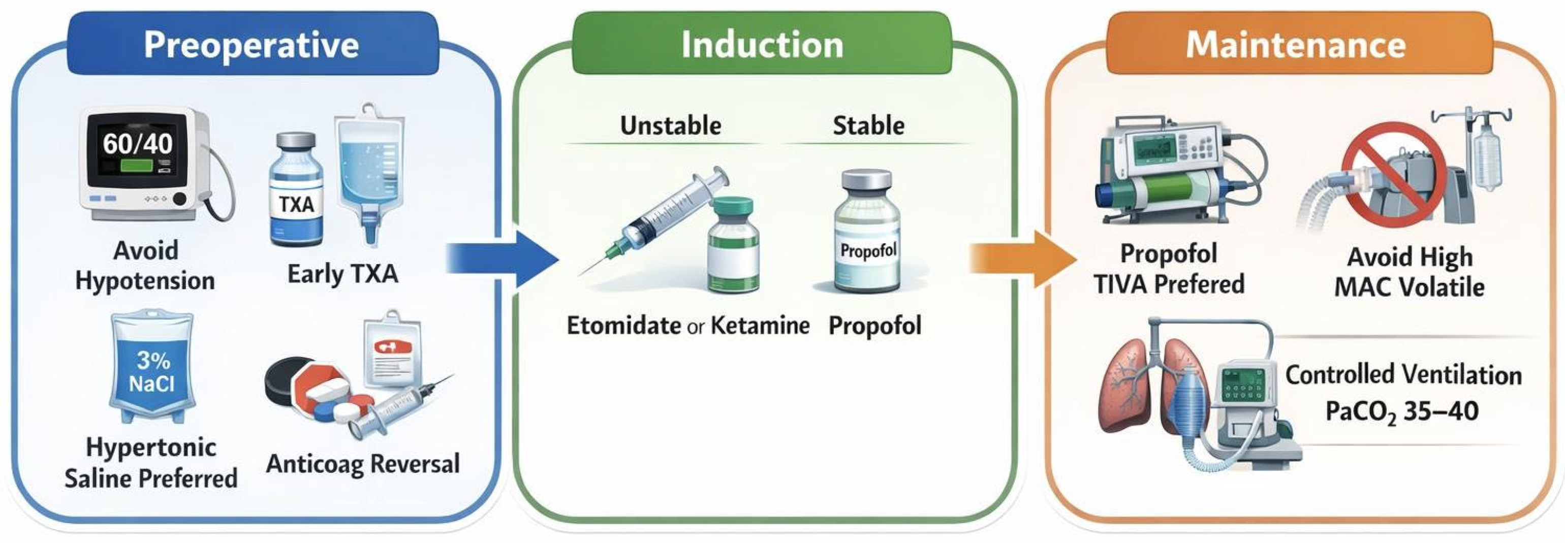
Temperature management is equally important in preventing secondary brain injury, with current recommendations of controlled normothermia (core 36.0–37.5 C) as part of Tier 1–2 ICP management for TBI [15]. Continuous core temperature monitoring using bladder, esophageal, or brain probes is preferred over intermittent or superficial measurement to reliably detect fever [15]. Therapeutic hypothermia (core ≤ 36.0 C) is reserved as a higher-tier option when standard Tier 1 and 2 measures fail to control ICP; however, target temperatures should remain as close to normothermia as possible given the complications of deeper cooling [15]. Rewarming must be slow and carefully titrated particularly in patients with labile ICP, and shivering should be actively treated with increased sedation or neuromuscular blockade to prevent surges in metabolic demand and ICP [15]. Within the broader framework of DCNS, integrating individualized CPP targets, vasopressor use, lung protective ventilation with cautious hyperventilation, and temperature management provides a coordinated strategy to minimize secondary insult in neurosurgical intervention [3,9,14,19,50,51,52,53] (Figure 1).

3.5. Special Considerations and Emerging Trends

Special populations undergoing DCNS introduce additional layers of physiologic complexity. In pediatric TBI, higher baseline cerebral metabolic demand and age-dependent autoregulation require age-adjusted CPP goals; the 2019 BTF pediatric guidelines and subsequent reviews recommend aggressive avoidance of hypotension and hypoxia, early ICP monitoring, and protocolized tiered therapy, but acknowledge limited high-quality evidence for many interventions [56]. Geriatric patients frequently present with frailty, multimorbidity, and pre-injury anticoagulant or antiplatelet use. Frailty indices have emerged as strong, independent predictors of mortality, prolonged hospitalization, and unfavorable discharge disposition after TBI [57]. This reinforces the importance of preoperative frailty assessment and conservative hemodynamic goals defined as avoidance of hypotension, rapid blood pressure fluctuations, and excessive vasopressor use to preserve cerebral perfusion while minimizing secondary injury during DCNS [57,58].
Pregnant patients pose a two-lives problem in which maternal resuscitation and brain protection must be balanced against uteroplacental perfusion, teratogenic risk, and fetal monitoring. Recent systematic reviews and neuroanesthesiology guidance highlight the absence of TBI thresholds specific to pregnancy and advocate individualized management that maintains maternal CPP while minimizing fetal exposure to hypotension, hypoxemia, and extreme hyperventilation [59,60].
Patients with substance use disorders frequently display altered hemodynamic responses, tolerance to sedatives and opioids, and increased risk of withdrawal or arrhythmia. Higher anesthetic requirements may be seen in tolerant patients, avoiding abrupt withdrawal, as well as meticulous perioperative cardiovascular monitoring.
Rapidly expanding research now explores artificial intelligence (AI) and machine learning as tools to anticipate secondary insults and guide management in neurotrauma. Models that analyze high-frequency ICP waveforms and physiologic data can forecast intracranial hypertension and rising ICP up to 30 min before clinical thresholds are crossed, creating opportunities for pre-emptive intervention in DCNS [61]. Systematic reviews highlight substantial progress in AI-based prediction of intracranial hypertension, outcome stratification, and TBI phenotyping, but still emphasize challenges related to data quality [62]. Advances in multimodal neuromonitoring, including combined ICP, CPP, brain tissue oxygen (PbtO2), cerebral blood flow, and EEG, aim to move from single-parameter threshold monitoring to a focus on overall physiology targets.
Biomarker-driven strategies are another emerging potential guide for anesthetic and neuroprotective management in DCNS. Serum and CSF markers such as S100B, GFAP, and neurofilament light have been linked to BBB disruption and axonal injury in TBI, and recent reviews suggest that biomarker panels may help identify patients at highest risk of malignant edema or delayed deterioration who may benefit from more aggressive intraoperative monitoring [5,63]. Overall, the combination of individualized CPPopt-guided therapy, biomarker-informed risk stratification, and AI prediction suggests that anesthetic management for DCNS is moving toward a more predictive perioperative paradigm.

3.6. Challenges, Limitations, and Knowledge Gaps

3.6.1. Lack of Standardization

A major limitation in advancing perioperative care for DCNS is the absence of standardized anesthetic and resuscitative protocols across trauma centers. Multicenter studies show substantial variability in how patients with severe TBI are monitored and prepared for surgery [64]. For example, in a survey of 66 European trauma centers caring for patients with moderate-to-severe TBI, Huijben et al. demonstrated substantial variability in coagulation and transfusion management including hemoglobin transfusion thresholds (<7 to ≥9 g/dL), platelet targets (50–100 × 109/L), indications for ICP monitoring, and approaches to antithrombotic reversal [64]. This variability reflects limited randomized evidence guiding hemostatic optimization in TBI and directly influences preoperative physiology, including cerebral oxygen delivery, bleeding risk, and operative readiness, all of which hinder the development of consistent resuscitative algorithms in DCNS.
Surgical decision making and anesthetic considerations are equally heterogeneous. A survey of 68 neurotrauma centers within the CENTER-TBI surveys showed variability in the indications and timing of decompressive craniectomy and mass lesion evacuation for sTBI, with centers differing in whether surgery was employed as an early intervention based on radiographic findings or reserved as salvage therapy for refractory intracranial hypertension [65]. In another multicenter observational study across 14 international centers, early sedation strategies varied considerably in agent selection, dosing, and escalation, with propofol and midazolam used in differing proportions and no consistent association between sedative choice and short-term outcomes [17].
Collectively these findings reveal the absence of clearly defined, evidence based perioperative pathways tailored to DCNS. Developing standardized, data-driven protocols aligned with modern trauma resuscitation principles remains essential for improving outcomes. It is important to acknowledge that high-quality randomized evidence specifically addressing anesthetic strategies in DCNS is limited. Much of the available literature derives from sTBI intensive care cohorts or elective neurosurgical populations. Therefore, several recommendations herein represent physiologic reasoning and extrapolation rather than direct DCNS-specific trial data.

3.6.2. Limited Randomized Control Trials

Despite advances in neurotrauma care, there remains a striking lack of high-quality randomized controlled trials evaluating specific anesthetic agents or perioperative anesthetic strategies for patients undergoing DCNS. Recent clinical trials in sTBI have predominantly focused on physiologic targets—such as ICP, CPP, brain tissue oxygenation, or coagulation interventions—rather than anesthetic drug selection. For example, randomized studies on sedative agents in TBI remain small and underpowered. Kurni et al. evaluated propofol and midazolam but failed to demonstrate definitive outcome differences due to limited sample sizes [66]. Trials investigating osmotherapy, including mannitol and HTS, similarly provide only indirect insight for DCNS, with most conducted in mixed or elective neurosurgical populations rather than true damage control settings [19,25]. Even large contemporary trauma trials, such as those evaluating TXA or prehospital resuscitation strategies do not address the anesthetic agents or intraoperative strategies used during emergent neurosurgical intervention [10,33]. These limitations highlight a critical need for well-designed, multicenter trials to determine which perioperative anesthetic approaches most effectively optimize neurologic outcomes in DCNS.

3.6.3. Short-Term Outcome Measures

A major barrier to evaluating perioperative anesthetic strategies in DCNS is the use of convenience end-points, namely short-term ICU length of stay or early functional outcomes. Although early functional status strongly predicts mortality and discharge disposition, centers use widely different tools and may not correlate perfectly with long-term outcome. Rydingsward et al. employed a nurse-assessed physical function score to predict 90-day mortality [67], whereas Ko et al. used a simplified bedside assessment of consciousness and mobility to predict long-term survival [68]. Mobility measures show similar inconsistency: the ICU Mobility Scale (IMS) predicts discharge home and improved 90-day survival [20], yet no single instrument (IMS, FSS-ICU, Barthel Index, or GOS) has been universally adopted in TBI cohorts. Neurotrauma data vary further, using BI at discharge, pediatric GOS, or combinations of GOS, FIM, and BI [54,69], making cross-study comparisons difficult.
These disparities highlight a critical knowledge gap. Current tools were developed for general ICU or heterogeneous TBI populations and fail to capture the unique physiology and instability of patients undergoing DCNS. Without validated, neurotrauma focused early outcome measures, existing studies cannot reliably link perioperative anesthetic strategies to meaningful short-term functional outcomes, highlighting the need for DCNS research standardization.

4. Conclusions

These conclusions reflect the synthesis of heterogeneous evidence, much of which is extrapolated from sTBI, ICU and elective neurosurgical populations, underscoring the need for DCNS-specific prospective trials. DCNS for sTBI presents a uniquely unstable physiologic environment in which anesthetic decisions directly influence CPP, ICP, and downstream secondary brain injury. Current evidence supports prioritizing propofol-based total intravenous anesthesia, informed use of ketamine or etomidate for hemodynamically fragile patients, hyperosmolar optimization with a growing role for hypertonic saline, individualized CPP targets, and lung- and brain-protective ventilation and temperature strategies. Integrating these approaches with damage control resuscitation principles, early hemostatic optimization, and neuromonitoring can facilitate timely neurologic assessment after DCNS. However, the field remains constrained by limited randomized data, substantial practice variation, and non-standardized outcome measures. Developing and validating DCNS-specific, physiology-guided anesthetic pathways represents a critical next step toward improving neurologic and systemic outcomes.

Author Contributions

Conceptualization: D.B., M.P. and M.K.; methodology: D.B. and M.P.; formal analysis: D.B., C.R. and D.M.; investigation, D.B., C.R. and D.M.; writing—original draft preparation: D.B., C.R. and D.M.; writing—review and editing: D.B., C.R., D.M., M.P. and M.K.; supervision: M.P. and M.K. All authors have read and agreed to the published version of the manuscript.

Funding

This research received no external funding.

Institutional Review Board Statement

Not applicable.

Informed Consent Statement

Not applicable.

Data Availability Statement

No new data were created or analyzed in this study. Data sharing is not applicable to this article.

Conflicts of Interest

The authors declare no conflicts of interest.

References

  1. Dewan, M.C.; Rattani, A.; Gupta, S.; Baticulon, R.E.; Hung, Y.-C.; Punchak, M.; Agrawal, A.; Adeleye, A.O.; Shrime, M.G.; Rubiano, A.M.; et al. Estimating the global incidence of traumatic brain injury. J. Neurosurg. 2019, 130, 1080–1097. [Google Scholar] [CrossRef]
  2. Kolias, A.G.; Adams, H.; Timofeev, I.; Czosnyka, M.; Corteen, E.A.; Pickard, J.D.; Turner, C.; Gregson, B.A.; Kirkpatrick, P.J.; Murray, G.D.; et al. Decompressive craniectomy following traumatic brain injury: Developing the evidence base. Br. J. Neurosurg. 2016, 30, 246–250. [Google Scholar] [CrossRef]
  3. Carney, N.; Totten, A.M.; O’Reilly, C.; Ullman, J.S.; Hawryluk, G.W.; Bell, M.J.; Bratton, S.L.; Chesnut, R.; Harris, O.A.; Kissoon, N.; et al. Guidelines for the Management of Severe Traumatic Brain Injury, Fourth Edition. Neurosurgery 2017, 80, 6–15. [Google Scholar] [CrossRef]
  4. Xiong, Y.; Mahmood, A.; Chopp, M. Current understanding of neuroinflammation after traumatic brain injury and cell-based therapeutic opportunities. Chin. J. Traumatol. 2018, 21, 137–151. [Google Scholar] [CrossRef]
  5. Dadas, A.; Washington, J.; Diaz-Arrastia, R.; Janigro, D. Biomarkers in traumatic brain injury (TBI): A review. Neuropsychiatr. Dis. Treat. 2018, 14, 2989–3000. [Google Scholar] [CrossRef]
  6. Hawryluk, G.W.J.; Aguilera, S.; Buki, A.; Bulger, E.; Citerio, G.; Cooper, D.J.; Arrastia, R.D.; Diringer, M.; Figaji, A.; Gao, G.; et al. A management algorithm for patients with intracranial pressure monitoring: The Seattle International Severe Traumatic Brain Injury Consensus Conference (SIBICC). Intensive Care Med. 2019, 45, 1783–1794. [Google Scholar] [CrossRef] [PubMed]
  7. Agrawal, S.; Smith, C.; Bogli, S.Y.; Placek, M.M.; Cabeleira, M.; White, D.; Daubney, E.; Young, A.; Beqiri, E.; Kayani, R.; et al. Status of cerebrovascular autoregulation relates to outcome in severe paediatric head injury: STARSHIP, a prospective multicentre validation study. eClinicalMedicine 2025, 81, 103077. [Google Scholar] [CrossRef]
  8. Beqiri, E.; Zeiler, F.A.; Ercole, A.; Placek, M.M.; Tas, J.; Donnelly, J.; Aries, M.J.H.; Hutchinson, P.J.; Menon, D.; Stocchetti, N.; et al. The lower limit of reactivity as a potential individualised cerebral perfusion pressure target in traumatic brain injury: A CENTER-TBI high-resolution sub-study analysis. Crit. Care 2023, 27, 194. [Google Scholar] [CrossRef] [PubMed]
  9. Bögli, S.Y.; Olakorede, I.; Beqiri, E.; Chen, X.; Lavinio, A.; Hutchinson, P.; Smielewski, P. Cerebral perfusion pressure targets after traumatic brain injury: A reappraisal. Crit. Care 2025, 29, 207. [Google Scholar] [CrossRef] [PubMed]
  10. Collaborators, C.T. Effects of tranexamic acid on death, disability, vascular occlusive events and other morbidities in patients with acute traumatic brain injury (CRASH-3): A randomised, placebo-controlled trial. Lancet 2019, 394, 1713–1723. [Google Scholar] [CrossRef]
  11. Hutchinson, P.J.; Adams, H.; Mohan, M.; Devi, B.I.; Uff, C.; Hasan, S.; Mee, H.; Wilson, M.H.; Gupta, D.K.; Bulters, D.; et al. Decompressive Craniectomy versus Craniotomy for Acute Subdural Hematoma. N. Engl. J. Med. 2023, 388, 2219–2229. [Google Scholar] [CrossRef] [PubMed]
  12. Jiang, Z.; Wu, Y.; Liang, F.; Jian, M.; Liu, H.; Mei, H.; Han, R. Brain relaxation using desflurane anesthesia and total intravenous anesthesia in patients undergoing craniotomy for supratentorial tumors: A randomized controlled study. BMC Anesthesiol. 2023, 23, 15. [Google Scholar] [CrossRef] [PubMed]
  13. Kim, H.; Min, K.T.; Lee, J.R.; Ha, S.H.; Lee, W.K.; Seo, J.H.; Choi, S.H. Comparison of Dexmedetomidine and Remifentanil on Airway Reflex and Hemodynamic Changes during Recovery after Craniotomy. Yonsei Med. J. 2016, 57, 980. [Google Scholar] [CrossRef]
  14. Laaksonen, M.; Rinne, J.; Rahi, M.; Posti, J.P.; Laitio, R.; Kivelev, J.; Saarenpää, I.; Laukka, D.; Frösen, J.; Ronkainen, A.; et al. Effect of xenon on brain injury, neurological outcome, and survival in patients after aneurysmal subarachnoid hemorrhage-study protocol for a randomized clinical trial. Trials 2023, 24, 417. [Google Scholar] [CrossRef]
  15. Lavinio, A.; Coles, J.P.; Robba, C.; Aries, M.; Bouzat, P.; Chean, D.; Frisvold, S.; Galarza, L.; Helbok, R.; Hermanides, J.; et al. Targeted temperature control following traumatic brain injury: ESICM/NACCS best practice consensus recommendations. Crit. Care 2024, 28, 170. [Google Scholar] [CrossRef]
  16. Ma, L.; Hu, X.; Song, L.; Chen, X.; Ouyang, M.; Billot, L.; Li, Q.; Malavera, A.; de Silva, A.; Thang, N.H.; et al. The third Intensive Care Bundle with Blood Pressure Reduction in Acute Cerebral Haemorrhage Trial (INTERACT3): An international, stepped wedge cluster randomised controlled trial. Lancet 2023, 402, 27–40. [Google Scholar] [CrossRef] [PubMed]
  17. Patel, P.P.; Egodage, T.; Martin, M.J. Decompressive craniectomy for traumatic brain injury: A review of recent landmark trials. Trauma Surg. Acute Care Open 2025, 10, e001784. [Google Scholar] [CrossRef]
  18. Russo, G.; Harrois, A.; Anstey, J.; Van Der Jagt, M.; Taccone, F.; Udy, A.; Citerio, G.; Duranteau, J.; Ichai, C.; Badenes, R.; et al. Early sedation in traumatic brain injury: A multicentre international observational study. Crit. Care Resusc. 2022, 24, 319–329. [Google Scholar] [CrossRef]
  19. Sokhal, N.; Rath, G.P.; Chaturvedi, A.; Singh, M.; Dash, H.H. Comparison of 20% mannitol and 3% hypertonic saline on intracranial pressure and systemic hemodynamics. J. Clin. Neurosci. 2017, 42, 148–154. [Google Scholar] [CrossRef]
  20. Tipping, C.J.; Bailey, M.J.; Bellomo, R.; Berney, S.; Buhr, H.; Denehy, L.; Harrold, M.; Holland, A.; Higgins, A.M.; Iwashyna, T.J.; et al. The ICU Mobility Scale Has Construct and Predictive Validity and Is Responsive. A Multicenter Observational Study. Ann. Am. Thorac. Soc. 2016, 13, 887–893. [Google Scholar] [CrossRef]
  21. Toro, C.; Temkin, N.; Barber, J.; Manley, G.; Jain, S.; Ohnuma, T.; Komisarow, J.; Foreman, B.; Korley, F.K.; Vavilala, M.S.; et al. Association of Vasopressor Choice with Clinical and Functional Outcomes Following Moderate to Severe Traumatic Brain Injury: A TRACK-TBI Study. Neurocrit. Care 2022, 36, 180–191. [Google Scholar] [CrossRef]
  22. Zhou, Z.; Ying, M.; Zhao, R. Efficacy and safety of sevoflurane vs propofol in combination with remifentanil for anesthesia maintenance during craniotomy: A meta-analysis. Medicine 2021, 100, e28400. [Google Scholar] [CrossRef]
  23. Zeiler, F.A.; Ercole, A.; Beqiri, E.; Cabeleira, M.; Thelin, E.P.; Stocchetti, N.; Steyerberg, E.W.; Maas, A.I.; Menon, D.K.; Czosnyka, M.; et al. Association between Cerebrovascular Reactivity Monitoring and Mortality Is Preserved When Adjusting for Baseline Admission Characteristics in Adult Traumatic Brain Injury: A CENTER-TBI Study. J. Neurotrauma 2020, 37, 1233–1241. [Google Scholar] [CrossRef]
  24. Bernard, F.; Barsan, W.; Diaz-Arrastia, R.; Merck, L.H.; Yeatts, S.; Shutter, L.A. Brain Oxygen Optimization in Severe Traumatic Brain Injury (BOOST-3): A multicentre, randomised, blinded-endpoint, comparative effectiveness study of brain tissue oxygen and intracranial pressure monitoring versus intracranial pressure alone. BMJ Open 2022, 12, e060188. [Google Scholar] [CrossRef]
  25. Ali, A.; Tetik, A.; Sabanci, P.A.; Altun, D.; Sivrikoz, N.; Abdullah, T.; Aydoseli, A.; Sencer, A.; Akinci, I.O. Comparison of 3% Hypertonic Saline and 20% Mannitol for Reducing Intracranial Pressure in Patients Undergoing Supratentorial Brain Tumor Surgery: A Randomized, Double-blind Clinical Trial. J. Neurosurg. Anesthesiol. 2018, 30, 171–178. [Google Scholar] [CrossRef] [PubMed]
  26. Hernández-Palazón, J.; Doménech-Asensi, P.; Fuentes-García, D.; Burguillos-López, S.; Piqueras-Pérez, C.; García-Palenciano, C. Comparison of 20% mannitol and 3% hypertonic saline for intraoperative brain relaxation during supratentorial brain tumour craniotomy in patients with a midline shift. Neurocir. Engl. Ed. 2023, 34, 273–282. [Google Scholar] [CrossRef]
  27. Singla, A.; Mathew, P.; Jangra, K.; Gupta, S.; Soni, S. A Comparison of Hypertonic Saline and Mannitol on Intraoperative Brain Relaxation in Patients with Raised Intracranial Pressure during Supratentorial Tumors Resection: A Randomized Control Trial. Neurol. India 2020, 68, 141. [Google Scholar] [CrossRef]
  28. Wu, M.; Yin, X.; Chen, M.; Liu, Y.; Zhang, X.; Li, T.; Long, Y.; Wu, X.; Pu, L.; Zhang, M.; et al. Effects of propofol on intracranial pressure and prognosis in patients with severe brain diseases undergoing endotracheal suctioning. BMC Neurol. 2020, 20, 394. [Google Scholar] [CrossRef] [PubMed]
  29. Akcil, E.F.; Dilmen, O.K.; Tunali, Y. Hypertonic saline versus mannitol for brain relaxation in supratentorial tumor surgery: A prospective randomized trial. Braz. J. Anesthesiol. Engl. Ed. 2025, 75, 844684. [Google Scholar] [CrossRef]
  30. Burgess, S.; Abu-Laban, R.B.; Slavik, R.S.; Vu, E.N.; Zed, P.J. A Systematic Review of Randomized Controlled Trials Comparing Hypertonic Sodium Solutions and Mannitol for Traumatic Brain Injury: Implications for Emergency Department Management. Ann. Pharmacother. 2016, 50, 291–300. [Google Scholar] [CrossRef]
  31. Ware, M.L.; Nemani, V.M.; Meeker, M.; Lee, C.; Morabito, D.J.; Manley, G.T. Effects of 23.4% sodium chloride solution in reducing intracranial pressure in patients with traumatic brain injury: A preliminary study. Neurosurgery 2005, 57, 727–736; discussion 727–736. [Google Scholar] [CrossRef]
  32. Patel, J.; Feghali, J.; Yang, W.; Rapaport, S.; Gami, A.; Sattari, S.A.; Tamargo, R.J.; Caplan, J.M.; Huang, J. Comparison of management approaches in deep-seated intracranial arteriovenous malformations: Does treatment improve outcome? J. Clin. Neurosci. 2021, 92, 191–196. [Google Scholar] [CrossRef]
  33. Rowell, S.E.; Meier, E.N.; McKnight, B.; Kannas, D.; May, S.; Sheehan, K.; Bulger, E.M.; Idris, A.H.; Christenson, J.; Morrison, L.J.; et al. Effect of Out-of-Hospital Tranexamic Acid vs Placebo on 6-Month Functional Neurologic Outcomes in Patients with Moderate or Severe Traumatic Brain Injury. JAMA 2020, 324, 961. [Google Scholar] [CrossRef]
  34. McKinley, W.I.; Rowell, S.E.; Mansour, A.; Hoefer, L.; Polcari, A.; Schreiber, M.; Benjamin, A.J. Tranexamic Acid, Mortality, and Intracranial Hemorrhage Type in Moderate or Severe Traumatic Brain Injury. JAMA Surg. 2023, 158, 1222–1224. [Google Scholar] [CrossRef]
  35. Acharya, P.; Amin, A.; Nallamotu, S.; Riaz, C.Z.; Kuruba, V.; Senthilkumar, V.; Kune, H.; Bhatti, S.S.; Sarlat, I.M.; Krishna, C.V.; et al. Prehospital tranexamic acid in trauma patients: A systematic review and meta-analysis of randomized controlled trials. Front. Med. 2023, 10, 1284016. [Google Scholar] [CrossRef]
  36. Greenberg, S.M.; Ziai, W.C.; Cordonnier, C.; Dowlatshahi, D.; Francis, B.; Goldstein, J.N.; Hemphill, J.C.; Johnson, R.; Mack, W.J.; Mocco, J.; et al. 2022 guideline for the management of patients with spontaneous intracerebral hemorrhage: A guideline from the American Heart Association/American Stroke Association. Stroke 2022, 53, e282–e361. [Google Scholar] [CrossRef]
  37. Baharoglu, M.I.; Cordonnier, C.; Salman, R.A.-S.; De Gans, K.; Koopman, M.M.; Brand, A.; Majoie, C.B.; Beenen, L.F.; A Marquering, H.; Vermeulen, M.; et al. Platelet transfusion versus standard care after acute stroke due to spontaneous cerebral haemorrhage associated with antiplatelet therapy (PATCH): A randomised, open-label, phase 3 trial. Lancet 2016, 387, 2605–2613. [Google Scholar] [CrossRef] [PubMed]
  38. Frontera, J.A.; Lewin, J.J.; Rabinstein, A.A.; Aisiku, I.P.; Alexandrov, A.W.; Cook, A.M.; del Zoppo, G.J.; Kumar, M.A.; Peerschke, E.I.B.; Stiefel, M.F.; et al. Guideline for Reversal of Antithrombotics in Intracranial Hemorrhage: A Statement for Healthcare Professionals from the Neurocritical Care Society and Society of Critical Care Medicine. Neurocrit. Care 2016, 24, 6–46. [Google Scholar] [CrossRef] [PubMed]
  39. Kvolik, S.; Koruga, N.; Skiljic, S. Analgesia in the Neurosurgical Intensive Care Unit. Front. Neurol. 2022, 12, 819613. [Google Scholar] [CrossRef] [PubMed]
  40. Nguyen, A.; Mandavalli, A.; Diaz, M.J.; Root, K.T.; Patel, A.; Casauay, J.; Perisetla, P.; Lucke-Wold, B. Neurosurgical Anesthesia: Optimizing Outcomes with Agent Selection. Biomedicines 2023, 11, 372. [Google Scholar] [CrossRef]
  41. Weerink, M.A.S.; Struys, M.M.R.F.; Hannivoort, L.N.; Barends, C.R.M.; Absalom, A.R.; Colin, P. Clinical Pharmacokinetics and Pharmacodynamics of Dexmedsetomidine. Clin. Pharmacokinet. 2017, 56, 893–913. [Google Scholar] [CrossRef]
  42. Gropper, M.A.; Cohen, N.H.; Eriksson, L.I.; Fleisher, L.A.; Leslie, K.; Johnson-Akeju, O. (Eds.) Miller’s Anesthesia, 2-Volume Set, 10th ed.; Elsevier: Amsterdam, The Netherlands, 2025. [Google Scholar]
  43. Sahinovic, M.M.; Struys, M.M.R.F.; Absalom, A.R. Clinical Pharmacokinetics and Pharmacodynamics of Propofol. Clin. Pharmacokinet. 2018, 57, 1539–1558. [Google Scholar] [CrossRef]
  44. Intarakhao, P.; Thiarawat, P.; Dhippayom, T. Comparison of propofol and inhalational anaesthetics for brain relaxation in neurosurgery: A systematic review and network meta-analysis. Indian J. Anaesth. 2025, 69, 770–778. [Google Scholar] [CrossRef] [PubMed]
  45. Knuf, K.; Maani, C.V. Nitrous Oxide. In StatPearls; StatPearls Publishing: Treasure Island, FL, USA, 2025. Available online: http://www.ncbi.nlm.nih.gov/books/NBK532922/ (accessed on 26 December 2025).
  46. Sciorilli, J.V.; Rossi, Y.I.; Dos Reis Schevz, R.; da Silva, D.B.P.; Lee, J.; Gallani, F.; Maegele, M. Is ketamine safe for traumatic brain injury? A systematic review and meta-analysis. J. Crit. Care 2026, 91, 155347. [Google Scholar] [CrossRef]
  47. Williams, L.M.; Boyd, K.L.; Fitzgerald, B.M. Etomidate. In StatPearls; StatPearls Publishing: Treasure Island, FL, USA, 2025. Available online: http://www.ncbi.nlm.nih.gov/books/NBK535364/ (accessed on 26 December 2025).
  48. Farrell, D.; Bendo, A.A. Perioperative Management of Severe Traumatic Brain Injury: What Is New? Curr. Anesthesiol. Rep. 2018, 8, 279–289. [Google Scholar] [CrossRef] [PubMed]
  49. Mathur, S.; Patel, J.; Goldstein, S.; Hendrix, J.M.; Jain, A. Bispectral Index. In StatPearls; StatPearls Publishing: Treasure Island, FL, USA, 2025. Available online: http://www.ncbi.nlm.nih.gov/books/NBK539809/ (accessed on 26 December 2025).
  50. Mokhtari, M.; Amirdosara, M.; Goharani, R.; Zangi, M.; Tafrishinejad, A.; Nashibi, M.; Dabbagh, A.; Sadeghi, H.; Nateghinia, S.; Hajiesmaeili, M.; et al. The Predictive Power of Near-Infrared Spectroscopy in Improving Cognitive Problems in Patients Undergoing Brain Surgeries: A Systematic Review. Anesthesiol. Pain Med. 2022, 12, e116637. [Google Scholar] [CrossRef] [PubMed]
  51. Mathieu, F.; Khellaf, A.; Ku, J.C.; Donnelly, J.; Thelin, E.P.; Zeiler, F.A. Continuous Near-infrared Spectroscopy Monitoring in Adult Traumatic Brain Injury: A Systematic Review. J. Neurosurg. Anesthesiol. 2020, 32, 288–299. [Google Scholar] [CrossRef]
  52. Shim, Y.; Kim, J.; Kim, H.S.; Oh, J.; Lee, S.; Ha, E.J. Intracranial Pressure Monitoring for Acute Brain Injured Patients: When, How, What Should We Monitor. Korean J. Neurotrauma 2023, 19, 149–161. [Google Scholar] [CrossRef]
  53. Sookplung, P.; Siriussawakul, A.; Malakouti, A.; Sharma, D.; Wang, J.; Souter, M.J.; Chesnut, R.M.; Vavilala, M.S. Vasopressor use and effect on blood pressure after severe adult traumatic brain injury. Neurocrit. Care 2011, 15, 46–54. [Google Scholar] [CrossRef]
  54. Zhang, Z.; Guo, Q.; Wang, E. Hyperventilation in neurological patients: From physiology to outcome evidence. Curr. Opin. Anaesthesiol. 2019, 32, 568–573. [Google Scholar] [CrossRef]
  55. Gouvea Bogossian, E.; Peluso, L.; Creteur, J.; Taccone, F.S. Hyperventilation in Adult TBI Patients: How to Approach It? Front. Neurol. 2020, 11, 580859. [Google Scholar] [CrossRef]
  56. Kochanek, P.M.; Tasker, R.C.; Bell, M.J.; Adelson, P.D.; Carney, N.; Vavilala, M.S.; Selden, N.R.; Bratton, S.L.; Grant, G.A.; Kissoon, N.; et al. Management of Pediatric Severe Traumatic Brain Injury: 2019 Consensus and Guidelines-Based Algorithm for First and Second Tier Therapies. Pediatr. Crit. Care Med. 2019, 20, 269–279. [Google Scholar] [CrossRef]
  57. Vetrano, D.L.; Palmer, K.; Marengoni, A.; Marzetti, E.; Lattanzio, F.; Roller-Wirnsberger, R.; Samaniego, L.L.; Rodríguez-Mañas, L.; Bernabei, R.; Onder, G. Frailty and Multimorbidity: A Systematic Review and Meta-analysis. J. Gerontol. Ser. A 2019, 74, 659–666. [Google Scholar] [CrossRef]
  58. Gardner, R.C.; Dams-O’Connor, K.; Morrissey, M.R.; Manley, G.T. Geriatric Traumatic Brain Injury: Epidemiology, Outcomes, Knowledge Gaps, and Future Directions. J. Neurotrauma 2018, 35, 889–906. [Google Scholar] [CrossRef]
  59. Lopez, C.E.; Salloum, J.; Varon, A.J.; Toledo, P.; Dudaryk, R. The Management of Pregnant Trauma Patients: A Narrative Review. Anesth. Analg. 2023, 136, 830–840. [Google Scholar] [CrossRef] [PubMed]
  60. Heller, C.; Kraft, M.; Martinez, M.; Mirmajlesi, A.S.; Janecka, M.; McCormack, C.; Thomason, M.E.; Weiss, T.; Arciniega, H. Complications After Maternal Traumatic Brain Injury During Pregnancy: A Systematic Review. JAMA Netw. Open 2025, 8, e2459877. [Google Scholar] [CrossRef] [PubMed]
  61. Tunthanathip, T.; Trakulpanitkit, A. Deep learning-based model for detection of intracranial waveforms with poor brain compliance in southern Thailand. Acute Crit. Care 2025, 40, 473–481. [Google Scholar] [CrossRef] [PubMed]
  62. Ahmed, A.E.; Alyami, R.M.; Al Ghazwi, F.H.; Hamzi, R.H.; Alshammari, N.K.; Jali, F.M.; A Al Alduwayh, A.; Almujami, T.M.; Alamri, A.S.; A Sabban, J.; et al. Artificial Intelligence in Traumatic Brain Injury: A Systematic Review of Prognostic, Diagnostic, and Monitoring Applications. Cureus 2025, 17, e96600. [Google Scholar] [CrossRef]
  63. Lindblad, C.; Nelson, D.W.; Zeiler, F.A.; Ercole, A.; Ghatan, P.H.; von Horn, H.; Risling, M.; Svensson, M.; Agoston, D.V.; Bellander, B.-M.; et al. Influence of Blood–Brain Barrier Integrity on Brain Protein Biomarker Clearance in Severe Traumatic Brain Injury: A Longitudinal Prospective Study. J. Neurotrauma 2020, 37, 1381–1391. [Google Scholar] [CrossRef]
  64. Huijben, J.A.; Van Der Jagt, M.; Cnossen, M.C.; A Kruip, M.J.H.; Haitsma, I.K.; Stocchetti, N.; Maas, A.I.R.; Menon, D.K.; Ercole, A.; Maegele, M.; et al. Variation in Blood Transfusion and Coagulation Management in Traumatic Brain Injury at the Intensive Care Unit: A Survey in 66 Neurotrauma Centers Participating in the Collaborative European NeuroTrauma Effectiveness Research in Traumatic Brain Injury Study. J. Neurotrauma 2018, 35, 323–332. [Google Scholar] [CrossRef]
  65. on behalf of the CENTER-TBI investigators; Cnossen, M.C.; Huijben, J.A.; van der Jagt, M.; Volovici, V.; van Essen, T.; Polinder, S.; Nelson, D.; Ercole, A.; Stocchetti, N.; et al. Variation in monitoring and treatment policies for intracranial hypertension in traumatic brain injury: A survey in 66 neurotrauma centers participating in the CENTER-TBI study. Crit. Care 2017, 21, 233. [Google Scholar] [CrossRef] [PubMed]
  66. Kurni, M.; Kaloria, N.; Hazarika, A.; Jain, K.; Gupta, S.K.; Walia, R. Comparison of Midazolam and Propofol Infusion to Suppress Stress Response in Patients with Severe Traumatic Brain Injury: A Prospective, Randomized Controlled Trial. Korean J. Neurotrauma 2023, 19, 70–81. [Google Scholar] [CrossRef] [PubMed]
  67. Rydingsward, J.E.; Horkan, C.M.; Mogensen, K.M.; Quraishi, S.A.; Amrein, K.; Christopher, K.B. Functional Status in ICU Survivors and out of Hospital Outcomes: A Cohort Study. Crit. Care Med. 2016, 44, 869–879. [Google Scholar] [CrossRef]
  68. Ko, R.-E.; Lee, H.; Jung, J.H.; Lee, H.O.; Sohn, I.; Yoo, H.; Ko, J.Y.; Suh, G.Y.; Chung, C.R. Simple functional assessment at hospital discharge can predict long-term outcomes of ICU survivors. PLoS ONE 2019, 14, e0214602. [Google Scholar] [CrossRef]
  69. Keleman, N.; Krasnik, R.; Mikov, A.; Dragičević-Cvjetković, D. Outcome of early rehabilitation of patients with traumatic brain injury during COVID-19 pandemic in The Republic of Srpska, Bosnia and Herzegovina. Front. Neurol. 2023, 14, 1269564. [Google Scholar] [CrossRef] [PubMed]
Figure 1. Legend: The figure depicts a phase-based perioperative approach emphasizing preoperative avoidance of hypotension, early tranexamic acid administration, a preference for hypertonic saline, and anticoagulation reversal. Induction agent selection is guided by hemodynamic stability, favoring etomidate or ketamine in unstable patients and propofol in stable patients. Intraoperative maintenance prioritizes propofol-based total intravenous anesthesia (TIVA), the avoidance of high minimum alveolar concentration (MAC) volatile agents, and controlled ventilation targeting normocapnia (PaCO2 35–40 mmHg) to limit secondary brain injury.
Traumacare 06 00005 g001
Table 1. Summary of key studies on perioperative strategies in DCNS (2015–2025).
Author (Year)Study TypeSample SizeEvidence Applicability to DCNS *Evidence Strength **Relevance to DCNS
Agrawal et al. (2025) [7]Prospective observational multicenter studyn = 135DirectModerateCerebral autoregulation following PRx
Beqiri et al. (2023) [8]Multicenter cohortn = 171IndirectLowMortality based on CPP
Bögli et al. (2025) [9]Observationaln = 809DirectModeratePressure reactivity index, CPP, upper/lower limit of autoregulation
CRASH-3 collaborators (2019) [10]Multicenter, randomized, placebo-controlled trialn = 12,737IndirectHighExtracranial bleeding, mortality, disability
Hutchinson et al. (2023) [11]Randomized Control Trialn = 450DirectHighDecompressive craniectomy may lead to better outcomes compared to a craniotomy due to better control of intracranial hypertension
Jiang et al. (2023) [12]Randomized controln = 369ExtrapolatedLowDesflurane vs. TIVA in brain relaxation
Kim et al. (2016) [13]Prospective, randomized, double-blind studyn = 74ExtrapolatedModerateDexmedetomidine vs. Remifentanil cough reflex, hemodynamic changes
Kolias et al. (2016) [2]Review of of randomized trialsNot applicableDirectModerateDirect DCNS surgical evidence
Laaksonen et al. (2023) [14]Study protocolNot applicableExtrapolatedLowGlutamate-mediated excitotoxicity, neuro apoptosis, ischemia–reperfusion injury, oxidant injury, and ICP of Xenon
Lavinio et al. (2024) [15]Survey of neuro-ICU expertsn = 18 experts surveyedIndirectLowTargeted temperature control
Ma et al. (2023) [16]Multicenter stepped-wedge cluster randomized controlled trialn = 122 hospitalsExtrapolatedModerateFunctional recovery
Patel at al. (2025) [17]ReviewNot applicableDirectModerateDepressive craniectomy timing for functional recovery
Russo et al. (2023) [18]Multicenter, retrospective observational studyn = 262IndirectLowMortality, ICU/hospital LOS, Glasgow Outcome Scale at discharge
Soktal et al. (2017) [19]Randomized controln = 40ExtrapolatedLowBrain relaxation
Tipping et al. (2016) [20]Prospective cohort studyn = 192ExtrapolatedLowICU mobility scale
Toro et al. (2021) [21]Retrospective cohortn = 156IndirectLowBlood pressure
* DCNS applicability. Direct = DCNS/emergent neurosurgical trauma cohort, perioperative decisions. Indirect = sTBI/neurocritical care evidence that informs DCNS but is not DCNS-specific, Extrapolated = elective craniotomy/ICH/animal/protocol/expert opinion. ** Evidence strength. High: large RCTs/meta-analyses with patient-centered outcomes. Moderate: smaller RCTs/cluster RCTs; strongly observational or review with consistent effects. Low: observational or primarily physiologic endpoints, expert opinion, protocols, surveys, or animal-only.
Disclaimer/Publisher’s Note: The statements, opinions and data contained in all publications are solely those of the individual author(s) and contributor(s) and not of MDPI and/or the editor(s). MDPI and/or the editor(s) disclaim responsibility for any injury to people or property resulting from any ideas, methods, instructions or products referred to in the content.