Abstract
In this narrative review, we explore the evolving role of machine learning (ML) in the diagnosis, prognosis, and clinical management of traumatic brain injury (TBI). The increasing prevalence of TBI necessitates advanced techniques for timely and accurate diagnosis, and ML offers promising tools to meet this challenge. Current research predominantly focuses on integrating clinical data, patient demographics, lab results, and imaging findings, but there remains a gap in fully harnessing the potential of image features. While advancements have been made in areas such as subdural hematoma segmentation and prognosis prediction, the translation of these techniques into clinical practice is still in its infancy. This is further compounded by challenges related to data privacy, clinician trust, and the interoperability of various health systems. Despite these hurdles, FDA-approved ML applications for TBI and their subsequent promising results underscore the potential of ML in revolutionizing TBI care. This review concludes by emphasizing the importance of bridging the gap between theoretical research and real-world clinical application and the necessity of addressing the ethical and privacy implications of integrating ML into healthcare.
1. Introduction
Traumatic brain injury (TBI) affects approximately 250 in 100,000 individuals globally, contributing to 30–50% of trauma-related mortalities, with adolescents, young adults, and the elderly being the most affected groups [1,2]. Mild TBI (mTBI) constitutes 80% of TBI cases, impacting around 42 million people annually, and can result in neuropsychiatric outcomes, impaired functionality, epilepsy, and neurodegenerative diseases [3,4,5,6]. Despite the rising interest in accurate TBI and mTBI diagnosis through fluid and imaging biomarkers [5,7], limited research has focused on the sole use of computed tomography (CT) or magnetic resonance imaging (MRI) in TBI diagnosis. These modalities are widely accessible yet often demonstrate low sensitivity, especially for mTBI [8].
Recent advancements have witnessed a surge in TBI research utilizing machine learning, aiding in predicting mortality, long-term outcomes, intracranial pressure, and various other clinical parameters [9,10,11,12,13,14,15,16,17,18,19,20,21,22,23,24,25,26,27,28,29,30,31,32,33,34,35,36,37,38,39,40,41,42,43,44,45,46,47,48,49,50,51,52,53,54,55]. Most of these studies have primarily used patient demographics, clinical data, physical examinations, and dictated imaging reports. Notably, there is a scarcity of research employing machine learning exclusively with CT or MRI images for TBI detection. This paper aims to provide a comprehensive review of machine learning’s role in diagnosing and predicting TBI and mTBI outcomes, emphasizing CT and MR imaging data. We aim to underscore the promising research avenues, pinpoint existing literature gaps, and suggest potential future directions for the field.
2. Machine Learning
Machine learning allows computers to learn from data, training algorithms to recognize patterns and make predictions on new, unseen data [56,57]. The typical workflow includes acquiring a dataset, addressing missing values, normalizing the data, choosing algorithms, selecting features, optimizing to avoid overfitting, and, finally, training, validating, testing, and choosing the optimal model [27].
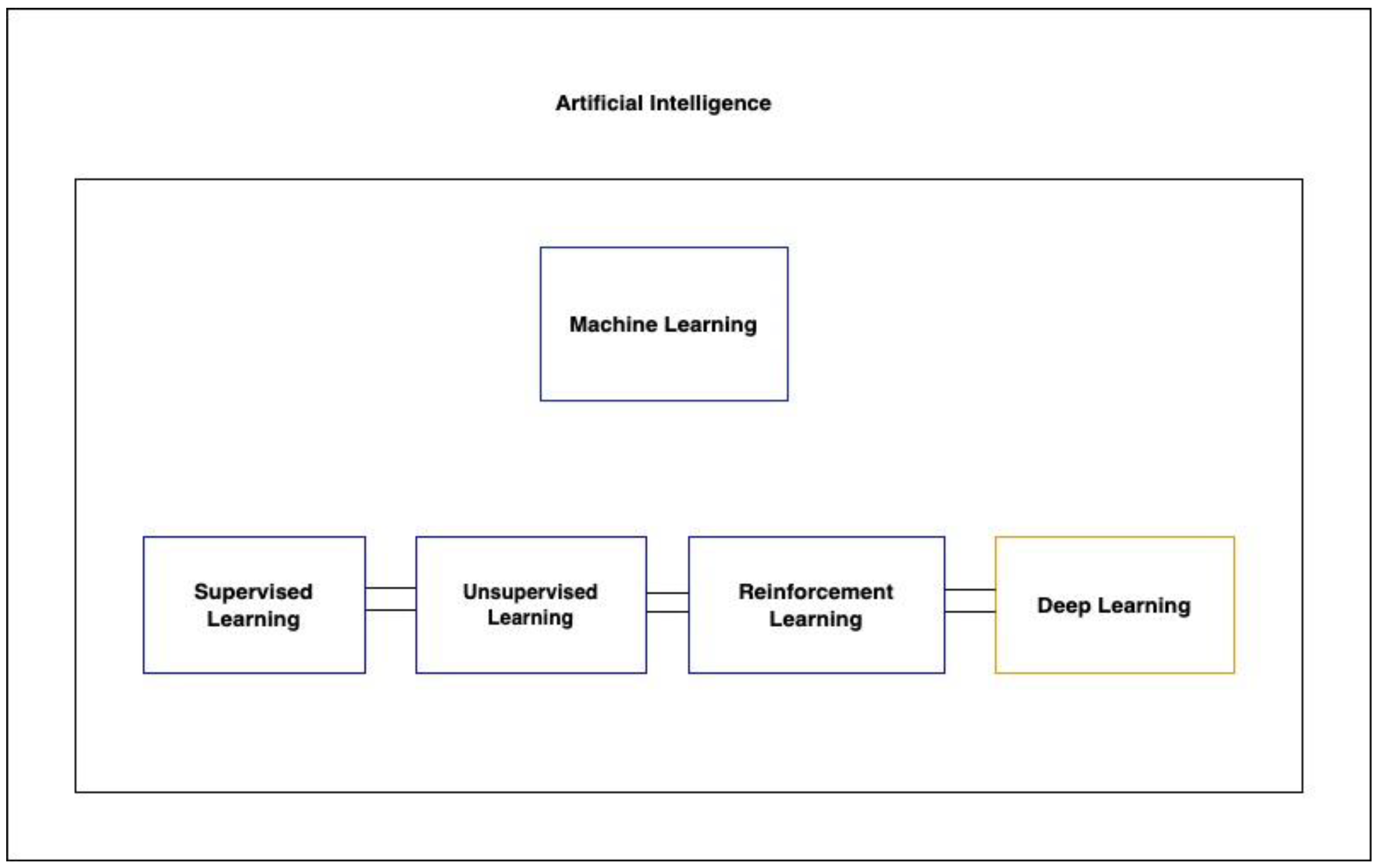
There are three primary types of machine learning algorithms: supervised, unsupervised, and reinforcement learning (Figure 1). Supervised learning operates on labeled datasets, while unsupervised learning, which is particularly useful for discovering hidden structures in data, does not require labels. This makes unsupervised learning beneficial, as it sidesteps the often laborious and costly process of data annotation [57]. A specialized subset of machine learning, deep learning, leverages multi-layered artificial neural networks to understand intricate patterns in vast amounts of data.

Figure 1.
Showcases the hierarchy of artificial intelligence techniques, highlighting the relationships between AI, machine learning, its types, and deep learning.
In the context of TBI research, various algorithms have been employed, such as support vector machines (SVMs), artificial neural networks (ANNs), multilayer perceptrons (MPNs), and more. The choice of algorithm often depends on the nature of the data and the desired outcome. While selecting the right algorithm is crucial, choosing pertinent and predictive variables is equally, if not more, vital [10,11,12,13,14,15,16,17,18,19,20,21,22,23,24,25,26,27,28,29,30,31,32,33,34,35,36,37,40,41,42,43,44,46,47,48,49,50,51,52,53,54,55,58,59]. Typically, the performance of machine learning models in TBI research is assessed using metrics like sensitivity, specificity, accuracy, and area under the receiver-operating curve (AUROC).
3. Identifying mTBI Using Functional Brain Activity
The difficulty of detecting mTBI through imaging alone has given rise to a substantial body of literature exploring machine learning as a tool for diagnosis, particularly given mTBI’s association with dysregulated neural network functioning [60].
Vergara’s work stands out in this domain. In 2017, Vergara employed dynamic functional network connectivity (dFNC) using fMRI data to differentiate mTBI patients from controls. The ground truth for mTBI diagnosis was based on criteria from the American Congress of Rehabilitation Medicine, which included a Glasgow Coma Scale (GCS) between 13 and 15. They identified four separate dFNC states that a patient could occupy during the five-minute fMRI scan period. Using a linear SVM, participants were classified within each dFNC state into mTBI patients and healthy controls. They were able to achieve a 92% AUROC among 48 patients when classifying using the optimal dFNC state and observed increased dFNC in the cerebellum compared to sensorimotor networks [61]. Another study by the same team in 2016 used resting-state functional network connectivity (rsFNC) from fMRI and fractional anisotropy from diffusion MRI (dMRI), attaining accuracies of 84.1% and 75.5%, respectively, with an SVM model. Increased rsFNC was noted in specific brain regions. Static connectivity, however, as opposed to dFNC, is not able to take into account the dynamic properties of the brain [62]. Later, using SVMs trained with dNFC data on a cohort of 50 mTBI patients, they reported an AUROC of 73% for mTBI prediction [63].
Other researchers have also leveraged fMRI. Luo (2021) used SVMs on resting-state fMRI (rs-fMRI) data to distinguish mTBI from controls among 48 patients, achieving notable diagnostic metrics and focusing on parameters like amplitude of low-frequency fluctuation, degree centrality, and voxel-mirrored homotopic connectivity. By combining multiple such parameters, they were able to distinguish patients with mTBI from controls with an AUROC of 77.8% and accuracy of 81.11% [64]. Fan (2021) and Rangaprakash (2017, 2018) made similar attempts with rs-fMRI and SVM models, yielding accuracies of 0.74 and up to 0.84, respectively [63,65,66].
Vedaei et al. concentrated on chronic mTBI, emphasizing its diagnostic significance for guiding treatment. Incorporating rs-fMRI with various metrics in their machine learning approach, they reported AUCs ranging from 80.42 to 93.33% in a study with 100 participants [67].
4. Detecting Axonal Injury with Machine Learning
Several studies have utilized machine learning with diffusor tensor imaging (DTI) metrics to detect axonal injury, which appears to be a key determinant of clinical outcome. Hellyer (2013) and Fagerholm (2016) trained SVM models, focusing on variables like fractional anisotropy and radial diffusivity, to differentiate patients with microbleeds suggestive of axonal injury from controls, achieving sensitivities and specificities often above 0.90. Microbleeds are often considered a surrogate marker of traumatic axonal injury and are visible on gradient-echo and susceptibility-weighted imaging, but patients without microbleeds can still have axonal injury. Both studies extended their initial analyses on microbleed patients to the larger group of non-microbleed patients to show generalizability in predicting cognitive function in the larger group of patients when compared to neuropsychological testing [68,69]. Mitra (2016) utilized RF classifiers focusing on fractional anisotropy features to identify diffuse axonal injury in mTBI patients. Among 325 TBI (mean GCS 13.1) and control participants, they achieved a mean classification accuracy of around 68% and sensitivity of 80% [70].
Stone (2016) employed RF classifiers to segment and quantify white-matter hyperintensities, which have shown prognostic value in TBI, on fluid-attenuated inversion recovery (FLAIR) MRI sequences. They used an RF framework to segment images from 24 patients, achieving an accuracy of 0.68 [70,71]. Other studies by Bai (2020), Cai (2018), Minaee (2017), and Senyukova (2011) employed SVM models with DTI metrics, yielding accuracies ranging from 0.68 to 0.96 [72,73,74,75]. Abdelrahman (2022) combined SVM with principal component analysis (PCA) on DTI indices to classify 52 total TBI patients and controls, achieving an accuracy of 90.5% and AUC of 0.93 [76].
Machine learning’s potential extends to the clinical management of TBI patients with diffuse axonal injury (DAI). Mohamed (2022) employed a CNN to predict favorable or unfavorable outcomes using the Glasgow Outcome Scale (GOS) from MRIs of 38 patients who sustained moderate or severe TBI with MRI evidence of DAI, achieving a sensitivity of 0.997 and AUROC of 0.917 [77]. Bohyn (2022) applied the FDA-cleared machine learning software icobrain to calculate brain lesion volumes in 20 DAI patients, correlating white-matter volume changes with GOS [78]. Tjerkaski (2022) used an RF model with gradient-echo and susceptibility-weighted imaging to develop a novel MRI-based traumatic axonal injury (TAI) grading system to discern between favorable and unfavorable outcomes, achieving an AUC of 0.72 [39].
5. Predicting TBI with CT
CT remains a primary imaging modality for suspected TBI. The urgency of detecting lesions has spurred interest in machine learning to enhance and expedite diagnosis. Automated image analysis through machine learning can streamline the process, reducing the time and potential variability introduced by manual reviews.
Recent studies have showcased the potential of convolutional neural networks (CNNs) in this domain. Monteiro (2020) used CNNs to identify and quantify various types of intracranial hemorrhages, achieving sensitivity and specificity up to 0.8 and 0.9, respectively, for small lesions [79]. Keshavamurthy (2017) and Salehinejad (2021) trained SVM models and generalizable machine learning models, respectively, both demonstrating high accuracies and sensitivities in detecting characteristic TBI lesions on CT [80,81].
Midline shift (MLS) in CT due to space-occupying lesions (typically hematomas from intracranial hemorrhage in TBI patients) is a prognostic factor in TBI. Studies indicate that TBI patients with MLS less than 10 mm have notably better outcomes than those with more significant shifts [82]. Machine learning’s automated MLS measurement can minimize observer variability. Both Nag (2021) and Yan (2022) employed CNNs for MLS estimation, achieving commendable accuracies greater than 85% and consistency across different types of intracranial hemorrhages when compared to hand-drawn MLSs by clinicians across a range of MLS values from a 2 mm cutoff value to greater than 10 mm [83,84].
Predicting long-term outcomes post-TBI can guide critical clinical decisions. Pease (2022) combined a CNN model analyzing CT data with a clinical model to forecast 6-month outcomes in severe TBI. This fusion model outperformed the standard IMPACT model when tested on their internal dataset and even surpassed the predictions of three neurosurgeons [85].
6. Detecting and Quantifying Subdural Hematomas with Machine Learning
Machine learning aids in the segmentation of pathology from normal structures, and in the context of TBI, it offers potential in delineating subdural hematomas (SDHs). Accurate segmentation enables both detection and volumetric analysis. While manual volumetric evaluation can be labor-intensive and is often omitted in clinical practice, machine learning provides a time-efficient alternative. This is vital because SDH volume is crucial for prognosis and surgical intervention considerations and as a predictor of post-operative recurrence [86,87].
Recent endeavors have demonstrated the capabilities of machine learning in this area. Farzaneh (2020) utilized a random forest model to detect and classify the severity of acute SDHs, achieving a sensitivity of 0.99 and specificity of 0.92 [88]. Chen (2022) employed a CNN for volumetric assessment, with the results closely mirroring manual segmentation with an AUROC of 0.83 [89]. Another CNN model designed for comprehensive SDH evaluation, including thickness, volume, and midline shift, showcased a sensitivity of 91.4% and specificity of 96.4% [90].
Chronic SDHs (cSDHs) present unique challenges due to their varied densities and resemblance to brain parenchyma in certain phases [91]. Kellogg (2021) trained CNN models for both pre- and post-operative cSDHs, achieving a DICE score of 0.83 in predicting cSDH volumes [92]. Moreover, insights from a study by Kung et al. highlight the capabilities of machine learning in predicting post-operative recurrence of SDHs by analyzing specific pathological features [93].
7. Clinical Applications of ML in Emergency Radiology: Achievements and Challenges
The true potential of machine learning (ML) techniques in TBI diagnosis and prognosis hinges on their demonstrated clinical utility. This encompasses tangible benefits for both patients and clinicians and requires addressing the barriers to its clinical implementation. Clinical utility is multifaceted, aiming to reduce morbidity, mortality, and costs through prompt diagnosis and intervention. It also seeks to optimize outcomes via enhanced diagnostic accuracy, reliable prognostication, and personalized treatment.
Emergency radiology’s current research landscape is vibrant, concentrating on worklist prioritization and automated pathology detection, which are particularly pertinent for conditions like TBI where timely diagnosis is important [94]. Companies such as Icometrix and Cortechs.ai have pioneered FDA-approved machine learning applications for TBI. For instance, Icometrix’s icobrain tbi for CT automates the detection and quantification of intracranial lesions, with correlations closely mirroring expert assessments [95]. Their MRI-focused counterpart, icobrain tbi for MRI, identifies traumatic axonal injuries and correlates lesion load with functional outcomes and brain volume changes [78]. Considering the challenges of assessing axonal injury using traditional imaging [96], machine learning provides a valuable augmentation.
Evidence suggests that patients with chronic mild-to-moderate TBI exhibit brain structural abnormalities, specifically, atypical hypertrophy in certain regions, believed to arise from compensatory mechanisms after ipsilateral cerebral white-matter injury, leading to the enlargement of contralateral brain regions [97]. Cortechs.ai’s solutions, NeuroQuant and NeuroGage, employ machine learning for quantitative MRI evaluation, providing objective assessments of brain anomalies [98].
8. Research Frontiers: Expanding the Role of ML in TBI Diagnosis and Prognosis
Current ML research on TBI integrates diverse data points, from patient demographics to radiologist-annotated imaging. However, there is a need to utilize image features independently when assessing TBI severity and predicting outcomes. Integrating ML-assessed imaging features with other clinical data can lead to a comprehensive patient profile, potentially improving diagnosis and tailoring treatments.
Khalili et al. (2023) used multiple ML and DL methods using patient demographics, laboratory data, and CT data to predict in-hospital and six-month outcomes, achieving around 82% accuracy for long-term survival. The status of the cisterns on CT was among the most reliable features for predicting in-hospital mortality, demonstrating the utility of incorporating independent imaging features into these models [91]. Further research is warranted in areas like chronic subdural hematomas (cSDHs) [92,93] and the application of structural MRI for TBI detection [99].
Emerging areas of interest include advanced imaging techniques like neurite orientation dispersion and density imaging [43], combined with serum biomarkers associated with TBI [5,7,72]. Predicting long-term outcomes for conditions like mTBI remains a challenge.
The application of ML research to clinical practice requires not only algorithm development but also validation in varied clinical environments [100]. For example, the CNN model developed by Pease et al. (2022) outperformed the IMPACT model in six-month-outcome prediction when tested on internal data but showed no improvement during external validation [85]. Ensuring these models’ robustness calls for high-quality, diverse datasets.
9. Ethical and Legal Crossroads in AI-Powered TBI Diagnosis
As the integration of AI and machine learning in TBI care continues to evolve, it encounters significant ethical, legal, and practical hurdles that could impede its progress. The use of vast amounts of sensitive medical data to fuel these models necessitates stringent protection measures and complete transparency with patients regarding the usage of their data.
Complicating matters further is the question of responsibility in the event of inaccurate diagnoses made by AI systems. As AI assumes a larger role in diagnostic processes, it becomes imperative to establish clear frameworks that delineate responsibility among healthcare providers, AI developers, and stakeholders. Another critical issue is potential bias within algorithms, which could lead to unfair disadvantages and poorer outcomes for certain patient groups. Continuous monitoring and refinement of these AI models are crucial to ensure equitable and accurate outcomes for all patients.
Moreover, there is a significant translational gap between the development and clinical implementation of AI in radiology, highlighted by regulatory challenges, data privacy concerns, issues around informed consent, intellectual property disputes, and the implications of AI-driven decisions in clinical settings [101]. This gap is further widened by inconsistencies in health records, imaging equipment, and protocols across different institutions [102,103]. Additionally, clinician skepticism toward AI, uncertainties in patient disclosure, and the unpredictable nature of machine learning prognostication also pose challenges [104].
Finally, the legal frameworks governing the medical use of AI must evolve in tandem with technological advancements. Existing medical regulations need to adapt to include AI applications for patient safety and the effectiveness of care. A collaborative approach involving ethicists, legal experts, clinicians, and technologists is essential to navigate these complexities and ensure the ethical integration of AI in TBI diagnosis and management.
Addressing these ethical, legal, and practical challenges is fundamental for the responsible and successful integration of AI in TBI care. As we approach this new frontier, careful consideration and collaboration are key to ensuring that the promise of AI translates into real, tangible improvements for patients affected by TBI. Notably, while conditions like intracranial hemorrhage may benefit from expedited diagnosis through AI, the tangible benefits for other conditions still require extensive research and validation.
10. Technical Hurdles
Technical limitations pose real-world challenges that need careful navigation. One major hurdle lies in the vast data required by these algorithms. Accurate predictions require extensive, diverse datasets encompassing the spectrum of TBI types, severities, and patient demographics. Gathering such data faces obstacles like limited access, logistical constraints, and privacy concerns. Moreover, meticulously annotating these data in supervised machine learning approaches is expensive and time-consuming.
Additionally, there exists the issue of overfitting, where models excel on training data but stumble on new cases. TBI’s inherent heterogeneity can lead algorithms to pick up on noise rather than meaningful patterns. Counteracting this involves techniques like cross-validation and regularization, but finding the right balance between model complexity and generalizability requires deliberate planning and careful tweaking.
Integrating AI into existing healthcare IT systems throws another wrench in the gears. Upgrading systems to accommodate these new tools and training clinicians and staff to use them can be costly and disruptive. Ensuring user-friendliness and seamless integration into clinical workflows will be important for widespread AI adoption and effectiveness.
Finally, data quality and standardization matter. Inconsistencies in imaging techniques and protocols can skew data, leading to biased or inaccurate models. Standardized data collection and preprocessing methods are essential for building robust and reliable ML applications in TBI.
11. Traditional Methods and the Rise of AI
CT is currently the standard diagnostic method for TBI. Unfortunately, it has a relatively poor ability for prognostication and is not sensitive to less severe cases of TBI [97,98]. The potential of ML in TBI extends from diagnosis to a comprehensive spectrum of patient care, encompassing prognostication, personalized treatment plans, recovery monitoring, and rehabilitation strategies.
Personalized treatment plans represent a significant shift toward precision medicine in TBI care, especially if supported by ML analysis of genomic data [105]. By leveraging genetic profiles, ML models can potentially predict individual susceptibility to TBI and response to specific treatments and assess long-term risks. This approach moves beyond the traditional one-size-fits-all methodology and can allow for treatments tailored to the unique genetic makeup of each patient.
The integration of wearable technology in TBI management is another promising avenue. These devices enable ML algorithms to monitor recovery progress, identify potential complications, and suggest timely interventions through continuous data collection.
In the realm of neuroimaging, ML is set to enhance the interpretation of traditional and emerging imaging modalities. Advanced imaging biomarkers, especially when analyzed using ML algorithms, are expected to provide information on the severity and prognosis of TBI [106]. This improved understanding will aid in formulating more accurate prognoses and tailoring rehabilitation protocols.
12. Cost Considerations
The incorporation of ML in TBI stands as not only a technological advancement but also a prudent long-term investment from a financial standpoint. The initial costs of ML, including the procurement of specialized software, the establishment of the necessary hardware infrastructure, and the imperative training of personnel, undoubtedly exceed those of traditional methods. Yet, when considering the broader financial picture, ML’s integration into TBI management may prove economically beneficial over the long term.
The principal advantage of ML resides in its improved efficiency in both diagnostic assistance and workflow improvement [100]. The potential future improvement in diagnostic times, workflow optimization, and treatment decision support will likely enhance patient throughput, reduce waiting times, and improve departmental efficiency. This is especially important in light of the significant shortage of radiologists. Such efficiency can lead to significant cost savings for healthcare systems. Additionally, information on prognostication can decrease ICU and overall hospital stay and inform when informed end-of-life decisions need to be considered, particularly in cases when ML algorithms support a minimal meaningful chance of patient recovery.
Traditional TBI management methods, though initially less expensive, accrue substantial costs over time. Repeated imaging, extended hospital stays, and frequent clinical assessments can collectively lead to higher overall healthcare expenditures. Importantly, ML’s ability to accurately predict complications enables healthcare providers to strategically allocate resources, thus avoiding the pitfalls of both the under- and overutilization of medical services and contributing to cost containment.
Beyond the financial aspects, ML promises to elevate the quality of TBI care. Its capacity for accurate and prompt diagnoses can inform more effective treatment plans, which may lead to enhanced patient outcomes and a decrease in long-term disability.
13. Global Perspective and Accessibility Issues
The global landscape of ML in TBI diagnosis and treatment is potentially marked by significant disparities in access and implementation. These disparities are primarily driven by differences in economic resources, healthcare infrastructure, and technical expertise across various regions. In high-income countries, the integration of ML in TBI care will likely be more advanced, as they benefit from robust healthcare systems, substantial funding, and access to the latest technologies. These nations are, therefore, expected to lead in developing and applying ML algorithms for TBI diagnosis and predictive analytics and creating personalized treatment approaches.
Conversely, low- and middle-income countries (LMICs) encounter substantial barriers to adopting ML technologies for TBI care. Challenges such as limited financial resources, inadequate healthcare infrastructure, and a dearth of technical expertise are prevalent. The high costs associated with ML technologies, including hardware, software, and maintenance, further exacerbate the issue and will likely make some of these advancements unaffordable for healthcare systems in these regions. Additionally, the effectiveness of ML applications is contingent on the availability and quality of data, which are often lacking in LMICs, hindering the development and accuracy of ML models.
It is important to be proactive in addressing these issues, especially considering that trauma is the leading cause of death worldwide [101]. To bridge these gaps, several strategies can be employed:
- (1).
- Collaborative International Research and Development: Promoting collaborative research efforts and technological exchanges between high-income countries and LMICs can aid in developing affordable and scalable ML solutions that are adaptable to various healthcare settings. Sharing data across nations will also build more robust and generalizable models.
- (2).
- Capacity Building: Investing in educational and training programs within LMICs is important for cultivating local expertise in ML. This initiative should focus on training healthcare professionals, data scientists, and technical staff to effectively manage and utilize ML systems.
- (3).
- Development of Open-Source and Low-Cost Tools: Encouraging the creation of open-source ML platforms and economical diagnostic tools can increase accessibility in resource-limited settings.
- (4).
- Standardization of Data and Protocols: Implementing standardized protocols for data collection, sharing, and processing can improve the quality and accessibility of data worldwide, which are essential for the development and implementation of effective ML models.
- (5).
- Policy and Funding Support: The role of governments and international organizations is pivotal in providing policy and funding support for integrating ML into healthcare systems, particularly in LMICs. This support could include grants, subsidies, and incentives for adopting ML technologies.
14. Conclusions
The exploration of ML in the diagnosis, prognosis, and clinical management of TBI reveals a landscape rich with potential yet fraught with challenges. As we advance in our understanding and application of ML in this domain, it becomes increasingly clear that this technology holds the key to significant improvements in patient care, particularly in the precision and efficiency of diagnosis and treatment strategies.
Despite the promise it holds, the integration of ML into TBI care is not without its hurdles. Ethical considerations, data privacy concerns, clinician trust, and the interoperability of health systems present substantial obstacles that must be overcome with care and diligence. The gap between theoretical research and clinical application further underscores the need for continued exploration and validation of these technologies in real-world settings.
Moreover, the discrepancies in access to ML technologies across different regions highlight a critical need for global collaboration, capacity building, and the development of cost-effective, scalable solutions that are accessible to all healthcare systems, regardless of economic status. Addressing these disparities is a matter not only of technological advancement but also of ethical responsibility to ensure that the benefits of ML in TBI care are available globally.
In conclusion, the journey of integrating ML into TBI diagnosis and treatment is an ongoing process that is marked by both remarkable achievements and significant challenges. The future of TBI care as elevated by ML promises enhanced diagnostic accuracy, improved patient outcomes, and more efficient healthcare delivery. However, realizing this future requires a concerted effort from researchers, clinicians, policymakers, and technologists to bridge the gap between innovation and practice and address the ethical, legal, and practical challenges inherent in this rapidly evolving field. By continuing to collaborate and innovate, we can ensure that the potential of ML in TBI care is fully realized and bring forth a new era in healthcare where precision, efficiency, and accessibility are the cornerstones of patient management.
Author Contributions
Conceptualization, K.P. and B.L.-W.; writing—original draft preparation, K.P., J.T., A.R., S.M.S.R., M.M. and A.P.; writing—review and editing, K.P. and B.L.-W.; supervision, B.L.-W.; project administration, K.P. and B.L.-W. All authors have read and agreed to the published version of the manuscript.
Funding
This research received no external funding.
Institutional Review Board Statement
Not applicable.
Informed Consent Statement
Not applicable.
Data Availability Statement
Data are contained within the article.
Conflicts of Interest
The authors declare no conflicts of interest.
References
- Fatuki, T.A.; Zvonarev, V.; Rodas, A.W. Prevention of Traumatic Brain Injury in the United States: Significance, New Findings, and Practical Applications. Cureus 2020, 12, e11225. [Google Scholar] [CrossRef]
- Rodríguez-Triviño, C.Y.; Torres Castro, I.; Dueñas, Z. Hypochloremia in Patients with Severe Traumatic Brain Injury: A Possible Risk Factor for Increased Mortality. World Neurosurg. 2019, 124, e783–e788. [Google Scholar] [CrossRef]
- McMahon, P.J.; Hricik, A.; Yue, J.K.; Puccio, A.M.; Inoue, T.; Lingsma, H.F.; Beers, S.R.; Gordon, W.A.; Valadka, A.B.; Manley, G.T.; et al. Symptomatology and Functional Outcome in Mild Traumatic Brain Injury: Results from the Prospective TRACK-TBI Study. J. Neurotrauma 2014, 31, 26–33. [Google Scholar] [CrossRef]
- Keret, A.; Bennett-Back, O.; Rosenthal, G.; Gilboa, T.; Shweiki, M.; Shoshan, Y.; Benifla, M. Posttraumatic epilepsy: Long-term follow-up of children with mild traumatic brain injury. J. Neurosurg. Pediatr. PED 2017, 20, 64–70. [Google Scholar] [CrossRef]
- Pierre, K.; Dyson, K.; Dagra, A.; Williams, E.; Porche, K.; Lucke-Wold, B. Chronic Traumatic Encephalopathy: Update on Current Clinical Diagnosis and Management. Biomedicines 2021, 9, 415. [Google Scholar] [CrossRef]
- Gardner, R.C.; Yaffe, K. Epidemiology of mild traumatic brain injury and neurodegenerative disease. Mol. Cell. Neurosci. 2015, 66, 75–80. [Google Scholar] [CrossRef]
- Wang, K.K.; Yang, Z.; Zhu, T.; Shi, Y.; Rubenstein, R.; Tyndall, J.A.; Manley, G.T. An update on diagnostic and prognostic biomarkers for traumatic brain injury. Expert. Rev. Mol. Diagn. 2018, 18, 165–180. [Google Scholar] [CrossRef]
- Shenton, M.E.; Hamoda, H.M.; Schneiderman, J.S.; Bouix, S.; Pasternak, O.; Rathi, Y.; Vu, M.A.; Purohit, M.P.; Helmer, K.; Koerte, I.; et al. A review of magnetic resonance imaging and diffusion tensor imaging findings in mild traumatic brain injury. Brain Imaging Behav. 2012, 6, 137–192. [Google Scholar] [CrossRef]
- Wu, X.; Sun, Y.; Xu, X.; Steyerberg, E.W.; Helmrich, I.R.A.R.; Lecky, F.; Guo, J.; Li, X.; Feng, J.; Mao, Q.; et al. Mortality Prediction in Severe Traumatic Brain Injury Using Traditional and Machine Learning Algorithms. J. Neurotrauma 2023, 40, 1366–1375. [Google Scholar] [CrossRef]
- Abujaber, A.; Fadlalla, A.; Gammoh, D.; Abdelrahman, H.; Mollazehi, M.; El-Menyar, A. Prediction of in-hospital mortality in patients on mechanical ventilation post traumatic brain injury: Machine learning approach. BMC Med. Inform. Decis. Mak. 2020, 20, 336. [Google Scholar] [CrossRef]
- Tunthanathip, T.; Oearsakul, T. Application of machine learning to predict the outcome of pediatric traumatic brain injury. Chin. J. Traumatol. 2021, 24, 350–355. [Google Scholar] [CrossRef]
- Abujaber, A.; Fadlalla, A.; Gammoh, D.; Abdelrahman, H.; Mollazehi, M.; El-Menyar, A. Prediction of in-hospital mortality in patients with post traumatic brain injury using National Trauma Registry and Machine Learning Approach. Scand. J. Trauma. Resusc. Emerg. Med. 2020, 28, 44. [Google Scholar] [CrossRef]
- Hsu, S.-D.; Chao, E.; Chen, S.-J.; Hueng, D.-Y.; Lan, H.-Y.; Chiang, H.-H. Machine Learning Algorithms to Predict In-Hospital Mortality in Patients with Traumatic Brain Injury. J. Pers. Med. 2021, 11, 1144. [Google Scholar] [CrossRef]
- Folweiler, K.A.; Sandsmark, D.K.; Diaz-Arrastia, R.; Cohen, A.S.; Masino, A.J. Unsupervised Machine Learning Reveals Novel Traumatic Brain Injury Patient Phenotypes with Distinct Acute Injury Profiles and Long-Term Outcomes. J. Neurotrauma 2020, 37, 1431–1444. [Google Scholar] [CrossRef]
- Hale, A.T.; Stonko, D.P.; Brown, A.; Lim, J.; Voce, D.J.; Gannon, S.R.; Le, T.M.; Shannon, C.N. Machine-learning analysis outperforms conventional statistical models and CT classification systems in predicting 6-month outcomes in pediatric patients sustaining traumatic brain injury. Neurosurg. Focus FOC 2018, 45, E2. [Google Scholar] [CrossRef]
- Hernandes Rocha, T.A.; Elahi, C.; Cristina da Silva, N.; Sakita, F.M.; Fuller, A.; Mmbaga, B.T.; Green, E.P.; Haglund, M.M.; Staton, C.A.; Nickenig Vissoci, J.R. A traumatic brain injury prognostic model to support in-hospital triage in a low-income country: A machine learning–based approach. J. Neurosurg. JNS 2020, 132, 1961–1969. [Google Scholar] [CrossRef]
- Vishwanath, M.; Jafarlou, S.; Shin, I.; Lim, M.M.; Dutt, N.; Rahmani, A.M.; Cao, H. Investigation of Machine Learning Approaches for Traumatic Brain Injury Classification via EEG Assessment in Mice. Sensors 2020, 20, 2027. [Google Scholar] [CrossRef]
- Farzaneh, N.; Williamson, C.A.; Gryak, J.; Najarian, K. A hierarchical expert-guided machine learning framework for clinical decision support systems: An application to traumatic brain injury prognostication. NPJ Digit. Med. 2021, 4, 78. [Google Scholar] [CrossRef]
- Raj, R.; Wennervirta, J.M.; Tjerkaski, J.; Luoto, T.M.; Posti, J.P.; Nelson, D.W.; Takala, R.; Bendel, S.; Thelin, E.P.; Luostarinen, T.; et al. Dynamic prediction of mortality after traumatic brain injury using a machine learning algorithm. NPJ Digit. Med. 2022, 5, 96. [Google Scholar] [CrossRef]
- Warman, P.I.; Seas, A.; Satyadev, N.; Adil, S.M.; Kolls, B.J.; Haglund, M.M.; Dunn, T.W.; Fuller, A.T. Machine Learning for Predicting In-Hospital Mortality After Traumatic Brain Injury in Both High-Income and Low- and Middle-Income Countries. Neurosurgery 2022, 90, 605–612. [Google Scholar] [CrossRef]
- Bruschetta, R.; Tartarisco, G.; Lucca, L.F.; Leto, E.; Ursino, M.; Tonin, P.; Pioggia, G.; Cerasa, A. Predicting Outcome of Traumatic Brain Injury: Is Machine Learning the Best Way? Biomedicines 2022, 10, 686. [Google Scholar] [CrossRef]
- Satyadev, N.; Warman, P.I.; Seas, A.; Kolls, B.J.; Haglund, M.M.; Fuller, A.T.; Dunn, T.W. Machine Learning for Predicting Discharge Disposition After Traumatic Brain Injury. Neurosurgery 2022, 90, 768–774. [Google Scholar] [CrossRef]
- Lang, E.; Neuschwander, A.; Favé, G.; Abback, P.-S.; Esnault, P.; Geeraerts, T.; Harrois, A.; Hanouz, J.-L.; Kipnis, E.; Leone, M.; et al. Clinical decision support for severe trauma patients: Machine learning based definition of a bundle of care for hemorrhagic shock and traumatic brain injury. J. Trauma Acute Care Surg. 2022, 92, 135–143. [Google Scholar] [CrossRef]
- Mohd Noor, N.S.E.; Ibrahim, H. Predicting Outcomes in Patients with Traumatic Brain Injury Using Machine Learning Models. In Proceedings of the Intelligent Manufacturing and Mechatronics, Melaka, Malaysia, 8 July 2019; Springer: Singapore, 2020; pp. 12–20. [Google Scholar]
- Radabaugh, H.L.; Bonnell, J.; Dietrich, W.D.; Bramlett, H.M.; Schwartz, O.; Sarkar, D. Development and Evaluation of Machine Learning Models for Recovery Prediction after Treatment for Traumatic Brain Injury. In Proceedings of the 2020 42nd Annual International Conference of the IEEE Engineering in Medicine & Biology Society (EMBC), Montreal, QC, Canada, 20–24 July 2020; pp. 2416–2420. [Google Scholar]
- Lee, S.H.; Lee, C.H.; Hwang, S.H.; Kang, D.H. A Machine Learning–Based Prognostic Model for the Prediction of Early Death After Traumatic Brain Injury: Comparison with the Corticosteroid Randomization After Significant Head Injury (CRASH) Model. World Neurosurg. 2022, 166, e125–e134. [Google Scholar] [CrossRef]
- Guimarães, K.A.A.; de Amorim, R.L.O.; Costa, M.G.F.; Costa Filho, C.F.F. Predicting early traumatic brain injury mortality with 1D convolutional neural networks and conventional machine learning techniques. Inform. Med. Unlocked 2022, 31, 100984. [Google Scholar] [CrossRef]
- Amorim, R.L.; Oliveira, L.M.; Malbouisson, L.M.; Nagumo, M.M.; Simoes, M.; Miranda, L.; Bor-Seng-Shu, E.; Beer-Furlan, A.; De Andrade, A.F.; Rubiano, A.M.; et al. Prediction of Early TBI Mortality Using a Machine Learning Approach in a LMIC Population. Front. Neurol. 2019, 10, 1366. [Google Scholar] [CrossRef]
- Signorini, D.F.; Andrews, P.J.; Jones, P.A.; Wardlaw, J.M.; Miller, J.D. Predicting survival using simple clinical variables: A case study in traumatic brain injury. J. Neurol. Neurosurg. Psychiatry 1999, 66, 20–25. [Google Scholar] [CrossRef]
- Pang, B.C.; Kuralmani, V.; Joshi, R.; Hongli, Y.; Lee, K.K.; Ang, B.T.; Li, J.; Leong, T.Y.; Ng, I. Hybrid Outcome Prediction Model for Severe Traumatic Brain Injury. J. Neurotrauma 2007, 24, 136–146. [Google Scholar] [CrossRef]
- Chong, S.L.; Liu, N.; Barbier, S.; Ong, M.E. Predictive modeling in pediatric traumatic brain injury using machine learning. BMC Med. Res. Methodol. 2015, 15, 22. [Google Scholar] [CrossRef]
- Adil, S.M.; Elahi, C.; Gramer, R.; Spears, C.A.; Fuller, A.T.; Haglund, M.M.; Dunn, T.W. Predicting the Individual Treatment Effect of Neurosurgery for Patients with Traumatic Brain Injury in the Low-Resource Setting: A Machine Learning Approach in Uganda. J. Neurotrauma 2020, 38, 928–939. [Google Scholar] [CrossRef] [PubMed]
- Daley, M.; Cameron, S.; Ganesan, S.L.; Patel, M.A.; Stewart, T.C.; Miller, M.R.; Alharfi, I.; Fraser, D.D. Pediatric severe traumatic brain injury mortality prediction determined with machine learning-based modeling. Injury 2022, 53, 992–998. [Google Scholar] [CrossRef]
- Nourelahi, M.; Dadboud, F.; Khalili, H.; Niakan, A.; Parsaei, H. A machine learning model for predicting favorable outcome in severe traumatic brain injury patients after 6 months. Acute Crit. Care 2022, 37, 45–52. [Google Scholar] [CrossRef]
- Matsuo, K.; Aihara, H.; Nakai, T.; Morishita, A.; Tohma, Y.; Kohmura, E. Machine Learning to Predict In-Hospital Morbidity and Mortality after Traumatic Brain Injury. J. Neurotrauma 2020, 37, 202–210. [Google Scholar] [CrossRef]
- Feng, J.; Wang, Y.; Peng, J.; Sun, M.; Zeng, J.; Jiang, H. Comparison between logistic regression and machine learning algorithms on survival prediction of traumatic brain injuries. J. Crit. Care 2019, 54, 110–116. [Google Scholar] [CrossRef]
- Noor, N.S.E.M.; Ibrahim, H. Machine Learning Algorithms and Quantitative Electroencephalography Predictors for Outcome Prediction in Traumatic Brain Injury: A Systematic Review. IEEE Access 2020, 8, 102075–102092. [Google Scholar] [CrossRef]
- Palacios, E.M.; Owen, J.P.; Yuh, E.L.; Wang, M.B.; Vassar, M.J.; Ferguson, A.R.; Diaz-Arrastia, R.; Giacino, J.T.; Okonkwo, D.O.; Robertson, C.S.; et al. The evolution of white matter microstructural changes after mild traumatic brain injury: A longitudinal DTI and NODDI study. Sci. Adv. 2020, 6, eaaz6892. [Google Scholar] [CrossRef]
- Tjerkaski, J.; Nyström, H.; Raj, R.; Lindblad, C.; Bellander, B.-M.; Nelson, D.W.; Thelin, E.P. Extended Analysis of Axonal Injuries Detected Using Magnetic Resonance Imaging in Critically Ill Traumatic Brain Injury Patients. J. Neurotrauma 2022, 39, 58–66. [Google Scholar] [CrossRef]
- Chen, W.; Cockrell, C.; Ward, K.R.; Najarian, K. Intracranial pressure level prediction in traumatic brain injury by extracting features from multiple sources and using machine learning methods. In Proceedings of the 2010 IEEE International Conference on Bioinformatics and Biomedicine (BIBM), Hong Kong, China, 18–21 December 2010; pp. 510–515. [Google Scholar]
- Ye, G.; Balasubramanian, V.; Li, J.K.J.; Kaya, M. Machine Learning-Based Continuous Intracranial Pressure Prediction for Traumatic Injury Patients. IEEE J. Transl. Eng. Health Med. 2022, 10, 4901008. [Google Scholar] [CrossRef]
- Chen, W.; Cockrell, C.H.; Ward, K.; Najarian, K. Predictability of intracranial pressure level in traumatic brain injury: Features extraction, statistical analysis and machine learning-based evaluation. Int. J. Data Min. Bioinform. 2013, 8, 480–494. [Google Scholar] [CrossRef]
- Tunthanathip, T.; Duangsuwan, J.; Wattanakitrungroj, N.; Tongman, S.; Phuenpathom, N. Comparison of intracranial injury predictability between machine learning algorithms and the nomogram in pediatric traumatic brain injury. Neurosurg. Focus 2021, 51, E7. [Google Scholar] [CrossRef]
- Molaei, S.; Korley, F.K.; Soroushmehr, S.M.R.; Falk, H.; Sair, H.; Ward, K.; Najarían, K. A machine learning based approach for identifying traumatic brain injury patients for whom a head CT scan can be avoided. In Proceedings of the 2016 38th Annual International Conference of the IEEE Engineering in Medicine and Biology Society (EMBC), Orlando, FL, USA, 16–20 August 2016; pp. 2258–2261. [Google Scholar]
- Li, Y.-C.; Liu, L.; Chiu, W.-T.; Jian, W.-S. Neural network modeling for surgical decisions on traumatic brain injury patients. Int. J. Med. Inform. 2000, 57, 1–9. [Google Scholar] [CrossRef]
- Abujaber, A.; Fadlalla, A.; Gammoh, D.; Abdelrahman, H.; Mollazehi, M.; El-Menyar, A. Using trauma registry data to predict prolonged mechanical ventilation in patients with traumatic brain injury: Machine learning approach. PLoS ONE 2020, 15, e0235231. [Google Scholar] [CrossRef]
- Abujaber, A.; Fadlalla, A.; Gammoh, D.; Al-Thani, H.; El-Menyar, A. Machine Learning Model to Predict Ventilator Associated Pneumonia in patients with Traumatic Brain Injury: The C.5 Decision Tree Approach. Brain Inj. 2021, 35, 1095–1102. [Google Scholar] [CrossRef]
- Marincowitz, C.; Paton, L.; Lecky, F.; Tiffin, P. Predicting need for hospital admission in patients with traumatic brain injury or skull fractures identified on CT imaging: A machine learning approach. Emerg. Med. J. 2022, 39, 394–401. [Google Scholar] [CrossRef]
- Abujaber, A.; Fadlalla, A.; Nashwan, A.; El-Menyar, A.; Al-Thani, H. Predicting prolonged length of stay in patients with traumatic brain injury: A machine learning approach. Intell.-Based Med. 2022, 6, 100052. [Google Scholar] [CrossRef]
- Prichep, L.S.; Jacquin, A.; Filipenko, J.; Dastidar, S.G.; Zabele, S.; Vodencarević, A.; Rothman, N.S. Classification of traumatic brain injury severity using informed data reduction in a series of binary classifier algorithms. IEEE Trans. Neural Syst. Rehabil. Eng. 2012, 20, 806–822. [Google Scholar] [CrossRef]
- Yadav, K.; Sarioglu, E.; Choi, H.A.; Cartwright Iv, W.B.; Hinds, P.S.; Chamberlain, J.M. Automated Outcome Classification of Computed Tomography Imaging Reports for Pediatric Traumatic Brain Injury. Acad. Emerg. Med. 2016, 23, 171–178. [Google Scholar] [CrossRef]
- Ellethy, H.; Chandra, S.S.; Nasrallah, F.A. The detection of mild traumatic brain injury in paediatrics using artificial neural networks. Comput. Biol. Med. 2021, 135, 104614. [Google Scholar] [CrossRef]
- Dhillon, N.S.; Sutandi, A.; Vishwanath, M.; Lim, M.M.; Cao, H.; Si, D. A Raspberry Pi-Based Traumatic Brain Injury Detection System for Single-Channel Electroencephalogram. Sensors 2021, 21, 2779. [Google Scholar] [CrossRef]
- Peacock, W.F.; Van Meter, T.E.; Mirshahi, N.; Ferber, K.; Gerwien, R.; Rao, V.; Sair, H.I.; Diaz-Arrastia, R.; Korley, F.K. Derivation of a Three Biomarker Panel to Improve Diagnosis in Patients with Mild Traumatic Brain Injury. Front. Neurol. 2017, 8, 641. [Google Scholar] [CrossRef]
- Rau, C.S.; Kuo, P.J.; Chien, P.C.; Huang, C.Y.; Hsieh, H.Y.; Hsieh, C.H. Mortality prediction in patients with isolated moderate and severe traumatic brain injury using machine learning models. PLoS ONE 2018, 13, e0207192. [Google Scholar] [CrossRef]
- Erickson, B.J.; Korfiatis, P.; Akkus, Z.; Kline, T.L. Machine Learning for Medical Imaging. Radiographics 2017, 37, 505–515. [Google Scholar] [CrossRef]
- Wagner, M.W.; Namdar, K.; Biswas, A.; Monah, S.; Khalvati, F.; Ertl-Wagner, B.B. Radiomics, machine learning, and artificial intelligence-what the neuroradiologist needs to know. Neuroradiology 2021, 63, 1957–1967. [Google Scholar] [CrossRef]
- Steyerberg, E.W.; Mushkudiani, N.; Perel, P.; Butcher, I.; Lu, J.; McHugh, G.S.; Murray, G.D.; Marmarou, A.; Roberts, I.; Habbema, J.D.F.; et al. Predicting Outcome after Traumatic Brain Injury: Development and International Validation of Prognostic Scores Based on Admission Characteristics. PLoS Med. 2008, 5, e165. [Google Scholar] [CrossRef]
- Perel, P.; Arango, M.; Clayton, T.; Edwards, P.; Komolafe, E.; Poccock, S.; Roberts, I.; Shakur, H.; Steyerberg, E.; Yutthakasemsunt, S. Predicting outcome after traumatic brain injury: Practical prognostic models based on large cohort of international patients. BMJ 2008, 336, 425–429. [Google Scholar] [CrossRef]
- Safar, K.; Zhang, J.; Emami, Z.; Gharehgazlou, A.; Ibrahim, G.; Dunkley, B.T. Mild traumatic brain injury is associated with dysregulated neural network functioning in children and adolescents. Brain Commun. 2021, 3, fcab044. [Google Scholar] [CrossRef]
- Vergara, V.M.; Mayer, A.R.; Kiehl, K.A.; Calhoun, V.D. Dynamic functional network connectivity discriminates mild traumatic brain injury through machine learning. NeuroImage Clin. 2018, 19, 30–37. [Google Scholar] [CrossRef]
- Vergara, V.M.; Mayer, A.R.; Damaraju, E.; Kiehl, K.A.; Calhoun, V. Detection of Mild Traumatic Brain Injury by Machine Learning Classification Using Resting State Functional Network Connectivity and Fractional Anisotropy. J. Neurotrauma 2017, 34, 1045–1053. [Google Scholar] [CrossRef]
- Rangaprakash, D.; Dretsch, M.N.; Venkataraman, A.; Katz, J.S.; Denney, T.S., Jr.; Deshpande, G. Identifying disease foci from static and dynamic effective connectivity networks: Illustration in soldiers with trauma. Hum. Brain Mapp. 2018, 39, 264–287. [Google Scholar] [CrossRef]
- Luo, X.; Lin, D.; Xia, S.; Wang, D.; Weng, X.; Huang, W.; Ye, H. Machine Learning Classification of Mild Traumatic Brain Injury Using Whole-Brain Functional Activity: A Radiomics Analysis. Dis. Markers 2021, 2021, 3015238. [Google Scholar] [CrossRef]
- Fan, L.; Xu, H.; Su, J.; Qin, J.; Gao, K.; Ou, M.; Peng, S.; Shen, H.; Li, N. Discriminating mild traumatic brain injury using sparse dictionary learning of functional network dynamics. Brain Behav. 2021, 11, e2414. [Google Scholar] [CrossRef]
- Rangaprakash, D.; Deshpande, G.; Daniel, T.A.; Goodman, A.M.; Robinson, J.L.; Salibi, N.; Katz, J.S.; Denney, T.S., Jr.; Dretsch, M.N. Compromised hippocampus-striatum pathway as a potential imaging biomarker of mild-traumatic brain injury and posttraumatic stress disorder. Hum. Brain Mapp. 2017, 38, 2843–2864. [Google Scholar] [CrossRef]
- Vedaei, F.; Mashhadi, N.; Zabrecky, G.; Monti, D.; Navarreto, E.; Hriso, C.; Wintering, N.; Newberg, A.B.; Mohamed, F.B. Identification of chronic mild traumatic brain injury using resting state functional MRI and machine learning techniques. Front. Neurosci. 2022, 16, 1099560. [Google Scholar] [CrossRef]
- Hellyer, P.J.; Leech, R.; Ham, T.E.; Bonnelle, V.; Sharp, D.J. Individual prediction of white matter injury following traumatic brain injury. Ann. Neurol. 2013, 73, 489–499. [Google Scholar] [CrossRef]
- Fagerholm, E.D.; Hellyer, P.J.; Scott, G.; Leech, R.; Sharp, D.J. Disconnection of network hubs and cognitive impairment after traumatic brain injury. Brain 2015, 138 Pt 6, 1696–1709. [Google Scholar] [CrossRef]
- Mitra, J.; Shen, K.-k.; Ghose, S.; Bourgeat, P.; Fripp, J.; Salvado, O.; Pannek, K.; Taylor, D.J.; Mathias, J.L.; Rose, S. Statistical machine learning to identify traumatic brain injury (TBI) from structural disconnections of white matter networks. NeuroImage 2016, 129, 247–259. [Google Scholar] [CrossRef]
- Stone, J.R.; Wilde, E.A.; Taylor, B.A.; Tate, D.F.; Levin, H.; Bigler, E.D.; Scheibel, R.S.; Newsome, M.R.; Mayer, A.R.; Abildskov, T.; et al. Supervised learning technique for the automated identification of white matter hyperintensities in traumatic brain injury. Brain Inj. 2016, 30, 1458–1468. [Google Scholar] [CrossRef]
- Bai, L.; Bai, G.; Wang, S.; Yang, X.; Gan, S.; Jia, X.; Yin, B.; Yan, Z. Strategic white matter injury associated with long-term information processing speed deficits in mild traumatic brain injury. Hum. Brain Mapp. 2020, 41, 4431–4441. [Google Scholar] [CrossRef] [PubMed]
- Cai, Y.; Wu, S.; Zhao, W.; Li, Z.; Wu, Z.; Ji, S. Concussion classification via deep learning using whole-brain white matter fiber strains. PLoS ONE 2018, 13, e0197992. [Google Scholar] [CrossRef] [PubMed]
- Minaee, S.; Wang, Y.; Chung, S.; Wang, X.H.; Fieremans, E.; Flanagan, S.; Rath, J.F.; Lui, Y.W. A Machine Learning Approach For Identifying Patients with Mild Traumatic Brain Injury Using Diffusion MRI Modeling. arXiv 2017, arXiv:1708.09000. [Google Scholar]
- Senyukova, O.; Galanine, V.; Krylov, A.; Petraikin, A.; Akhadov, T.; Sidorin, S. Diffuse Axonal Injury Lesion Segmentation Using Contouring Algorithm. In Proceedings of the 21st International Conference on Computer Graphics and Vision, GraphiCon’2011-Conference Proceedings, Moscow, Russia, 26–30 September 2011. [Google Scholar]
- Abdelrahman, H.A.F.; Ubukata, S.; Ueda, K.; Fujimoto, G.; Oishi, N.; Aso, T.; Murai, T. Combining Multiple Indices of Diffusion Tensor Imaging Can Better Differentiate Patients with Traumatic Brain Injury from Healthy Subjects. Neuropsychiatr. Dis. Treat. 2022, 18, 1801–1814. [Google Scholar] [CrossRef]
- Mohamed, M.; Alamri, A.; Mohamed, M.; Khalid, N.; O’Halloran, P.; Staartjes, V.; Uff, C. Prognosticating outcome using magnetic resonance imaging in patients with moderate to severe traumatic brain injury: A machine learning approach. Brain Inj. 2022, 36, 353–358. [Google Scholar] [CrossRef]
- Bohyn, C.; Vyvere, T.V.; Keyzer, F.; Sima, D.M.; Demaerel, P. Morphometric evaluation of traumatic axonal injury and the correlation with post-traumatic cerebral atrophy and functional outcome. Neuroradiol. J. 2022, 35, 468–476. [Google Scholar] [CrossRef]
- Monteiro, M.; Newcombe, V.F.J.; Mathieu, F.; Adatia, K.; Kamnitsas, K.; Ferrante, E.; Das, T.; Whitehouse, D.; Rueckert, D.; Menon, D.K.; et al. Multiclass semantic segmentation and quantification of traumatic brain injury lesions on head CT using deep learning: An algorithm development and multicentre validation study. Lancet Digit. Health 2020, 2, e314–e322. [Google Scholar] [CrossRef]
- Keshavamurthy, K.N.; Leary, O.P.; Merck, L.H.; Kimia, B.B.; Collins, S.; Wright, D.W.; Allen, J.W.; Brock, J.F.; Merck, D. Machine learning algorithm for automatic detection of CT-identifiable hyperdense lesions associated with traumatic brain injury. In Proceedings of the Medical Imaging 2017: Computer-Aided Diagnosis, Orlando, FL, USA, 13–16 February 2017. [Google Scholar]
- Salehinejad, H.; Kitamura, J.; Ditkofsky, N.; Lin, A.; Bharatha, A.; Suthiphosuwan, S.; Lin, H.M.; Wilson, J.R.; Mamdani, M.; Colak, E. A real-world demonstration of machine learning generalizability in the detection of intracranial hemorrhage on head computerized tomography. Sci. Rep. 2021, 11, 17051. [Google Scholar] [CrossRef]
- Puffer, R.C.; Yue, J.K.; Mesley, M.; Billigen, J.B.; Sharpless, J.; Fetzick, A.L.; Puccio, A.; Diaz-Arrastia, R.; Okonkwo, D.O. Long-term outcome in traumatic brain injury patients with midline shift: A secondary analysis of the Phase 3 COBRIT clinical trial. J. Neurosurg. 2018, 131, 596–603. [Google Scholar] [CrossRef]
- Nag, M.K.; Gupta, A.; Hariharasudhan, A.S.; Sadhu, A.K.; Das, A.; Ghosh, N. Quantitative analysis of brain herniation from non-contrast CT images using deep learning. J. Neurosci. Methods 2021, 349, 109033. [Google Scholar] [CrossRef]
- Yan, J.L.; Chen, Y.L.; Chen, M.Y.; Chen, B.A.; Chang, J.X.; Kao, C.C.; Hsieh, M.C.; Peng, Y.T.; Huang, K.C.; Chen, P.Y. A Robust, Fully Automatic Detection Method and Calculation Technique of Midline Shift in Intracranial Hemorrhage and Its Clinical Application. Diagnostics 2022, 12, 693. [Google Scholar] [CrossRef] [PubMed]
- Pease, M.; Arefan, D.; Barber, J.; Yuh, E.; Puccio, A.; Hochberger, K.; Nwachuku, E.; Roy, S.; Casillo, S.; Temkin, N.; et al. Outcome Prediction in Patients with Severe Traumatic Brain Injury Using Deep Learning from Head CT Scans. Radiology 2022, 304, 385–394. [Google Scholar] [CrossRef] [PubMed]
- Stanišić, M.; Hald, J.; Rasmussen, I.A.; Pripp, A.H.; Ivanović, J.; Kolstad, F.; Sundseth, J.; Züchner, M.; Lindegaard, K.F. Volume and densities of chronic subdural haematoma obtained from CT imaging as predictors of postoperative recurrence: A prospective study of 107 operated patients. Acta Neurochir. 2013, 155, 323–333; discussion 333. [Google Scholar] [CrossRef] [PubMed]
- Bullock, M.R.; Chesnut, R.; Ghajar, J.; Gordon, D.; Hartl, R.; Newell, D.W.; Servadei, F.; Walters, B.C.; Wilberger, J.E. Surgical management of acute subdural hematomas. Neurosurgery 2006, 58, S16–S24; discussion Si-iv. [Google Scholar] [CrossRef] [PubMed]
- Farzaneh, N.; Williamson, C.A.; Jiang, C.; Srinivasan, A.; Bapuraj, J.R.; Gryak, J.; Najarian, K.; Soroushmehr, S.M.R. Automated Segmentation and Severity Analysis of Subdural Hematoma for Patients with Traumatic Brain Injuries. Diagnostics 2020, 10, 773. [Google Scholar] [CrossRef]
- Chen, D.; Bian, L.; He, H.Y.; Li, Y.D.; Ma, C.; Mao, L.G. Evaluation of Traumatic Subdural Hematoma Volume by Using Image Segmentation Assessment Based on Deep Learning. Comput. Math. Methods Med. 2022, 2022, 3830245. [Google Scholar] [CrossRef]
- Colasurdo, M.; Leibushor, N.; Robledo, A.; Vasandani, V.; Luna, Z.A.; Rao, A.S.; Garcia, R.; Srinivasan, V.M.; Sheth, S.A.; Avni, N.; et al. Automated detection and analysis of subdural hematomas using a machine learning algorithm. J. Neurosurg. 2023, 138, 1077–1084. [Google Scholar] [CrossRef] [PubMed]
- Khalili, H.; Rismani, M.; Nematollahi, M.A.; Masoudi, M.S.; Asadollahi, A.; Taheri, R.; Pourmontaseri, H.; Valibeygi, A.; Roshanzamir, M.; Alizadehsani, R.; et al. Prognosis prediction in traumatic brain injury patients using machine learning algorithms. Sci. Rep. 2023, 13, 960. [Google Scholar] [CrossRef] [PubMed]
- Kellogg, R.T.; Vargas, J.; Barros, G.; Sen, R.; Bass, D.; Mason, J.R.; Levitt, M. Segmentation of Chronic Subdural Hematomas Using 3D Convolutional Neural Networks. World Neurosurg. 2021, 148, e58–e65. [Google Scholar] [CrossRef]
- Kung, W.M.; Lin, M.S. CT-Based Quantitative Analysis for Pathological Features Associated With Postoperative Recurrence and Potential Application Upon Artificial Intelligence: A Narrative Review With a Focus on Chronic Subdural Hematomas. Mol. Imaging 2020, 19, 1536012120914773. [Google Scholar] [CrossRef] [PubMed]
- Cellina, M.; Cè, M.; Irmici, G.; Ascenti, V.; Caloro, E.; Bianchi, L.; Pellegrino, G.; D’Amico, N.; Papa, S.; Carrafiello, G. Artificial Intelligence in Emergency Radiology: Where Are We Going? Diagnostics 2022, 12, 3223. [Google Scholar] [CrossRef] [PubMed]
- Jain, S.; Vyvere, T.V.; Terzopoulos, V.; Sima, D.M.; Roura, E.; Maas, A.; Wilms, G.; Verheyden, J. Automatic Quantification of Computed Tomography Features in Acute Traumatic Brain Injury. J. Neurotrauma 2019, 36, 1794–1803. [Google Scholar] [CrossRef]
- van Eijck, M.M.; Schoonman, G.G.; van der Naalt, J.; de Vries, J.; Roks, G. Diffuse axonal injury after traumatic brain injury is a prognostic factor for functional outcome: A systematic review and meta-analysis. Brain Inj. 2018, 32, 395–402. [Google Scholar] [CrossRef]
- Yu, H.; Ande, S.R.; Batoo, D.; Linton, J.; Shankar, J. Prognostic Value of Initial Diagnostic Imaging Findings for Patient Outcomes in Adult Patients with Traumatic Brain Injury: A Systematic Review and Meta-Analysis. Tomography 2023, 9, 509–528. [Google Scholar] [CrossRef]
- Shafie, M.; Mahmoodkhani, M.; Salehi, I.; Dehghan, A. Clinical predictors of abnormal brain computed tomography findings in mild traumatic brain injury: A cross-sectional study. Medicine 2023, 102, e34167. [Google Scholar] [CrossRef]
- Bigler, E.D.; Abildskov, T.J.; Goodrich-Hunsaker, N.J.; Black, G.; Christensen, Z.P.; Huff, T.; Wood, D.M.; Hesselink, J.R.; Wilde, E.A.; Max, J.E. Structural Neuroimaging Findings in Mild Traumatic Brain Injury. Sports Med. Arthrosc. Rev. 2016, 24, e42–e52. [Google Scholar] [CrossRef] [PubMed]
- Pierre, K.; Haneberg, A.G.; Kwak, S.; Peters, K.R.; Hochhegger, B.; Sananmuang, T.; Tunlayadechanont, P.; Tighe, P.J.; Mancuso, A.; Forghani, R. Applications of Artificial Intelligence in the Radiology Roundtrip: Process Streamlining, Workflow Optimization, and Beyond. Semin. Roentgenol. 2023, 58, 158–169. [Google Scholar] [CrossRef]
- Rossiter, N.D. Trauma-the forgotten pandemic? Int. Orthop. 2022, 46, 3–11. [Google Scholar] [CrossRef] [PubMed]
- Tariq, A.; Purkayastha, S.; Padmanaban, G.P.; Krupinski, E.; Trivedi, H.; Banerjee, I.; Gichoya, J.W. Current Clinical Applications of Artificial Intelligence in Radiology and Their Best Supporting Evidence. J. Am. Coll. Radiol. 2020, 17, 1371–1381. [Google Scholar] [CrossRef] [PubMed]
- Winter, J.S.; Davidson, E. Governance of artificial intelligence and personal health information. Digit. Policy Regul. Gov. 2019, 21, 280–290. [Google Scholar] [CrossRef]
- Jungmann, F.; Jorg, T.; Hahn, F.; Pinto Dos Santos, D.; Jungmann, S.M.; Düber, C.; Mildenberger, P.; Kloeckner, R. Attitudes Toward Artificial Intelligence Among Radiologists, IT Specialists, and Industry. Acad. Radiol. 2021, 28, 834–840. [Google Scholar] [CrossRef]
- Kals, M.; Kunzmann, K.; Parodi, L.; Radmanesh, F.; Wilson, L.; Izzy, S.; Anderson, C.D.; Puccio, A.M.; Okonkwo, D.O.; Temkin, N.; et al. A genome-wide association study of outcome from traumatic brain injury. EBioMedicine 2022, 77, 103933. [Google Scholar] [CrossRef]
- Wilde, E.A.; Wanner, I.B.; Kenney, K.; Gill, J.; Stone, J.R.; Disner, S.; Schnakers, C.; Meyer, R.; Prager, E.M.; Haas, M.; et al. A Framework to Advance Biomarker Development in the Diagnosis, Outcome Prediction, and Treatment of Traumatic Brain Injury. J. Neurotrauma 2022, 39, 436–457. [Google Scholar] [CrossRef]
Disclaimer/Publisher’s Note: The statements, opinions and data contained in all publications are solely those of the individual author(s) and contributor(s) and not of MDPI and/or the editor(s). MDPI and/or the editor(s) disclaim responsibility for any injury to people or property resulting from any ideas, methods, instructions or products referred to in the content. |
© 2024 by the authors. Licensee MDPI, Basel, Switzerland. This article is an open access article distributed under the terms and conditions of the Creative Commons Attribution (CC BY) license (https://creativecommons.org/licenses/by/4.0/).