Review

Strength in Weakness: The Mutable Collagenous Tissue of Echinoderms

by
Iain C. Wilkie
1,* and
M. Daniela Candia Carnevali
2
1
School of Biodiversity, One Health and Veterinary Medicine, University of Glasgow, Glasgow G12 8QQ, UK
2
Department of Environmental Science and Policy, University of Milan, 20133 Milan, Italy
*
Author to whom correspondence should be addressed.
Encyclopedia 2025, 5(4), 185; https://doi.org/10.3390/encyclopedia5040185
Submission received: 13 July 2025 / Revised: 17 September 2025 / Accepted: 24 October 2025 / Published: 3 November 2025
(This article belongs to the Section Biology & Life Sciences)

Abstract

Mutable collagenous tissue (MCT) is a type of connective tissue that is characterized by its capacity to undergo rapid, nervously mediated changes in mechanical properties. In terms of both the magnitude of these changes and the timescale within which they occur (less than one second to a few minutes), this tissue appears to be unique to the phylum Echinodermata and, as it is ubiquitous in all five extant echinoderm classes, it represents one of the four major defining features of the phylum, together with pentaradial symmetry, endoskeletal stereom (calcite meshwork), and the water vascular system. MCT has been the subject of intensive scientific investigation for over 50 years. The primary aim of this contribution is to provide a comprehensive and definitive survey of the current state of knowledge of this remarkable tissue. After outlining the history of the scientific investigation of MCT, we review current information on its anatomical distribution, organization at the histological, ultrastructural and molecular levels, and physiology—focusing on its mechanical behavior and the regulation of this behavior; its significance for echinoderm biology, including pathology; and biomedical and other applications that exploit MCT-derived components or biological principles. We conclude by drawing attention to more serious deficiencies in the current knowledge base and suggesting how these should be rectified.

1. Introduction

It has long been recognized that the collagens constitute the most abundant and widely distributed protein family in the animal kingdom [1]. According to a classification based on domain structure and supramolecular organization, 28 distinct collagen proteins have been identified, the most prevalent of which occur as parallel fibril-like aggregates or three-dimensional meshworks in the extracellular matrix (ECM) of connective tissue [2,3]. Connective tissue consisting mainly of fibril- or meshwork-forming collagens is present in all but the simplest multicellular animals. It is by far the most important structural material in the bodies of all non-arthropods [4,5], has essential regulatory functions by virtue of its involvement in cell adhesion, differentiation, and intercellular signaling [6,7,8], and in some animals may function as a body-wide mechanosensitive signaling network [9].
As a structural material, the roles of collagenous connective tissue are mainly to resist, transmit, and dissipate mechanical forces and to store and release elastic strain energy [10]. However, the specific tensile properties of different collagenous structures vary according to the functional demands for which these structures are adapted [3,11,12,13]. The tensile properties of individual structures may also change during development and aging and during homeostatic and other physiological processes. Such changes usually occur within timescales of hours to years, are due primarily to the modulation of the composition and supramolecular organization of the ECM, and are controlled proximally by endocrine, paracrine, and gene regulatory mechanisms [13,14,15,16]. By way of contrast, collagenous structures in echinoderms (starfish, sea-urchins, and their close relatives) can undergo drastic changes in mechanical properties within timescales of less than one second to a few minutes, which result mainly from the neural regulation of factors responsible for cohesion between their collagen fibrils [13,17].
The term ‘mutable’ was first applied to echinoderm collagenous tissue evincing such variable tensility by J.P. Eylers in 1979 (in litt.). It is also known as ‘catch connective tissue’ [18,19], a designation that the present authors avoid because it invites comparison with the ‘catch muscle’ of bivalve molluscs [20] and encourages misconceptions about the possible involvement of actin-myosin-based active contractility in the mechanical adaptability of mutable collagenous structures. It is also inappropriate for certain such structures that exhibit only irreversible destabilization and are likely to lack ‘catch’-like activity, i.e., reversible stiffening and destiffening (see Section 3).
Mutable collagenous tissue (MCT) occurs widely throughout the five extant echinoderm classes, i.e., Asteroidea (starfish), Crinoidea (sea-lilies and featherstars), Echinoidea (sea-urchins), Holothuroidea (sea-cucumbers), and Ophiuroidea (brittlestars and basket stars) [17], and there is indirect evidence for its presence in extinct Paleozoic echinoderm classes [21,22]. Since it also appears to be unique to the Echinodermata, it represents one of the major defining features of the phylum, together with pentaradial symmetry, endoskeletal stereom (calcite meshwork), and the water vascular system [23]. MCT plays an important role in many facets of echinoderm biology including locomotion, defense, alimentation, and reproduction, and is conjectured to have been a major contributor to the evolutionary success of the phylum [24,25,26].
This review first outlines the history of the scientific investigation of MCT, then summarizes current knowledge of its anatomical distribution, organization at the histological, ultrastructural, and molecular levels and physiology—focusing on its mechanical behavior and the regulation of this behavior. It continues with a consideration of the pervasive significance of MCT for the biology of echinoderms in health and disease, and an overview of biomedical and other applications that exploit MCT-derived components or biological principles. It concludes by drawing attention to the more serious deficiencies in current knowledge of MCT biology and suggesting how these should be rectified. The review cites 140 papers on MCT biology, around 60% of the total published since interest in this subject was reawakened in the 1960s (see Section 2). Parts of Section 4 and Section 5 are adapted and updated from refs. [13,17].

2. History of MCT Research

The history of the scientific investigation of MCT began over 150 years ago when advances in optical microscopy and staining technology allowed the histological organization of animals to be examined systematically [27]. This revealed an apparent mismatch between the morphology and the functioning of certain echinoderm anatomical components. Jickeli [28] proposed that in featherstars the active motility of the appendages known as cirri and the downward power-stroke of their swimming arms could result only from the action of structures that had the histological appearance and staining properties of connective tissue. Perrier [29] called these structures ‘hyaline muscles’ and provided histological evidence for their innervation, which was questioned by Bosshard [30] but confirmed by Reichensperger [31], who also attributed the autotomy (defensive self-detachment) of featherstar cirri and arms to the ‘hypercontraction’ of such tissues. Lindemann [32] investigated the physicochemical mechanism underlying the changes in consistency—hardening, softening, and liquefaction—exhibited by the dermis of certain sea-cucumbers. He described this phenomenon as being ‘… a process sui generis which is very probably unique in the whole animal kingdom’ (translated). In vitro experiments on strips of sea-cucumber body wall enabled Jordan [33,34] to demonstrate nervous influences on its mechanical behavior. He concluded that it was ‘… midway between connective and muscular tissue’ (translated), although connective tissue alone was revealed by his histological investigations. Von Uexküll [35] regarded sea-cucumber dermis unequivocally as a catch musculature (‘Sperrmuskulatur’). Von Uexküll [36] also had no doubts that, since it could immobilize the spine-test joint, the spine ligament of sea-urchins was a form of catch musculature, thus disagreeing with the view of other authors that it was connective tissue [37,38]. These soft tissue structures in featherstars, sea-cucumbers, and sea-urchins thus demonstrated a discrepancy between morphological data, which indicated they were conventional connective tissue, and physiological data, which suggested the presence of contractile cells.
After the 1920s, this conundrum was neglected for 40 years, until publications by Serra-von Buddenbrock on sea-cucumber dermis [39], Takahashi on the sea-urchin spine ligament [40,41], and Meyer on featherstar ligaments [42] stimulated a revival of interest that led to a steady expansion in knowledge of various aspects of MCT and the eventual recognition of its importance for the biology of all echinoderms [43].
Whilst research activity on MCT over the subsequent 50 years has concentrated on its basic biology, with a persistent and continuing focus on the molecular mechanism(s) modulating its tensility [43,44,45] and on the involvement of specialized neurons—juxtaligamental cells—as effectors of tensile change [46,47], consideration has also been given to the potential biomedical and engineering applications of MCT. There has been growing interest in MCT as a source of components for the synthesis of new materials intended for therapeutic interventions and as inspiration for the design of entirely artificial materials and devices with adjustable mechanical properties [27,48,49,50].

3. Anatomical Distribution of MCT

Table 1 lists those echinoderm collagenous structures in which the capacity for variable tensility has been either observed directly or detected experimentally. An example of the former is afforded by the disintegration of brittlestar intervertebral ligaments during arm autotomy [51]. The experimental demonstration of variable tensility has usually involved the in vitro mechanical testing of isolated tissue preparations, for example before and after their treatment with agents that act on the nervous system, such as elevated [K+], anesthetics, and neurotransmitter agonists and antagonists (see Section 5). Table 2 lists the small number of echinoderm collagenous structures that have not yielded evidence for variable tensility when tested in physiological experiments.
Table 1 and Table 2 demonstrate that the vast majority of echinoderm connective tissue structures that have been investigated have been found to show mechanical adaptability. These mutable collagenous structures represent a range of morphofunctional types, including (in all five living classes) the body wall dermis and a variety of ligaments, and (in brittlestars alone) tendons. Whilst it is obvious that a wide diversity of echinoderm collagenous structures has been found to be mutable, data on individual structures have been obtained from, in most cases, a very small number of species. In view of this taxonomic limitation, and the possibility that investigated species are not representative, it is unsafe to make assumptions about the pervasiveness of the mutability phenomenon. A cautionary example is provided by the central spine-joint ligament of sea-urchins, with that of Diadema setosum (belonging to Subclass Euechinoidea) being mutable, but that of Eucidaris tribuloides (belonging to Subclass Cidaroidea) lacking mutability [63,81].

4. Organization of MCT

4.1. Extracellular Components

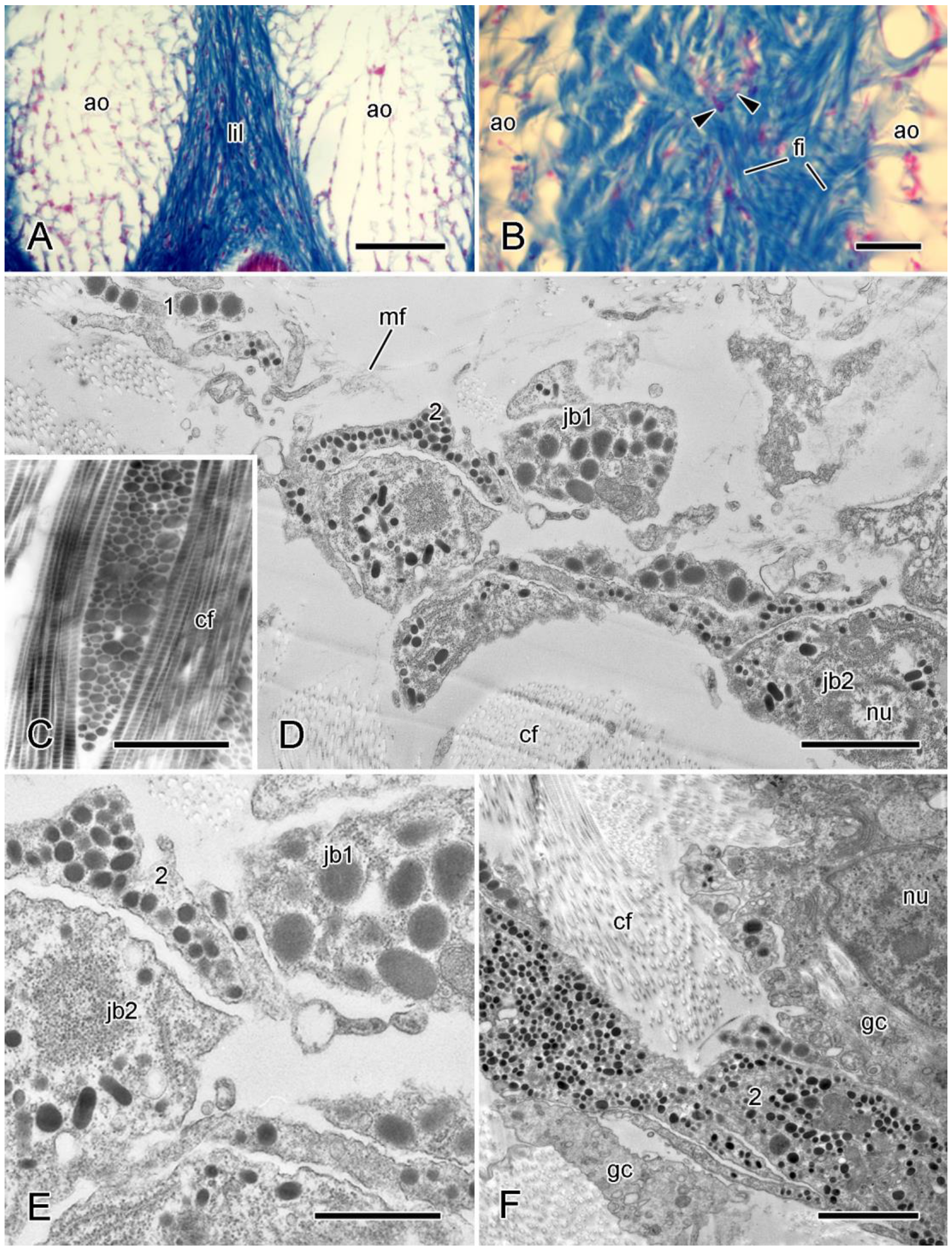
MCT consists predominantly of extracellular materials and contains a relatively small volume fraction of cellular constituents. In histological sections viewed in the light microscope, the extracellular components of most mutable collagenous structures are seen as aggregations of fibers of varying diameter, conformation, and spatial arrangement (Figure 1A,B and Figure 2A,B). Some, e.g., interossicular ligaments, are predominantly simple parallel fiber arrays, although even these may display structural variability, particularly at insertion regions [65,82]. Others consist of stacks of parallel fiber sheets with the fiber orientation varying between adjacent sheets: the genital bar-lateral arm plate ligament of amphiurid brittlestars is a simple example of this [77], whereas the dermis of both starfish dorsolateral body wall and sea-urchin peristomial membrane has three interconnected but structurally distinct layers dominated by orthogonal fiber arrays [83,84]. Another arrangement is seen in the MCT of sea-urchin and starfish tube feet, which consists of an inner sheath of longitudinal fibers and an outer sheath of crossed-fiber helical arrays [55]. MCT thus demonstrates a microstructural diversity comparable to that of vertebrate fibrous connective tissue [85].
The extracellular fibers of MCT are, with one exception, parallel bundles of transversely banded collagen fibrils accompanied by interfibrillar proteoglycans and loose arrangements of microfibrils (Figure 1C,D and Figure 2C,D). The exception is certain tendon fibers at the autotomy planes of brittlestar arms (see below). The banded collagen fibrils of MCT are parallel assemblages of trimeric collagen molecules arranged in a regular staggered array with adjacent molecules offset by 40 to 80 nm. In most echinoderm fibrils, the collagen molecules consist of two fibrillar α chains (1α and 2α) which form (1α)22α heterotrimers [87,88,89]. Some collagen molecules in the fibrils of sea-urchin MCT include a third fibrillar chain (5α) and form (1α)25α heterotrimers [87]. The N-propeptide of the 5α chain shows two peculiarities: (1) it is retained after fibril assembly, which is not the case in the echinoderm 2α chain and in all vertebrate fibrillar procollagens [90], and remains at the surface of the fibrils; (2) it contains 11 SURF (‘sea-urchin fibrillar’) modules, which are also present in the 2α N-propeptide and in fibrosurfin—an interfibrillar component of sea-urchin MCT; fibrosurfin is structurally related to the fibropellins that constitute the apical lamina surrounding sea-urchin embryos [91,92].
Analyses of the Strongylocentrotus purpuratus genome and the transcriptome of the body wall of the sea-cucumber Cladolabes schmeltzii have not uncovered any features of the echinoderm ECM that could be correlated with its variable tensility [93,94,95]. These analyses, as well as a targeted proteomic investigation of the body wall of the sea-cucumber Apostichopus japonicus [96] and a comparative genomic analysis of four echinoderm classes [97], have also shown the presence of fibrillin genes and their transcripts or gene products. This complements biochemical and immunological evidence that the microfibrils occurring in MCT and non-mutable echinoderm ligaments [78,80,98] consist at least partly of fibrillin-like proteins [99]. The function of these microfibrils is unclear. They may facilitate slippage between adjacent fibril bundles during MCT deformation, maintain the orientation of fibrils during deformation, and contribute to passive elastic recoil after the removal of external forces.
Other interfibrillar molecules contribute to the linkage between adjacent collagen fibrils and for that reason must have a major influence on interfibrillar stress transfer and the mechanical properties of MCT. Biochemical evidence indicates that proteoglycans, which consist of a protein core and glycosaminoglycan (GAG) sidechains, act as binding sites for other molecules that are inferred to form interfibrillar crossbridges (discussed in Section 5.2). After staining with the cationic dyes cuprolinic blue or cupromeronic blue, which label GAG sidechains, electron-dense deposits are visualized as granules on the fibrillar surface or as interfibrillar bridges, both arranged regularly within each fibril D-period (Figure 3) [86,98,99,100,101]. Fucosylated chondroitin sulfate is the major GAG of sea-cucumber dermis. It has been found only in this tissue, is covalently associated with collagen fibrils, and consists of a chondroitin sulfate core with branches of the sulfated sugar α-L-fucose. The functional significance of the fucose side-branches is not understood, although it is known that their enzyme-inhibitory activity is crucial for the ‘exogenous’ anticoagulant, antithrombotic, and antidiabetic properties of fucosylated chondroitin sulfate [49,101,102].
MCT contains other molecules that contribute to interfibrillar cohesion but whose extracellular disposition is unknown; these are discussed in Section 5.2.
The tendon fibers at the autotomy planes of brittlestar arms are continuous with the basement membrane (BM) of the intervertebral muscles (Figure 4). Their histochemistry and the ultrastructural similarities between their junctions and the muscles and conventional (non-mutable) BM–muscle complexes of echinoderms and vertebrates confirm that they are BM derivatives [13]. Genomic analysis has demonstrated that echinoderms express the full BM extracellular ‘toolkit’—collagen IV, the glycoproteins laminin and nidogen, and the heparan sulfate proteoglycan perlecan [93,95,97]. There is no direct information on the spatial arrangement of these molecules in brittlestar autotomy tendons or conventional echinoderm BMs. Some of the molecules are very similar to their vertebrate homologs, particularly, in the case of collagen IV and nidogen, with respect to domains involved in intermolecular bonding [103,104]. The configuration of echinoderm BMs (including brittlestar autotomy tendons) is therefore likely to resemble that detected in all investigated vertebrates and invertebrate BMs, which consist of separate collagen IV and laminin networks linked to each other and to cell surfaces by molecules that include nidogen and perlecan [105].

4.2. Cellular Components

4.2.1. Juxtaligamental Cells

The most abundant cellular components of MCT are neurosecretory-like cell processes containing large (diameter > 100 nm), membrane-bounded dense-core vesicles (LDCVs) (Figure 1C,E, Figure 2D,E and Figure 4B). Those present in brittlestar intervertebral ligaments were the first to be described and were named ‘juxtaligamental cells’ (JLCs) because their somata are located outside but close to the ligaments [106]. JLCs are the subject of a recent comprehensive review [46] on which the following account is based.
All investigated mutable collagenous structures are permeated by, or adpressed against, cell processes containing LDCVs [46]. In all classes except the Echinoidea there is usually more than one type of process distinguishable mainly by the size and shape of their LDCVs (Table 3; Figure 1E and Figure 2D). The processes occur mostly in compact bundles of two or more, which always comprise more than one type. Although most sea-cucumber juxtaligamental components appear to be separated from the ECM by a basement membrane [107,108,109,110], no such basement membrane is present in the other classes (apart from single starfish and brittlestar exceptions [111,112]).
In all classes, the LDCVs of some juxtaligamental cell bodies and their processes are strongly acidophilic in histological sections (Figure 1B and Figure 2B), a feature that may serve as a marker for juxtaligamental tissue [46]. Ultrastructural evidence from all classes indicates that the LDCVs are produced by the Golgi complex. The consistent presence of microtubules in JLCs and their labeling by antibodies against acetylated tubulin, somatostatin, and synaptotagmin B [78,117,118] suggest that JLCs are neurons. Those with smaller LDCVs in the 100–300 nm size range may be regular aminergic or peptidergic neurons. Those with larger LDCVs > 300 nm in diameter are the main reason why JLCs are often described as being ‘neurosecretory-like’, since LDCVs of a comparable size are often present in the neurosecretory cells of other phyla [119,120,121].
The juxtaligamental somata of all classes other than the Holothuroidea occur in aggregations. Large aggregations—called juxtaligamental nodes—are associated with all brittlestar mutable collagenous structures (Figure 1A,F) and some ligaments of sea-lilies, featherstars and sea-urchins [106,122,123]. Large aggregations are always located outside the MCT into which their juxtaligamental processes project and they have a central neuropil-like region (Figure 1G). The latter intimates that they may have ganglion-like integrative and coordinative functions, which is supported by the presence in brittlestars of a capsule of glia-like cells, which compartmentalize the juxtaligamental somata (Figure 1H) [78]. Starfish MCT contains smaller aggregations of cell bodies that have a micro-ganglion-like organization including a central cluster of cell processes and a loose outer layer of glia-like cells (Figure 2D–F) [86]. Cells sharing features with the radial glial cells of echinoderm radial nerve cords are in close contact with the JLCs of sea-urchin tooth ligaments [124,125]. JLC aggregations appear to be absent from sea-cucumbers. Glia-like cells ensheathing bundles of cell processes have been seen in the MCT of one sea-cucumber [109].

4.2.2. Other Cells

Fibrillar MCT usually includes a sparse population of cell bodies containing heterogeneous vacuoles that resemble lysosomes and enclose cytoplasmic debris or collagen fibrils (Figure 1C) (see, e.g., [65,78,110,126]). They occur also in non-mutable echinoderm connective tissue [127] and, given the dearth of obvious fibroblasts in MCT, at least some may be pluripotential cells that can adopt a fibrogenic phenotype.
Morula cells are abundant in, and peculiar to, sea-cucumber dermis. These contain very large (diameter 3–6 µm) membrane-bounded vesicles containing material of variable electron density, and may synthesize molecules that participate in fibrogenesis [110,128].
Possible regular neuronal processes that lack LDCVs occur in mutable collagenous structures of all five classes [86]. In sea-cucumber dermis, such processes are associated with glia-like cells [110,128].
Myocytes have been observed in several starfish, sea-urchin, and sea-cucumber structures, in most of which they occupy a small proportion of the total cross-sectional area (Table 4).

5. Physiology of MCT

5.1. Passive Mechanical Properties

5.1.1. Baseline Mechanical Behavior

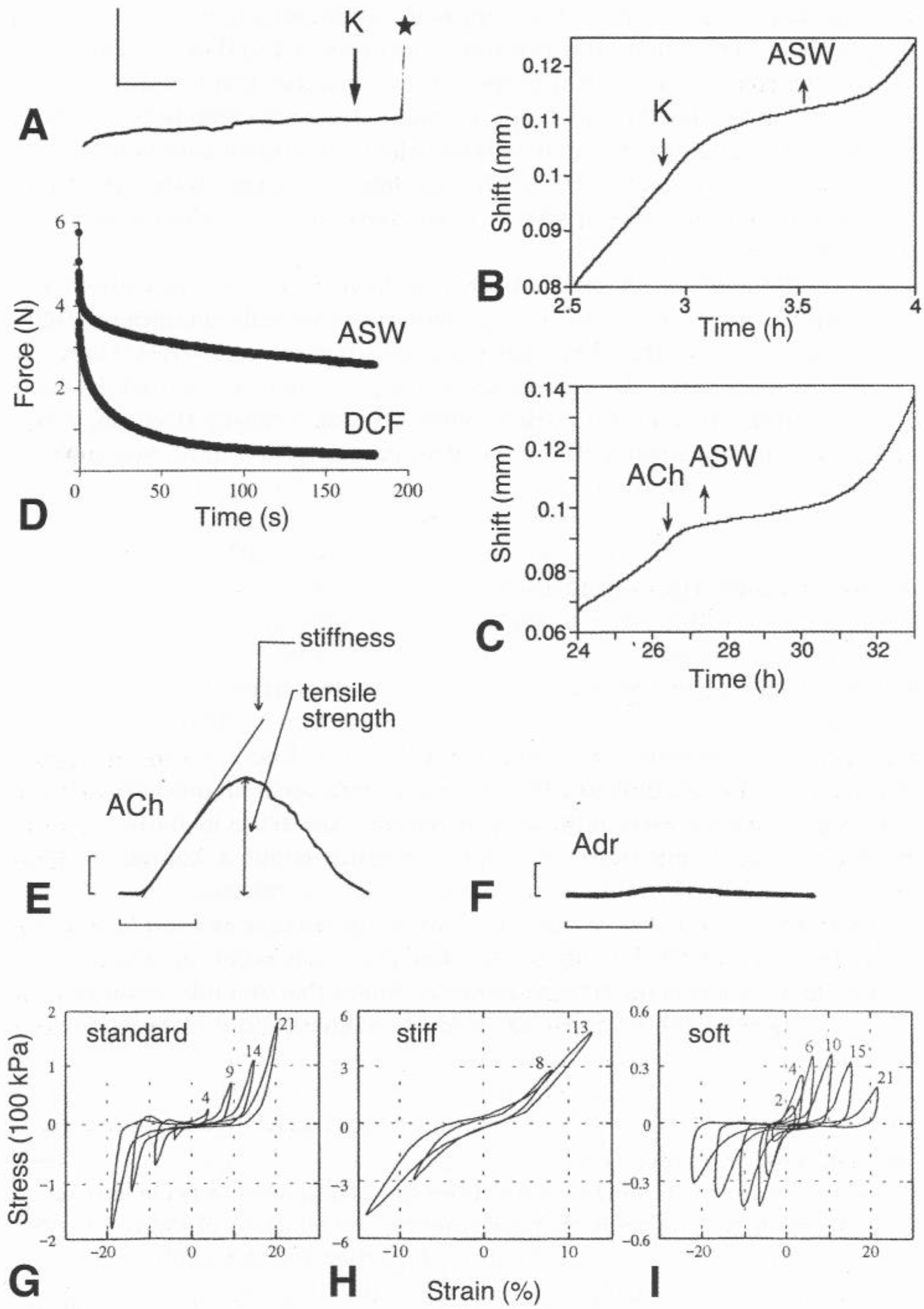
It was established more than 40 years ago [73] that fibrillar MCT can show passive mechanical behavior that generally resembles that of the fibrillar collagenous tissue of vertebrates. Like the latter [131,132], MCT is, at least in certain physiological states, a viscoelastic material that demonstrates tensile creep (elongation) under constant load (Figure 5A–C), stress relaxation (force decay when stretched to and maintained at a fixed length) (Figure 5D), stress–strain curves with non-linear toe regions (Figure 5E,F), and time-dependence—for example, its elastic stiffness (slope of the stress–strain curve) increases with increasing strain rate [61,73,83,133].
Table 5 and Table 6 provide data on the creep and stress–strain behavior of mutable collagenous structures and a selection of mammalian collagenous structures. With respect to all the featured mechanical properties (viscosity, elastic stiffness, etc.), there is considerable quantitative variation between different mutable collagenous structures, some of which may be attributable to differences in ECM composition and microarchitecture: dermis consists of a 3-D fiber meshwork, whereas ligaments are parallel fiber arrays. There is also considerable quantitative variation between homologous mutable collagenous structures in different species, which in some cases may reflect the varying functional demands for which they are adapted (compare data for Actinopyga echinites and Holothuria leucospilota in Table 5, and Anthocidaris crassispina and Paracentrotus lividus in Table 6). In comparison with mammalian collagenous tissue, MCT exhibits a much wider range of values for viscosity, generally lower values for elastic stiffness and ultimate stress but much higher values for ultimate strain.

5.1.2. Tensile Change

The capacity for tensile change varies qualitatively between different mutable collagenous structures. Each shows one of four patterns of tensile change (Table 1): (1) only irreversible destabilization (as occurs during autotomy), (2) irreversible destabilization, as well as reversible stiffening and destiffening, (3) only reversible stiffening and destiffening, and (4) only irreversible stiffening [44,155].
The dramatic and irreversible loss of tensile strength occurring at autotomy can be mimicked experimentally and quantified using isolated tissue preparations undergoing creep tests (Table 5). Treatment with neuro-active agents such as elevated [K+] causes an abrupt decrease in viscosity and tissue rupture (Figure 5A).
Reversible changes in viscosity have been demonstrated in creep tests and stress relaxation tests (Table 6; Figure 5B,C). Reversible changes in tensile strength, tensile stiffness and other parameters have been investigated by means of standard stress–strain tests (Figure 5E,F) and by dynamic testing methods in which samples are subjected to oscillating strain (Figure 5G–I). The so far unique example of irreversible stiffening—exhibited by sea-cucumber Cuvierian organs—has been quantified in stress–strain tests (Figure 6). Data from biomechanical studies have been used to generate a number of models that aim to specify the contribution of different extracellular components to mechanical behavior and reveal their role, if any, in variable tensility. The currently prevalent model, derived originally from work on sea-cucumber dermis, is based on the concept that the tissue can adopt three mechanical states—standard, stiff and soft (Figure 5G–I). These have qualitatively different features: only dermis in the soft state shows stress-induced softening; dermis in the soft and standard states is characterized by J-shaped stress–strain curves with prominent toe regions, while the stress–strain curve of stiff dermis is linear [19,73,135].

5.2. Molecular Basis of Tensile Change

Variation in the capacity of MCT for tensile change is probably enabled by an important difference between MCT and most collagenous tissues of vertebrates: cohesion between adjacent collagen fibrils in the latter is highly dependent on permanent, covalent molecular interactions and is relatively stable [156,157], in contrast to MCT, in which interfibrillar cohesion is less dependent on covalent interactions and is labile. This explains the observations that echinoderm collagen fibrils can be extracted from whole tissue by mild chemical and mechanical methods [158,159], whereas the isolation of intact collagen fibrils from normal adult vertebrate collagenous tissues requires harsher treatments such as enzymolysis and gross mechanical manipulation [160,161,162]. A major aim of MCT research has been the elucidation of the determinants of interfibrillar cohesion and how these determinants are modulated.
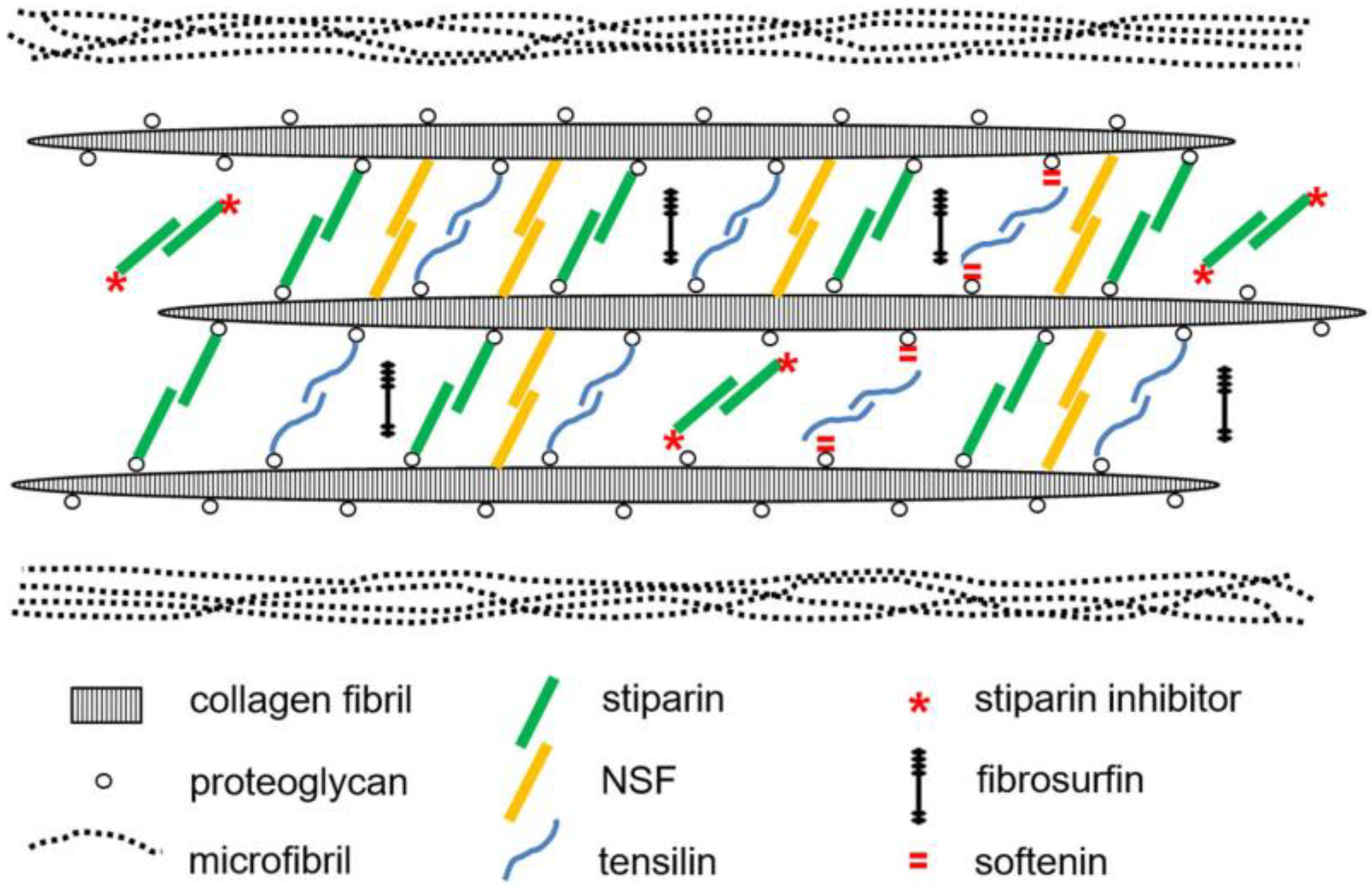
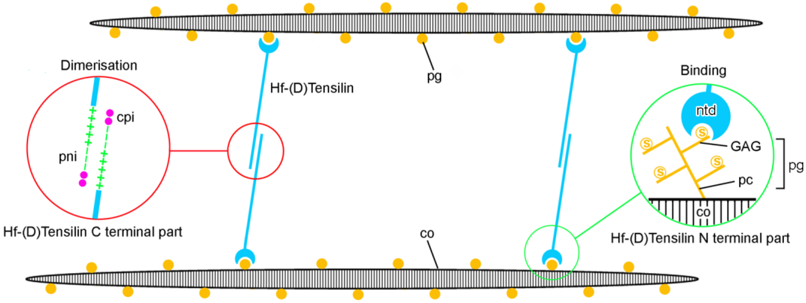
During the past 50 years, several hypotheses for the molecular basis of variable tensility have been proposed and subsequently superseded (as recounted in previous reviews [25,73,81,99,163,164,165,166]). Current ideas on possible molecular mechanisms are derived primarily from investigations of reversible changes in sea-cucumber dermis, assume the premises of the three-state paradigm mentioned above (see Section 5.1.2), and focus on chemical factors that can be isolated from the dermis and that influence its mechanical behavior in vitro (Figure 7), though their specific roles in vivo have not been determined with certainty. The tensilins are the most thoroughly investigated of these factors. Tensilins have a high degree of sequence identity to TIMP (tissue inhibitor of metalloproteinase) proteins and can be isolated from dermis only by procedures that cause cell lysis, which suggests they are present in intracellular stores. They stiffen samples of whole dermis and aggregate isolated collagen fibrils in vitro [108]. A comparison of the properties of intact and truncated recombinant tensilin suggested that the N-terminal TIMP-like domain interacts strongly with GAGs attached to the surface of collagen fibrils, and that interfibrillar linkages may involve the dimerization or oligomerization of tensilin mediated by its C-terminal regions [44] (Figure 8).
In vitro experiments have indicated that sea-cucumber tensilins have only a partial stiffening effect in that they convert dermis from the soft to the standard state but not from the standard to the stiff state [167]. The phylum-wide importance of tensilin is also questionable, in view of the inconsistent effect of sea-urchin tensilin-like protein on the mutable compass depressor ligament of the sea-urchin Paracentrotus lividus [136]. In contrast to the tensilins, a second stiffening protein—‘novel stiffening factor’ (NSF)—switches sea-cucumber dermis from the standard to the stiff state, has no effect on soft dermis, and does not aggregate isolated collagen fibrils [168]. The different actions of tensilin and NSF are compatible with the idea that separate molecular mechanisms are responsible for the soft-to-standard and standard-to-stiff transitions. Another protein—stiparin—aggregates collagen fibrils, but, unlike the tensilins, does not affect the whole dermis. It can be extracted by prolonged immersion of dermis in seawater alone and may be a constitutive component that contributes to a basal level of interfibrillar cohesion [169].
Three destiffening molecules have been extracted from sea-cucumber dermis and partially characterized: ‘stiparin inhibitor’, a 62 kDa sulfated glycoprotein that binds stiparin and inhibits its fibril aggregating activity; ‘plasticiser’, a small (<15 kDa) protein that directly destiffens dermal ECM and is released only after cytolytic treatments [169]; and softenin, a ca. 20 kDa protein that can be extracted without cell lysis. Softenin may compete for tensilin binding sites on the collagen fibrils, since it disaggregates tensilin-aggregated collagen fibrils in vitro and reversibly destiffens cell-dead dermis that is in the standard state [170].
Other possible MCT destiffeners include matrix metalloproteinases (MMPs), which have important roles in ECM remodeling associated with echinoderm development and regeneration [171,172,173]. The synthetic MMP inhibitor galardin stiffens sea-urchin compass depressor ligaments in all three mechanical states, the effect being much weaker on stiff ligaments than on soft and standard ligaments. One explanation for these findings is that ligament stiffness is modified through crosslink degradation by constitutive MMP activity regulated by the cellular release of an endogenous MMP inhibitor [174].
As discussed below in Section 5.3.3, several endogenous peptides have indirect, cell-mediated effects on the mechanical behavior of MCT preparations. However, it has been speculated that holokinin-1 and holokinin-2, whose heptapeptide sequences are present in the C-terminal domain of 5α collagen (see Section 4.1), may destiffen the dermis by directly disrupting as yet unidentified interfibrillar linkages involving 5α collagen [175].
Further evidence that separate mechanisms produce the soft-to-standard and standard-to-stiff changes has been acquired by comparing the water content and distribution in MCT samples that are in different mechanical states. In the case of sea-cucumber dermis and sea-urchin compass depressor ligaments, it has been demonstrated that water moves out of the tissues during the standard-to-stiff shift but not during the soft-to-standard shift [176,177], and that the standard-to-stiff transition alone is accompanied by a significant increase in the collagen fibril packing density [98,178]. In both these transitions in sea-cucumber dermis there is also a significant increase in the number of crossbridges connecting adjacent collagen fibrils, as revealed by transmission electron microscopy [178]. On the basis of the latter observations, Tamori et al. [178] proposed that dermal stiffening is achieved by three mechanisms: (1) increased crossbridge formation in both transitions; (2) tensilin-dependent stiffening of collagen fibrils (through subfibril fusion) in the soft-to-standard transition; and (3) in the standard-to-stiff transition, increased bonding within the proteoglycan matrix between fibril bundles, which increases fibril packing density and squeezes out water.
The model of Tamori et al. [178] incorporates the hypothesis that the mechanical adaptability of sea-cucumber dermis is achieved partly by changes in the stiffness of collagen fibrils consequent on their disaggregation into subfibrils and subsequent reaggregation. A similar mechanism was previously proposed on the basis of ultrastructural evidence for fibril disaggregation in featherstar ligaments [100]; however, this has not been reported for any other mutable collagenous structure. More recently, in a series of investigations using time-resolved synchrotron small-angle X-ray scattering (SAXS) to compare the behavior of sea-cucumber dermis in three mechanical states, Gupta and co-workers moved from an initial model in which the variable tensility of the tissue was attributed to modulation of interfibrillar matrix stiffness alone [179,180,181] to one that also incorporates changes in fibril stiffness mediated by a chemoelastic process involving the reversible scission of collagen chains (at unspecified chemical bonds) [45]. It is thus apparent that, despite sea-cucumber dermis being the most intensively investigated mutable collagenous structure, the molecular mechanisms responsible for its variable tensility have yet to be established with certainty. At a more general level, the phylum-wide applicability of information and concepts derived from research on sea-cucumber dermis is at present obscured by the scarcity of relevant information on the MCT of other echinoderm classes [43].

5.3. Regulation of Mechanical Properties

5.3.1. Juxtaligamental Cells

Circumstantial evidence suggests that JLCs are the effectors that directly alter the tensile properties of MCT: JLC processes extend into and terminate within MCT, have no feasible cellular targets in the tissue, and constitute a pathway between the ECM and the motor nervous system, and they have not been found in non-mutable echinoderm collagenous structures [75,80,81]. In addition, possible effector molecules occur in sea-cucumber JLCs—the stiffening proteins tensilin and stiparin—have been immunolocalized to LDCVs in the dermis [169] and tensilin has been detected by immunohistochemistry and in situ hybridization in JLCs of Cuvierian tubule connective tissue [71]. Crucially, it has yet to be demonstrated that dermal stiffening is preceded or accompanied by the release of tensilin, or any other putative effector agent, from JLCs. A correlation between alterations of tensile state and changes in JLC ultrastructure has been observed in a minority of mutable collagenous structures. Such changes have been associated mainly with irreversible destabilization or stiffening and usually include evidence that LDCVs or their contents are released into the extracellular compartment [60,71,98,182].
LDCV-containing processes tend to occur as clusters in which usually two types of process are arranged in parallel and in close proximity (see Section 4.2.1), possibly indicating a functional relationship [78,113,147,182]. Whilst this might be a reciprocal effector system, with one cell-type destabilizing the ECM and the other type stabilizing it, the possibility that processes containing LDCVs in the smaller size range (see Table 3) are aminergic or peptidergic neurons suggests that their role may be to regulate the effector activity of neurosecretory-like JLCs. However, the LDCVs of vertebrate neurosecretory neurons are known to handle a variety of molecules involved in a wide range of biological processes, including ECM degradation [183,184,185]. It is therefore feasible that each of the different cell-types in MCT could have both effector and regulatory functions.

5.3.2. Nervous System

There is a considerable amount of morphological and physiological information on the neural regulation of MCT tensility. As discussed above (see Section 4.2.1), the JLCs, which are associated with all mutable collagenous structures, are likely to be neurons. In all classes there is morphological evidence for a functional linkage between juxtaligamental components and the central nervous system, which at the histological level is represented by the extension into juxtaligamental tissue of neural pathways from the central nervous system [46,186,187]. The best characterized of these are the complex supply of motor nerves from the radial nerve cord to brittlestar juxtaligamental nodes [106,118,182] and the simpler pathways serving sea-lily, featherstar, and sea-urchin JLC aggregations [122,123]. Ultrastructural investigations have revealed possible synaptic junctions between JLCs and exogenous neuronal processes in sea-lilies, featherstars, sea-urchins, and brittlestars [78,122,188,189]. There is variation between the echinoderm classes regarding which neural subsystems provide motor pathways to JLCs: the hyponeural subsystem is involved in starfish and brittlestars [106,187]; the aboral (apical) subsystem in sea-lilies and featherstars [122]; the ectoneural subsystem in sea-cucumbers [186,190]; and the ectoneural and hyponeural subsystems in sea-urchins [191].
The neuropharmacology of MCT has attracted considerable interest. Acetylcholine (ACh) is the main excitatory transmitter in echinoderm motor systems [192]. In vitro pharmacological experiments have shown different patterns of cholinergic responses. ACh and nicotinic receptor agonists cause an initial increase in viscosity and later decrease in the viscosity of sea-cucumber dermis, whereas muscarinic agonists cause a decrease in viscosity [193]. These findings suggest that both stiffening and destiffening of sea-cucumber dermis are controlled by cholinergic motoneurons. Sea-urchin capsular spine ligaments are stiffened by ACh (Figure 5E) and by both nicotinic and muscarinic agonists, the latter having a slower effect [194]. In contrast, both types of cholinomimetic agonists destiffen the cirral ligaments of a sea-lily, the muscarinic effect again being slower [133]. The body wall of a starfish is stiffened by ACh and muscarinic agonists, but not by nicotinic agonists [53].
Several other neurotransmitters have been shown to affect the mechanical behavior of isolated MCT preparations in vitro. The catecholamines adrenaline, noradrenaline, and dopamine cause a biphasic (destiffening then stiffening) response in sea-urchin spine ligaments, as does β-phenylethylamine, although its derivatives, tyramine and octopamine, cause only destiffening [194]. Glutamate-like immunoreactivity has been demonstrated in neuron-like cells and within mutable ligaments in the arms of a featherstar. L-glutamate causes irreversible destiffening of MCT-containing ligaments at syzygies (interossicular junctions specialized for autotomy) in the arms of this animal, which provides evidence that the neural control of autotomy in featherstars involves a glutamatergic pathway. ACh depresses the response of syzygial preparations to L-glutamate [59].
It is to be expected that the nervous system both controls the mechanical properties of MCT and enables changes in these properties to be coordinated with muscle activity. Such coordination has been demonstrated using the sea-urchin spine joint as a model system [195]. Each spine joint is encircled by an inner ring of mutable capsular ligament and an outer ring of muscle. Mechanical stimulation of a spine base results in the stiffening of its capsular ligament and the inclination of adjacent spines towards the stimulated spine; inclination of adjacent spines can occur only through the coordinated contraction of their muscles and destiffening of their ligaments.

5.3.3. Neuropeptides and Other Peptides

Extracts of the sea-cucumber Apostichopus japonicus contain myoactive neuropeptides that cause muscle relaxation or contraction and change the stiffness of sea-cucumber dermis: the amidated pentapeptide NGIWYamide causes stiffening and the cyclic peptide stichopin (DRQGWPACYDSKGNYKC) suppresses the stiffening effect of ACh on the dermis [196]. NGIWYamide- and stichopin-like immunoreactivity has been detected in nerve fibers located within the dermis [190,197]. Stichopin-like immunoreactivity was also detected in cells located in the dermis and in other collagenous tissue [197]. The vasopressin/oxytocin-type neuropeptide crinotocin causes nervously mediated destiffening of ligaments at the mobile diarthrial arm joints of the featherstar Antedon mediterranea but has no effect on the ligaments at the syzygial joints where autotomy occurs. Cells expressing crinotocin precursor transcripts are present in the main motor nerve of the arm and at the base of nerve branches that innervate the arm muscles and diarthrial ligaments [198]. The injection of another neuropeptide—ArSK/CCK1—into the starfish Asterias rubens promotes arm autotomy, which is likely to be due partly to its contractant effect on the tourniquet muscle that constricts the arm at the detachment plane during autotomy. However, as ArSK/CCK1-immunoreactive cells are present in the inter-ossicular collagenous tissue of the body wall, it may also be involved in initiating the destabilization of this tissue during autotomy [187,199].
The heptapeptides holokinin-1 and holokinin-2 (PLGYMFR and an oxidized derivative, respectively) cause destiffening of A. japonicus dermis. Analysis of A. japonicus transcriptome sequence data has revealed that the holokinins are not neuropeptides, as originally supposed [196], but fragments of the C-terminal region of a 5α type collagen [175]. As mentioned above (see Section 5.2), this may help to explain how holokinins influence the mechanical properties of MCT. However, the physiological relevance of holokinins in sea-cucumbers is unclear, since they could be a product of collagen degradation generated when body wall extracts are prepared.

5.3.4. Celomic Factors

The celomic fluid of intact and untreated echinoderms contains separate chemical factors that stiffen or destiffen isolated MCT preparations in vitro. These factors have been detected in representatives of all classes except the Crinoidea (no members of which have been investigated), and their effects appear not to be species- or class-specific [52,69,139]. The chemical nature, source, and physiological significance of these factors remain unknown. It has been conjectured that they are neurotransmitters that modulate the activity of JLCs [52].
The above factors have been reported only in sea-cucumber species from the orders Synallactida and Holothuriida. Sea-cucumbers from the order Dendrochirotida demonstrate defensive auto-evisceration of the retractable anterior portion of the body (the ‘introvert’) and the internal organs. This requires the destabilization of the introvert dermis and of tendons connecting the introvert retractor muscles to the longitudinal body wall muscles. Celomic fluid from eviscerating dendrochirotids contains a factor that induces evisceration when injected into intact animals and which destabilizes the introvert dermis and retractor tendons. This ‘evisceration factor’ is absent from the celomic fluid of intact animals and is a small molecule with a molecular mass of ca. 150 Da [200,201].
The celomic fluid of scalded starfish induces autotomy when injected into intact starfish, an effect that is not produced by celomic fluid from intact and untreated animals [202]. An ‘autotomy-promoting factor’ (APF) has been isolated from the celomic fluid of scalded starfish and partly characterized. It is likely to be a peptide with a molecular mass of 1100–1200 Da [203,204] and therefore possibly an ortholog of the autotomy-promoting neuropeptide ArSK/CCK1 (molecular mass 1613 Da) (see Section 5.3.3) [198]. Like that neuropeptide, starfish APF may activate MCT destabilization, since it causes the nervously mediated rupture of isolated preparations of the basal arm autotomy plane of Pycnopodia helianthoides [54]. As these investigations have involved only starfish species belonging to the family Asteriidae, the wider significance of APF is unclear.

5.4. Force Generation

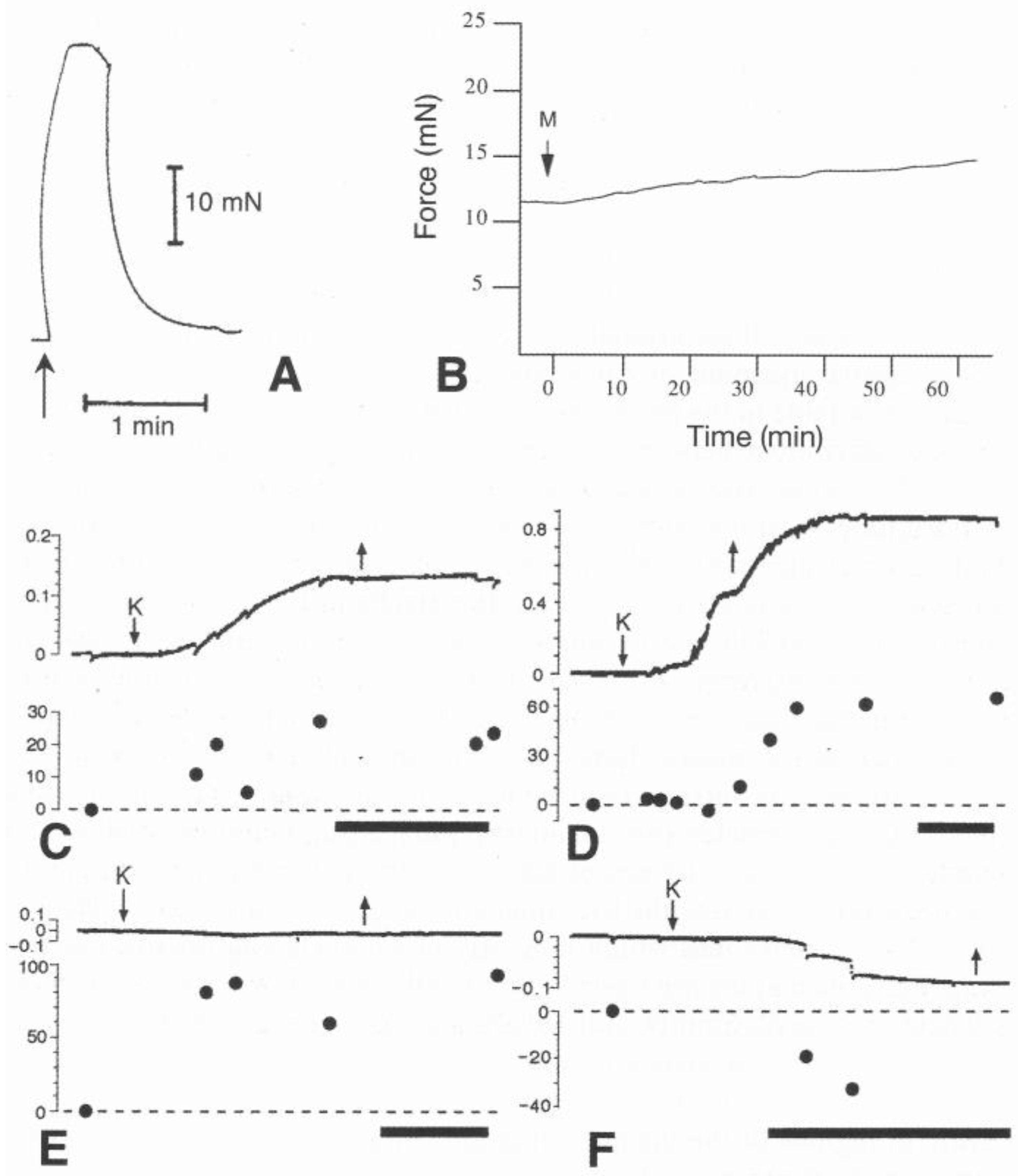
It has been demonstrated that certain mutable collagenous structures in sea-urchins, sea-lilies and featherstars can generate contractile force. As well as increasing the tensile strength and stiffness of the capsular ligament of the sea-urchin spine joint (see Section 5.3.2), ACh also causes isolated preparations of it to shorten and develop tensile force [61,205]. Both the contraction and relaxation phases of the contractile response are very fast, the contraction phase reaching up to 90% of the maximum value in under 1 s (Figure 9A) [206]. Force generation in this case depends on myocytes that are distributed between the bundles of collagen fibrils and constitute 1–3% of the total cross-sectional area of the ligament. The functional significance of the myocytes and their contractile activity has not been established. It has been demonstrated experimentally that they are not responsible for varying the passive stiffness of the capsular ligament [207]. They may help to reshorten extended ligament fibers or they may operate synergistically with the spine muscle, for example during re-erection of the spine.
Though lacking myocytes, mutable ligaments in the cirri and arms of sea-lilies and featherstars have the capacity for active contractility. Cirri are claw-like appendages that have an endoskeleton consisting of a single series of interarticulating ossicles connected by ligaments. They are attached to the aboral surface of featherstars and the stalk of some sea-lilies. Those connected to the stalk of the sea-lily Metacrinus rotundus bend upwards when the stalk or the cirri themselves are stimulated mechanically. Cholinergic agonists induce slow force generation in isolated cirri undergoing stress relaxation (Figure 9B). This is not due to the passive elastic recoil of a structural component that was previously in an extended state, since cirral preparations can develop force even if they show partial stress relaxation before they are stimulated [133,209,210].
The bending of sea-lily and featherstar arms occurs at mobile hinge joints. Within each joint, there is a fulcral ridge, below which is a single ligament and above which are paired ligaments and paired muscles. Although contraction of the muscles bends the arm upwards, the power stroke that brings about locomotion by swimming, crawling, or climbing depends on the downward flexion of the arm and must therefore be accomplished by the ligaments. The aboral (sub-fulcral) ligament of featherstars has been shown to shorten slowly and generate a force of up to ca. 5.6 kPa [210,211]. By measuring stiffness and shortening simultaneously in the aboral ligament of a sea-lily, Motokawa et al. [208] demonstrated the independence of its passive mechanical properties and contractile activity (Figure 9C–F). However, as contracting ligaments are usually in the destiffened state, their passive and active mechanical behaviors appear to be coordinated.
No information is available on the mechanism of active force generation in featherstar and sea-lily ligaments. Birenheide & Motokawa [210] speculated that this could be related to the contraction of hydrogels that results from changes in their fluid phase [212].

6. Biological Significance of MCT

6.1. Energy-Sparing Postural Fixation

Since echinoderm collagenous tissue was first shown to undergo reversible stiffening and destiffening, it has been conjectured that this capacity is used to maintain the posture of the body or its appendages for extended periods of time as an alternative to tonic muscular contraction, and that MCT thus serves an energy-sparing function [24,25,138,163]. Prolonged postural fixation is employed primarily in feeding mechanisms and as a defensive strategy. Sea-lilies, featherstars, basket stars, brittlestars and starfish belonging to the family Brisingidae are suspension feeders with long arms, which, when collecting food items, are extended in fixed postures often for hours. In addition, during such feeding activity, sea-lilies and featherstars are anchored to their substrate by articulated appendages: a stalk or claw-like cirri, respectively [213,214,215,216,217]. Examples of defensive postural fixation are provided by sea-cucumbers in the family Stichopodidae that survive months of dormancy sheltering under stones while in a contracted and stiffened state [218,219]; sea-urchins that wedge themselves into holes and crevices in rock by means of their immobilized spines [73,220]; and brittlestars whose whole body ‘freezes’ and becomes rigid when they are severely disturbed [221].
There can be no doubt that MCT alone is responsible for the postural fixation of those structures that lack muscles (apart from myocytes associated with epithelia), i.e., sea-lily stalks, featherstar cirri and stichopodid sea-cucumber dermis [107,222,223]. However, although mutable ligaments are present at sea-urchin spine joints and in the arms of sea-lilies, featherstars, brittlestars, and starfish (Table 1), these anatomical structures also contain substantial muscles that actuate their movement and, theoretically, could also maintain posture by tonic contraction. This can be discounted in the case of sea-urchin spine joints, since the defensive ‘freeze response’, in which direct touch invokes prolonged immobilization of the spine in the upright position, persists after the spine muscle has been completely transected and thus must be achieved only through the stiffening of the capsular ligament [195]. Electrophysiological evidence also indicates that the rigidity of the arms of brittlestars undergoing defensive ‘freezing’ is unlikely to depend on continuous contraction of their intervertebral muscles. Intracellular recording has shown that, after initial excitatory post-synaptic activity in hyponeural motor neurons, the frozen posture is maintained in the absence of any apparent neural activity [221].
Protracted MCT stiffening also has a role in sea-urchin dental mechanics. Each sea-urchin tooth is attached to a ‘jaw’ by a tooth ligament. To compensate for wear caused by scraping hard surfaces, the tooth grows by the addition of new material at its dorsal end and, when the animal is not feeding, moves slowly in a ventral direction, which is permitted by the destiffened state of the tooth ligament. When the animal is feeding, the tooth is subjected to mechanical loading and, to prevent its dorsal displacement, the ligament switches to a stiffened state [64,124,134].
Investigations comparing the oxygen consumption rates of MCT and muscle have confirmed that fixing posture by the passive stiffening of MCT rather than muscle contraction is likely to confer considerable energetic advantages. As shown in Table 7, the VO2 of the contracted longitudinal body wall muscle of a sea-cucumber was found to be 7–10 times higher than that of its stiffened dermis, and the VO2 of the contracted longitudinal carinal muscle of a starfish was nearly 40 times higher than that of its stiffened dermis. More precise comparisons of energy costs, taking into account the stress that contracted muscle and stiffened MCT can support (inferred from their mechanical stiffness), suggested that postural fixation of the body by the dermis would require only 1.4% of the energy needed if the same supporting force were provided by muscle alone in the sea-cucumber and 0.03% of the energy needed by muscle alone in the starfish [224,225].

6.2. Autotomy and Fission

The term ‘autotomy’ is used herein to denote the adaptive detachment of animal body parts where this serves a defensive (usually anti-predator) function, is achieved by an intrinsic breakage mechanism, and is neurally mediated [155,226]. The capacity for autotomy occurs widely throughout all five extant echinoderm classes and involves the disconnection of a diverse range of anatomical structures—from small appendages like featherstar cirri and sea-urchin pedicellariae, to complex body regions such as the arms of stellate echinoderms (including brittlestars, featherstars and starfish), and to potentially more than half of the body of apodid sea-cucumbers [86,155,182,227].
Echinoderm autotomy planes always transect mechanically significant collagenous components. In all investigated cases, which include examples from each echinoderm class, it has been found that these components consist of MCT that undergoes irreversible destabilization during autotomy (Table 1) [155]. Evidence for such a detachment mechanism has been obtained both from the direct observation of autotomizing structures [51,227,228,229] and by mechanical testing methods applied to isolated MCT preparations in vitro [53,59,70,76].
In all confirmed examples of echinoderm autotomy, anatomical structures are detached quickly (within less than a second to a few minutes) at a preformed, morphologically distinguishable autotomy plane at which there may be adaptations for minimizing tissue damage, sealing fluid compartments, assisting wound repair, and promoting regeneration [155]. There are, however, examples of echinoderm detachment phenomena occurring in response to external agents at sites that lack any autotomy-specific features but are associated with mutable collagenous structures normally involved in only reversible changes in stiffness. For example, although there are no reports of their spines undergoing rapid defensive self-detachment, brittlestars and sea-urchins can shed spines to which tags have been attached for experimental purposes, a process that can take hours to days and results from the drastic destiffening of their spine-joint ligaments [62,78]. Such occurrences have been described as ‘opportunistic self-detachment’ to distinguish them from autotomy sensu stricto [155].
Fission—asexual reproduction by division of the body—occurs in certain adult brittlestars, starfish, and sea-cucumbers [230] and in certain larval starfish [231]. Direct observation of splitting animals and experimental evidence obtained from isolated tissue preparations in vitro indicate that fission in adult echinoderms is likely to be assisted by the destiffening of MCT—predominantly body wall dermis—at the plane of separation [72,232,233]. This has also been confirmed by a comparative transcriptomic analysis of tissues from intact specimens of a fissiparous sea-cucumber and from specimens undergoing fission [93]. It is notable that the latter investigation [93] hints that proteases (other than MMPs) may have a greater role in fission-related dermal destabilization than has been proposed for reversible changes in dermal stiffness (see Section 5.2). There is morphological evidence that at least one form of asexual reproduction in starfish larvae (detachment of the anterior portion of the pre-oral lobe in bipinnariae of Luidia spp.) occurs at a preformed breakage plane [231]. However, although this process must involve the rupture of ECM structures in the larval body wall [234], it is not known if this is facilitated by a pre-weakening mechanism.

6.3. Sea-Cucumber Dermal Autolysis

Sea-cucumber dermal autolysis (SCDA), also referred to as ‘liquefaction’, ‘local degeneration’, and ‘melting’, is observed when certain sea-cucumbers are touched firmly or gripped. After an initial stiffening reaction, the stimulated area softens progressively over a period of minutes until it can be penetrated easily, allowing extrusion of the internal organs (evisceration) [25,235]. In contrast to such localized responses, persistent mechanical stimulation or exposure to UV radiation, high temperatures, or irritant chemicals can convert large areas of the body wall into a viscous flowing mass (Figure 10) [236,237,238]. SCDA is a serious cause of deterioration in the quality of commercially harvested edible sea-cucumbers, especially if they are subjected to prolonged transportation, insensitive handling, or unsuitable storage conditions [239].
SCDA has been regarded as a form of autotomy [25,236]. However, autotomy by definition involves anatomical detachment (see previous Section) and this is not an outcome of SCDA. In addition, some species that demonstrate SCDA—notably Stichopus chloronotus and S. horrens—also have the capacity to detach discrete areas of (non-autolyzed) body wall in response to predator attack. During this process the mesentery surrounding the internal viscera is not punctured and evisceration does not occur [240,241]. This is undoubtedly a defensive autotomy response and is clearly a separate process from SCDA.
Because of its economic implications, the biological pathways responsible for SCDA have been the subject of intense scrutiny, with most of the focus being on proteolytic mechanisms. The activity of several endogenous proteases increases in disintegrating dermis [242], though autolysis is blocked most effectively by MMP inhibitors [243] and only MMP activity accomplishes the complete disaggregation of collagen fibers into smaller fibril bundles and fibrils (together with the degradation of interfibrillar bridges), which appears to be the basis of the destabilization process [243,244,245,246,247]. As several MMP genes are also significantly upregulated in autolyzed dermis [248], it seems that MMPs are the key endogenous proteases responsible for SCDA [243].
The sources of the proteases involved in SCDA include dermal cells containing multivesicular bodies: one of the implicated enzymes—cathepsin L—is released from these ‘vacuole cells’ during autolysis in Apostichopus japonicus [249]. Though not reported for A. japonicus, ‘vacuole cells’ in a related species—Stichopus chloronotus—have processes that are distributed throughout the dermis, mostly isolated from other cellular components, though some occur in bundles with juxtaligamental and other neuronal processes to which they are closely adjacent [250]. These ultrastructural observations complement physiological evidence that SCDA is nervously mediated: pretreatment with anesthetic agents blocks autolysis in Isostichopus badionotus [251].
Three aspects of SCDA suggest it has affinities with ‘conventional’ mutability phenomena and is not a pathological process. (1) It results from targeted proteolysis that disaggregates both collagen fibers and their component fibrils but does not damage monomeric collagen [245,246]. This resonates with the model of Tamori et al. [178], which proposed that the reversible destiffening of holothurian dermis involves collagen fiber and fibril disaggregation (see Section 5.2). (2) MMPs contribute to SCDA and are hypothesized to have a role in the reversible destiffening of mutable collagenous structures in general [174]. (3) There is morphological and physiological evidence that SCDA, like the routine tensile changes undergone by MCT, is nervously mediated. It is therefore feasible that, as surmised by Motokawa [25], localized dermal autolysis evolved as an adaptive predation-avoidance tactic that makes it difficult for an attacker to achieve a firm grip, whereas the non-localized, potentially fatal expression of SCDA that affects wide areas of body wall is caused by the non-adaptive over-activation of the proteolytic softening mechanism.

6.4. Regeneration

Echinoderms are renowned for their regenerative abilities and have provided several productive models for regeneration research [252,253,254,255]. Although available evidence is sparse, it suggests that autotomy is the most common proximate cause of structural loss in echinoderms in their natural environment [155]. This implies that most echinoderm regeneration events are preceded by autotomy and occur at the retained side of a fractured autotomy plane. As a result of the presence of MCT at all echinoderm autotomy planes, the post-autotomy wound surface invariably includes disrupted MCT. There is evidence that the JLCs in disrupted MCT can have a role in regeneration.
The visceral mass of featherstars is connected to the endoskeleton by mesentery-like septa. It is highly likely that collagenous tissue in the septa has mutable properties that facilitate defensive detachment of the visceral mass [60]. It has been reported that, after loss of the visceral mass in Himerometra robustipinna and Dichrometra (previously Lamprometra) palmata, JLCs migrate from the ruptured septa to the wound surface and start a process of direct transdifferentiation (there being no intermediate stage of dedifferentiation). They undergo a series of ultrastructural changes that, after 24 h, transform them into enterocyte (gut epithelial cell) precursors with developing microvilli on their apical surface and secretory granules characteristic of mature enterocytes in their apical region [256,257]. In D. palmata, JLCs also transform into epidermal cells in the regenerating body wall of the visceral mass [257]. The presence of a reservoir of cells with such transformation potential close to the regeneration site may account for the rapid restoration of a functioning visceral mass, which can be accomplished in as little as 4–7 days after autotomy [256,257]. Furthermore, a comparative investigation of visceral mass regeneration in a taxonomically diverse range of 15 featherstar species identified a positive correlation between JLC abundance and regeneration rate [258].
The cell plasticity implied by these observations is plausible, since mammalian neurons can transform into epithelial cells in vitro, though this involves a dedifferentiation stage [259]. However, JLCs do not contribute to visceral mass regeneration in all featherstars, since the enterocyte precursors in Antedon mediterranea are derived from celothelial cells that dedifferentiate then redifferentiate [260]. In addition, there are no reports of JLCs being involved in the regeneration of any other echinoderm structures. On the other hand, the cellular mechanisms of only a very small proportion of all echinoderm regeneration processes have been studied in detail, and many of these relate to events following surgical amputation rather than induced autotomy, a procedure that damages the autotomy plane or avoids it altogether and is known to result in anomalous regeneration [261,262].

6.5. Pathology

6.5.1. Sea Star Wasting Disease

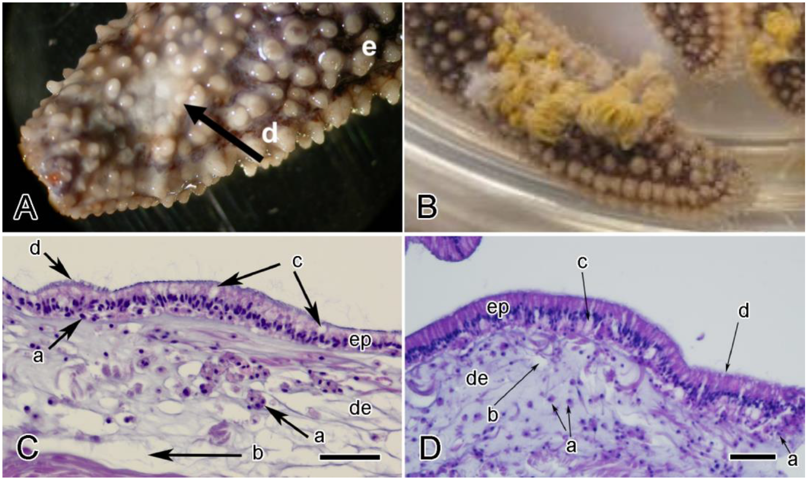
The condition known as sea star wasting disease (or syndrome) (SSWD) is characterized by a set of grossly abnormal signs that include body deflation or loss of turgor, arm loss, body wall lesions, and abnormal behavior (Figure 11A,B). It has been reported for over 100 years, affects dozens of starfish species, and may be common, but, despite over two decades of intensive research, distinct causative agents have not been identified [263,264].
Whatever its etiology, the pathogenesis of SSWD involves the starfish body wall, including its mutable dermis. One of the earliest signs of SSWD is ‘inflammation’ of the dermal MCT, which refers to its infiltration by hemocytes and its edematous appearance resulting from increased spacing between its constituent collagen fibers (Figure 11C,D). These changes precede epidermal ulceration and the softening and eventual disintegration (‘melting’) of the body wall [266,267,268]. Gene expression studies comparing symptomatic and asymptomatic animals have revealed differences in the expression of collagen, cell adhesion, and tissue remodeling genes (including increased expression of MMPs), suggesting enhancement of degradative processes in the MCT of diseased individuals [269,270]. These investigations have also provided evidence that the nervous system is affected. For example, in Pycnopodia helianthoides, amongst other changes, reduced expression of acetylcholinesterase and a muscarinic receptor indicate that ACh signaling pathways may be disrupted [271]. It has also been surmised that the upregulation of RNA processing genes detected in symptomatic Pisaster ochraceus could be a sign of neurological dysfunction [270]. On the basis of such findings, it has been repeatedly conjectured that the body wall changes observed in SSWD result from disturbance of neural pathways controlling MCT tensility [263,267,268,270,271]. This is highly speculative and leaves unexplained the pathogenetic connection between putative neurological dysfunction and the inflammatory and degradative processes occurring in the initial stages of SSWD.

6.5.2. Sea-Cucumber Skin Ulceration Disease

Skin ulceration disease (or syndrome) (SKUD) was first reported in 1998 and has been observed worldwide mainly in farmed edible sea-cucumbers, causing up to 100% mortality in cultured stocks [272,273]. It is therefore, like sea-cucumber dermal autolysis (see Section 6.3), of great commercial importance.
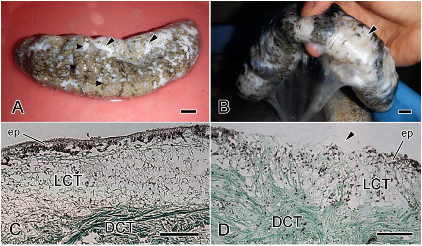
The symptomatology of SKUD resembles that of SSWD in that the integument of affected animals develops white lesions where destruction of the cuticle, epidermis, and outer dermis has exposed the deeper dermis (Figure 12). The lesions widen and the dermis undergoes extreme softening prior to being completely punctured [273,274,275]. Further similarities to SSWD have been revealed by a histopathological investigation of Isostichopus badionotus, which demonstrated that changes in the dermis of lesions—disorganization of its collagen fiber microarchitecture, ‘liquefactive necrosis’, and ‘anhistic’ areas (possibly indicating edema)—can occur before the epidermis is damaged [276].
Whilst there is strong evidence that some cases of SKUD are caused by highly infectious bacterial or viral pathogens, the initial causes of others are uncertain and may be abiotic, with opportunistic pathogens infecting lesions secondarily [273,275]. A direct role for MCT in bacterial pathogenesis has been suggested by a proteomic analysis of the body wall of Apostichopus japonicus specimens in which SKUD was induced experimentally by exposure to Vibrio splendidus bacteria. This showed that 5α collagen was one of only two proteins that were downregulated at all stages of disease progression [277]. Since tissue colonization and the establishment of infections in mammals can involve interactions between bacterial pathogens and ECM components [278,279], it has been argued that 5α collagen might be a target protein of causative pathogens, its downregulation representing a protective response [277]. However, if 5α collagen is involved in interfibrillar crosslinking (see Section 5.2), its downregulation could also be a factor in the ECM disorganization that contributes to dermal lesion formation.

7. Applications

Interest in the potential biomedical and other applications of MCT has been steadily growing since Trotter et al. [169] first introduced the concept of MCT-inspired synthetic systems. Two types of applications have emerged: biomimetic applications, which exploit information on MCT organization and the mechanisms underpinning its mechanical adaptability but do not incorporate any MCT components; and biotechnological applications, which use extracted MCT components for deployment in synthetic materials [17].

7.1. Biomimetic Applications Inspired by MCT

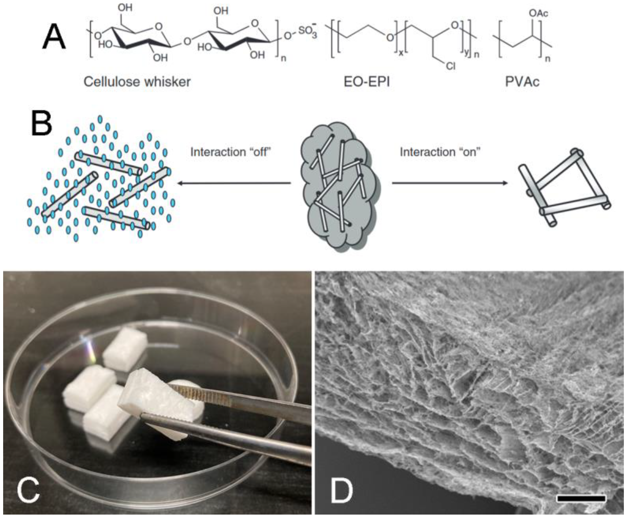
The system envisaged by Trotter et al. [169] was a ‘hybrid’ biomaterial comprising fibrils extracted from holothurian dermis and a synthetic interfibrillar matrix. Capadona et al. [280] were the first to assemble and test a wholly synthetic mechanically adaptive material inspired by sea-cucumber dermis, which could be used in the construction of implanted intracortical microelectrodes. The model employed tunicate (sea-squirt)-derived cellulose nanofibers immersed in a polymeric matrix (ethylene oxide-epichlorohydrin 1:1 polymer or polyvinyl acetate). Cellulose nanofibers tend to aggregate via hydrogen bonding due to the high density of strongly interacting surface hydroxyl groups [281]. This interaction can be switched off by water through competitive hydrogen bonding that reduces hydrogen bonding between the nanofibers and destiffens the composite reversibly. Restiffening occurs when water is removed (Figure 13A,B) [280,282]. In contrast to the reversible stiffening/destiffening system of this first MCT-inspired synthetic composite, irreversible stiffening (which is shown by sea-cucumber Cuvierian tubules: see Section 5.1.2) is the key property of one of the most recent—a calcium phosphate-based cement designed for bone repair, which incorporates cellulose nanofibers and polyvinyl alcohol. This can be injected as a hydrated slurry in which water prevents hydrogen bonding between the nanofibers and the polyvinyl alcohol. When the slurry solidifies, the consequent reduction in the water phase permits hydrogen bonding between the additives, resulting in a material with high strength and toughness [283].
The sea-cucumber dermis has served repeatedly as a conceptual model for the development of hydrogels, which are three-dimensional crosslinked polymeric networks swollen with water. Hydrogels can respond to external and internal physical or chemical stimuli, such as temperature, electric fields, chemical compounds, and pH (for review, see [285]). They have been employed as injectable biomaterials, tunable surfaces for cell sheet engineering, sensors, and actuators (see, e.g., [286]). As indicated by in vitro cytotoxicity tests and preliminary subcutaneous implantation, supramolecular polymer hydrogels can be both biocompatible and autolytic in vivo, and could be used for such purposes as intestinal drug delivery or injectable filling to facilitate the suturing of small vessels.
Ideas derived from sea-cucumber dermis mutability have also assisted the development of shape-memory polymer-cellulose nanofiber composites that can adopt one or more temporary shapes while retaining the ability to return to their original shape [282,287]. Early models employed water movement to regulate hydrogen-bonding interactions in the nanocomposite framework, while later models responded to environmental stimuli, such as temperature or pH changes [288,289,290].
At the most general level, the fast stiffness modulation of MCT, achieved by adjusting interfibrillar stress transfer, has also inspired models of stiffness tuning that involve completely unrelated molecular mechanisms, such as reversible thermoplastic softening, as demonstrated by polymers for coating heating elements [291] and the thermoplastic mesh of a shape-changing material that mimics starfish dermis [50].
MCT has been a source of design concepts for soft actuators with uses in diverse fields including flexible electronics, soft robotics, and biomedicine [212]. Of particular interest are water-driven self-operating actuators that perform movements by means of a swelling/deswelling process. Choi et al. [292] developed a dynamic poly(N-isopropylacrylamide) hydrogel whose elastic modulus could be altered via hydration/dehydration and which had shape-memory properties. This was incorporated into a programmable and ultrafast water-driven self-operating soft actuator (SITuator), which was based on hydrogel stiffness changes obtained by varying crosslinker concentration.
Other synthetic analogs of sea-cucumber dermis have been trialed for employment in soft robotic applications. One such example [293] is an impact-protective supramolecular polymeric material (SPM) characterized by impact-hardening and reversible softness-stiffness switchability. This SPM consists of chains of a triblock copolymer (poly(ethylene glycol)-block-poly(propylene glycol)-block-poly(ethylene glycol) to which 2-ureido-4-pyrimidone (UPy) dimers are attached that are stabilized by quadruple hydrogen bonding. The UPy dimers form crosslinked domains that, when subjected to a sudden impact, undergo reversible stiffening, thereby reducing deformation, possibly through the rapid reconstitution of hydrogen bonds after initial impact-induced hydrogen bond disruption.

7.2. Biotechnological Applications Employing MCT Components

Collagen obtained from animal sources is used widely in the production of biomedical materials for the management of human diseases and defects, particularly where tissue repair and regeneration need to be promoted. Extracted collagen can be assembled to form 2D membranes and 3D meshes, which are subjected to crosslinking procedures to increase their mechanical stability and resistance to enzymatic degradation. Whilst most of the collagen is currently mammalian in origin, there is increasing interest in replacing this with collagen from marine invertebrates, including echinoderms, for various reasons, such as marine collagen’s relative cheapness, ready availability in large quantities, immunological advantages, and low risk of disease transmission [43,294,295]. Since it is very difficult to isolate intact fibrils from the collagenous tissue of postfetal vertebrates (see Section 5.2), most mammalian collagen is used in its hydrolyzed (acid-solubilized) form, which compromises its mechanical performance. Echinoderm collagen, however, can be extracted as intact fibrils using mild, non-denaturing methods that leave in place D-periodically distributed surface GAGs, which may help to stabilize synthetic assemblages of the fibrils [284,295,296].
The main source of echinoderm collagen for prospective biotechnological investigations has been sea-urchin peristomial membranes (PMs), which consist largely of a mutable dermal layer [66,84] and are available in abundance as a waste-product left by the harvesting of sea-urchin gonads for human consumption. Both 2D membranes and 3D scaffolds derived from PM collagen have been investigated as potential alternatives to commercially available bovine collagen-based biomaterials (Figure 13C,D). PM-derived sheets have been shown to have good biocompatibility in terms of enabling the growth of mammalian mesenchymal stromal cells and dermal fibroblasts and could be used for cell culture and human health applications such as guided tissue regeneration [295,296]. Thin films of fibrils extracted from another MCT—sea-cucumber dermis—have reversible stimulus-responsive behavior. At 25 °C the films are translucent and soft; when subjected to an increasing temperature, they become opaque and brittle at 100 °C, reverting to their original state when the temperature drops back to 25 °C [297].
PM-derived collagen is also being used in the development of a new bilayered skin substitute intended as an alternative management option to skin grafting for wound repair. This consists of a membrane that functionally mimics epidermis by reducing water loss and preventing bacterial infiltration, and a scaffold that functions as a mechanically stable dermis. This ‘dermal template’ has been shown to be an effective substrate for fibroblast infiltration, survival, and proliferation [298] and to promote experimental wound healing in sheep and rats, resulting in outcomes similar to those achieved by commercially available artificial skin that incorporates bovine collagen [299,300].
A range of decellularized bioscaffolds that retain their extracellular structural framework and are derived from human and other mammalian tissues is now available and widely applied in tissue engineering and regenerative medicine [301]. Regarding MCT, Goh and Holmes [302] provided an exhaustive theoretical framework pertaining specifically to the use of sea-urchin structures for tissue engineering purposes. However, this application area has been explored so far only in pilot studies employing decellularized dermis from sea-urchin PMs. These PM-derived scaffolds have been investigated with the goal of advancing the development of echinoderm cell cultures, a research field that is still largely unexplored. It is possible that they could eventually have a role in human therapeutics [303].

8. Gaps in Knowledge

The nervously mediated mechanical adaptability of echinoderm collagenous tissue is one of the most unusual phenomena yet encountered in the field of animal biology. However, despite 50 years of research effort, the understanding of it is far from complete and many aspects remain unexamined or poorly characterized. This applies both to the extracellular components of MCT and its key cellular components—the juxtaligamental cells.

8.1. Extracellular Components and Mechanisms of Tensile Change

Significant progress in elucidating the molecular basis of the unique mechanical behavior of MCT—and in realizing its full potential for biomedical and technological exploitation—is unlikely to be made unless there is an exhaustive characterization of its molecular and supramolecular organization, particularly with regard to those components responsible for interfibrillar cohesion and stress transfer. This has not been achieved so far for any mutable collagenous structure. Whilst several possible constitutive or regulatory factors (e.g., stiparin and tensilin, respectively) have been isolated from sea-cucumber dermis, their exact contributions to interfibrillar crosslinking and its dynamics have yet to be ascertained. To put this in context, however, it should be remembered that there is still uncertainty as to which molecular components transfer interfibrillar shear forces in mammalian tendons [157].
Concerning mechanisms of passive tensile change, the three-state model of Motokawa and Tsuchi [135] remains an inspirational paradigm that gave rise to the specific stiffening hypothesis of Tamori et al. [178]. It needs to be emphasized that the three-state model was derived from the mechanical behavior of sea-cucumber dermis and that most of the endogenous factors that influence MCT properties have been isolated from this tissue alone. The relevance of the three-state model for other mutable collagenous structures that show reversible changes is therefore unclear. It is certainly not applicable to those structures that show only irreversible stiffening or destabilization, least of all to the non-fibrillar basement membrane-derived autotomy tendons of brittlestars. It has also become apparent that sea-cucumber dermis cannot be regarded as a representative fibrillar MCT. This can be inferred from the unique presence of fucosylated chondroitin sulfate [49] and of morula cells [110,128], and the unusual features of its juxtaligamental supply: separation from the ECM by a basement membrane, lack of JLC aggregations, and ectoneural innervation [46]. The discovery that sea-cucumbers possess many more TIMP genes than do the other four echinoderm classes [304] may be a further indication of the distinctiveness of their collagenous tissue. These considerations underscore the need for a radical shift in emphasis to other mutable collagenous structures that are amenable to both comparative ‘-omics’ approaches and in vitro functional investigations. Potential experimental models include the body walls of asteriid starfish [86], brittlestar ventral arm plate ligaments [76], and featherstar cirral ligaments [56].
The ability of some featherstar and sea-lily ligaments to develop contractile force adds another layer of complexity to the MCT phenomenon. However, this aspect appears to have been totally neglected for 20 years [208] and the mechanism of force generation remains unknown. Information derived from biomimetic analogs, i.e., contractile hydrogels [292], together with the evidence from different mutable collagenous structures that alterations in passive tensile properties can be accompanied by changes in ECM water content and fibril packing density [98,176,177,178], could be the foundation of testable hypotheses.

8.2. Juxtaligamental Cells

The ontogenesis of JLCs has not been investigated. Elucidation of their developmental origins in different classes would clarify their relationship with the nervous system and help to determine if they are homologous across the phylum [46]. Several experimental models are potentially suitable for this purpose, including the normal (i.e., not regenerating) growing tips of starfish, featherstar and brittlestar arms, the early post-metamorphic stages of sea-urchins [305] and sea-cucumbers [306], and the intrabursal juveniles of viviparous brittlestars such as Amphipholis squamata [307]. Concerning the morphogenesis of juxtaligamental systems, nothing is known of the processes by which mutable collagenous structures come to be populated with JLCs. ECM components are known to regulate the proliferation, differentiation, and migration direction of vertebrate and invertebrate neurons [308,309], and echinoderms possess many homologs of the participating neural adhesion proteins [95]. Insight into this aspect could be obtained by comparative gene expression analyses, taking advantage of experimental models in which a mutable collagenous structure (that contains JLCs) is adjacent to a non-mutable collagenous structure (that lacks JLCs). Accessible and tractable examples of such models are the capsular and central spine ligaments of cidaroid sea-urchins [81] and the autotomy and non-autotomy tendons of brittlestar intervertebral muscles [75].
Whilst circumstantial evidence strongly implicates JLCs as the effectors responsible for the rapid modulation of MCT tensility, a crucial piece of evidence is missing: the demonstration of a correlation between externally provoked, nervously mediated tensile change and the release from JLCs of a previously stored chemical factor that produces the same tensile change when isolated preparations are treated with it in vitro. This is symptomatic of the very limited information that is available on JLC activities and functions, current concepts of which have been derived almost exclusively from ultrastructural observations. This deficiency could be partly redressed by means of transcriptomic and/or metabolomic profiling, an important aim of such an approach being to define JLC secretomes [310,311]. A recent transcriptome-wide gene expression investigation of the brittlestar Ophiocoma wendti is a first step in this direction. A comparison of this animal’s inner arm core (which is enriched in JLCs) with its whole arm (containing a basal level of JLCs) and its stomach (lacking JLCs) led to the identification of 16 genes whose expression may be specific to JLCs [47]. It is feasible that this methodology could be applied to isolated JLCs because juxtaligamental nodes in the arms of some brittlestar species are surgically accessible [46,106].

Author Contributions

Conceptualization, I.C.W. and M.D.C.C.; writing—original draft preparation, I.C.W.; writing—review and editing, I.C.W. and M.D.C.C. All authors have read and agreed to the published version of the manuscript.

Funding

This research received no external funding.

Institutional Review Board Statement

Not applicable.

Informed Consent Statement

Not applicable.

Data Availability Statement

No new data were created or analyzed in this study. Data sharing is not applicable to this article.

Conflicts of Interest

The authors declare no conflicts of interest.

Abbreviations

AChacetylcholine
APFautotomy-promoting factor
BMbasement membrane
ECMextracellular matrix
GAGglycosaminoglycan
JLCjuxtaligamental cell
LDCVlarge dense-core vesicle
LMlight micrograph
MCTmutable collagenous tissue
MMPmatrix metalloproteinase
PMperistomial membrane
SCDAsea-cucumber dermal autolysis
SKUDskin ulceration disease
SPMsupramolecular polymeric material
SSWDsea star wasting disease
TEMtransmission electron micrograph
TIMPtissue inhibitor of matrix metalloproteinase

References

  1. Baccetti, B. Collagen and animal phylogeny. In Biology of Invertebrate and Lower Vertebrate Collagens; Bairati, A., Garrone, R., Eds.; Plenum Press: New York, NY, USA, 1985; pp. 29–47. [Google Scholar]
  2. Gordon, M.K.; Hahn, R.A. Collagens. Cell Tissue Res. 2010, 339, 247–257. [Google Scholar] [CrossRef] [PubMed]
  3. Mienaltowski, M.J.; Gonzales, N.L.; Beall, J.M.; Pechanec, M.Y. Basic structure, physiology, and biochemistry of connective tissues and extracellular matrix collagens. In Progress in Heritable Soft Connective Tissue Diseases, 2nd ed.; Halper, J., Ed.; Springer: Cham, Switzerland, 2021; pp. 5–43. [Google Scholar] [CrossRef]
  4. Rahman, M.A. Collagen of extracellular matrix from marine invertebrates and its medical applications. Mar. Drugs 2019, 17, 118. [Google Scholar] [CrossRef]
  5. Jayadev, R.; Sherwood, D.R. Basement membranes. Current Biol. 2017, 27, R207–R211. [Google Scholar] [CrossRef]
  6. Heinoa, J.; Huhtalab, M.; Käpyläa, J.; Johnson, M.S. Evolution of collagen-based adhesion systems. Int. J. Biochem. Cell Biol. 2009, 41, 341–348. [Google Scholar] [CrossRef]
  7. Rasmussen, C.H.; Petersen, D.R.; Moeller, J.B.; Hansson, M.; Dufva, M. Collagen type I improves the differentiation of human embryonic stem cells towards definitive endoderm. PLoS ONE 2015, 10, e0145389. [Google Scholar] [CrossRef]
  8. Wareham, L.K.; Baratta, R.O.; Del Buono, B.J.; Schlumpf, E.; Calkins, D.J. Collagen in the central nervous system: Contributions to neurodegeneration and promise as a therapeutic target. Mol. Neurodegener. 2024, 19, 11. [Google Scholar] [CrossRef]
  9. Langevin, H.M. Connective tissue: A body-wide signaling network? Med. Hypotheses 2006, 66, 1074–1077. [Google Scholar] [CrossRef] [PubMed]
  10. Silver, F.H.; Landis, W.J. Viscoelasticity, energy storage and transmission and dissipation by extracellular matrices in vertebrates. In Collagen: Structure and Mechanics; Fratzl, P., Ed.; Springer: New York, NY, USA, 2008; pp. 133–154. [Google Scholar]
  11. Jung, H.J.; Fisher, M.B.; Woo, S.L.Y. Role of biomechanics in the understanding of normal, injured, and healing ligaments and tendons. Sports Med. Arthrosc. Rehab. Ther. Technol. 2009, 1, 9. [Google Scholar] [CrossRef] [PubMed]
  12. Matson, A.; Konow, N.; Miller, S.; Konow, P.P.; Roberts, T.J. Tendon material properties vary and are interdependent among turkey hindlimb muscles. J. Exp. Biol. 2012, 215, 3552–3558. [Google Scholar] [CrossRef]
  13. Wilkie, I.C. Basement membranes, brittlestar tendons, and their mechanical adaptability. Biology 2024, 13, 375. [Google Scholar] [CrossRef]
  14. Svensson, R.B.; Heinemeier, K.M.; Couppé, C.; Kjaer, M.; Magnusson, S.P. Effect of aging and exercise on the tendon. J. Appl. Physiol. 2016, 121, 1237–1246. [Google Scholar] [CrossRef] [PubMed]
  15. Yoshida, K.; Jayyosi, C.; Lee, N.; Mahendroo, M.; Myers, K.M. Mechanics of cervical remodelling: Insights from rodent models of pregnancy. Interface Focus 2019, 9, 20190026. [Google Scholar] [CrossRef] [PubMed]
  16. Khalilgharibi, N.; Mao, Y. To form and function: On the role of basement membrane mechanics in tissue development, homeostasis and disease. Open Biol. 2021, 11, 200360. [Google Scholar] [CrossRef]
  17. Candia Carnevali, M.D.; Sugni, M.; Bonasoro, F.; Wilkie, I.C. Mutable collagenous tissue: A concept generator for biomimetic materials and devices. Mar. Drugs 2024, 22, 37. [Google Scholar] [CrossRef]
  18. Motokawa, T. Skin of sea cucumbers: The smart connective tissue that alters mechanical properties in response to external stimuli. J. Aero Aqua Bio-Mech. 2019, 8, 2–5. [Google Scholar] [CrossRef]
  19. Tamori, M.; Yamada, A. Possible mechanisms of stiffness changes induced by stiffeners and softeners in catch connective tissue of echinoderms. Mar. Drugs 2023, 21, 140. [Google Scholar] [CrossRef]
  20. Sugi, H.; Ohno, T.; Moriya, M. Mechanism and function of the catch state in molluscan smooth muscle: A historical perspective. Int. J. Mol. Sci. 2020, 21, 7576. [Google Scholar] [CrossRef]
  21. Saleh, F.; Lefebvre, B.; Dupichaud, C.; Martin, E.L.O.; Nohejlová, M.; Spaccesi, L. Skeletal elements controlled soft-tissue preservation in echinoderms from the Early Ordovician Fezouata Biota. Geobios 2023, 81, 51–66. [Google Scholar] [CrossRef]
  22. Waters, J.A.; Bohatý, J.; Macurda, D.B. Feeding postures as indicators of mutable collagenous tissue in extinct echinoderms. Comm. Biol. 2024, 7, 1516. [Google Scholar] [CrossRef]
  23. Rahman, I.A.; Zamora, S. Origin and early evolution of echinoderms. Ann. Rev. Earth Planet. Sci. 2024, 52, 295–320. [Google Scholar] [CrossRef]
  24. Emson, R.H. Bone idle—A recipe for success? In Echinodermata; Keegan, B.F., O’Connor, B.D.S., Eds.; A.A. Balkema: Rotterdam, The Netherlands, 1985; pp. 25–30. [Google Scholar]
  25. Motokawa, T. Catch connective tissue: A key character for echinoderms’ success. In Echinoderm Biology; Burke, R.D., Mladenov, P.V., Lambert, P., Parsley, R.L., Eds.; A.A. Balkema: Rotterdam, The Netherlands, 1988; pp. 39–54. [Google Scholar]
  26. Wilkie, I.C.; Emson, R.H. Mutable collagenous tissues and their significance for echinoderm palaeontology and phylogeny. In Echinoderm Phylogeny and Evolutionary Biology; Paul, C.R.C., Smith, A.B., Eds.; Clarendon Press: Oxford, UK, 1988; pp. 311–330. [Google Scholar]
  27. Wollman, A.J.M.; Nudd, R.; Hedlund, E.G.; Leake, M.C. From Animaculum to single molecules: 300 years of the light microscope. Open Biol. 2015, 5, 150019. [Google Scholar] [CrossRef] [PubMed]
  28. Jickeli, C.F. Vorläufige Mittheilungen über den Bau der Echinodermen. Zool. Anz. 1884, 7, 346–349, 366–370. [Google Scholar]
  29. Perrier, E. Mémoire sur l’organisation et le développement de la comatule de la Méditerranée (Antedon rosacea Linck). Part 3. Organisation de l’Antedon adulte. I–IX. Nouv. Arch. Mus. D’Hist. Nat. Paris (3) 1889, 1, 169–286. [Google Scholar]
  30. Bosshard, H. Zur Kenntnis der Verbindungsweise der Skelettstücke der Arme und Ranken von Antedon rosacea Linck (Comatula mediterranea Lam.). Jena. Z. Naturw. 1900, 34, 65–112. [Google Scholar]
  31. Reichensperger, A. Beiträge zur Histologie und zum Verlauf der Regeneration bei Crinoiden. Z. Wiss. Zool. 1912, 101, 1–69. [Google Scholar]
  32. Lindemann, W. Über einige Eigenschaften der Holothurienhaut. Z. Biol. 1900, 39, 18–36. [Google Scholar]
  33. Jordan, H. Über ‘reflexarme’ Tiere. IV. Die Holothurien. 1 Mitteil. Die Reizbarkeit und der Einfluss des zentralen Nervensystems auf die Muskulatur und die muskulähnlichen Fasern der Haut (auf Erregbarkeit und Tonusfunktion). Zool. Jarhrb. Jena. Allg. Zool. Physiol. 1914, 34, 365–436. [Google Scholar]
  34. Jordan, H. Über ‘reflexarme’ Tiere. IV. Die Holothurien. 2 Mitteil. Die Holothurien als hohlorganartige Tiere und die Tonusfunktion ihrer Muskulatur. Zool. Jarhrb. Jena. Allg. Zool. Physiol. 1919, 36, 109–156. [Google Scholar]
  35. von Uexküll, J. Die Sperrmuskulatur der Holothurien. Pflüg. Arch. Ges. Physiol. 1926, 212, 1–14. [Google Scholar] [CrossRef]
  36. von Uexküll, J. Die Physiologie des Seeigelstachels. Z. Biol. 1900, 39, 73–112. [Google Scholar]
  37. Hamann, O. Beiträge zur Histologie der Echinodermen. Jena. Z. Med. Naturw. 1887, 21, 87–266. [Google Scholar]
  38. Prouho, H. Recherches sur le Dorocidaris papillata et quelques autres échinides de la Méditerranée. Arch. Zool. Exp. Gén. 1887, 5, 213–380. [Google Scholar]
  39. Serra-von Buddenbrock, E. Etudes physiologiques et histologiques sur le tégument des holothuries (Holothuria tubulosa). Vie Milieu 1963, 14, 55–70. [Google Scholar]
  40. Takahashi, K. The catch apparatus of the sea-urchin spine I. Gross histology. J. Fac. Sci. Univ. Tokyo Sec. IV 1967, 11, 110–120. [Google Scholar]
  41. Takahashi, K. The catch apparatus of the sea-urchin spine II. Responses to stimuli. J. Fac. Sci. Univ. Tokyo Sec. IV 1967, 11, 122–130. [Google Scholar]
  42. Meyer, D.L. The collagenous nature of problematical ligaments in crinoids (Echinodermata). Mar. Biol. 1971, 9, 235–241. [Google Scholar] [CrossRef]
  43. Wilkie, I.C.; Sugni, M.; Gupta, H.S.; Candia Carnevali, M.D.; Elphick, M.R. The mutable collagenous tissue of echinoderms: From biology to biomedical applications. In Soft Matter for Biomedical Applications; Azevedo, H.S., Mano, J.F., Borges, J., Eds.; The Royal Society of Chemistry: Cambridge, UK, 2021; pp. 3–33. [Google Scholar]
  44. Bonneel, M.; Hennebert, E.; Aranko, A.S.; Hwang, D.S.; Lefevre, M.; Pommier, V.; Wattiez, R.; Delroisse, J.; Flammang, P. Molecular mechanisms mediating stiffening in the mechanically adaptable connective tissues of sea cucumbers. Matrix Biol. 2022, 108, 39–54. [Google Scholar] [CrossRef]
  45. Barbieri, E.; Gupta, H.S. Is stress relaxation in sea cucumber dermis chemoelastic? Mar. Drugs 2023, 21, 610. [Google Scholar] [CrossRef]
  46. Wilkie, I.C.; Candia Carnevali, M.D. The juxtaligamental cells of echinoderms and their role in the mechano-effector function of connective tissue. In Frontiers in Invertebrate Physiology: A Collection of Reviews, Volume 3: Annelida and Echinodermata; Saleuddin, A.S., Leys, S., Roer, R., Wilkie, I.C., Eds.; Apple Academic Press: Point Pleasant, NJ, USA, 2024; pp. 345–430. [Google Scholar]
  47. Nouri, R.; Mashanov, V.; Harris, A.; New, G.; Taylor, W.; Janies, D.; Reid, R.W.; Machado, D.J. Unveiling putative modulators of mutable collagenous tissue in the brittle star Ophiomastix wendtii: An RNA-Seq analysis. BMC Genom. 2024, 25, 1013. [Google Scholar] [CrossRef]
  48. Romano, G.; Almeida, M.; Varela Coelho, A.; Cutignano, A.; Gonçalves, L.G.; Hansen, E.; Khnykin, D.; Mass, T.; Ramšak, A.; Rocha, M.S.; et al. Biomaterials and bioactive natural products from marine invertebrates: From basic research to innovative applications. Mar. Drugs 2022, 20, 219. [Google Scholar] [CrossRef]
  49. Atanassova, M.R.; Kolden Midtbø, L.; Mildenberger, J.; Friðjónsson, Ó.H. Novel biomaterials and biotechnological applications derived from North Atlantic sea cucumbers: A systematic review. In The World of Sea Cucumbers; Mercier, A., Hamel, J.F., Suhrbier, A., Pearce, C., Eds.; Academic Press: Cambridge, MA, USA, 2024; pp. 585–609. [Google Scholar] [CrossRef]
  50. Raman; Labisch, S.; Dirks, J.H. A starfish-inspired 4D self-healing morphing structure. Sci. Rep. 2024, 14, 22024. [Google Scholar] [CrossRef]
  51. Wilkie, I.C. Arm autotomy in brittlestars. J. Zool. Lond. 1978, 186, 311–330. [Google Scholar] [CrossRef]
  52. Motokawa, T. Rapid change in mechanical properties of echinoderm connective tissues caused by coelomic fluid. Comp. Biochem. Physiol. C 1982, 73, 223–229. [Google Scholar] [CrossRef]
  53. Wilkie, I.C.; Griffiths, G.V.R.; Glennie, S.F. Morphological and physiological aspects of the autotomy plane in the aboral integument of Asterias rubens L. (Echinodermata). In Echinoderm Research; De Ridder, C., Dubois, P., Lahaye, M., Jangoux, M., Eds.; A.A. Balkema: Rotterdam, The Netherlands, 1990; pp. 301–313. [Google Scholar]
  54. Wilkie, I.C.; Emson, R.H.; Mladenov, P.V. Autotomy mechanism and its control in the starfish Pycnopodia helianthoides (Brandt). In Echinoderm Research 1995; Emson, R.H., Smith, A.B., Campbell, A.C., Eds.; A.A. Balkema: Rotterdam, The Netherlands, 1995; pp. 137–146. [Google Scholar]
  55. Santos, R.; Haesaerts, D.; Jangoux, M.; Flammang, P. The tube feet of sea urchins and sea stars contain functionally different mutable collagenous tissues. J. Exp. Biol. 2005, 208, 2277–2288. [Google Scholar] [CrossRef] [PubMed]
  56. Wilkie, I.C. Nervously mediated change in the mechanical properties of the cirral ligaments of a crinoid. Mar. Behav. Physiol. 1983, 9, 229–248. [Google Scholar] [CrossRef]
  57. Wilkie, I.C.; Candia Carnevali, M.D.; Bonasoro, F. Evidence for the ‘cellular regulation hypothesis’ from ‘simple’ mutable collagenous structures: The brachial and cirral syzygial ligaments of Antedon mediterranea (Lam.). In Echinoderm Research 1998; Candia Carnevali, M.D., Bonasoro, F., Eds.; A.A. Balkema: Rotterdam, The Netherlands, 1999; pp. 119–125. [Google Scholar]
  58. Wilkie, I.C.; Emson, R.H.; Young, C.M. Variable tensility of the ligaments in the stalk of a sea-lily. Comp. Biochem. Physiol. A 1994, 109, 633–641. [Google Scholar] [CrossRef]
  59. Wilkie, I.C.; Barbaglio, A.; Maclaren, W.M.; Candia Carnevali, M.D. Physiological and immunocytochemical evidence that glutamatergic neurotransmission is involved in the activation of arm autotomy in the featherstar Antedon mediterranea (Echinodermata: Crinoidea). J. Exp. Biol. 2010, 213, 2104–2115. [Google Scholar] [CrossRef]
  60. Bobrovskaya, N.V.; Dolmatov, I.Y. Autotomy of the visceral mass in the feather star Himerometra robustipinna (Crinoidea, Comatulida). Biol. Bull. 2014, 226, 81–91. [Google Scholar] [CrossRef] [PubMed]
  61. Hidaka, M.; Takahashi, K. Fine structure and mechanical properties of the catch apparatus of the sea-urchin spine, a collagenous connective tissue with muscle-like holding capacity. J. Exp. Biol. 1983, 103, 1–14. [Google Scholar] [CrossRef]
  62. Rodríguez-Barreras, R.; Sabat, A.M. Evaluation of three tagging methods in the sea urchin Diadema antillarum. J. Mar. Biol. Ass. UK 2015, 95, 1255–1260. [Google Scholar] [CrossRef]
  63. Motokawa, T. Mechanical properties and structure of the spine-joint central ligament of the sea urchin, Diadema setosum (Echinodermata, Echinoidea). J. Zool. Lond. 1983, 201, 223–235. [Google Scholar] [CrossRef]
  64. Birenheide, R.; Tsuchi, A.; Motokawa, T. To be stiff or to be soft—The dilemma of the echinoid tooth ligament. II. Mechanical properties. Biol. Bull. 1996, 190, 231–236. [Google Scholar] [CrossRef]
  65. Wilkie, I.C.; Candia Carnevali, M.D.; Bonasoro, F. The compass depressors of Paracentrotus lividus (Echinodermata: Echinoida): Ultrastructural and mechanical aspects of their variable tensility and contractility. Zoomorphology 1992, 112, 142–153. [Google Scholar] [CrossRef]
  66. Wilkie, I.C.; Candia Carnevali, M.D.; Andrietti, F. Variable tensility of the peristomial membrane of the sea-urchin Paracentrotus lividus (Lamarck). Comp. Biochem. Physiol. A 1993, 105, 493–501. [Google Scholar] [CrossRef]
  67. Seagar, L. The Mechanical Properties of the Sea-Urchin Periproct. Bachelor’s Thesis, Glasgow Caledonian University, Glasgow, Scotland, 1994. [Google Scholar]
  68. Bretschneider, A.; Pomory, C.M. Autotomy of globiferous pedicellariae in the sea urchin Lytechinus variegatus (Echinodermata: Echinoidea). Gulf. Carib. Res. 2024, 35, 16–22. [Google Scholar] [CrossRef]
  69. Motokawa, T. The stiffness change of the holothurian dermis caused by chemical and electrical stimulation. Comp. Biochem. Physiol. C 1981, 73, 41–48. [Google Scholar] [CrossRef]
  70. Byrne, M. The mechanical properties of the autotomy tissues of the holothurian Eupentacta quinquesemita and the effects of certain physico-chemical agents. J. Exp. Biol. 1985, 117, 69–86. [Google Scholar] [CrossRef]
  71. Demeuldre, M.; Hennebert, E.; Bonneel, M.; Lengerer, B.; Van Dyck, S.; Wattiez, R.; Ladurner, P.; Flammang, P. Mechanical adaptability of sea cucumber Cuvierian tubules involves a mutable collagenous tissue. J. Exp. Biol. 2017, 220, 2108–2119. [Google Scholar] [CrossRef]
  72. Wilkie, I.C.; Emson, R.H.; Mladenov, P.V. Morphological and mechanical aspects of fission in Ophiocomella ophiactoides. Zoomorphology 1984, 104, 310–322. [Google Scholar] [CrossRef]
  73. Motokawa, T. Connective tissue catch in echinoderms. Biol. Rev. 1984, 59, 255–270. [Google Scholar] [CrossRef]
  74. Wilkie, I.C. Design for disaster: The ophiuroid intervertebral ligament as a typical mutable collagenous structure. In Echinoderm Biology; Burke, R.D., Mladenov, P.V., Lambert, P., Parsley, R.L., Eds.; A.A. Balkema: Rotterdam, The Netherlands, 1988; pp. 25–38. [Google Scholar]
  75. Wilkie, I.C.; Emson, R.H. The tendons of Ophiocomina nigra and their role in autotomy (Echinodermata, Ophiuroida). Zoomorphology 1987, 107, 33–44. [Google Scholar] [CrossRef]
  76. Wilkie, I.C. Variable tensility of the oral arm plate ligaments of the brittlestar Ophiura ophiura (Echinodermata: Ophiuroidea). J. Zool. Lond. 1992, 228, 5–26. [Google Scholar] [CrossRef]
  77. Dobson, W.E.; Turner, R.L. Morphology and histology of the disc autotomy plane in Ophiophragmus filograneus (Echinodermata, Ophiurida). Zoomorphology 1989, 108, 323–332. [Google Scholar] [CrossRef]
  78. Wilkie, I.C. Functional morphology of the arm spine joint and adjacent structures of the brittlestar Ophiocomina nigra (Echinodermata: Ophiuroidea). PLoS ONE 2016, 11, e0167533. [Google Scholar] [CrossRef]
  79. Candia Carnevali, M.D.; Bonasoro, F.; Wilkie, I.C. Structural and mechanical aspects of the mouth-frame of the brittlestar Ophioderma longicaudum (Retz.). In Echinoderms through Time; David, B., Guille, A., Féral, J.P., Roux, M., Eds.; A.A. Balkema: Rotterdam, The Netherlands, 1988; pp. 387–392. [Google Scholar]
  80. Wilkie, I.C.; McKew, M.; Candia Carnevali, M.D. Functional morphology of the compass-rotular ligament of Echinus esculentus (Echinodermata: Echinoida): A non-mutable collagenous component of Aristotle’s lantern. Zoomorphology 2005, 124, 9–26. [Google Scholar] [CrossRef]
  81. Del Castillo, J.; Smith, D.S.; Vidal, A.M.; Sierra, C. Catch in the primary spines of the sea urchin Eucidaris tribuloides: A brief review and a new interpretation. Biol. Bull. 1995, 188, 120–127. [Google Scholar] [CrossRef]
  82. Smith, D.S.; del Castillo, J.; Morales, M.; Luke, B. The attachment of collagenous ligament to stereom in primary spine of the sea-urchin Eucidaris tribuloides. Tissue Cell 1990, 22, 157–176. [Google Scholar] [CrossRef]
  83. O’Neill, P.L. Structure and mechanics of starfish body wall. J. Exp. Biol. 1989, 147, 53–89. [Google Scholar] [CrossRef]
  84. Wilkie, I.C.; Candia Carnevali, M.D.; Andrietti, F. Microarchitecture and mechanics of the sea-urchin peristomial membrane. Boll. Zool. 1994, 61, 39–51. [Google Scholar] [CrossRef]
  85. Fratzl, P. Collagen: Structure and Biomechanics; Springer: Berlin, Germany, 2008. [Google Scholar]
  86. Wilkie, I.C.; Candia Carnevali, M.D. Morphological and physiological aspects of mutable collagenous tissue at the autotomy plane of the starfish Asterias rubens L. (Echinodermata, Asteroidea): An echinoderm paradigm. Mar. Drugs 2023, 21, 138. [Google Scholar] [CrossRef]
  87. Cluzel, C.; Lethias, C.; Garrone, R.; Exposito, J.-Y. Distinct maturations of N-propeptide domains in fibrillar procollagen molecules involved in the formation of heterotypic fibrils in adult sea urchin collagenous tissues. J. Biol. Chem. 2004, 279, 9811–9817. [Google Scholar] [CrossRef]
  88. Cui, F.; Xue, C.; Li, Z.; Zhang, Y.; Dong, P.; Fu, X.; Gao, X. Characterization and subunit composition of collagen from the body wall of sea cucumber Stichopus japonicus. Food Chem. 2007, 100, 1120–1125. [Google Scholar] [CrossRef]
  89. Tian, M.; Xue, C.; Chang, Y.; Shen, J.; Zhang, Y.; Li, Z.; Wang, Y. Collagen fibrils of sea cucumber (Apostichopus japonicus) are heterotypic. Food Chem. 2020, 316, 126272. [Google Scholar] [CrossRef]
  90. Hulmes, D.J.S. Collagen diversity, synthesis and assembly. In Collagen: Structure and Mechanics; Fratzl, P., Ed.; Springer: Boston, MA, USA, 2008; pp. 15–47. ISBN 978-0-387-73906-9. [Google Scholar]
  91. Bisgrove, B.W.; Andrews, M.E.; Raff, R.A. Fibropellins, products of an EGF repeat-containing gene, form a unique extracellular matrix structure that surrounds the sea-urchin embryo. Dev. Biol. 1991, 146, 89–99. [Google Scholar] [CrossRef]
  92. Cluzel, C.; Lethias, C.; Humbert, F.; Garronne, R.; Exposito, J.-Y. Characterization of fibrosurfin, an interfibrillar component of sea urchin catch connective tissues. J. Biol. Chem. 2001, 276, 18108–18114. [Google Scholar] [CrossRef]
  93. Dolmatov, I.Y.; Afanasyev, S.V.; Boyko, A.V. Molecular mechanisms of fission in echinoderms: Transcriptome analysis. PLoS ONE 2018, 13, e0195836. [Google Scholar] [CrossRef]
  94. Sea Urchin Genome Sequencing Consortium; Sodergren, E.; Weinstock, G.M.; Davidson, E.H.; Cameron, R.A.; Gibbs, R.A.; Angerer, R.C.; Angerer, L.M.; Arnone, M.I.; Burgess, D.R.; et al. The genome of the sea urchin Strongylocentrotus purpuratus. Science 2006, 314, 941–952. [Google Scholar] [CrossRef]
  95. Whittaker, C.A.; Bergeron, K.-F.; Whittle, J.; Brandhorst, B.P.; Burke, R.D.; Hynes, R.O. The echinoderm adhesome. Dev. Biol. 2006, 300, 252–266. [Google Scholar] [CrossRef]
  96. Dolmatov, I.Y.; Nizhnichenko, V.A. Extracellular matrix of echinoderms. Mar. Drugs 2023, 21, 417. [Google Scholar] [CrossRef]
  97. Shi, F.; Liu, K.; Chen, G.; Chang, Y.; Xue, C. Investigation of the presence of fibrillin in sea cucumber (Apostichopus japonicus) body wall by utilizing targeted proteomics and visualization strategies. J. Agr. Food Chem. 2024, 72, 8798–8804. [Google Scholar] [CrossRef]
  98. Ribeiro, A.R.; Barbaglio, A.; Benedetto, C.D.; Ribeiro, C.C.; Wilkie, I.C.; Candia Carnevali, M.D.; Barbosa, M.A. New insights into mutable collagenous tissue: Correlations between the microstructure and mechanical state of a sea-urchin ligament. PLoS ONE 2011, 6, e24822. [Google Scholar] [CrossRef]
  99. Wilkie, I.C. Mutable collagenous tissue: Overview and biotechnological perspective. In Echinodermata (Progress in Molecular and Subcellular Biology 39); Matranga, V., Ed.; Springer: Berlin/Heidelberg, Germany, 2005; pp. 221–250. ISBN 978-3-540-24402-8. [Google Scholar] [CrossRef]
  100. Erlinger, R.; Welsch, U.; Scott, J.E. Ultrastructural and biochemical observations on proteoglycans and collagen in the mutable connective tissue of the feather star Antedon bifida (Echinodermata, Crinoidea). J. Anat. 1993, 183, 1–11. [Google Scholar]
  101. Wang, J.; Chang, Y.; Wu, F.; Xu, X.; Xue, C. Fucosylated chondroitin sulfate is covalently associated with collagen fibrils in sea cucumber Apostichopus japonicus body wall. Carbohyd. Polym. 2018, 186, 439–444. [Google Scholar] [CrossRef]
  102. Felix, A.L.; Penno, S.M.; Bezerra, F.F.; Mourão, P.A.S. Fucosylated chondroitin sulfate, an intriguing polysaccharide from sea cucumber: Past, present, and future. Glycobiology 2025, 35, cwae098. [Google Scholar] [CrossRef]
  103. Exposito, J.Y.; D’Alessio, M.; Di Liberto, M.; Ramirez, F. Complete primary structure of a sea urchin type IV collagen α chain and analysis of the 5’ end of its gene. J. Biol. Chem. 1993, 268, 5249–5254. [Google Scholar] [CrossRef]
  104. Bechtel, M.; Keller, M.V.; Bloch, W.; Sasaki, T.; Boukamp, P.; Zaucke, F.; Paulsson, M.; Nischt, R. Different domains in nidogen-1 and nidogen-2 drive basement membrane formation in skin organotypic cocultures. FASEB J. 2012, 26, 3637–3648. [Google Scholar] [CrossRef]
  105. Hohenester, E.; Yurchenco, P.D. Laminins in basement membrane assembly. Cell Adhes. Migr. 2013, 7, 56–63. [Google Scholar] [CrossRef]
  106. Wilkie, I.C. The juxtaligamental cells of Ophiocomina nigra and their possible role in mechano-effector function of collagenous tissue. Cell Tissue Res. 1979, 197, 515–530. [Google Scholar] [CrossRef]
  107. Motokawa, T. Fine structure of the dermis of the body wall of the sea cucumber, Stichopus chloronotus, a connective tissue which changes its mechanical properties. Galaxea 1982, 1, 55–64. [Google Scholar]
  108. Koob, T.J.; Koob-Emunds, M.M.; Trotter, J.A. Cell-derived stiffening and plasticizing factors in sea cucumber (Cucumaria frondosa) dermis. J. Exp. Biol. 1999, 202, 2291–2301. [Google Scholar] [CrossRef]
  109. Byrne, M. The morphology of autotomy structures in the sea cucumber Eupentacta quinquesemita before and during evisceration. J. Exp. Biol. 2001, 204, 848–863. [Google Scholar] [CrossRef]
  110. Byrne, M. Morphological, physiological and mechanical features of the mutable collagenous tissues associated with autotomy and evisceration in dendrochirotid holothuroids. Mar. Drugs 2023, 21, 134. [Google Scholar] [CrossRef]
  111. Chia, F.S.; Koss, R. Asteroidea. In Microscopic Anatomy of Invertebrates, Volume 14. Echinodermata; Harrison, F.W., Chia, F.S., Eds.; Wiley-Liss: New York, NY, USA, 1994; pp. 169–245. [Google Scholar]
  112. Byrne, M. Ophiuroidea. In Microscopic Anatomy of Invertebrates, Volume 14. Echinodermata; Harrison, F.W., Chia, F.S., Eds.; Wiley-Liss: New York, NY, USA, 1994; pp. 247–343. [Google Scholar]
  113. Hennebert, E.; Haesaerts, D.; Dubois, P.; Flammang, P. Evaluation of the different forces brought into play during tube foot activities in sea stars. J. Exp. Biol. 2010, 213, 1162–1174. [Google Scholar] [CrossRef]
  114. Welsch, U.; Heinzeller, T.; Cobb, J.L.S. Ultrastructure and innervation of ligament connective tissue in Antedon bifida and Decametra sp. In Echinoderm Research; De Ridder, C., Dubois, P., LaHaye, M.C., Jangoux, M., Eds.; A.A. Balkema: Rotterdam, The Netherlands, 1990; pp. 295–300. [Google Scholar]
  115. Grimmer, J.C.; Holland, N.D.; Hayami, I. Fine structure of the stalk of an isocrinid sea lily (Metacrinus rotundus) (Echinodermata, Crinoidea). Zoomorphologie 1985, 105, 39–50. [Google Scholar] [CrossRef]
  116. Smith, D.S.; Wainwright, S.A.; Baker, J.; Cayer, M.L. Structural features associated with movement and ‘catch’ of sea-urchin spines. Tissue Cell 1981, 13, 299–320. [Google Scholar] [CrossRef]
  117. Welsch, U.; Heinzeller, T.; Cobb, J.L.S. Histochemical and finestructural observations on the nervous tissue of Antedon bifida and Decametra spec. (Echinodermata: Crinoidea). Biomed. Res. 1989, 10, 145–154. [Google Scholar]
  118. Zueva, O.; Khoury, M.; Heinzeller, T.; Mashanova, D.; Mashanov, V. The complex simplicity of the brittle star nervous system. Front. Zool. 2018, 15, 1. [Google Scholar] [CrossRef]
  119. Kreiner, T.; Sossin, W.; Scheller, R.H. Localization of Aplysia neurosecretory peptides to multiple populations of dense core vesicles. J. Cell Biol. 1986, 102, 769–782. [Google Scholar] [CrossRef]
  120. Plattner, H.; Artalejo, A.R.; Neher, E. Ultrastructural organization of bovine chromaffin cell cortex—Analysis by cryofixation and morphometry of aspects pertinent to exocytosis. J. Cell Biol. 1997, 139, 1709–1717. [Google Scholar] [CrossRef]
  121. Smith, C.L.; Varoqueaux, F.; Kittelmann, M.; Azzam, R.N.; Cooper, B.; Winters, C.A.; Eitel, M.; Fasshauer, D.; Reese, T.S. Novel cell types, neurosecretory cells, and body plan of the early-diverging metazoan Trichoplax adhaerens. Current Biol. 2014, 24, 1565–1572. [Google Scholar] [CrossRef]
  122. Welsch, U.; Lange, A.; Bals, R.; Heinzeller, T. Juxtaligamental cells in feather stars and isocrinids. In Echinoderm Research 1995; Emson, R.H., Smith, A.B., Campbell, A.C., Eds.; A.A. Balkema: Rotterdam, The Netherlands, 1995; pp. 129–135. [Google Scholar]
  123. Peters, B.H. The innervation of spines in the sea-urchin Echinus esculentus L. An electron-microscopic study. Cell Tissue Res. 1985, 239, 219–228. [Google Scholar] [CrossRef]
  124. Birenheide, R.; Motokawa, T. To be stiff or to be soft—The dilemma of the echinoid tooth ligament. I. Morphology. Biol. Bull. 1996, 190, 218–230. [Google Scholar] [CrossRef]
  125. Mashanov, V.; Zueva, O.; Rubilar, T.; Epherra, L.; García-Arrarás, J.E. Echinodermata. In Structure and Evolution of Invertebrate Nervous Systems; Schmidt-Rhaesa, A., Harzsch, S., Purschke, G., Eds.; Oxford University Press: Oxford, UK, 2016; pp. 665–688. [Google Scholar]
  126. Grimmer, J.C.; Holland, N.D.; Kubota, H. The fine structure of the stalk of the pentacrinoid larva of a feather star, Comanthus japonica. Acta Zool. Stockh. 1984, 65, 41–58. [Google Scholar] [CrossRef]
  127. Wilkie, I.C.; McKew, M.; Candia Carnevali, M.D. Unusual morphological features of the compass-rotular ligament of Echinus esculentus L. In Echinoderms: München; Heinzeller, T., Nebelsick, J.H., Eds.; Taylor & Francis Group: London, UK, 2004; pp. 379–385. [Google Scholar]
  128. Byrne, M. The ultrastructure of the morula cells of Eupentacta quinquesemita (Echinodermata: Holothuroidea) and their role in the maintenance of the extracellular matrix. J. Exp. Biol. 1986, 188, 179–189. [Google Scholar] [CrossRef]
  129. Motokawa, T. Morphology of spines and spine joint in the crown-of-thorns starfish Acanthaster planci (Echinodermata, Asteroida). Zoomorphology 1986, 106, 247–253. [Google Scholar] [CrossRef]
  130. Wilkie, I.C.; Candia Carnevali, M.D.; Bonasoro, F. Organization and mechanical behaviour of myocyte-ligament composites in a sea-urchin lantern: The compass depressors of Stylocidaris affinis. Zoomorphology 1998, 118, 87–101. [Google Scholar] [CrossRef]
  131. Frank, C.B.; Shrive, N.G. Ligament. In Biomechanics of the Musculo-Skeletal System; Nigg, B.M., Herzog, W., Eds.; Wiley: Chichester, NH, USA, 1994; pp. 106–131. [Google Scholar]
  132. Wang, J.H.C. Mechanobiology of tendon. J. Biomech. 2006, 39, 1563–1582. [Google Scholar] [CrossRef]
  133. Birenheide, R.; Yokoyama, K.; Motokawa, T. Cirri of the stalked crinoid Metacrinus rotundus: Neural elements and the effect of cholinergic agonists on mechanical properties. Proc. R. Soc. Lond. B 2000, 267, 7–16. [Google Scholar] [CrossRef]
  134. Ellers, O.; Telford, M. Advancement mechanics of growing teeth in sand dollars (Echinodermata, Echinoidea): A role for mutable collagenous tissue. Proc. R. Soc. Lond. B 1996, 263, 39–44. [Google Scholar]
  135. Motokawa, T.; Tsuchi, A. Dynamic mechanical properties of the body-wall dermis in various mechanical states and their implications for the behavior of sea cucumbers. Biol. Bull. 2003, 205, 261–275. [Google Scholar] [CrossRef]
  136. Wilkie, I.C.; Fassini, D.; Cullorà, E.; Barbaglio, A.; Tricarico, S.; Sugni, M.; Del Giacco, L.; Candia Carnevali, M.D. Mechanical properties of the compass depressors of the sea-urchin Paracentrotus lividus (Echinodermata, Echinoidea) and the effects of enzymes, neurotransmitters and synthetic tensilin-like protein. PLoS ONE 2015, 10, e0120339. [Google Scholar] [CrossRef]
  137. Motokawa, T. Viscoelasticity of holothurian body wall. J. Exp. Biol. 1984, 109, 63–75. [Google Scholar] [CrossRef]
  138. Motokawa, T. The viscosity change of the body-wall dermis of the sea cucumber Stichopus japonicus caused by mechanical and chemical stimulation. Comp. Biochem. Physiol. A 1984, 77, 419–423. [Google Scholar]
  139. Motokawa, T. Factors regulating the mechanical properties of holothurian dermis. J. Exp. Biol. 1982, 99, 29–41. [Google Scholar] [CrossRef]
  140. Eylers, J.P. Ion-dependent viscosity of holothurian body wall and its implications for the functional morphology of echinoderms. J. Exp. Biol. 1982, 99, 1–8. [Google Scholar] [CrossRef]
  141. Pioletti, D.P.; Rakotomanana, L.R.; Benvenuti, J.F.; Leyvraz, P.F. Viscoelastic constitutive law in large deformations: Application to human knee ligaments and tendons. J. Biomech. 1998, 31, 753–757. [Google Scholar] [CrossRef]
  142. Del Prete, Z.; Antoniucci, S.; Hoffman, A.H.; Grigg, P. Viscoelastic properties of skin in Mov-13 and Tsk mice. J. Biomech. 2004, 37, 1491–1497. [Google Scholar] [CrossRef]
  143. Harkness, M.L.R.; Harkness, R.D. Changes in the physical properties of the uterine cervix of the rat during pregnancy. J. Physiol. 1959, 148, 524–547. [Google Scholar] [CrossRef]
  144. Silver, F.H.; Freeman, J.W.; DeVore, D. Viscoelastic properties of human skin and processed dermis. Skin Res. Technol. 2001, 7, 18–23. [Google Scholar] [CrossRef]
  145. Marrs, J.; Wilkie, I.C.; Sköld, M.; Maclaren, W.M.; McKenzie, J.D. Size-related aspects of arm damage, tissue mechanics, and autotomy in the starfish Asterias rubens. Mar. Biol. 2000, 137, 59–70. [Google Scholar] [CrossRef]
  146. O’Neill, P.L.; Withers, P.C. An analysis of the load curve of the body wall of Coscinasterias calamaria (Echinodermata: Asteroidea). Mar. Fresh. Behav. Physiol. 1995, 25, 245–260. [Google Scholar] [CrossRef]
  147. Motokawa, T. Mechanical mutability in connective tissue of starfish body wall. Biol. Bull. 2011, 221, 280–291. [Google Scholar] [CrossRef]
  148. Birenheide, R.; Motokawa, T. Morphological basis and mechanics of arm movement in the stalked crinoid Metacrinus rotundus (Echinodermata, Crinoida). Mar. Biol. 1994, 121, 273–283. [Google Scholar] [CrossRef]
  149. Szulgit, G.K.; Shadwick, R.E. The effects of calcium chelation and cell perforation on the mechanical properties of sea urchin ligaments. In Echinoderms Through Time; David, B., Guille, A., Féral, J.P., Roux, M., Eds.; A.A. Balkema: Rotterdam, The Netherlands, 1988; pp. 887–892. [Google Scholar]
  150. Trotter, J.A.; Koob, T.J. Collagen and proteoglycan in a sea urchin ligament with mutable mechanical properties. Cell Tissue Res. 1989, 258, 527–539. [Google Scholar] [CrossRef]
  151. Legerlotz, K.; Riley, G.P.; Screen, H.R.C. GAG depletion increases the stress-relaxation response of tendon fascicles, but does not influence recovery. Acta Biomater. 2013, 9, 6860–6866. [Google Scholar] [CrossRef]
  152. Silver, F.H.; Seehra, G.P.; Freeman, J.W.; DeVore, D.P. Viscoelastic properties of young and old human dermis: A proposed molecular mechanism for elastic energy storage in collagen and elastin. J. App. Polym. Sci. 2002, 86, 1978–1985. [Google Scholar] [CrossRef]
  153. Mattucci, S.F.E.; Moulton, J.A.; Chandrashekar, N.; Cronin, D.S. Strain rate dependent properties of younger human cervical spine ligaments. J. Mech. Behav. Biomed. Mater. 2012, 10, 216–226. [Google Scholar] [CrossRef]
  154. Kastelic, J.; Baer, E. Deformation in tendon collagen. Symp. Soc. Exp. Biol. 1980, 34, 397–435. [Google Scholar]
  155. Wilkie, I.C. Autotomy as a prelude to autotomy in echinoderms. Microscop. Res. Technol. 2001, 55, 369–396. [Google Scholar] [CrossRef]
  156. Ciarletta, P.; Ben Amar, M. A finite dissipative theory of temporary interfibrillar bridges in the extracellular matrix of ligaments and tendons. J. R. Soc. Interface 2009, 6, 909–924. [Google Scholar] [CrossRef]
  157. Szczesny, S.E.; Elliott, D.M. Interfibrillar shear stress is the loading mechanism of collagen fibrils in tendon. Acta Biomater. 2014, 10, 2582–2590. [Google Scholar] [CrossRef]
  158. Tricarico, S.; Barbaglio, A.; Burlini, N.; Del Giacco, L.; Ghilardi, A.; Sugni, M.; Di Benedetto, C.; Bonasoro, F.; Wilkie, I.C.; Candia Carnevali, M.D. New insights into the mutable collagenous tissue of Paracentrotus lividus: Preliminary results. Zoosymposia 2012, 7, 279–285. [Google Scholar] [CrossRef]
  159. Barbaglio, A.; Tricarico, S.; Ribeiro, A.R.; Di Benedetto, C.; Barbato, M.; Dessì, D.; Fugnanesi, V.; Magni, S.; Mosca, F.; Sugni, M.; et al. Ultrastructural and biochemical characterization of mechanically adaptable collagenous structures in the edible sea urchin Paracentrotus lividus. Zoology 2015, 118, 147–160. [Google Scholar] [CrossRef]
  160. Birk, D.E.; Nurminskaya, M.V.; Zycband, E.I. Collagen fibrillogenesis in situ: Fibril segments undergo post-depositional modifications resulting in linear and lateral growth during matrix development. Dev. Dyn. 1995, 202, 229–243. [Google Scholar] [CrossRef] [PubMed]
  161. deVente, J.E.; Lester, G.E.; Trotter, J.A.; Dahners, L.E. Isolation of intact collagen fibrils from healing ligament. J. Electron Microsc. 1997, 46, 353–356. [Google Scholar] [CrossRef]
  162. Liu, Y.; Andarawis-Puri, N.; Eppell, S.J. Method to extract minimally damaged collagen fibrils from tendon. J. Biol. Methods 2016, 3, e54. [Google Scholar] [CrossRef]
  163. Wilkie, I.C. Variable tensility in echinoderm collagenous tissues: A review. Mar. Behav. Physiol. 1984, 11, 1–34. [Google Scholar] [CrossRef]
  164. Wilkie, I.C. Mutable collagenous tissues: Extracellular matrix as mechano-effector. In Echinoderm Studies 5; Jangoux, M., Lawrence, J.M., Eds.; A.A. Balkema: Rotterdam, The Netherlands, 1996; pp. 61–102. [Google Scholar]
  165. Wilkie, I.C. Is muscle involved in the mechanical adaptability of echinoderm mutable collagenous tissue? J. Exp. Biol. 2002, 205, 159–165. [Google Scholar] [CrossRef] [PubMed]
  166. Bonneel, M.; Hennebert, E.; Byrne, M.; Flammang, P. Mutable collagenous tissues in sea cucumbers. In The World of Sea Cucumbers; Mercier, A., Hamel, J.-F., Suhrbier, A., Pearce, C., Eds.; Academic Press: Cambridge, MA, USA, 2024; pp. 573–584. [Google Scholar] [CrossRef]
  167. Tamori, M.; Yamada, A.; Nishida, N.; Motobayashi, Y.; Oiwa, K.; Motokawa, T. Tensilin-like stiffening protein from Holothuria leucospilota does not induce the stiffest state of catch connective tissue. J. Exp. Biol. 2006, 209, 1594–1602. [Google Scholar] [CrossRef] [PubMed]
  168. Yamada, A.; Tamori, M.; Iketani, T.; Oiwa, K.; Motokawa, T. A novel stiffening factor inducing the stiffest state of holothurian catch connective tissue. J. Exp. Biol. 2010, 213, 3416–3422. [Google Scholar] [CrossRef]
  169. Trotter, J.A.; Tipper, J.; Lyons-Levy, G.; Chino, K.; Heuer, A.H.; Liu, Z.; Mrksich, M.; Hodneland, C.; Dillmore, W.S.; Koob, T.J.; et al. Towards a fibrous composite with dynamically controlled stiffness: Lessons from echinoderms. Biochem. Soc. Trans. 2000, 28, 357–362. [Google Scholar] [CrossRef]
  170. Takehana, Y.; Yamada, A.; Tamori, M.; Motokawa, T. Softenin, a novel protein that softens the connective tissue of sea cucumbers through inhibiting interaction between collagen fibrils. PLoS ONE 2014, 9, e85644. [Google Scholar] [CrossRef]
  171. Ingersoll, E.P.; Wilt, F.H. Matrix metalloproteinase inhibitors disrupt spicule formation by primary mesenchyme cells in the sea urchin embryo. Dev. Biol. 1998, 196, 95–106. [Google Scholar] [CrossRef]
  172. Sharpe, C.; Robinson, J.J. Characterization of matrix metalloprotease activities induced in the sea urchin extraembryonic matrix, the hyaline layer. Biochem. Cell Biol. 2001, 79, 461–468. [Google Scholar] [CrossRef]
  173. Quiñones, J.L.; Rosa, R.; Ruiz, D.L.; García-Arrarás, J.E. Extracellular matrix remodeling and metalloproteinase involvement during intestine regeneration in the sea cucumber Holothuria glaberrima. Dev. Biol. 2002, 250, 181–197. [Google Scholar] [CrossRef]
  174. Ribeiro, A.R.; Barbaglio, A.; Oliveira, M.J.; Ribeiro, C.C.; Wilkie, I.C.; Candia Carnevali, M.D.; Barbosa, M.A. Matrix metalloproteinases in a sea urchin ligament with adaptable mechanical properties. PLoS ONE 2012, 7, e49016. [Google Scholar] [CrossRef]
  175. Elphick, M.R. The protein precursors of peptides that affect the mechanics of connective tissue and/or muscle in the echinoderm Apostichopus japonicus. PLoS ONE 2012, 7, e44492. [Google Scholar] [CrossRef]
  176. Tamori, M.; Takemae, C.; Motokawa, T. Evidence that water exudes when holothurian connective tissue stiffens. J. Exp. Biol. 2010, 213, 1960–1966. [Google Scholar] [CrossRef]
  177. Ribeiro, A.R.; Barbaglio, A.; Oliveira, M.J.; Santos, R.; Coelho, A.V.; Ribeiro, C.C.; Wilkie, I.C.; Candia Carnevali, M.D.; Barbosa, M.A. Correlations between the biochemistry and mechanical states of a sea-urchin ligament: A mutable collagenous structure. Biointerphases 2012, 7, 38. [Google Scholar] [CrossRef]
  178. Tamori, M.; Ishida, K.; Matsuura, E.; Ogasawara, K.; Hanasaka, T.; Takehana, Y.; Motokawa, T.; Osawa, T. Ultrastructural changes associated with reversible stiffening in catch connective tissue of sea cucumbers. PLoS ONE 2016, 11, e0155673. [Google Scholar] [CrossRef]
  179. Mo, J.; Prévost, S.F.; Blowes, L.M.; Egertová, M.; Terrill, N.J.; Wang, W.; Elphick, M.R.; Gupta, H.S. Interfibrillar stiffening of echinoderm mutable collagenous tissue demonstrated at the nanoscale. Proc. Natl. Acad. Sci. USA 2016, 113, E6362–E6371. [Google Scholar] [CrossRef]
  180. Gupta, H.S.; Barbieri, E.; Inamdar, S.R.; Mo, J. Synchrotron X-ray imaging combined with multiscale modelling applied to biological soft tissues. In Soft Matter for Biomedical Applications; Azevedo, H.S., Mano, J.F., Borges, J., Eds.; The Royal Society of Chemistry: Cambridge, UK, 2021; pp. 34–60. [Google Scholar]
  181. Barbieri, E.; Mo, J.; Gupta, H.S. Chemoviscoelasticity of the interfibrillar matrix of the dermis of the black sea cucumber Holothuria atra. Mech. Mater. 2022, 168, 104252. [Google Scholar] [CrossRef]
  182. Charlina, N.A.; Dolmatov, I.Y.; Wilkie, I.C. Juxtaligamental system of the disc and oral frame of the ophiuroid Amphipholis kochii (Echinodermata: Ophiuroidea) and its role in autotomy. Invert. Biol. 2009, 128, 145–156. [Google Scholar] [CrossRef]
  183. Scalettar, B.A. How neurosecretory vesicles release their cargo. Neuroscientist 2006, 12, 164–176. [Google Scholar] [CrossRef]
  184. Imamura, Y.; Morita, S.; Nakatani, Y.; Okada, K.; Ueshima, S.; Matsuo, O.; Miyata, S. Tissue plasminogen activator and plasminogen are critical for osmotic homeostasis by regulating vasopressin secretion. J. Neurosci. Res. 2010, 88, 1995–2006. [Google Scholar] [CrossRef] [PubMed]
  185. Bai, H.; Nangia, S.; Parmer, R.J. The plasminogen activation system and the regulation of catecholaminergic function. J. Biomed. Biotechnol. 2012, 2012, 721657. [Google Scholar] [CrossRef]
  186. Díaz-Balzac, C.A.; Santacana-Laffitte, G.; San Miguel-Ruíz, J.E.; Tossas, K.; Valentín-Tirado, G.; Rives-Sánchez, M.; Mesleh, A.; Torres, I.I.; García-Arrarás, J.E. Identification of nerve plexi in connective tissues of the sea cucumber Holothuria glaberrima by using a novel nerve-specific antibody. Biol. Bull. 2007, 213, 28–42. [Google Scholar] [CrossRef] [PubMed]
  187. Tinoco, A.B.; Barreiro-Iglesias, A.; Yañez-Guerra, L.A.; Delroisse, J.; Zhang, Y.; Gunner, E.F.; Zampronio, C.G.; Jones, A.M.; Egertová, M.; Elphick, M.R. Ancient role of sulfakinin/cholecystokinin-type signalling in inhibitory regulation of feeding processes revealed in an echinoderm. eLife 2021, 10, e65667. [Google Scholar] [CrossRef]
  188. Cobb, J.L.S. The motor innervation of the oral plate ligament in the brittlestar Ophiura ophiura. Cell Tissue Res. 1985, 242, 685–688. [Google Scholar] [CrossRef]
  189. Cobb, J.L.S. Neurobiology of the Echinodermata. In Nervous Systems in Invertebrates; Ali, M.A., Ed.; Plenum Press: New York, NY, USA, 1987; pp. 483–525. [Google Scholar]
  190. Inoue, M.; Birenheide, R.; Koizumi, O.; Kobayakawa, Y.; Muneoka, Y.; Motokawa, T. Localization of the neuropeptide NGIWYamide in the holothurian nervous system and its effects on muscular contraction. Proc. R. Soc. Lond. B 1999, 266, 993–1000. [Google Scholar] [CrossRef]
  191. Cobb, J.L.S.; Begbie, K.M. Aspects of the hyponeural nervous system. In Echinoderms Through Time; David, B., Guille, A., Féral, J.P., Roux, M., Eds.; A.A. Balkema: Rotterdam, The Netherlands, 1988; pp. 25–29. [Google Scholar]
  192. Díaz-Balzac, C.A.; García-Arrarás, J.E. Echinoderm nervous system. In Oxford Research Encyclopedia, Neuroscience; Sherman, S.M., Ed.; Oxford University Press: New York, NY, USA, 2019; pp. 1–31. [Google Scholar]
  193. Motokawa, T. Cholinergic control of the mechanical properties of the catch connective tissue in the holothurian body wall. Comp. Biochem. Physiol. C 1987, 86, 333–337. [Google Scholar] [CrossRef]
  194. Morales, M.; Sierra, C.; Vidal, A.; Del Castillo, J.; Smith, D.S. Pharmacological sensitivity of the articular capsule of the primary spines of Eucidaris tribuloides. Comp. Biochem. Physiol. C 1993, 105, 25–30. [Google Scholar] [CrossRef]
  195. Motokawa, T.; Fuchigama, Y. Coordination between catch connective tissue and muscles through nerves in the spine joint of the sea urchin Diadema setosum. J. Exp. Biol. 2015, 218, 703–710. [Google Scholar] [CrossRef]
  196. Birenheide, R.; Tamori, T.; Motokawa, T.; Ohtani, M.; Iwakoshi, E.; Muneoka, Y.; Fujita, T.; Minakata, H.; Nomoto, K. Peptides controlling stiffness of connective tissue in sea cucumbers. Biol. Bull. 1998, 194, 253–259. [Google Scholar] [CrossRef]
  197. Tamori, M.; Saha, A.K.; Matsuno, A.; Noskor, S.C.; Koizumi, O.; Kobayakawa, Y.; Nakajima, Y.; Motokawa, T. Stichopin-containing nerves and secretory cells specific to connective tissues of the sea cucumber. Proc. R. Soc. B 2007, 274, 2279–2285. [Google Scholar] [CrossRef]
  198. Aleotti, A.; Wilkie, I.C.; Yañez-Guerra, L.A.; Gattoni, G.; Rahman, T.A.; Wademan, R.; Ahmad, Z.; Ivanova, D.A.; Semmens, D.C.; Delroisse, J.; et al. Discovery and functional characterization of neuropeptides in crinoid echinoderms. Front. Neurosci. 2022, 16, 1006594. [Google Scholar] [CrossRef]
  199. Tinoco, A.B.; Kirupakaran, V.; Capatina, D.; Egertová, M.; Elphick, M.R. Discovery of a neuropeptide that acts as an autotomy-promoting factor. Curr. Biol. 2024, 34, 4325–4331. [Google Scholar] [CrossRef]
  200. Smith, G.N.; Greenberg, M.J. Chemical control of the evisceration process in Thyone briareus. Biol. Bull. 1973, 144, 421–436. [Google Scholar] [CrossRef]
  201. Byrne, M. Induction of evisceration in the holothurian Eupentacta quinquesemita and evidence for the existence of an endogenous evisceration factor. J. Exp. Biol. 1986, 120, 25–40. [Google Scholar] [CrossRef]
  202. Chaet, A.B. A toxin in the coelomic fluid of scalded starfish (Asterias forbesi). Proc. Soc. Exp. Biol. Med. 1962, 109, 791–794. [Google Scholar] [CrossRef]
  203. Mladenov, P.V.; Igdoura, S.; Asotra, S.; Burke, R.D. Purification and partial characterization of an autotomy-promoting factor from the sea star Pycnopodia helianthoides. Biol. Bull. 1989, 176, 169–175. [Google Scholar] [CrossRef]
  204. Murphy, C.J.; Mladenov, P.V. Autotomy promoting factor (APF) in south west Pacific sea stars. In Troisième Symposium Sur Substances Naturelles D’Intérét Biologique de la Région Pacifique-Asie; Debitus, C., Amade, P., Laurent, D., Cosson, J.P., Eds.; ORSTOM: Paris, France, 1992; pp. 411–420. [Google Scholar]
  205. Morales, M.; del Castillo, J.; Smith, D.S. Acetylcholine sensitivity of the spine-test articular capsule of the sea urchin Eucidaris tribuloides. Comp. Biochem. Physiol. C 1989, 94, 547–554. [Google Scholar] [CrossRef]
  206. Vidal, A.M.; del Castillo, J.; Smith, D.S. Contractile properties of the articular capsule or ligament, in the primary spines of the sea urchin Eucidaris tribuloides. Comp. Biochem. Physiol. C 1993, 106, 643–647. [Google Scholar] [CrossRef]
  207. Takemae, N.; Motokawa, T. Mechanical properties of the isolated catch apparatus of the sea urchin spine joint: Muscle fibers do not contribute to passive stiffness changes. Biol. Bull. 2005, 208, 29–35. [Google Scholar] [CrossRef]
  208. Motokawa, T.; Shintani, O.; Birenheide, R. Contraction and stiffness changes in collagenous arm ligaments of the stalked crinoid Metacrinus rotundus (Echinodermata). Biol. Bull. 2004, 206, 4–12. [Google Scholar] [CrossRef]
  209. Birenheide, R.; Motokawa, T. Motility and stiffness of cirri of the stalked crinoid Metacrinus rotundus. In Echinoderm Research 1995; Emson, R.H., Smith, A.B., Campbell, A.C., Eds.; A.A. Balkema: Rotterdam, The Netherlands, 1995; pp. 91–94. [Google Scholar]
  210. Birenheide, R.; Motokawa, T. Crinoid ligaments: Catch and contractility. In Echinoderms: San Francisco; Mooi, R., Telford, M., Eds.; A.A. Balkema: Rotterdam, The Netherlands, 1998; pp. 139–144. [Google Scholar]
  211. Birenheide, R.; Motokawa, T. Contractile connective tissue in crinoids. Biol. Bull. 1996, 191, 1–4. [Google Scholar] [CrossRef]
  212. Li, W.; Guan, Q.; Li, M.; Saiz, E.; Hou, X. Nature-inspired strategies for the synthesis of hydrogel actuators and their applications. Prog. Polym. Sci. 2023, 140, 101665. [Google Scholar] [CrossRef]
  213. Meyer, D.L. Feeding behavior and ecology of shallow-water unstalked crinoids (Echinodermata) in the Caribbean Sea. Mar. Biol. 1973, 22, 105–129. [Google Scholar] [CrossRef]
  214. Macurda, D.B.; Meyer, D.L. Feeding posture of modern stalked crinoids. Nature 1974, 247, 394–396. [Google Scholar] [CrossRef]
  215. Meyer, D.L.; Veitch, M.; Messing, C.G.; Stevenson, A. Crinoid Feeding Strategies: New Insights from Subsea Video and Time-Lapse; Cambridge University Press: Cambridge, UK, 2021. [Google Scholar]
  216. Warner, G.F. Food and feeding mechanisms: Ophiuroidea. In Echinoderm Nutrition; Jangoux, M., Lawrence, J.M., Eds.; A.A. Balkema: Rotterdam, The Netherlands, 1982; pp. 161–181. [Google Scholar]
  217. Emson, R.H.; Young, C.M. Feeding mechanism of the brisingid starfish Novodinia antillensis. Mar. Biol. 1994, 118, 433–442. [Google Scholar] [CrossRef]
  218. Mitsukuri, K. Notes on the habits and life-history of Stichopus japonicus Selenka. Annot. Zool. Japon. 1903, 5, 1–21. [Google Scholar]
  219. Chen, M.; Li, X.; Zhu, A.; Storey, K.B.; Sun, L.; Gao, T.; Wang, T. Understanding mechanism of sea cucumber Apostichopus japonicus aestivation: Insights from TMT-based proteomic study. Comp. Biochem. Physiol. D 2016, 19, 78–89. [Google Scholar] [CrossRef]
  220. Otter, G.W. Rock-burrowing echinoids. Biol. Bull. 1932, 7, 89–107. [Google Scholar] [CrossRef]
  221. Cobb, J.L.S. A physiological study of the correlation between motor output in hyponeural nerves and arm movements in the brittlestar Ophiura ophiura. Mar. Behav. Physiol. 1990, 17, 147–157. [Google Scholar] [CrossRef]
  222. Grimmer, J.C.; Holland, N.D.; Messing, C.G. Fine structure of the stalk of the bourgueticrinid sea lily Democrinus conifer (Echinodermata: Crinoidea). Mar. Biol. 1984, 81, 163–176. [Google Scholar] [CrossRef]
  223. Holland, N.D.; Grimmer, J.C. Fine structure of the cirri and a possible mechanism for their motility in stalkless crinoids (Echinodermata). Cell Tissue Res. 1981, 214, 207–217. [Google Scholar] [CrossRef]
  224. Takemae, N.; Nakaya, F.; Motokawa, T. Low oxygen consumption and high body content of catch connective tissue contribute to low metabolic rate of sea cucumbers. Biol. Bull. 2009, 216, 45–54. [Google Scholar] [CrossRef]
  225. Motokawa, T.; Sato, E.; Umeyama, K. Energy expenditure associated with softening and stiffening of echinoderm connective tissue. Biol. Bull. 2012, 222, 150–157. [Google Scholar] [CrossRef] [PubMed]
  226. Fredericq, L. Sur l’autotomie ou mutilation par voie refléxe comme moyen de défense chez les animaux. Arch. Zool. Exp. Gén. 1883, 1, 413–426. [Google Scholar]
  227. Kubota, T.; Iwamuro, H.; Miyahara, R. Autotomy of the apodid sea cucumber Polycheira fusca (rufescens): Observations and surgical experiments. Rep. Fac. Sci. Kagoshima Univ. 2001, 34, 25–33. [Google Scholar]
  228. Anderson, J.M. Observations on autotomy in the starfish, Asterias forbesi. Biol. Bull. 1956, 111, 297. [Google Scholar]
  229. Jobson, S.; Penney, H.D.; Hamel, J.F.; Mercier, A. Split personality. Front. Ecol. Environ. 2020, 18, 557. [Google Scholar] [CrossRef]
  230. Emson, R.H.; Wilkie, I.C. Fission and autotomy in echinoderms. Oceanogr. Mar. Biol. Ann. Rev. 1980, 18, 155–250. [Google Scholar]
  231. Jaeckle, W.B. Multiple modes of asexual reproduction by tropical and subtropical sea star larvae: An unusual adaptation for genet dispersal and survival. Biol. Bull. 1994, 186, 62–71. [Google Scholar] [CrossRef]
  232. Uthicke, S. The process of asexual reproduction by transverse fission in Stichopus chloronotus (greenfish). SPC Bech-De-Mer Inform. Bull. 2001, 14, 23–25. [Google Scholar]
  233. Rubilar, T.; Pastor, C.; Díaz de Vivar, E. Timing of fission in the starfish Allostichaster capensis (Echinodermata: Asteroidea) in the laboratory. Rev. Biol. Trop. 2005, 53 (Suppl. S3), 299–303. [Google Scholar]
  234. Crawford, B.J. Changes in the arrangement of the extracellular matrix, larval shape, and mesenchyme cell migration during asteroid larval development. J. Morphol. 1990, 206, 147–161. [Google Scholar] [CrossRef]
  235. Hill, R.B. Role of Ca2+ in excitation-contraction coupling in echinoderm muscle: Comparison with role in other tissues. J. Exp. Biol. 2001, 204, 897–908. [Google Scholar] [CrossRef] [PubMed]
  236. Junqueira, L.C.U.; Montes, G.S.; Mourão, P.A.S.; Carneiro, J.; Salles, L.M.M.; Bonetti, S.S. Collagen-proteoglycans interaction during autotomy in the sea cucumber, Stichopus badionotus. Rev. Can. Biol. 1980, 39, 157–164. [Google Scholar]
  237. Hill, R.B. Propylene phenoxetol as a “preservative” for living holothurians. Nature 1966, 211, 204–205. [Google Scholar] [CrossRef]
  238. Liu, Z.Q.; Zhou, D.Y.; Liu, Y.X.; Yu, M.M.; Liu, B.; Song, L.; Dong, X.P.; Qi, H.; Shahidi, F. Inhibitory effect of natural metal ion chelators on the autolysis of sea cucumber (Stichopus japonicus) and its mechanism. Food Res. Int. 2020, 133, 109205. [Google Scholar] [CrossRef]
  239. Fan, X.; Wu, K.; Tian, X.; Benjakul, S.; Li, Y.; Sang, X.; Zhao, Q.; Zhang, J. Endogenous proteases in sea cucumber (Apostichopus japonicus): Deterioration and prevention during handling, processing, and preservation. Foods 2024, 13, 2153. [Google Scholar] [CrossRef]
  240. Kropp, R.K. Responses of five holothurian species to attacks by a predatory gastropod, Tonna perdix. Pacific Sci. 1982, 36, 445–452. [Google Scholar]
  241. Muzahar, M.; Wulandari, R.; Putri, D.S.; Yulianto, T.; Irawan, H.; Bakkara, O.R. Fission technique on sea cucumbers (Holothuroidea): An alternative method for seed production. BIO Web Conf. 2024, 134, 06005. [Google Scholar] [CrossRef]
  242. Zhu, B.; Zheng, J.; Zhang, Z.; Dong, X.; Zhao, L.; Tada, M. Autophagy plays a potential role in the process of sea cucumber body wall “melting” induced by UV irradiation. Wuhan Univ. J. Nat. Sci. 2008, 13, 232–238. [Google Scholar] [CrossRef]
  243. Liu, Z.Q.; Zhou, D.Y.; Liu, Y.X.; Liu, X.Y.; Liu, Y.; Liu, B.; Song, L.; Shahidi, F. In vivo mechanism of action of matrix metalloprotease (MMP) in the autolysis of sea cucumber (Stichopus japonicus). J. Food Process. Preserv. 2020, 44, e14383. [Google Scholar] [CrossRef]
  244. Sun, L.M.; Wang, T.T.; Zhu, B.W.; Niu, H.L.; Zhang, R.; Hou, H.M.; Zhang, G.L.; Murata, Y. Effect of matrix metalloproteinase on autolysis of sea cucumber Stichopus japonicus. Food Sci. Biotechnol. 2013, 22, 1–3. [Google Scholar] [CrossRef]
  245. Liu, Y.X.; Zhou, D.Y.; Liu, Z.Q.; Lu, T.; Song, L.; Li, D.M.; Dong, X.P.; Qi, H.; Zhu, B.W.; Shahidi, F. Structural and biochemical changes in dermis of sea cucumber (Stichopus japonicus) during autolysis in response to cutting the body wall. Food Chem. 2018, 240, 1254–1261. [Google Scholar] [CrossRef]
  246. Liu, Z.; Liu, Y.; Zhou, D.; Liu, X.; Dong, X.; Li, D.; Shahidi, F. The role of matrix metalloprotease (MMP) to the autolysis of sea cucumber (Stichopus japonicus). J. Sci. Food Agric. 2019, 99, 5752–5759. [Google Scholar] [CrossRef]
  247. Yan, L.J.; Sun, L.; Cao, K.Y.; Chen, Y.L.; Zhang, L.J.; Liu, G.M.; Jin, T.; Cao, M.J. Type I collagen from sea cucumber (Stichopus japonicus) and the role of matrix metalloproteinase-2 in autolysis. Food Biosci. 2021, 41, 100959. [Google Scholar] [CrossRef]
  248. Yan, T.; Sun, J.; Zheng, J.; Yang, J. An analysis combining proteomics and transcriptomics revealed a regulation target of sea cucumber autolysis. Comp. Biochem. Physiol. D 2024, 52, 101274. [Google Scholar] [CrossRef]
  249. Liu, Y.X.; Zhou, D.Y.; Ma, D.D.; Liu, Y.F.; Li, D.M.; Dong, X.P.; Tan, M.Q.; Du, M.; Zhu, B.W. Changes in collagenous tissue microstructures and distributions of cathepsin L in body wall of autolytic sea cucumber (Stichopus japonicus). Food Chem. 2016, 212, 341–348. [Google Scholar] [CrossRef] [PubMed]
  250. Matsuno, A.; Motokawa, T. Evidence for calcium translocation in catch connective tissue of the sea cucumber Stichopus chloronotus. Cell Tissue Res. 1992, 267, 307–312. [Google Scholar] [CrossRef]
  251. Hill, R.B.; Reinschmidt, D. Relative importance of the antioxidant and anesthetic properties of propylene phenoxetol in its action as a “preservative” for living holothurians. J. Invert. Pathol. 1976, 28, 131–135. [Google Scholar] [CrossRef]
  252. Candia Carnevali, M.D.; Bonasoro, F. Microscopic overview of crinoid regeneration. Microscop. Res. Technol. 2001, 55, 403–426. [Google Scholar] [CrossRef] [PubMed]
  253. Biressi, A.C.M.; Zou, T.; Dupont, S.; Dahlberg, C.; Di Benedetto, C.; Bonasoro, F.; Thorndyke, M.C.; Candia Carnevali, M.D. Wound healing and arm regeneration in Ophioderma longicaudum and Amphiura filiformis (Ophiuroidea, Echinodermata): Comparative morphogenesis and histogenesis. Zoomorphology 2010, 129, 1–19. [Google Scholar] [CrossRef]
  254. Ben Khadra, Y.; Sugni, M.; Ferrario, C.; Bonasoro, F.; Coelho, A.V.; Martinez, P.; Candia Carnevali, M.D. An integrated view of asteroid regeneration: Tissues, cells and molecules. Cell Tissue Res. 2017, 370, 13–28. [Google Scholar] [CrossRef]
  255. Ferrario, C.; Sugni, M.; Somorjai, I.M.L.; Ballarin, L. Beyond adult stem cells: Dedifferentiation as a unifying mechanism underlying regeneration in invertebrate deuterostomes. Front. Cell Dev. Biol. 2020, 8, 587320. [Google Scholar] [CrossRef]
  256. Kalacheva, N.V.; Eliseikina, M.G.; Frolova, I.Y.; Dolmatov, I.Y. Regeneration of the digestive system in the crinoid Himerometra robustipinna occurs by transdifferentiation of neurosecretory-like cells. PLoS ONE 2017, 12, e0182001. [Google Scholar] [CrossRef]
  257. Kalacheva, N.V.; Kamenev, Y.O.; Dolmatov, I.Y. Regeneration of the digestive system in the crinoid Lamprometra palmata (Mariametridae, Comatulida). Cell Tissue Res. 2021, 391, 87–109. [Google Scholar] [CrossRef] [PubMed]
  258. Dolmatov, I.Y.; Kalacheva, N.V.; Mekhova, E.S.; Frolova, L.T. Autotomy and regeneration of the visceral mass in feather stars. Zoomorphology 2020, 139, 171–187. [Google Scholar] [CrossRef]
  259. Liu, Y.; Jiang, X.; Yu, M.K.; Dong, J.; Zhang, X.; Tsang, L.L.; Chung, Y.W.; Li, T.; Chan, H.C. Switching from bone marrow-derived neurons to epithelial cells through dedifferentiation and translineage redifferentiation. Cell Biol. Int. 2010, 34, 1075–1083. [Google Scholar] [CrossRef]
  260. Mozzi, D.; Dolmatov, I.Y.; Bonasoro, F.; Candia Carnevali, M.D. Visceral regeneration in the crinoid Antedon mediterranea: Basic mechanisms, tissues and cells involved in gut regrowth. Central Eur. J. Biol. 2006, 1, 609–635. [Google Scholar] [CrossRef]
  261. Candia Carnevali, M.D.; Bonasoro, F. Arm regeneration and pattern formation in crinoids. In Echinoderm Research 1995; Emson, R.H., Smith, A.B., Campbell, A.C., Eds.; A.A. Balkema: Rotterdam, The Netherlands, 1995; pp. 245–253. [Google Scholar]
  262. Byrne, M. The link between autotomy and CNS regeneration: Echinoderms as non-model species for regenerative biology. BioEssays 2020, 42, 1900219. [Google Scholar] [CrossRef]
  263. Oulhen, N.; Byrne, M.; Duffin, P.; Gomez-Chiarri, M.; Hewson, I.; Hodin, J.; Konar, B.; Lipp, E.K.; Miner, B.G.; Newton, A.L.; et al. A review of asteroid biology in the context of sea star wasting: Possible causes and consequences. Biol. Bull. 2022, 243, 50–75. [Google Scholar] [CrossRef]
  264. Hewson, I.; Johnson, M.R.; Reyes-Chavez, B. Lessons learned from the sea star wasting disease investigation. Annu. Rev. Mar. Sci. 2025, 17, 257–279. [Google Scholar] [CrossRef]
  265. Bucci, C.; Francoeur, M.; McGreal, J.; Smolowitz, R.; Zazueta-Novoa, V.; Wessel, G.M.; Gomez-Chiarri, M. Sea star wasting disease in Asterias forbesi along the Atlantic Coast of North America. PLoS ONE 2017, 12, e0188523. [Google Scholar] [CrossRef]
  266. Núñez-Pons, L.; Work, T.M.; Angulo-Preckler, C.; Moles, J.; Avila, C. Exploring the pathology of an epidermal disease affecting a circum-Antarctic sea star. Sci. Rep. 2018, 8, 11353. [Google Scholar] [CrossRef] [PubMed]
  267. Work, T.M.; Weatherby, T.M.; DeRito, C.M.; Besemer, R.M.; Hewson, I. Sea star wasting disease pathology in Pisaster ochraceus shows a basal-to-surface process affecting color phenotypes differently. Dis. Aquat. Org. 2021, 145, 21–33. [Google Scholar] [CrossRef]
  268. Smith, S.; Kunc, H.P.; Hewson, I.; Collins, P.C. Elevated temperature linked to signs associated with sea star wasting disease in a keystone European, Asterias rubens. Mar. Ecol. Prog. Ser. 2023, 724, 97–109. [Google Scholar] [CrossRef]
  269. Gudenkauf, B.M.; Hewson, I. Metatranscriptomic analysis of Pycnopodia helianthoides (Asteroidea) affected by sea star wasting disease. PLoS ONE 2015, 10, e0128150. [Google Scholar] [CrossRef]
  270. Pespeni, M.H.; Lloyd, M.M. Sea stars resist wasting through active immune and collagen systems. Proc. R. Soc. B 2023, 290, 20230347. [Google Scholar] [CrossRef]
  271. Fuess, L.E.; Eisenlord, M.E.; Closek, C.J.; Tracy, A.M.; Mauntz, R.; Gignoux-Wolfsohn, S.; Moritsch, M.M.; Yoshioka, R.; Burge, C.A.; Harvell, C.D.; et al. Up in arms: Immune and nervous system response to sea star wasting disease. PLoS ONE 2015, 10, e0133053. [Google Scholar] [CrossRef]
  272. Deng, H.; He, C.; Zhou, Z.; Liu, C.; Tan, K.; Wang, N.; Jiang, B.; Gao, X.; Liu, W. Isolation and pathogenicity of pathogens from skin ulceration disease and viscera ejection syndrome of the sea cucumber Apostichopus japonicus. Aquaculture 2009, 287, 18–27. [Google Scholar] [CrossRef]
  273. Delroisse, J.; Wayneberghe, K.V.; Flammang, P.; Gillan, D.; Gerbaux, P.; Opina, N.; Todinanahary, G.G.B.; Eeckhaut, I. Epidemiology of Skin Ulceration Disease in the sea cucumber Holothuria scabra with a review on the SKUDs in Holothuroidea (Echinodermata). Sci. Rep. 2020, 10, 22150. [Google Scholar] [CrossRef]
  274. Becker, P.; Gillan, D.; Lanterbecq, D.; Jangoux, M.; Rasolofonirina, R.; Rakotovao, J.; Eeckhaut, I. The skin ulceration disease in cultivated juveniles of Holothuria scabra (Holothuroidea, Echinodermata). Aquaculture 2004, 242, 13–30. [Google Scholar] [CrossRef]
  275. Scott, M.M.F.; Ma, K.C.K.; Wolvin, S.; Hamel, J.F.; Mercier, A. First report of skin ulceration disease from temperate waters of the Northwest Atlantic: The case of the sea cucumber Cucumaria frondosa. SPC Beche-de-mer Inform. Bull. 2024, 44, 22–26. [Google Scholar]
  276. Arjona-Cambranes, K.A.; Olvera-Novoa, M.A.; Cerqueda-García, D.; Arjona-Torres, M.G.; Aguirre-Macedo, M.L.; Vidal-Martínez, V.M.; García-Maldonado, J.Q. Characterization of microbiota and histology of cultured sea cucumber Isostichopus badionotus juveniles during an outbreak of skin ulceration syndrome. PLoS ONE 2024, 19, e0303480. [Google Scholar] [CrossRef]
  277. Zhao, Z.; Jiang, J.; Pan, Y.; Sun, H.; Guan, X.; Gao, S.; Chen, Z.; Dong, Y.; Zhou, Z. Proteomic analysis reveals the important roles of alpha-5-collagen and ATP5β during skin ulceration syndrome progression of sea cucumber Apostichopus japonicus. J. Proteomics 2018, 175, 136–143. [Google Scholar] [CrossRef]
  278. Westerlund, B.; Korhonen, T.K. Bacterial proteins binding to the mammalian extracellular matrix. Mol. Microbiol. 1993, 9, 687–694. [Google Scholar] [CrossRef]
  279. Arora, S.; Gordon, J.; Hook, M. Collagen binding proteins of gram-positive pathogens. Front. Microbiol. 2021, 12, 628798. [Google Scholar] [CrossRef]
  280. Capadona, J.R.; Shanmuganathan, K.; Tyler, D.J.; Rowan, S.J.; Weder, C. Stimuli-responsive polymer nanocomposites inspired by the sea cucumber dermis. Science 2008, 319, 1370–1374. [Google Scholar] [CrossRef]
  281. Wohlert, M.; Benselfelt, T.; Wågberg, L.; István, F.; Berglund, L.A.; Wohlert, J. Cellulose and the role of hydrogen bonds: Not in charge of everything. Cellulose 2022, 29, 1–23. [Google Scholar] [CrossRef]
  282. Mendez, J.; Annamalai, P.K.; Eichhorn, S.J.; Rusli, R.; Rowan, S.J.; Foster, E.J.; Weder, C. Bioinspired mechanically adaptive polymer nanocomposites with water-activated shape-memory effect. Macromolecules 2011, 44, 6827–6835. [Google Scholar] [CrossRef]
  283. Wu, Z.; Wang, Z.; Cai, Z.; Qu, L.; Yu, T. Self-reinforced calcium phosphate cement inspired by sea cucumber dermis. J. Wuhan Univ. Technol.-Mater. Sci. Ed. 2024, 39, 682–688. [Google Scholar] [CrossRef]
  284. Ciulla, M.G.; Massironi, A.; Sugni, M.; Ensign, M.A.; Marzorati, S.; Forouharshad, M. Recent advances in the development of biomimetic materials. Gels 2023, 9, 833. [Google Scholar] [CrossRef]
  285. Lim, H.L.; Hwang, Y.; Kar, M.; Varghese, S. Smart hydrogels as functional biomimetic systems. Biomater. Sci. 2014, 2, 603–618. [Google Scholar] [CrossRef]
  286. Gao, F.; Zhang, Y.; Li, Y.; Xu, B.; Cao, Z.; Liu, W. Sea cucumber-inspired autolytic hydrogels exhibiting tunable high mechanical performances, repairability, and reusability. ACS Appl. Mater. Interfaces 2016, 8, 8956–8966. [Google Scholar] [CrossRef]
  287. Studart, A.R.; Erb, R.M. Bioinspired materials that self-shape through programmed microstructures. Soft Matter 2014, 10, 1284–1294. [Google Scholar] [CrossRef]
  288. Honda, S.; Toyota, T. Photo-triggered solvent-free metamorphosis of polymeric materials. Nat. Commun. 2017, 8, 502. [Google Scholar] [CrossRef]
  289. Montero De Espinosa, L.; Meesorn, W.; Moatsou, D.; Weder, C. Bioinspired polymer systems with stimuli-responsive mechanical properties. Chem. Rev. 2017, 117, 12851–12892. [Google Scholar] [CrossRef]
  290. Moatsou, D.; Weder, C. Mechanically adaptive nanocomposites inspired by sea cucumbers. In Bio-Inspired Polymers; Bruns, N., Kilbinger, A.F.M., Eds.; The Royal Society of Chemistry: Cambridge, UK, 2017; pp. 402–428. [Google Scholar]
  291. Charaklias, D.; Qiang, D.; Dorey, R.; Mohagheghian, I. Rapid de-stiffening of multilayer transparent structures using controlled thermoplastic softening. Smart Mater. Struct. 2023, 32, 115020. [Google Scholar] [CrossRef]
  292. Choi, A.; Han, H.; Kim, D.S. A programmable powerful and ultra-fast water-driven soft actuator inspired by the mutable collagenous tissue of the sea cucumber. J. Mater. Chem. A 2021, 9, 15937–15947. [Google Scholar] [CrossRef]
  293. Liu, K.; Cheng, L.; Zhang, N.; Pan, H.; Fan, X.; Li, G.; Zhang, Z.; Zhao, D.; Zhao, J.; Yang, X.; et al. Biomimetic impact protective supramolecular polymeric materials enabled by quadruple H-bonding. J. Am. Chem. Soc. 2021, 143, 1162–1170. [Google Scholar] [CrossRef]
  294. Subhan, F.; Ikram, M.; Shehzad, A.; Ghafoor, A. Marine collagen: An emerging player in biomedical applications. J. Food Sci. Technol. 2015, 52, 4703–4707. [Google Scholar] [CrossRef]
  295. Ferrario, C.; Leggio, L.; Leone, R.; Di Benedetto, C.; Guidetti, L.; Coccè, V.; Ascagni, M.; Bonasoro, F.; La Porta, C.A.M.; Candia Carnevali, M.D.; et al. Marine-derived collagen biomaterials from echinoderm connective tissues. Mar. Environ. Res. 2017, 128, 46–57. [Google Scholar] [CrossRef]
  296. Di Benedetto, C.; Barbaglio, A.; Martinello, T.; Alongi, V.; Fassini, D.; Cullorà, E.; Patruno, M.; Bonasoro, F.; Barbosa, M.A.; Candia Carnevali, M.D.; et al. Production, characterization and biocompatibility of marine collagen matrices from an alternative and sustainable source: The sea urchin Paracentrotus lividus. Mar. Drugs 2014, 12, 4912–4933. [Google Scholar] [CrossRef]
  297. Sisican, K.M.D.; Torreno, V.P.M.; Yu, E.T.; Conato, M.T. Physicochemical and biochemical characterization of collagen from Stichopus cf. horrens tissues for use as stimuli-responsive thin films. ACS Omega 2023, 8, 35791–35799. [Google Scholar] [CrossRef]
  298. Ferrario, C.; Rusconi, F.; Pulaj, A.; Macchi, R.; Landini, P.; Paroni, M.; Colombo, G.; Martinello, T.; Melotti, L.; Gomiero, C.; et al. From food waste to innovative biomaterial: Sea urchin-derived collagen for applications in skin regenerative medicine. Mar. Drugs 2020, 18, 414. [Google Scholar] [CrossRef]
  299. Melotti, L.; Martinello, T.; Perazzi, A.; Iacopetti, I.; Ferrario, C.; Sugni, M.; Sacchetto, R.; Patruno, M. A prototype skin substitute, made of recycled marine collagen, improves the skin regeneration of sheep. Animals 2021, 11, 1219. [Google Scholar] [CrossRef]
  300. Carolo, A.; Melotti, L.; Zivelonghi, G.; Sacchetto, R.; Akyürek, E.E.; Martinello, T.; Venerando, A.; Iacopetti, I.; Sugni, M.; Martinelli, G.; et al. Mutable collagenous tissue isolated from echinoderms leads to the production of a dermal template that is biocompatible and effective for wound healing in rats. Mar. Drugs 2023, 21, 506. [Google Scholar] [CrossRef]
  301. Golebiowska, A.A.; Intravaia, J.T.; Sathe, V.M.; Kumbar, S.G.; Nukavarapu, S.P. Decellularized extracellular matrix biomaterials for regenerative therapies: Advances, challenges and clinical prospects. Bioactive Mater. 2024, 32, 98–123. [Google Scholar] [CrossRef]
  302. Goh, K.L.; Holmes, D.F. Collagenous extracellular matrix biomaterials for tissue engineering: Lessons from the common sea urchin tissue. Int. J. Mol. Sci. 2017, 18, 901. [Google Scholar] [CrossRef]
  303. Di Benedetto, C. Progenitor Cells and Regenerative Potential in Echinoderms: An In Vivo and In Vitro Approach; Lambert Academic Publishing: Saarbrücken, Germany, 2011. [Google Scholar]
  304. Clouse, R.M.; Linchangco, G.V.; Kerr, A.M.; Reid, R.W.; Janies, D.A. Phylotranscriptomic analysis uncovers a wealth of tissue inhibitor of metalloproteinases variants in echinoderms. R. Soc. Open Sci. 2015, 2, 150377. [Google Scholar] [CrossRef]
  305. Emlet, R.B. Morphological evolution of newly metamorphosed sea urchins—A phylogenetic and functional analysis. Integr. Comp. Biol. 2010, 50, 571–588. [Google Scholar] [CrossRef]
  306. Hamel, J.F.; Mercier, A. Early development, settlement, growth, and spatial distribution of the sea cucumber Cucumaria frondosa (Echinodermata: Holothuroidea). Can. J. Fish. Aquat. Sci. 1996, 53, 253–271. [Google Scholar] [CrossRef]
  307. Formery, L.; Peluso, P.; Rank, D.R.; Rokhsar, D.S.; Lowe, C.J. Anterio-posterior patterning in the brittle star Amphipholis squamata and the evolution of echinoderm body plans. EvoDevo 2025, 16, 7. [Google Scholar] [CrossRef]
  308. Brown, N.H. Extracellular matrix in development: Insights from mechanisms conserved between invertebrates and vertebrates. Cold Spring Harb. Perspect. Biol. 2011, 3, a005082. [Google Scholar] [CrossRef]
  309. Long, K.R.; Huttner, W.B. How the extracellular matrix shapes neural development. Open Biol. 2019, 9, 180216. [Google Scholar] [CrossRef]
  310. Nemes, P.; Knolhoff, A.M.; Rubakhin, S.S.; Sweedler, J.V. Single-cell metabolomics: Changes in the metabolome of freshly isolated and cultured neurons. ACS Chem. Neurosci. 2012, 3, 782–792. [Google Scholar] [CrossRef]
  311. McGrath, L.L.; Vollmer, S.V.; Kaluziak, S.T.; Ayers, J. De novo transcriptome assembly for the lobster Homarus americanus and characterization of differential gene expression across nervous system tissues. BMC Genom. 2016, 17, 63. [Google Scholar] [CrossRef] [PubMed]
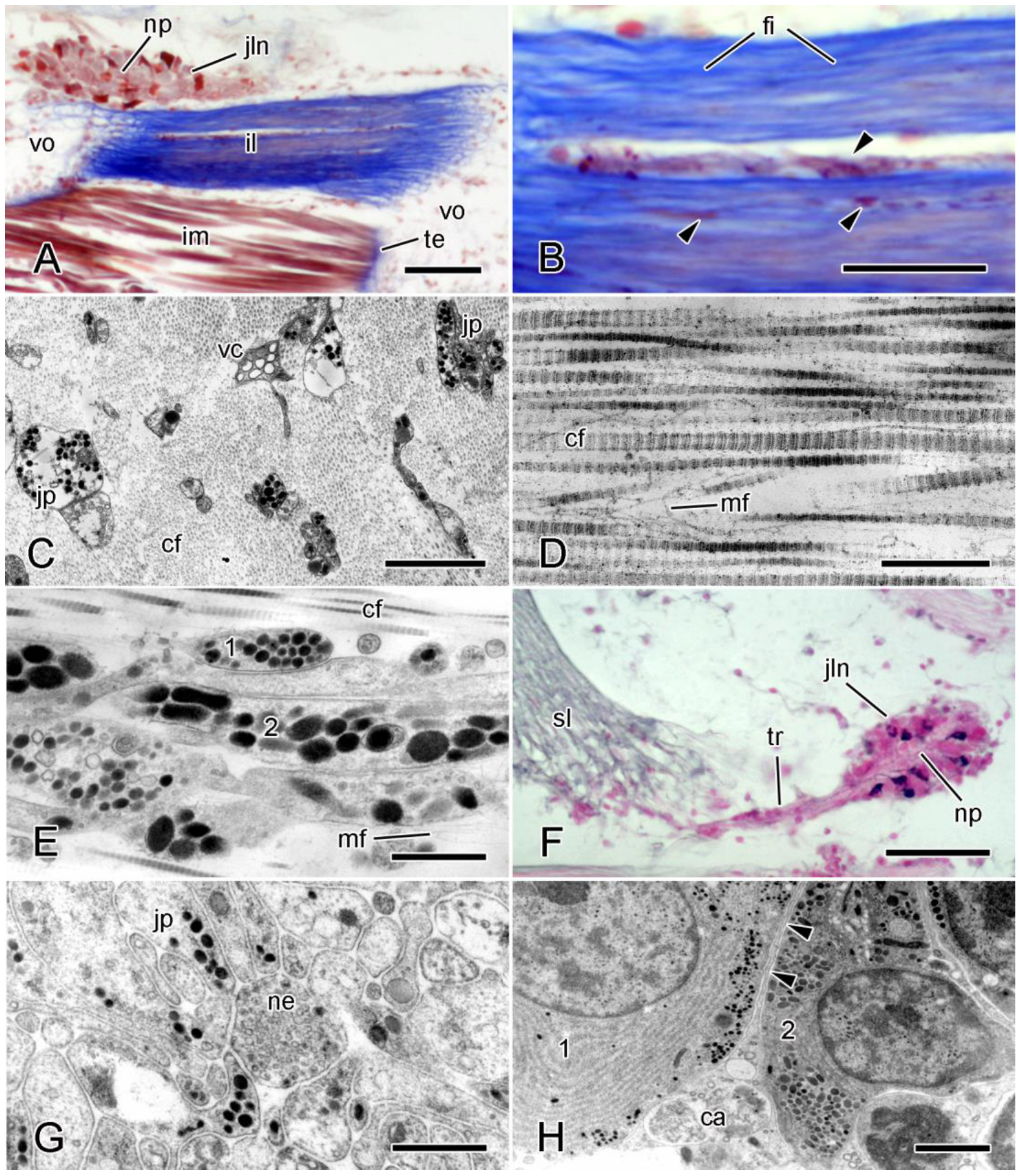
Figure 1. Organization of MCT. Parallel-fiber ligaments. (AD) Intervertebral ligament (il) of brittlestar Ophiocomina nigra. (A,B) Light micrographs (LMs) of horizontal sections stained with Milligan’s trichrome. (A) Ventral region of ligament and adjacent structures: intervertebral muscle (im) attached to vertebral ossicles (vo) by tendons (te), and juxtaligamental node (jln) with central neuropil-like region (np). Scalebar = 50 µm. (B) More magnified view of ligament, showing collagen fibers (fi) and cellular components (arrowheads). Scalebar = 20 µm. (C,D) Transmission electron micrographs (TEMs). (C) Transverse section that includes collagen fibrils (cf), juxtaligamental cell processes (jp), and heterogeneous vacuole-containing cells (vc). Scalebar = 2 µm. (D) Longitudinal section with collagen fibrils and microfibrils (mf). Scalebar = 0.5 µm. (EH) Spine ligament of O. nigra. (E) TEM (longitudinal section) of ligament showing two types of LDCV-containing juxtaligamental cell processes (1, 2). Scalebar = 1 µm. (F) LM (transverse section) of juxtaligamental node and tract of juxtaligamental cell processes (tr) extending into spine ligament (sl); stained with chrome hematoxylin and phloxine. Scalebar = 50 µm. (G,H) TEMs of juxtaligamental node. (G) Neuropil-like region containing juxtaligamental processes and neuronal processes (ne). Scalebar = 1 µm. (H) Edge of node showing two types of juxtaligamental cell (1, 2) and outer capsule (ca) forming intercellular partition (arrowheads). Scalebar = 2 µm. ((A,B) Previously unpublished. (C,E,F,H) Adapted from ref. [17]. (D) Adapted with permission from ref. [51]. Copyright 1978 John Wiley & Sons-Books. (G) Adapted from ref. [78]).
Encyclopedia 05 00185 g001
Figure 2. Organization of MCT. Crossed-fiber dermal structure. Longitudinal interambulacral ligament (lil) of starfish Asterias rubens. (A,B) LMs of horizontal sections stained with Milligan’s trichrome. (A) Ligament connecting adjacent ambulacral ossicles (ao). Scalebar = 0.1 mm. (B) Ligament consists of collagen fibers (fi) and cellular components (arrowheads). Scalebar = 20 µm (CF) TEMs. (C) Longitudinal and transverse sections of collagen fibers, each fiber comprising a tightly packed bundle of fibrils (cf). Scalebar = 1 µm. (D) Cellular aggregation. Two types (1, 2) of JLC processes and cell bodies (jb) are present; mf, microfibrils; nu nucleus. Scalebar = 2 µm. (E) Enlarged details of JLC components in (D). Scalebar = 1 µm. (F) Cellular aggregation that includes presumptive gliocyte components (gc) adjacent to type 2 JLC processes. Scalebar = 2 µm. ((A,B) Adapted from ref. [86]. (CF) Reprinted from ref. [86]).
Encyclopedia 05 00185 g002
Figure 3. Proteoglycan histochemistry. TEMs of sections stained with cupromeronic blue. Note electron-dense deposits (arrowheads) arranged regularly at each D-period of the collagen fibrils (cf) or as interfibrillar bridges. (A) Brachial ligament of featherstar Antedon bifida. Scalebar = 0.2 µm. (B) Inner dermis of dorsolateral body wall of starfish Asterias rubens. Scalebar = 0.2 µm. ((A) Adapted from ref. [17]. (B) Adapted from ref. [86]).
Encyclopedia 05 00185 g003
Figure 4. Autotomy tendons of intervertebral muscles of brittlestar Ophiocomina nigra. Horizontal sections. (A) LM. Horizontal section stained with Milligan’s trichrome. Intervertebral muscle (im) is attached to vertebral ossicle (vo) by tendons (stained blue) which forms loops (arrowheads) that enclose (decalcified) bars of skeletal stereom. Scalebar = 10 µm. (B,C) TEMs. (B) Intact tendons (te), which are extensions of basement membrane of muscle cells. Arrowheads indicate JLC components. Scalebar = 1 µm. (C) Elongating tendon fixed during autotomy. JLC process represented by row of vesicle-like structures (arrowhead). Scalebar = 0.5 µm. ((A) Adapted from ref. [13]. (B,C) Adapted with permission from ref. [75]. Copyright 1987 John Wiley & Sons-Books).
Encyclopedia 05 00185 g004
Figure 5. Passive mechanical behavior of MCT. (AC) Creep tests (elongation under constant load). (A) Syzygial ligament of featherstar Antedon mediterranea. Treatment with seawater containing 100 mmol L−1 K+ ions (K) causes sudden decrease in viscosity and rupture (star). Scalebars = 1 min (horizontal) and 1 mm (vertical). (B,C) Dental ligament of sea-urchin Diadema setosum. Seawater containing 100 mmol L−1 K+ ions or 1 mmol L−1 acetylcholine (ACh) causes reversible increase in viscosity; ASW, artificial seawater. (D) Stress relaxation test (force decay under constant deformation). Average force relaxation curves of 20 dental ligaments of sea-urchin Dendraster excentricus treated with ASW and 15 ligaments treated with divalent cation-free seawater (DCF). (E,F) Stress–strain tests (force recorded while sample stretched at fixed extension rate). Spine ligament of sea-urchin Anthocidaris crassispina. (E) Ligament treated with 0.1 mmol L−1 acetylcholine. Scalebars = 10% strain (horizontal) and 10 MPA (vertical). (F) Ligament treated with 0.1 mmol L−1 adrenaline. Scalebars = 10% strain (horizontal) and 1 MPA (vertical). (GI) Dynamic stress–strain tests (force recorded while sample subjected to oscillating strain). Hysteresis loops obtained by repeated testing of dermis of sea-cucumber Actinopyga mauritiana in three mechanical states. During these tests, maximum strain (indicated by number above each curve) was increased incrementally. ((A) Adapted with permission from ref. [57]. Copyright 1999 Taylor & Francis Group. (B,C) Adapted with permission from ref. [64]. Copyright 1996 University of Chicago Press. (D) Adapted with permission from ref. [134]. Copyright 1996 The Royal Society. (E,F) Adapted with permission from ref. [61]. Copyright 1983 The Company of Biologists. (GI) Adapted with permission from ref. [135]. Copyright 2003 University of Chicago Press).
Encyclopedia 05 00185 g005
Figure 6. Stress–strain tests on Cuvierian tubules from sea-cucumber Holothuria forskali. (A) Typical stress–strain curve of quiescent tubule. (B) Typical stress–strain curve of elongated tubule. (Adapted with permission from ref. [71]. Copyright 2017 The Company of Biologists).
Encyclopedia 05 00185 g006
Figure 7. Molecular organization of MCT. Conjectural model derived mainly from sea-cucumber dermis. Stiparin, novel stiffening factor, and tensilin are assumed to form interfibrillar crossbridges by dimerization. The function and microstructural disposition of fibrosurfin, which has been detected only in sea-urchin MCT, are unknown. (Reprinted with permission from ref. [99]. Copyright 2005 Springer Nature BV).
Encyclopedia 05 00185 g007
Figure 8. Diagrammatic representation of molecular interactions between Hf-(D)Tensilin and collagen fibrils in dermis of sea-cucumber Holothuria forskali. Collagen fibrils (co) are crosslinked (1) at the C-terminal part of tensilin (red circles) by the dimerization or oligomerization of tensilin molecules via positively and negatively charged amino acid interactions (pni) and non-covalent cation-π interactions (cpi), and (2) at the N-terminal part of tensilin, by the binding of the NTR-TIMP-like domain (ntd) with sulfates (s) on glycosaminoglycan (GAG) sidechains of proteoglycans (pg) attached via their protein core (pc) to the surface of collagen fibrils (green circles). (Redrawn and modified from ref. [44]. Reprinted from ref. [17]).
Encyclopedia 05 00185 g008
Figure 9. Force generation by MCT. (A) Contraction of spine ligament of sea-urchin Eucidaris tribuloides in response to 0.1 mmol L−1 acetylcholine (arrow) which was removed as soon as force peaked. (B) Contraction of cirral ligaments of sea-lily Metacrinus rotundus in response to 0.1 mmol L−1 methacholine (M). (CF) Responses to seawater containing 100 mmol L−1 K+ (K) of arm ligaments of M. rotundus. Upper traces show upward displacement (in mm) of arm tip (caused by shortening of ligaments) and lower traces show stiffness changes (as percentages) in ligament. Horizontal scalebars = 3 min. (C) Contraction accompanied by stiffening. (D) Contraction without stiffness change in seawater containing 100 mmol L−1 K+ and contraction with stiffening when excess K+ was removed. (E) No contraction, but with marked stiffening. (F) No contraction, but with destiffening. ((A) Adapted with permission from ref. [206]. Copyright 1993 Elsevier Science & Technology Journals. (B) Adapted with permission from ref. [133]. Copyright 2000 The Royal Society. (CF) Adapted with permission from ref. [208]. Copyright 2004 University of Chicago).
Encyclopedia 05 00185 g009
Figure 10. Sea-cucumber dermal autolysis. (A) Unstimulated specimen of Isostichopus badionotus. Scalebar = 5 cm. (B) Specimen of S. badionotus 60 min after irritant (formalin or Bouin’s fluid) was painted over posterior third of body wall. (Adapted from ref. [236]).
Encyclopedia 05 00185 g010
Figure 11. Sea star wasting disease (SSWD) in Asterias forbesi. (A,B) External appearance of affected animals. (A) Small ulcer (d) and disturbed spine orientation (e). (B) Severe ulceration resulting in exposure of internal organs. (C,D) LMs. Histological sections of affected outer body wall in early stages of disease. Stained with hematoxylin and eosin. de, dermis, ep, epidermis. Scalebars = 100 µm (C) and 50 µm (D). Morphological changes include: a, influx of hemocytes; b, edema; c, epidermal vacuolation; d, cuticular disruption. (Adapted from ref. [265]).
Encyclopedia 05 00185 g011
Figure 12. Skin ulceration disease (SKUD) in sea-cucumber Holothuria scabra. (A,B) External appearance of affected animals, showing ulcers (arrowheads). Scalebars = 1 cm. (A) Stage III. (B) Stage IV. (C,D) LMs. Histological sections of outer body wall stained with Masson’s trichrome. Scalebars = 100 µm. (C) Healthy body wall. (D) SKUD-affected body wall. Arrowhead, ulcer; ep, epidermis; DCT, dense connective tissue; LCT loose connective tissue. (Adapted from ref. [273]).
Encyclopedia 05 00185 g012
Figure 13. Applications. (A,B) Biomimetic application: nanocomposite with dynamic mechanical properties. (A) This consists of cellulose whiskers in a matrix of ethylene oxide-epichlorhydrin 1:1 copolymer (EO-EPI) or poly(vinyl acetate) (PVAc). (B) Switching mechanism: in the ‘on’ stiffened state, hydrogen bonds between the cellulose nanofibers maximize stress transfer; in the ‘off’ destiffened state, these interactions are disrupted by the introduction of a chemical regulator, such as water, which introduces competitive hydrogen bonding. (C,D) Biotechnological application: 3-D scaffold assembled from collagen obtained from sea-urchin peristomial membranes. (C) External view. (D) Scanning electron microscope image. Scalebar = 500 µm. ((A,B) Adapted with permission from ref. [280]. Copyright 2008 AAAS. (C,D) Adapted from ref. [284]).
Encyclopedia 05 00185 g013
Table 1. Mutable collagenous structures. The list is limited to those structures in which the capacity for variable tensility has been demonstrated experimentally or confirmed by direct observation. Tensile changes: Id, irreversible destiffening associated with autotomy; Id*, irreversible destiffening associated with opportunistic self-detachment (see Section 6.2); Is, irreversible stiffening; R, reversible stiffening and destiffening. APL, arm plate ligament; IM, intervertebral muscle; LI, longitudinal interambulacral; PRM-LBWM, pharyngeal retractor muscle–longitudinal body wall muscle; PTG, peripheral through-going; SP, spine-pedicel.
Class and SpeciesAnatomical
Component
Mutable Collagenous
Structure
Tensile
Change
Reference
Asteroidea
Acanthaster planciSpine jointDermis and SP ligamentR[52]
Asterias rubensBody wallDorsolateral dermisR,Id[53]
Pycnopodia helianthoidesBody wallLI ligamentR,Id[54]
Marthasterias glacialisTube footDermisR[55]
Crinoidea
Antedon bifidaCirrusSynathrial ligamentR[56]
Antedon mediterraneaCirrusSynostosal ligamentId[57]
Cenocrinus asteriusStalkSymplexal/PTG ligamentsR[58]
Cenocrinus asteriusStalkSynostosal ligamentId[58]
Antedon mediterraneaArmDiarthrial ligamentR[59]
Antedon mediterraneaArmSyzygial ligamentId[59]
Himerometra robustipinnaVisceral massTegmen and septaId[60]
Echinoidea
Anthocidaris crassispinaSpine jointCapsular ligamentR[61]
Diadema antillarumSpine jointCapsular ligamentId*[62]
Diadema setosumSpine jointCentral ligamentR[63]
Diadema setosumAristotle’s lanternTooth ligamentR[64]
Paracentrotus lividusAristotle’s lanternCompass depressor ligamentR[65]
Paracentrotus lividusTube footDermisR[55]
Paracentrotus lividusBody wallPeristomial membraneR[66]
Echinus esculentusBody wallPeriproctal dermis R[67]
Lytechinus variegatusGlobiferous pedicellariaHead-stalk ligamentId[68]
Holothuroidea
Stichopus chloronotusMain body wallDermisR[69]
Stichopus chloronotusMain body wallDermisId*[25]
Eupentacta quinquesemitaIntrovert body wallDermisId[70]
Eupentacta quinquesemitaLBWMPRM-LBWM tendonId[70]
Holothuria forskaliCuvierian tubuleInner connective tissue layerIs[71]
Ophiuroidea
Ophiocomella ophiactoidesDorsal disk body wallDermisId[72]
Ophiomastix lütkeniArmIntervertebral ligamentR[73]
Ophiocomina nigraArmIntervertebral ligamentR,Id[74]
Ophiocomina nigraArmIM autotomy tendonId[75]
Ophiura ophiuraArmDistal ventral APLR,Id[76]
Ophiura ophiuraArmProximal ventral APLR[76]
Ophiophragmus filograneusArmGenital plate-lateral APLId[77]
Ophiocomina nigraArm spine jointSpine ligamentR,Id*[78]
Ophioderma longicaudumVentral disk body wallOral shield-oral plate ligamentR[79]
Table 2. Non-mutable collagenous structures. The list comprises structures that failed to evince the capacity for variable tensility in physiological experiments.
Class and SpeciesAnatomical
Component
Collagenous
Structure
Reference
Echinoidea
Echinus esculentusAristotle’s lanternCompass-rotular ligament[80]
Eucidaris tribuloidesSpine jointCentral ligament[81]
Ophiuroidea
Ophiocomina nigraArmIntervertebral muscle non-autotomy tendon[75]
Ophioderma longicaudumInterbrachial frameRadial and interradial oral plate ligaments[79]
Table 3. Ultrastructure of juxtaligamental large dense-core vesicles (LDCVs) associated with two examples of collagenous structures from each class. The placement of LDCV data from a particular species in the columns headed ‘LDCV Type’ is not intended to imply homology with LDCVs of other species in the same column. c, circular; CL, capsular ligament; cr, crescentic; CSL, central spine ligament; CT, connective tissue; IL, intervertebral ligament; JLN, juxtaligamental node; LIL, longitudinal interambulacral ligament; o, oval; TF, tube-foot.
Class and SpeciesJLC ComponentLocationLDCV Type (a)LDCV Type (b)Reference
ShapeSize (nm)ShapeSize (nm)
Asteroidea
Asterias
rubens
SomataLILc-o≤250 × 380c≤540[86]
ProcessesLILc-o≤230 × 360c≤540
Marthasterias
glacialis
SomataTF outer CT sheath ca. 150 ca. 250[55]
[113]
ProcessesTF outer CT sheathc-oca. 150 ca. 250
Crinoidea
Decametra
sp.
SomataStereom spacescca. 100oca. 180[114]
ProcessesDiarthrial arm ligamentscca. 100oca. 180
Metacrinus
rotundus
SomataStereom spacesc150–200c600[115]
ProcessesStalk ligamentsc150–200c600
Echinoidea
Arbacia
punctulata
SomataInside + outside CLc-o≤320 [116]
ProcessesInside + outside CLc-o≤220/330
Diadema
setosum
SomataInside CSLc-o100–500 [63]
ProcessesInside CSLc-o100–500
Holothuroidea
Holothuria
forskali
SomataCuvierian tubule CTc200–400c-o300–600[71]
ProcessesCuvierian tubule CTc200–400c-o300–600
Stichopus
chloronotus
SomataBody wall dermiso-cr≤1400 [107]
ProcessesBody wall dermisc-o≤300 × 700
Ophiuroidea
Ophiocomina
nigra
SomataIL JLNsc≤160c-o≤210 × 460[74,106]
ProcessesIntervertebral ligamentc≤300–400c-o≤250 × 750
Ophiocomina
nigra
SomataSpine ligament JLNc≤150c-o≤220 × 310[78]
ProcessesSpine ligamentc≤200c-o≤270 × 520
Table 4. Mutable collagenous structures that contain myocytes. CSA, proportion of the total cross-sectional area occupied by myocytes; SP, spine-pedicel.
Class and SpeciesAnatomical
Component
Collagenous
Structure
CSA (%)Reference
Asteroidea
Acanthaster planciSpine jointDermis and SP ligament<2–ca. 30 1[129]
Echinoidea
Anthocidaris crassispinaSpine jointCapsular ligament3[61]
Eucidaris tribuloidesSpine jointCapsular ligament1.5[81]
Paracentrotus lividusAristotle’s lanternCompass depressor ligament8[65]
Stylocidaris affinisAristotle’s lanternCompass depressor ligament13[130]
Holothuroidea
Eupentacta quinquesemitaIntrovert body wallDermis1–4[110]
1 CSA varies from <2% in the dermis that ensheathes the joint to ca. 30% in the ligament between the spine base and the pedicel ossicle of the body wall [129].
Table 5. Mechanical properties of (A) echinoderm mutable collagenous structures and, for comparison, (B) selected mammalian collagenous structures: creep data. Values shown are means or ranges. Coefficient of viscosity is strain rate/stress. CL, cruciate ligament; DW, distilled water; K, elevated [K+]; ---, no data.
Class and SpeciesMutable Collagenous
Structure
Stress 1
MPa
Coefficient of
Viscosity MPa·s
Strain at
Breakage
Reference
A. Echinodermata
Asteroidea
Asterias rubensBasal aboral body wall 2---0.5---[53]
Asterias rubensDistal aboral body wall 2---1.4---[53]
Echinoidea
Diadema setosumCentral spine ligamentca. 0.120–60000.8–3.7[63]
Paracentrotus lividusCompass depressor ligament0.17–1.16104–14771.41–4.54[136]
Holothuroidea
Actinopyga echinitesDermis0.056100---[137]
Apostichopus japonicusDermis0.3–3.00.076–35---[138]
Eupentacta quinquesemitaIntrovert dermis0.03ca. 100≤9[70]
Holothuria leucospilotaDermis0.05611---[137]
Stichopus chloronotusDermis (untreated, K-stiffened)6 × 10−50.06, 1.25---[139]
Thyone inermisDermis 3 (untreated, DW-stiffened)---5100, 15,800---[140]
Ophiuroidea
Ophiocomella ophiactoidesAboral disk body wall 2---ca. 1000---[72]
Ophiocomina nigraIntervertebral ligament0.2–1.1500–71000.6–3.0[74]
B. Chordata
Mammalia
Homo sapiensAnterior CL, posterior CL---39.29, 48.67---[141]
Homo sapiensPatellar tendon---438.1---[141]
Mus musculusSkin 4---1375---[142]
Rattus norvegicusUterine cervix (pregnant)0.029128.9---[143]
1 In some of the cited investigations, ‘stress’ is initial stress, i.e., load/initial cross-sectional area; in others, it is constant stress, achieved by continuously adjusting the load to compensate for the progressive decline in cross-sectional area during sample extension. 2 The mechanical properties of these structures are determined mainly by the dermis. This, however, consists partly of endoskeletal ossicles, which confounds comparison with purely collagenous structures. 3 The experimental animals were immersed in distilled water for at least 30 min before the excision, treatment (further exposure to distilled water), and mechanical testing of dermis samples, which may explain the anomalously high ‘untreated’ viscosity value. 4 The mechanical properties of mammalian skin are determined mainly by the dermis [144].
Table 6. Mechanical properties of (A) echinoderm mutable collagenous structures and, for comparison, (B) selected mammalian collagenous structures: stress–strain data and ultimate properties. Values shown are means or ranges. Elastic stiffness is the slope of the linear portion of the stress–strain curve. Ultimate stress (=tensile strength) is the maximum stress observed during a stress–strain test. Ultimate strain is the strain occurring at maximum stress. ACh, acetylcholine; K, elevated [K+]; ---, no data.
Class and SpeciesMutable Collagenous
Structure
Strain
Rate s−1
Elastic
Stiffness MPa
Ultimate
Stress MPa
Ultimate
Strain
Reference
A. Echinodermata
Asteroidea
Asterias rubensAboral body wall 1 (anesthetized)0.032.480.680.49[145]
Echinaster spinulosusAboral body wall 1 (stiffened by handling)0.01–0.02249–35337–4512–15[83]
Coscinasterias calamariaAboral body wall 1 (anesthetized)0.09–0.248.6–10.63.270.43[146]
Linckia laevigataDermis 10.0004, 0.00420.9, 36.03.65, 6.2730.5, 23.0[147]
Crinoidea
Metacrinus rotundusAboral brachial ligament---0.4–15.9------[148]
Echinoidea
Anthocidaris crassispinaCapsular spine ligament (ACh-stiffened)ca. 0.003–0.29230–42018–38ca. 0.3[61]
Eucidaris tribuloidesCapsular spine ligament0.006258---[149]
Eucidaris tribuloidesCapsular spine ligament (stiff state)---200------[150]
Paracentrotus lividusCompass depressor ligament0.003–0.2503.3–44.21.5–23.20.56–6.50[136]
Holothuroidea
Actinopyga echinitesDermis (untreated, K-stiffened)1.51.67, 2.08------[138]
Holothuria forskaliCuvierian tubule (quiescent, elongated)---0.044, 19.70.88, 3.22.0, 0.27[71]
Holothuria leucospilotaDermis (untreated, K-stiffened)1.50.42, 0.59------[138]
Stichopus chloronotusDermis (untreated, K-stiffened)1.50.024, 0.1280.03, 0.115>12–13, ---[139]
Ophiuroidea
Ophiocomina nigraIntervertebral ligament (anesthetized)------6.17---[74]
B. Chordata
Mammalia
Bos taurusDigital extensor tendon0.163995.70.231[151]
Homo sapiensDermis (cryopreserved)0.1, 1015–41, 7–45------[152]
Homo sapiensSpine anterior longitudinal ligament0.550321.15[153]
Rattus norvegicusTail tendon0.0013130440–800.05–0.17[154]
1 See Table 5, Footer 2.
Table 7. Oxygen consumption rate (VO2) of the dermis and longitudinal body wall muscle of the sea-cucumber Actinopyga mauritiana and of the dermis and longitudinal carinal muscle of the starfish Linckia laevigata. Dermis samples in the standard state and relaxed muscle preparations were in normal artificial seawater (ASW); dermis samples in the stiff state (see Section 5.1.2) and contracted muscle preparations were in ASW containing 100 mmol L−1 K+ ions; in addition, a second batch of contracted sea-cucumber muscle preparations (asterisk) was in ASW containing 1 mmol L−1 acetylcholine. Data from refs. [224,225].
SpeciesAverage VO2 (µL·g−1·h−1) ± SD
DermisMuscle
StandardStiffRelaxedContracted
Actinopyga mauritiana1.61 ± 0.512.45 ± 1.349.21 ± 3.2823.5 ± 5.8; 18.4 ± 3.6 *
Linckia laevigata0.56 ± 0.120.78 ± 0.1912.44 ± 5.5429.30 ± 17.09
Disclaimer/Publisher’s Note: The statements, opinions and data contained in all publications are solely those of the individual author(s) and contributor(s) and not of MDPI and/or the editor(s). MDPI and/or the editor(s) disclaim responsibility for any injury to people or property resulting from any ideas, methods, instructions or products referred to in the content.