Entry

In Vivo Dosimetry in Radiotherapy: Techniques, Applications, and Future Directions

by
James C. L. Chow
1,2,3,* and
Harry E. Ruda
3,4
1
Radiation Medicine Program, Princess Margaret Cancer Centre, University Health Network, Toronto, ON M5G 1X6, Canada
2
Department of Radiation Oncology, University of Toronto, Toronto, ON M5T 1P5, Canada
3
Department of Materials Science and Engineering, University of Toronto, Toronto, ON M5S 3E4, Canada
4
Centre of Advance Nanotechnology, Faculty of Applied Science and Engineering, University of Toronto, Toronto, ON M5S 3E4, Canada
*
Author to whom correspondence should be addressed.
Encyclopedia 2025, 5(1), 40; https://doi.org/10.3390/encyclopedia5010040
Submission received: 31 January 2025 / Revised: 14 March 2025 / Accepted: 18 March 2025 / Published: 20 March 2025
(This article belongs to the Section Medicine & Pharmacology)

Definition

:
In vivo dosimetry (IVD) is a vital component of modern radiotherapy, ensuring accurate and safe delivery of radiation doses to patients by measuring dose parameters during treatment. This paper provides a comprehensive overview of IVD, covering its fundamental principles, historical development, and the technologies used in clinical practice. Key techniques, including thermoluminescent dosimeters (TLDs), optically stimulated luminescent dosimeters (OSLDs), diodes, metal-oxide-semiconductor field-effect transistors (MOSFETs), and electronic portal imaging devices (EPIDs), are discussed, highlighting their clinical applications, advantages, and limitations. The role of IVD in external beam radiotherapy, brachytherapy, and pediatric treatments is emphasized, particularly its contributions to quality assurance, treatment validation, and error mitigation. Challenges such as measurement uncertainties, technical constraints, and integration into clinical workflows are explored, along with potential solutions and emerging innovations. The paper also addresses future perspectives, including advancements in artificial intelligence, adaptive radiotherapy, and personalized dosimetry systems. This entry underscores the critical role of IVD in enhancing the precision and reliability of radiotherapy, advocating for ongoing research and technological development.

1. Introduction

In vivo dosimetry (IVD) refers to the measurement of the radiation dose delivered directly to the patient during radiotherapy treatments. Unlike pre-treatment dose calculations or phantom-based measurements, IVD provides real-time or near-real-time data on the actual dose received by the patient, ensuring that it aligns with the planned dose distribution [1]. By directly monitoring the radiation delivery process, IVD plays a significant role in maintaining the accuracy and reliability of radiotherapy treatments [2,3].
The primary purpose of IVD is threefold: to ensure precise dose delivery to the target area, to enhance patient safety by identifying and mitigating potential deviations from the treatment plan, and to validate treatment plans by providing feedback for quality assurance (QA) protocols [4]. Through these functions, IVD acts as a safeguard against errors arising from equipment malfunctions [5], patient positioning inaccuracies [6], or anatomical changes during treatment [7].
In radiotherapy, where techniques like total body irradiation (TBI) [8], intensity-modulated radiotherapy (IMRT) [9], volumetric modulated arc therapy (VMAT) [10], and stereotactic body radiotherapy (SBRT) [11] demand high levels of precision, the role of IVD has become more significant. Its integration into clinical workflows aligns with international QA standards and guidelines, such as those established by the International Atomic Energy Agency (IAEA) [12] and the American Association of Physicists in Medicine (AAPM) [13]. By ensuring the consistent and accurate delivery of prescribed radiation doses, IVD contributes to improving treatment outcomes and patient safety, making it an essential component of contemporary radiotherapy practice.
In addition to providing a comprehensive overview of IVD techniques and applications, this entry paper also addresses recent advancements and future directions in the field, building upon the foundational work of previous reviews. Notable among these are the studies by Olaciregui-Ruiz et al., which outline the requirements and future directions for IVD in external beam photon radiotherapy [14]; those by Esposito et al., which review in vivo measurement methods for dose delivery accuracy in stereotactic body radiation therapy [15]; and those by Houlihan et al., which discuss IVD in pelvic brachytherapy [16]. By integrating these perspectives, in this paper, we aim to provide a more holistic understanding of the current state and future potential of IVD in radiotherapy.

2. Background and Overview

The practice of dosimetry in radiotherapy has evolved significantly over the years, transitioning from rudimentary point measurements to advanced systems capable of real-time monitoring and high-precision data acquisition [17]. Early approaches relied on simple ionization chambers or film dosimetry to estimate the radiation dose delivered to patients [18]. While these methods provided valuable initial insights, they lacked the accuracy and adaptability needed for the increasingly complex techniques in radiotherapy.
A major milestone in the evolution of dosimetry was the adoption of thermoluminescent dosimeters (TLDs) in the mid-20th century. TLDs offered a compact and reusable solution for measuring doses, making them a standard tool in both clinical and research settings. Their ability to provide cumulative dose measurements with relatively high accuracy marked a significant advancement in ensuring treatment quality [19].
Subsequent innovations introduced electronic dosimeters, such as diodes [20] and metal-oxide-semiconductor field-effect transistors (MOSFETs) [21], which enabled real-time monitoring of radiation doses during treatment. These devices addressed the limitations of passive systems by offering instantaneous feedback, thus paving the way for more responsive QA practices. The development of electronic portal imaging devices (EPIDs) further enhanced dosimetric capabilities, integrating imaging and dose measurement to streamline workflows in radiotherapy [22].
Parallel to these technological advancements, regulatory frameworks and guidelines emerged to standardize in vivo dosimetry practices and ensure consistent QA across clinical settings [23]. Organizations such as the IAEA and AAPM have been instrumental in promoting the adoption of IVD techniques [24]. Their recommendations emphasized the critical role of dosimetry in reducing errors, improving patient outcomes, and ensuring compliance with safety standards.

3. Principles of In Vivo Dosimetry

IVD is based on fundamental dosimetric principles, including dose, energy, and spatial distribution. The dose, defined as the amount of energy deposited per unit mass of tissue, is a critical parameter for assessing the accuracy of radiation delivery. The energy of the radiation beam determines its penetration depth and interaction with tissues [25], while the spatial distribution ensures that the prescribed dose is accurately delivered to the target area with minimal exposure to surrounding healthy tissues [26]. Together, these principles provide the foundation for the precise measurement and validation of radiation treatments in clinical practice.
IVD utilizes both passive and active dosimeters, each with unique characteristics and applications. Passive dosimeters, such as TLDs and film dosimeters, are widely used for point-dose and spatial dose distribution measurements. TLDs store energy in their crystal lattices; this energy is released as light upon heating, providing cumulative dose readings with high accuracy and reusability [27]. Film dosimeters, on the other hand, offer high-resolution visual representations of dose distributions but are single-use and require labor-intensive processing [28]. Active dosimeters, including diodes, MOSFETs, and OSLDs, provide real-time or near-real-time dose measurements. Diodes are highly sensitive and commonly used for point-dose verification, while MOSFETs are particularly advantageous for small treatment fields due to their compact size and precision. OSLDs, which use light to stimulate the dosimetric material for dose readouts, are favored for their sensitivity, stability, and reusability [29].
The accuracy of IVD is influenced by beam characteristics and patient-specific factors. Beam energy and quality play a significant role in determining the depth and uniformity of dose deposition. For example, higher-energy beams enable deeper penetration but may result in greater uncertainties at shallow depths. Additionally, advanced techniques like IMRT and VMAT introduce dose and intensity variations that add complexity to measurements [30]. Anatomical factors such as tissue heterogeneity and patient movement also impact measurement accuracy. Differences in tissue composition, such as the presence of bone, soft tissue, or air cavities, can alter dose absorption and scattering [31]. Similarly, patient movement during treatment, such as respiratory motion or organ shifts, can lead to deviations from the planned dose [32]. Addressing these challenges requires careful selection of dosimeters, motion management strategies, and advanced monitoring techniques.
By understanding these principles and the factors influencing IVD, clinicians can optimize its application in radiotherapy, ensuring precise dose delivery and improved patient safety. TLDs and OSLDs are particularly valuable in specialized treatments like TBI, where accurate dose verification is critical due to the large treatment fields and the need for uniform dose delivery throughout the entire body [33,34]. These dosimeters are often placed at specific anatomical reference points, such as the head, chest, and extremities, to monitor dose uniformity and ensure compliance with clinical protocols [35,36]. Incorporating these advanced dosimetric techniques into routine practice enhances the reliability of radiation treatments, contributing to better patient outcomes and adherence to quality assurance standards.

4. Techniques and Tools

A variety of tools are employed in IVD to ensure accurate radiation dose measurements, each tailored to specific clinical needs and applications. These tools range from well-established devices like TLDs to cutting-edge advancements such as fiber-optic dosimeters.
TLDs are widely used passive devices that measure cumulative radiation dose. They work by trapping energy in their crystal structures when exposed to radiation. When heated, this energy is released as light, which is proportional to the radiation dose received. Clinically, TLDs are employed in applications such as point-dose verification, total skin electron therapy [37], and TBI [38]. Their benefits include their compact size, reusability, and relatively high accuracy. However, TLDs have limitations, including the need for specialized equipment for readout and the lack of real-time feedback [39].
Diodes, on the other hand, are active dosimeters that provide real-time data during radiotherapy. These semiconductor devices are highly sensitive and capable of measuring radiation doses instantaneously, making them ideal for monitoring treatment sessions [40]. They are commonly used for point-dose verification in external beam radiotherapy, especially in techniques requiring high precision, such as IMRT. While diodes offer the advantage of real-time measurement, their sensitivity to temperature and angular dependence can pose challenges [41].
MOSFETs are another category of active dosimeters, valued for their miniaturization and ease of use. Their small size allows them to be placed in hard-to-reach areas, making them particularly useful for pediatric treatments and small-field dosimetry [42]. MOSFETs provide real-time dose measurements and are relatively easy to calibrate, but they can be more sensitive to cumulative radiation damage compared to other devices [43].
EPIDs represent a significant advancement in real-time dosimetry during treatment. Originally designed for imaging, EPIDs have been adapted for dosimetric purposes by analyzing the radiation beam’s intensity profile as it exits the patient. They are particularly effective for verifying dose distribution in complex treatment plans, such as VMAT [44]. EPIDs integrate seamlessly into the treatment workflow, providing both imaging and dosimetric capabilities [45], though their spatial resolution can be limited compared to other tools.
OSLDs are a newer addition to the field of IVD, offering several advantages over traditional tools [46]. OSLDs measure doses by stimulating a radiation-sensitive material with light, which releases stored energy in the form of luminescence. They provide highly accurate cumulative dose measurements, are reusable, and exhibit excellent stability over time. OSLDs are particularly useful for monitoring dose distributions in treatments like TBI [47] and small-field radiotherapy [48]. In TBI, using OSLDs to measure entrance, exit, and midline doses is crucial to verify dose uniformity, assess tissue attenuation, and ensure the prescribed dose is accurately delivered throughout the body. This helps minimize treatment errors, validate beam symmetry, and improve patient outcomes by ensuring effective tumor control while protecting healthy tissues. Figure 1 shows the TBI setup, with a medical linear accelerator and solid water phantom, and the measurement setup entrance, exit, and midline doses, with solid water phantom. OSLDs are also commonly used in intraoperative radiation therapy (IORT). These dosimeters provide real-time measurements and are particularly useful for monitoring the skin dose during breast IORT [49].
Newer technologies have further expanded the capabilities of IVD. Radiochromic films, for instance, provide high spatial resolution and are ideal for measuring complex dose distributions. These films change color in response to radiation, and their dose–response characteristics can be analyzed with optical scanners. Despite their high precision, radiochromic films are single-use and require careful handling to ensure accuracy [50].
Fiber-optic dosimeters represent another cutting-edge innovation, offering real-time dose measurements with minimal perturbation of the radiation field [51]. These dosimeters are highly flexible, making them suitable for placement in challenging anatomical locations. They are also resistant to electromagnetic interference, making them a promising tool for use in advanced radiotherapy techniques [52]. Table 1 provides an overview of various in vivo dosimeters used in radiotherapy, highlighting their unique characteristics, clinical applications, advantages, and limitations. It includes both well-established devices, such as TLDs and diodes, as well as newer technologies like fiber-optic dosimeters and radiochromic films.

5. Applications of IVD

IVD plays a key role in ensuring the accuracy, safety, and effectiveness of radiotherapy across various treatment modalities. Its applications extend from EBRT to brachytherapy and specialized cases such as pediatric radiotherapy, addressing unique clinical challenges and enhancing patient-specific QA.

5.1. EBRT

In EBRT, IVD is important for verifying complex treatment plans, particularly for advanced techniques like IMRT and VMAT [53]. These highly conformal approaches demand precise dose delivery to irregularly shaped targets while sparing surrounding healthy tissues, making in vivo measurements critical for ensuring treatment accuracy. IVD is frequently used in patient-specific QA for head-and-neck [54], thoracic [55], and pelvic treatments [56], where anatomical complexity increases the potential for deviations from planned dose distributions. Incorporating IVD into patient-specific QA protocols enhances the reliability of radiotherapy treatments, helping to improve treatment outcomes and patient safety.
IVD is also employed to monitor and minimize exposure for radiation sensitive devices and structures during treatment. For instance, it is used to measure and ensure safe doses are delivered to pacemakers in patients undergoing thoracic irradiation [57] or to monitor gonadal doses during seminoma irradiation to reduce the risk of fertility complications [58]. Additionally, IVD facilitates the measurement of peripheral doses—radiation received by tissues outside the primary treatment field—using devices such as OSLDs or TLDs [59].
In addition to its applications in conventional EBRT, IVD is also crucial in heavy-particle radiotherapy, including proton and carbon ion therapy. These modalities offer distinct advantages due to their unique physical and biological properties, such as the Bragg peak, which allows for precise dose deposition within a tumor while sparing surrounding healthy tissues. However, the accurate delivery of these high-dose treatments necessitates robust dosimetric verification. For instance, Carnicer et al. developed an indirect in vivo dosimetry system for ocular proton therapy, demonstrating its effectiveness in ensuring accurate dose delivery to sensitive ocular structures [60]. Similarly, Cheng et al. investigated the dosimetric characteristics of a single-use MOSFET dosimeter for in vivo dosimetry in proton therapy, highlighting its potential for real-time dose verification and its suitability for clinical use [61]. These studies underscore the importance of IVD in heavy-particle radiotherapy, providing essential feedback for treatment validation and enhancing patient safety.

5.2. TBI

In TBI, accurate dose delivery throughout the entire body is essential to achieve uniform therapeutic effects while avoiding excessive toxicity to critical organs. TLDs and OSLDs are widely used for patient-specific QA in TBI, providing reliable dose measurements at key anatomical reference points such as the head, chest, and extremities [62]. These measurements help ensure dose uniformity and adherence to clinical protocols, minimizing potential treatment-related complications.

5.3. Brachytherapy

In brachytherapy, where radioactive sources are placed directly within or near the treatment area, IVD is vital for ensuring accurate dose delivery. This is particularly important for interstitial, intracavitary, and prostate treatments, where dose gradients can be steep, and minor deviations can significantly impact therapeutic outcomes [63]. Figure 2 shows the in vivo dosimetry for prostate brachytherapy using three different methods. A computed tomography (CT) scan of a prostate cancer patient undergoing treatment, highlighting the bladder (orange), rectum (green), and target area (red contour) is shown in Figure 2A. The radioactive source is marked in red with a 3D sketch of the CT scan. In the first method, the three circles (yellow, green, and purple) represent the catheters used for detector placement. Source tracking with point detectors is carried out by positioning the detector(s) inside or attaching them to the patient’s body (Figure 2A). The second method involves a flat-panel detector placed externally to the patient, which captures photons emitted by the source, as shown in Figure 2B. The third method uses a collimator placed over the imaging panel to function as a slit camera (Figure 2C).
In high-dose-rate (HDR) brachytherapy, IVD serves as a critical safety measure, providing real-time or near-real-time dose verification to prevent over- or under-dosing of target tissues or adjacent organs at risk [64].

5.4. Pediatric Radiotherapy

Pediatric radiotherapy presents unique challenges due to developing tissues’ heightened sensitivity to radiation and the need for smaller treatment fields. IVD is especially important in this context to ensure precise dose delivery and minimize the risk of long-term side effects [65,66]. Techniques such as TLDs, OSLDs, and MOSFETs are frequently used to measure doses in pediatric cases, accounting for factors like patient movement and tissue heterogeneity. The use of IVD in pediatric radiotherapy enhances treatment safety and helps clinicians achieve optimal therapeutic outcomes while preserving the quality of life of young patients.

6. Challenges and Limitations

While IVD has become an integral component of radiotherapy, its implementation is not without challenges and limitations. These challenges range from measurement uncertainties and technical constraints to practical issues affecting clinical workflows and costs.

6.1. Measurement Uncertainties

Accurate dose measurements in IVD can be affected by several factors, leading to uncertainties in the results. Calibration errors, arising from improper or inconsistent device calibration, can compromise the reliability of dosimetric readings [67]. Positional deviations, such as incorrect placement of dosimeters on a patient, further contribute to inaccuracies, especially in treatments with steep dose gradients or small fields [68]. Moreover, patient anatomy variability, including changes in tissue composition, density, or positioning during treatment, can alter dose distributions and affect the accuracy of in vivo measurements [69]. These uncertainties highlight the need for rigorous calibration protocols and meticulous dosimeter placement to achieve reliable results. Another source of uncertainty in IVD is the perturbation caused by the dosimeters themselves, particularly in small fields. This perturbation can affect the accuracy of dose measurements, as the presence of the dosimeter can alter the radiation beam’s characteristics. Studies have shown that in vivo diodes and TLDs can cause significant beam perturbations, leading to measurement inaccuracies. For instance, Ketabi et al. conducted a phantom-based experimental and Monte Carlo study, demonstrating the suitability and limitations of in vivo diodes and TLDs for entrance dosimetry in small-to-medium sized 6 MV photon fields [70]. Similarly, Sen et al. quantitatively assessed the beam perturbations caused by silicon diodes used for in vivo dosimetry, highlighting the potential for these devices to introduce uncertainties in dose measurements [71].

6.2. Technical Limitations

IVD systems are also subject to technical limitations that can impact their performance and applicability. For example, TLDs are sensitive to environmental conditions, such as temperature and humidity, which can affect their stored signals and lead to measurement errors [72]. Similarly, many IVD tools, including OSLDs and diodes, offer limited spatial resolution, making it challenging to capture detailed dose distributions in regions with steep dose gradients or complex anatomy [73,74]. These limitations necessitate careful selection of dosimeters based on specific clinical requirements and the use of complementary dosimetric techniques to address gaps in measurement resolution.
Moreover, the technical limitations of dosimetry devices significantly impact the accuracy and precision of IVD. Various factors, such as direction, energy, dose rate, and temperature, can influence IVD readings. For instance, the angular dependence of dosimeters can lead to inaccuracies if the device is not aligned correctly with the radiation beam. Similarly, the energy dependence of dosimeters means that their responses can vary with the energy of the radiation beam, potentially leading to errors in dose measurements. Dose rate dependence is another critical factor, as some dosimeters may not respond linearly to changes in dose rate, affecting their accuracy in treatments with varying dose rates. Temperature fluctuations can also impact dosimeter readings, as some devices are sensitive to changes in environmental conditions. Kim et al. highlighted the importance of addressing these dependencies in their study on a compact and real-time radiation dosimeter using silicon photomultipliers for in vivo dosimetry [75]. Furthermore, Mosleh-Shirazi et al. conducted a Monte Carlo and experimental investigation into the dosimetric behavior of low- and medium-perturbation diodes, emphasizing the need to account for these technical limitations to ensure accurate dose measurements [76].

6.3. Clinical Integration

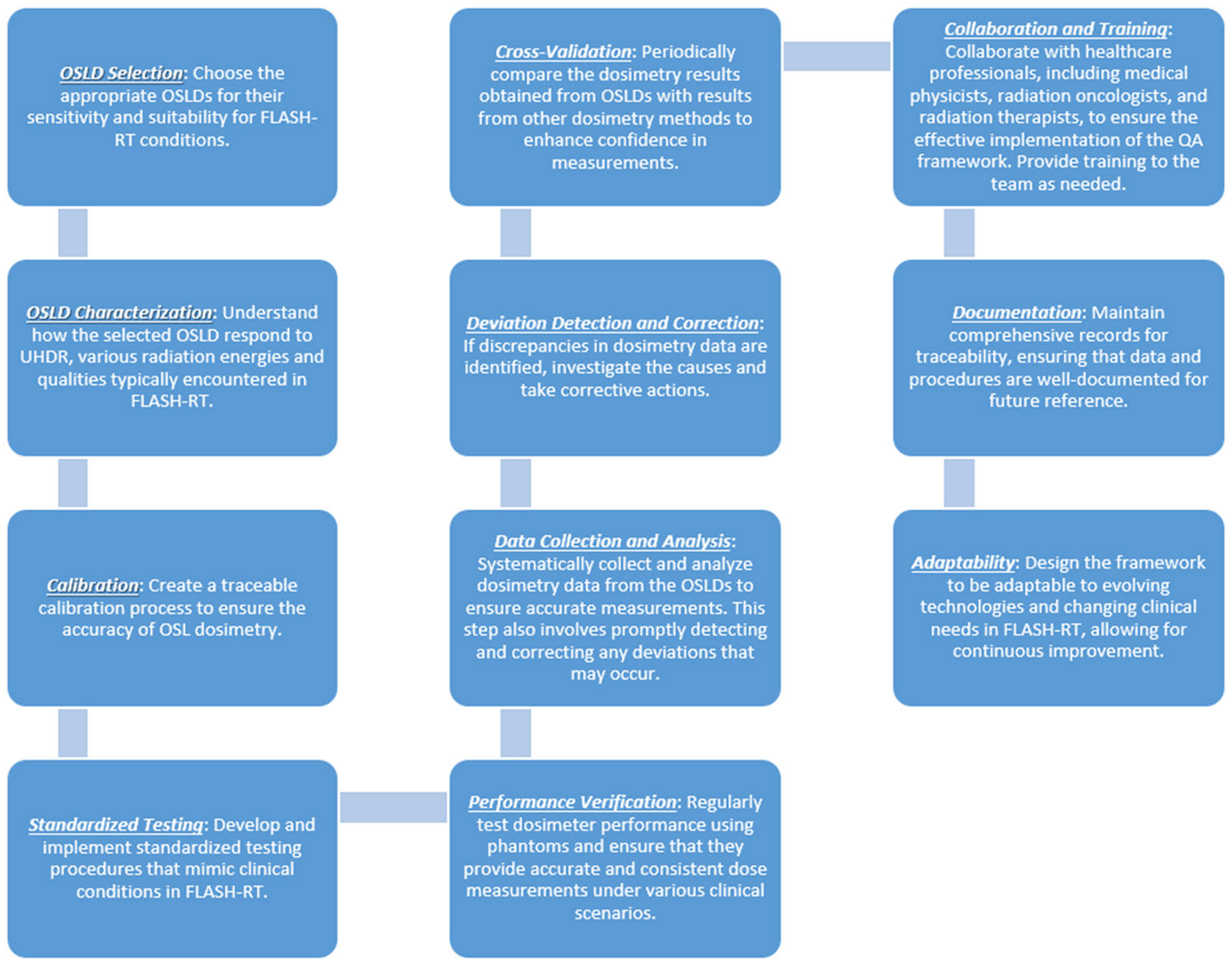
The integration of IVD into routine clinical workflows can present practical challenges. The additional steps required for dosimeter preparation, placement, and data analysis may interrupt standard workflows and increase treatment times. These interruptions can be particularly problematic in busy clinical settings, where efficiency is crucial [77]. Given the sometimes time-consuming nature of IVD, as well as its costs, there is a trend in some centers to reduce the frequency of performing IVD. The aim of this selective de-implementation approach is to balance the benefits of IVD with the practical constraints of clinical workflows and financial considerations [78]. Furthermore, the cost of acquiring, maintaining, and calibrating IVD equipment can pose financial barriers, particularly for resource-limited facilities [79]. Passive dosimeters like TLDs and OSLDs, while cost-effective per use, require specialized equipment for readouts, adding to the overall expense [80]. Active dosimeters, such as MOSFETs and diodes, may offer real-time measurements, but they often come with higher initial investment and maintenance costs [81]. Figure 3 shows a comprehensive QA framework for radiation dosimetry in FLASH radiotherapy using OSLDs, which involves a structured, multi-step approach. The process begins with dosimeter selection and characterization, where the composition of the OSLDs is optimized for their sensitivity and suitability under FLASH radiotherapy conditions, followed by the characterization of their radiation responses at ultra-high dose rates. Next is the calibration-and-standardized-testing step, which focuses on developing a traceable calibration process and creating standardized testing procedures to simulate clinical conditions. Performance verification is then conducted regularly by testing dosimeter performance using phantoms to verify dose accuracy and consistency across varying clinical scenarios. Systematic data collection and analysis is another crucial step, enabling the prompt detection and correction of deviations. Cross-validation is performed periodically, comparing OSL dosimetry with other methods to enhance confidence in the measurements. Collaboration and training play an essential role, as coordination with healthcare professionals, including medical physicists and physics associates, and proper training ensure the framework’s effective implementation. Finally, thorough documentation and adaptability are emphasized to maintain traceability and design the framework in a way that it can evolve alongside advancements in FLASH technologies.
While there are several tools available for verifying the dosimetric accuracy of radiotherapy treatments, such as quality checks on equipment and accessories, patient-specific dosimetric checks, pre-treatment imaging, and log-file analysis, IVD remains a common tool in certain specific situations. These tools can often provide more comprehensive verification of treatment accuracy, particularly in inverse planning treatments with fluence modulation, where point dosimeters may only be partially exposed to the direct field. However, in potentially critical treatments conducted under special irradiation conditions, such as TBI with open fields, IORT [82], brachytherapy, and total-skin irradiation, IVD is still important. In these scenarios, IVD provides real-time or near-real-time feedback on the actual dose received by the patient, ensuring precise dose delivery and enhancing patient safety. Despite being time-consuming, the use of IVD in these treatments can significantly impact treatment duration in terms of Monitor Units, making it an essential component of quality assurance protocols.

7. Future Perspectives

The future of IVD is poised for transformative advancements, driven by emerging technologies, innovative materials, and evolving clinical practices. These developments promise to enhance the precision, efficiency, and personalization of radiotherapy, aligning with the broader trend toward patient-centric treatment approaches.

7.1. Emerging Technologies

AI and machine learning are poised to revolutionize IVD by enabling predictive dose analysis and real-time adaptive feedback [83]. Machine learning algorithms can analyze vast datasets from dosimetric measurements and patient imaging to identify patterns, predict potential dose deviations, and optimize treatment plans accordingly [84]. These tools also offer the potential to improve quality assurance by automating data analysis, reducing human error, and enhancing decision-making capabilities during treatment planning and execution [85,86].
Similarly, the integration of 3D dosimetry systems into clinical workflows provides the ability to visualize radiation dose distributions in greater detail, enhancing the accuracy of dose delivery and ensuring treatment precision [87]. High-resolution 3D imaging and dosimetric data can be used to identify potential areas of concern, improve treatment planning, and provide a more comprehensive understanding of the dose delivered to tissues across complex anatomical structures [88].
Monte Carlo simulations, a powerful computational tool, are increasingly being integrated into clinical dosimetry workflows [89,90]. These simulations model the interaction of radiation with matter at a very detailed level, allowing for highly accurate dose calculations in complex treatment scenarios. Monte Carlo methods can be used to predict dose distributions in heterogeneous tissues, verify treatment plans, and assess the impact of uncertainties such as patient movement or variations in anatomy [91,92]. The incorporation of Monte Carlo simulations into IVD allows clinicians to verify dose delivery with greater confidence, providing a valuable tool for quality assurance and personalized treatment adaptation.

7.2. Advancements in Detector Materials and Nanotechnology

The development of advanced detector materials and the application of nanotechnology are unlocking new possibilities for IVD [93,94]. Nanowire-based dosimeters, for example, offer superior sensitivity and precision compared to traditional dosimeters [95,96]. These next-generation detectors are capable of measuring minute variations in radiation doses with improved spatial resolution, making them particularly useful for complex treatment modalities like stereotactic radiosurgery (SRS) and TBI. In addition, the miniaturization of dosimetric devices enabled by nanotechnology facilitates their use in anatomically challenging areas, expanding the scope of in vivo measurements [97].

7.3. Integration with Adaptive Radiotherapy

Adaptive radiotherapy, which involves modifying treatment plans in response to changes in patient anatomy or tumor characteristics, is a growing area of focus in modern oncology [98]. IVD is expected to play an important role in this paradigm by providing real-time dose feedback that informs treatment adjustments [99]. The seamless integration of IVD with adaptive radiotherapy systems could significantly enhance the precision and effectiveness of treatment, ensuring that dose delivery remains optimal despite anatomical variations or tumor shrinkage during therapy.

7.4. Integration with FLASH Radiotherapy

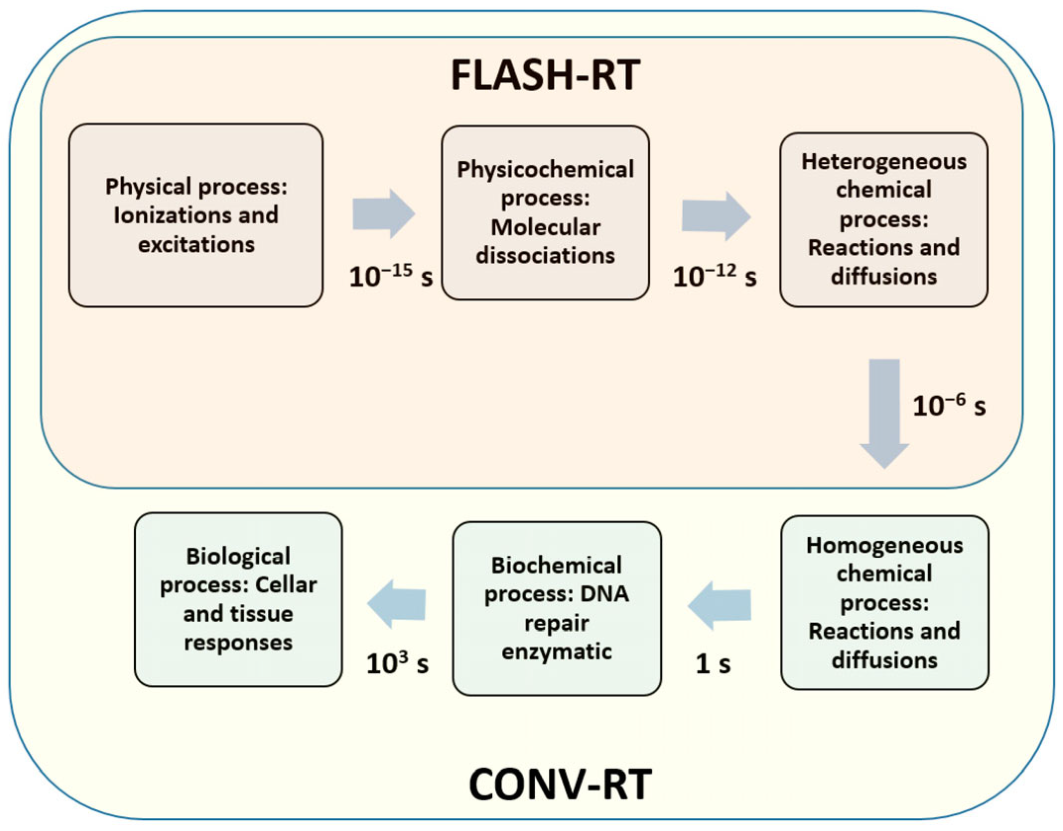
One of the most exciting advancements in radiotherapy is the development of FLASH radiotherapy, which involves delivering ultra-high dose rates in a fraction of the time compared to conventional therapies [100,101]. As FLASH radiotherapy gains momentum, accurate IVD becomes even more critical, given the unique challenges posed by the high-dose-rate beams [102]. Figure 4 compares the timelines of physicochemical processes in FLASH radiotherapy (FLASH-RT) and conventional radiotherapy (CONV-RT), emphasizing that FLASH irradiation is approximately 1000 times faster than conventional irradiation. At such an ultra-high dose rate exceeding 40 Gy/s, a dosimeter with an exceptionally fast response rate is essential for accurate FLASH-based in vivo dosimetry.
Nanowire-based OSLDs are emerging as promising tools for measuring radiation dose in FLASH radiotherapy. These next-generation dosimeters offer enhanced sensitivity and response rates, making them ideal for detecting the rapid dose delivery typical of FLASH treatments. The use of nanowire-based OSLDs allows clinicians to measure ultra-high-dose-rate radiation with high spatial resolution, ensuring that doses are accurately delivered to the target while minimizing potential damage to surrounding healthy tissues. This integration of advanced dosimetric technologies with FLASH radiotherapy is paving the way for a new era of faster, more precise, and potentially more effective radiation treatments [103].

7.5. Individualized Patient Dosimetry and Real-Time Feedback

The move toward personalized medicine is also influencing the future direction of IVD. Advances in dosimetric technology are paving the way for individualized patient dosimetry, where measurements are tailored to the unique characteristics of each patient’s anatomy and treatment plan [104]. Real-time feedback systems, supported by innovations in sensor technology and AI, could allow instantaneous dose verification, enabling clinicians to make on-the-fly adjustments during treatment sessions [105,106]. This capability would not only enhance treatment accuracy but also improve patient safety by identifying and addressing potential errors before they affect clinical outcomes.

8. Conclusions

IVD plays a crucial role in ensuring the accuracy, safety, and quality of radiotherapy, providing essential real-time verification of dose delivery during treatment. By directly measuring the radiation dose received by the patient, IVD allows clinicians to confirm that the prescribed dose has been accurately delivered to the target while minimizing exposure to surrounding healthy tissues. This is particularly important for advanced treatment modalities such as IMRT, VMAT, and emerging techniques like FLASH radiotherapy.
Ongoing advancements in dosimetric technologies, including the integration of AI, Monte Carlo simulations, and the development of novel materials such as nanowire-based OSLDs, hold tremendous potential to further enhance clinical outcomes. These innovations enable more precise, adaptive, and personalized radiotherapy treatments, improving both the effectiveness and safety of radiotherapy.
However, to fully realize the benefits of these advancements, continued research and standardization are essential. Ensuring consistency in dosimetric practices, developing standardized protocols for various clinical applications, and addressing challenges related to measurement uncertainties and technical limitations will be critical in maximizing the impact of IVD. With ongoing efforts to refine and expand the capabilities of IVD, the field of radiotherapy will continue to evolve, providing patients with safer, more effective treatment options.

Author Contributions

Conceptualization, J.C.L.C. and H.E.R.; methodology, J.C.L.C. and H.E.R.; resources, J.C.L.C. and H.E.R.; writing—original draft preparation, J.C.L.C.; writing—review and editing, J.C.L.C. and H.E.R.; visualization, J.C.L.C.; project administration, J.C.L.C. and H.E.R.; funding acquisition, J.C.L.C. and H.E.R. All authors have read and agreed to the published version of the manuscript.

Funding

This research was supported by the Government of Canada’s New Frontiers in Research Fund—Exploration (Grant number: NFRFE-2022-00707), through the three federal research funding agencies (CIHR, NSERC, and SSHRC).

Institutional Review Board Statement

Not applicable.

Informed Consent Statement

Not applicable.

Data Availability Statement

No new data were created.

Conflicts of Interest

The authors declare no conflicts of interest.

References

  1. Ismail, A.; Giraud, J.Y.; Lu, G.N.; Sihanath, R.; Pittet, P.; Galvan, J.M.; Balosso, J. Radiotherapy quality insurance by individualized in vivo dosimetry: State of the art. Cancer/Radiothér. 2009, 13, 182–189. [Google Scholar] [CrossRef] [PubMed]
  2. Leunens, G.; Van Dam, J.; Dutreix, A.; Van der Schueren, E. Quality assurance in radiotherapy by in vivo dosimetry. 2. Determination of the target absorbed dose. Radiother. Oncol. 1990, 19, 73–87. [Google Scholar] [CrossRef] [PubMed]
  3. Leunens, G.; Van Dam, J.; Dutreix, A.; Van der Schueren, E. Quality assurance in radiotherapy by in vivo dosimetry. 1. Entrance dose measurements, a reliable procedure. Radiother. Oncol. 1990, 17, 141–151. [Google Scholar] [CrossRef] [PubMed]
  4. Mijnheer, B. State of the art of in vivo dosimetry. Radiat. Prot. Dosim. 2008, 131, 117–122. [Google Scholar]
  5. Essers, M.; Mijnheer, B. In vivo dosimetry during external photon beam radiotherapy. Int. J. Radiat. Oncol. Biol. Phys. 1999, 43, 245–259. [Google Scholar]
  6. Fiorino, C.; Corletto, D.; Mangili, P.; Broggi, S.; Bonini, A.; Cattaneo, G.M.; Parisi, R.; Rosso, A.; Signorotto, P.; Villa, E.; et al. Quality assurance by systematic in vivo dosimetry: Results on a large cohort of patients. Radiother. Oncol. 2000, 56, 85–95. [Google Scholar]
  7. Rozendaal, R.A.; Mijnheer, B.J.; Hamming-Vrieze, O.; Mans, A.; Van Herk, M. Impact of daily anatomical changes on EPID-based in vivo dosimetry of VMAT treatments of head-and-neck cancer. Radiother. Oncol. 2015, 116, 70–74. [Google Scholar]
  8. Wills, C.; Cherian, S.; Yousef, J.; Wang, K.; Mackley, H.B. Total body irradiation: A practical review. Appl. Radiat. Oncol. 2016, 5, 11–17. [Google Scholar]
  9. Staffurth, J. A review of the clinical evidence for intensity-modulated radiotherapy. Clin. Oncol. 2010, 22, 643–657. [Google Scholar]
  10. Hunte, S.O.; Clark, C.H.; Zyuzikov, N.; Nisbet, A. Volumetric modulated arc therapy (VMAT): A review of clinical outcomes—What is the clinical evidence for the most effective implementation? Br. J. Radiol. 2022, 95, 20201289. [Google Scholar]
  11. Ma, C.M. Physics and Dosimetric Principles of SRS and SBRT. Mathews J. Cancer Sci. 2019, 4, 22. [Google Scholar]
  12. Healy, B.J.; Budanec, M.; Ourdane, B.; Peace, T.; Petrovic, B.; Sanz, D.E.; Scanderbeg, D.J.; Tuntipumiamorn, L. An IAEA survey of radiotherapy practice including quality assurance extent and depth. Acta Oncol. 2020, 59, 503–510. [Google Scholar] [CrossRef] [PubMed]
  13. Dogan, N.; Mijnheer, B.J.; Padgett, K.; Nalichowski, A.; Wu, C.; Nyflot, M.J.; Olch, A.J.; Papanikolaou, N.; Shi, J.; Holmes, S.M.; et al. Use of electronic portal imaging devices for pre-treatment and in vivo dosimetry patient-specific IMRT and VMAT QA: Report of AAPM Task Group 307. Med. Phys. 2023, 50, e865. [Google Scholar] [CrossRef]
  14. Olaciregui-Ruiz, I.; Beddar, S.; Greer, P.; Jornet, N.; McCurdy, B.; Paiva-Fonseca, G.; Mijnheer, B.; Verhaegen, F. In vivo dosimetry in external beam photon radiotherapy: Requirements and future directions for research, development, and clinical practice. Phys. Imaging Radiat. Oncol. 2020, 15, 108–116. [Google Scholar] [CrossRef] [PubMed]
  15. Esposito, M.; Villaggi, E.; Bresciani, S.; Cilla, S.; Falco, M.D.; Garibaldi, C.; Russo, S.; Talamonti, C.; Stasi, M.; Mancosu, P. Estimating dose delivery accuracy in stereotactic body radiation therapy: A review of in-vivo measurement methods. Radiother. Oncol. 2020, 149, 158–167. [Google Scholar] [CrossRef]
  16. Houlihan, O.A.; Workman, G.; Hounsell, A.R.; Prise, K.M.; Jain, S. In vivo dosimetry in pelvic brachytherapy. Br. J. Radiol. 2022, 95, 20220046. [Google Scholar] [CrossRef]
  17. Wernli, C. A short history and critical review of individual monitoring. Radiat. Prot. Dosim. 2016, 170, 4–7. [Google Scholar] [CrossRef]
  18. Jennings, W.A. Evolution over the past century of quantities and units in radiation dosimetry. J. Radiol. Prot. 2007, 27, 5. [Google Scholar] [CrossRef]
  19. Sinclair, S.A.; Pech-Canul, M.I. Development feasibility of TLD phosphors and thermoluminescent composite materials for potential applications in dosimetry: A review. Chem. Eng. J. 2022, 443, 136522. [Google Scholar] [CrossRef]
  20. Rosenfeld, A.B. Electronic dosimetry in radiation therapy. Radiat. Meas. 2006, 41, S134–S153. [Google Scholar] [CrossRef]
  21. Asensio, L.J.; Carvajal, M.A.; Lopez-Villanueva, J.A.; Vilches, M.; Lallena, A.M.; Palma, A.J. Evaluation of a low-cost commercial mosfet as radiation dosimeter. Sens. Actuators A Phys. 2006, 125, 288–295. [Google Scholar] [CrossRef]
  22. Van Elmpt, W.; McDermott, L.; Nijsten, S.; Wendling, M.; Lambin, P.; Mijnheer, B. A literature review of electronic portal imaging for radiotherapy dosimetry. Radiother. Oncol. 2008, 88, 289–309. [Google Scholar] [PubMed]
  23. Alecu, R.; Loomis, T.; Alecu, J.; Ochran, T. Guidelines on the implementation of diode in vivo dosimetry programs for photon and electron external beam therapy. Med. Dosim. 1999, 24, 5–12. [Google Scholar] [CrossRef] [PubMed]
  24. Meghzifene, A.; Followill, D.; Dewaraja, Y.K.; Allisy, P.J.; Kessler, C.; van der Merwe, D. International symposium on standards, applications and quality assurance in medical radiation dosimetry (IDOS 2019): Highlights of an IAEA meeting. Med. Phys. Int. 2019, 7, 342–360. [Google Scholar]
  25. Chow, J.C. Depth dose enhancement on flattening-filter-free photon beam: A Monte Carlo study in nanoparticle-enhanced radiotherapy. Appl. Sci. 2020, 10, 7052. [Google Scholar] [CrossRef]
  26. Mohan, R.; Barest, G.; Brewster, L.J.; Chui, C.S.; Kutcher, G.J.; Laughlin, J.S.; Fuks, Z. A comprehensive three-dimensional radiation treatment planning system. Int. J. Radiat. Oncol. Biol. Phys. 1988, 15, 481–495. [Google Scholar] [CrossRef]
  27. Rivera, T. Thermoluminescence in medical dosimetry. Appl. Radiat. Isot. 2012, 71, 30–34. [Google Scholar]
  28. Pai, S.; Das, I.J.; Dempsey, J.F.; Lam, K.L.; LoSasso, T.J.; Olch, A.J.; Palta, J.R.; Reinstein, L.E.; Ritt, D.; Wilcox, E.E. TG-69: Radiographic film for megavoltage beam dosimetry. Med. Phys. 2007, 34, 2228–2258. [Google Scholar]
  29. McKeever, S.W. Optically stimulated luminescence: A brief overview. Radiat. Meas. 2011, 46, 1336–1341. [Google Scholar]
  30. Chow, J.C.; Jiang, R.; Kiciak, A.; Markel, D. Dosimetric comparison between the prostate intensity-modulated radiotherapy (IMRT) and volumetric-modulated arc therapy (VMAT) plans using the planning target volume (PTV) dose–volume factor. J. Radiother. Pract. 2016, 15, 263–268. [Google Scholar]
  31. Chow, J.C.; Owrangi, A.M. Dosimetric dependences of bone heterogeneity and beam angle on the unflattened and flattened photon beams: A Monte Carlo comparison. Radiat. Phys. Chem. 2014, 101, 46–52. [Google Scholar]
  32. Chow, J.C. Cone-beam CT dosimetry for the positional variation in isocenter: A Monte Carlo study. Med. Phys. 2009, 36, 3512–3520. [Google Scholar] [PubMed]
  33. Butson, M.; Haque, M.; Smith, L.; Butson, E.; Odgers, D.; Pope, D.; Gorjiana, T.; Whitaker, M.; Morales, J.; Hong, A.; et al. Practical time considerations for optically stimulated luminescent dosimetry (OSLD) in total body irradiation. Australas. Phys. Eng. Sci. Med. 2017, 40, 167–171. [Google Scholar] [CrossRef] [PubMed]
  34. Sánchez-Doblado, F.; Terrón, J.A.; Sánchez-Nieto, B.; Arráns, R.; Errazquin, L.; Biggs, D.; Lee, C.; Núñez, L.; Delgado, A.; Muñiz, J.L. Verification of an on line in vivo semiconductor dosimetry system for TBI with two TLD procedures. Radiother. Oncol. 1995, 34, 73–77. [Google Scholar]
  35. Nibhanupudy, J.R.; De Jesus, M.A.; Fujita, M.; Goldson, A.L. Radiation dose monitoring in a breast cancer patient with a pacemaker: A case report. J. Natl. Med. Assoc. 2001, 93, 278. [Google Scholar]
  36. Miften, M.; Mihailidis, D.; Kry, S.F.; Reft, C.; Esquivel, C.; Farr, J.; Followill, D.; Hurkmans, C.; Liu, A.; Gayou, O.; et al. Management of radiotherapy patients with implanted cardiac pacemakers and defibrillators: A report of the AAPM TG-203. Med. Phys. 2019, 46, e757–e788. [Google Scholar]
  37. Dhivya, S.; Anuradha, C.; Murali, V.; Ramasubramanian, V. In-vivo dosimetry in total skin electron therapy: Literature review. J. Radiother. Pract. 2021, 20, 357–360. [Google Scholar] [CrossRef]
  38. Rodriguez-Cortes, J.; Rivera-Montalvo, T.; Navarro, L.V.; Flores-López, O.; Roman, J.; Hernandez-Oviedo, J.O. Thermoluminescent dosimetry in total body irradiation. Appl. Radiat. Isot. 2012, 71, 35–39. [Google Scholar]
  39. Kron, T. Applications of thermoluminescence dosimetry in medicine. Radiat. Prot. Dosim. 1999, 85, 333–340. [Google Scholar]
  40. Bruzzi, M. Novel silicon devices for radiation therapy monitoring. Nucl. Instrum. Methods Phys. Res. Sect. A Accel. Spectrom. Detect. Assoc. Equip. 2016, 809, 105–112. [Google Scholar]
  41. Colussi, V.C.; Beddar, A.S.; Kinsella, T.J.; Sibata, C.H. In vivo dosimetry using a single diode for megavoltage photon beam radiotherapy: Implementation and response characterization. J. Appl. Clin. Med. Phys. 2001, 2, 210–218. [Google Scholar] [CrossRef] [PubMed]
  42. Chow, J.C.; Leung, M.K. Monte Carlo simulation of MOSFET dosimeter for electron backscatter using the GEANT4 code. Med. Phys. 2008, 35, 2383–2390. [Google Scholar] [CrossRef] [PubMed]
  43. Jong, W.L.; Ung, N.M.; Tiong, A.H.; Rosenfeld, A.B.; Wong, J.H. Characterisation of a MOSFET-based detector for dose measurement under megavoltage electron beam radiotherapy. Radiat. Phys. Chem. 2018, 144, 76–84. [Google Scholar] [CrossRef]
  44. Bakhtiari, M.; Kumaraswamy, L.; Bailey, D.W.; De Boer, S.; Malhotra, H.K.; Podgorsak, M.B. Using an EPID for patient-specific VMAT quality assurance. Med. Phys. 2011, 38, 1366–1373. [Google Scholar]
  45. Blake, S.J.; McNamara, A.L.; Deshpande, S.; Holloway, L.; Greer, P.B.; Kuncic, Z.; Vial, P. Characterization of a novel EPID designed for simultaneous imaging and dose verification in radiotherapy. Med. Phys. 2013, 40, 091902. [Google Scholar]
  46. Al-Senan, R.M.; Hatab, M.R. Characteristics of an OSLD in the diagnostic energy range. Med. Phys. 2011, 38, 4396–4405. [Google Scholar] [CrossRef]
  47. Choi, C.H.; Park, J.M.; Park, S.Y.; Chun, M.; Han, J.H.; Cho, J.D.; Kim, J.I. Prediction of midline dose from entrance and exit dose using OSLD measurements for Total body irradiation. J. Radiat. Prot. Res. 2017, 42, 77–82. [Google Scholar] [CrossRef]
  48. Jursinic, P.A. Characterization of optically stimulated luminescent dosimeters, OSLDs, for clinical dosimetric measurements. Med. Phys. 2007, 34, 4594–4604. [Google Scholar]
  49. Brodin, N.P.; Mehta, K.J.; Basavatia, A.; Goddard, L.C.; Fox, J.L.; Feldman, S.M.; McEvoy, M.P.; Tomé, W.A. A skin dose prediction model based on in vivo dosimetry and ultrasound skin bridge measurements during intraoperative breast radiation therapy. Brachytherapy 2019, 18, 720–726. [Google Scholar] [CrossRef]
  50. Butson, M.J.; Peter, K.N.; Cheung, T.; Metcalfe, P. Radiochromic film for medical radiation dosimetry. Mater. Sci. Eng. R Rep. 2003, 41, 61–120. [Google Scholar]
  51. O’Keeffe, S.; McCarthy, D.; Woulfe, P.; Grattan, M.W.; Hounsell, A.R.; Sporea, D.; Mihai, L.; Vata, I.; Leen, G.A.; Lewis, E. A review of recent advances in optical fibre sensors for in vivo dosimetry during radiotherapy. Br. J. Radiol. 2015, 88, 20140702. [Google Scholar] [CrossRef] [PubMed]
  52. Zhuang, Q.; Yaosheng, H.; Yu, M.; Wenhui, Z.; Weimin, S.; Daxin, Z.; Ziyin, C.; Elfed, L. Embedded structure fiber-optic radiation dosimeter for radiotherapy applications. Opt. Express 2016, 24, 5172–5185. [Google Scholar] [CrossRef] [PubMed]
  53. Mijnheer, B.; Olaciregui-Ruiz, I.; Rozendaal, R.; Sonke, J.J.; Spreeuw, H.; Tielenburg, R.; Van Herk, M.; Vijlbrief, R.; Mans, A. 3D EPID-based in vivo dosimetry for IMRT and VMAT. J. Phys. Conf. Ser. 2013, 444, 012011. [Google Scholar] [CrossRef]
  54. Qi, Z.Y.; Deng, X.W.; Huang, S.M.; Shiu, A.; Lerch, M.; Metcalfe, P.; Rosenfeld, A.; Kron, T. Real-time in vivo dosimetry with MOSFET detectors in serial tomotherapy for head and neck cancer patients. Int. J. Radiat. Oncol. Biol. Phys. 2011, 80, 1581–1588. [Google Scholar] [CrossRef]
  55. McCurdy, B.M.; McCowan, P.M. In vivo dosimetry for lung radiotherapy including SBRT. Phys. Med. 2017, 44, 123–130. [Google Scholar] [CrossRef]
  56. Esposito, M.; Ghirelli, A.; Pini, S.; Alpi, P.; Barca, R.; Fondelli, S.; Leonulli, B.G.; Paoletti, L.; Rossi, F.; Bastiani, P.; et al. Clinical implementation of 3D in vivo dosimetry for abdominal and pelvic stereotactic treatments. Radiother. Oncol. 2021, 154, 14–20. [Google Scholar] [CrossRef]
  57. Peet, S.C.; Wilks, R.; Kairn, T.; Crowe, S.B. Measuring dose from radiotherapy treatments in the vicinity of a cardiac pacemaker. Phys. Med. 2016, 32, 1529–1536. [Google Scholar]
  58. Gruber, G.; Schwegler, N. Low-dose testicular irradiation in seminoma patients. In-vivo dosimetry: In-vivo-dosimetrie. Strahlenther. Onkol. 1999, 175, 185–189. [Google Scholar] [CrossRef]
  59. Banaee, N.; Nedaie, H.A.; Esmati, E.; Nosrati, H.; Jamali, M. Dose measurement outside of radiotherapy treatment field (Peripheral dose) using thermoluminesent dosimeters. Int. J. Radiat. Res. 2014, 12, 355–359. [Google Scholar]
  60. Carnicer, A.; Letellier, V.; Rucka, G.; Angellier, G.; Sauerwein, W.; Hérault, J. An indirect in vivo dosimetry system for ocular proton therapy. Radiat. Prot. Dosim. 2014, 161, 373–376. [Google Scholar] [CrossRef]
  61. Cheng, C.W.; Wolanski, M.; Zhao, Q.; Fanelli, L.; Gautam, A.; Pack, D.; Das, I.J. Dosimetric characteristics of a single use MOSFET dosimeter for in vivo dosimetry in proton therapy. Med. Phys. 2010, 37, 4266–4273. [Google Scholar] [PubMed]
  62. Patel, R.P.; Warry, A.J.; Eaton, D.J.; Collis, C.H.; Rosenberg, I. In vivo dosimetry for total body irradiation: Five-year results and technique comparison. J. Appl. Clin. Med. Phys. 2014, 15, 306–315. [Google Scholar]
  63. Fonseca, G.P.; Johansen, J.G.; Smith, R.L.; Beaulieu, L.; Beddar, S.; Kertzscher, G.; Verhaegen, F.; Tanderup, K. In vivo dosimetry in brachytherapy: Requirements and future directions for research, development, and clinical practice. Phys. Imaging Radiat. Oncol. 2020, 16, 1–11. [Google Scholar] [CrossRef] [PubMed]
  64. Jayakody, M.; Jeyasugiththan, J.; Rajasooriyar, C.; Chougule, A. Dosimetry procedure to verify dose in High Dose Rate (HDR) brachytherapy treatment of cancer patients: A systematic review. Phys. Med. 2022, 96, 70–80. [Google Scholar] [PubMed]
  65. Stathopoulos, I.; Ploussi, A.; Syrgiamiotis, V.; Makri, T.; Hatzigiorgi, C.; Carinou, E.; Sakellaropoulos, G.; Panayiotakis, G.S.; Efstathopoulos, E.P. In vivo dosimetry for head CT examinations in paediatric patients. Phys. Med. 2016, 32, 205–206. [Google Scholar]
  66. Bonato, C.C.; Dias, H.B.; Alves, M.D.; Duarte, L.O.; Dias, T.M.; Dalenogare, M.O.; Viegas, C.C.; Elnecave, R.H. In vivo dosimetry of thyroid doses from different irradiated sites in children and adolescents: A cross-sectional study. Radiat. Oncol. 2014, 9, 40. [Google Scholar]
  67. Herbert, C.E.; Ebert, M.A.; Joseph, D.J. Feasible measurement errors when undertaking in vivo dosimetry during external beam radiotherapy of the breast. Med. Dosim. 2003, 28, 45–48. [Google Scholar]
  68. Noel, A.; Aletti, P.; Bey, P.; Malissard, L. Detection of errors in individual patients in radiotherapy by systematic in vivo dosimetry. Radiother. Oncol. 1995, 34, 144–151. [Google Scholar]
  69. Mans, A.; Wendling, M.; McDermott, L.N.; Sonke, J.J.; Tielenburg, R.; Vijlbrief, R.; Mijnheer, B.; Van Herk, M.; Stroom, J.C. Catching errors with in vivo EPID dosimetry. Med. Phys. 2010, 37, 2638–2644. [Google Scholar]
  70. Ketabi, A.; Karbasi, S.; Faghihi, R.; Mosleh-Shirazi, M.A. A phantom-based experimental and Monte Carlo study of the suitability of in-vivo diodes and TLD for entrance in-vivo dosimetry in small-to-medium sized 6 MV photon fields. Radiat. Phys. Chem. 2022, 201, 110411. [Google Scholar] [CrossRef]
  71. Sen, A.; Parsai, E.I.; McNeeley, S.W.; Ayyangar, K.M. Quantitative assessment of beam perturbations caused by silicon diodes used for in vivo dosimetry. Int. J. Radiat. Oncol. Biol. Phys. 1996, 36, 205–211. [Google Scholar] [CrossRef] [PubMed]
  72. Kirby, T.H.; Hanson, W.F.; Johnston, D.A. Uncertainty analysis of absorbed dose calculations from thermoluminescence dosimeters. Med. Phys. 1992, 19, 1427–1433. [Google Scholar] [CrossRef] [PubMed]
  73. Wong, C.J.; Ackerly, T.; He, C.; Patterson, W.; Powell, C.E.; Qiao, G.; Solomon, D.H.; Meder, R.; Geso, M. Small field size dose-profile measurements using gel dosimeters, gafchromic films and micro-thermoluminescent dosimeters. Radiat. Meas. 2009, 44, 249–256. [Google Scholar] [CrossRef]
  74. Muñoz, I.D.; Gamboa-deBuen, I.; Avila, O.; Brandan, M.E. Dosimetry in a mammography phantom using TLD-300 dosimeters. Med. Phys. 2018, 45, 4287–4296. [Google Scholar] [CrossRef]
  75. Kim, J.; Park, J.; Park, B.; Kim, Y.; Park, B.; Park, S.H. Compact and Real-Time Radiation Dosimeter Using Silicon Photomultipliers for In Vivo Dosimetry in Radiation Therapy. Sensors 2025, 25, 857. [Google Scholar] [CrossRef]
  76. Mosleh-Shirazi, M.A.; Ketabi, A.; Karbasi, S.; Faghihi, R. A Monte Carlo and experimental investigation of the dosimetric behavior of low- and medium-perturbation diodes used for entrance in vivo dosimetry in megavoltage photon beams. J. Appl. Clin. Med. Phys. 2012, 13, 3917. [Google Scholar] [CrossRef]
  77. Bossuyt, E.; Nevens, D.; Weytjens, R.; Mokaddem, A.T.; Verellen, D. Assessing the impact of adaptations to the clinical workflow in radiotherapy using transit in vivo dosimetry. Phys. Imaging Radiat. Oncol. 2023, 25, 100420. [Google Scholar] [CrossRef]
  78. Mao, S.P.; Han-Oh, S.; Moore, J.; Huang, E.; McNutt, T.R.; Souranis, A.N.; Briner, V.; Halthore, A.; Alcorn, S.R.; Meyer, J.J.; et al. Selective de-implementation of routine in vivo dosimetry. J. Appl. Clin. Med. Phys. 2023, 24, e13953. [Google Scholar] [CrossRef]
  79. Falco, M.D.; Giancaterino, S.; De Nicola, A.; Adorante, N.; De Lorenzo, R.G.; Di Tommaso, M.; Vinciguerra, A.; Trignani, M.; Perrotti, F.; Allajbej, A.; et al. A feasibility study for in vivo dosimetry procedure in routine clinical practice. Technol. Cancer Res. Treat. 2018, 17, 1533033818779201. [Google Scholar] [CrossRef]
  80. Kry, S.F.; Alvarez, P.; Cygler, J.E.; DeWerd, L.A.; Howell, R.M.; Meeks, S.; O’Daniel, J.; Reft, C.; Sawakuchi, G.; Yukihara, E.G.; et al. AAPM TG 191: Clinical use of luminescent dosimeters: TLDs and OSLDs. Med. Phys. 2020, 47, e19–e51. [Google Scholar] [CrossRef]
  81. Chow, J.C.; Grigorov, G.N.; Barnett, R.B. Study on surface dose generated in prostate intensity-modulated radiation therapy treatment. Med. Dosim. 2006, 31, 249–258. [Google Scholar] [PubMed]
  82. Istituto Superiore di Sanità. Istisan Report 23/5. Available online: https://www.iss.it/documents/20126/6682486/23-5+web.pdf/05ce834b-d81a-9530-e156-45456b3b1406?t=1685451301943 (accessed on 13 March 2025).
  83. Bossuyt, E.; Weytjens, R.; Nevens, D.; De Vos, S.; Verellen, D. Evaluation of automated pre-treatment and transit in-vivo dosimetry in radiotherapy using empirically determined parameters. Phys. Imaging Radiat. Oncol. 2020, 16, 113–129. [Google Scholar] [CrossRef] [PubMed]
  84. Siddique, S.; Chow, J.C. Artificial intelligence in radiotherapy. Rep. Pract. Oncol. Radiother. 2020, 25, 656–666. [Google Scholar] [PubMed]
  85. Chow, J.C. Artificial intelligence in radiotherapy and patient care. In Artificial Intelligence in Medicine; Springer International Publishing: Cham, Switzerland, 2021; pp. 1–13. [Google Scholar]
  86. Chow, J.C. Quantum Computing and Machine Learning in Medical Decision-Making: A Comprehensive Review. Algorithms 2025, 18, 156. [Google Scholar] [CrossRef]
  87. Dong, J.; Li, Z.; Huang, W.; Kong, F.; Chen, L.; Zhang, M.; Huang, S.; Yan, H.; Xu, X. Preliminary application of EPID three-dimensional dose reconstruction in in vivo dose verification of breast cancer intensity-modulated radiation therapy. Phys. Med. 2025, 129, 104884. [Google Scholar]
  88. Negrete-Hernandez, I.M.; Lozano, I.B.; Roman-Lopez, J.; Guzman-Castañeda, J.I. Implementation of OSL nanoDot dosimetry in different treatment techniques for head and neck cancer. Radiat. Prot. Dosim. 2025, 201, 70–77. [Google Scholar] [CrossRef]
  89. Huy, B.N.; Van Dung, P.; Tinh, H.T.; Ha, N.T.; Duc, N.M. Photon energy estimation in diagnostic radiology using OSL dosimeters: Experimental validation and Monte Carlo simulations. Radiat. Meas. 2025, 180, 107342. [Google Scholar]
  90. de Andrade, E.M.; Paixão, L.; Mendes, B.M.; Fonseca, T.C. Monte Carlo modeling and simulation of a new 3D printed phantom for WBC calibration with ballistic gel as a tissue substitute. Appl. Radiat. Isot. 2025, 215, 111565. [Google Scholar] [CrossRef]
  91. Chow, J.C.; Owrangi, A.M. Mucosal dosimetry on unflattened photon beams: A Monte Carlo phantom study. Biomed. Phys. Eng. Express 2018, 5, 015007. [Google Scholar]
  92. Spina, A.; Chow, J.C. Dosimetric impact on the flattening filter and addition of gold nanoparticles in radiotherapy: A Monte Carlo study on depth dose using the 6 and 10 MV FFF photon beams. Materials 2022, 15, 7194. [Google Scholar] [CrossRef]
  93. Ali, N.; Sahare, P.D. Dosimetry characteristics of NaLi2PO4: Tb3+ OSLD phosphor. J. Radioanal. Nucl. Chem. 2021, 330, 941–950. [Google Scholar] [CrossRef]
  94. Cohen, J.M.; DeLoid, G.M.; Demokritou, P. A critical review of in vitro dosimetry for engineered nanomaterials. Nanomedicine 2015, 10, 3015–3032. [Google Scholar] [PubMed]
  95. Li, Y.; Dong, X.; Gao, J.; Hei, D.; Zhou, X.; Zhang, H. A highly sensitive γ-radiation dosimeter based on the CeO2 nanowires. Phys. E Low-Dimens. Syst. Nanostruct. 2009, 41, 1550–1553. [Google Scholar] [CrossRef]
  96. Li, Y.Y.; Dong, X.; Zhang, H.Q. CeO2 nanowires aqueous γ-radiation dosimeter for low dose sensitively detecting. Procedia Eng. 2013, 52, 202–207. [Google Scholar] [CrossRef]
  97. Chaikh, A.; Beuve, M.; Balosso, J. Nanotechnology in radiation oncology: The need for implantable nano dosimeters for in-vivo real time measurements. Int. J. Cancer Ther. Oncol. 2015, 3, 3217. [Google Scholar]
  98. Lim-Reinders, S.; Keller, B.M.; Al-Ward, S.; Sahgal, A.; Kim, A. Online adaptive radiation therapy. Int. J. Radiat. Oncol. Biol. Phys. 2017, 99, 994–1003. [Google Scholar]
  99. Moradi, F.; Bradley, D.A.; Tarif, Z.H.; Khodaei, A.; Basaif, A.; Ibrahim, S.A.; Abdul-Rashid, H.A. Time-resolved optical fiber measurements: A review of scintillator materials and applications. Radiat. Detect. Technol. Methods 2025, 9, 1–6. [Google Scholar] [CrossRef]
  100. Chow, J.C.; Ruda, H.E. Flash radiotherapy: Innovative cancer treatment. Encyclopedia 2023, 3, 808–823. [Google Scholar] [CrossRef]
  101. Chow, J.C.; Ruda, H.E. Mechanisms of Action in FLASH Radiotherapy: A Comprehensive Review of Physicochemical and Biological Processes on Cancerous and Normal Cells. Cells 2024, 13, 835. [Google Scholar] [CrossRef]
  102. Siddique, S.; Ruda, H.E.; Chow, J.C. FLASH radiotherapy and the use of radiation dosimeters. Cancers 2023, 15, 3883. [Google Scholar] [CrossRef]
  103. Chow, J.C.; Ruda, H.E. Impact of Scattering Foil Composition on Electron Energy Distribution in a Clinical Linear Accelerator Modified for FLASH Radiotherapy: A Monte Carlo Study. Materials 2024, 17, 3355. [Google Scholar] [CrossRef]
  104. Lassmann, M.; Eberlein, U. The relevance of dosimetry in precision medicine. J. Nucl. Med. 2018, 59, 1494–1499. [Google Scholar] [CrossRef] [PubMed]
  105. Hou, B.; Yi, L.; Hu, D.; Luo, Z.; Gao, D.; Li, C.; Xing, B.; Wang, J.W.; Lee, C.N.; Zhang, R.; et al. A swallowable X-ray dosimeter for the real-time monitoring of radiotherapy. Nat. Biomed. Eng. 2023, 7, 1242–1251. [Google Scholar] [CrossRef] [PubMed]
  106. Avanzo, M.; Pirrone, G.; Mileto, M.; Massarut, S.; Stancanello, J.; Baradaran-Ghahfarokhi, M.; Rink, A.; Barresi, L.; Vinante, L.; Piccoli, E.; et al. Prediction of skin dose in low-kV intraoperative radiotherapy using machine learning models trained on results of in vivo dosimetry. Med. Phys. 2019, 46, 1447–1454. [Google Scholar] [CrossRef] [PubMed]
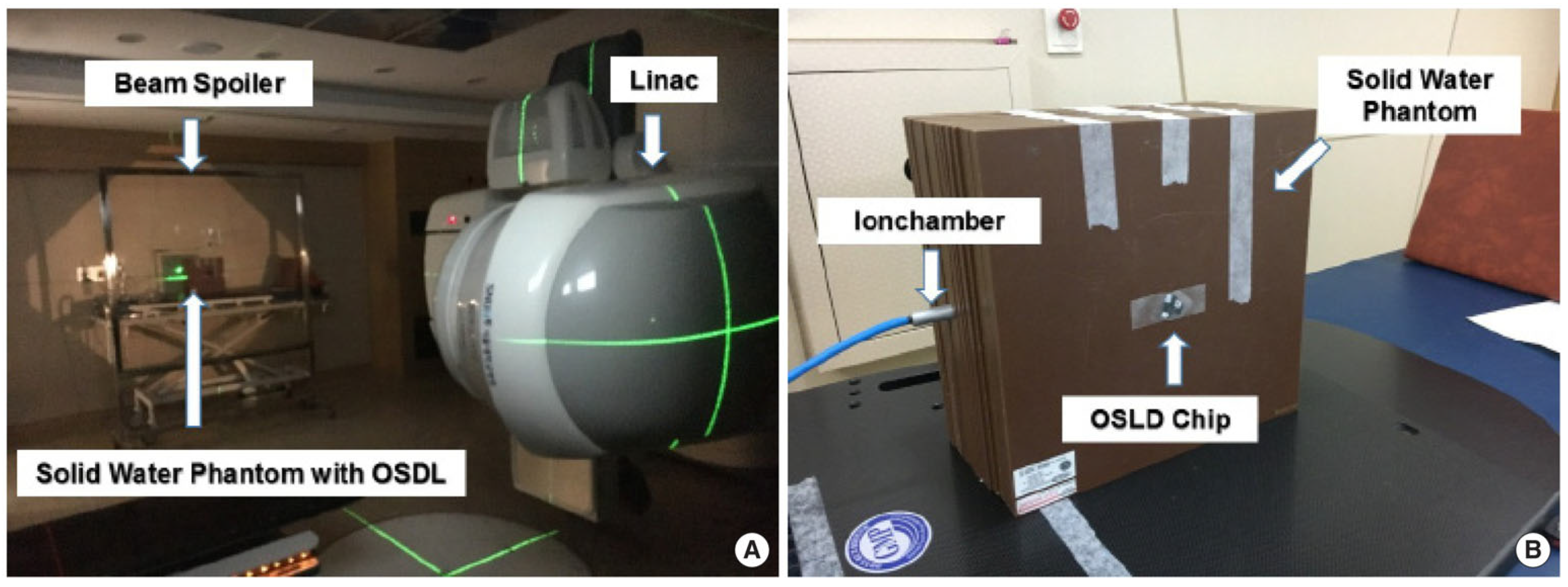
Figure 1. Total-body-irradiation setup with a linear accelerator and solid water phantom (A), and measurement setup for determining entrance, exit, and midline doses using the solid water phantom (B). Reproduced from reference [47] under the Creative Commons Attribution 4.0 International License (https://creativecommons.org/licenses/by/4.0/ (accessed on 10 January 2025)).
Encyclopedia 05 00040 g001
Figure 2. Brachytherapy source tracking measured via (A) CT scan, (B) flat-panel detector, and (C) imaging panel with collimator. Reproduced from reference [63] under the Creative Commons Attribution 4.0 International License (https://creativecommons.org/licenses/by/4.0/ (accessed on 10 January 2025)).
Encyclopedia 05 00040 g002
Figure 3. Framework of OSL dosimetry in FLASH radiotherapy.
Encyclopedia 05 00040 g003
Figure 4. A schematic diagram illustrating the primary physicochemical and biological reactions triggered by cellular and tissue exposure to radiation. Conventional radiotherapy (CONV-RT) disrupts both chemical and biological processes, whereas FLASH radiotherapy (FLASH-RT), with its ultra-high dose rate, bypasses engagement with biochemical pathways. This extreme dose rate also poses challenges to the response rate of an in vivo dosimeter (IVD). Schematic was reproduced from reference [101] under the Creative Commons Attribution 4.0 International License (https://creativecommons.org/licenses/by/4.0/ (accessed on 10 January 2025)).
Encyclopedia 05 00040 g004
Table 1. Overview of in vivo dosimeters: characteristics, applications, advantages, and limitations.
Dosimeter TypeCharacteristicsApplicationsAdvantagesLimitationsReferences
Thermoluminescent Dosimeters (TLDs)Passive devices, measuring cumulative dose via light emitted during heating.Point-dose verification, total-skin electron therapy, and TBI.Compact size, reusability, high accuracy.Requires specialized readout equipment, does not provide real-time feedback.[37,38,39]
DiodesActive semiconductor devices, providing real-time dose measurements.Point-dose verification in IMRT.Real-time data, high sensitivity.Temperature and angular dependence.[40,41]
MOSFETsActive dosimeters, miniaturized and versatile.Pediatric treatments, small-field dosimetry.Small size, easy to use, real-time measurements.Sensitive to cumulative radiation damage.[42,43]
EPIDsOriginally designed for imaging, they have been adapted for dosimetry by analyzing exit-beam intensity profiles.Dose distribution verification in VMAT.Integrated imaging and dosimetry, seamless workflow.Limited spatial resolution compared to other tools.[44,45]
OSLDsMeasure dose via luminescence released upon light stimulation of radiation-sensitive material.TBI monitoring, small-field radiotherapy, IORT.High accuracy, reusability, excellent stability.Requires specialized stimulation and readout equipment.[46,47,48,49]
Radiochromic FilmsMeasure dose via color change proportional to radiation, analyzed using optical scanners.Complex dose distributions, total-skin electron therapy.High spatial resolution, ideal for complex measurements.Single-use, requires careful handling.[50]
Fiber-Optic DosimetersReal-time measurements using flexible fiber-optic sensors; they induce minimal perturbation of radiation field.Challenging anatomical locations.Resistant to electromagnetic interference, provide real-time data.Specialized fabrication and calibration needed.[51,52]
Disclaimer/Publisher’s Note: The statements, opinions and data contained in all publications are solely those of the individual author(s) and contributor(s) and not of MDPI and/or the editor(s). MDPI and/or the editor(s) disclaim responsibility for any injury to people or property resulting from any ideas, methods, instructions or products referred to in the content.