Review

Chytrids in Soil Environments: Unique Adaptations and Distributions

by
Deirdre G. Hanrahan-Tan
1,*,
Osu Lilje
2 and
Linda Henderson
3,*
1
Department of Primary Industries, Elizabeth Macarthur Agricultural Institute, Menangle, NSW 2568, Australia
2
School of Life and Environmental Sciences, The University of Sydney, Camperdown, NSW 2006, Australia
3
Department of Planning and Environment, Locked Bag 5022, Parramatta, NSW 2124, Australia
*
Authors to whom correspondence should be addressed.
Encyclopedia 2023, 3(2), 642-664; https://doi.org/10.3390/encyclopedia3020046
Submission received: 23 February 2023 / Revised: 25 April 2023 / Accepted: 12 May 2023 / Published: 18 May 2023
(This article belongs to the Collection Encyclopedia of Fungi)

Abstract

:
Chytridiomycota (zoosporic true fungi) have a consistent presence in soils and have been frequently identified within many diverse terrestrial environments. However, Chytridiomycota and other early-diverging fungi have low representation in whole-genome sequencing databases compared to Dikarya. New molecular techniques have provided insights into the diversity and abundance of chytrids in soils and the changes in their populations both spatially and temporally. Chytrids complete their life cycle within rapidly changing soil environments where they may be more common within micropores due to protection from predation, desiccation, and extreme temperatures. Reproductive and morphological changes occur in response to environmental changes including pH, fluctuating nutrient concentrations, and metals at levels above toxic thresholds. Rhizoids share some features of hyphae, including the spatial regulation of branching and the ability to attach, adapt to, and proliferate in different substrates, albeit on a microscale. Soil chytrids provide a pool of novel enzymes and proteins which enable a range of lifestyles as saprotrophs or parasites, but also can be utilised as alternative tools with some biotechnological applications. Thus, 3D live-cell imaging and micromodels such as MicroCT may provide insight into zoospore functions and rhizoid plasticity, respectively, in response to various conditions. A combination of classical techniques of soil chytrid baiting with simultaneous molecular and ecological data will provide insights into temporal population changes in response to environmental change. The authors emphasise the need to review and improve DNA-based methodologies for identifying and quantifying chytrids within the soil microbiome to expand our knowledge of their taxonomy, abundance, diversity, and functionality within soil environments.

1. Introduction

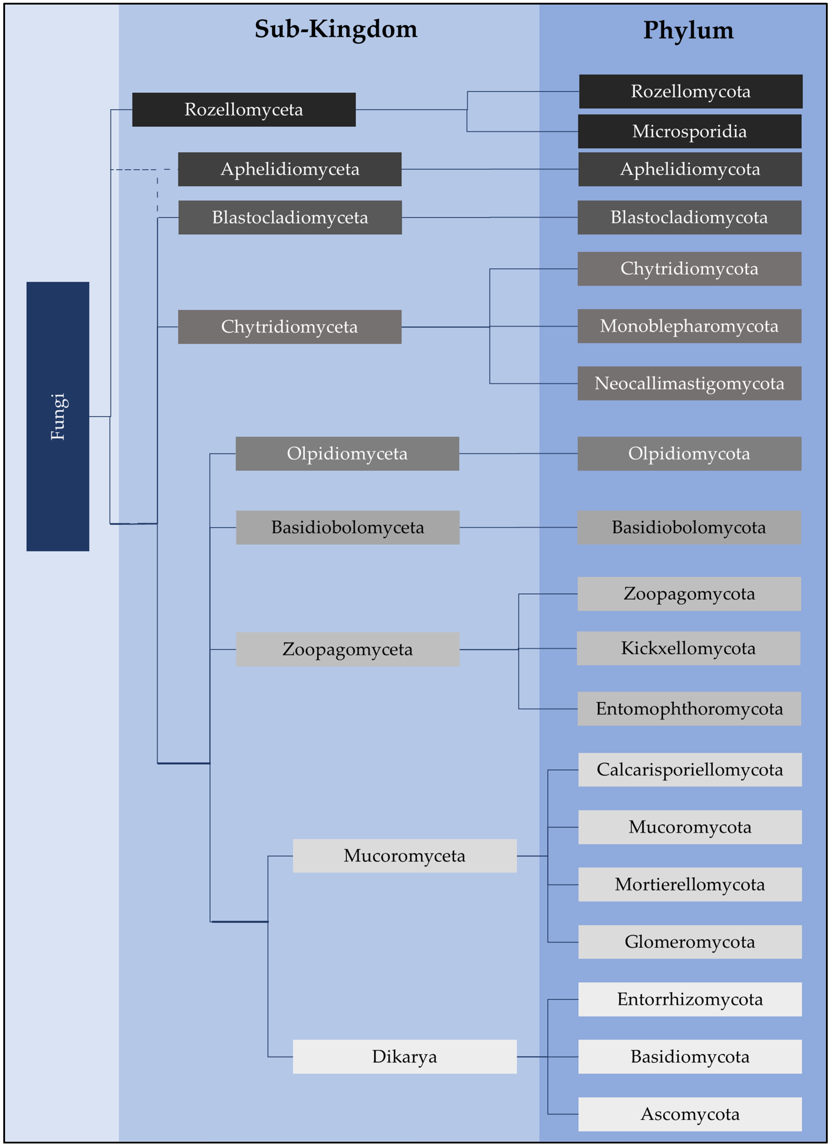
Chytridiomycota have been isolated from many environments, studied, and described [1,2]; however, their ecological importance within soil environments has been neglected. Despite this, it is apparent that these fungi play a keystone role in many aquatic environments [3]. The taxonomic classification and organisation of the phylogenetic tree of the fungal kingdom is under constant review and evolution [4,5,6,7]. In particular, significant caveats in phylogenetic and evolutionary knowledge concerning basal lineages of fungi still exist. Early classification of zoosporic true fungi (chytrids) combined the group of fungus-like species within the aquatic phycomycetes (sensu Sparrow 1960) [1]. Further revision of the taxonomy of eucaryotic organisms assigned chytrids to the supergroup Opisthokonta [8,9]. Later, zoosporic true fungi were split from the fungus-like zoosporic members of the Opisthokonta, with chytrids being assigned to the kingdom Fungi [10]. Chytridiomycota (chytrids), Neocallimastigomycota (neocallimastigos), and Monoblepharidomycota (monoblephs) are zoosporic early-diverging phyla under the sub-kingdom Chytridiomyceta [11,12] (Figure 1), although the exact relationships between these phyla are still to be determined [7].
Within Chytridiomycota there are currently 14 described orders: Caulochytriales, Chytridiales, Cladochytriales, Gromochytriales, Lobulomycetales, Mesochytriales, Polychytriales, Polyphagales, Rhizophydiales, Rhizophlyctidales, Spizellomycetales, Synchytriales, Zygophlyctidales, and Zygorhizidiales [7]. However, more than half of these orders still lack sequence data. Nephridiophagids have also been recently shown to sit within the Chytridiomycota phylum [13] and may form an additional order. For the anaerobic fungi phylum Neocallimastigomycota, there is only one order, Neocallimastigales. For the Monoblepheridiomycota there is also one order, Monoblepheridales. Previously, Blastocladiomycota was grouped within Chytridiomyceta but is now recognised as phylogenetically distinct based on morphological and molecular analyses [14,15,16,17]. However, the position of Blastocladiomycota on the fungal evolutionary tree has not yet been resolved [7]. More work is needed to fully understand the taxonomy of early-diverging fungi. Recently, increased phylogenetic diversity within the early-diverging taxa [7,11] has further highlighted the need to resolve these relationships, including within Chytridiomyceta. This review focuses on Chytridiomycota.
Figure 1. Fungi classification at the higher taxonomic ranks of sub-kingdom and phylum as per Powell and Letcher 2014 [12], Tedersoo et al. (18S and 28S rRNA sequence data) (2018) [11], and James et al. (2020) [7]. Although our current understanding of classifications has been updated, James et al. (2006a, 2006b) [15,18] has provided foundational phylogenetic analyses using the 18S, 28S, 5.8S, ITS, EF1α, RPB1 and RPB2 markers. The exact branching of Aphelidomyceta is still unclear (dotted lines).
Encyclopedia 03 00046 g001
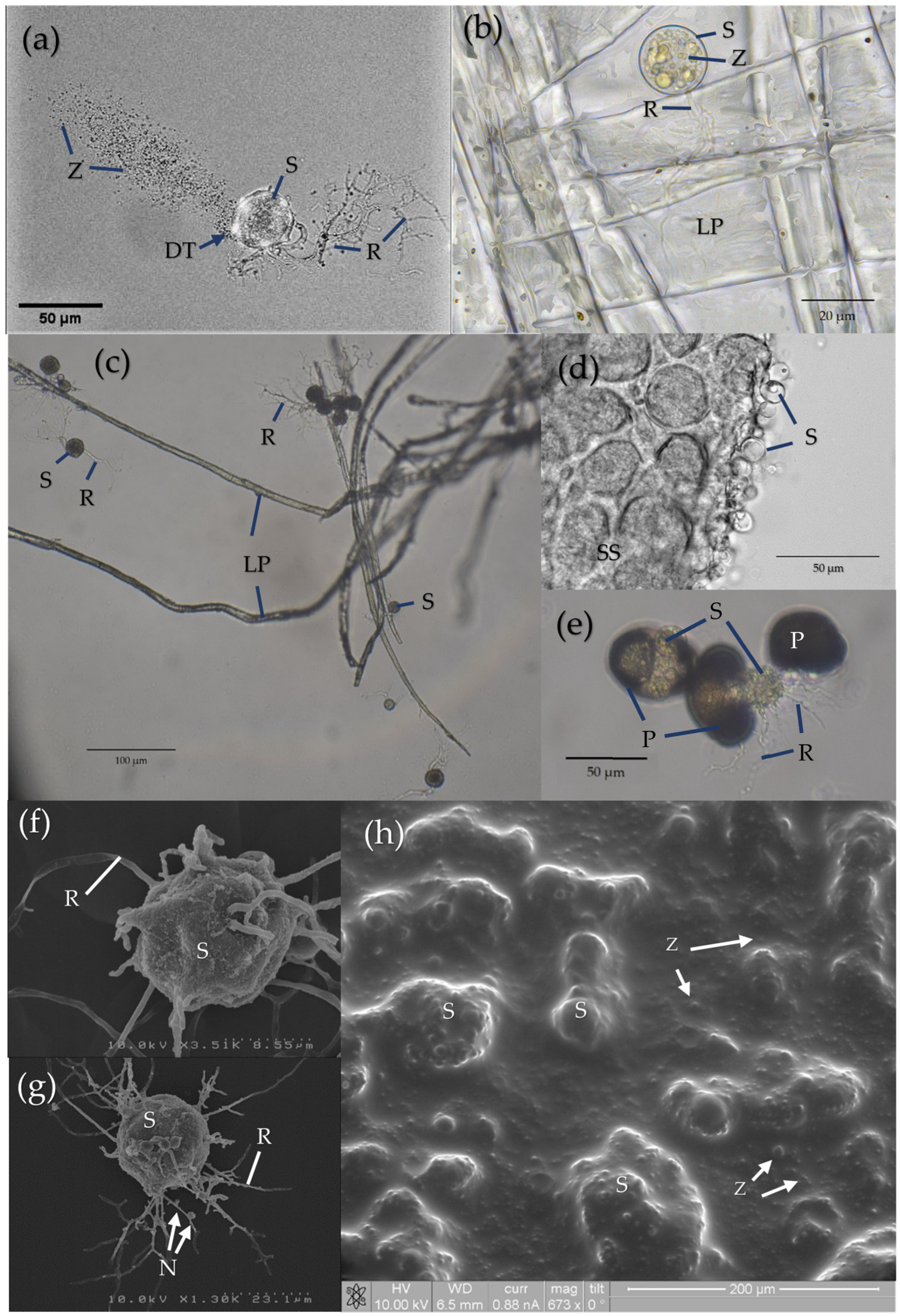
The morphological and physiological characteristics which allow chytrids to grow and disperse in soil environments are distinct and have been previously discussed [19]. The characteristic features of chytrids include chemotactic responses [20,21], motile zoospores propelled by a posterior flagellum [1,22], the presence of cholesterol rather than ergosterol as the major sterol [23], and morphological adaptations to environmental cues at different growth stages of the life cycle (Figure 2). These adaptations include changes in zoospore size, shape, and release during zoosporulation, changes in the size of the sporangium, and changes in the length and branching of the rhizoids after encystment [19] (Figure 3). A further feature is the ability of the rhizoid to attach to and penetrate highly resistant solid substrates, such as the hard outer casings of diatoms [24] and pollen grains [25] (Figure 3e). Stress tolerance and ruderal ecological strategies may both be adopted by individual chytrid species [19] in order to survive, grow, and complete their life cycle under a wide range of environmental conditions. There are a number of species in Chytridiomycota that are pathogenic in aquatic environments. Batrachochytrium dendrobatidis (order Rhizophydiales) can be considered one of the most important infectious diseases impacting the biodiversity of the amphibian population [26,27,28,29,30]. Members of Chytridiomycota have been associated with phytoplankton, including diatoms, dinoflagellates, and cyanobacteria, as microparasites [31,32,33,34]. They are well documented in bloom events in lakes [35,36] and algal blooms in the Mediterranean Sea [37]. Data indicate that fungal parasites can potentially control the fate of phytoplankton-derived organic matter by enhancing remineralisation and reducing sedimentation in freshwater and coastal systems [31].
This review examines the recent evidence for the presence and relative abundance of chytrids in different soil environments. The authors examine the soil environment and the functions and adaptive strategies of soil chytrid populations, particularly in light of the recent additional knowledge on their morphology and functionality. The authors then highlight the current research discussing potential applications in biotechnology. Finally, the authors recommend innovative methods for further investigating the distribution and ecology of chytrids in soil environments.

2. Chytrid Presence and Diversity in Soils

The relative abundance of chytrid sequences as a percentage of total fungal sequences is high in high elevation soils, which was revealed through targeting the 18S rRNA marker gene [41], as well as the 28S [5] and ITS regions [42], and were found to be the dominant fungal phylum in unvegetated soils above a 5100 m elevation in the Himalayas of Nepal [41]. Relative chytrid abundance, determined by targeting the ITS2 marker region, was the greatest at the highest elevation (3536 m) on Xinglong Mountain in Northwest China [43]. Chytrids have a high relative abundance (11% and greater) compared to total fungal sequences targeting 18S rRNA using pyrosequencing in arctic tundra soils [44] and are at least as abundant as Dikarya in association with arctic plant roots in the Archipelago Svalbard, Norway, when comparing the relative abundance of total fungal sequences [45]. Chytrid populations may be among the dominant fungal phyla in the rhizosphere rather than the bulk soil [46,47], but these populations are also sensitive to temporal factors and higher chytrid populations have been noted within the rhizosphere at early stages of plant development [46,48]. The ability of chytrids from soil environments to survive and grow in a range of salinity levels is known to vary by species and isolate [49]. In a comparison of community composition in marshland sediments, the highest relative abundance of chytrids was found in the salt marsh sample (7%) compared to brackish (3%) and freshwater swamps (1%) [50].
Chytrids may also contribute to the dominant fungal phyla in microhabitats, such as in biological soil crusts where they have a higher relative abundance in the soil beneath the crust based on the ITS marker analysis [51]. In biological soil crusts in the coastal and central deserts of Oman, bare soil areas compared to cyanobacteria and lichen-dominated crusts had a higher relative abundance of chytrids, being up to 26% as a proportion of total fungal abundance, compared to chytrid sequence abundances of less than 7%. The dominant genus found using the ITS marker was Rhizophlyctis (order Rhizophlyctidales) with a relative abundance from 8 to 25% as compared to the total fungal abundance in bare sandy soils from the Omanian regions of Muscat, Sur, and Haat [52]. Increases in chytrid populations using ITS2 markers occur in fire-affected biocrusts (genera Phlyctochytrium and Spizellomyces) [53]; these genera are within the order Chytridiales and Spizellomycetales, respectively, and their members are known to grow at high temperatures in vitro [54].
Molecular techniques have assisted with the identification of chytrids within soils from a wide diversity of forest ecosystems. Earlier studies using baiting techniques found chytrid diversity was higher in subtropical rain forest compared to open heath [55]. Relatively low chytrid abundances (1.6%) were found in Norway spruce (Picea abies) forest rhizosphere soils when using the ITS2 markers [56]. The three most common species were within the genera Gaertneriomyces (order Spizellomycetales), Rhizophydium (including R. globosum) (order Rhizophydiales), and Spizellomyces. Of the 42 taxa identified as chytrid, half had no cultured representative in the GenBank (NCBI) database [56]. A chronosequence in the subtropical Xishuangbanna forest of southwestern China also found proportionately low chytrid relative abundances in soil. A trend of increasing chytrid relative abundance was observed as forest age increased, and a negative correlation was found using the ITS marker between chytrid populations and the water-soluble carbon and nitrogen content of soils [57]. In contrast, in zinc/lead-contaminated Masson pine forest soil in Hunan, China, chytrids were among the most abundant fungal phyla in both the bulk soil and rhizosphere, with a relative abundance greater than 50% determined using the 18S rRNA gene [58]. Soil inoculated with the ectomycorrhizal fungus Suillus luteus also led to increases in relative chytrid abundance [58]. Chytrids from Spizellomycetales and Chytridiales, which are commonly found in terrestrial soils, have also been isolated from tree canopy soils of lowland rainforests in Australia and New Zealand [59,60]. Fungi that utilise spore dispersal (which may include zoospores) over mycelial development may contribute to the distinct fungal profile of the canopy of soils [61].
With recent advancements in high-throughput sequencing technologies and bioinformatic platforms, not only is the number and variety of soil chytrid genera being revealed, but also the functional community in which chytrids belong may be discerned with future work. Despite the array of methods applied in determining soil microbial communities and describing individual species, the setbacks and biases of each can limit the information obtained. PCR applications, for example, are reliant on existing primer sets which may discriminate against some fungal taxa. While fingerprinting DGGE or TGGE methods are limited to capturing the dominant phyla, usually with an abundance >1% [62], metabarcoding at a sufficient sequencing depth reveals the relative abundance of rare (<0.1% abundance) species [63].
Predominantly, soil fungal community analysis has focused on internal transcribed spacer (ITS) markers [42,47,51,52,64,65,66,67,68,69], while chytrid aquatic fungal community diversity has been elucidated by targeting the 18S rRNA gene marker [41,70,71]. Non-Dikarya lineages are at a particular disadvantage due to the lack of information about the diversity within various groups [72], and the suitability of current markers is, therefore, likely hampered. In particular, ITS markers are known to present biases regarding amplicon length, taxonomy, and primer mismatch [73]. Relatively few studies have undertaken a comparative analysis of metabarcoding methods for determining the fungal community composition of soils, but nonetheless, a few studies suggest variable capabilities to identify diverse fungal clades. A comparison of root-associated fungal communities in the arctic tundra using both the 18S rRNA and ITS2 gene markers found ITS2 data were largely missing the Chytridiomycota and Mucoromycota taxa, which were abundant in the 18S rRNA data [45]. Analysis of organic sediment-rich cave ice samples found Ascomycota dominated the total fungal community composition based on ITS2 Illumina sequencing [74], while Illumina shotgun sequencing found Chytridiomycota to be the dominant taxon [75]. Furthermore, Tedersoo et al. (2020) [76] resolved the issue of previously unidentified fungal taxa at the class level by re-analysing a set of 214 global soil samples using the 18S rRNA marker in combination with the ITS marker. Previously, the sample collection had only been analysed by targeting the ITS region.
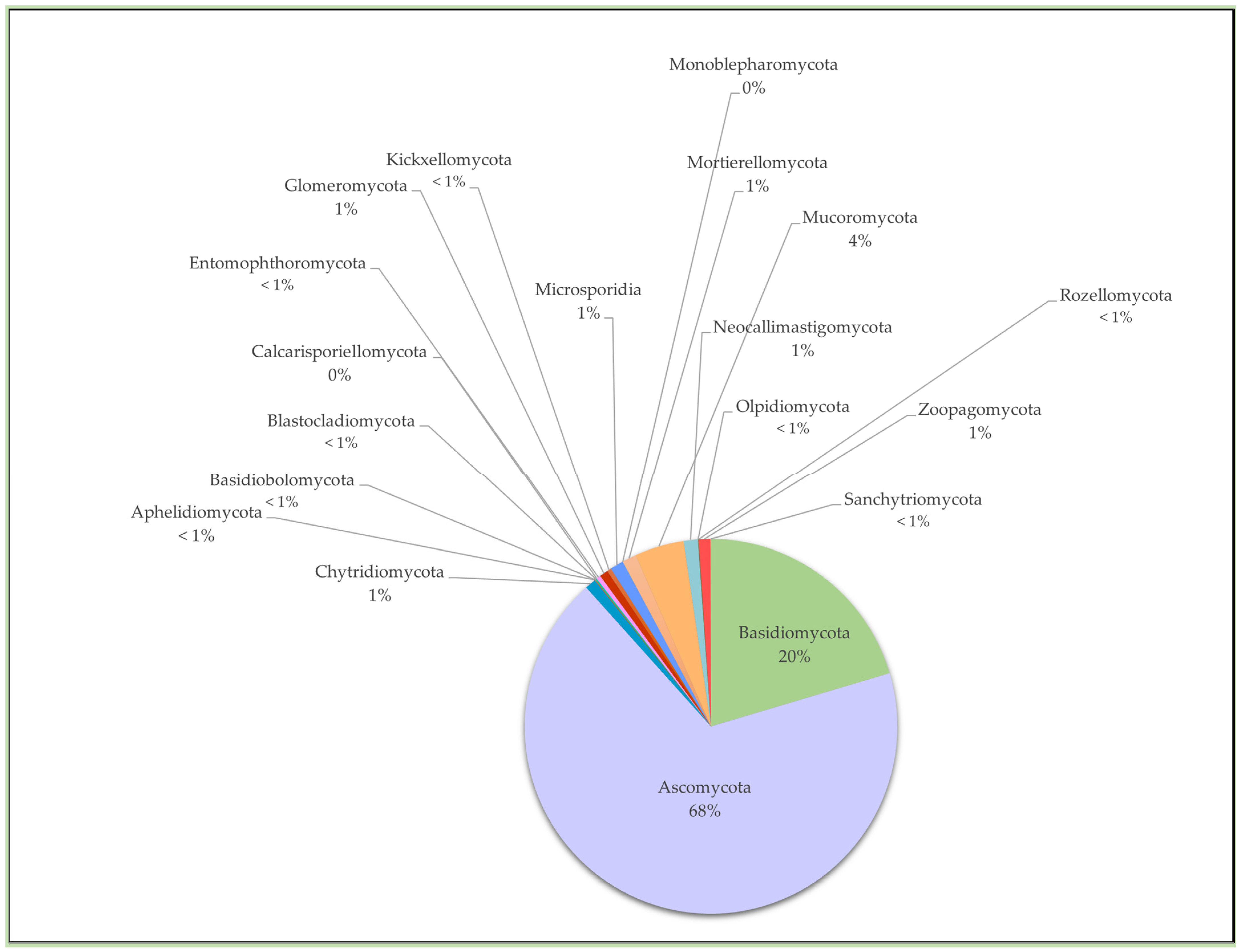
When examining the Global Biodiversity Information Facility (GBIF), Blaalid and Khomich (2021) [77] observed that Synchytrium anemones (order Synchytriales) was the most commonly recorded species in Norway, Sweden, and Finland, constituting 30% of the entire genus record. As highlighted, this particular chytrid can be observed macroscopically by eye due to its distinct phenotypic presentation as a plant parasite. It also highlights the emphasis often placed on microorganisms that are agriculturally relevant compared to those which may have less conspicuous roles. Data from the Global Soil Organisms project (distribution of occurrences dataset) further highlights the vast difference between the number of records of Dikarya phyla (>2 million records for both Ascomycota and Basidiomycota) compared to Chytridiomycota (<1 million chytrid records) [78]. Of all the fungi currently recognised, less than 10% are described, and the mass of data from genomic investigations, such as molecular operational taxonomic units, provides a likely pool of information regarding undescribed taxa [79]. For example, in a study of dust in an urban area of the Negev, Israel, half of the unclassified eukaryotic sequences identified with the 18S rRNA marker were similar to Rhizophlyctis rosea (order Rhizophlyctidales), while others were closely related to known Chytridiomycota genera including, but not limited to, Gaertneriomyces, Spizellomyces, and Powellomyces [80]. These genera are within the order Spizellomycetales, the members of which are able to survive desiccation and temperatures of up to 90 °C [81]. There is a need for more Chytridiomycota and Blastocladiomycota genomes to be sequenced and assembled as these taxa are currently poorly represented in genome databases (Figure 4). Only 1% of the currently described fungi are from Chytridiomycota and less than 1% are from Blastocladiomycota. For marine benthic environments where chytrids have been identified as dominant OTUs in ITS datasets, they are rare in the associated metatranscriptome data [82,83]. As “dark matter” fungi represent unexplored fungal diversity in marine environments [84], chytrids are likely to have unexplored diversity in soils. Of the unclassified taxa isolated from Gelisol soils in Alaska, 19.5% were identified at the order level with the 28S rRNA marker as belonging to Chytridiomycota [85]. This is becoming a common observation throughout the literature [13,76,77,85,86,87].

3. The Soil Environment at the Microscale: Chytrid Distribution and Adaptations

Unlike aquatic environments, soil environments experience continuous and sometimes sudden changes in conditions; wetting and drying cycles cause changes in soil pH [88], phosphorus levels [89], and availability of micronutrients and other metals at the microscale. Likewise, soil structural components are not static. Soil is composed of solid, liquid, and gaseous components, with the solid component consisting of particles of different sizes arranged into microaggregates (53–212 µm) [90] containing pore spaces of various sizes and connectivities. The ultrafine connected pores within aggregates are continually changing and evolving, with repeated wetting–drying cycles resulting in blocking, reforming, and reconnecting of pore networks [91]. The soil pore network fundamentally determines the ability of microbes to seek resources and evade predators [92]; however, it is also continually evolving. Pore size and connectivity also regulate the availability and transport of organic matter, water, and oxygen within microbial hotspots [93], while pore networks and fissures in the micro- to nanosize range are important reservoirs of soil organic carbon [91]. The size and physiology of soil microbes are expected to affect dispersal patterns in soil environments, and therefore, affect the composition of soil species assemblages at the microscale. For example, filamentous fungi are more likely to populate large air-filled pores >100 µm [94], and bacteria populate micropores <1.2 µm [95]. In forest soils, Chytridiomycota abundance was positively correlated with an average pore diameter in the <20 µm range [96]. Soil pore space affects predation by protists [97], and hence it is possible that chytrid sporangia and zoospores may be protected in smaller soil pores due to the limitation of predator body size. Rhizarian testate amoebae such as Euglypha (Silicofilosea; Rhizaria) can reach 150 µm and are unlikely to reach into small spaces due to a hard inflexible shell [97]. Instead, they are able to use thin filopodia (pseudopodia) to forage within soil aggregates [98]. The size of zoospores and mature sporangia vary with growth conditions [99]. However, it is not uncommon for rhizoids to be uniformly 1.5 µm in diameter and zoospores to be as small as 2–3 µm in diameter, such as described for the soil chytrid Rhizophydium brooksianum [100]. Smaller pores also have higher resource availability. Soil pores of 0.2–720 μm in diameter were found to be strongly correlated with the total soil organic carbon content [101]. The availability of organic matter, water, and oxygen within micropores may render them microbial hotspots [93], where chytrids may extract resources and also be protected from predation.
Recently, fungal community profiling of soil microaggregates targeting the 18S rRNA gene found a shift in the community structure at the phylum level as soil aggregate size decreased from 250 to 2 µm [102]. Blastocladiomycota were predominantly associated with the 20–63 µm aggregate size and Chytridiomycota were most abundant in association with the 2–20 µm aggregate size. There is a negative relationship between aggregate size and the number of soil pores, where a reduced aggregate size is associated with a significantly greater soil pore number [103]. Chytrid association with the smaller soil aggregate size and pore diameter is consistent with sporangial and rhizoidal size and the mineral particle size range, which they are expected to interact with.
The biological functions of rhizoids in soil, including rhizoidal interactions with substrates, are only beginning to be explored. Fungal hyphae, however, are known to regulate the soil environment [92]. Arbuscular mycorrhizal (AM) fungi increase soil aggregation [90,104,105,106,107,108] and slow the rate of macroaggregate turnover [90]. The hyphae of AM fungi have the ability to change soil particle orientation at the µm scale [92] and significantly increase soil mesopores (30–75 μm) and micropores (<30 μm) [109]. In clay loam soil, the smaller aggregates (0.5 mm) contained 88% more pores than larger aggregates (2–4 mm), and in sandy loam soil it was 92% more pores [109]. Recently, evidence has emerged of the ability of Rhizoclosmatium globosum (order Chytridiales) rhizoids to adapt by changing their morphology as they encounter different substrates, allowing them to attach and proliferate where suitable substrates are found [110]. Similar to hyphae, rhizoids exhibit polarised apical growth and fractal-like growth patterns which are modified by environmental conditions such as resource availability [110]. The spatial regulation of rhizoidal branching is controlled by the actin cytoskeleton which forms dynamic cellular structures [111]. Although chytrid rhizoids are known to attach to numerous substrates, including sand grains [19], the potential for chytrids to create pore spaces and assemble microaggregates as soil microhabitat engineers is unknown.
Chytrids may also adapt to physical soil disturbance. Due to their smaller size as compared to hyphae of arbuscular mycorrhizal fungi (AMF) (3–30 m g−1) [112], chytrid rhizoids (µm scale) may be less vulnerable when exposed to tillage which is otherwise destructive to AMF hyphae, as observed through population shifts in situ [113]. In addition to the negative mechanical disruption, tillage likely leads to drier soils in which the desiccation-resistant sporangia structure is the primary defence mechanism [81], therefore allowing population increases in some chytrid species under tillage [67,113].

4. Chytrid Mechanisms and Adaptations to Extreme Conditions and Environmental Gradients in Soil

Chytrids have been isolated from arid to semi-arid soils of the Western coastal regions of Saudi Arabia [68] and dunes of the Namib desert [114]. It is of interest that Rhizophlyctis sp. occurrence in desert soil is correlated with female but not male plants of the desert gymnosperm Welwitschia mirabilis. This was postulated to be due to soil chemical changes resulting from nectar accumulation in soil under the female plants [115]. In addition to heat and desiccation tolerance, some chytrids have been shown to survive after exposure to subzero temperatures [116]. It is not surprising then that chytrids have been isolated from polar regions including sub- and periglacial environments in the Arctic and Antarctic [41,86,117,118,119], as well as from Gelisol soils in Alaska [85]. Although considerable variability between the population abundances observed across studies is to be expected, there could be a notable distinction between the two terrestrial climates of deserts and polar regions. Low soil moisture content may be one factor contributing to overall lower population abundances (0.002–0.004%) in drier soils, such as in Saudi Arabia [68]. However, chytrid populations can increase as the soil water content increases, as was observed in arid soil in China [120]. Chytrids are reliant upon the presence of moisture to stimulate zoosporulation and, subsequently, aid in dispersal. Recovery through rehydration was used to determine the viability of dried chytrid thalli in vitro [81]. Due to the nature of periglacial and arctic environments with seasonal freeze–thaw cycles, the soil under ice layers is regularly saturated with water. The soil moisture content, therefore, fluctuates between 10 and 40% and chytrid populations shift accordingly (0.4–7.0%) [41]. In Antarctic soils, chytrids in the order Chytridiales were most abundant at higher soil moisture contents of 8% [119]. Chytrid sequences found in the soil at Mars Oasis in the southern maritime Antarctic were found to have close similarity to Chytridiales in soils with a relatively high moisture content in proximity to a meltwater pond [119]. In one study, a positive correlation between the presence of the sea ice-associated diatom Fragiliaropsis nitzschia and chytrid populations was observed [87]. Some chytrids, including Powellomyces sp., grow within pollen grains, which can then act as a protective casing in extreme environments [121].
Changes in nutrient levels produce changes in the morphology and reproductive strategies of chytrids at various stages of the chytrid life cycle. Unique responses amongst chytrid species even within the same order have been observed [38]. In one study, nitrogen starvation increased zoospore numbers for Gaertneriomyces semiglobifer but reduced numbers for R. rosea when compared with nitrogen-replete conditions in vitro [38]. Elevated phosphorus levels increased R. rosea zoospore numbers, while phosphorus scarcity reduced zoospore lipid content as well as motility [38]. It is clear that fluctuations in soil nutrient levels, such as through the addition of soil fertilisers, can lead to considerable soil microbiome shifts where some species are resilient while others are sensitive [67]. R. rosea sporangia exposed to toxic levels of zinc significantly reduced biomass production but also significantly increased zoospore numbers [122], indicating a shift in resources to zoospore production. Nutrients may change the morphology of rhizoids and sporangia—for example, R. rosea rhizoidal branching increased in the presence of elevated levels of copper [40].
Metals, including those which are not essential for biological functioning, may affect morphological development. As lead levels increased, rhizoid length and rhizoid number per sporangium increased for R. rosea. This observation corresponded with an increase in the number of sporangia attached to cellulosic substrates [39]. It may be physiologically advantageous for the chytrid to attach to substrates or to form colonies in order to survive toxic metal conditions, which may frequently occur in soil environments. The presence of extracellular polymeric substances may also provide an additional barrier on the fungal surface, thereby mitigating heavy metal toxicity [123]. A recent study looking at heavy metal effects on fungal community diversity supports the notion that chytrids are resilient to various heavy metals, but demonstrates the potential upper thresholds of this resilience, as chytrid OTUs were not isolated from the contaminated site where lead and chromium were most concentrated (42.9 mg Kg−1 and 45.9 mg Kg−1, respectively) [124].
As aquatic chytrids are known to quickly adapt to sudden high nutrient inputs, resulting in chytrid epidemics [125,126], so too can chytrids from soil environments. For example, under laboratory conditions, R. rosea zoospore production increased with increasing phosphate concentrations [38]. Rapid increases in some zoosporic true fungi have been identified in natural environments [1,127]. Some chytrids adopt ruderal strategies with rapid generation times, and are able to complete their life cycle in 48 h [128]. For example, R. rosea rapidly colonises cellulose baits [129]. When favourable conditions are present, the population density of R. rosea has also been observed to increase rapidly in soil environments [129]. Spore motility and sensing allow for rapid, energy-efficient dispersal, as well as survival in resource-scarce and resource-patchy environments.
Changes in soil pH also affect chytrid development in the soil. In one study under low pH and high Eh, soil conditions were ideal for supporting the chytrid infection of pollen grains [130]. Chytrid tolerance to more extreme pH ranges has been further shown in vitro [131], but this tolerance is species specific and does not account for the influence of other soil factors. For example, chytrid populations have been observed to have a positive correlation with soil factors, including for properties such as fine texture, electric conductivity, sulphate concentration, and soil phosphorus, nitrogen, and carbon content [120,132]. The methods used in Zhou et al. (2021) [66] clearly mapped out both the microbiological and biochemical changes in a soil ecosystem and provide an experimental guide for future investigations. These observations suggest a need to continue unveiling the relationships between abiotic factors and living microbial communities, including Chytridiomycota, to evaluate risks and develop strategies for building resilient natural and agricultural terrestrial environments.

5. Chytrid Roles in Soil Communities

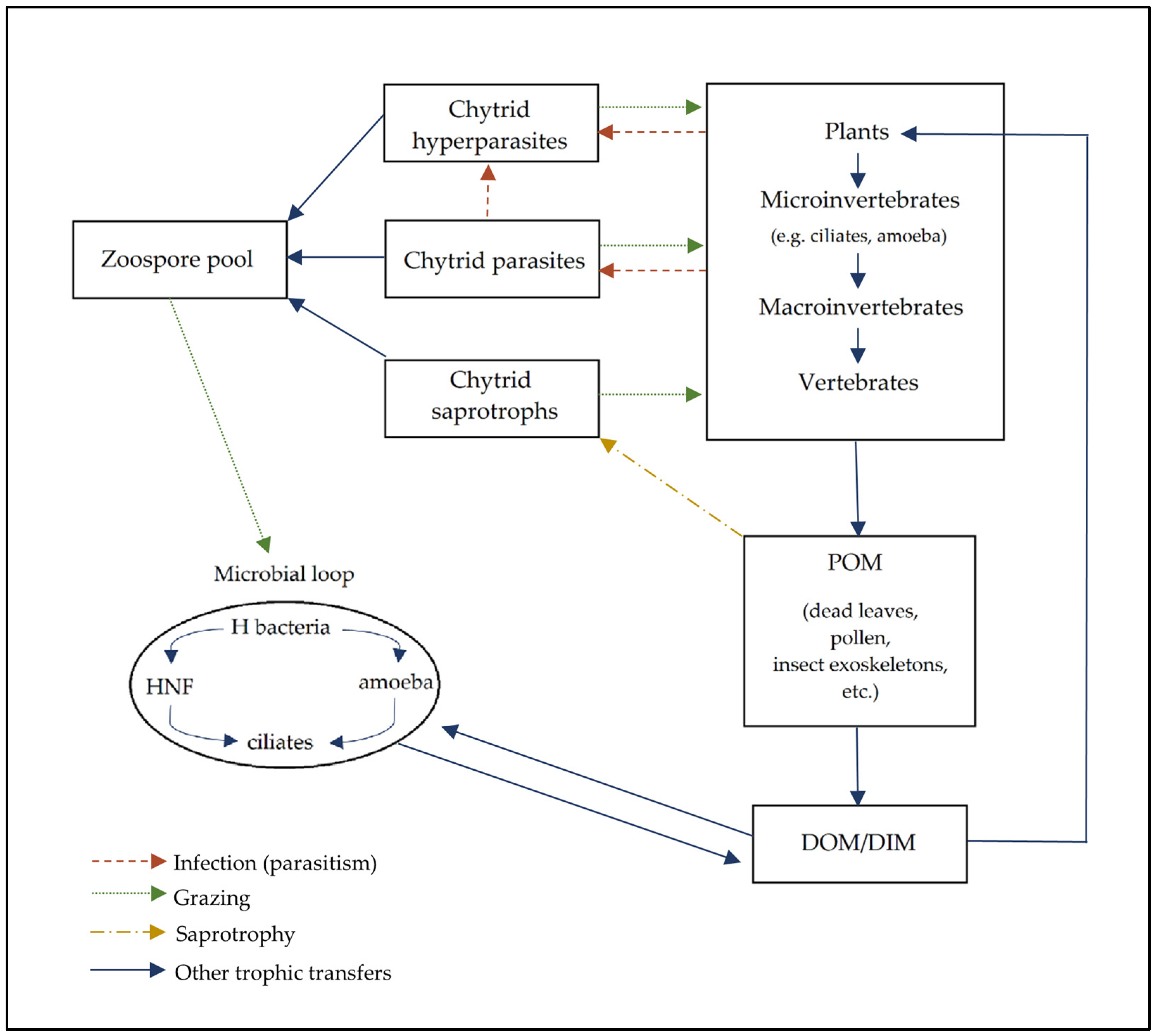
Chytrids have a number of consumer strategies, including parasitism, saprotrophism, and mutualism [133], which may alter the flow of energy and nutrients, change interspecific competition, and promote community diversity (Figure 5). Chytrids are able to penetrate resistant structures, such as pollen grains (Figure 3), releasing recalcitrant carbon. Saprotrophic chytrids efficiently digest cellulose, chitin, and protein typically found in soil particulate matter, which is solubilised to become dissolved organic matter and dissolved inorganic matter [134]. Soil chytrids may be hyperparasites of oomycetes and other chytrid species and parasites or facultative parasites of plants, oomycetes, chytrids [135], and vertebrates in soil systems, which increases the energy transfer and food web complexity. These organic nutrients which are transferred to zoospores (termed the “mycoloop”) [136] are, in turn, a valuable food resource for higher trophic levels as they contain organic phosphorus, nitrogen, sulphur, and mineral ions and vitamins [134]. Members of the Blastocladiomycota, especially the genera Coelomomyces and Catenaria, are common parasites of the soil-dwelling Crustacea, Hexapoda (Diptera), and Nematoda [137,138]. Yet, the supposed contribution of chytrids as parasites and saprotrophs to the soil fungal community and soil food web is relatively unknown.
The genus Synchytrium is predominantly composed of pathogens of terrestrial plants and algae and contains over 200 species [139]. Of the agriculturally important chytrids, Synchytrium endobioticum is recognised globally as a quarantine pest responsible for potato wart disease. Synchytrium anemones is a parasite of Anemone, a genus of flowering plants including buttercups, which causes purple-brown lesions on the leaves and stems [140]. The leguminous Desmodium plants are also susceptible to wart infections by Synchytrium desmodii [141]. In the rhizosphere of Fritaillaria taipaiensis (a medicinal flowering plant species in the family Liliaceae), the Chytridiomycota genera, including Synchytrium, were in the top 20 relative abundances of rhizospheric soil populations in the first cultivation year but decreased significantly in abundance by the fifth year [66]. However, viable resting spores of S. endobioticum can lay dormant in soil for decades, presenting a continuous risk to crops [142,143]. Some chytrid species are parasites that do not lead to pathogenic symptoms, including Rhizophydium gramanis which is a parasite of mono- and dicotyledonous plant roots and was first observed in 1936 [144,145]. Less well-known examples include the hyperparasitic Chytridium parasiticum (order Chytridiales). C. parasiticum is a biotroph of Septosperma rhizophydii which is, in turn, a parasite of the plant chytrid Rhizophydium macrosporum [146]. Nephridiophagids, which have recently been assigned to Chytridiomycota, are parasites of insects, including cockroaches (order Dictyoptera) and beetles (order Coleoptera) [147]. The Spizellomycetallean soil-dwelling G. semiglobifer has been found to live parasitically on azygospores of Entomophaga maimaiga, a fungal parasite of the gypsy moth [148]. G. semiglobifer is also known to be a parasite of the oospores of downy mildew (Peronosporomycetes) [149]. G. semiblobifer rhizoids are able to penetrate the highly resistant entomophthoralean azygospores, which are persistent and accumulate in soil due to their thick double wall. Early observations also indicated the potential for the diverse parasitic interactions of other early-diverging fungi. For instance, Catenaria anguillulae of the Blastocladiomycota may be a less well-known endobiotic parasite of Phytophthora cinnamomi and P. parasitica, which are important agricultural pests [150], and Catenaria allomycis has been described as a mycoparasite of Allomyces arbuscula [151]. The chytrid species Rhizidiomyces japonicus and Canteriomyces stigeoclonii are known hyperparasites of Phytophthora megasperma [152]. Research in the field of chytrid suppression of terrestrial plant diseases is generally lacking [135], but deserves attention with a focus on biocontrol routes and the aid of current molecular technologies.
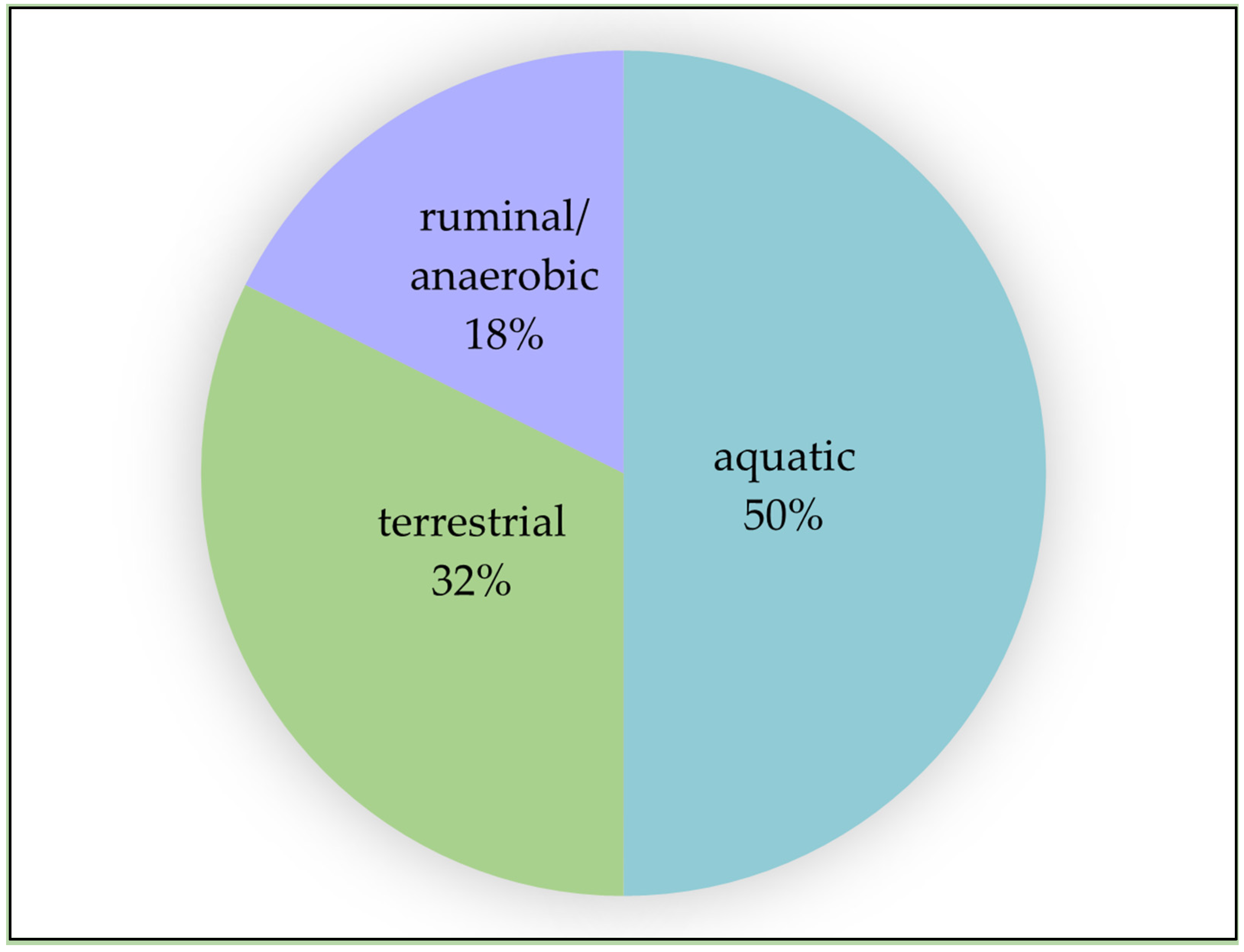
Chytridiomyceta and Blastocladiomycota have low representation in whole-genome sequencing databases compared to fungi generally (Figure 4), while less than a third of the NCBI chytrid genomes are from terrestrial chytrids (Figure 6). R. rosea and numerous other species are commonly isolated from soil using well-known baiting techniques. R. rosea was isolated from four agricultural soil types and from various land uses and vegetation types in Eastern Australia [55,129]. Often, studies attribute lower relative abundances of chytrids in soil to the belief that they are predominantly found in aquatic environments, which are thought to be more favourable to their growth. Chytrids may occur as relatively rare taxa in soil environments; however, they perform functional roles as decomposers, biological controllers, and potentially, as ecosystem regulators underpinning soil community resilience [41,153]. Soil fungal communities are known to modify rare taxa in response to changes in local soil environments, including in carbon to nitrogen ratios [154]. Community changes in response to environmental stress may also favour chytrids—for example, chytrid abundance significantly increased with saline irrigation in desert soil [155] and with increased carbon dioxide content in black clay soil [69]. Recently, chytrids have been implicated in fungal community resistance to conditions expected under climate change. Chytridiomycota dominated the fungal community composition of 900-year-old ice cores containing high organic sediment content from Scarisoara Cave, Romania [75]. Of the Chytridiomycota, Rhizophydium was the dominant genus. Heat shock treatment caused a 55% increase in the relative abundance of Chytridiomycota after seven days of incubation. The relative abundance of Blastocladiomycota also increased from 5 to 11% seven days after heat shock treatment. In soils subjected to seven years of simulated drought conditions, Chytridiomycota were correlated with the maintenance of soil functionality, specifically, the resistance of the soil nitrogen mineralisation rate and both resistance and resilience of soil phosphorus enzymes [156].
The relationships between communities of saprotrophic microbes are beginning to be explored. For example, the co-occurrence of bacterial and fungal decomposers of leaf litter produces accelerated decomposition in aquatic ecosystems [157]. Synergistic interactions are common between bacteria and fungi [158]. However, the study of synergistic and other interactions between fungi and bacteria in soil systems is rudimentary compared to the study of fungi–bacteria interactions in aquatic systems. Nonetheless, soil chytrids may play keystone roles within terrestrial communities. The aquatic chytrid Rhizoclosmatium globosum, through the colonisation of chitin microbeads, increased the diversity of the subsequent bacterial community potentially through the provision of dissolved organic carbon [159] and may have influenced bacterial community succession. The authors contend that chytrids in soil environments may have similar roles.
Figure 6. Percentage (%) of assembled Chytridiomyceta (Chytridiomycota and Neocallimastigomycota) genomes available in the NCBI Taxonomy Browser (n = 36) based on primary habitat (aquatic, terrestrial, ruminal/anaerobic). Neocallimastigomycota are only resolved at the class level in NCBI and classified under the phylum Chytridiomycota. No genome sequence data are available on NCBI for Monoblepharomycota. Data from the NCBI Taxonomy Browser were accessed on 21 November 2022. Genomes belonging to the Chytridiomycota phylum were searched for at a filter level of one. The genome data from the Entrez records were accessed and the primary habitat of each assembled chytrid species was determined by location from which the species was isolated or its known mode of life [149,160,161,162,163,164,165,166,167,168,169,170].
Encyclopedia 03 00046 g006

6. Further Research

Combinations of molecular and imaging techniques, such as confocal microscopy, along with culture-based analysis may further elucidate the roles of chytrids in soil environments and their interactions with other organisms. A study of population size changes within soil could use the methods adopted by Van den Wyngaert et al. (2022) [35] modified for the study of soil chytrids. This would involve classical techniques of soil chytrid baiting with simultaneous molecular and ecological data, allowing the study of temporal population changes in response to environmental change.
The 3D live-cell imaging of zoospores with laser-scanning confocal microscopy may provide further insights into the attachment of zoospores to host cells, including the location and production of adhesives. The variation in size and shape of zoospores [171] in response to substrates and environmental factors, as well as the ameboid-like nature of movement once in contact with substrates, has not been sufficiently examined. The variation in zoospore motility, chemotaxis, the length of time of motility, and the cues for the attachment, encystment, and germination of zoospores remain unelucidated [19]. Live-cell imaging, such as the use of the fluorescence staining of zoospore lipids, allows for the measurement of zoospore mobility [38] and is another promising technique (Supplementary Video S1a–c). Exploration of chytrid photoreceptors and the degree of phototaxis has also progressed slowly. Early observations of the parasitic Blastocladiomycota fungi Coelomomyces dodgei indicated light-sensitive spore release [172]. Allomyces reticulatus zoospores (Blastocladiomycota) are also known to be guided by phototaxis due to the presence of rhodopsins [173]. Unlike the Dikarya, which contain Type I opsins (bacteriorhodopsins), chytrids are now understood to contain rhodopsins homologous to type II opsins, which are classically of metazoan origin [174]. Not only does this finding highlight the distinct proteome of chytrids compared to other fungi, but also provides an alternate route for investigating biotechnology applications such as in optogenetics [175,176]. The aquatic chytrid Rhizoclosmatium globosum has been found to house an unexplored rhodopsin, Neorhodopsin (NeoR) [176]. This rhodopsin is of biotechnological interest as it is bistable and photoswitchable due to its geometric and chemical fluidity when exposed to either near-infrared light (NIR) or non-fluorescent UV light. Bioengineering of this chytrid rhodopsin could allow for the development of non-invasive tools for neuronal stimulation as it is not limited by the low penetration depths of current rhodopsin models [175,176].
While advances have recently been made in understanding the morphological development of the rhizoid [110] and the functions of the cytoskeleton [111], further investigation is necessary into the characteristics of rhizoids during different stages of the life cycle, including attachment. Study of the rhizoid can be undertaken in relatively small substrate volumes and could inform our understanding of rhizoidal development. MicroCT has been developed for 3D imaging of zoosporic and other fungi within a polystyrene bead matrix [177]. The introduction of resource heterogeneity or abiotic gradients into these matrices is also possible. Another potential method to examine rhizoid penetration is with a soil-like micromodel involving a siloxane polymer PDMS coated with O2 plasma [178]. Utilisation of cell staining for photomicrography and image processing software could also provide an insightful tool for measuring rhizoid growth and development [112].
A focus on progressing DNA-based methodologies, including the primers and markers specifically relevant to Chytridiomycota, is essential to conduct soil microbiome investigations [179] and better our ability to combat the current challenges of soil security in line with both agricultural and natural ecologies. Whole-genome sequencing can provide information about soil chytrid secretomes, and therefore, the likely functions of the fungi. The combination of 5.8S with ITS2 markers may allow for greater discernment of chytrid taxonomy [180].
Pectinase, cellulolytic, and xylanolytic enzymes have been identified from various chytrids, and those from aerobic chytrids share a common ancestor to the plant cell wall-degrading enzymes from Dikarya fungi [181,182,183]. The soil chytrid R. rosea is known to house an array of these enzymes and holds the potential to be a model organism for studying the secretomes of chytrids and other early-diverging fungi [181]. Comparative analyses of genomes between chytrids and other early-diverging, zoosporic fungi are becoming more accessible with the latest databases, models, and proteomic analysis methods [184]. Identifying the types of enzymes in secretomes would not only provide valuable information on the different ways the chytrids have adapted to different environments, but also regarding their potential applications [184].
Anaerobic fungi in the phyla Chytridiomycota and Rozellomycota (an early-diverging lineage [7,11,79]) have been found to significantly promote lignin degradation in subsurface sediment [185]. This is a significant observation and has implications in terms of bioremediation. The accumulation and deposition of organic matters in freshwater lakes has become a serious problem over the last few decades. The use of lignocellulose-producing species from these phyla could be used to degrade recalcitrant organic matter [185]. The presence and survival of Chytridiomycota and other key phyla in heavy metal soil samples suggest the potential use of tolerant species in bioremediation [124]. It was observed that fungal communities, including the taxa of Chytridiomycota, in rivers impacted by rare-earth-element acid mine drainage respond more robustly compared to prokaryotic networks [186]. These observations suggest that our further understanding of what mechanisms influence changes in microbial communities is required to reduce the impact of these pollutants on the microbial diversity of an environment. There are also potential “green” biotechnology benefits—in terms of the production of biofuels, biopolymers, and chemicals—in optimising these resources and processes [187,188]. Identifying the range of enzymes produced by microorganisms such as chytrids would provide more opportunities to understand and utilise these resources. Continuing to build on this knowledge is essential for both our comprehension of chytrid ecology in soil environments and for exploring potential biotechnological applications.

7. Conclusions

Soil chytrids are ubiquitous in terrestrial environments and are commonly found within many diverse soil microbiomes with a range of abundances. They have novel secretomes and play roles in nutrient cycling in the transformation of nutrients from organic to inorganic forms and in carbon cycling in the breakdown of complex organic materials. Many chytrids from soil environments are also parasites of plants and animals and may exert top-down control of prey populations and facilitate energy transfer within their ecosystem. The zoospore is a key unique characteristic of chytrids which allows for the dispersal and location, through chemotaxis, of suitable resources in order to perpetuate survival. Here, the authors reviewed new information on chytrid population distributions in soil environments that was discovered using recent advances in molecular techniques. The current focus on soil fungal community analysis by targeting internal transcribed spacer (ITS) markers may underestimate chytrid taxa and abundances, while the 18S gene marker could provide greater discernment. There is also a requirement for more sequenced genomes of terrestrial chytrids as they are currently poorly represented in genome databases. Furthermore, a more extensive database would allow for an in-depth investigation into gene–function relations. The authors also discussed new information on the soil environment and the implications for the chytrid life cycle. There is evidence that chytrid populations are distributed uniquely at the microscale, increasing in abundance in the rhizosphere and within microaggregates and micropores. The role of chytrid rhizoids in this process is promising but still unclear. The potential contributions of chytrids in soil microbial communities are only beginning to be explored. The authors encourage a combination of traditional and emerging techniques for research into the ecological roles of these understudied soil microorganisms.

Supplementary Materials

The following supporting information can be downloaded at: https://www.mdpi.com/article/10.3390/encyclopedia3020046/s1, Video S1a–c. Fluorescent confocal microscopy of nile red stained zoospores.

Author Contributions

Conceptualisation, L.H.; formal analysis, D.G.H.-T.; investigation, L.H. and D.G.H.-T.; writing—original draft preparation, L.H., D.G.H.-T. and O.L.; writing—review and editing, L.H., D.G.H.-T. and O.L.; visualisation, D.G.H.-T.; supervision, L.H. and O.L.; project administration, L.H. All authors have read and agreed to the published version of the manuscript.

Funding

This research received no external funding.

Data Availability Statement

Genome sequence data uploaded to NCBI Taxonomy Browser (https://www.ncbi.nlm.nih.gov/Taxonomy/Browser/wwwtax.cgi) were accessed on 21 November 2022. Data obtained from the Global Biodiversity Information Facility (https://www.gbif.org/) were accessed on 26 January 2023.

Conflicts of Interest

The authors declare no conflict of interest.

References

  1. Sparrow, F.K. Aquatic Phycomycetes, 2nd ed.; University of Michigan Press: Ann Arbor, MI, USA, 1960. [Google Scholar]
  2. Willoughby, L.G. The Ecology of some lower fungi at Esthwaite water. Trans. Br. Mycol. Soc. 1961, 44, 305–332, IN1–IN2. [Google Scholar] [CrossRef]
  3. Kagami, M.; de Bruin, A.; Ibelings, B.W.; Van Donk, E. Parasitic chytrids: Their effects on phytoplankton communities and food-web dynamics. Hydrobiologia 2007, 578, 113–129. [Google Scholar] [CrossRef]
  4. Naranjo-Ortiz, M.A.; Gabaldón, T. Fungal evolution: Diversity, taxonomy and phylogeny of the Fungi. Biol. Rev. Camb. Philos. Soc. 2019, 94, 2101–2137. [Google Scholar] [CrossRef]
  5. Tedersoo, L.; Bahram, M.; Puusepp, R.; Nilsson, R.H.; James, T. Novel soil-inhabiting clades fill gaps in the fungal tree of life. Microbiome 2017, 5, 42. [Google Scholar] [CrossRef] [PubMed]
  6. Li, Y.; Steenwyk, J.L.; Chang, Y.; Wang, Y.; James, T.Y.; Stajich, J.E.; Spatafora, J.W.; Groenewald, M.; Dunn, C.W.; Hittinger, C.T.; et al. A genome-scale phylogeny of the kingdom Fungi. Curr. Biol. 2021, 31, 1653–1665.e5. [Google Scholar] [CrossRef] [PubMed]
  7. James, T.Y.; Stajich, J.E.; Hittinger, C.T.; Rokas, A. Toward a Fully Resolved Fungal Tree of Life. Annu. Rev. Microbiol. 2020, 74, 291–313. [Google Scholar] [CrossRef] [PubMed]
  8. Baldauf, S.L. The Deep Roots of Eukaryotes. Sci. Am. Assoc. Adv. Sci. 2003, 300, 1703–1706. [Google Scholar] [CrossRef]
  9. Baldauf, S.L. An overview of the phylogeny and diversity of eukaryotes. J. Syst. Evol. 2008, 46, 263–273. [Google Scholar]
  10. Ruggiero, M.A.; Gordon, D.P.; Orrell, T.M.; Bailly, N.; Bourgoin, T.; Brusca, R.C.; Cavalier-Smith, T.; Guiry, M.D.; Kirk, P.M. A higher level classification of all living organisms. PLoS ONE 2015, 10, e0119248. [Google Scholar] [CrossRef]
  11. Tedersoo, L.; Sánchez-Ramírez, S.; Kõljalg, U.; Bahram, M.; Döring, M.; Schigel, D.; May, T.; Ryberg, M.; Abarenkov, K. High-level classification of the Fungi and a tool for evolutionary ecological analyses. Fungal Div. 2018, 90, 135–159. [Google Scholar] [CrossRef]
  12. Powell, M.J.; Letcher, P.M. Chytridiomycota, Monoblepharidomycota, and Neocallimastigomycota. In The Mycota, 2nd ed.; Esser, K., McLaughlin, D.J., Spatafora, J.W., Eds.; Springer: Berlin/Heidelberg, Germany, 2014; Volume 7, pp. 141–175. [Google Scholar] [CrossRef]
  13. Strassert, J.F.H.; Wurzbacher, C.; Hervé, V.; Antany, T.; Brune, A.; Radek, R. Long rDNA amplicon sequencing of insect-infecting nephridiophagids reveals their affiliation to the Chytridiomycota and a potential to switch between hosts. Sci. Rep. 2021, 11, 396. [Google Scholar] [CrossRef] [PubMed]
  14. Hibbett, D.S.; Binder, M.; Bischoff, J.F.; Blackwell, M.; Cannon, P.F.; Eriksson, O.E.; Huhndorf, S.; James, T.; Kirk, P.M.; Lücking, R.; et al. A higher-level phylogenetic classification of the Fungi. Mycol. Res. 2007, 111, 509–547. [Google Scholar] [CrossRef] [PubMed]
  15. James, T.Y.; Letcher, P.M.; Longcore, J.E.; Mozley-Standridge, S.E.; Porter, D.; Powell, M.J.; Griffith, G.W.; Vilgalys, R. A molecular phylogeny of the flagellated fungi (Chytridiomycota) and description of a new phylum (Blastocladiomycota). Mycologia 2006, 98, 860–871. [Google Scholar] [CrossRef] [PubMed]
  16. Powell, M.J. Chytridiomycota. In Handbook of the Protists, 2nd ed.; Archibald, J., Simpson, A., Slamovits, C., Eds.; Springer International Publishing: Cham, Switzerland, 2017; pp. 1523–1558. [Google Scholar] [CrossRef]
  17. Porter, T.M.; Martin, W.; James, T.Y.; Longcore, J.E.; Gleason, F.H.; Adler, P.H.; Letcher, P.M.; Vilgalys, R. Molecular phylogeny of the Blastocladiomycota (Fungi) based on nuclear ribosomal DNA. Fungal Biol. 2011, 115, 381–392. [Google Scholar] [CrossRef] [PubMed]
  18. James, T.Y.; Kauff, F.; Schoch, C.L.; Matheny, P.B.; Hofstetter, V.; Cox, C.J.; Celio, G.; Gueidan, C.; Fraker, E.; Miadlikowska, J. Reconstructing the early evolution of Fungi using a six-gene phylogeny. Nature 2006, 443, 818–822. [Google Scholar] [CrossRef]
  19. Gleason, F.H.; Crawford, J.W.; Neuhauser, S.; Henderson, L.E.; Lilje, O. Resource seeking strategies of zoosporic true fungi in heterogeneous soil habitats at the microscale level. Soil Biol. Biochem. 2012, 45, 79–88. [Google Scholar] [CrossRef]
  20. Held, A.A. Attraction and attachment of zoospores of the parasitic chytrid Rozella allomycis in response to host-dependent factors. Arch. Microbiol. 1974, 95, 97–114. [Google Scholar] [CrossRef]
  21. Mitchell, R.T.; Deacon, J.W. Selective accumulation of zoospores of chytridiomycetes and oomycetes on cellulose and chitin. Trans. Br. Mycol. Soc. 1986, 86, 219–223. [Google Scholar] [CrossRef]
  22. Gleason, F.H.; Lilje, O. Structure and function of fungal zoospores: Ecological implications. Fungal Ecol. 2009, 2, 53–59. [Google Scholar] [CrossRef]
  23. Weete, J.D.; Laseter, J.L. Distribution of sterols in the fungi I. Fungal spores. Lipids 1974, 9, 575–581. [Google Scholar] [CrossRef]
  24. Beakes, G.W.; Canter, H.M.; Jaworski, G.H.M. Comparative ultrastructural ontogeny of zoosporangia of Zygorhizidium affluens and Z. planktonicum, chytrid parasites of the diatom Asterionella formosa. Mycol. Res. 1992, 96, 1047–1059. [Google Scholar] [CrossRef]
  25. Barr, D.J.S. Phlyctochytrium reinboldtae (Chytridiales): Morphology and physiology. Can. J. Bot. 1970, 48, 479–484. [Google Scholar] [CrossRef]
  26. Scheele, B.C.; Skerratt, L.F.; Grogan, L.F.; Hunter, D.A.; Clemann, N.; McFadden, M.; Newell, D.; Hoskin, C.J.; Gillespie, G.R.; Heard, G.W. After the epidemic: Ongoing declines, stabilizations and recoveries in amphibians afflicted by chytridiomycosis. Biol. Conserv. 2017, 206, 37–46. [Google Scholar] [CrossRef]
  27. Scheele, B.C.; Pasmans, F.; Skerratt, L.F.; Berger, L.; Martel, A.; Beukema, W.; Acevedo, A.A.; Burrowes, P.A.; Carvalho, T.; Catenazzi, A.; et al. Amphibian fungal panzootic causes catastrophic and ongoing loss of biodiversity. Science 2019, 363, 1459–1463. [Google Scholar] [CrossRef] [PubMed]
  28. Tessa, G.; Sotgiu, G.; Bovero, S.; Angelini, C.; Favelli, M.; Gazzaniga, E.; Giacoma, C.; Garner, T.W.J. Cryptic but direct costs of an epidemic caused by Batrachochytrium dendrobatidis in the endangered Sardinian newt Euproctus platycephalus (Amphibia, Caudata). Amphibia-Reptilia 2023, 44, 83–94. [Google Scholar] [CrossRef]
  29. O’Hanlon, S.J.; Rieux, A.; Farrer, R.A.; Rosa, G.M.; Waldman, B.; Bataille, A.; Kosch, T.A.; Murray, K.; Brankovics, B.; Fumagalli, M.; et al. A 20th Century Out-of-Asia Origin of a panzootic threat to global amphibian biodiversity. Science 2018, 360, 621–627. [Google Scholar] [CrossRef] [PubMed]
  30. Weldon, C.; Channing, A.; Misinzo, G.; Cunningham, A.A. Disease driven extinction in the wild of the Kihansi spray toad, Nectophrynoides Asperginis. Afr. J. Herpetol. 2020, 69, 151–164. [Google Scholar] [CrossRef]
  31. Klawonn, I.; Van den Wyngaert, S.; Iversen, M.H.; Walles, T.J.W.; Flintrop, C.M.; Cisternas-Novoa, C.; Nejstgaard, J.C.; Kagami, M.; Grossart, H.-P. Fungal parasitism on diatoms alters formation and bio–physical properties of sinking aggregates. Commun. Biol. 2023, 6, 206. [Google Scholar] [CrossRef]
  32. Kagami, M.; Gurung, T.; Yoshida, T.; Urabe, J. To sink or to be lysed? Contrasting fate of two large phytoplankton species in Lake Biwa. Limnol. Oceanogr. 2006, 51, 2775–2786. [Google Scholar] [CrossRef]
  33. Gerphagnon, M.; Colombet, J.; Latour, D.; Sime-Ngando, T. Spatial and temporal changes of parasitic chytrids of cyanobacteria. Sci. Rep. 2017, 7, 6056. [Google Scholar] [CrossRef]
  34. Ibelings, B.W.; Gsell, A.S.; Mooij, W.M.; Van Donk, E.; Van Den Wyngaert, S.; De Senerpont Domis, L.N. Chytrid infections and diatom spring blooms: Paradoxical effects of climate warming on fungal epidemics in lakes. Freshw. Biol. 2011, 56, 754–766. [Google Scholar] [CrossRef]
  35. Van den Wyngaert, S.; Ganzert, L.; Seto, K.; Rojas-Jimenez, K.; Agha, R.; Berger, S.A.; Woodhouse, J.; Padisak, J.; Wurzbacher, C.; Kagami, M.; et al. Seasonality of parasitic and saprotrophic zoosporic fungi: Linking sequence data to ecological traits. ISME J. 2022, 16, 2242–2254. [Google Scholar] [CrossRef] [PubMed]
  36. Gsell, A.S.; Wolinska, J.; Preuß, K.; Teurlincx, S.; Özkundakci, D.; Hilt, S.; van Donk, E.; Ibelings, B.W.; Adrian, R. Long-term trends and seasonal variation in host density, temperature, and nutrients differentially affect chytrid fungi parasitising lake phytoplankton. Freshw. Biol. 2022, 67, 1532–1542. [Google Scholar] [CrossRef]
  37. Lepelletier, F.; Karpov, S.A.; Alacid, E.; Le Panse, S.; Bigeard, E.; Garcés, E.; Jeanthon, C.; Guillou, L. Dinomyces arenysensis gen. et sp. nov. (Rhizophydiales, Dinomycetaceae fam. nov.), a Chytrid Infecting Marine Dinoflagellates. Protist 2014, 165, 230–244. [Google Scholar] [CrossRef] [PubMed]
  38. Hanrahan-Tan, D.G.; Henderson, L.; Kertesz, M.A.; Lilje, O. The Effects of Nitrogen and Phosphorus on Colony Growth and Zoospore Characteristics of Soil Chytridiomycota. J. Fungi 2022, 8, 341. [Google Scholar] [CrossRef] [PubMed]
  39. Henderson, L.; Marano, A.V.; Truszewski, E.; Gleason, F.H.; Lilje, O. Copper (II), lead (ll) and zinc (ll) reduce the rate of attachment in three zoosporic true fungi from soils of NSW, Australia. Nova Hedwig. 2019, 108, 435–447. [Google Scholar] [CrossRef]
  40. Henderson, L.E.; Lilje, E.; Robinson, K.; Gleason, F.H.; Lilje, O. Effects of Toxic Metals on Chytrids, Fungal-Like Organisms, and Higher Fungi. In The Fungal Community: Its organisation and Role in the Ecosystem, 4th ed.; Dighton, J., White, J.F., Eds.; CRC Press: Boca Raton, FL, USA, 2017; Volume 32, pp. 487–512. [Google Scholar]
  41. Freeman, K.R.; Martin, A.P.; Karki, D.; Lynch, R.C.; Mitter, M.S.; Meyer, A.F.; Longcore, J.E.; Simmons, D.R.; Schmidt, S.K. Evidence That Chytrids Dominate Fungal Communities in High-Elevation Soils. Proc. Natl. Acad. Sci. USA 2009, 106, 18315–18320. [Google Scholar] [CrossRef]
  42. Liu, H.; Cheng, J.; Jin, H.; Xu, Z.; Yang, X.; Min, D.; Xu, X.; Shao, X.; Lu, D.; Qin, B. Characterization of Rhizosphere and Endophytic Microbial Communities Associated with Stipa purpurea and Their Correlation with Soil Environmental Factors. Plants 2022, 11, 363. [Google Scholar] [CrossRef]
  43. Khan, S.; Chen, N.; Zhang, C.; Wang, L.; Han, C.; Lu, K.; Li, Y.; Rafiq, M.; Iqbal, A.; Zhao, C. Soil fungal taxonomic diversity along an elevation gradient on the semi-arid Xinglong Mountain, Northwest China. Arch. Microbiol. 2020, 202, 2291–2302. [Google Scholar] [CrossRef]
  44. Shi, Y.; Xiang, X.; Shen, C.; Chu, H.; Neufeld, J.D.; Walker, V.K.; Grogan, P. Vegetation-associated impacts on arctic tundra bacterial and microeukaryotic communities. Appl. Environ. Microbiol. 2015, 81, 492–501. [Google Scholar] [CrossRef]
  45. Botnen, S.S.; Thoen, E.; Eidesen, P.B.; Krabberød, A.K.; Kauserud, H. Community composition of arctic root-associated fungi mirrors host plant phylogeny. FEMS Microbiol. Ecol. 2020, 96, 1. [Google Scholar] [CrossRef] [PubMed]
  46. An, Z.; Guo, F.; Chen, Y.; Bai, G.; Chen, Z. Rhizosphere bacterial and fungal communities during the growth of Angelica sinensis seedlings cultivated in an Alpine uncultivated meadow soil. PeerJ 2020, 2020, e8541. [Google Scholar] [CrossRef] [PubMed]
  47. Wang, S.; Wang, J.; Zhou, Y.; Huang, Y.; Tang, X. Comparative Analysis on Rhizosphere Soil and Endophytic Microbial Communities of Two Cultivars of Cyperus esculentus L. Var. Sativus. J. Soil Sci. Plant Nutr. 2022, 22, 2156–2168. [Google Scholar] [CrossRef]
  48. Sun, R.; Yi, Z.; Fu, Y.; Liu, H. Dynamic changes in rhizosphere fungi in different developmental stages of wheat in a confined and isolated environment. Appl. Microbiol. Biotechnol. 2022, 106, 441–453. [Google Scholar] [CrossRef] [PubMed]
  49. Gleason, F.H.; Midgley, D.J.; Letcher, P.M.; McGee, P.A. Can soil Chytridiomycota survive and grow in different osmotic potentials? Mycol. Res. 2006, 110, 869–875. [Google Scholar] [CrossRef] [PubMed]
  50. Mohamed, D.J.; Martiny, J.B.H. Patterns of fungal diversity and composition along a salinity gradient. ISME J. 2011, 5, 379–388. [Google Scholar] [CrossRef]
  51. Wang, J.; Huang, R.; Zhu, L.; Guan, H.; Lin, L.; Fang, H.; Yang, M.; Ji, S.; Zou, X.; Li, X. The Effects of Biochar on Microbial Community Composition in and Beneath Biological Soil Crusts in a Pinus massoniana Lamb. Plantation. Forests 2022, 13, 1141. [Google Scholar] [CrossRef]
  52. Abed, R.M.M.; Tamm, A.; Hassenrück, C.; Al-Rawahi, A.N.; Rodríguez-Caballero, E.; Fiedler, S.; Maier, S.; Weber, B. Habitat-dependent composition of bacterial and fungal communities in biological soil crusts from Oman. Sci. Rep. 2019, 9, 6468. [Google Scholar] [CrossRef]
  53. García-Carmona, M.; Lepinay, C.; García-Orenes, F.; Baldrian, P.; Arcenegui, V.; Cajthaml, T.; Mataix-Solera, J. Moss biocrust accelerates the recovery and resilience of soil microbial communities in fire-affected semi-arid Mediterranean soils. Sci. Total Environ. 2022, 846, 157467. [Google Scholar] [CrossRef]
  54. Gleason, F.H.; Letcher, P.M.; Commandeur, Z.; Jeong, C.E.; McGee, P.A. The growth response of some Chytridiomycota to temperatures commonly observed in the soil. Mycol. Res. 2005, 109, 717–722. [Google Scholar] [CrossRef]
  55. Letcher, P.M.; McGee, P.A.; Powell, M.J. Distribution and diversity of zoosporic fungi from soils of four vegetation types in New South Wales, Australia. Can. J. Bot. 2004, 82, 1490–1500. [Google Scholar] [CrossRef]
  56. Marčiulynas, A.; Marčiulynienė, D.; Mishcherikova, V.; Franić, I.; Lynikienė, J.; Gedminas, A.; Menkis, A. High Variability of Fungal Communities Associated with the Functional Tissues and Rhizosphere Soil of Picea abies in the Southern Baltics. Forests 2022, 13, 1103. [Google Scholar] [CrossRef]
  57. Bai, Z.; Wu, X.; Lin, J.-J.; Xie, H.-T.; Yuan, H.-S.; Liang, C. Litter-, soil- and C:N-stoichiometry-associated shifts in fungal communities along a subtropical forest succession. Catena Giess. 2019, 178, 350–358. [Google Scholar] [CrossRef]
  58. Yu, P.; Ning, C.; Chen, J.; Zhu, F.; Sun, Y.; Shen, A.; Zeng, W.; Jiang, L. The Effects of Suillus luteus Inoculation on the Diversity of Fungal Communities and Their Structures in the Soil under Pinus massoniana Located in a Mining Area. Forests 2022, 13, 2162. [Google Scholar] [CrossRef]
  59. Longcore, J.E. Zoosporic fungi from Australian and New Zealand tree-canopy detritus. Aust. J. Bot. 2005, 53, 259–272. [Google Scholar] [CrossRef]
  60. Letcher, P.M.; Longcore, J.E.; Powell, M.J. Dendrochytridium crassum gen. et sp. nov., a taxon in Chytridiales with unique zoospore ultrastructure. Mycologia 2014, 106, 145–153. [Google Scholar] [CrossRef]
  61. Orlovich, D.A.; Draffin, S.J.; Daly, R.A.; Stephenson, S.L. Piracy in the high trees: Ectomycorrhizal fungi from an aerial ‘canopy soil’ microhabitat. Mycologia 2013, 105, 52–60. [Google Scholar] [CrossRef]
  62. van Elsas, J.D.; Boersma, F.G.H. A review of molecular methods to study the microbiota of soil and the mycosphere. Eur. J. Soil Biol. 2011, 47, 77–87. [Google Scholar] [CrossRef]
  63. Joos, L.; Beirinckx, S.; Haegeman, A.; Debode, J.; Vandecasteele, B.; Baeyen, S.; Goormachtig, S.; Clement, L.; De Tender, C. Daring to be differential: Metabarcoding analysis of soil and plant-related microbial communities using amplicon sequence variants and operational taxonomical units. BMC Genom. 2020, 21, 733. [Google Scholar] [CrossRef]
  64. Tedersoo, L.; Bahram, M.; Polme, S.; Koljalg, U.; Yorou, N.S.; Wijesundera, R.; Villarreal Ruiz, L.; Vasco, A.; Pham Quang, T.; Suija, A.; et al. Global diversity and geography of soil fungi. Sci. Am. Assoc. Adv. Sci. 2014, 346, 1078. [Google Scholar] [CrossRef]
  65. Leff, J.W.; Jones, S.E.; Prober, S.M.; Barberán, A.; Borer, E.T.; Firn, J.L.; Harpole, W.S.; Hobbie, S.E.; Hofmockel, K.S.; Knops, J.M.H.; et al. Consistent responses of soil microbial communities to elevated nutrient inputs in grasslands across the globe. Proc. Natl. Acad. Sci. USA 2015, 112, 10967–10972. [Google Scholar] [CrossRef] [PubMed]
  66. Zhou, N.; Mu, M.; Xie, H.; Wu, Y.; Zhou, Y.; Li, W. Rhizospheric fungal diversities and soil biochemical factors of Fritillaria taipaiensis over five cultivation years. Horticulturae 2021, 7, 560. [Google Scholar] [CrossRef]
  67. Hannula, S.E.; Di Lonardo, D.P.; Christensen, B.T.; Crotty, F.V.; Elsen, A.; Erp, P.J.; Hansen, E.M.; Rubæk, G.H.; Tits, M.; Toth, Z.; et al. Inconsistent effects of agricultural practices on soil fungal communities across 12 European long-term experiments. Eur. J. Soil Sci. 2021, 72, 1902–1923. [Google Scholar] [CrossRef]
  68. Moussa, T.A.A.; Al-Zahrani, H.S.; Almaghrabi, O.A.; Abdelmoneim, T.S.; Fuller, M.P. Comparative metagenomics approaches to characterize the soil fungal communities of western coastal region, Saudi Arabia. PLoS ONE 2017, 12, e0185096. [Google Scholar] [CrossRef]
  69. Procter, A.C.; Ellis, J.C.; Fay, P.A.; Polley, H.W.; Jackson, R.B. Fungal community responses to past and future atmospheric CO2 differ by soil type. Appl. Environ. Microbiol. 2014, 80, 7364–7377. [Google Scholar] [CrossRef]
  70. Beng, K.C.; Wolinska, J.; Funke, E.; Van den Wyngaert, S.; Gsell, A.S.; Monaghan, M.T. Temporal dynamics of freshwater planktonic parasites inferred using a DNA metabarcoding time-series. Parasitology 2021, 148, 1602–1611. [Google Scholar] [CrossRef] [PubMed]
  71. Comeau, A.M.; Vincent, W.F.; Bernier, L.; Lovejoy, C. Novel chytrid lineages dominate fungal sequences in diverse marine and freshwater habitats. Sci. Rep. 2016, 6, 30120. [Google Scholar] [CrossRef] [PubMed]
  72. Lindahl, B.D.; Nilsson, R.H.; Tedersoo, L.; Abarenkov, K.; Carlsen, T.; Kjøller, R.; Kõljalg, U.; Pennanen, T.; Rosendahl, S.; Stenlid, J.; et al. Fungal community analysis by high-throughput sequencing of amplified markers—A user’s guide. New Phytol. 2013, 199, 288–299. [Google Scholar] [CrossRef]
  73. Bellemain, E.; Carlsen, T.; Brochmann, C.; Coissac, E.; Taberlet, P.; Kauserud, H. ITS as an environmental DNA barcode for fungi: An in silico approach reveals potential PCR biases. BMC Microbiol. 2010, 10, 189. [Google Scholar] [CrossRef]
  74. Mondini, A.; Donhauser, J.; Itcus, C.; Marin, C.; Perșoiu, A.; Lavin, P.; Frey, B.; Purcarea, C. High-throughput sequencing of fungal communities across the perennial ice block of Scărișoara Ice Cave. Ann. Glaciol. 2018, 59, 134–146. [Google Scholar] [CrossRef]
  75. Mondini, A.; Anwar, M.Z.; Anwar, M.Z.; Ellegaard-Jensen, L.; Lavin, P.; Lavin, P.; Jacobsen, C.S.; Purcarea, C. Heat Shock Response of the Active Microbiome From Perennial Cave Ice. Front. Microbiol. 2022, 12, 809076. [Google Scholar] [CrossRef] [PubMed]
  76. Tedersoo, L.; Anslan, S.; Bahram, M.; Kõljalg, U.; Abarenkov, K. Identifying the ‘unidentified’ fungi: A global-scale long-read third-generation sequencing approach. Fungal Div. 2020, 103, 273–293. [Google Scholar] [CrossRef]
  77. Blaalid, R.; Khomich, M. Current knowledge of Chytridiomycota diversity in Northern Europe and future research needs. Fungal Biol. Rev. 2021, 36, 42–51. [Google Scholar] [CrossRef]
  78. PlutoF. GLOBAL Soil Organisms Occurrence Dataset. Available online: https://www.gbif.org/dataset/9f0e1ca6-fb08-4c72-9a4a-1e3b7a528c10/metrics (accessed on 27 January 2023).
  79. Voigt, K.; James, T.Y.; Kirk, P.M.; Santiago, A.L.C.M.d.A.; Waldman, B.; Griffith, G.W.; Fu, M.; Radek, R.; Strassert, J.F.H.; Wurzbacher, C.; et al. Early-diverging fungal phyla: Taxonomy, species concept, ecology, distribution, anthropogenic impact, and novel phylogenetic proposals. Fungal Divers. 2021, 109, 59–98. [Google Scholar] [CrossRef]
  80. Katra, I.; Arotsker, L.; Krasnov, H.; Zaritsky, A.; Kushmaro, A.; Ben-Dov, E. Richness and diversity in dust stormborne biomes at the southeast mediterranean. Sci. Rep. 2014, 4, 5265. [Google Scholar] [CrossRef]
  81. Gleason, F.H.; Letcher, P.M.; McGee, P.A. Some Chytridiomycota in soil recover from drying and high temperatures. Mycol. Res. 2004, 108, 583–589. [Google Scholar] [CrossRef]
  82. Ortega-Arbulú, A.S.; Pichler, M.; Vuillemin, A.; Orsi, W.D. Effects of organic matter and low oxygen on the mycobenthos in a coastal lagoon. Environ. Microbiol. 2019, 21, 374–388. [Google Scholar] [CrossRef]
  83. Nguyen, N.H.; Song, Z.; Bates, S.T.; Branco, S.; Tedersoo, L.; Menke, J.; Schilling, J.S.; Kennedy, P.G. FUNGuild: An open annotation tool for parsing fungal community datasets by ecological guild. Fungal Ecol. 2016, 20, 241–248. [Google Scholar] [CrossRef]
  84. Grossart, H.-P.; Wurzbacher, C.; James, T.Y.; Kagami, M. Discovery of dark matter fungi in aquatic ecosystems demands a reappraisal of the phylogeny and ecology of zoosporic fungi. Fungal Ecol. 2016, 19, 28–38. [Google Scholar] [CrossRef]
  85. Penton, C.R.; Louis, D.S.; Cole, J.R.; Yiqi, L.U.O.; Liyou, W.U.; Schuur, E.A.G.; Jizhong, Z.; Tiedje, J.M. Fungal Diversity in Permafrost and Tallgrass Prairie Soils under Experimental Warming Conditions. Appl. Environ. Microbiol. 2013, 79, 7063–7072. [Google Scholar] [CrossRef]
  86. Perini, L.; Gostinčar, C.; Gunde-Cimerman, N. Fungal and bacterial diversity of Svalbard subglacial ice. Sci. Rep. 2019, 9, 20230. [Google Scholar] [CrossRef]
  87. Kilias, E.S.; Junges, L.; Šupraha, L.; Leonard, G.; Metfies, K.; Richards, T.A. Chytrid fungi distribution and co-occurrence with diatoms correlate with sea ice melt in the Arctic Ocean. Commun. Biol. 2020, 3, 183. [Google Scholar] [CrossRef] [PubMed]
  88. Kim, M.; Or, D. Microscale pH variations during drying of soils and desert biocrusts affect HONO and NH3 emissions. Nat. Commun. 2019, 10, 3944. [Google Scholar] [CrossRef] [PubMed]
  89. Khan, S.U.; Hooda, P.S.; Blackwell, M.S.A.; Busquets, R. Microbial Biomass Responses to Soil Drying-Rewetting and Phosphorus Leaching. Front. Environ. Sci. 2019, 7, 1–9. [Google Scholar] [CrossRef]
  90. Morris, E.K.; Morris, D.J.P.; Vogt, S.; Gleber, S.C.; Bigalke, M.; Wilcke, W.; Rillig, M.C. Visualizing the dynamics of soil aggregation as affected by arbuscular mycorrhizal fungi. ISME J. 2019, 13, 1639–1646. [Google Scholar] [CrossRef]
  91. Smucker, A.J.M.; Park, E.J.; Dorner, J.; Horn, R. Soil Micropore Development and Contributions to Soluble Carbon Transport within Macroaggregates. Vadose Zone J. 2007, 6, 282–290. [Google Scholar] [CrossRef]
  92. Ritz, K.; Young, I.M. Interactions between soil structure and fungi. Mycologist 2004, 18, 52–59. [Google Scholar] [CrossRef]
  93. Rabbi, S.M.F.; Daniel, H.; Lockwood, P.V.; Macdonald, C.; Pereg, L.; Tighe, M.; Wilson, B.R.; Young, I.M. Physical soil architectural traits are functionally linked to carbon decomposition and bacterial diversity. Sci. Rep. 2016, 6, 33012. [Google Scholar] [CrossRef] [PubMed]
  94. Otten, W.; Hall, D.; Harris, K.; Ritz, K.; Young, I.M.; Gilligan, C.A. Soil Physics, Fungal Epidemiology and the Spread of Rhizoctonia Solani. New Phytol. 2001, 151, 459–468. [Google Scholar] [CrossRef]
  95. Hassink, J.; Bouwman, L.A.; Zwart, K.B.; Brussaard, L. Relationships between habitable pore space, soil biota and mineralization rates in grassland soils. Soil Biol. Biochem. 1993, 25, 47–55. [Google Scholar] [CrossRef]
  96. Meng, M.; Chen, H.Y.H.; Lin, J.; Liu, X.; Guo, X.; Yuan, Y.; Zhang, J. Long term forest conversion affected soil nanoscale pores in subtropical China. Catena Giess. 2020, 185, 104289. [Google Scholar] [CrossRef]
  97. Geisen, S.; Mitchell, E.A.D.; Adl, S.; Bonkowski, M.; Dunthorn, M.; Ekelund, F.; Fernández, L.D.; Jousset, A.; Krashevska, V.; Singer, D.; et al. Soil protists: A fertile frontier in soil biology research. FEMS Microbiol. Rev. 2018, 42, 293–323. [Google Scholar] [CrossRef] [PubMed]
  98. Meisterfeld, R. Testate amoebae with filopodia. In The llustrated Guide to the Protozoa; Lee, J.J.L.G., Bradbury, P., Eds.; Society of Protozoologists: Lawrence, KS, USA, 2002; Volume 2, pp. 827–860. [Google Scholar]
  99. Paterson, R.A. Observations on two species of Rhizophydium from Northern Michigan. Trans. Br. Mycol. Soc. 1963, 46, 530–536, IN9. [Google Scholar] [CrossRef]
  100. Longcore, J.E. Rhizophydium brooksianum sp. nov., a multipored chytrid from soil. Mycologia 2004, 96, 162–171. [Google Scholar] [CrossRef] [PubMed]
  101. Fukumasu, J.; Jarvis, N.; Koestel, J.; Kätterer, T.; Larsbo, M. Relations between soil organic carbon content and the pore size distribution for an arable topsoil with large variations in soil properties. Eur. J. Soil Sci. 2022, 73, e13212. [Google Scholar] [CrossRef]
  102. Keuschnig, C.; Martins, J.M.F.; Navel, A.; Simonet, P.; Larose, C. Micro-fractionation shows microbial community changes in soil particles below 20 μm. Front. Ecol. Evol. 2022, 10, 1–15. [Google Scholar] [CrossRef]
  103. Mangalassery, S.; Sjögersten, S.; Sparkes, D.L.; Sturrock, C.J.; Mooney, S.J. The effect of soil aggregate size on pore structure and its consequence on emission of greenhouse gases. Soil Tillage Res. 2013, 132, 39–46. [Google Scholar] [CrossRef]
  104. Miller, R.M.; Jastrow, J.D. Mycorrhizal Fungi Influence Soil Structure. In Arbuscular Mycorrhizas: Physiology and Function; Kapulnik, Y., Douds, D.D., Eds.; Springer: Dordrecht, The Netherlands, 2000; pp. 3–18. [Google Scholar] [CrossRef]
  105. Rillig, M.C.; Mummey, D.L. Mycorrhizas and Soil Structure. New Phytol. 2006, 171, 41–53. [Google Scholar] [CrossRef]
  106. Rillig, M.C.; Mardatin, N.F.; Leifheit, E.F.; Antunes, P.M. Mycelium of arbuscular mycorrhizal fungi increases soil water repellency and is sufficient to maintain water-stable soil aggregates. Soil Biol. Biochem. 2010, 42, 1189–1191. [Google Scholar] [CrossRef]
  107. Thomas, R.S.; Franson, R.L.; Bethlenfalvay, G.J. Separation of vesicular-arbuscular mycorrhizal fungus and root effects on soil aggregation. Soil Sci. Soc. Am. J. 1993, 57, 77–81. [Google Scholar] [CrossRef]
  108. Wilson, G.W.T.; Rice, C.W.; Rillig, M.C.; Springer, A.; Hartnett, D.C. Soil aggregation and carbon sequestration are tightly correlated with the abundance of arbuscular mycorrhizal fungi: Results from long-term field experiments. Ecol. Lett. 2009, 12, 452–461. [Google Scholar] [CrossRef] [PubMed]
  109. Samaei, F.; Asghari, S.; Aliasgharzad, N. The effects of two arbuscular mycorrhizal fungi on some physical properties of a sandy loam soil and nutrients uptake by spring barley. J. Soil Environ. 2015, 1, 1–9. [Google Scholar]
  110. Laundon, D.; Chrismas, N.; Wheeler, G.; Cunliffe, M. Chytrid rhizoid morphogenesis resembles hyphal development in multicellular fungi and is adaptive to resource availability. Proc. R. Society. B Biol. Sci. 2020, 287, 20200433. [Google Scholar] [CrossRef]
  111. Prostak, S.M.; Robinson, K.A.; Titus, M.A.; Fritz-Laylin, L.K. The actin networks of chytrid fungi reveal evolutionary loss of cytoskeletal complexity in the fungal kingdom. Curr. Biol. 2021, 31, 1192–1205.e6. [Google Scholar] [CrossRef]
  112. Shen, Q.; Kirschbaum, M.U.F.; Hedley, M.J.; Arbestain, M.C. Testing an alternative method for estimating the length of fungal hyphae using photomicrography and image processing. PLoS ONE 2016, 11, e0157017. [Google Scholar] [CrossRef] [PubMed]
  113. Lienhard, P.; Terrat, S.; Prévost-Bouré, N.C.; Nowak, V.; Régnier, T.; Sayphoummie, S.; Panyasiri, K.; Tivet, F.; Mathieu, O.; Levêque, J.; et al. Pyrosequencing evidences the impact of cropping on soil bacterial and fungal diversity in Laos tropical grassland. Agron. Sustain. Dev. 2014, 34, 525–533. [Google Scholar] [CrossRef]
  114. van Der Walt, A.J.; Johnson, R.M.; Cowan, D.A.; Seely, M.; Ramond, J.-B.; Kelly, R.M. Unique microbial phylotypes in Namib Desert dune and gravel plain fairy circle soils. Appl. Environ. Microbiol. 2016, 82, 4592–4601. [Google Scholar] [CrossRef]
  115. Doniger, T.; Kerfahi, D.; Wachtel, C.; Marais, E.; Maggs-Kölling, G.; Sherman, C.; Adams, J.M.; Steinberger, Y. Plant Gender Affects Soil Fungal Microbiota Associated with Welwitschia mirabilis, an Unusual Desert Gymnosperm. Microb. Ecol. 2022. [Google Scholar] [CrossRef]
  116. Gleason, F.H.; Letcher, P.M.; McGee, P.A. Freeze tolerance of soil chytrids from temperate climates in Australia. Mycol Res 2008, 112, 976–982. [Google Scholar] [CrossRef]
  117. Booth, T.; Barrett, P. Occurrence and distribution of zoosporic fungi from Devon Island, Canadian Eastern Arctic. Can. J. Bot. 1971, 49, 359–369. [Google Scholar] [CrossRef]
  118. Zhang, T.; Wang, N.-F.; Zhang, Y.-Q.; Liu, H.-Y.; Yu, L.-Y. Diversity and distribution of aquatic fungal communities in the Ny-Ålesund Region, Svalbard (High Arctic). Microb. Ecol. 2016, 71, 543–554. [Google Scholar] [CrossRef] [PubMed]
  119. Bridge, P.D.; Newsham, K.K. Soil fungal community composition at Mars Oasis, a southern maritime Antarctic site, assessed by PCR amplification and cloning. Fungal Ecol. 2009, 2, 66–74. [Google Scholar] [CrossRef]
  120. Li, W.; Jiang, L.; Zhang, Y.; Teng, D.; Wang, H.; Wang, J.; Lv, G. Structure and driving factors of the soil microbial community associated with Alhagi sparsifolia in an arid desert. PLoS ONE 2021, 16, e0254065. [Google Scholar] [CrossRef]
  121. Longcore, J.E.; Barr, D.J.S.; Désaulniers, N. Powellomyces, a new genus in the Spizellomycetales. Can. J. Bot. 1995, 73, 1385–1390. [Google Scholar] [CrossRef]
  122. Henderson, L.; Pilgaard, B.; Gleason, F.H.; Lilje, O. Copper (II) lead (II), and zinc (II) reduce growth and zoospore release in four zoosporic true fungi from soils of NSW, Australia. Fungal Biol. 2015, 119, 648–655. [Google Scholar] [CrossRef] [PubMed]
  123. Jia, T.; Wang, R.; Fan, X.; Chai, B. A comparative study of fungal community structure, diversity and richness between the soil and the phyllosphere of native grass species in a copper tailings dam in Shanxi Province, China. Appl. Sci. 2018, 8, 1297. [Google Scholar] [CrossRef]
  124. Passarini, M.R.Z.; Ottoni, J.R.; Costa, P.E.d.S.; Hissa, D.C.; Falcão, R.M.; Melo, V.M.M.; Balbino, V.Q.; Mendonça, L.A.R.; Lima, M.G.d.S.; Coutinho, H.D.M.; et al. Fungal community diversity of heavy metal contaminated soils revealed by metagenomics. Arch. Microbiol. 2022, 204, 255. [Google Scholar] [CrossRef]
  125. Canter, H.M.; Lund, J.W.G. Studies on plankton parasites III. Examples of the interaction between parasitism and other factors determining the growth of diatoms. Ann. Bot. 1951, 15, 359–371. [Google Scholar]
  126. Ibelings, B.W.; De Bruin, A.; Kagami, M.; Rijkeboer, M.; Brehm, M.; Donk, E.V. Host parasite interactions between freshwater phytoplankton and chytrid fungi (Chytridiomycota). J. Phycol. 2004, 40, 437–453. [Google Scholar] [CrossRef]
  127. Gleason, F.; Macarthur, D. The chytrid epidemic revisited. Inoculum 2008, 59, 1–3. [Google Scholar]
  128. Ward, M.W. Observations on Rhizophlyctis rosea. J. Elisha Mitchell Sci. Soc. 1939, 55, 353–360. [Google Scholar]
  129. McGee, P.A.; Daynes, C.N.; Gleason, F.H.; Marano, A.V.; Barrera, M.D.; Steciow, M.M. Rhizophlyctis rosea (Rhizophlyctidales, Chytridiomycota) in soil: Frequency, abundance and density of colonization of lens paper baits. Nova Hedwig. 2011, 93, 73–84. [Google Scholar] [CrossRef]
  130. Phuphumirat, W.; Gleason, F.H.; Phongpaichit, S.; Mildenhall, D.C. The infection of pollen by zoosporic fungi in tropical soils and its impact on pollen preservation: A preliminary study. Nova Hedwig. 2011, 92, 233–244. [Google Scholar] [CrossRef]
  131. Gleason, F.H.; Daynes, C.N.; McGee, P.A. Some zoosporic fungi can grow and survive within a wide pH range. Fungal Ecol. 2010, 3, 31–37. [Google Scholar] [CrossRef]
  132. Fernandes, M.L.P.; Bastida, F.; Jehmlich, N.; Martinović, T.; Větrovský, T.; Baldrian, P.; Delgado-Baquerizo, M.; Starke, R. Functional soil mycobiome across ecosystems. J. Proteom. 2022, 252, 104428. [Google Scholar] [CrossRef] [PubMed]
  133. Picard, K.T.; Letcher, P.M.; Powell, M.J. Evidence for a facultative mutualist nutritional relationship between the green coccoid alga Bracteacoccus sp. (Chlorophyceae) and the zoosporic fungus Rhizidium phycophilum (Chytridiomycota). Fungal Biol. 2013, 117, 319–328. [Google Scholar] [CrossRef]
  134. Gleason, F.H.; Kagami, M.; Lefevre, E.; Sime-Ngando, T. The ecology of chytrids in aquatic ecosystems: Roles in food web dynamics. Fungal Biol. Rev. 2008, 22, 17–25. [Google Scholar] [CrossRef]
  135. Kagami, M.; Miki, T.; Takimoto, G. Mycoloop: Chytrids in aquatic food webs. Front. Microbiol. 2014, 5, 166. [Google Scholar] [CrossRef]
  136. Gleason, F.H.; Lilje, O.; Marano, A.V.; Sime-Ngando, T.; Sullivan, B.K.; Kirchmair, M.; Neuhauser, S. Ecological functions of zoosporic hyperparasites. Front. Microbiol. 2014, 5, 244. [Google Scholar] [CrossRef]
  137. Deacon, J.W.; Saxena, G. Orientated zoospore attachment and cyst germination in Catenaria anguillulae, a facultative endoparasite of nematodes. Mycol. Res. 1997, 101, 513–522. [Google Scholar] [CrossRef]
  138. Gleason, F.H.; Marano, A.V.; Johnson, P.; Martin, W.W. Blastocladian parasites of invertebrates. Fungal Biol. Rev. 2010, 24, 56–67. [Google Scholar] [CrossRef]
  139. Longcore, J.E.; Simmons, D.R.; Letcher, P.M. Synchytrium microbalum sp. nov. is a saprobic species in a lineage of parasites. Fungal Biol. 2016, 120, 1156–1164. [Google Scholar] [CrossRef]
  140. Dijk, L.J.A.; Ehrlén, J.; Tack, A.J.M. The relationship between pathogen life-history traits and metapopulation dynamics. New Phytol. 2022, 233, 2585–2598. [Google Scholar] [CrossRef] [PubMed]
  141. Lange, L.; Lenne, J.M.; Olson, L.W. Ultrastructural studies of zoosporangium and resting sporangium of Synchytrium Desmodii. J. Phytopathol. 1989, 125, 361–371. [Google Scholar] [CrossRef]
  142. Przetakiewicz, J. The viability of winter sporangia of Synchytrium endobioticum (Schilb.) Perc. from Poland. Am. J. Potato Res. 2015, 92, 704–708. [Google Scholar] [CrossRef]
  143. Laidlaw, W.M.R. A method for the detection of the resting sporangia of potato wart disease (Synchytrium endobioticum) in the soil of old outbreak sites. Potato Res. 1985, 28, 223–232. [Google Scholar] [CrossRef]
  144. Barr, D.J. Rhizophydium graminis (Chytridiales): Morphology, host range, and temperature effect. Can. Plant Dis. Surv. 1973, 53, 191–193. [Google Scholar]
  145. Ledingham, G. Rhizophydium graminis n. sp., a parasite of wheat roots. Can. J. Res. 2011, 14, 117–121. [Google Scholar] [CrossRef]
  146. Karling, J.S. Parasitism Among the Chytrids. II Chytriomyces verrucosus sp. nov. and Phlyctochytrium Synchytrii. Bull. Torrey Bot. Club 1960, 87, 326–336. [Google Scholar] [CrossRef]
  147. Kaczmarek, A.; Boguś, M.I. Fungi of entomopathogenic potential in Chytridiomycota and Blastocladiomycota, and in fungal allies of the Oomycota and Microsporidia. IMA Fungus 2021, 12, 29. [Google Scholar] [CrossRef]
  148. Hajek, A.E.; Longcore, J.E.; Rabern Simmons, D.; Peters, K.; Humber, R.A. Chytrid mycoparasitism of entomophthoralean azygospores. J. Invertebr. Pathol. 2013, 114, 333–336. [Google Scholar] [CrossRef]
  149. Wakefield, W.S.; Powell, M.J.; Letcher, P.M.; Barr, D.J.S.; Churchill, P.F.; Longcore, J.E.; Chen, S.-F. A molecular phylogenetic evaluation of the Spizellomycetales. Mycologia 2010, 102, 596–604. [Google Scholar] [CrossRef] [PubMed]
  150. Daft, G.C.; Tsao, P.H. Parasitism of Phytophthora cinnamomi and P. parasitica spores by Catenaria anguillulae in a soil environment. Trans. Br. Mycol. Soc. 1984, 82, 485–490. [Google Scholar] [CrossRef]
  151. Sykes, E.E.; Porter, D. Infection and Development of the Obligate Parasite Catenaria allomycis on Allomyces Arbuscula. Mycologia 1980, 72, 288–300. [Google Scholar] [CrossRef]
  152. Sneh, B. Parasitism of Oospores of Phytophthora megasperma var. sojae, P. cactorum, Pythium sp., and Aphanomyces euteiches in Soil by Oomycetes, Chytridiomycetes, Hyphomycetes, Actinomycetes, and Bacteria. Phytopathology 1977, 77, 622. [Google Scholar] [CrossRef]
  153. Jiao, S.; Wang, J.; Wei, G.; Chen, W.; Lu, Y. Dominant role of abundant rather than rare bacterial taxa in maintaining agro-soil microbiomes under environmental disturbances. Chemosphere 2019, 235, 248–259. [Google Scholar] [CrossRef] [PubMed]
  154. Wang, W.; Li, J.; Ye, Z.; Wang, J.; Qu, L.; Zhang, T. Spatial factors and plant attributes influence soil fungal community distribution patterns in the lower reaches of the Heihe River Basin, Northwest China. Environ. Microbiol. 2021, 23, 2499–2508. [Google Scholar] [CrossRef] [PubMed]
  155. Guo, H.N.; Huang, Z.J.; Li, M.Q.; Min, W. Response of soil fungal community structure and diversity to saline water irrigation in alluvial grey desert soils. Appl. Ecol. Environ. Res. 2020, 18, 4969–4985. [Google Scholar] [CrossRef]
  156. Dacal, M.; García-Palacios, P.; Asensio, S.; Wang, J.; Singh, B.K.; Maestre, F.T. Climate change legacies contrastingly affect the resistance and resilience of soil microbial communities and multifunctionality to extreme drought. Funct. Ecol. 2022, 36, 908–920. [Google Scholar] [CrossRef]
  157. Zhao, B.; Xing, P.; Wu, Q.L. Interactions between bacteria and fungi in macrophyte leaf litter decomposition. Environ. Microbiol. 2021, 23, 1130–1144. [Google Scholar] [CrossRef]
  158. Rousk, J.; Bååth, E. Growth of saprotrophic fungi and bacteria in soil. FEMS Microbiol. Ecol. 2011, 78, 17–30. [Google Scholar] [CrossRef] [PubMed]
  159. Roberts, C.; Allen, R.; Bird, K.E.; Cunliffe, M. Chytrid fungi shape bacterial communities on model particulate organic matter. Biol. Lett. 2020, 16, 20200368. [Google Scholar] [CrossRef] [PubMed]
  160. Busi, S.B.; Bourquin, M.; Fodelianakis, S.; Michoud, G.; Kohler, T.J.; Peter, H.; Pramateftaki, P.; Styllas, M.; Tolosano, M.; De Staercke, V.; et al. Genomic and metabolic adaptations of biofilms to ecological windows of opportunity in glacier-fed streams. Nat. Commun. 2022, 13, 2168. [Google Scholar] [CrossRef]
  161. St Wilken, E.; Monk, J.M.; Leggieri, P.A.; Lawson, C.E.; Lankiewicz, T.S.; Seppälä, S.; Daum, C.G.; Jenkins, J.; Lipzen, A.M.; Mondo, S.J.; et al. Experimentally Validated Reconstruction and Analysis of a Genome-Scale Metabolic Model of an Anaerobic Neocallimastigomycota Fungus. mSystems 2021, 6, e00002-21. [Google Scholar] [CrossRef] [PubMed]
  162. Van de Vossenberg, B.T.L.H.; Warris, S.; Nguyen, H.D.T.; van Gent-Pelzer, M.P.E.; Joly, D.L.; van de Geest, H.C.; Bonants, P.J.M.; Smith, D.S.; Lévesque, A.C.; van der Lee, T.A.J. Comparative genomics of chytrid fungi reveal insights into the obligate biotrophic and pathogenic lifestyle of Synchytrium Endobioticum. Sci. Rep. 2019, 9, 8672. [Google Scholar] [CrossRef]
  163. Olive, L.S. Caulochytrium protostelioides sp. nov., a new chytrid with aerial sporangia. Am. J. Bot. 1980, 67, 568–574. [Google Scholar] [CrossRef]
  164. Ahrendt, S.R.; Quandt, C.A.; Ciobanu, D.; Clum, A.; Salamov, A.; Andreopoulos, B.; Cheng, J.-F.; Woyke, T.; Pelin, A.; Henrissat, B.; et al. Leveraging single-cell genomics to expand the fungal tree of life. Nat. Microbiol. 2018, 3, 1417–1428. [Google Scholar] [CrossRef]
  165. Kadłubowska, J.Z. Rare species of fungi parasiting on algae I. Parasites of Spirogyra and Mougeotia. Acta Mycol. 1998, 33, 247–254. [Google Scholar] [CrossRef]
  166. Haitjema, C.H.; Gilmore, S.P.; Henske, J.K.; Solomon, K.V.; de Groot, R.; Kuo, A.; Mondo, S.J.; Salamov, A.A.; LaButti, K.; Zhao, Z.; et al. A parts list for fungal cellulosomes revealed by comparative genomics. Nat. Microbiol. 2017, 2, 17087. [Google Scholar] [CrossRef]
  167. Forget, L.; Ustinova, J.; Wang, Z.; Huss, V.A.R.; Lang, B.F. Hyaloraphidium curvatum: A linear mitochondrial genome, tRNA editing, and an evolutionary link to lower fungi. Mol. Biol. Evol. 2002, 19, 310–319. [Google Scholar] [CrossRef]
  168. Simmons, D.R. Phylogeny of Powellomycetaceae fam. nov. and description of Geranomyces variabilis gen. et comb. nov. Mycologia 2011, 103, 1411–1420. [Google Scholar] [CrossRef]
  169. Vélez, C.G.; Letcher, P.M.; Schultz, S.; Mataloni, G.; Lefèvre, E.; Powell, M.J. Three new genera in Chytridiales from aquatic habitats in Argentina. Mycologia 2013, 105, 1251–1265. [Google Scholar] [CrossRef]
  170. Wang, S.-K.; Zuo, X.-A.; Zhao, X.-Y.; Li, Y.-Q.; Zhou, X.; Lv, P.; Luo, Y.-Q.; Yun, J.-Y. Responses of soil fungal community to the sandy grassland restoration in Horqin Sandy Land, northern China. Environ. Monit. Assess. 2016, 188, 21. [Google Scholar] [CrossRef]
  171. Bernstein, L.B. A Biosystematic Study of Rhizophlyctis rosea with Emphasis on Zoospore Variability. J. Elisha Mitchell Sci. Soc. 1968, 84, 84–93. [Google Scholar]
  172. Federici, B.A. Species-specific gating of gametangial dehiscence as a temporal reproductive isolating mechanism in Coelomomyces. Proc. Natl. Acad. Sci. USA 1983, 80, 604–607. [Google Scholar] [CrossRef]
  173. Saranak, J.; Foster, K.W. Rhodopsin guides fungal phototaxis. Nature 1997, 387, 465–466. [Google Scholar] [CrossRef]
  174. Ahrendt, S.R.; Medina, E.M.; Chang, C.-E.A.; Stajich, J.E. Exploring the binding properties and structural stability of an opsin in the chytrid using comparative and molecular modeling. PeerJ 2017, 5, e3206. [Google Scholar] [CrossRef]
  175. Broser, M. Far-Red Absorbing Rhodopsins, Insights From Heterodimeric Rhodopsin-Cyclases. Front. Mol. Biosci. 2021, 8, 806922. [Google Scholar] [CrossRef]
  176. Broser, M.; Spreen, A.; Konold, P.E.; Peter, E.; Adam, S.; Borin, V.; Schapiro, I.; Seifert, R.; Kennis, J.T.M.; Bernal Sierra, Y.A.; et al. NeoR, a near-infrared absorbing rhodopsin. Nat. Commun. 2020, 11, 5682. [Google Scholar] [CrossRef]
  177. Lilje, O.; Lilje, E.; Marano, A.V.; Gleason, F.H. Three dimensional quantification of biological samples using micro-computer aided tomography (microCT). J. Microbiol. Methods 2013, 92, 33–41. [Google Scholar] [CrossRef]
  178. Soufan, R.; Delaunay, Y.; Gonod, L.V.; Shor, L.M.; Garnier, P.; Otten, W.; Baveye, P.C. Pore-scale monitoring of the effect of microarchitecture on fungal growth in a two-dimensional soil-like micromodel. Front. Environ. Sci. 2018, 6, 1–11. [Google Scholar] [CrossRef]
  179. Schloter, M.; Nannipieri, P.; Sorensen, S.J.; van Elsas, J.D. Microbial indicators for soil quality. Biol. Fertil. Soils 2018, 54, 1–10. [Google Scholar] [CrossRef]
  180. Heeger, F.; Bourne, E.C.; Baschien, C.; Yurkov, A.; Bunk, B.; Spröer, C.; Overmann, J.; Mazzoni, C.J.; Monaghan, M.T. Long-read DNA metabarcoding of ribosomal RNA in the analysis of fungi from aquatic environments. Mol. Ecol. Resour. 2018, 18, 1500–1514. [Google Scholar] [CrossRef] [PubMed]
  181. Lange, L.; Pilgaard, B.; Herbst, F.-A.; Busk, P.K.; Gleason, F.; Pedersen, A.G. Origin of fungal biomass degrading enzymes: Evolution, diversity and function of enzymes of early lineage fungi. Fungal Biol. Rev. 2019, 33, 82–97. [Google Scholar] [CrossRef]
  182. Chang, Y.; Wang, S.; Sekimoto, S.; Aerts, A.L.; Choi, C.; Clum, A.; LaButti, K.M.; Lindquist, E.A.; Yee Ngan, C.; Ohm, R.A.; et al. Phylogenomic Analyses Indicate that Early Fungi Evolved Digesting Cell Walls of Algal Ancestors of Land Plants. Genome Biol. Evol. 2015, 7, 1590–1601. [Google Scholar] [CrossRef]
  183. Huang, Y.; Zheng, X.; Pilgaard, B.; Holck, J.; Muschiol, J.; Li, S.; Lange, L. Identification and characterization of GH11 xylanase and GH43 xylosidase from the chytridiomycetous fungus, Rhizophlyctis Rosea. Appl. Microbiol. Biotechnol. 2019, 103, 777–791. [Google Scholar] [CrossRef]
  184. Lange, L.; Barrett, K.; Pilgaard, B.; Gleason, F.; Tsang, A. Enzymes of early-diverging, zoosporic fungi. Appl. Microbiol. Biotechnol. 2019, 103, 6885–6902. [Google Scholar] [CrossRef]
  185. Song, N.; Xu, H.; Yan, Z.; Yang, T.; Wang, C.; Jiang, H.-L. Improved lignin degradation through distinct microbial community in subsurface sediments of one eutrophic lake. Renew. Energy 2019, 138, 861–869. [Google Scholar] [CrossRef]
  186. Chen, Z.; Fei, Y.-h.; Liu, W.-S.; Ding, K.; Lu, J.; Cai, X.; Cui, T.; Tang, Y.-T.; Wang, S.; Chao, Y.; et al. Untangling microbial diversity and assembly patterns in rare earth element mine drainage in South China. Water Res. 2022, 225, 119172. [Google Scholar] [CrossRef]
  187. Ravindran, R.; Jaiswal, A.K. A comprehensive review on pre-treatment strategy for lignocellulosic food industry waste: Challenges and opportunities. Bioresour. Technol. 2016, 199, 92–102. [Google Scholar] [CrossRef]
  188. Wongwilaiwalin, S.; Rattanachomsri, U.; Laothanachareon, T.; Eurwilaichitr, L.; Igarashi, Y.; Champreda, V. Analysis of a thermophilic lignocellulose degrading microbial consortium and multi-species lignocellulolytic enzyme system. Enzym. Microb. Technol. 2010, 47, 283–290. [Google Scholar] [CrossRef]
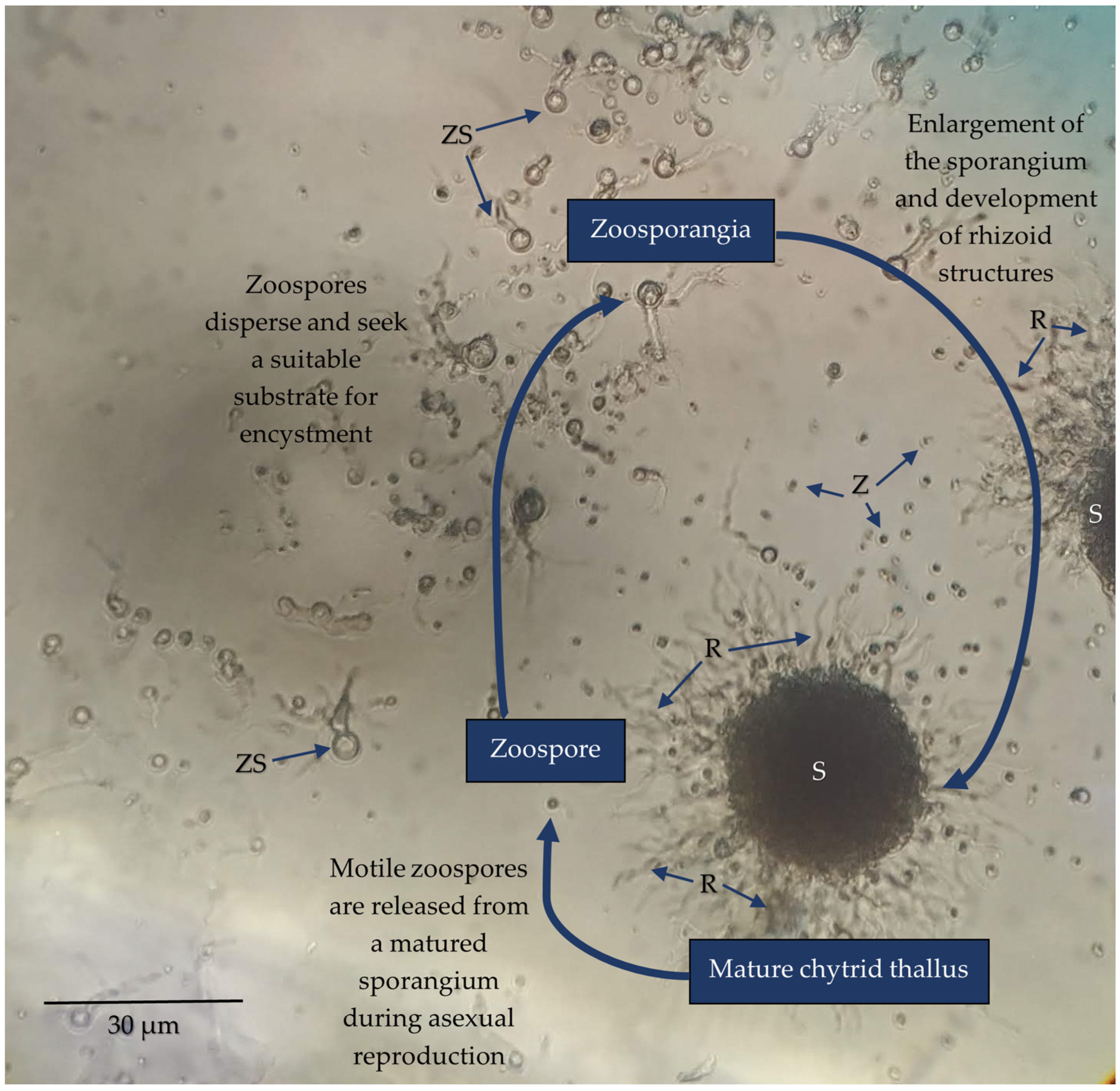
Figure 2. Light micrograph of Spizellomyces sp. (order Spizellomycetales) culture capturing three characteristic stages of development; a mature thallus containing a large sporangium (S) and rhizoid (R) extensions (not all chytrid species produce rhizoids), sporulation to disperse motile zoospores (Z), and zoospore encystment which initiates maturation from a zoosporangium (ZS) into a mature thallus. Strains were revived from long-term stock cultures by aseptically removing a segment of agar containing mature sporangium and placing it inoculated side down onto fresh peptone, yeast, and glucose (PYG) medium (2.5 mM peptone, 4.56 mM yeast extract, 27.7 mM glucose, 2% w/v agar). A few drops of sterile deionised water were then added over the inoculated agar to promote sporulation. Cultures were incubated at room temperature over the course of a week to observe the chytrid life cycle.
Encyclopedia 03 00046 g002
Figure 3. Microscopic images of Chytridiomycota (a) captured with a confocal microscope, (be) taken using a light microscope, and (fh) captured via scanning electron microscopy (SEM). As per Figure 2, strains were cultured and maintained on PYG. (a) Spizellomyces sp. (order Spizellomycetales) chytrid during sporulation, identifying thallus structures including the sporangium (S), rhizoids (R), discharge tube (DT), and released zoospores (Z). Spizellomyces sp. was cultured and prepared for microscopy as per Hanrahan-Tan et al. (2019) [38]. (b) Rhizophlyctis rosea (order Rhizophlyctidales) attached to lens paper (LP) as a cellulose substrate. Adapted with permission from Henderson et al. (2019) [39]. Copyright 2019 Nova Hedwigia. (c) Spizellomyces sp. attached to lens paper as a cellulose substrate. Culture plates were flooded with sterile deionised water from which an aliquot of active zoospores could be taken approximately 2 h later. The aliquot was then used to inoculate a liquid medium containing autoclaved lens paper baits (1.5 mm diameter). (d) Terramyces sp. (order Rhizophydiales) attached to snake skin (SS) as a keratin substrate. (e) Spizellomyces sp. thalli attached to and penetrating the tough exine layer of pollen grains (P) to access nutrients. As per (c), an inoculum of fresh zoospores was added to liquid medium containing autoclaved pollen grains. (f) R. rosea thallus under normal conditions on PYG prepared as per Henderson et al. (2017, 2019) [39,40]. Adapted with permission from Henderson et al. (2017, 2019) [39,40] and reproduced with permission from The Licensor through PLSclear. Copyright 2017 Taylor and Francis Group LLC (Books) US and copyright 2019 Nova Hedwigia. (g) R. rosea thallus. Incubation on media containing 60 ppm copper over 5 days shortens the length of rhizoids, increases the degree of rhizoid branching, and can lead to nodulation (N). Image sourced from Henderson et al. (2017) [40]. Reproduced with permission from The Licensor through PLSclear. Copyright 2017 Taylor and Francis Group LLC (Books) US. (h) Environment SEM of Terramyces sp. colony grown on PYG medium. Sporangia appear as larger bumps (>20 µm) under the surface of the biofilm while zoospores are encysted as small spots (<10 µm) in the biofilm surface.
Encyclopedia 03 00046 g003
Figure 4. Percentage (%) of genomes representing each fungal phylum available in the NCBI Taxonomy Browser (n = 4211). Data from the NCBI Taxonomy Browser were accessed on 21 November 2022. Each fungal phylum was searched for at a filter level of one. The number of assembled genomes compiled in the Entrez records table was recorded for each phylum and the percentage of genomes representing each phylum was calculated. The 17 phyla recognised are included to highlight the limited genomic data available across multiple lineages of early-diverging fungi [11].
Encyclopedia 03 00046 g004
Figure 5. Possible roles of soil chytrids in a hypothesised food web. All arrows indicate energy transfer from one source (origin of line) to another (arrowhead). The zoospore pool is a food source containing macromolecules (lipids) which can be consumed by grazers (ciliates, amoeba, heterotrophic bacteria (H Bacteria)) and heterotrophic nanoflagellates (HNF)) in soil ecosystems. Chytrids are both parasites and hyperparasites (red dotted lines), but may also be grazed on (green dotted lines). Some chytrids are saprotrophs (yellow line) and contribute to the decomposition of particulate organic matter (POM) including both allochthonous (leaf litter, pollen, other debris) and autochthonous (dead insects, exoskeletons), into dissolved organic matter (DOM) and dissolved inorganic matter (DIM) which can then be utilised by plants and other microorganisms.
Encyclopedia 03 00046 g005
Disclaimer/Publisher’s Note: The statements, opinions and data contained in all publications are solely those of the individual author(s) and contributor(s) and not of MDPI and/or the editor(s). MDPI and/or the editor(s) disclaim responsibility for any injury to people or property resulting from any ideas, methods, instructions or products referred to in the content.