Entry

Oxidative Stress in Relation to Aging and Exercise

1
Biochemistry Department, Thomas More Kempen, Thomas More University, Kleinhoefstraat 4, 2440 Geel, Belgium
2
Ciências Biomédicas Laboratoriais, Escola Superior de Tecnologia da Saúde de Coimbra (ESTeSC), Politécnico de Coimbra, Rua 5 de Outubro, 3046-854 Coimbra, Portugal
3
Laboratório de Investigação em Ciências Aplicadas à Saúde (LABINSAÚDE), Escola Superior de Tecnologia da Saúde de Coimbra (ESTeSC), Instituto Politécnico de Coimbra, Rua 5 de Outubro, 3046-854 Coimbra, Portugal
4
Unidade I&D Química-Física Molecular, Faculdade de Ciências e Tecnologia, Universidade de Coimbra, 3004-535 Coimbra, Portugal
*
Author to whom correspondence should be addressed.
Encyclopedia 2022, 2(3), 1545-1558; https://doi.org/10.3390/encyclopedia2030105
Submission received: 16 May 2022 / Revised: 17 August 2022 / Accepted: 19 August 2022 / Published: 25 August 2022
(This article belongs to the Section Medicine & Pharmacology)

Definition

:
Oxidative stress is an imbalance between oxidants and antioxidants in favor of the oxidants, resulting in disruption of redox signaling and control and causing molecular damage. Oxidative stress is related to a variety of diseases, for example, cardiovascular diseases, neurodegenerative diseases, infections, and cancer. It might be that oxidative stress, and, more specifically, reactive oxygen species (ROS), affects longevity in a subtle way through signaling. Possible therapies to reduce oxidative stress in the elderly are nutritional intervention (for example, caloric restriction (CR)) and exercise. Exercise is associated with favorable changes in the expression of antioxidant enzymes and the oxidative stress status in general. A diet with CR also seems to be a promising way to reduce oxidative stress by decreasing oxidant emission and improving antioxidant mechanisms. A better understanding of where the antioxidant mechanisms in the elderly fail could be a big step forward in developing new therapies (such as exercise or diet) that prevent oxidative damage and cellular dysfunction with age.

1. Introduction

Aging is a major challenge in our modern society, both socially and economically. Global average life expectancy rose by 5.5 years between 2000 and 2016. The cause of this increase was a decline in infectious diseases resulting in death. Across 28 European member states, life expectancy is estimated at 80.9 years, while, in the USA, life expectancy is estimated at 78.9 years [1,2]. Augmented life expectancy results in increased vulnerability to the development of chronic pathological conditions, such as cardiovascular diseases (CVD), cancer, and neurodegenerative diseases [3]. A major contributor to aging and those degenerative diseases is oxidative stress [4].
According to data, oxidative stress gradually develops during aging. Oxidative stress contributes to an augmented chance of morbidity and mortality. The free radical theory of aging was postulated as early as 1956 by Harman [5,6].
This theory states that the reactive oxygen species (ROS) that are constantly formed as by-products of essential metabolic processes are the source of molecular damage due to oxidative stress. The ROS are mainly formed by oxidative phosphorylation, which happens in all eukaryotic species. The accumulation of this oxidative damage caused by ROS over time contributes to age-related retrogression. Damage to mitochondrial DNA (mtDNA) is a central aspect of the free radical theory. According to this theory, increased ROS production accelerates aging, whereas antioxidant enzymes or other factors protecting against ROS retard the aging process. However, nowadays, the free radical theory of aging is questioned due to many conflicting data [7,8].
Both control of body weight by caloric restriction (CR) and exercise are able to ameliorate the oxidative stress state. High consumption of vegetables and fruits diminishes oxidative stress due to the antioxidant properties of the phenolic compounds [9].

2. Oxidative Stress

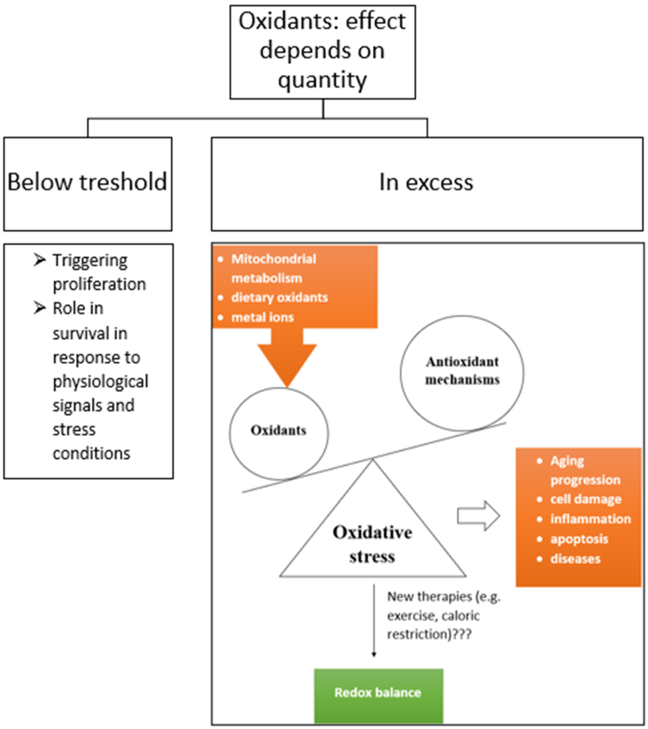
Oxidative stress is the result of an imbalance between the generation of ROS, such as the superoxide radical ( O 2 . ) , and the endogenous antioxidant capacity in a living organism. Oxidative stress originates when ROS generation exceeds the antioxidant capacity [10]. Oxidative stress is also defined as the perturbation of the cell redox balance [11]. The principle of oxidative stress is shown in Figure 1.
In the mitochondria, adenosine triphosphate (ATP) is generated during oxidative metabolism. Nicotinamide adenine dinucleotide (NAD) or flavin adenine dinucleotide (FAD) donates electrons down the electron transfer chain (ETC). The process in the ETC is called oxidative phosphorylation, as oxygen is used to generate ATP. Oxidative phosphorylation is comprised of two parts: the ETC and chemiosmosis. In the ETC, electrons pass through a series of redox reactions, releasing energy. In chemiosmosis, ATP is synthesized using the proton gradient over the inner mitochondrial membrane (IMM) [10,12,13]. Disruption of the ETC leads to increased generation of ROS and altered redox balance. Electrons dissociate (<0.1%) from the ETC because the transfer of electrons is usually not completely coupled. This causes a partial reduction of molecular oxygen (O2), forming superoxide ( O 2 . ) , hydrogen peroxide (H2O2), and hydroxyl radicals ( H O . ) . H2O2 can also be formed as a product of catalytic reactions with enzymes (e.g., monoamine oxidase and nicotinamide adenine dinucleotide phosphate oxidase 4). Mitochondria, through their constituent antioxidant enzymes and cytochrome c, is also capable of removing ROS with antioxidant mechanisms [10,12].
Figure 1. Oxidative stress: an imbalance between oxidants and antioxidant mechanisms in favor of the oxidants. Oxidants are formed due to mitochondrial metabolism, dietary oxidants, and metal ions [14]. In low-to-moderate quantities, oxidants have important functions in the body, such as triggering proliferation or survival responses to physiological signals and stress conditions. However, in excess, they provoke oxidative stress [15]. The oxidative stress results in various pathologies. Different therapies, such as exercise or caloric restriction, are being studied to understand how they might lower oxidative stress and restore redox balance.
Encyclopedia 02 00105 g001

2.1. Reactive Oxygen Species

ROS are compounds with extra electrons derived from O2 as a result of a partial chemical reduction. Examples of ROS are O 2 . , H2O2, H O . , and nitric oxide (NO). Free radicals that are not directly derived from oxygen (e.g., those derived from oxidation of an unsaturated lipid) can also be considered ROS. O 2 . is the first species produced in many biological oxidative cascades and reacts moderately with most biological compounds. H2O2 is also moderately reactive and has a long half-life time. It can diffuse across cell membranes. The highly reactive H O . can be formed from H2O2. This is a redox reaction with reduced metal (for example, iron or copper): H 2 O 2 + F e 2 +     O H . + O H + F e 3 + [16].
Another biologically important radical is NO. It is formed from arginine and oxygen by nitric oxide synthase (NOS). NO has an important role in vasoconstriction and acts as a regulator of blood pressure and also as a signal molecule in the nervous system. In addition, it is a defense against infections. NO is present in many different cell types (such as nervous, immune, and cardiovascular cells) and species (such as mammals and plants) [16,17,18,19].
The majority of intracellular ROS production occurs in the mitochondria; however, the cytosolic enzyme family of nicotinamide adenine dinucleotide phosphate oxidases (NOX), which can generate O 2 . , also produces ROS in smaller amounts. ROS production is potentiated by mitochondrial respiratory inhibition and Ca2+ accumulation [16,20].
The major generators of ROS in the cytosol are NOX and xanthine oxidase (XOD) [11]. ROS are important for routine cellular and mitochondrial signaling and functionality. ROS give signals for modifying proteins and lipid species within mitochondria. In addition, they modulate mitochondrial and cardiomyocyte functions. Normal levels of ROS regulate cell growth and division, the apoptotic pathway, and mitochondrial fusion–fission dynamics. ROS play an important role in the redox reactions in cells in low-to-moderate concentrations. ROS are also involved in the defense against invading microorganisms. In excessive quantities, they result in oxidative stress. ROS are very reactive entities that are capable of damaging nearly any molecular structure with which they interact. Each ROS has specific chemical properties and, therefore, also different reactivity with cellular macromolecules [7,10,12,16,21].

2.2. Antioxidants and Mechanisms of Their Action

Antioxidants protect the body against oxidative damage. They can prevent formation of ROS or they can quench ROS before they react with other biomolecules. There are two groups of antioxidants. The first type of antioxidant is based on enzymatic activities, including superoxide dismutase (SOD), catalase (CAT), glutathione peroxidase (GPx), and glutathione reductase (GluRed). The second type consists of a variety of nonenzymatic antioxidants, for example, vitamins E, C, and A and melatonin, selenium, and uric acid. Additionally, some molecules, such as circulating high-density lipoproteins, have intrinsic antioxidant activities. Albumin and other proteins contribute to the total antioxidant capacity in the blood [16,22,23].

2.2.1. Superoxide Dismutase

SOD accelerates the neutralization of the toxic O 2 . by catalyzing through the following reaction: 2 O 2 . + 2 H + H 2 O 2 + O 2 . SOD is present in all aerobic organisms. SOD catalyzes the rate production of hydrogen peroxide by 10.000X compared to the rate of production without SOD. Therefore, the cell is free from O 2 . [16].
SOD metalloenzymes are classified into different groups according to the metal cofactors at the active site that are involved in the redox reaction. Other metals can be incorporated in the enzymes, but it results in inactivation of the enzyme [24].
For example, mammals have three types of SOD: copper–zinc containing SOD (SOD1), present in the cytosol, manganese containing SOD (SOD2), found in the mitochondrial matrix, and manganese containing extracellular SOD (SOD3). Manganese containing superoxide dismutase (MnSOD) is a primary regulator of the O 2 . concentration in the mitochondrial matrix. In the intermembrane space, copper–zinc superoxide dismutase (CuZnSOD) reduces O 2 . to H2O2. The highest expression of SOD takes place in the renal tubules of healthy kidneys [11,12].
Recently, there have been various studies that revealed the different potential applications of antioxidants and free radical manipulations in the prevention or control of disease [22]. For example, Zn, Cu, and Mn supplementation can decrease oxidative stress and increase the levels of SOD. These minerals are essential for the activity of the antioxidant enzyme system (MnSOD, Cu/ZnSOD), which catalyzes the dismutation of O 2 . to H2O2 and O2. In addition, it may be that Fe ions are used in reactions aimed at maintaining the oxidant/antioxidant balance [25].
SOD possibly plays a role in the life extension of organisms by removing the toxic O 2 . , which is involved in a wide range of diseases. Therefore, SOD is interesting in relation to therapeutic interventions [24]. SOD is used as treatment for arthritis, burns, and inflammatory diseases. Therefore, measurement of SOD enzymatic activity, is a way to evaluate the efficiency of a treatment [26]. SOD incorporated into mixed lipid vesicles specifically developed for transdermal delivery is shown to be able to promote non-invasive treatment of induced arthritis when applied onto a skin area not necessarily in proximity to the inflamed tissue [27]. In addition, the antioxidant potential of new drugs can be determined this way. The effects of sports on ROS damage and the protective effects of SOD can also be determined with a SOD activity test [26].

2.2.2. Glutathione Peroxidase, Glutathione Reductase, and Glutathione S-Transferase

GPx contains selenium and catalyzes the reduction of H2O2 or other organic hydroperoxides to water or corresponding alcohols. GPx consumes reduced glutathione (GSH) and eliminates the excess of H2O2. GPx is the major antioxidant for eliminating H2O2 produced during the dismutation of O 2 . . GluRed reduces oxidized glutathione (GSSG) via nicotinamide adenine dinucleotide phosphate (NADPH) [11,16,22].
The following reactions take place:
2   G S H + H 2 O 2   G P x   G S S G + 2 H 2 O G S S G + N A D P H + H +   G l u R e d   2   G S H + N A D P +
Glutathione S-transferase (GST) enzymes are encoded by two supergene families, and their function is associated with the detoxication of electrophiles by glutathione conjugation. These enzymes play a critical role in cellular protection against oxidative stress and toxic chemicals. GST enzymes detoxify a variety of electrophilic compounds, such as the by-products of ROS, oxidized lipids, and DNA. The catalytic properties of GST also suggest that they play a single role in the biotransformation of drugs [28,29,30].
The determination of the enzyme activities of GPx and GluRed is used for assessing mitochondrial ROS (mtROS) production [12].

2.2.3. Oxidized and Reduced Glutathione

GSH is the major non-protein thiol found in most mammalian tissues. GSH is a small molecule antioxidant that quenches free radicals. It is a tripeptide (γ-Glu-Cys-Gly) with an unusual γ-glutamyl peptide bond and a cysteine which provides the thiol group. The thiol groups are oxidized to disulfide bonds. It possesses a thiol (R-SH) disulfide (R-S-S-R) pair, which provides a convenient redox couple used to destroy ROS, to regenerate other antioxidants, and to signal cellular redox status [11,16]. L-glutamate is transformed into GSH by two enzymes, γ-glutamylcysteine synthetase (γ-GCS) and glutathione synthetase (GS) [31].
The majority of intracellular GSH is present in the cytosol, about 1–11 mM. Cytosol is the principal location of GSH biosynthesis [31]. The intracellular GSH/GSSG is reported to be 100:1. Glutathione is mostly present in a reduced form in biological tissues in concentrations of 3100 µg/g tissue. GSSG is observed in lower concentrations, in a range of 0 to 244 µg/g tissue. In case of oxidative stress, the GSH/GSSG ratio is reduced to 10:1. Therefore, the determination of this ratio is important for determining the cellular redox status. Increased GSSG presence is a signal of an increased oxidative environment. When cells are exposed to oxidative stress, GSSG accumulates, and the GSH/GSSG ratio diminishes. Prolonged oxidative stress is also responsible for a decreased amount of free GSH, which also leads to a reduced GSH/GSSG ratio [14,32,33].
The endoplasmic reticulum is more oxidizing than the cytosol and has GSH/GSSG ratios of 1:1 to 3:1. Compared to the overall ratio in the cell, usually ≥100:1, it is very low [31].
Extracellular GSH is present in lower amounts than intracellular GSH (100 to 1000 times less). The function of extracellular GSH is thought to be detoxifying and provides protection against oxidant injury [31].
Some cells are able to export GSH or GSSG into plasma. GSSG is formed as a result of oxidative stress. This causes a shift in the GSH/GSSG ratio, which results in a more positive potential. If the potential rises too much, it is harmful to the cell. Exporting GSSG prevents the shift in the ratio. So, the ability to export GSSG could be an important factor in the sensitivity of cells to oxidative stress [31].
This thiol plays a major role in maintaining redox balance by neutralizing ROS, as shown in Figure 2 [14]. GSH is involved in xenobiotic metabolism, intracellular signal transduction, and gene regulation, among other things. Abnormal GSH levels are associated with many diseases, for example hepatic injury, Alzheimer’s disease, diabetes, human immunodeficiency virus (HIV) infection, and cancer [34].
A method based on fluorescent measurement with use of o-phthalaldehyde (OPT) as a fluorescent reagent was developed for both GSSG and GSH. GSH reacts with OPT at pH 8, while GSSG reacts with OPT at pH 12. Interference during the measurement of GSSG can be avoided by complexing GSH to N-ethylmaleimide [32].
In addition, a lot of other methods are available for the determination of the GSH/GSSG ratio in plasma, urine, saliva, and tissue. Many GSSG measurements are based upon the reduction of the disulfide followed by measurement of GSH. Electrochemical detection can be used to directly and simultaneously measure GSH and GSSG in plasma without sample pre-treatment. Unfortunately, due to the low endogenous concentration of GSSG, this technique is often not sensitive enough for an accurate determination [14].

2.2.4. Catalase

Catalase (CAT) hydrolyzes H2O2 into water and oxygen.
2 H 2 O 2   C A T   2 H 2 O + O 2
CAT contains a ferriheme. It only converts H2O2 and no other peroxides. This enzyme is expressed in almost all types of cell, but the highest amounts of CAT are found in the liver and erythrocytes [11,16,35].

2.2.5. Others

In the mitochondria, beyond GPx, H2O2 can also be reduced to water by peroxiredoxin 3 and 5 and thioredoxin pathways. In the heart mitochondria, CAT is used to neutralize H2O2. H2O2 can diffuse out of the matrix into the cytosol if not neutralized by the antioxidant enzymes in the matrix. In the cytosol, it is neutralized by CAT or cytosolic isozymes in the glutathione and thioredoxin pathways [12,36,37].
The level of antioxidant response is representative of the free radical activity and can be used to determine the amount of ROS [22]. GPx and SOD activity are positive correlated. When GPx activity increases, SOD activity also rises [38].
In relation to age, the nonenzymatic antioxidants decrease, and the enzymatic antioxidant activities increase. Remarkably, this is vice versa in centenarians. The extreme longevity of centenarians seems to be related to a high-level profile of vitamin A and vitamin E. Plasma GPx activity is equal among all age groups [6].
Higher concentrations of α-tocopherol plasma or serum carotenoids or ascorbic acid in mammalian species are linked to a higher life expectancy. Vitamins E and A are of particular importance for longevity. Aging and decreased levels of molecular antioxidants are associated. In addition, in senescent animals, the levels of SOD, CAT, and GPx are decreased in various types of tissue [5].
Recently, there have been big developments relating to new ways to augment antioxidant levels: gene therapy to increase antioxidant production, GMO plants (genetically modified organisms), synthetic antioxidant enzymes, novel biomolecules, and the use of functional foods (food with certain beneficial effects on one or more target functions in the body beyond the basic nutritional effects with, as a result, an improved health state and improved well-being or a reduction in risk of diseases) enriched with antioxidants [22,39].

2.3. Effects of Oxidative Stress

Oxidative stress has many adverse effects on the body. It can result in tissue and cell damage, leading to inflammation and more oxidative stress. Mild or moderate stressors alter calcium homeostasis (homeostasis denotes the maintenance, or regulation, of vital internal variables in a state of relative constancy) and ROS signaling, resulting in tolerance to and overcoming of the stressors [10,40]. By exposure to severe stressors, cell death pathways (apoptosis and necrosis) are activated [10].
Normally, migration of mitochondrial proteins through the IMM is not possible. However, calcium overload or excessive ROS signaling can induce its permeabilization. As a consequence, the proton gradient across the IMM is disturbed, leading to a loss in membrane potential and less ATP production. During the necrosis of the cell, ATP consumption is unchanged in other cell compartments, resulting in depletion of ATP. There is swelling in the mitochondrial matrix (osmosis due to the high concentration of solutes in the matrix), which can result in mitochondrial rupture [10].
Apoptosis is less destructive than necrosis, as apoptogens are released to ensure controlled resorption of the cell. Apoptosis is induced by permeabilization of the outer mitochondrial membrane (OMM) [10].
Oxidative stress is an important pathological mechanism in ischemia (reduction of blood flow, leading to a decrease in oxygen supply), heart failure, and diabetic cardiomyopathy. ROS excess can also provoke CVD and irreversible damage to mitochondria. Oxidative stress is also involved in the development of neurodegenerative diseases, such as Alzheimer’s disease, Parkinson’s disease, and Huntington’s disease, and can cause retinal degeneration. The pathophysiology of diseases, such as respiratory, obesity, metabolic syndrome, recurrent aphthous stomatitis, and diabetes mellitus, is associated with oxidative stress [10,20,22,23,41].

3. Oxidative Stress in Relation to Aging

3.1. Free Radical Theory of Aging

In recent decades, the free radical theory of aging has gained more recognition. The theory states that ROS are produced as a normal by-product of aerobic life. These ROS accumulate and cause oxidative damage, inducing fundamental changes found in senescence. Free radicals, especially those of mitochondrial origin, are the main cause of aging. The ROS presence forms a steady-state condition, as the antioxidant mechanisms are not completely efficient. Cells are constantly under oxidative stress. This accumulation of oxidative damage results in the aging phenotype and limits lifespan. This theory states that mechanisms that limit ROS or repair oxidative damage to macromolecules should increase longevity. Increased ROS neutralization should also increase longevity according to this theory [12,16,42,43].
A general cause of aging is considered to be the time-dependent accumulation of cellular damage. ROS are involved in this accumulation of cellular damage, but a lot of other factors are also involved, for example, physical, chemical, and biological agents, DNA replication errors, and spontaneous hydrolytic reactions. Most mtDNA mutations in adult or aged cells seem to be caused by replication errors early in life and not by oxidative damage. For this reason, the free radical theory of aging might be questioned [8].
First of all, there are a lot of controversial results about this theory. The results about the correlation between longevity and ROS production/neutralization are not so straightforward. The balance between regulation and damage by ROS and how this correlates with aging and longevity is still unclear [12].
Additionally, there are some difficulties in carrying out research about this topic. The isolated mitochondria have several dissimilar features in comparison to the in vivo conditions. This can have a sizeable impact, resulting in an overestimation of endogenous levels of ROS production. In addition, for measuring ROS flux in vivo, the possibilities for direct measurement are limited. O 2 . and H2O2 can be measured by fluorescent probes in vivo, but the results are difficult to interpret because of the high noise-to-signal ratios. Alternatively, markers of oxidative stress can be measured, but these do not account for repair mechanisms, some of which are themselves sensitive to changes in ROS concentrations [12].
Changes in animals at maximum lifespan occur at the cellular level, including accumulation of oxidative damage in cellular macromolecules (particularly DNA and proteins). This is in line with the free radical theory of aging, as oxidative stress provokes cellular dysfunction and cell loss with aging. Addressing a specific determinant of the aging rate, such as O 2 . production, it is predicted that the rate is reduced in longer-lived animal species. There has been researched in multiple species (for example, in mice), a variety of tissue types, and respiratory substrates in vertebrates. No clear trend between mitochondrial H2O2 production and longevity was found. The lifespans of the mice also did not increase consistently when the antioxidant enzymes were overexpressed. Reducing the activities of key antioxidant enzymes in mice also did not result in the expected lifespan reductions. Thus, the theory is not strongly supported. Nevertheless, it is possible that ROS might affect longevity in a more subtle way through signaling [12].
Nowadays, the nine hallmarks of aging (genomic instability, telomere attrition, epigenetic alterations, loss of proteostasis, deregulated nutrient sensing, mitochondrial dysfunction, cellular senescence, stem cell exhaustion, and altered intercellular communication) are a largely accepted framework for studying aging [8].

3.2. Telomere Shortening

Replicative senescence (irreversible loss of division potential) is an appropriate model for aging. The replicative potential in vitro and the age of the donor/maximal lifespan are correlated [44]. Telomeres are sensors for oxidative damage in the genome. These segments of repetitive nucleotide sequences at each end of a chromosome protect the chromosome against decline of the chromosome ends. Oxidative stress and inflammation are the main reasons for telomere shortening, which induces cellular senescence and apoptosis. Cell senescence provokes mitotic arrest and the senescence-associated secretory phenotype (SASP). A large quantity of proinflammatory and growth factors are secreted. This triggers inflammation and production of ROS. Consequently, there is even more cellular senescence, resulting in a vicious circle. Telomere shortening has the potential to be a biomarker of aging, as it reflects the cumulative amount of oxidative damage to the organism [10,44].

3.3. Cellular Senescence

ROS can accelerate telomere shortening, as telomeres are very rich in guanine nucleotides, which is the reason why they are especially sensitive to oxidative stress, followed by DNA damage response and senescence. Senescence results in further ROS generation via SASP. A characteristic of cellular senescence is proliferative arrest [10].
Cellular senescence can be divided into two types: replicative or stress-induced premature senescence. Replicative senescence is driven by cell division, while stress-induced premature senescence is induced by oxidative stress, mitogens, oncogenes, irradiation, and other factors and is normally not related to telomere attrition [10].
SASP results in degenerative and proliferative age-related tissue alterations. These lead to chronic inflammation, remodeling, and tissue repair. This provokes more oxidative stress, and, therefore, more cells become premature senescent cells [10].
Telomere shortening and dysfunction are not only affected by age (oxidative stress and inflammation); there are a lot of other factors that have their influence on telomere shortening. Some examples of environmental factors that promote telomere shortening are smoking, alcohol abuse, obesity, air pollution, and mental stress. Divergent metabolisms, vitamins C, D, and E, omega-3 fatty acids, and estrogen treatment are examples of environmental factors counteracting telomere shortening. These factors are modifiable. Endogenous factors that counteract telomere shortening are telomerase, estrogen, and endogenous antioxidants. Telomerase replaces telomere repeats by catalyzing DNA synthesis, preventing the shortening of telomers in stem and germ cells. In addition, in cancer cells, a high activity of telomerase is noticeable. Adversely, in somatic cells, there is low or undetectable telomerase activity. Estrogen activates telomerase. Additionally, telomere shortening is linked with CVD, genetic and genomic perturbation, cell division, mitogenic signals, and nontelomeric damage [10].

4. Oxidative Stress in Relation to Lifestyle

Changes in lifestyle, such as CR and exercise training, contribute to controlling mtROS production or breakdown. In addition, they help to prevent apoptosis, necrosis, and inflammation. There is an association between longer leukocyte telomere length (LTL) and higher levels of physical activity [10].
Aerobic exercise and caloric restriction are much researched as possible therapeutic strategies to prevent the decline of mitochondrial and skeletal muscle health with advancing age. Sedentary individuals were found to have lower mitochondrial function compared to active individuals [45,46].
Via the respiration in mitochondria, ROS are formed. In excess, they result in progressive damage to mitochondrial constituents (DNA, proteins, and lipids). Due to mutations in the mtDNA, the synthesis of the ETC subunits is impaired, and there is a reduction in oxidative phosphorylation. This causes mitochondrial dysfunction, and, in addition, mitochondrial dysfunction causes further increases in ROS and oxidative damage. This forms a vicious circle, resulting in a decline in muscle mass and strength (sarcopenia), reduced physical function, and aging [45,46,47].
Aging affects the mitochondria negatively through higher levels of oxidative stress, which contributes to sarcopenia. Aging skeletal muscle possesses fewer enzymes in its oxidative metabolism or ETC. Between the decline of mitochondrial and skeletal muscle health with age exists a temporal and functional connection. The reduced mitochondrional function results in less energy, which results in the hindrance of energy-demanding processes such as skeletal muscle protein turnover. This process is critical for skeletal muscle health with advancing age because it maintains protein quality [45,46].
Hindered protein turnover and excess ROS combined leads to an environment that stimulates the aging of skeletal muscle. The damaged mtDNA may produce dysfunctional proteins within the ETC, resulting in more ROS production and oxidative damage. It is important to mention that mutations in mtDNA are rare. They only have physiological significance in people aged 80 years and older. Sarcopenia and other age-related diseases start earlier [46].

4.1. Exercise

ROS have a negative effect on the body; however, recently, it was discovered that exercise-induced ROS can upregulate several enzymatic and nonenzymatic antioxidants in the biological system, resulting in less oxidative stress [48]. Exercise causes the production of ROS in the ETC and the system of XOD. All tissues use their reserve of antioxidants to counteract the ROS effects. Physical exercise shows positive effects on the redox environment in different systems of the body. Exercise diminishes the levels of oxidative stress markers due to activation of antioxidant enzymes [49]. Several studies showed an increase in antioxidant enzymes with endurance exercise training and/or high-intensity training. Exercise training increased both SOD1 and SOD2 in trained skeletal muscles by 20–110% and GPX1 in skeletal muscles by 20–180%. Results about the influence of exercise training on the levels of CAT were contradictive [15].
To cope with the oxidative stress elicited by high-level metabolism during exercise, human cells may have developed an ubiquitous antioxidant defense system [9].
Muscular activity promotes oxidant production in contracting skeletal muscle fibers [47]. This contraction-induced ROS generation is associated with oxidant damage in several tissues, accelerated muscle fatigue, and activation of biochemical signaling pathways that contribute to exercise-induced adaptation in the contracting muscle fibers [15]. These ROS have an important role in muscle force production [47]. The effect of ROS on the muscle force production is dependent on the level of ROS within the fiber. Both decreasing and increasing ROS when the ROS level is at the optimum result in a decrease in the muscles’ ability to generate force. Elevated levels of oxidants in skeletal muscle associated with prolonged exercise can lead to damaging the proteins involved in excitation–contraction coupling, resulting in reduced muscle force production. The exact cause of the reduction of muscle force is not clear yet. Possible explanations are changes in both free calcium levels in the muscle and myofibrillar sensitivity to calcium and the ROS-mediated decrease in Na+/K+ pump activity [15].
The major source of ROS production during exercise is skeletal muscle. Mitochondria are not the major site of production of ROS in the skeletal muscle. Studies showed that they produce less O 2 . during active state 3 respiration compared to basal state 4 respiration [15].
Active elderly people show less oxidative stress than the sedentary elderly of the same age [49]. Physical activity can reduce oxidative stress, resulting in a good therapy for reducing age-induced oxidative stress. Regular exercise may be an anti-aging factor that functions by augmenting antioxidant systems. Combining normal physical activity with aerobic and resistance training can increase antioxidants and reduce oxidative damage in the elderly. Due to exercise, the formation of ROS alters the signaling pathways and/or causes molecular damage that can produce adaptive responses to withstand further stress [48,50]. However, there is a lot of controversy about the benefits of exercise in terms of oxidative stress in the literature. Sometimes there is an increase in the formation of oxidants and inflammatory mediators, leading to augmented oxidative stress. In other cases, exercise causes an augmentation of antioxidant enzymes, resulting in less oxidative stress. The scenario depends on the exercise type and intensity and the training status of the individual. For better insight into exercise-induced oxidative stress benefits and/or consequences, the following aspects should be considered: exercise type, intensity, training status, and individual variability [48].
In general, relatively high-intensity, prolonged aerobic exercise (i.e., 65–75% V ˙ O 2 max) results in greater ROS production compared to low-intensity (i.e., <40% V ˙ O 2 max), short-duration exercise. Additionally, higher levels of muscle temperature result in higher levels of ROS production. Short-duration (<1min) and low-intensity (∼30% V ˙ O 2 max) exercise does not promote oxidative stress, whereas acute bouts of prolonged and high-intensity endurance exercise in untrained humans results in increases in biomarkers of oxidative stress (for example, increased protein oxidation and lipid peroxidation) [15].
However, rigorous and prolonged exercise training seems not to cause oxidative damage that is harmful to human cells. This can be explained by exercise-induced hormesis (a phenomenon whereby moderate stress proves beneficial) [7,15]. This means that by low-to-moderate levels of exercise-induced ROS production, essential adaptions in skeletal muscle take place, while high levels of ROS production cause damage to the muscle. Regarding the relationship between the cellular redox state and skeletal muscle force, an optimum level of ROS is required for maximal force production during concentric contractions. A further increase in exercise-induced ROS production would cause damage and decline in the exercise-induced adaptations. Production of ROS in detrimental amounts through high-intensity exercise is unlikely because of several reasons. First, the duration of exercise is limited by both the cardiovascular system’s ability to provide blood to the working muscles and the impact of ROS production on muscle fatigue. This limitation for exercise also limits the muscle ROS production during an exercise bout. In addition, the mitochondrial coupling is higher during exercise respiration, reducing electron spill and ROS production by the mitochondria during exercise. Third, acute exercise leads to an increased expression of uncoupling proteins. These move protons from the intermembrane space into the matrix, resulting in a diminished, high mitochondrial proton gradient that favors O 2 . formation. Last, regular exercise causes increases in antioxidant enzymes in skeletal muscle. This improves the muscle fibers’ ability to remove ROS formed during exercise [15].
A physically active lifestyle is associated with the partial compensatory preservation of mitochondrial biogenesis and cellular oxidative and antioxidant capacity in the skeletal muscle of older adults. In contrast, a sedentary lifestyle leads to reduced mitochondrial function and dysregulation of cellular redox status. A sedentary lifestyle is, furthermore, associated with chronic systemic inflammation. This inflammation makes the skeletal muscle susceptible to ROS-mediated toxicity. A sedentary lifestyle can result in frailty; nevertheless, physical activity intervention can ameliorate the physical performance of the sedentary elderly [51].
Independent of age, the mitochondrial molecular regulation and protein content are increased after 12–16 weeks of exercise. This suggest that exercise training is also beneficial for older individuals. The effect of different training programs (aerobic training = any activities that raise heart rate and make breathing somewhat harder vs. resistance training = overall fitness program composed of various exercise types such as aerobic training, flexibility training, strength training, and balance exercises vs. concurrent training = combination of resistance and endurance training in a periodized program to maximize all aspects of physical performance) on mitochondria and skeletal muscle function is still unclear, but one can conclude that exercise can improve or prevent the loss of mitochondrial health during sedentary aging [46,52,53,54].
During aging, cellular pathways responsible for the regulation of mitochondrial turnover, including biogenesis, dynamics, and autophagy, may become dysregulated. When homeostasis is not re-established, cell death occurs, resulting in muscle atrophy. With aging, excess ROS are produced, and there is a decrease in protein turnover (protein degradation and synthesis). The combination of these two changes in the cells can result in reduced protein quality and function. Acute and chronic exercise can counteract these symptoms. Exercise contributes to restoring mitochondrial turnover and promoting a healthier mitochondrial pool. The oxidatively damaged proteins are replaced by newly synthesized functional proteins. As a result, the muscles are better preserved. Studies report that old muscle has a lower adaptability than young muscle, although the causes of the diminished training responses are still uncertain [45,46].
Nutrition (increased supply) and/or reduced physical activity leads to a buildup of protons, resulting in a high membrane potential. This situation could cause the electron flow through the ETC to stop, leading to an increase in oxidant production. The membrane potential (and, consequently, the oxidant production) decreases with any mechanism to allow protons to flow back to the mitochondrial matrix (for example, ATP synthesis or uncoupling) [46].
Both the GPx activity and GluRed activity values rise after the practice of hydrotherapy. Therefore, regular and moderate practice of exercise is a positive stimulus in the antioxidant activity of aged individuals. Exercising could lead to a higher quality of life in older individuals [55].
Oxidative responses owing to exercise depend on the exercise intensity and the time course of exercise recovery. For example, anaerobic exercise can increase oxidative stress and decrease or increase antioxidants [48,56].

4.2. Diet

Another way to alleviate the effects of aging and prolong lifespan is CR, which means a 20–40% reduction in caloric intake, because it causes a decrease in oxidant emission and an increase in antioxidant scavenging. Consequently, the oxidative damage to macromolecules, such as DNA and proteins, are reduced. By protecting the macromolecules through CR, the mitochondrial function is preserved. CR retards age-associated changes and extends the maximum lifespan in mammals. The effect of CR is quickly noticeable [43,57].
Overnutrition can generate free radicals. Inadequate or excess nutrient supply causes oxidative stress. Consequences are the disruption of oxidative homeostasis, activation of a cascade of molecular pathways, and alterations of the metabolic status of various tissues [58].
CR lowers mtROS production and oxidative damage to mtDNA. Therefore, a CR of around 40% extends the mean and maximum lifespan of a wide range of species. Protein restriction and methionine restriction delay the aging effect by means of reducing mtROS production [42].
CR showed a temporary increase in ROS formation in C. elegans. This led to increased antioxidant activity, causing a prolongation of survival. The phenomenon whereby moderate stress proves beneficial is called hormesis [7].
A CR of 20% in a healthy adult human subject reduces blood pressure and blood cholesterol in a similar manner to specific drugs used to treat vascular disease. CR is a good therapy to counter the aging effects in overweight persons related to overeating combined with a sedentary lifestyle [59].
A diet rich in vitamins, minerals, fibers, fatty acids or amino acids, and/or food supplements can provide a series of antioxidants. Examples are vitamin A (retinoids, carotenes), vitamins C and E (tocopherols), lycopene, lutein, ubiquinone, glutathione, polyphenols (flavonoids and nonflavonoids), and N-acetylcysteine. These components are often not present in the necessary amounts in the diet [11].
It seems that the supplementation of fresh orange juice has a positive effect on the efficiency of several specific antioxidant systems in elderly women that undergo exhaustive exercise [60]. In addition, epidemiologic studies show that consumption of large amounts of vegetables and fruits is related with a lower risk of cancer, neurodegenerative disorder, and aging induced by oxidative stress [9,60].

5. Conclusions and Prospects

The relation between ROS and age-related dysfunction needs to be further investigated. A better understanding of the dynamics of accumulation and removal of molecular damage can help us to better understand the role of free radicals in aging. It is important to mention that oxidative stress is one of many mechanisms that drive the aging process [7]. ROS signaling is one of the mechanisms responsible for the development of cell senescence and organismal aging. ROS signaling is considered to be a further development of the free radical theory of aging [61].
As antioxidant mechanisms counteract oxidative damage, it is important to know more about the antioxidant capacity at the systemic, skeletal muscle, and mitochondrial compartments. Current results about the antioxidant capacity in the elderly are paradoxical. A better understanding of where the antioxidant mechanisms in the elderly fail could be a big step forward in developing new therapies (for example, exercise or antioxidant supplementation) that prevent oxidative damage and cellular dysfunction with age [46].
If the influence of exercise on oxidative stress in relation to aging is better understood, it will help to create better training programs that improve healthy aging. More sensitive techniques could help to gain more insight into this subject. Nowadays, techniques including colorimetry, fluorescence, photoluminescence, and electrochemistry are used for GSH and GluRed activity determination. These techniques are insensitive and vulnerable. There is a colorimetric method in development which has gained interest due to the advantages of convenient instrumentation and low cost and to the fact that it allows direct bare-eye detection, and it can be used in point-of-care diagnostics. This colorimetric method uses hemin/G-quadruplex DNAzyme, which has high thermal stability, ease of synthesis, and low cost. This technique is promising for the development of colorimetric biosensors that give better results than the current methods [34]. Using this new method, it is easier to monitor people. This is essential for obtaining more insight into the influences of exercise or other parameters on oxidative stress. In addition, this can be promising for developing personal, tailored exercise programs according to individual needs.
In conclusion, both CR and exercise could be valuable therapies for improving the oxidative stress status of the elderly, resulting in healthy aging. However, further research is necessary to explore the possibilities and advantages/disadvantages of these therapies.

Author Contributions

Writing—original draft preparation, D.V.; writing—review and editing, A.C.; writing—review and editing, K.S.; writing—review and editing, N.O. All authors have read and agreed to the published version of the manuscript.

Funding

This research received no external funding.

Data Availability Statement

Not applicable.

Conflicts of Interest

The authors declare no conflict of interest.

References

  1. Longo, M.; Bellastella, G.; Maiorino, M.I.; Meier, J.J.; Esposito, K.; Giugliano, D. Diabetes and Aging: From Treatment Goals to Pharmacologic Therapy. Front. Endocrinol. 2019, 10, 45. [Google Scholar] [CrossRef] [PubMed]
  2. Caseiro, A.; Rocha, C.; Silva, A.M.; Ferreira, C.; Silva, I.; Clemente, M.; Cipriano, I.; Saraiva, M.; Barreira, R.; Azenha, J.; et al. Effects of A Personalized Intervention Program on the Biochemical and Hematological Profile in Community Dwelling Old Adults-The AGA@4life Intervention Model. Int. J. Env. Res Public Health 2020, 17, 718. [Google Scholar] [CrossRef] [PubMed]
  3. Ovadya, Y.; Krizhanovsky, V. Strategies targeting cellular senescence. J. Clin. Invest 2018, 128, 1247–1254. [Google Scholar] [CrossRef] [PubMed]
  4. Ames, B.N.; Shigenaga, M.K.; Hagen, T.M. Oxidants, Antioxidants, and the Degenerative Diseases of Aging. Proc. Natl. Acad. Sci. USA 1993, 90, 7915–7922. [Google Scholar] [CrossRef]
  5. Junqueira, V.B.C.; Barros, S.B.M.; Chan, S.S.; Rodrigues, L.; Giavarotti, L.; Abud, R.L.; Deucher, G.P. Aging and oxidative stress. Mol. Asp. Med. 2004, 25, 5–16. [Google Scholar] [CrossRef] [PubMed]
  6. Mecocci, P.; Polidori, M.C.; Troiano, L.; Cherubini, A.; Cecchetti, R.; Pini, G.; Straatman, M.; Monti, D.; Stahl, W.; Sies, H.; et al. Plasma antioxidants and longevity: A study on healthy centenarians. Free Radic. Biol. Med. 2000, 28, 1243–1248. [Google Scholar] [CrossRef]
  7. Kirkwood, T.B.L.; Kowald, A. The free-radical theory of ageing--older, wiser and still alive: Modelling positional effects of the primary targets of ROS reveals new support. Bioessays 2012, 34, 692–700. [Google Scholar] [CrossRef]
  8. López-Otín, C.; Blasco, M.A.; Partridge, L.; Serrano, M.; Kroemer, G. The Hallmarks of Aging. Cell 2013, 153, 1194–1217. [Google Scholar] [CrossRef]
  9. Li, C.; Feng, F.; Xiong, X.; Li, R.; Chen, N. Exercise coupled with dietary restriction reduces oxidative stress in male adolescents with obesity. J. Sports Sci. 2017, 35, 663–668. [Google Scholar] [CrossRef]
  10. Sack, M.N.; Fyhrquist, F.Y.; Saijonmaa, O.J.; Fuster, V.; Kovacic, J.C. Basic Biology of Oxidative Stress and the Cardiovascular System: Part 1 of a 3-Part Series. J. Am. Coll. Cardiol. 2017, 70, 196–211. [Google Scholar] [CrossRef]
  11. Pisoschi, A.M.; Pop, A. The role of antioxidants in the chemistry of oxidative stress: A review. Eur. J. Med. Chem. 2015, 97, 55–74. [Google Scholar] [CrossRef] [PubMed]
  12. Robb, E.L.; Christoff, C.A.; Maddalena, L.A.; Stuart, J.A. Mitochondrial reactive oxygen species (ROS) in animal cells: Relevance to aging and normal physiology. Can. J. Zool. 2014, 92, 603–613. [Google Scholar] [CrossRef]
  13. Ahmad, M.; Wolberg, A.; Kahwaji, C.I. Biochemistry, Electron Transport Chain; StatPearls Publishing: Treasure Island, FL, USA, 2021. [Google Scholar]
  14. Childs, S.; Haroune, N.; Williams, L.; Gronow, M. Determination of cellular glutathione:glutathione disulfide ratio in prostate cancer cells by high performance liquid chromatography with electrochemical detection. J. Chromatogr. A 2016, 1437, 67–73. [Google Scholar] [CrossRef]
  15. Powers, S.K.; Deminice, R.; Ozdemir, M.; Yoshihara, T.; Bomkamp, M.P.; Hyatt, H. Exercise-induced oxidative stress: Friend or foe? J. Sport Health Sci. 2020, 9, 415–425. [Google Scholar] [CrossRef] [PubMed]
  16. Close, D.C.; Hagerman, A.E. Oxidative Stress, Exercise, and Aging; Imperial College Press: London, UK; Distributed by World Scientific Pub: Hackensack, NJ, USA, 2006. [Google Scholar]
  17. Ignarro, L.J. Nitric Oxide: Biology and Pathobiology, 1st ed.; Academic Press: San Diego, MA, USA, 2000. [Google Scholar]
  18. Blaise, G.A.; Gauvin, D.; Gangal, M.; Authier, S. Nitric oxide, cell signaling and cell death. Toxicology 2005, 208, 177–192. [Google Scholar] [CrossRef]
  19. Wendehenne, D.; Pugin, A.; Klessig, D.F.; Durner, J. Nitric oxide: Comparative synthesis and signaling in animal and plant cells. Trends Plant Sci. 2001, 6, 177–183. [Google Scholar] [CrossRef]
  20. Simpkins, J.W.; Wang, J.; Wang, X.; Perez, E.; Prokai, L.; Dykens, J.A. Mitochondria play a central role in estrogen-induced neuroprotection. Curr. Drug Targets CNS Neurol. Disord. 2005, 4, 69–83. [Google Scholar] [CrossRef]
  21. Wu, W.; Chen, M.; Luo, T.; Fan, Y.; Zhang, J.; Zhang, Y.; Zhang, Q.; Sapin-Minet, A.; Gaucher, C.; Xia, X. ROS and GSH-responsive S-nitrosoglutathione functionalized polymeric nanoparticles to overcome multidrug resistance in cancer. Acta Biomater 2020, 103, 259–271. [Google Scholar] [CrossRef]
  22. Gupta, I.; Shetti, A.; Keluskar, V.; Bagewadi, A. Assessment of Serum Enzymatic Antioxidant Levels in Patients with Recurrent Aphthous Stomatitis: A Case Control Study. Enzym. Res. 2014, 2014, 340814–340819. [Google Scholar] [CrossRef]
  23. Aztatzi-Aguilar, O.G.; Sierra-Vargas, M.P.; Ortega-Romero, M.; Jiménez-Corona, A.E. Osteopontin’s relationship with malnutrition and oxidative stress in adolescents. A pilot study. PLoS ONE 2021, 16, e0249057. [Google Scholar] [CrossRef]
  24. Phillips, N.H. Superoxide Dismutase (SOD): Sources, Therapeutic Uses and Health Benefits; Nova Biomedical: New York, NY, USA, 2016. [Google Scholar]
  25. Żwierełło, W.; Styburski, D.; Maruszewska, A.; Piorun, K.; Skórka-Majewicz, M.; Czerwińska, M.; Maciejewska, D.; Baranowska-Bosiacka, I.; Krajewski, A.; Gutowska, I. Bioelements in the treatment of burn injuries—The complex review of metabolism and supplementation (copper, selenium, zinc, iron, manganese, chromium and magnesium). J. Trace Elem. Med. Biol. 2020, 62, 126616. [Google Scholar] [CrossRef] [PubMed]
  26. RANDOX. Superoxide Dismutase (Ransod). Available online: https://www.randox.com/superoxide-dismutase-ransod/?highlight=Ransod# (accessed on 11 February 2022).
  27. Simões, S.I.; Delgado, T.C.; Lopes, R.M.; Jesus, S.; Ferreira, A.A.; Morais, J.A.; Cruz, M.E.M.; Corvo, M.L.; Martins, M.B.F. Developments in the rat adjuvant arthritis model and its use in therapeutic evaluation of novel non-invasive treatment by SOD in Transfersomes. J. Control Release 2005, 103, 419–434. [Google Scholar] [CrossRef] [PubMed]
  28. Hayes, J.D.; Strange, R.C. Glutathione S-transferase polymorphisms and their biological consequences. Pharmacology 2000, 61, 154–166. [Google Scholar] [CrossRef] [PubMed]
  29. Strange, R.C.; Spiteri, M.A.; Ramachandran, S.; Fryer, A.A. Glutathione-S-transferase family of enzymes. Mutat. Res. 2001, 482, 21–26. [Google Scholar] [CrossRef]
  30. Kanwal, R.; Pandey, M.; Bhaskaran, N.; Maclennan, G.T.; Fu, P.; Ponsky, L.E.; Gupta, S. Protection against oxidative DNA damage and stress in human prostate by glutathione S-transferase P1. Mol. Carcinog. 2014, 53, 8–18. [Google Scholar] [CrossRef]
  31. Schafer, F.Q.; Buettner, G.R. Redox environment of the cell as viewed through the redox state of the glutathione disulfide/glutathione couple. Free Radic. Biol. Med. 2001, 30, 1191–1212. [Google Scholar] [CrossRef]
  32. Hissin, P.J.; Hilf, R. A fluorometric method for determination of oxidized and reduced glutathione in tissues. Anal. Biochem. 1976, 74, 214–226. [Google Scholar] [CrossRef]
  33. Fitri, L.E.; Iskandar, A.; Sardjono, T.W.; Erliana, U.D.; Rahmawati, W.; Candradikusuma, D.; Saputra, U.B.; Suhartono, E.; Setiawan, B.; Sulistyaningsih, E. Plasma glutathione and oxidized glutathione level, glutathione/oxidized glutathione ratio, and albumin concentration in complicated and uncomplicated falciparum malaria. Asian Pac. J. Trop. Biomed. 2016, 6, 646–650. [Google Scholar] [CrossRef]
  34. Lai, Y.; Li, M.; Liao, X.; Zou, L. Smartphone-Assisted Colorimetric Detection of Glutathione and Glutathione Reductase Activity in Human Serum and Mouse Liver Using Hemin/G-Quadruplex DNAzyme. Molecules 2021, 26, 5016. [Google Scholar] [CrossRef] [PubMed]
  35. Nannenga, B.L.; Shi, D.; Hattne, J.; Reyes, F.E.; Gonen, T. Structure of catalase determined by MicroED. Elife 2014, 3, e03600. [Google Scholar] [CrossRef]
  36. Iskusnykh, I.Y.; Zakharova, A.A.; Pathak, D. Glutathione in Brain Disorders and Aging. Molecules 2022, 27, 324. [Google Scholar] [CrossRef]
  37. Yamawaki, H.; Haendeler, J.; Berk, B.C. Thioredoxin: A Key Regulator of Cardiovascular Homeostasis. Circ. Res. 2003, 93, 1029–1033. [Google Scholar] [CrossRef]
  38. Dworzański, J.; Strycharz-Dudziak, M.; Kliszczewska, E.; Kiełczykowska, M.; Dworzańska, A.; Drop, B.; Polz-Dacewicz, M. Glutathione peroxidase (GPx) and superoxide dismutase (SOD) activity in patients with diabetes mellitus type 2 infected with Epstein-Barr virus. PLoS ONE 2020, 15, e0230374. [Google Scholar] [CrossRef] [PubMed]
  39. Goldberg, I. Functional Foods; IntechOpen: London, UK, 2018. [Google Scholar]
  40. Asarian, L.; Gloy, V.; Geary, N. Homeostasis. In Encyclopedia of Human Behavior, 2nd ed.; Ramachandran, V.S., Ed.; Academic Press: San Diego, MA, USA, 2012; pp. 324–333. [Google Scholar] [CrossRef]
  41. Vukovic, D.; Kiyan, V. Myocardial Ischemia: Causes, Symptoms and Treatment; Nova Science Publishers: Hauppauge, NY, USA, 2010. [Google Scholar]
  42. Sanchez-Roman, I.; Barja, G. Regulation of longevity and oxidative stress by nutritional interventions: Role of methionine restriction. Exp. Gerontol. 2013, 48, 1030–1042. [Google Scholar] [CrossRef] [PubMed]
  43. Sohal, R.S.; Weindruch, R. Oxidative Stress, Caloric Restriction, and Aging. Science 1996, 273, 59–63. [Google Scholar] [CrossRef] [PubMed]
  44. Saretzki, G.; von Zglinicki, T. Replicative senescence as a model of aging: The role of oxidative stress and telomere shortening--an overview. Z. Für Gerontol. Und Geriatr. 1999, 32, 69–75. [Google Scholar] [CrossRef]
  45. Joseph, A.M.; Adhihetty, P.J.; Leeuwenburgh, C. Beneficial effects of exercise on age-related mitochondrial dysfunction and oxidative stress in skeletal muscle. J. Physiol. 2016, 594, 5105–5123. [Google Scholar] [CrossRef]
  46. Konopka, A.R.; Sreekumaran Nair, K. Mitochondrial and skeletal muscle health with advancing age. Mol. Cell Endocrinol. 2013, 379, 19–29. [Google Scholar] [CrossRef] [PubMed]
  47. Powers, S.K.; Hogan, M.C. Exercise and oxidative stress. J. Physiol. 2016, 594, 5079–5080. [Google Scholar] [CrossRef] [PubMed] [Green Version]
  48. Thirupathi, A.; Wang, M.; Lin, J.K.; Fekete, G.; István, B.; Baker, J.S.; Gu, Y. Effect of Different Exercise Modalities on Oxidative Stress: A Systematic Review. Bio. Med. Res. Int. 2021, 2021, 1947928. [Google Scholar] [CrossRef]
  49. Galli, D.; Carubbi, C.; Masselli, E.; Vaccarezza, M.; Presta, V.; Pozzi, G.; Ambrosini, L.; Gobbi, G.; Vitale, M.; Mirandola, P. Physical Activity and Redox Balance in the Elderly: Signal Transduction Mechanisms. Appl. Sci. 2021, 11, 2228. [Google Scholar] [CrossRef]
  50. Kozakiewicz, M.; Rowiński, R.; Kornatowski, M.; Dąbrowski, A.; Kędziora-Kornatowska, K.; Strachecka, A. Relation of Moderate Physical Activity to Blood Markers of Oxidative Stress and Antioxidant Defense in the Elderly. Oxidative Med. Cell. Longev. 2019, 2019, 5123628. [Google Scholar] [CrossRef] [PubMed]
  51. Safdar, A.; Hamadeh, M.J.; Kaczor, J.J.; Raha, S.; Debeer, J.; Tarnopolsky, M.A. Aberrant mitochondrial homeostasis in the skeletal muscle of sedentary older adults. PLoS ONE 2010, 5, e10778. [Google Scholar] [CrossRef]
  52. Aerobic Training. Available online: https://www.healthychildren.org/English/healthy-living/fitness/Pages/Aerobic-Training.aspx#:~:text=Aerobic%20training%20exercises%20are%20any,Jogging%20or%20running (accessed on 20 June 2022).
  53. AUSactive. Resistance Training–Health Benefits. Available online: https://www.betterhealth.vic.gov.au/health/healthyliving/resistance-training-health-benefits (accessed on 23 June 2022).
  54. Harper, C.; Gopalan, V.; Goh, J. Exercise rescues mitochondrial coupling in aged skeletal muscle: A comparison of different modalities in preventing sarcopenia. J. Transl. Med. 2021, 19, 71. [Google Scholar] [CrossRef] [PubMed]
  55. Valado, A.; Fortes, S.; Morais, M.; Rosado, J.; Figueiredo, J.P.; Barreira, R.; Caseiro, A. Hydrotherapy in the evaluation of enzymatic antioxidants in an elderly population. Eur. J. Public Health 2020, 30 (Suppl. 2), ckaa040-056. [Google Scholar] [CrossRef]
  56. Kliszczewicz, B.; John, Q.C.; Daniel, B.L.; Gretchen, O.D.; Michael, E.R.; Kyle, T.J. Acute Exercise and Oxidative Stress: CrossFit™ vs. Treadmill Bout. J. Hum. Kinet 2015, 47, 81–90. [Google Scholar] [CrossRef]
  57. Lanza, I.R.; Zabielski, P.; Klaus, K.A.; Morse, D.M.; Heppelmann, C.J.; Bergen, H.R.; Dasari, S.; Walrand, S.; Short, K.R.; Johnson, M.L.; et al. Chronic Caloric Restriction Preserves Mitochondrial Function in Senescence without Increasing Mitochondrial Biogenesis. Cell Metab. 2012, 16, 777–788. [Google Scholar] [CrossRef]
  58. Saha, S.K.; Lee, S.B.; Won, J.; Choi, H.Y.; Kim, K.; Yang, G.-M.; Dayem, A.A.; Cho, S.-G. Correlation between Oxidative Stress, Nutrition, and Cancer Initiation. Int. J. Mol. Sci 2017, 18, 1544. [Google Scholar] [CrossRef]
  59. Everitt, A.V.; Roth, G.S.; Le Couteur, D.G.; Hilmer, S.N. Caloric restriction versus drug therapy to delay the onset of aging diseases and extend life. Age 2005, 27, 39–48. [Google Scholar] [CrossRef] [Green Version]
  60. Rapisarda, P.; Fanella, F.; Maccarone, E. Reliability of Analytical Methods for Determining Anthocyanins in Blood Orange Juices. J. Agric. Food Chem. 2000, 48, 2249–2252. [Google Scholar] [CrossRef]
  61. Afanas’ev, I. Signaling and Damaging Functions of Free Radicals in Aging-Free Radical Theory, Hormesis, and TOR. Aging Dis. 2010, 1, 75–88. [Google Scholar] [PubMed]
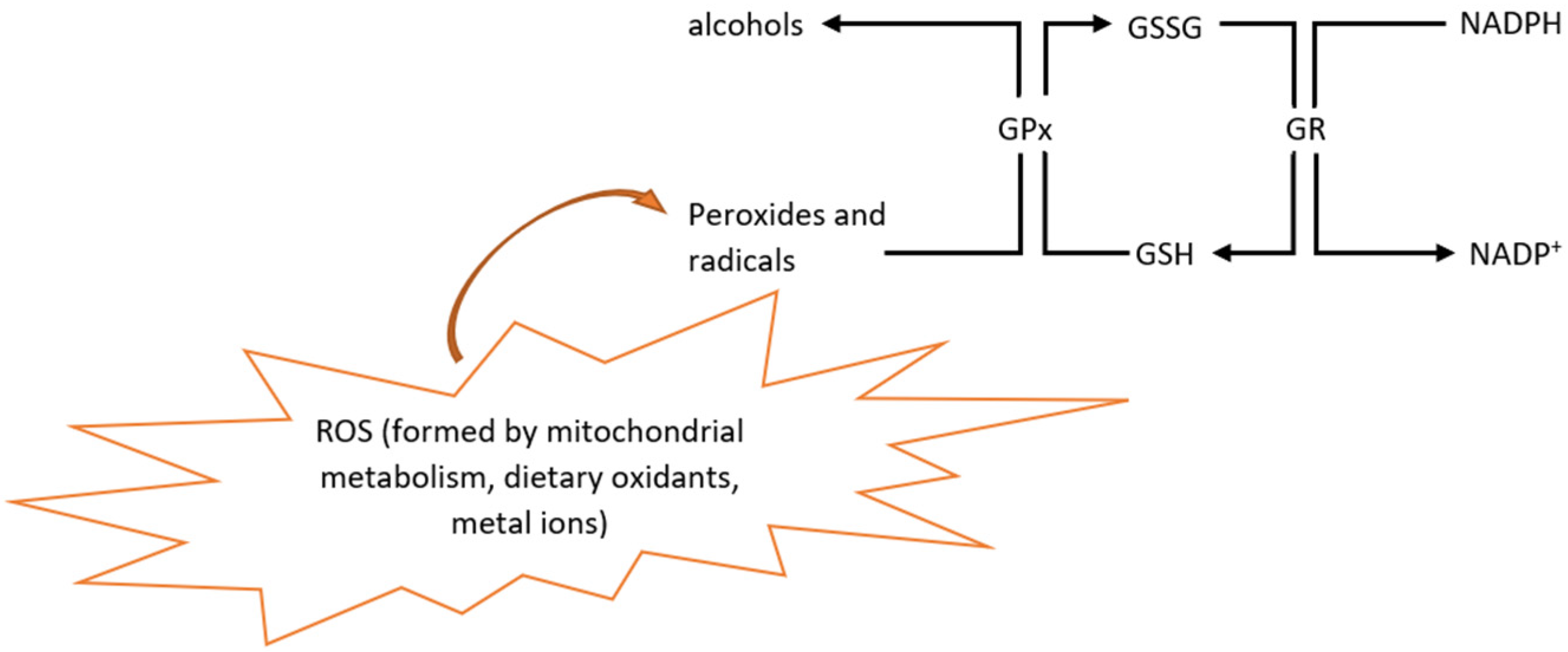
Figure 2. Glutathione mechanism for the neutralization of ROS. GPx neutralizes peroxides and radicals. In this process, GSH is oxidized to GSSG. GR reduces GSSG again to GSH in the presence of NADPH. GPx = glutathione peroxidase; GR = glutathione reductase; GSH = reduced glutathione; GSSG = oxidized glutathione; ROS = reactive oxygen species; NADPH = nicotinamide adenine dinucleotide phosphate.
Encyclopedia 02 00105 g002
Publisher’s Note: MDPI stays neutral with regard to jurisdictional claims in published maps and institutional affiliations.