Entry

The Applications of Microphysiological Systems in Biomedicine: Impact on Urologic and Orthopaedic Research

1
Department of Urology, University Medicine Greifswald, 17475 Greifswald, Germany
2
Center for Orthopaedics, Trauma Surgery and Rehabilitation Medicine, University Medicine Greifswald, 17475 Greifswald, Germany
*
Author to whom correspondence should be addressed.
Encyclopedia 2022, 2(2), 1128-1137; https://doi.org/10.3390/encyclopedia2020075
Submission received: 29 April 2022 / Revised: 25 May 2022 / Accepted: 7 June 2022 / Published: 7 June 2022
(This article belongs to the Section Medicine & Pharmacology)

Definition

:
Microphysiological systems (MPSs) are in vitro models that can incorporate dynamic stimuli such as flow, pressure and contraction in cell culture, enabling the formation of cellular architectures and retrieving physiological function often absent in conventional 2D-cell culture. MPS applications saw a substantial growth in recent years, drawing attention from industry as a strategy to optimize pre-clinical drug-development purposes, as well as from biomedical research, to fill a gap between in vivo and in vitro models. Several MPS platforms are now available and are employed in the development of bone and kidney complex systems for urologic and orthopaedic research. These advances have enabled, for example, the in vitro modelling of bone regeneration and renal drug secretion, and have dramatic potential to improve research into both orthopaedic and urology cancers.

1. Introduction

The pursuit of improved and ever more physiologically representative in vitro models began with the development and implementation of mammalian cell culture in the second half of the 20th century. Cell lines enabled major breakthroughs in biomedical research, introducing standardized study models that can be transferred and scaled-up with ease. The development of appropriate culture solutions that could feed and maintain cells ex vivo were crucial in the early days of cell culture [1,2]. Cancer cells, given their resilience and high turnover, were key to the successful generation of the first cell lines, which are still extensively used today, with HeLa cells as the prototypical example [3]. Nowadays, virtually every cell type from any organ system can be available for in vitro testing. Advances in culture media formulations enable the growth and expansion of viable and high-quality primary cell cultures [4]. Genetically manipulated cells can be made to overexpress or suppress genes of interest, which are invaluable tools to investigate the function of individual proteins and their overall physiological impact. Stem cells and reprogramed somatic cells, widely referred to as induced pluripotent stem cells (iPSCs), can be used to generate different adult cell lines from a single donor, maintaining the genetic background [5]. However, immortalized cell lines, characterized by their distinct stability, are still extensively used for highly reproducible models representative of healthy tissues or disease conditions.
Despite their widespread use, with thousands of cell lines catalogued, and invaluable contributions to molecular biology, biomedicine and life sciences in general, cell lines have significant limitations. The artificial culture conditions that isolated cells are subject to is dramatically different from their native environment [6]. Ex vivo, cells adapt their physiology to the culture conditions. Extensive characterization studies have shown the transformations that cells undergo in vitro. Among the many phenotypic changes, noteworthy are the rewriting of regulatory pathways, loss of polarization, reduced protein expression and altered metabolic activity [7,8,9]. In traditional in vitro culture systems, such as flasks and microplates, cells grow in an adherent surface and are covered by culture media. In this format, cells have one surface attached to a rigid substrate and the other exposed to the aqueous environment of the growth media. Cells are usually maintained at 37 °C in a humidified chamber at atmospheric pressure. Arguably, these conditions are dramatically different from the physiological milieu where cells thrive and fulfil their functions. Nonetheless, these set-ups can successfully sustain cells and are very powerful and cost-effective research tools [10].
Three-dimensional (3D) cell culture formats can introduce additional levels of cellular complexity. Hydrogels consist of biological (e.g., collagens) or synthetic (e.g., polyethylene glycol) polymers that form a matrix where cells can be embedded and experience a bespoke microenvironment [11]. Cell culture media easily diffuses through the gels, reaching the embedded cells and providing an aqueous environment. Gel properties, such as stiffness, pore size and adhesion can be designed to meet the needs of specific cells in order to optimize growth, viability and differentiation, therefore improving the phenotype by mimicking an extracellular matrix (ECM) [12]. Three-dimensional cultures enable better cell polarization, and cells can also shape the gel microenvironment by secreting their own matrix proteins. Not all cell lines are suitable for 3D culture, and usually cells that retain the ability to form structures such as epitheliums are best suited for hydrogel cultures. Certain cells can also spontaneously generate 3D structure, such as spheroids, in conventional culture, without the need for an ECM [13]. Conventional and 3D cell cultures are static, another stark difference relative to native physiology where cells experience a multitude of stimuli, to which they react. Allegedly, these static cell culture technologies have reached the limit of the physiology they can extract from cells. The next generation of in vitro culture systems is set to introduce further cellular complexity by incorporating external dynamic stimulus [14].

2. Microphysiological Systems and Their Applications

Microphysiological systems (MPSs) is a term employed to describe culture platforms that differ from conventional formats by incorporating perfusion and culture conditions that can enable complex cellular architectures [15,16]. MPSs are often referred to as organs-on-chip, a somewhat misleading terminology since MPSs, despite their advanced physiology, do not recreate fully functional organs per se. Generally, current MPSs consist of modular and enclosed platforms incorporating microfluidic circuits that enable perfusion through a culture compartment [17]. Flow is usually driven externally and these MPSs often consist of the culture platforms, coupled to perfusion units. Hence, these culture platforms/units are commonly designed as culture chips.
Advances in microfabrication and microfluidics facilitated the development of multiple MPS designs and applications in the past 20 years [17]. Interest in MPS saw a dramatic increase in recent years. A search in the National Library of Medicine database (pubmed.org) for the key work ‘organ-on-a-chip’ shows an increase from 106 entries in 2011 to 641 entries in 2021. Initial studies showed the feasibility of such systems and their potential to improve cell physiology, filling a gap between in vitro models and animal experimentation [18]. This contributed to catalyze interest from the pharmaceutical industry as well as initiatives and funding bodies aiming at reducing animal testing [19].

2.1. Perspective on MPS Applications in Drug Development

The wide use of MPSs in preclinical drug testing is still in the future. Nonetheless, several recent applications in the areas of safety/toxicology (Table 1) and drug metabolism and pharmacokinetics (DMPK) illustrate the advantages of MPSs relative to conventional models [20]. The kidneys are secretory organs that rely on highly polarized epithelial barriers populated with arrays of specialized membrane carrier proteins. The proximal tubules are the most DMPK- and safety-relevant renal functional unit. Proximal tubule epithelial cells are responsible for clearing drugs from circulation against steep concentration gradients, and their activity is crucial for accessing renal clearance and nephrotoxicity during drug development [21]. In conventional culture, renal epithelial cells lose the expression of key update and efflux carriers, and have poorly differentiated apical and basal membranes [8]. The use of transwells, where cells grow in a semipermeable membrane in contact with culture media from both sides, improves polarity and cells can effectively form a barrier between the compartments separated by the semipermeable membrane. Nonetheless, membrane carrier expression remains limited and secretory activity is poorly representative of native renal physiology. MPSs can overcome these limitations by providing renal epithelial cells with a more native microenvironment [22]. An example of a dedicated MPS that can recreate renal proximal tubule physiology is the ParVivo platform by Nortis Inc., Seattle, WA, USA (Figure 1A,B). This model comprises perfused micro-tubules imbedded in an ECM, and cells grow in a membrane-free tubular surface, enabling the formation of a continuous monolayer where the basal membrane attaches to the ECM and the apical membrane delineates the lumen of the renal tubule [23]. Continuous flow is sensed by the cells and upregulates the activity of membrane drug carriers and promotes cellular adhesion. With the renal tubule entirely surrounded by ECM, epithelial cells secrete and assemble their own basal membrane, rich in glycoproteins, that anchor the tubule to the matrix and improve structure integrity. Together, these features enable epithelial cells in MPSs to recreate active renal clearance absent in conventional culture [24], namely, the secretion of low permeability anionic and cationic drugs, which account for the majority of drugs cleared by the proximal tubules [25]. Employing MPSs during pre-clinical development can potentially improve the accuracy of pharmacokinetics predictions (e.g., renal clearance) and, therefore, help to optimize drug testing [26].
Another interesting aspect of MPSs in drug development are their prospective uses in drug discovery [19]. Human health and disease models that are easily accessible and can be studied under controlled conditions are valuable tools to identify disease mechanisms and drug targets. The identification of targets for rational drug design requires either native physiology or highly representative models that precisely recapitulate the activity of regulatory pathways [27]. Cell lines and conventional models have considerable physiological gaps that are not appropriate, in particular, for the development of new drug modalities which aim to have high target specificity and therapeutic efficacy [28].
MPSs can also potentially reduce the use of animals for drug testing. Accurate MPS-derived predictions may eliminate the need for extensive drug testing in pre-clinical species. Regulatory agencies (e.g., Federal Drug Administration—FDA and European Medicines Agency—EMA) will play a major role in the adoption of MPSs in drug testing and, therefore, minimizing the usage of animal models [29]. The introduction of a regulatory framework dictating which MPS applications will be accepted for the certification of new molecules will fast track the implementation of MPSs and related workflows during drug development.

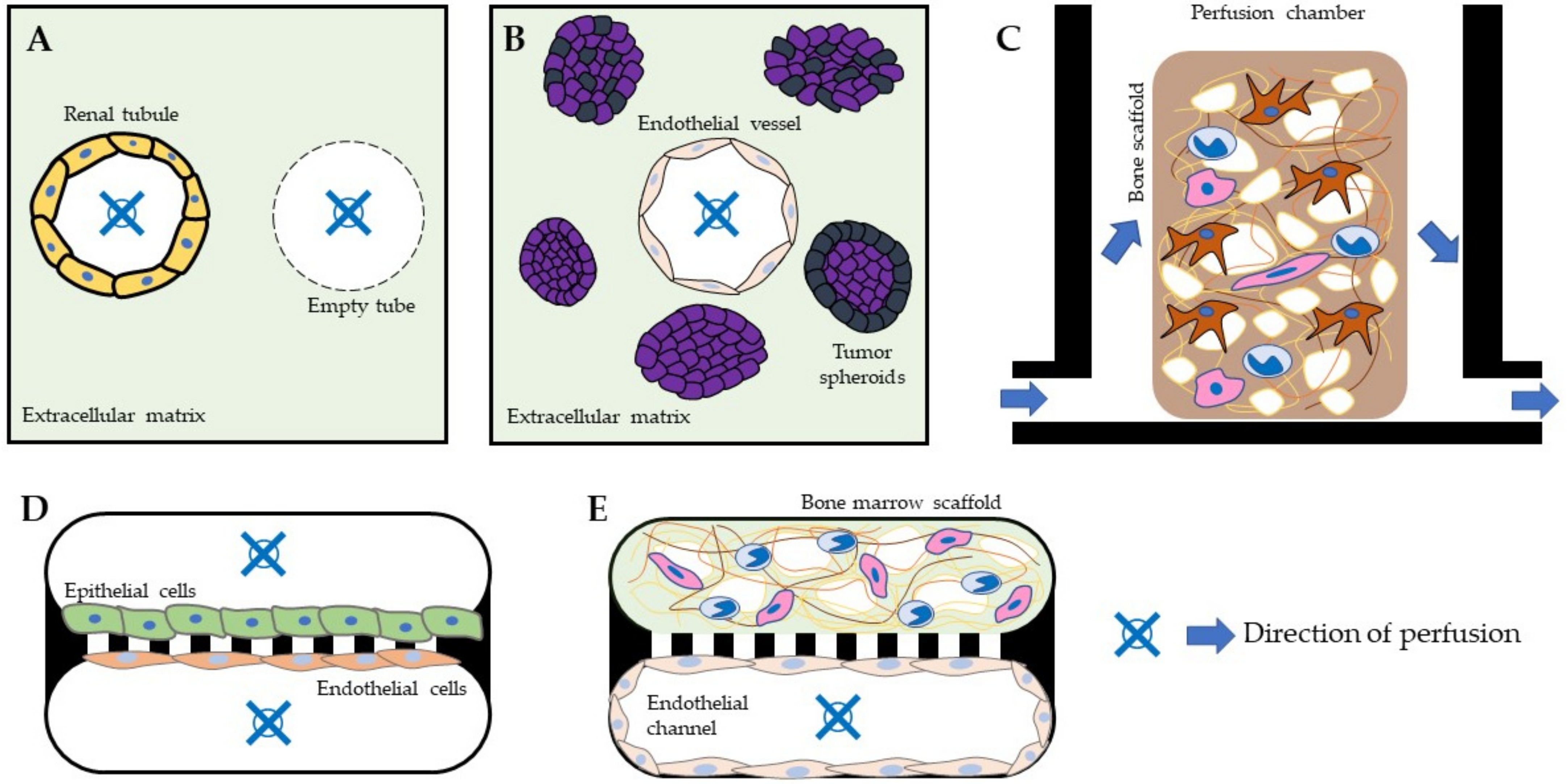
2.2. Perspective on MPS Applications in Biomedical Research

While the industry is far more focused on the specific applications of MPS that can improve drug testing, in academia MPS applications cover a broader spectrum (Table 1). Academic research has played a central role in the development, characterization and validation of MPS models. Bespoke MPS models are widely employed in fundamental research. However, their use is often restricted to the research institutions that promoted their development.
Renal models, such as the abovementioned system, can be translatable to applications that require the use of epithelial cells and the formation of tubular structures within a matrix. This is important for the implementation of 3D models in urological research, since most cell types of interest, namely kidney and prostate cancer cells, are of epithelial origin [30]. Modelling cancer-specific events, such as angiogenesis in renal cell carcinoma mediated by vascular endothelial growth factor secretion, is important to replicate the pathophysiology of kidney cancer in vitro [31]. An important aspect of using an MPS cancer model is the possibility to investigate cancer-specific biomarkers that are secreted into biological fluids, a feature that static cultures lack, given their lack of flow [32]. Beyond the use of cell lines, microphysiological systems can also enable the cultivation of patient-derived cancer cells or cancer tissue in a controlled system [33]. Primary tissue cultures are relevant in oncology research given the enormous heterogenicity found within the same cancer types, and cell lines can only offer a window into the specific particular subject they were created from. Moreover, current prostate and prostate cancer in vitro models are poorly representative given the de-differentiation that cells undergo in culture, losing critical prostate markers. Growing prostate cells under fluidic conditions may overcome such limitations, however, to date, little evidence is found regarding the development of prostate-MPS models.
Commercially available MPS models provide standardized platforms where protocols and applications can be exchanged and implemented by different users. An example of a versatile MPS model used in industry and academia is the HUMIMIC platform by TissUse GmbH, Berlin (Figure 1C). This MPS platform allows for cell culture under pulsating circulation enabled through sequentially moved on-chip polydimethylsiloxane (PDMS) membranes constituting micropumps controlled by externally generated air pressure and vacuum [34,35]. Several studies indicate the applicability of the system to culture cell lines, iPSCs and primary cells in 2D and fabricated 3D constructs, as well as human biopsies, to emulate specific function of human organs such as liver, kidney, testis, skin and lung [36,37,38,39,40,41]. Moreover, a bone marrow model was previously introduced and suggested for preclinical drug testing [42]. This MPS, showing a 4-week survival of hematopoietic stem and progenitor cells (HSPCs), is based on the co-culture of human bone marrow mesenchymal stromal cells (MSCs) seeded on a hydroxyapatite-coated, trabecular ceramic scaffold and HSPCs from umbilical cord blood. Based on this model, a bone-on-a-chip system was developed that contains human trabecular bone and the overall population of human bone marrow mononuclear cells (BM-MNCs) [43,44]. This system is set up by sequential seeding on decellularized human trabecular bone scaffolds with MSCs, osteoblasts and osteoclast precursors, and the addition of BM-MNCs from the same donor after formation of the intertrabecular matrix under pulsing microfluid flow. Using this in vitro model, it was possible to reproduce that, in particular in chromium, which can be released from orthopaedic implants, and accumulates in the reticular bone marrow matrix. This bone model represents a promising tool to investigate foreign body reactions and the possible toxic effects of implant materials and their degradation products in vitro. The development of this bone model is a good example of how commercially available MPSs can be facilitated for basic biomedical research, and is also promising for other applications, as it aims to represent both active bone metabolism and the individual immunocompetence of the respective cell donor. A functional immune system is closely associated with physiological bone homeostasis and bone healing [45,46]. Local immune cells play a crucial role in the development and progression of bone pathologies such as osteoporosis [47,48] and aseptic periprosthetic osteolysis [49]. In addition to inserting immune cells into multicellular bone models, other important parameters noted for the development of bone-on-a-chip models are controllable mechanical load and oxygen levels, since these parameters are of distinct importance for physiological bone metabolism [50]. For this reason, there have been efforts to simulate mechanical forces in vitro using classical bioreactor-based models to study their precise influence on the physiology and pathophysiology of bone. Early models used, e.g., pneumatic load of a 3D bone organoid [51,52]. Other models use piezoelectric actuation, which allows the applied mechanical forces to be regulated very precisely [53]. In order to better emulate the biomechanics in the mature bone, systems were developed that allow mechanical compression with high forces and simultaneous perfusion of the bone organoid [54,55]. Organ-on-a-chip models are the further development of classical bioreactor systems, which are based on the use of microfluidic platforms and are prone to introduce the regulation of organ- and tissue-specific parameters, such as mechanical load or oxygen levels [16,56]. Despite the frequently described organ chips, there are still only a few MPSs that emulate mature bone [16,50]. Among these bone-on-a-chip models, only a few systems are dedicated to the biomechanics essential for bone metabolism, e.g., similar to some classical bioreactor systems, pneumatic load is used to study the differentiation of MSCs under controllable mechanical load [57]. Other models emulate the shear stress in the canaliculi of the bone to study their influence on osteocytes [58].
The complexity of bone-on-a-chip models is being further advanced in terms of controllable parameters and cellular and matrix-specific composition to be able to investigate physiological and pathophysiological processes in vitro. Nevertheless, limitations of advanced in vitro models will remain and must be understood through characterization.

3. Conclusions and Prospects

Nowadays, several MPS platforms from different providers are available [68,69] and the two examples presented, describing different application for bone and kidney research, illustrate the overall capabilities of advanced in vitro models incorporating microfluidic conditions. MPSs have proven to deliver enhanced physiology and surpass the limitations of conventional cell culture.
The technical complexities of such systems are still at a bottleneck in their implementation, given the dedicated equipment and training required. Nonetheless, as interest grows and translational research shifts away from animal models [70], MPSs are finding a foothold in industry and academic labs worldwide.
As the field of MPS platforms begins to consolidate, standardization similar to that existing in 2D cell culture formats may be beneficial to the further dissemination of these technologies, as well as facilitating the comparison of similar studies performed across different institutions. As the complexity of MPS increase, so can the biology they replicate, and we can expect that the next generation of advanced cell culture platforms will be a step closer to organ-like cellular organization, incorporating multiple cell types and even multiple interconnected organ systems. Substantial progress has been made in the field of 3D bone models, as well as renal models (Figure 1), and these lessons can be translated to implanting MPS applications in the wider filed of urology research, focusing on cancer models. While validated renal MPS models are now in use, examples of further organs in the urogenital tract are lacking, with limited literature available on prostate and bladder models [61,62]. An interesting and valuable research avenue is the interaction of healthy and tumor cells, since fluidic conditions enable an intricate connection between different cells. The development of cancer metastasis is still poorly understood, and recreating it in vitro can propel the development of new therapies and diagnostic tools.
Ultimately, when developing in vitro models for biomedical research, it is important to always keep the target application clearly in mind and to challenge whether the respective model is suitable to answer the scientific question at hand.

Author Contributions

P.C.-P. conceptualized this short review and J.S. contributed with his extensive expertise in the development of MPS applications. All authors have read and agreed to the published version of the manuscript.

Funding

This research received no external funding.

Conflicts of Interest

The authors declare no conflict of interest.

References

  1. Arora, M. Cell Culture Media: A Review. Mater. Methods 2013, 3, 175. [Google Scholar] [CrossRef]
  2. Yao, T.; Asayama, Y. Animal-cell culture media: History, characteristics, and current issues. Reprod. Med. Biol. 2017, 16, 99–117. [Google Scholar] [CrossRef] [PubMed]
  3. Masters, J.R. HeLa cells 50 years on: The good, the bad and the ugly. Nat. Rev. Cancer 2002, 2, 315–319. [Google Scholar] [CrossRef]
  4. Richter, M.; Piwocka, O.; Musielak, M.; Piotrowski, I.; Suchorska, W.M.; Trzeciak, T. From Donor to the Lab: A Fascinating Journey of Primary Cell Lines. Front. Cell Dev. Biol. 2021, 9, 1869. [Google Scholar] [CrossRef]
  5. Shi, Y.; Inoue, H.; Wu, J.C.; Yamanaka, S. Induced pluripotent stem cell technology: A decade of progress. Nat. Rev. Drug Discov. 2017, 16, 115–130. [Google Scholar] [CrossRef] [PubMed]
  6. Allen, S.L.; Marshall, R.N.; Edwards, S.J.; Lord, J.M.; Lavery, G.G.; Breen, L. The effect of young and old ex vivo human serum on cellular protein synthesis and growth in an in vitro model of aging. Am. J. Physiol. Cell Physiol. 2021, 321, C26–C37. [Google Scholar] [CrossRef]
  7. Verhulst, A.; Sayer, R.; De Broe, M.E.; D’Haese, P.C.; Brown, C.D.A. Human proximal tubular epithelium actively secretes but does not retain rosuvastatin. Mol. Pharmacol. 2008, 74, 1084–1091. [Google Scholar] [CrossRef] [PubMed]
  8. Jenkinson, S.E.; Chung, G.W.; van Loon, E.; Bakar, N.S.; Dalzell, A.M.; Brown, C.D.A. The limitations of renal epithelial cell line HK-2 as a model of drug transporter expression and function in the proximal tubule. Pflug. Arch. 2012, 464, 601–611. [Google Scholar] [CrossRef]
  9. Jeske, R.; Yuan, X.; Fu, Q.; Bunnell, B.A.; Logan, T.M.; Li, Y. In Vitro Culture Expansion Shifts the Immune Phenotype of Human Adipose-Derived Mesenchymal Stem Cells. Front. Immunol. 2021, 12, 46. [Google Scholar] [CrossRef]
  10. Lorian, V.; Satta, G. Differences between in vitro and in vivo studies. Antimicrob. Agents Chemother. 1988, 32, 1600–1601. [Google Scholar] [CrossRef]
  11. Pampaloni, F.; Reynaud, E.G.; Stelzer, E.H.K. The third dimension bridges the gap between cell culture and live tissue. Nat. Rev. Mol. Cell Biol. 2007, 8, 839–845. [Google Scholar] [CrossRef]
  12. Mahinroosta, M.; Jomeh Farsangi, Z.; Allahverdi, A.; Shakoori, Z. Hydrogels as intelligent materials: A brief review of synthesis, properties and applications. Mater. Today Chem. 2018, 8, 42–55. [Google Scholar] [CrossRef]
  13. Langhans, S.A. Three-dimensional in vitro cell culture models in drug discovery and drug repositioning. Front. Pharmacol. 2018, 9, 6. [Google Scholar] [CrossRef] [PubMed]
  14. Jensen, C.; Teng, Y. Is It Time to Start Transitioning From 2D to 3D Cell Culture? Front. Mol. Biosci. 2020, 7, 33. [Google Scholar] [CrossRef]
  15. Van Ness, K.P.; Chang, S.Y.; Weber, E.J.; Zumpano, D.; Eaton, D.L.; Kelly, E.J. Microphysiological systems to assess nonclinical toxicity. Curr. Protoc. Toxicol. 2017, 2017, 28. [Google Scholar] [CrossRef]
  16. Bhatia, S.N.; Ingber, D.E. Microfluidic organs-on-chips. Nat. Biotechnol. 2014, 32, 760–772. [Google Scholar] [CrossRef]
  17. Low, L.A.; Mummery, C.; Berridge, B.R.; Austin, C.P.; Tagle, D.A. Organs-on-chips: Into the next decade. Nat. Rev. Drug Discov. 2021, 20, 345–361. [Google Scholar] [CrossRef]
  18. Van Berlo, D.; van de Steeg, E.; Amirabadi, H.E.; Masereeuw, R. The potential of multi-organ-on-chip models for assessment of drug disposition as alternative to animal testing. Curr. Opin. Toxicol. 2021, 27, 8–17. [Google Scholar] [CrossRef]
  19. Ramsden, D. Leveraging microphysiological systems to address challenges encountered during development of oligonucleotide therapeutics. ALTEX Altern. Anim. Exp. 2021, 39, 273–296. [Google Scholar] [CrossRef]
  20. Ishida, S. Organs-on-a-chip: Current applications and consideration points for in vitro ADME-Tox studies. Drug Metab. Pharmacokinet. 2018, 33, 49–54. [Google Scholar] [CrossRef]
  21. Caetano-Pinto, P.; Janssen, M.J.; Gijzen, L.; Verscheijden, L.; Wilmer, M.J.G.; Masereeuw, R. Fluorescence-Based Transport Assays Revisited in a Human Renal Proximal Tubule Cell Line. Mol. Pharm. 2016, 13, 933–944. [Google Scholar] [CrossRef]
  22. Van Ness, K.P.; Kelly, E.J.; Cesar, F. Microphysiological systems in adsorption, distribution, metabolism and elimination sciences. Clin. Transl. Sci. 2022, 15, 9–42. [Google Scholar] [CrossRef] [PubMed]
  23. Weber, E.J.; Chapron, A.; Chapron, B.D.; Voellinger, J.L.; Lidberg, K.A.; Yeung, C.K.; Wang, Z.; Yamaura, Y.; Hailey, D.W.; Neumann, T.; et al. Development of a microphysiological model of human kidney proximal tubule function. Kidney Int. 2016, 90, 627–637. [Google Scholar] [CrossRef]
  24. Imaoka, T.; Huang, W.; Shum, S.; Hailey, D.W.; Chang, S.-Y.; Chapron, A.; Yeung, C.K.; Himmelfarb, J.; Isoherranen, N.; Kelly, E.J. Bridging the gap between in silico and in vivo by modeling opioid disposition in a kidney proximal tubule microphysiological system. Sci. Rep. 2021, 11, 21356. [Google Scholar] [CrossRef] [PubMed]
  25. Scotcher, D.; Jones, C.; Rostami-Hodjegan, A.; Galetin, A. Novel minimal physiologically-based model for the prediction of passive tubular reabsorption and renal excretion clearance. Eur. J. Pharm. Sci. 2016, 94, 59–71. [Google Scholar] [CrossRef]
  26. Van Ness, K.P.; Cesar, F.; Yeung, C.K.; Himmelfarb, J.; Kelly, E.J. Microphysiological Systems in ADME Sciences. Clin. Transl. Sci. 2021, 15, 9–42. [Google Scholar] [CrossRef] [PubMed]
  27. Moffat, J.G.; Vincent, F.; Lee, J.A.; Eder, J.; Prunotto, M. Opportunities and challenges in phenotypic drug discovery: An industry perspective. Nat. Rev. Drug Discov. 2017, 16, 531–543. [Google Scholar] [CrossRef]
  28. Valeur, E.; Guéret, S.M.; Adihou, H.; Gopalakrishnan, R.; Lemurell, M.; Waldmann, H.; Grossmann, T.N.; Plowright, A.T. New Modalities for Challenging Targets in Drug Discovery. Angew. Chemie. Int. Ed. 2017, 56, 10294–10323. [Google Scholar] [CrossRef]
  29. Isoherranen, N.; Madabushi, R.; Huang, S.M. Emerging Role of Organ-on-a-Chip Technologies in Quantitative Clinical Pharmacology Evaluation. Clin. Transl. Sci. 2019, 12, 113–121. [Google Scholar] [CrossRef]
  30. Pinto, P.C.; Rönnau, C.; Burchardt, M.; Wolff, I. Kidney Cancer and Chronic Kidney Disease: Too Close for Comfort. Biomedicines 2021, 9, 1761. [Google Scholar] [CrossRef]
  31. Chapron, A.; Chapron, B.D.; Hailey, D.W.; Chang, S.Y.; Imaoka, T.; Thummel, K.E.; Kelly, E.; Himmelfarb, J.; Shen, D.; Yeung, C.K. An Improved Vascularized, Dual-Channel Microphysiological System Facilitates Modeling of Proximal Tubular Solute Secretion. ACS Pharmacol. Transl. Sci. 2020, 3, 496–508. [Google Scholar] [CrossRef]
  32. Nieskens, T.T.G.; Magnusson, O.; Andersson, P.; Söderberg, M.; Persson, M.; Sjögren, A.K. Nephrotoxic antisense oligonucleotide SPC5001 induces kidney injury biomarkers in a proximal tubule-on-a-chip. Arch. Toxicol. 2021, 95, 2123–2136. [Google Scholar] [CrossRef]
  33. Miller, C.P.; Tsuchida, C.; Zheng, Y.; Himmelfarb, J.; Akilesh, S. A 3D Human Renal Cell Carcinoma-on-a-Chip for the Study of Tumor Angiogenesis. Neoplasia 2018, 20, 610–620. [Google Scholar] [CrossRef]
  34. Ramme, A.P.; Koenig, L.; Hasenberg, T.; Schwenk, C.; Magauer, C.; Faust, D.; Lorenz, A.K.; Krebs, A.C.; Drewell, C.; Schirrmann, K.; et al. Autologous induced pluripotent stem cell-derived four-organ-chip. Futur. Sci. OA 2019, 5, FSO413. [Google Scholar] [CrossRef] [PubMed]
  35. Maschmeyer, I.; Lorenz, A.K.; Schimek, K.; Hasenberg, T.; Ramme, A.P.; Hübner, J.; Lindner, M.; Drewell, C.; Bauer, S.; Thomas, A.; et al. A four-organ-chip for interconnected long-term co-culture of human intestine, liver, skin and kidney equivalents. Lab Chip 2015, 15, 2688–2699. [Google Scholar] [CrossRef] [PubMed]
  36. Lin, N.; Zhou, X.; Geng, X.; Drewell, C.; Hübner, J.; Li, Z.; Zhang, Y.; Xue, M.; Marx, U.; Li, B. Repeated dose multi-drug testing using a microfluidic chip-based coculture of human liver and kidney proximal tubules equivalents. Sci. Rep. 2020, 10, 8879. [Google Scholar] [CrossRef] [PubMed]
  37. Tao, T.P.; Brandmair, K.; Gerlach, S.; Przibilla, J.; Géniès, C.; Jacques-Jamin, C.; Schepky, A.; Marx, U.; Hewitt, N.J.; Maschmeyer, I.; et al. Demonstration of the first-pass metabolism in the skin of the hair dye, 4-amino-2-hydroxytoluene, using the Chip2 skin–liver microphysiological model. J. Appl. Toxicol. 2021, 41, 1553–1567. [Google Scholar] [CrossRef]
  38. Kühnl, J.; Tao, T.P.; Brandmair, K.; Gerlach, S.; Rings, T.; Müller-Vieira, U.; Przibilla, J.; Genies, C.; Jaques-Jamin, C.; Schepky, A.; et al. Characterization of application scenario-dependent pharmacokinetics and pharmacodynamic properties of permethrin and hyperforin in a dynamic skin and liver multi-organ-chip model. Toxicology 2021, 448, 152637. [Google Scholar] [CrossRef]
  39. Tavares, R.S.N.; Phuong-Tao, T.; Maschmeyer, I.; Maria-Engler, S.S.; Schäfer-Korting, M.; Winter, A.; Zoschke, C.; Lauster, R.; Marx, U.; Gaspar, L.R. Toxicity of topically applied drugs beyond skin irritation: Static skin model vs. Two organs-on-a-chip. Int. J. Pharm. 2020, 589, 119788. [Google Scholar] [CrossRef]
  40. Baert, Y.; Ruetschle, I.; Cools, W.; Oehme, A.; Lorenz, A.; Marx, U.; Goossens, E.; Maschmeyer, I. A multi-organ-chip co-culture of liver and testis equivalents: A first step toward a systemic male reprotoxicity model. Hum. Reprod. 2020, 35, 1029–1044. [Google Scholar] [CrossRef]
  41. Schimek, K.; Frentzel, S.; Luettich, K.; Bovard, D.; Rütschle, I.; Boden, L.; Rambo, F.; Erfurth, H.; Dehne, E.M.; Winter, A.; et al. Human multi-organ chip co-culture of bronchial lung culture and liver spheroids for substance exposure studies. Sci. Rep. 2020, 10, 7865. [Google Scholar] [CrossRef]
  42. Sieber, S.; Wirth, L.; Cavak, N.; Koenigsmark, M.; Marx, U.; Lauster, R.; Rosowski, M. Bone marrow-on-a-chip: Long-term culture of human haematopoietic stem cells in a three-dimensional microfluidic environment. J. Tissue Eng. Regen. Med. 2018, 12, 479–489. [Google Scholar] [CrossRef] [PubMed]
  43. Schoon, J.; Hesse, B.; Rakow, A.; Ort, M.J.; Lagrange, A.; Jacobi, D.; Winter, A.; Huesker, K.; Reinke, S.; Cotte, M.; et al. Metal-Specific Biomaterial Accumulation in Human Peri-Implant Bone and Bone Marrow. Adv. Sci. 2020, 7, 2000412. [Google Scholar] [CrossRef]
  44. Ingber, D.E. Human organs-on-chips for disease modelling, drug development and personalized medicine. Nat. Rev. Genet. 2022, 1–25. [Google Scholar] [CrossRef] [PubMed]
  45. Arron, J.R.; Choi, Y. Bone versus immune system. Nature 2000, 408, 535–536. [Google Scholar] [CrossRef]
  46. Tsukasaki, M.; Takayanagi, H. Osteoimmunology: Evolving concepts in bone–immune interactions in health and disease. Nat. Rev. Immunol. 2019, 19, 626–642. [Google Scholar] [CrossRef] [PubMed]
  47. Saxena, Y.; Routh, S.; Mukhopadhaya, A. Immunoporosis: Role of Innate Immune Cells in Osteoporosis. Front. Immunol. 2021, 12, 3168. [Google Scholar] [CrossRef]
  48. Fischer, V.; Haffner-Luntzer, M. Interaction between bone and immune cells: Implications for postmenopausal osteoporosis. Semin. Cell Dev. Biol. 2022, 123, 14–21. [Google Scholar] [CrossRef]
  49. Perino, G.; De Martino, I.; Zhang, L.; Xia, Z.; Gallo, J.; Natu, S.; Langton, D.; Huber, M.; Rakow, A.; Schoon, J.; et al. The contribution of the histopathological examination to the diagnosis of adverse local tissue reactions in arthroplasty. EFORT Open Rev. 2021, 6, 399–419. [Google Scholar] [CrossRef]
  50. Scheinpflug, J.; Pfeiffenberger, M.; Damerau, A.; Schwarz, F.; Textor, M.; Lang, A.; Schulze, F. Journey into bone models: A review. Genes 2018, 9, 247. [Google Scholar] [CrossRef]
  51. Matziolis, D. Osteogenic Predifferentiation of Human Bone Marrow-Derived Stem Cells by Short-Term Mechanical Stimulation. Open Orthop. J. 2011, 5, 1–6. [Google Scholar] [CrossRef]
  52. Matziolis, G.; Tuischer, J.; Kasper, G.; Thompson, M.; Bartmeyer, B.; Krocker, D.; Perka, C.; Duda, G. Simulation of cell differentiation in fracture healing: Mechanically loaded composite scaffolds in a novel bioreactor system. Tissue Eng. 2006, 12, 201–208. [Google Scholar] [CrossRef]
  53. Petersen, A.; Joly, P.; Bergmann, C.; Korus, G.; Duda, G.N. The impact of substrate stiffness and mechanical loading on fibroblast-induced scaffold remodeling. Tissue Eng. Part A 2012, 18, 1804–1817. [Google Scholar] [CrossRef] [PubMed]
  54. Hoffmann, W.; Feliciano, S.; Martin, I.; de Wild, M.; Wendt, D. Novel Perfused Compression Bioreactor System as an in vitro Model to Investigate Fracture Healing. Front. Bioeng. Biotechnol. 2015, 3. [Google Scholar] [CrossRef]
  55. Richards, R.G.; Simpson, A.E.; Jaehn, K.; Furlong, P.I.; Stoddart, M.J. Establishing a 3D ex vivo culture system for investigations of bone metabolism and biomaterial interactions. ALTEX Altern. Tierexp. 2007, 24, 56–59. [Google Scholar]
  56. Kodzius, R.; Schulze, F.; Gao, X.; Schneider, M.R. Organ-on-chip technology: Current state and future developments. Genes 2017, 8, 266. [Google Scholar] [CrossRef] [PubMed]
  57. Park, S.H.; Sim, W.Y.; Min, B.H.; Yang, S.S.; Khademhosseini, A.; Kaplan, D.L. Chip-Based Comparison of the Osteogenesis of Human Bone Marrow- and Adipose Tissue-Derived Mesenchymal Stem Cells under Mechanical Stimulation. PLoS ONE 2012, 7, e46689. [Google Scholar] [CrossRef]
  58. Sun, Q.; Choudhary, S.; Mannion, C.; Kissin, Y.; Zilberberg, J.; Lee, W.Y. Ex vivo replication of phenotypic functions of osteocytes through biomimetic 3D bone tissue construction. Bone 2018, 106, 148–155. [Google Scholar] [CrossRef] [PubMed]
  59. Nieskens, T.T.G.; Persson, M.; Kelly, E.J.; Sjögren, A.K. A multicompartment human kidney proximal tubule-on-a-chip replicates cell polarization–dependent cisplatin toxicity. Drug Metab. Dispos. 2020, 48, 1303–1311. [Google Scholar] [CrossRef] [PubMed]
  60. Jang, K.J.; Mehr, A.P.; Hamilton, G.A.; McPartlin, L.A.; Chung, S.; Suh, K.Y.; Ingber, D.E. Human kidney proximal tubule-on-a-chip for drug transport and nephrotoxicity assessment. Integr. Biol. 2013, 5, 1119–1129. [Google Scholar] [CrossRef]
  61. Jiang, L.; Ivich, F.; Tahsin, S.; Tran, M.; Frank, S.B.; Miranti, C.K.; Zohar, Y. Human stroma and epithelium co-culture in a microfluidic model of a human prostate gland. Biomicrofluidics 2019, 13, 064116. [Google Scholar] [CrossRef] [PubMed]
  62. Sharma, K.; Dhar, N.; Thacker, V.V.; Simonet, T.M.; Signorino-Gelo, F.; Knott, G.; McKinney, J.D. Dynamic persistence of intracellular bacterial communities of uropathogenic escherichia coli in a human bladder-chip model of urinary tract infections. Elife 2021, 10, e66481. [Google Scholar] [CrossRef] [PubMed]
  63. Lin, H.; Lozito, T.P.; Alexander, P.G.; Gottardi, R.; Tuan, R.S. Stem cell-based microphysiological osteochondral system to model tissue response to interleukin-1Β. Mol. Pharm. 2014, 11, 2203–2212. [Google Scholar] [CrossRef] [PubMed]
  64. Iordachescu, A.; Hughes, E.A.B.; Joseph, S.; Hill, E.J.; Grover, L.M.; Metcalfe, A.D. Trabecular bone organoids: A micron-scale ‘humanised’ prototype designed to study the effects of microgravity and degeneration. npj Microgravity 2021, 7, 17. [Google Scholar] [CrossRef]
  65. Chou, D.B.; Frismantas, V.; Milton, Y.; David, R.; Pop-Damkov, P.; Ferguson, D.; MacDonald, A.; Vargel Bölükbaşı, Ö.; Joyce, C.E.; Moreira Teixeira, L.S.; et al. On-chip recapitulation of clinical bone marrow toxicities and patient-specific pathophysiology. Nat. Biomed. Eng. 2020, 4, 394–406. [Google Scholar] [CrossRef] [PubMed]
  66. Agrawal, G.; Aung, A.; Varghese, S. Skeletal muscle-on-a-chip: An in vitro model to evaluate tissue formation and injury. Lab Chip 2017, 17, 3447–3461. [Google Scholar] [CrossRef]
  67. Ortega, M.A.; Fernández-Garibay, X.; Castaño, A.G.; De Chiara, F.; Hernández-Albors, A.; Balaguer-Trias, J.; Ramón-Azcón, J. Muscle-on-a-chip with an on-site multiplexed biosensing system for: In situ monitoring of secreted IL-6 and TNF-α. Lab Chip 2019, 19, 2568–2580. [Google Scholar] [CrossRef]
  68. Peel, S.; Jackman, M. Imaging microphysiological systems: A review. Am. J. Physiol. Cell Physiol. 2021, 320, C669–C680. [Google Scholar] [CrossRef]
  69. Caetano-Pinto, P.; Stahl, S.H. Perspective on the application of microphysiological systems to drug transporter studies. Drug Metab. Dispos. 2018, 46, 1647–1657. [Google Scholar] [CrossRef]
  70. Ingber, D.E. Is it Time for Reviewer 3 to Request Human Organ Chip Experiments Instead of Animal Validation Studies? Adv. Sci. 2020, 7, 2002030. [Google Scholar] [CrossRef]
Figure 1. Examples of commercially available microphysiological system designs used in experimental urology and orthopaedic research. (A) The Nortis ParVivo dual-channel chip design incorporates two parallel micro-perfusion channels, embedded in an extracellular matrix (ECM). Epithelial proximal tubule cells seeded in one channel assemble a tight renal epithelial tubule and are continuously perfused via the lumen. The parallel empty channel is used to load compounds/drugs into the ECM that are exposed to the renal tubule from the outside. This model recreates the apical-basolateral dynamic of the renal proximal tubules with continuous flow through the inside and outside of the tubule. (B) The ParVivo single-channel design, incorporating a single channel in an ECM, is used as a kidney cancer model, mimicking the interaction of renal cell carcinoma (RCC) spheroids and the vasculature. RCCs are embedded in the ECM, proliferating and secreting factors into the extracellular space, which is populated by a continuously perfused endothelial vessel, formed by human umbilical vein endothelial cells (HUVECs). (C) The TissUse Humimic chips consist of interconnected perfusion chambers where culture media is recirculated using cycling pulsation. This platform enables structures in the chambers to experience both contraction and flow, and it is a preferred platform for bone modelling. A ceramic scaffold populated with osteoclasts, mesenchymal cells and bone marrow cells subjected to these dynamic conditions is able to differentiate into mature bone structures. (D) The Emulate Organ-Chip platform is widely used to recapitulate functional organ units and consists of two adjacent continuously perfused channels separated by a semipermeable membrane. Different cell types can be grown on both sides of the membrane, and kidney, prostate and bladder models established using this platform are based on this design, with epithelial cells of the respective organ seeded on the top channel and endothelial or supporting cells on the lower channel. (E) In the bone-marrow model based on the Emulate platform, endothelial cells grow along the entire surface of the lower channel, replicating a blood vessel, while the top channel is completely filled with ECM containing hematopoietic stem cells. Perfusion is driven through the endothelial channel and the ECM mimics the niche in the bone marrow where stem cells differentiate into different hematopoietic cells.
Encyclopedia 02 00075 g001
Table 1. Representative examples of current MPS models and applications used in biomedical research, namely in experimental urology and orthopaedics.
Organ/TissueCell TypeMPS ModelStimulusPurposeRef
KidneyHuman renal epithelial proximal tubule cellsNortis a
ParVivo
Dual-chip
Continuous flowImprove renal drug secretion studies in vitro[23,31,59]
Human renal epithelial proximal tubule cellsEmulate a
Organ-Chip
Continuous flowImprove nephrotoxicity evaluation in vitro[60]
ProstateHuman benign epithelial prostate cellsBespoke bContinuous flowEvaluate prostate paracrine secretion in vitro[61]
Kidney CancerHuman renal cell carcinoma/endothelial cellsNortis a
ParVivo
Single-chip
Continuous flowRecapitulate kidney cancer derived angiogenesis in vitro[33]
BladderHuman bladder epithelial and endothelial cells/E. coliEmulate a
Organ-Chip
Continuous flow/cyclic compressionRecreate bladder infections conditions and recapitulate immune response in vitro[62]
BoneHuman mesenchymal stromal cells/osteoblasts/bone marrow mononuclear cellsTissUse a Humimic-Chip 2Pulsating cyclic flowStudy heavy metal nanotoxicity derived from medical bone implants[43]
Human bone marrow mononuclear cellsBespoke bContinuous flowRecapitulate osteoarthritis in vitro[63]
Human bone marrow mononuclear cellsBespoke bCyclic compressionImprove the study of the bone healing process in vitro[54]
Human osteoblasts/osteoclastsNASA-Synthecon bRotating centrifugal force (artificial gravity)Study bone loss in a microgravity environment[64]
Bone marrowHuman bone marrow mononuclear cells/hematopoietic stem cellsEmulate aContinuous flowImprove the study of hematopoietic defects[65]
Skeletal muscleMouse myoblast cell line C2C12Bespoke bTensionRecreate muscle injury in vitro[66]
Mouse myoblast cell line C2C12Bespoke bContinuous flowImprove muscle metabolic studies in vitro[67]
a Commercially available platform; b custom-designed, non-commercial.
Publisher’s Note: MDPI stays neutral with regard to jurisdictional claims in published maps and institutional affiliations.

