
Article

Prognostic Biomarkers in COVID-19: Prediction of Critical Outcomes and Mortality
by
Joise Wottrich
,
Lucas Machado Sulzbacher
,
Maicon Machado Sulzbacher
,
Vítor Antunes de Oliveira
,
Pauline Brendler Goettems Fiorin
,
Mirna Stela Ludwig
,
Thiago Gomes Heck
and
Matias Nunes Frizzo
*
Graduate Program in Comprehensive Health Care (PPGAIS), Regional University of the Northwest of the State of Rio Grande do Sul (UNIJUÍ), Ijuí 98700-000, RS, Brazil
*
Author to whom correspondence should be addressed.
COVID 2026, 6(8), 140; https://doi.org/10.3390/covid6080140
Submission received: 1 April 2026 / Revised: 28 July 2026 / Accepted: 30 July 2026 / Published: 4 August 2026
(This article belongs to the Section COVID Clinical Manifestations and Management)

Abstract

COVID-19, caused by the SARS-CoV-2 coronavirus, emerged as a global pandemic in 2020, with profound impacts on public health and a substantial burden on intensive care units (ICUs). Because critically ill patients are at increased risk of rapid clinical deterioration and death, there is a need for reliable early prognostic biomarkers to support risk stratification and optimize clinical management. Therefore, this study investigated whether clinical and laboratory parameters obtained at hospital admission could serve as prognostic biomarkers for endotracheal intubation and mortality in critically ill patients with COVID-19 admitted to ICUs. A retrospective analytical study was conducted using the medical records of 107 critically ill patients with confirmed COVID-19 admitted to ICUs. Patients were classified according to clinical outcome as survivors (n = 22) or non-survivors (n = 85). Clinical characteristics and laboratory biomarkers obtained at hospital admission were analyzed. D-dimer, lactate dehydrogenase (LDH), red cell distribution width (RDW), the AST/ALT ratio, and age were independently associated with mortality. In addition, D-dimer and LDH demonstrated moderate discriminatory performance for predicting the need for endotracheal intubation. These findings suggest that routinely available clinical and laboratory parameters obtained at hospital admission may serve as complementary prognostic biomarkers to support early risk stratification and clinical decision-making in critically ill patients with COVID-19. Nevertheless, prospective multicenter studies are warranted to externally validate these findings and further establish their clinical applicability.

1. Introduction

In December 2019, an outbreak of pneumonia caused by a novel coronavirus—officially designated by the World Health Organization as coronavirus disease 2019 (COVID-19)—was identified in Wuhan, Hubei Province, China. Following the initial outbreak, the disease spread rapidly worldwide, leading to an estimated global death toll in the millions [1].
Typical clinical manifestations of COVID-19 include fever, cough, fatigue, myalgia, and dyspnea. Disease severity ranges from mild symptoms to respiratory failure requiring mechanical ventilation and multiorgan dysfunction. Approximately 5% of patients progress to severe or critical illness requiring intensive care and invasive mechanical ventilation (IMV), with reported mortality rates varying from 16% to 78%. Accordingly, early clinical assessment is crucial for triage and risk stratification [2,3,4], which may be enhanced by the assessment of hematological and biochemical variables that serve as biomarkers of the inflammatory process and tissue injury in COVID-19 [5].
The disease course is closely linked to an exacerbated inflammatory response with multisystem involvement. Pro-inflammatory cytokines trigger early disturbances in hemostasis and circulation, promoting platelet activation and intravascular coagulation, which may increase residual protein fragments resulting from the degradation of these clots (D-dimer), as well as erythrocyte deformities, raising circulating levels of red cell distribution width (RDW). When detected early, such alterations can serve as potential biomarkers for predicting clinical deterioration, such as elevated circulating levels of the enzyme lactate dehydrogenase (LDH) [2,5,6,7].
Given that SARS-CoV-2 primarily targets nasal and bronchial epithelial cells and pneumocytes, biomarkers of lung injury may help quantify tissue damage and anticipate complications and disease progression [7]. Early laboratory evaluation of liver function, and markers of injury in the respective tissue, such as aspartate aminotransferase (AST) and alanine aminotransferase (ALT), may also contribute to prognostication in COVID-19 [5,6,7,8].
Despite the growing body of evidence on prognostic biomarkers in COVID-19, most studies have focused on isolated laboratory parameters or biomarkers that are not routinely available in all healthcare settings. During the first waves of the COVID-19 pandemic (2020–2021), healthcare systems faced an unprecedented demand for hospital and intensive care resources, while standardized therapeutic protocols were still evolving and specific vaccines were not yet widely available. In this scenario, the early identification of patients at increased risk of clinical deterioration was essential to support timely clinical decision-making and optimize the allocation of limited healthcare resources [9].
The present study addresses this clinical need by investigating the prognostic performance of readily available and low-cost laboratory biomarkers routinely measured in hospital practice, including D-dimer, lactate dehydrogenase (LDH), red cell distribution width (RDW), and the AST/ALT ratio. Because these biomarkers reflect different pathophysiological mechanisms involved in COVID-19, such as coagulation abnormalities, systemic inflammation, tissue injury, and hepatic involvement, their combined evaluation may improve early risk stratification in critically ill patients.
Therefore, the primary objective of this study was to evaluate the prognostic value of clinical and laboratory parameters obtained at hospital admission for in-hospital mortality among critically ill patients with COVID-19 admitted to intensive care units. As secondary outcomes, we investigated their association with the need for endotracheal intubation and determined their ability to discriminate patients at higher risk of clinical deterioration.

2. Materials and Methods

2.1. Study Design

This was a retrospective, analytical study based on data obtained from the medical records of critically ill patients with confirmed COVID-19 admitted to the Intensive Care Unit (ICU). Clinical characteristics and laboratory biomarkers obtained at hospital admission (baseline assessment) were evaluated as potential prognostic markers.

2.2. Study Outcomes

The primary outcome was in-hospital mortality, defined as death occurring during hospitalization after ICU admission. The secondary outcome was the need for endotracheal intubation and invasive mechanical ventilation during ICU stay. Clinical and laboratory parameters obtained at hospital admission were evaluated as potential prognostic biomarkers for these outcomes.

2.3. Study Setting

The study was conducted in the COVID-19 Intensive Care Unit of a medium-sized, high-complexity hospital located in the Northwest region of the State of Rio Grande do Sul, Brazil, comprising 10 ICU beds.

2.4. Ethics

The study complied with Resolution 466/2012 of the Brazilian National Health Council and was approved by the UNIJUÍ Research Ethics Committee (approval number 5.439.229, 30 May 2022). The requirement for informed consent was waived, as the study involved only the retrospective review of medical records without direct patient contact. Brazilian national legislation considers the approval of the Research Ethics Committee (CEP) and the Institution as appropriate measures to ensure compliance with ethical research principles for studies involving exclusively the collection of secondary data from medical records, without direct patient identification and without any intervention or interaction with them. This procedure aligns with Resolution 466/2012 of the National Health Council, which regulates research involving human subjects in the country [10].

2.5. Population and Sample

A total of 136 medical records of patients treated in the ICU for COVID-19 at the hospital, between 1 April 2020, and 31 December 2021, were selected by convenience according to the following criteria.
-
Inclusion criteria: all medical records from the COVID-19 ICU during the study period that were fully completed regarding clinical and laboratory parameters.
-
Exclusion criteria: medical records from outside the predefined study period or with incomplete information on clinical and laboratory parameters.
After applying the inclusion and exclusion criteria, the final sample comprised 107 records, stratified into two outcome groups:
-
Hospital discharge group: 22 records;
-
Death group: 85 records.

2.6. Procedures

Data Collection

Data from medical records were entered into a Microsoft Excel 365 (Microsoft Corporation, Redmond, WA, USA) spreadsheet and organized into clinical and laboratory parameters.
-
Clinical variables: sex, age, number and description of comorbidities, total length of hospital stay, length of ICU stay for COVID-19, and duration of mechanical ventilation.
-
Laboratory variables: erythrocyte count, hemoglobin, hematocrit, mean corpuscular volume (MCV), mean corpuscular hemoglobin (MCH), mean corpuscular hemoglobin concentration (MCHC), RDW, platelet count, total leukocyte count, band neutrophils, segmented neutrophils, eosinophils, basophils, monocytes, lymphocytes, prothrombin time (PT), prothrombin activity (%), international normalized ratio (INR), activated partial thromboplastin time (aPTT), D-dimer, quantitative C-reactive protein (CRP), lactate, LDH, creatinine, urea, sodium, potassium, gamma-glutamyl transferase (GGT), AST, ALT, pH, partial pressure of carbon dioxide (PaCO2), partial pressure of oxygen (PaO2), and bicarbonate (HCO3).

2.7. Quantitative Assessment of Leukocyte Ratios

-
Non-segmented/segmented neutrophil ratio: A differential count of 200 neutrophils was performed, classifying cells as non-segmented and segmented to determine the ratio.
-
Band/segmented neutrophil ratio: A differential count of 200 neutrophils was performed, classifying cells as band and segmented to determine the ratio.
-
Neutrophil-to-lymphocyte ratio (NLR): Calculated using absolute neutrophil and lymphocyte counts (per mm3).
-
Monocyte-to-lymphocyte ratio (MLR): Calculated using absolute monocyte and lymphocyte counts (per mm3).
-
Platelet-to-lymphocyte ratio (PLR): Calculated using platelet count and absolute lymphocyte count (mm3).
-
AST/ALT ratio (De Ritis): Calculated using aspartate aminotransferase (AST) and alanine aminotransferase (ALT) results expressed in international units per liter (U/L).

2.8. Respiratory Support Strategy and Algorithm

During the study period, respiratory support followed the institutional protocol adopted for patients with COVID-19 and was based on contemporary national and international recommendations. Patients presenting with hypoxemia or respiratory distress initially received conventional oxygen therapy through a nasal cannula or face mask, with oxygen supplementation titrated to maintain peripheral oxygen saturation (SpO2) between 93% and 96% in patients without chronic hypercapnic respiratory failure and between 88% and 92% in those with type II respiratory failure.
Patients who remained hypoxemic despite conventional oxygen therapy were preferentially managed with a high-flow nasal cannula (HFNC), provided there was no immediate indication for endotracheal intubation. HFNC settings were adjusted according to oxygen saturation, respiratory rate, work of breathing, arterial blood gas analysis, and overall clinical evolution.
During the initial phase of the COVID-19 pandemic, noninvasive ventilation (NIV) was not routinely employed because of concerns regarding aerosol generation, a high probability of treatment failure, and delayed endotracheal intubation. As clinical experience increased and biosafety protocols evolved, NIV was incorporated into the institutional respiratory support algorithm for selected patients who were cooperative, hemodynamically stable, and without an immediate indication for airway protection. Patients receiving HFNC or NIV underwent continuous monitoring and frequent reassessment to promptly identify treatment failure.
The decision to perform endotracheal intubation and initiate invasive mechanical ventilation was based on an integrated clinical assessment rather than on isolated physiological thresholds. Indicators of noninvasive respiratory support failure included persistent or progressive hypoxemia despite optimized therapy, increasing respiratory rate and work of breathing, use of accessory respiratory muscles or respiratory fatigue, progressive hypercapnia with respiratory acidosis, altered level of consciousness, inability to protect the airway, hemodynamic instability, or evidence of evolving organ dysfunction. Patients receiving HFNC or NIV remained under continuous surveillance in an environment where early and controlled endotracheal intubation could be promptly performed whenever clinical deterioration occurred.

2.9. Criteria for Intubation

The physician performed a clinical assessment in conjunction with the evaluation of laboratory parameters, which were used as criteria for orotracheal intubation of the patients:
-
Refractory hypoxemia;
-
SpO2 < 90% (or PaO2 < 60 mmHg) despite high-flow oxygen (HFNC) or well-conducted noninvasive ventilation (NIV);
-
PaO2/FiO2 ratio < 100 with clinical deterioration;
-
Signs of respiratory fatigue;
-
Marked use of accessory muscles;
-
Paradoxical breathing;
-
Persistent tachypnea (generally >35–40 breaths per minute);
-
Inability to speak full sentences;
-
Exhaustion;
-
Altered level of consciousness;
-
Drowsiness, confusion, or significant agitation due to hypoxia or hypercapnia;
-
Inability to protect the airway;
-
Progressive hypercapnia with acidosis;
-
pH < 7.25 associated with CO2 retention, especially with clinical worsening;
-
Hemodynamic instability;
-
Shock or increasing requirement for vasopressor;
-
Respiratory arrest or impending arrest;
-
Apnea, gasping, or very rapid deterioration.

2.10. Statistical Analysis

Data are expressed as mean ± standard deviation. After assessing the distribution of variables, comparative analyses of means were performed using the t-test or Mann–Whitney test, and one- or two-way ANOVA, as appropriate, along with effect size estimation. A two-sided p-value < 0.05 was considered statistically significant.

3. Results

Our results show, among clinical parameters, that age was higher (p = 0.0005) in the death group, with a mean of 70.38 (±15.11) years. The other clinical parameters did not differ between patients in the discharge and death outcome groups, as shown in Table 1.
Regarding laboratory parameters including hematology (complete blood count), liver enzymes, nitrogenous compounds, electrolytes, and blood gas analysis, there were no differences at the time of hospital admission, nor were they associated with intubation or death, as described in Table 2.
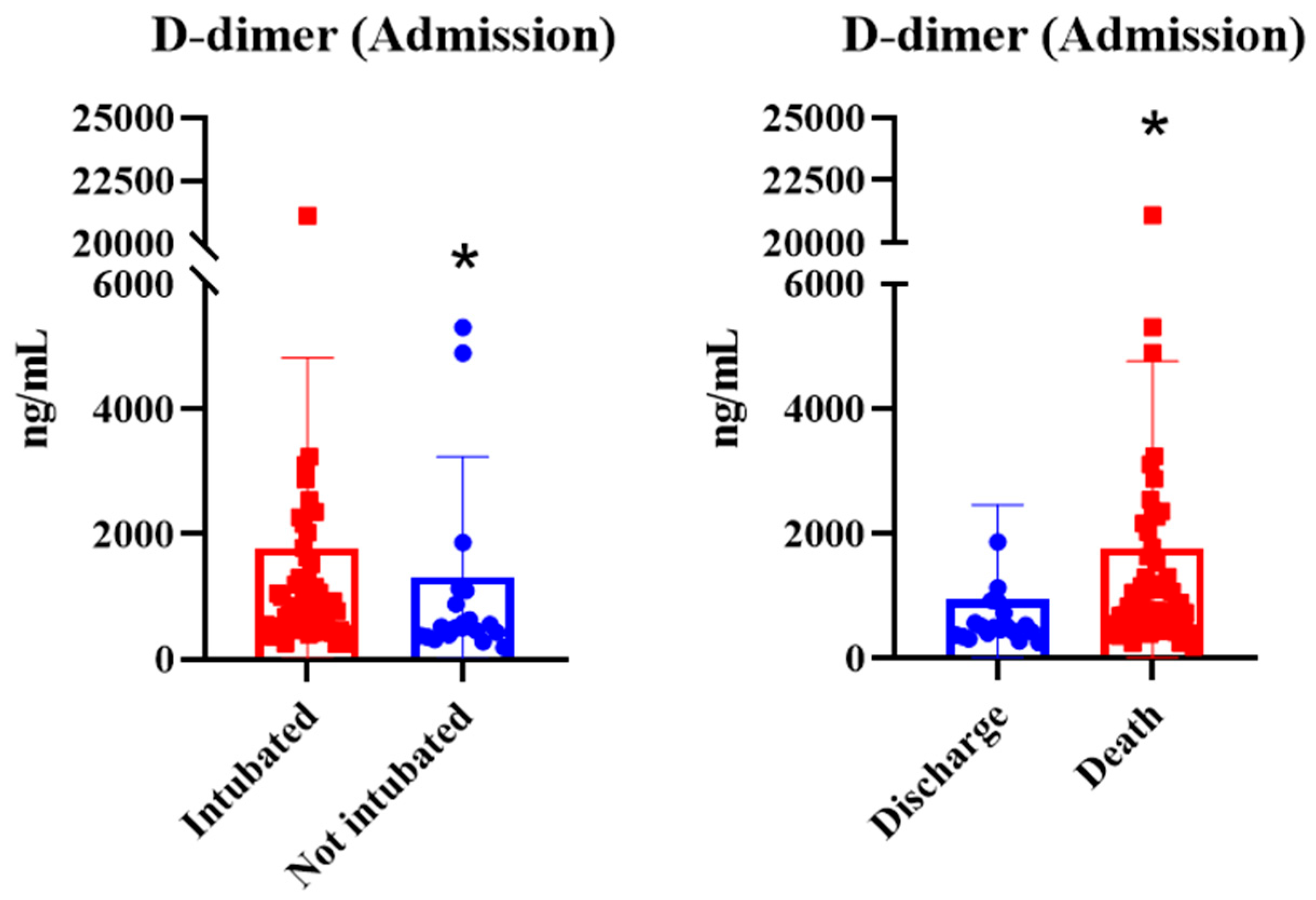
D-dimer concentrations at hospital admission were significantly higher among patients who subsequently required endotracheal intubation and among those who died during hospitalization (p = 0.0471 for intubation and p = 0.0057 for mortality), as shown in Figure 1.
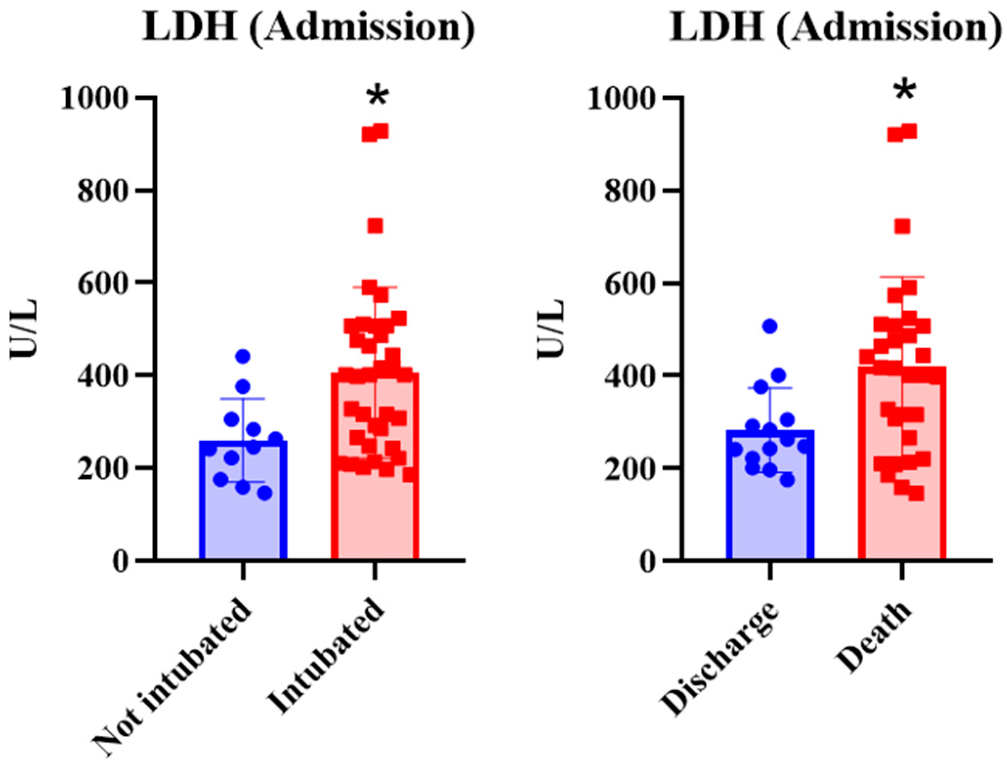
Similarly to D-dimer, LDH concentrations at hospital admission were higher in patients who were intubated (p = 0.0118), as well as in those in the death group (p = 0.0060), as shown in Figure 2.
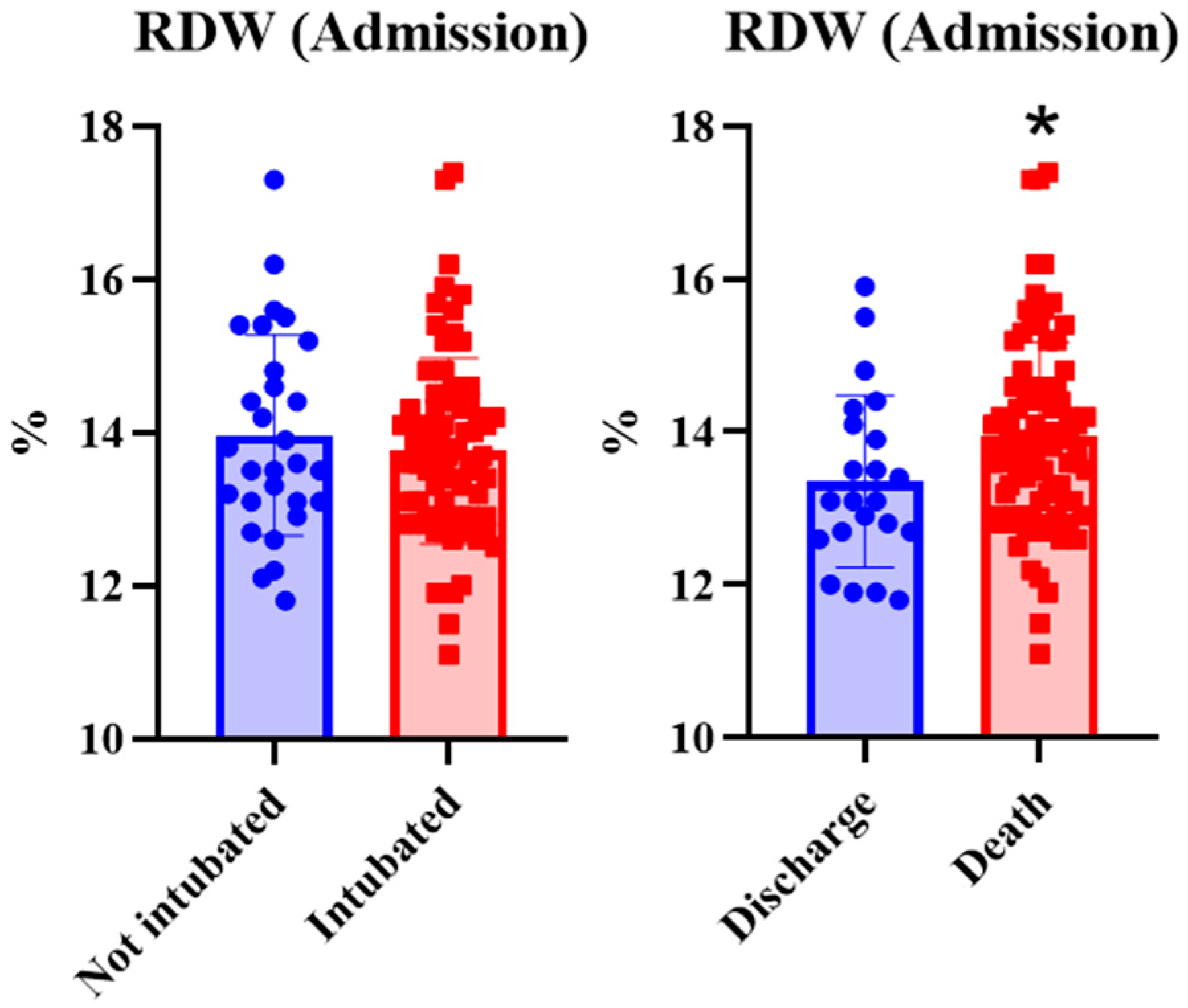
Considering the red cell distribution width (RDW), we identified that values at hospital admission were higher in the death group (p = 0.0417). However, this biomarker did not differ between intubation groups (p = 0.5172) (Figure 3).
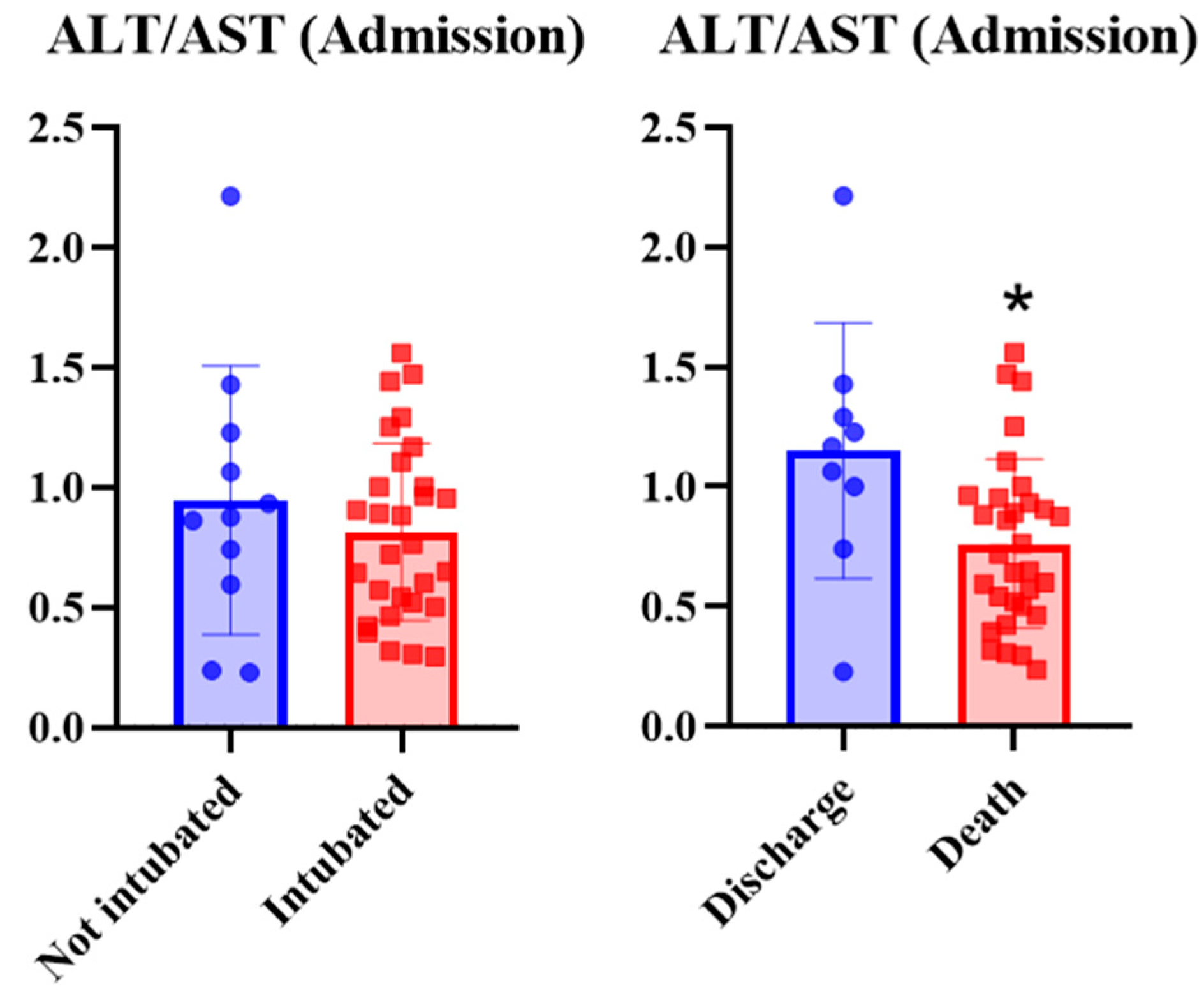
Likewise, the ALT/AST (Figure 4) ratio was significantly lower in patients who died during hospitalization (p = 0.0138). It is noteworthy that, as shown in Table 2, isolated AST and ALT values were not relevant; however, their ratio predicted death.
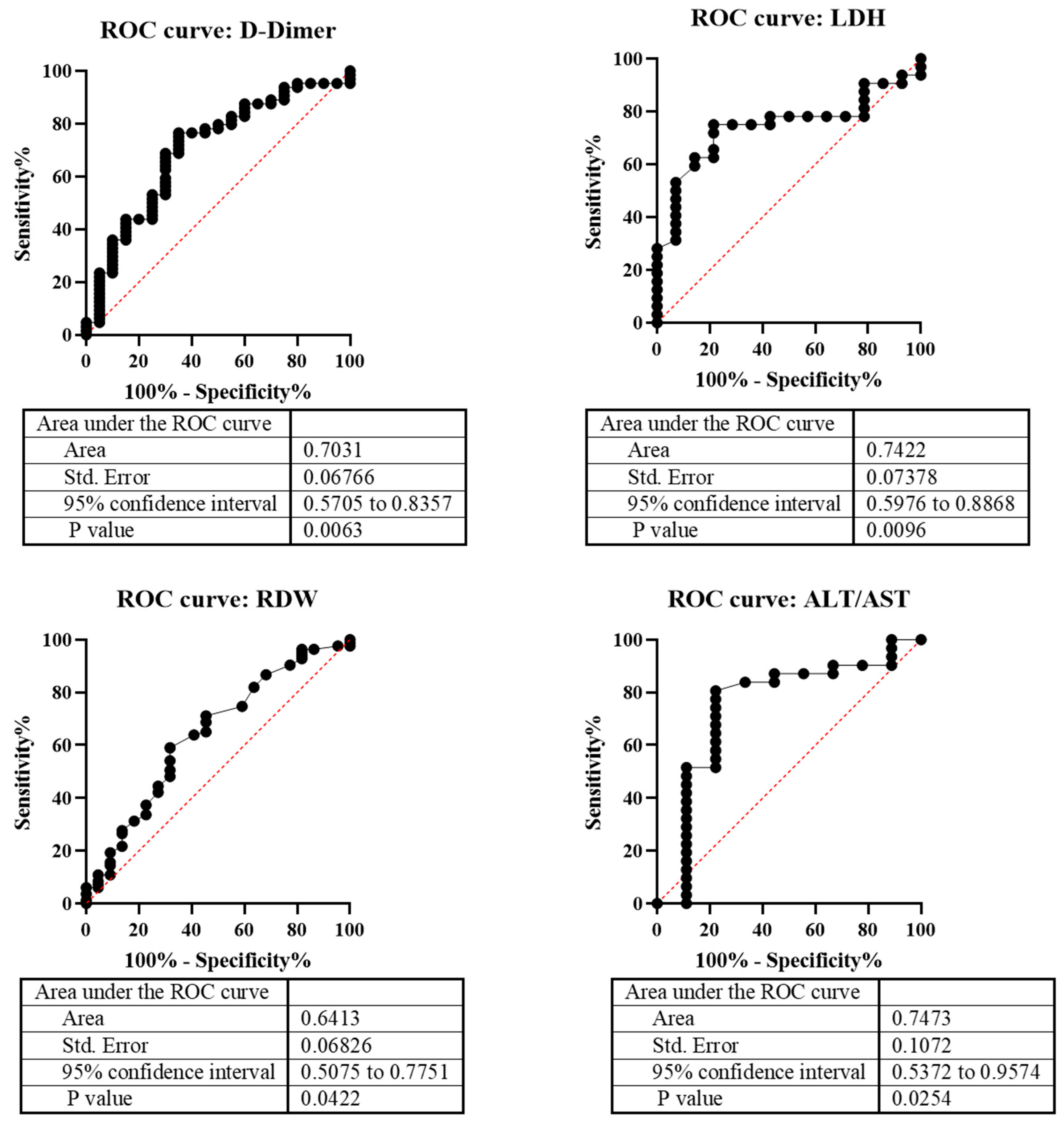
Biomarkers that were significantly associated with in-hospital mortality were subsequently evaluated using receiver operating characteristic (ROC) curve analysis to assess their discriminatory performance and determine optimal cutoff values (Figure 5). Overall, the biomarkers demonstrated moderate discriminative ability for predicting mortality.
For D-dimer, the optimal cutoff value was >572.9 ng/mL, yielding an area under the ROC curve (AUC) of 0.7031 (95% CI: 0.5705–0.8357), with 68.75% sensitivity and 70.00% specificity (p = 0.0063). LDH showed an optimal cutoff of >306.5 U/L, with an AUC of 0.7422 (95% CI: 0.5976–0.8868), 75.00% sensitivity, and 78.57% specificity (p = 0.0096). For RDW, the optimal threshold was >13.15%, corresponding to an AUC of 0.6413 (95% CI: 0.5075–0.7751), with 71.88% sensitivity and 54.55% specificity (p = 0.0422). The ALT/AST ratio (reported as TGP/TGO) demonstrated the best cutoff at <0.9815, with an AUC of 0.7473 (95% CI: 0.5372–0.9574), achieving 80.65% sensitivity and 77.78% specificity (p = 0.0254).

4. Discussion

The present study demonstrated that age, D-dimer, LDH, RDW, and the ALT/AST ratio were associated with adverse clinical outcomes among critically ill patients with COVID-19. Overall, these findings reinforce the concept that biomarkers reflecting thromboinflammation, tissue injury, hematological alterations, and organ dysfunction may contribute to early risk stratification when interpreted together with the patient’s clinical condition. Although the discriminatory performance of the evaluated biomarkers was moderate, their widespread availability, low cost, and routine use in hospital practice make them attractive complementary tools for identifying patients at increased risk of unfavorable outcomes [11,12]. Studies indicate that D-dimer levels above 1000 ng/mL are associated with a significantly higher mortality rate, reinforcing the importance of this biomarker in risk stratification [12]. In the present study, D-dimer levels >572.9 ng/mL at hospital admission were associated with an increased likelihood of subsequent respiratory deterioration requiring endotracheal intubation. However, elevated D-dimer levels should be interpreted as markers of overall disease severity and systemic inflammatory and thrombotic burden, rather than as independent predictors of the need for invasive mechanical ventilation.
D-dimer, a fibrin degradation product, is one of the most extensively studied prognostic biomarkers in COVID-19. Elevated D-dimer concentrations have consistently been associated with thromboembolic events, exaggerated inflammatory responses, disease severity, and increased mortality. Zhou et al. (2020) [11] demonstrated that patients with severe COVID-19 exhibited significantly higher D-dimer levels than those with non-severe disease, highlighting its value for the early identification of thromboembolic complications. Likewise, Thachil et al. (2020) [13] reported that elevated D-dimer concentrations are consistently associated with poor clinical outcomes and may serve as a reliable indicator of disease severity. Jee (2020) [14] further demonstrated that increased D-dimer levels are associated with a greater likelihood of adverse outcomes, including the need for mechanical ventilation and ICU admission.
Our findings are consistent with these previous reports. In the present study, D-dimer concentrations above 572.9 ng/mL at hospital admission were associated with subsequent progression to endotracheal intubation, supporting its role as an early prognostic biomarker for clinical deterioration. Although Tang et al. (2020) [12] proposed a higher threshold (>1000 ng/mL) for predicting disease progression, differences in patient characteristics, disease severity, and study design may explain the lower cutoff identified in our cohort. Importantly, the moderate discriminatory performance observed in our ROC analysis suggests that D-dimer should not be interpreted as an isolated predictor but rather as a complementary biomarker integrated with clinical assessment and other laboratory findings.
Beyond its association with coagulation abnormalities, elevated D-dimer may also reflect the intense inflammatory response observed in severe COVID-19. Battaglini et al. (2022) [15] suggested that D-dimer may represent not only a marker of thrombotic complications but also an indirect indicator of cytokine storm and endothelial dysfunction, reinforcing its biological relevance during disease progression. Collectively, these findings support the incorporation of D-dimer into the initial laboratory assessment of hospitalized patients, particularly for early risk stratification and identification of individuals at increased risk of respiratory deterioration.
Similarly, LDH has been consistently recognized as a marker of tissue injury and systemic inflammation in patients with COVID-19. Elevated LDH concentrations reflect cellular damage, hypoxia, and pulmonary injury, all of which are characteristic features of severe disease [16,17,18]. We identified that elevated LDH levels at hospital admission were associated with subsequent clinical deterioration, including progression to respiratory failure requiring endotracheal intubation and an increased risk of death during ICU stay. Nevertheless, this association likely reflects the greater severity of pulmonary and systemic tissue injury in critically ill patients, and LDH should be interpreted as a complementary prognostic biomarker rather than as an independent determinant of the need for invasive mechanical ventilation.
Our results are in agreement with previous studies. Aloisio et al. (2020) [16] demonstrated that patients with LDH concentrations above 250 U/L had significantly higher mortality rates, emphasizing the usefulness of this biomarker for identifying individuals at increased risk of unfavorable outcomes. Likewise, the meta-analysis conducted by Li et al. (2020) [18] showed that LDH levels were consistently higher among patients with severe COVID-19 than among those with milder disease, suggesting that LDH may serve as an early marker of clinical deterioration. Similar observations were reported by Jee (2020) [14], who found that elevated LDH concentrations were associated with worse clinical outcomes, including mechanical ventilation and mortality.
Although the ROC analysis demonstrated only moderate discriminatory performance, the observed prognostic value of LDH is biologically plausible. As discussed by Lopes-Pacheco et al. (2021) [17], LDH reflects not only systemic inflammation but also the extent of tissue damage, particularly pulmonary injury, which plays a central role in the pathophysiology of severe COVID-19. Battaglini et al. (2022) [15] further emphasized that LDH may contribute to the assessment of systemic inflammatory responses and cytokine storm, supporting its use as a complementary biomarker for early risk stratification. Therefore, rather than serving as a standalone predictor, LDH should be interpreted together with clinical findings and other laboratory parameters to improve prognostic assessment at hospital admission.
Liver involvement has been consistently described in patients with COVID-19 and is thought to result from multiple mechanisms, including direct viral cytopathic effects, immune-mediated injury, systemic inflammation, hypoxia, and drug-induced hepatotoxicity [19]. Because hepatocytes and cholangiocytes express angiotensin-converting enzyme 2 (ACE2), SARS-CoV-2 infection may directly contribute to hepatic dysfunction [20]. Nevertheless, abnormalities in liver enzymes should be interpreted cautiously, particularly during hospitalization, when antiviral agents, corticosteroids, immunomodulatory therapies, and other medications may influence biochemical parameters. In the present study, however, the laboratory biomarkers included in the prognostic analyses were obtained at hospital admission, before the evaluated clinical outcomes occurred, thereby minimizing the potential influence of therapies initiated during hospitalization.
Among the hepatic biomarkers evaluated, isolated AST and ALT concentrations were not associated with mortality or the need for endotracheal intubation. In contrast, the ALT/AST ratio was significantly associated with mortality, suggesting that the relationship between these enzymes may provide more clinically relevant information than isolated transaminase values. Similar observations have been reported by Zinellu et al. (2020) [19], who demonstrated that the De Ritis ratio (AST/ALT) was significantly higher among non-survivors with COVID-19. Although our study employed the inverse calculation (ALT/AST), both investigations indicate that the relative balance between AST and ALT, rather than their absolute concentrations, better reflects disease severity and adverse prognosis [20].
These findings are further supported by Pranata et al. (2021) [21] and Drácz et al. (2022) [22], who reported that alterations in the AST/ALT ratio are associated with severe clinical outcomes and increased mortality in COVID-19. The biological rationale underlying these observations probably extends beyond liver injury alone, reflecting systemic inflammation, hypoxic tissue damage, and multisystem involvement frequently observed in critically ill patients [20]. Therefore, the ALT/AST ratio may represent a simple and widely available complementary biomarker for mortality risk stratification, although its clinical interpretation should always be integrated with the patient’s overall clinical condition.
RDW was also associated with in-hospital mortality in our cohort, although no significant association was observed with the subsequent need for endotracheal intubation. RDW has emerged as a readily available hematological biomarker reflecting anisocytosis, ineffective erythropoiesis, systemic inflammation, oxidative stress, and impaired tissue oxygenation [23]. Increasing evidence suggests that elevated RDW is associated with adverse outcomes across a wide spectrum of acute and chronic diseases, including COVID-19. Foy et al. (2020) [24] demonstrated that increased RDW at hospital admission was independently associated with mortality among hospitalized patients with COVID-19, while Lee et al. (2021) [25] further reinforced its value as an indicator of disease severity and systemic inflammatory response.
The findings of the present study are consistent with these observations, suggesting that RDW may identify patients presenting a more pronounced inflammatory state and greater physiological impairment at hospital admission. However, unlike D-dimer and LDH, RDW was not associated with the need for endotracheal intubation, indicating that its prognostic value in our cohort was restricted to mortality. This distinction reinforces the importance of interpreting each biomarker according to the specific clinical outcome evaluated rather than assuming a uniform prognostic role across different endpoints. Moreover, the moderate discriminatory performance observed in the ROC analysis indicates that RDW should be interpreted as a complementary biomarker integrated with other laboratory findings and the overall clinical assessment. Previous studies have similarly demonstrated that elevated RDW reflects chronic inflammation, dysregulated hematopoiesis, and progressive organ dysfunction, supporting its incorporation into early clinical risk stratification in hospitalized patients with COVID-19 [13,18,21,24,25].
Age remained one of the strongest clinical factors associated with mortality in the present study, reinforcing findings consistently reported since the beginning of the COVID-19 pandemic. Older individuals exhibit greater susceptibility to severe disease due to immunosenescence, chronic low-grade inflammation, endothelial dysfunction, and the higher prevalence of chronic comorbidities, all of which contribute to impaired physiological reserve and reduced ability to respond to acute systemic infections [16,26,27,28]. In agreement with previous epidemiological studies, patients who died in our cohort were substantially older than survivors.
Our findings are consistent with those reported by Zhou et al. (2020) [11], Msemburi et al. (2023) [26], Wiemken et al. (2023) [27], and Paterson et al. (2022) [28], all of whom identified advanced age as one of the principal determinants of adverse outcomes in COVID-19. Although hypertension, diabetes mellitus, and obesity were frequently observed in our cohort, these comorbidities were not independently associated with mortality or endotracheal intubation in the univariate analyses [16]. This observation does not diminish their established clinical relevance but suggests that, within our study population, age represented the most prominent demographic characteristic associated with poor prognosis.
The findings of the present study suggest that routinely available clinical and laboratory biomarkers obtained at hospital admission may support the early identification of critically ill patients with COVID-19 who are at increased risk of adverse outcomes. Because these biomarkers are inexpensive, widely available, and already incorporated into routine hospital practice, they may represent practical complementary tools for early risk stratification and clinical decision-making, particularly in settings with high patient demand or limited healthcare resources. Nevertheless, their moderate discriminatory performance indicates that they should not be used in isolation but rather interpreted in conjunction with clinical assessment and other diagnostic information to support individualized patient management. Future studies should further investigate and externally validate these biomarkers, as well as explore multimarker approaches capable of improving prognostic accuracy and optimizing therapeutic decision-making [15,17].
This study has several limitations that should be considered when interpreting its findings. First, its retrospective, single-center design may limit the generalizability of the results to other populations and healthcare settings. In addition, patients were included through convenience sampling based on the availability of eligible medical records, which may have introduced selection bias and reduced the representativeness of the study population. Furthermore, the marked imbalance between outcome groups and the absence of multivariable analyses preclude conclusions regarding the independent prognostic value of the evaluated biomarkers. Complete and standardized information regarding treatments administered during hospitalization, including corticosteroids, antiviral agents, and immunomodulatory therapies, was not consistently available, preventing adjustment for these potential confounders. Although these interventions may influence inflammatory biomarkers and liver enzyme levels, all clinical and laboratory variables included in the prognostic analyses were obtained at hospital admission, reflecting the patients’ baseline clinical status before the effects of in-hospital therapies became fully established. Finally, the study was conducted during the first waves of the COVID-19 pandemic (2020–2021), a period characterized by the absence of vaccination, evolving therapeutic protocols, and unprecedented demand on healthcare systems, factors that should be considered when extrapolating these findings to the current clinical scenario. Therefore, prospective, multicenter studies with larger cohorts, standardized treatment protocols, and adjusted multivariable analyses are warranted to externally validate these findings and further establish the clinical applicability of these readily available biomarkers.

5. Conclusions

The present study demonstrated that higher admission levels of D-dimer, LDH, RDW, the ALT/AST ratio and advanced age were associated with increased in-hospital mortality among critically ill patients with COVID-19. In addition, higher admission concentrations of D-dimer and LDH were associated with subsequent endotracheal intubation, suggesting that these biomarkers may reflect greater disease severity and an increased likelihood of respiratory deterioration.
Overall, these findings support the potential value of routinely available clinical and laboratory biomarkers obtained at hospital admission as complementary tools for early risk stratification. Given their low cost and broad availability, these biomarkers may assist clinical decision-making when interpreted alongside clinical assessment and respiratory evaluation. However, their moderate discriminatory performance and the retrospective nature of this study indicate that they should not be considered standalone prognostic markers. Prospective, multicenter studies incorporating multivariable analyses and standardized respiratory severity assessment are needed to externally validate these findings before broader clinical implementation.

Author Contributions

J.W.: conceptualization, formal analysis, investigation, methodology, and writing—original draft; P.B.G.F., V.A.d.O., M.S.L. and T.G.H.: investigation and methodology; L.M.S. and M.M.S.: formal analysis, writing—original draft; M.N.F.: investigation, methodology, project administration, supervision, writing—original draft. All authors have read and agreed to the published version of the manuscript.

Funding

This research received no external funding.

Institutional Review Board Statement

The study subsequently received approval from the Research Ethics Committee of Regional University of Northwestern Rio Grande do Sul State (UNIJUÍ), under approval number 5.439.229 (approved 30 May 2022), in accordance with the ethical principles outlined by Resolution 466/2012 of the National Health Council. Additionally, we ensured that all data reported and analyzed were handled confidentially and anonymized, safeguarding patient privacy.

Informed Consent Statement

The requirement for informed consent was waived, as the study involved only the retrospective review of medical records without direct patient contact. Brazilian national legislation considers the approval of the Research Ethics Committee (CEP) and the Institution as appropriate measures to ensure compliance with ethical research principles for studies exclusively involving the collection of secondary data from medical records, without direct patient identification and without any intervention or interaction with them. This procedure aligns with Resolution 466/2012 of the National Health Council, which regulates research involving human subjects in the country.

Data Availability Statement

The original contributions presented in the study are included in the article, and further inquiries can be directed to the corresponding author.

Acknowledgments

The authors would like to thank the colleagues from the Research Group in Physiology (UNIJUÍ) and the hospital where the study was conducted.

Conflicts of Interest

The authors declare no conflicts of interest.

References

  1. Richardson, S.; Hirsch, J.S.; Narasimhan, M.; Crawford, J.M.; McGinn, T.; Davidson, K.W.; the Northwell COVID-19 Research Consortium; Barnaby, D.P.; Becker, L.B.; Chelico, J.D.; et al. Presenting characteristics, comorbidities, and outcomes among 5700 patients hospitalized with COVID-19 in the New York city area. JAMA 2020, 323, 2052–2059. [Google Scholar] [CrossRef] [Scilit] [PubMed]
  2. Guan, W.J.; Ni, Z.Y.; Hu, Y.; Liang, W.H.; Ou, C.Q.; He, J.X.; Liu, L.; Shan, H.; Lei, C.L.; Hui, D.S.C.; et al. Clinical characteristics of coronavirus disease 2019 in China. N. Engl. J. Med. 2020, 382, 1708–1720. [Google Scholar] [CrossRef] [Scilit] [PubMed] [PubMed Central]
  3. Grasselli, G.; Zangrillo, A.; Zanella, A.; Antonelli, M.; Cabrini, L.; Castelli, A.; Cereda, D.; Coluccello, A.; Foti, G.; Fumagalli, R.; et al. Baseline characteristics and outcomes of 1591 patients infected with SARS-CoV-2 admitted to ICUs of the Lombardy region, Italy. JAMA 2020, 323, 1574–1581. [Google Scholar] [CrossRef] [Scilit] [PubMed]
  4. Mitra, A.R.; Fergusson, N.A.; Lloyd-Smith, E.; Wormsbecker, A.; Foster, D.; Karpov, A.; Crowe, S.; Haljan, G.; Chittock, D.R.; Kanji, H.D.; et al. Baseline characteristics and outcomes of patients with COVID-19 admitted to intensive care units in Vancouver, Canada: A case series. Can. Med. Assoc. J. 2020, 192, 694–701. [Google Scholar] [CrossRef] [Scilit] [PubMed] [PubMed Central]
  5. Liu, Y.; Yang, Y.; Zhang, C.; Huang, F.; Wang, F.; Yuan, J.; Wang, Z.; Li, J.; Li, J.; Feng, C.; et al. Clinical and biochemical indexes from 2019-nCoV infected patients linked to viral loads and lung injury. Sci. China Life Sci. 2020, 63, 364–374. [Google Scholar] [CrossRef] [Scilit] [PubMed] [PubMed Central]
  6. Liu, H.; Hu, T.; Zhang, C.; Chen, X.; Zhang, S.; Li, M.; Jing, H.; Wang, C.; Hu, T.; Shi, J. Mechanisms of COVID-19 thrombosis in an inflammatory environment and new anticoagulant targets. Am. J. Transl. Res. 2021, 13, 3925–3941. [Google Scholar] [CrossRef] [Scilit] [PubMed] [PubMed Central]
  7. Okechukwu, C.E. Potential Biomarkers for Defining the Severity, Progression, and Survival from COVID-19. Int. J. Prev. Med. 2021, 12, 85. [Google Scholar] [CrossRef] [Scilit] [PubMed] [PubMed Central]
  8. Herold, T.; Jurinovic, V.; Arnreich, C.; Lipworth, B.J.; Hellmuth, J.C.; von Bergwelt-Baildon, M.; Klein, M.; Weinberger, T. Elevated levels of IL-6 and CRP predict the need for mechanical ventilation in COVID-19. J. Allergy Clin. Immunol. 2020, 146, 128–136. [Google Scholar] [CrossRef] [Scilit] [PubMed] [PubMed Central]
  9. Zhou, C.; Huang, Z.; Tan, W.; Li, X.; Yin, W.; Xiao, Y.; Tao, Z.; Geng, S.; Hu, Y. Predictive factors of severe coronavirus disease 2019 in previously healthy young adults: A single-center, retrospective study. Respir. Res. 2020, 21, 157. [Google Scholar] [CrossRef] [Scilit] [PubMed] [PubMed Central]
  10. Brazilian National Health Council. Resolution No. 466 of 12 December 2012: Guidelines and Regulatory Standards for Research Involving Human Beings. 2012. Available online: https://www.gov.br/conselho-nacional-de-saude/pt-br/atos-normativos/resolucoes/2012/resolucao-no-466.pdf (accessed on 19 July 2026).
  11. Zhou, F.; Yu, T.; Du, R.; Fan, G.; Liu, Y.; Liu, Z.; Xiang, J.; Wang, Y.; Song, B.; Gu, X.; et al. Clinical course and risk factors for mortality of adult inpatients with COVID-19 in Wuhan, China: A retrospective cohort study. Lancet 2020, 395, 1054–1062. [Google Scholar] [CrossRef] [Scilit] [PubMed]
  12. Tang, N.; Li, D.; Wang, X.; Sun, Z. Abnormal coagulation parameters are associated with poor prognosis in patients with novel coronavirus pneumonia. J. Thromb. Haemost. 2020, 18, 844–847. [Google Scholar] [CrossRef] [Scilit] [PubMed] [PubMed Central]
  13. Thachil, J.; Tang, N.; Gando, S.; Falanga, A.; Cattaneo, M.; Levi, M.; Clark, C.; Iba, T. ISTH interim guidance on recognition and management of coagulopathy in COVID-19. J. Thromb. Haemost. 2020, 18, 1023–1026. [Google Scholar] [CrossRef] [Scilit] [PubMed] [PubMed Central]
  14. Jee, Y. WHO International Health Regulations Emergency Committee for the COVID-19 outbreak. Epidemiol. Health 2020, 42, e2020013. [Google Scholar] [CrossRef] [Scilit] [PubMed] [PubMed Central]
  15. Battaglini, D.; Lopes-Pacheco, M.; Castro-Faria-Neto, H.C.; Pelosi, P.; Rocco, P.R.M. Laboratory biomarkers for diagnosis and prognosis in COVID-19. Front. Immunol. 2022, 13, 857573. [Google Scholar] [CrossRef] [Scilit] [PubMed] [PubMed Central]
  16. Aloisio, E.; Chibireva, M.; Serafini, L.; Pasqualetti, S.; Falvella, F.S.; Dolci, A.; Panteghini, M. A comprehensive appraisal of laboratory biochemistry tests as major predictors of COVID-19 severity. Arch. Pathol. Lab. Med. 2020, 144, 1457–1464. [Google Scholar] [CrossRef] [Scilit] [PubMed]
  17. Lopes-Pacheco, M.; Silva, P.L.; Cruz, F.F.; Battaglini, D.; Robba, C.; Pelosi, P.; Morales, M.M.; Caruso Neves, C.; Rocco, P.R.M. Pathogenesis of multiple organ injury in COVID-19 and potential therapeutic strategies. Front. Physiol. 2021, 12, 593223. [Google Scholar] [CrossRef] [Scilit] [PubMed] [PubMed Central]
  18. Li, C.; Ye, J.; Chen, Q.; Hu, W.; Wang, L.; Fan, Y.; Lu, Z.; Chen, J.; Chen, Z.; Chen, S.; et al. Elevated lactate dehydrogenase (LDH) level as an independent risk factor for the severity and mortality of COVID-19. Aging 2020, 12, 15670–15681. [Google Scholar] [CrossRef] [Scilit] [PubMed] [PubMed Central]
  19. Zinellu, A.; Arru, F.; De Vito, A.; Sassu, A.; Valdes, G.; Scano, V.; Zinellu, E.; Perra, R.; Madeddu, G.; Carru, C.; et al. The De Ritis ratio as prognostic biomarker of in-hospital mortality in COVID-19 patients. Eur. J. Clin. Investig. 2020, 51, e13427. [Google Scholar] [CrossRef] [Scilit] [PubMed] [PubMed Central]
  20. Ali, N. Relationship between COVID-19 infection and liver injury: A review of recent data. Front. Med. 2020, 7, 458. [Google Scholar] [CrossRef] [Scilit] [PubMed] [PubMed Central]
  21. Pranata, R.; Huang, I.; Lim, M.A.; Yonas, E.; Vania, R.; Lukito, A.A.; Nasution, S.A.; Siswanto, B.B.; Kuswardhani, R.A.T. Elevated de ritis ratio is associated with poor prognosis in COVID-19: A systematic review and meta-analysis. Front. Med. 2021, 8, 676581. [Google Scholar] [CrossRef] [Scilit] [PubMed] [PubMed Central]
  22. Drácz, B.; Czompa, D.; Müllner, K.; Hagymási, K.; Miheller, P.; Székely, H.; Papp, V.; Horváth, M.; Hritz, I.; Szijártó, A.; et al. The elevated de ritis ratio on admission is independently associated with mortality in COVID-19 patients. Viruses 2022, 14, 2360. [Google Scholar] [CrossRef] [Scilit] [PubMed] [PubMed Central]
  23. Wang, C.; Zhang, H.; Cao, X.; Deng, R.; Ye, Y.; Fu, Z.; Gou, L.; Shao, F.; Li, J.; Fu, W.; et al. Red cell distribution width (RDW): A prognostic indicator of severe COVID-19. Ann. Transl. Med. 2020, 8, 1230. [Google Scholar] [CrossRef] [Scilit] [PubMed] [PubMed Central]
  24. Foy, B.H.; Carlson, J.C.T.; Reinertsen, E.; Padros IValls, R.; Pallares Lopez, R.; Palanques-Tost, E.; Mow, C.; Westover, M.B.; Aguirre, A.D.; Higgins, J.M. Association of red blood cell distribution width with mortality risk in hospitalized adults with SARS-CoV-2 infection. JAMA 2020, 3, e2022058. [Google Scholar] [CrossRef] [Scilit] [PubMed] [PubMed Central]
  25. Lee, J.J.; Montazerin, S.M.; Jamil, A.; Jamil, U.; Marszalek, J.; Chuang, M.L.; Chi, G. Association between red blood cell distribution width and mortality and severity among patients with COVID-19: A systematic review and meta-analysis. J. Med. Virol. 2021, 93, 2513–2522. [Google Scholar] [CrossRef] [Scilit] [PubMed] [PubMed Central]
  26. Msemburi, W.; Karlinsky, A.; Knutson, V.; Aleshin-Guendel, S.; Chatterji, S.; Wakefield, J. The WHO estimates of excess mortality associated with the COVID-19 pandemic. Nature 2023, 613, 130–137. [Google Scholar] [CrossRef] [Scilit] [PubMed] [PubMed Central]
  27. Wiemken, T.L.; Khan, F.; Puzniak, L.; Yang, W.; Simmering, J.; Polgreen, P.; Nguyen, J.L.; Jodar, L.; McLaughlin, J.M. Seasonal trends in COVID-19 cases, hospitalizations, and mortality in the United States and Europe. Sci. Rep. 2023, 13, 3886. [Google Scholar] [CrossRef] [Scilit] [PubMed] [PubMed Central]
  28. Paterson, C.; Davis, D.; Roche, M.; Bissett, B.; Roberts, C.; Turner, M.; Baldock, E.; Mitchell, I. What are the long-term holistic health consequences of COVID-19 among survivors? An umbrella systematic review. J. Med. Virol. 2022, 94, 5653–5668. [Google Scholar] [CrossRef] [Scilit] [PubMed] [PubMed Central]
Figure 1. D-dimer at hospital admission, significantly associated with death and the need for intubation. Legend: Statistical analysis by the Mann–Whitney test; discharge group (n = 22) and death group (n = 85); significance level of 5%. Asterisks (*) indicate statistically significant differences between groups (p < 0.05).
Covid 06 00140 g001
Figure 2. LDH at hospital admission, significantly associated with death and the need for intubation. Legend: Statistical analysis by the Mann–Whitney test; discharge group (n = 22) and death group (n = 85); significance level of 5%. Asterisks (*) indicate statistically significant differences between groups (p < 0.05).
Covid 06 00140 g002
Figure 3. RDW at hospital admission was associated with mortality but not with endotracheal intubation. Legend: Statistical analysis by the Mann–Whitney test; discharge group (n = 22) and death group (n = 85); significance level of 5%. Asterisks (*) indicate statistically significant differences between groups (p < 0.05).
Covid 06 00140 g003
Figure 4. ALT/AST ratio at hospital admission was significantly associated with in-hospital mortality. Legend: Statistical analysis by the Mann–Whitney test; discharge group (n = 22) and death group (n = 85); significance level of 5%. Asterisks (*) indicate statistically significant differences between groups (p < 0.05).
Covid 06 00140 g004
Figure 5. ROC curves for D-dimer, LDH, RDW, and ALT/AST ratio. Legend: ROC curve analysis; discharge group (n = 22) and death group (n = 85); significance level of 5%. The red dashed line represents the reference value/cut-off value used in the analysis.
Covid 06 00140 g005
Table 1. Clinical parameters in outcome prognosis.
ParametersDischarge (n = 22)Death (n = 85)p-Value
SexMale—63.6% (14)
Female—33.4% (8)
Male—56.5% (48)
Female—43.5% (37)
0.632
Age (years)56.27 ± 16.9770.38 ± 15.110.0005
Number of comorbidities1.81 ± 1.501.72 ± 1.150.785
Type of comorbidityHypertension (63.33%); Type 2 diabetes (36.36%); Obesity (31.81%)Hypertension (51.76%); Type 2 diabetes (25.88%); Obesity (24.70%)
Total length of stay (days)27.86 ± 17.9919.31 ± 13.460.045
ICU length of stay (days)15.95 ± 13.9813.35 ± 9.900.696
Mechanical ventilation (days)11.32 ± 15.0315.32 ± 13.910.068
Legend: Fisher’s exact test was used for sex. For the other clinical parameters, the Mann–Whitney test was used, with a significance level of 5%.
Table 2. Laboratory parameters in outcome prognosis.
ParametersDischarge (n = 22)Death (n = 85)p-Value
Erythrocytes (106/mm3)4.23 ± 0.624.17 ± 0.690.748
Hemoglobin (g/dL)12.84 ± 1.8012.74 ± 1.980.732
Hematocrit (%)37.96 ± 5.3437.86 ± 5.690.758
MCV (fL)89.97 ± 5.3590.94 ± 5.570.465
MCH (pg)30.42 ± 1.6630.56 ± 1.960.751
MCHC (%)33.82 ± 0.9933.61 ± 0.990.41
Platelets (103/mm3)220.90 ± 101.70222.50 ± 119.600.668
Total Leukocyte (/mm3)10,030 ± 744710,223 ± 57570.671
Band Neutrophils (/mm3)691.70 ± 685.1760.50 ± 785.900.958
Segmented Neutrophils (/mm3)7519 ± 56847431 ± 46910.945
Eosinophils (/mm3)98.14 ± 164.3072.69 ± 102.100.465
Basophils (/mm3)0.00 ± 0.000.49 ± 2.750.600
Monocyte (/mm3)611.70 ± 610.00663.60 ± 580.400.553
Lymphocyte (/mm3)1073.00 ± 549.001298 ± 905.200.325
NLR8.78 ± 4.857.97 ± 6.580.126
MLR0.59 ± 0.320.54 ± 0.290.370
PLR0.23 ± 0.110.22 ± 0.150.358
NT/WBC0.80 ± 0.600.77 ± 0.100.364
LYN/WBC0.12 ± 0.060.14 ± 0.080.121
PT (s)11.23 ± 0.8515.21 ± 15.570.413
PT activity (%)91.63 ± 16.7581.72 ± 25.130.413
INR1.04 ± 0.081.39 ± 1.370.413
aPTT (s)24.70 ± 0.0032.89 ± 21.410.999
CRP (mg/dL)84.68 ± 70.53120.70 ± 139.200.180
Lactate (mg/dL)14.18 ± 5.4014.47 ± 8.370.599
Creatinine (mg/dL)1.83 ± 2.401.54 ± 3.070.763
Urea (mg/dL)61.75 ± 53.9662.34 ± 41.260.270
Sodium (mEq/L)139.30 ± 6.28137.3 ± 4.320.292
Potassium (mEq/L)4.21 ± 0.964.12 ± 0.690.746
GGT (U/L)90 ± 49.49174.00 ± 125.400.999
AST (U/L)29.56 ± 8.6373.53 ± 131.90.125
ALT (U/L)34.10 ± 17.7439.42 ± 48.360.616
pH7.42 ± 0.047.41 ± 0.090.737
PaCO2 (mmHg)32.36 ± 3.4534.72 ± 11.420.346
PaO2 (mmHg)91.29 ± 45.5281.89 ± 33.220.590
pHCO3 (mEq/L)21.16 ± 2.9827.41 ± 29.710.536
Legend: For erythrocyte count, the unpaired t-test was used. For the other laboratory parameters, the Mann–Whitney test was used, with a significance level of 5%.
Disclaimer/Publisher’s Note: The statements, opinions and data contained in all publications are solely those of the individual author(s) and contributor(s) and not of MDPI and/or the editor(s). MDPI and/or the editor(s) disclaim responsibility for any injury to people or property resulting from any ideas, methods, instructions or products referred to in the content.