Systematic Review

Association between SARS-CoV-2 Infection and Neuropsychiatric Manifestations

by
Aranza Llorente Vidrio
1,
Humberto Nicolini
1,
Carlos Tovilla Zarate
2,
Thelma Gonzales Castro
3,
Isela Juárez Rojop
4,
Jaime Martínez Magaña
1,
Nicolás Martínez López
5 and
Alma Delia Genis Mendoza
1,6,*
1
Laboratory of Genomics of Psychiatric and Neurodegenerative Diseases, National Institute of Genomic Medicine, Mexico City 14610, Mexico
2
Multidisciplinary Academic Division of Comalcalco, Juárez Autonomous University of Tabasco, Comalcalco 86658, Mexico
3
Multidisciplinary Academic Division of Jalpa de Méndez, Juárez Autonomous University of Tabasco, Jalpa de Méndez 86205, Mexico
4
Academic Division of Health Sciences, Juárez Autonomous University of Tabasco, Jalpa de Méndez 86205, Mexico
5
National Institute of Psychiatry “Ramón de la Fuente Muñiz”, México City 14370, Mexico
6
Children’s Psychiatric Hospital “Juan N. Navarro”, México City 14080, Mexico
*
Author to whom correspondence should be addressed.
COVID 2022, 2(9), 1270-1286; https://doi.org/10.3390/covid2090094
Submission received: 14 July 2022 / Revised: 26 August 2022 / Accepted: 29 August 2022 / Published: 9 September 2022
(This article belongs to the Special Issue How COVID-19 and Long COVID Changed Individuals and Communities)

Abstract

:
Coronaviruses are neurotropic viruses capable of entering the brain through various mechanisms and generating an important inflammatory response that is capable of triggering neuropsychiatric manifestations. Several reports describe the appearance of different conditions, such as sleep problems, anxiety and depression disorders, acute psychotic disorders, encephalitis, and delirium, among others, associated with COVID-19 infection. We performed a literature review in PubMed, Springer, Nature, MDPI, and other scientific journals on the relationship between COVID-19 infection with the development and aggravation of neuropsychiatric manifestations explained by molecular changes secondary to SARS-CoV-2 where it was found that there is a relationship between the virus and the development of these manifestations. Prospective neuropsychiatric follow-up of people exposed to SARS-CoV-2 at different points in their lives, as well as their neuroimmunological status, is necessary to fully understand the long-term impact of COVID-19 on mental health. It is required to identify the risk of developing neuropsychiatric problems due to COVID-19 infection to provide better medical care from a multidisciplinary team and improve the prognosis of these patients as well as the treatment of long-term sequelae.

1. Introduction

The severe acute respiratory syndrome coronavirus 2 (SARS-CoV-2) corresponds to a new variant of beta-coronavirus that is commonly known as Coronavirus disease 2019 (COVID-19). Being highly infectious, it quickly spread throughout the world. As of 13 July 2022, around 555,446,890 confirmed cases of COVID-19 are reported worldwide, with more than 6.3 million deaths. In México, around 6.2 million confirmed cases and 326,097 deaths have been reported [1].
The impact of COVID-19 on mental health is growing, and it is suggested that a significant number of people affected by COVID-19: The general population, but especially health workers, ethnic minorities living in conditions of overcrowding, people with pre-existing medical conditions including mental disorders; who as if that were not enough, also among these people there is a greater fear of getting vaccinated [2], and people exposed to the virus by the nature of their employment. In general, the most economically and medically disadvantaged are at higher risk of exposure to the virus and developing mental health sequelae secondary to COVID-19 [3,4]. Among health workers, it was found that the most frequently reported short-term specific mental health problems were; depression, generalized anxiety, insomnia, and post-traumatic stress disorder (PTSD) [5]. In contrast, the main medium and long-term mental health problems include the consequences of sustained anguish, such as burnout syndrome [5]. The increase in the probability of this will depend mainly on the severity of the disease, the administration of medications that may have a psychogenic effect, such as corticosteroids, and psychosocial variables [6,7].
Although most of the time, COVID-19 infection is related to respiratory symptoms, it has been observed that it can cause neuropsychiatric complications, and although these may be due to stressful, social, and psychological factors related to the impact of the pandemic, there is evidence that the virus itself can trigger the onset of neuropsychiatric syndromes [6]. Several studies have shown that coronavirus group (CoV), such as severe acute respiratory syndrome (SARS-CoV-1) and Middle East Respiratory Syndrome (MERS-CoV), together with COVID-19, can cause neuropsychiatric manifestations during acute illness in patients with and without a previous psychiatric history [7,8]. In 2009, it was shown that many SARS-CoV-1 survivors developed psychiatric morbidity that persisted at 4 years of follow-up, with 42.5% of such patients experiencing at least one psychiatric illness after SARS-CoV-1 infection. The main diagnoses included: 54.5% PTSD, 39% depression, 32.5% panic disorder, and 15.6% obsessive-compulsive disorder (OCD) [9]. Similar findings have been described in H1N1 influenza outbreaks in Europe, the Zika outbreak in Brazil, and the earlier Nipah infection in Singapore [10]. Influenza pandemics, in particular, have been characterized by an increased incidence of various neuropsychiatric symptoms [10]. Studies of past respiratory viral pandemics suggest that SARS-CoV-2 possibly also carries the same risk [6,11,12]. Various types of neuropsychiatric symptoms may arise in the setting of an acute viral infection or after variable periods of time after infection [10].
In 2021, Oxford University researchers analyzed a sample of 236,379 patients with a confirmed diagnosis of COVID-19 and found that 33.62% of patients had a psychiatric or neurological diagnosis at the 6-month follow-up period. People were diagnosed with neuropsychiatric disorders ranging from anxiety to psychotic episodes [13]. The currently available literature on the mental health impact of COVID-19 continues to emerge in first-world countries, and information from low-income and developing countries are beginning to appear, but at a considerably slower rate than in developing countries [14]. To date, the evidence on the psychological impact of the COVID-19 pandemic continues to be scarce in developing countries [13].
Immunity is known to play an important role in the genesis and severity of COVID-19 infection [3]. Supporting evidence includes the inverse relationship between neutrophil and lymphocyte counts, elevated C-reactive protein (CRP) levels, erythrocyte sedimentation rate (ESR), and severity of infection [15]. The notion that stress-induced inflammation can induce affective symptomatology was first highlighted by the finding of the use of INF-α to treat infectious diseases and cancer-induced depressive symptoms [16]. The neuroinvasive and proinflammatory properties of the virus have been related to the appearance of de novo neuropsychiatric disorders, as well as other systemic complications and increased mortality secondary to more severe stages of the disease [3,12].
The objective of this review is to explain the relationship between COVID-19 infection and the development of neuropsychiatric disorders through biomolecular changes secondary to a neuroinflammatory process; among them, we highlight: sleep disorders, anxiety and depression, delirium, anosmia and ageusia, post-traumatic stress disorder, psychosis, and encephalitis in patients with and without predisposing risk factors.

2. Materials and Methods

The search of references for this review was identified through searches of PubMed, Medscape, Nature, Academic Google, SpringerLink, The Lancet, among others, of published articles, combining the terms “COVID-19” and “psychiatric disorder“ with the terms “coronaviruses”, “psychosis”, “mental health”, “sequel”, “inflammation”, “cytokine storm” and “neurocovid”. Articles resulting from these searches and relevant references cited were reviewed on those items. Articles with novel or recent content and observational studies that reported the prevalence of sequelae of COVID-19 were included.

3. COVID-19 and the Cascade of Cytokines

These recent decades of coronavirus infections share similar immunological features and pathological impact, which can be classified into four main aspects: viral replication in innate immune cells, dysregulated immune response, cytokine storm, and antibody-mediated response [17].
The SARS-CoV-2 infection causes a varied clinical picture of infection that ranges from its asymptomatic course to critical illness that can consist of SARS and multi-organ failure [18]. Although observational studies reported older age and the presence of comorbidities as risk factors for increased disease severity, it quickly became clear that severe disease can also occur in younger patients without underlying medical conditions. Higher levels of inflammatory markers in the blood (including CRP, ferritin, and D-dimer), a higher ratio of neutrophils to lymphocytes, and elevated serum levels of several cytokines, as well as proinflammatory chemokines, have been associated with the severity of the disease illness and death from COVID-19 [18]. This reminds us of the critical role of an effective host immune response and the devastating effect of immune dysregulation in SARS-CoV-2 infection [19]. As seen since the 1918 influenza pandemic, a cytokine storm secondary to hyperinflammation has been suggested to be responsible for this deleterious course of the disease, leading to terminal outcomes in both the young and elderly, with and without pre-existing comorbidities [6].

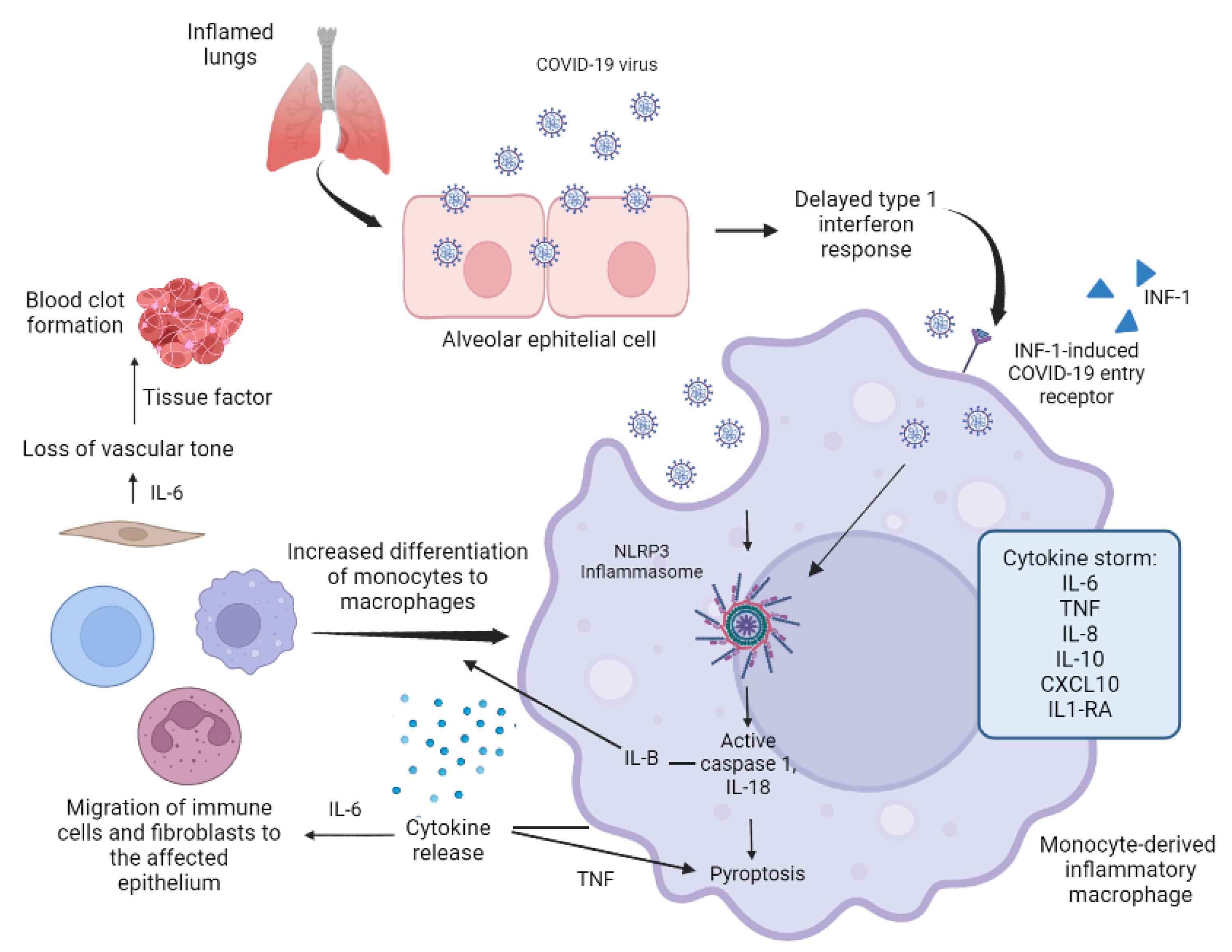
3.1. Pathogenesis of Cytokine Storm

The cytokine storm or cytokine release syndrome (CRS) is a life-threatening systemic inflammatory syndrome involving elevated levels of circulating cytokines and hyperactivation of immune cells. A cytokine profile resembling CRS is also associated with COVID-19 [18]. Regarding SARS-CoV-2, the cytokines that are important in hyperinflammation include: interleukin n (IL)-6, IL-8, IL-9, IL-12, IL-17, IL-18, IL-33, IL1-β, MCP-1, GCS-F, IP 10, CCL1–3, IFN-γ, TNF-α, among others [18,19,20]. Of all these, IL-6 and TNF-α are considered the main proinflammatory cytokines in the pathogenesis of CRS due to COVID-19. IL-6 is an acute-phase interleukin capable of promoting macrophage activation syndrome (MAS), which consists of the massive and poorly controlled differentiation of monocytes into macrophages, triggering the massive production of proinflammatory cytokines and inducing the migration of neutrophils and fibroblasts towards the affected epithelium [20]. It also causes deleterious induced endothelial activation and infiltration of neutrophils, resulting in nitric oxide (NO)-mediated changes in vascular permeability and loss of vascular tone [19]. Clinically it is reflected by the increased ratio of neutrophils to T lymphocytes (LT) and the development of septic shock [21]. In the same way, TNF-α has a similar role with proinflammatory actions in COVID-19. Increased serum levels of TNF-α are associated with greater severity of CRS, [18] in addition to inducing lymphocyte apoptosis, producing lymphopenia; a commonly reported clinical finding of severe COVID-19 [13,20,22].
Another important contributor to CRS is delayed or insufficient secretion of type 1 interferon (IFN-1) early in the immune response, as this has been associated with increased MAS and worsening of the cytokine storm [13,23]. It is possible that IFN-1 induces the expression of SARS-CoV-2 entry receptors, allowing the virus to access the cytoplasm of macrophages and stimulate inflammasomes; a group of multimeric protein complexes that respond to pathogen-associated molecular patterns (PAMPs), damage-associated molecular patterns (DAMPs), and environmental toxins [17]. In COVID-19, the nucleotide-binding domain inflammasome, leucine-rich repeat (LRR), and pyrin domain-containing protein 3 (NLRP3) are activated in response to infection. NLRP3 is an intracellular sensor that is activated by cellular stress through PAMPs (bacterial, viral, and fungal infections) and DAMPs, resulting in the formation of the NLRP3 inflammasome with elevated levels of caspase-1, IL-1, and IL-18 and increased pyroptosis, an inflammatory form of cell death [13,24] IL-1β secretion can amplify the activation of monocyte-derived macrophages in an autocrine or paracrine fashion, enhancing MAS and further contributing to the massive production of proinflammatory cytokines [13] (Figure 1).
Image created from: Merad, M., 2022. Figure 1: Possible pathways contributing to hyperactivation of monocyte-derived macrophages and hyperinflammation in COVID-19.|Nature Reviews Immunology. [online] Nature.com. Available at: https://www.nature.com/articles/s41577-020-0331-4/figures/1 (accessed on 15 April 2020).
On the other hand, the initiation of inflammation-induced coagulation is almost always mediated by the expression of tissue factor (TF). TF is expressed in response to proinflammatory cytokines (primarily IL-6) as well as on vascular endothelial cells, and its expression is known to lead to fibrin-based blood clot formation. Clinically, elevated D-dimer is observed, which predicts a poor prognosis of COVID-19 infection. This occurs initially within the microcirculation to prevent pathogenic material from moving into larger vessels. Unfortunately, this increases microvascular complications, including microthrombi and capillary hemorrhage [19]. Additionally, major natural anticoagulant pathways are almost always affected during inflammation, further contributing to the spread and formation of these clots. In the absence of vascular injury, the initiation of coagulation is entirely dependent on the recruitment of TF-expressing inflammatory monocytes by activated endothelial cells [13]. SARS-CoV-2 infection is known to enter a critical phase when multi-organ damage and circulatory collapse develop. In some cases, myocardial infarction in COVID-19 can be triggered by cytokine-induced microvascular dysfunction [18]. In addition to microvascular events, COVID-19 is associated with increased arterial thrombosis and an increased incidence of venous thromboembolic events, such as pulmonary embolism. It is known that the CNS can synthesize its own cytokines through type 1 astrocytes and microglia. A cytokine storm might trigger an acute hemorrhagic-necrotizing encephalopathy resulting from COVID-19, a rare complication that has also been observed in influenza and other viral infections and is postulated to be associated with intracranial cytokines storm [18]. Severe systemic inflammation coupled with cytokine storm is the main cause of tissue injury in COVID-19, which can lead to SARS, organ failure, and death [24].
Alterations in the expression of immune genes also play an important role in the increase in systemic inflammation. A study carried out in female rhesus macaques exposed to chronic psychosocial stress associated with social subordination shows that low social status causally alters the expression profiles of immune genes of NK cells, B lymphocytes, T helper lymphocytes, and T cytotoxic lymphocytes towards expression profiles denoting further lymphocyte disruption, increased innate immune responses, and increased cytokine responses. Following this context, a variety of chronic stressors have been found in humans, including low socioeconomic status, social isolation, or stress generated by the current pandemic [16].

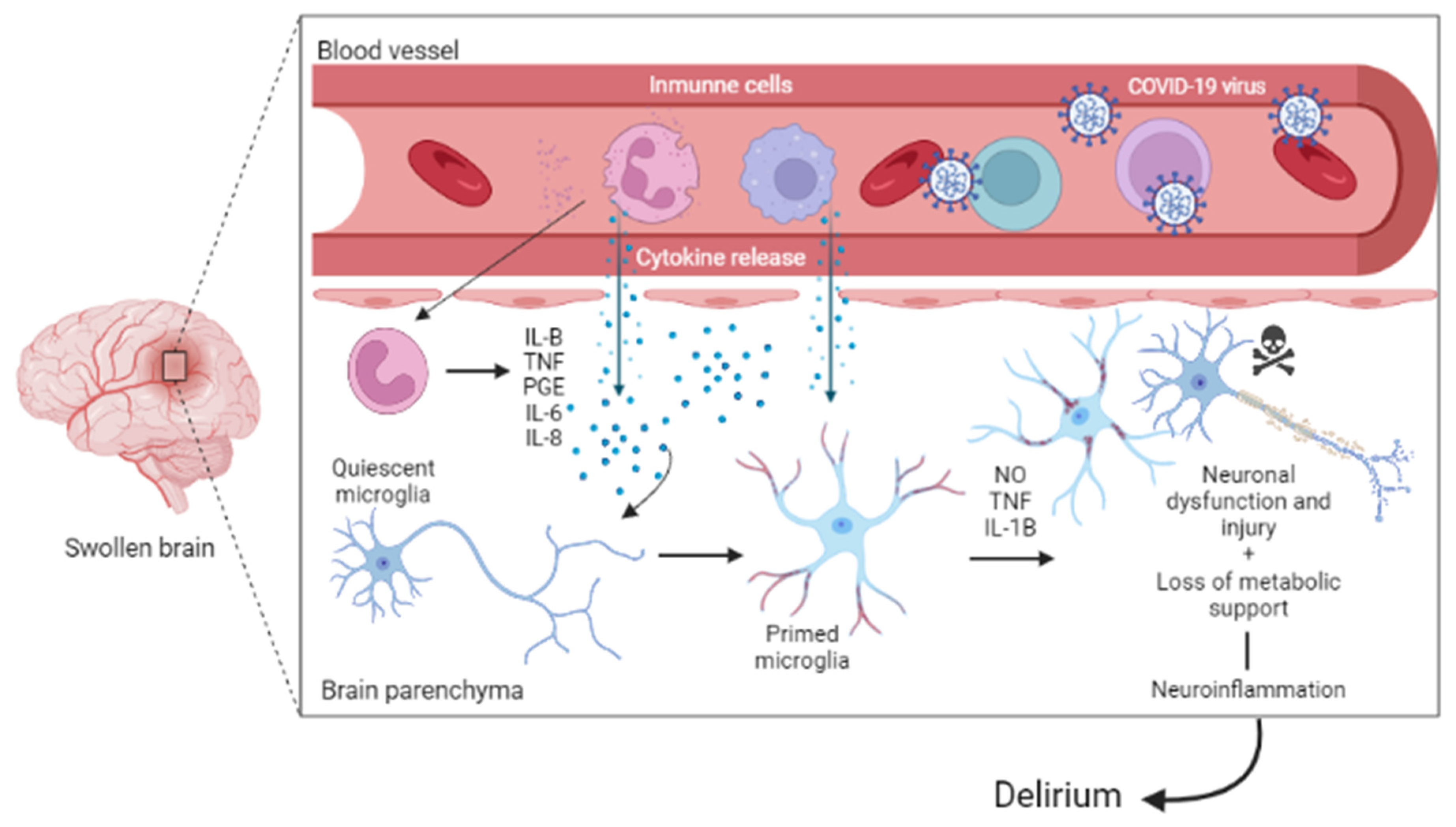
3.2. COVID-19 and the Central Nervous System

The new variant of the SARS-CoV-2 coronavirus belongs to the CoV group. This group is considered neurotropic, of which there are mainly two routes of invasion to the central nervous system (CNS): neuroinvasion through the bloodstream (hematogenous retrograde route) and neuroinvasion through peripheral nerves (neuronal route) [23].
In the hematogenous retrograde route, the virus can pass the blood-brain barrier (BBB) to the nervous tissue by a transendothelial mechanism. Systemic viral infection can lead to BBB breakdown induced by microglia activation; these innate immune cells release factors, such as: proinflammatory cytokines, eicosanoids/prostanoids, NO, and neurotropic factors, causing a proinflammatory environment [25]. More recent data indicate that stress-induced activation of NF-κB and TNF signaling pathways in endothelial cells of the nucleus accumbens locally reduces BBB integrity, leading to increased BBB permeability and compromise [16], a mechanism that allows the infiltration of inflammatory mediators and pathogens that reside in the circulation. Once in the brain, cytokines can influence behavior through their ability to alter the metabolism of neurotransmitters, including monoamines and glutamate. These effects of central cytokines on neurotransmitters are mediated by the effects on the synthesis, release, and reuptake of neurotransmitters, decreasing the levels of some of these, such as serotonin and dopamine, but can also generate an increase in oxidative stress secondary to excitotoxicity of some other neurotransmitters such as glutamate [16] further exacerbating inflammation in the CNS, causing the destruction of healthy tissue and, finally, deterioration of brain function if the acute inflammation does not cease [23,25].
The release of proinflammatory cytokines such as IL-6, IL-8, IL-10, IL-18, TNF-α, IL-1β, and GM-CSF; have been found in patients with the first episode of psychosis [3,12]. In one study, high levels of IL-6 were reported to have been detected in plasma, cerebrospinal fluid (CSF), and postmortem prefrontal cortex of subjects with suicidal ideation, nonfatal suicide attempts, and completed suicides. Circulating IL-6 levels have also been correlated with suicidal behaviors, personality disorders, aggressiveness, and impulsiveness [22,26]. This proves the role of cytokines in the regulation of emotions and behaviors through the interaction with specific areas of the brain and different neural pathways, demonstrating the impact of COVID-19 on the brain [26,27].
SARS-CoV-2 is believed to enter the CNS through this pathway where the structural protein S of the coronavirus binds to the angiotensin-converting enzyme 2 (ACE2) receptor, the cellular input receptor in both neurons and glia [3]. Although the general levels of this receptor within the CNS are low compared to other organs (vascular endothelium or the lungs) [23], its involvement in the pathogenesis of neuropsychiatric symptoms in patients with coronavirus has been proposed. Within the CNS, the general expression of ACE2 receptors is relatively high in dopaminergic, serotonergic nuclei, and the substantia nigra (basal ganglia), lateral ventricles, and glutamatergic neurons (cerebral cortex), which are areas implicated in schizophrenia [3]. One possible hypothesis is that these areas, due to their relatively high concentration of ACE2 receptors, are more vulnerable to the virus [3].
Neuroimaging studies have shown that the creation of this proinflammatory environment, as well as endogenous inflammation in patients with a history of depression, can alter the functional connectivity and activation of brain regions involved in the pathophysiology of stress-related psychopathology including the prefrontal cortex, striatum, dorsal anterior cingulate cortex, and amygdala. These studies indicate that inflammation decreases functional connectivity between the prefrontal cortex and the striatum in a way that predicts reward deficits, anhedonia, and psychomotor slowing [16].
One longitudinal imaging study investigated brain imaging changes in UK Biobank participants before being infected by SARS-CoV-2 and, on average, 38 months later after having tested positive for COVID-19. The study revealed a significant deleterious impact associated with SARS-CoV-2, such as longitudinal reduction in gray matter thickness and tissue contrast in the orbitofrontal cortex and the parahippocampal gyrus, greater changes in markers of tissue damage in regions that are functionally connected to the primary olfactory cortex, and a greater reduction in global brain size in the SARS-CoV-2 cases among COVID-19 survivors [27].
Basal ganglia dysfunction and related disorders appear to be present as a result, as the basal ganglia and other structures are likely to be affected by aberrant hemorrhagic or primarily neuroinflammatory processes in the CNS [28].
Regarding neuroinvasion through peripheral nerves, the neuronal route consists of some viruses infecting and migrating through peripheral nerves as a second entry route to the CNS. In this process, neurons play an essential role since these cells innervate peripheral organs, and thus, viruses use them as a gateway to the CNS. Another possible route involves hematogenous spread to the CNS via infected leukocytes [3]. This mechanism is known as a “Trojan horse” [12].
In this sense, research carried out in mouse models shows that the OC43 strain (HCoV-OC43) of the coronavirus has a preferential tropism to infect neurons compared to other neural cells (oligodendrocytes, astrocytes, microglia). Mice inoculated with HCoV-OC43 developed signs of acute encephalitis secondary to neuronal apoptosis. These mice were found to have behavioral abnormalities with viral RNA persistence in the brain for several months. These findings suggest that respiratory viruses with neuroinvasive potential can induce neuronal cell death and consequent psychosis in vulnerable individuals [3,8].
Finally, other factors that are predisposed to neuroinfection by COVID-19 include patients with a psychiatric history, unfavorable psychosocial conditions (for example, stress, fear of the pandemic, insufficient social support, reduced physical activity, and affectations during isolation such as irritability, changes in emotional control, loss of expectations in the future, motivation and attention span) [2], and suffering from adverse drug reactions (for example, high doses of corticosteroids, hydroxychloroquine, ivermectin), as well as being a carrier of SARS-CoV-2 strains HKU1 and NL63 (as patient IgG levels for these coronavirus strains were significantly higher in patients with psychotic symptoms in compared to controls, suggesting that these two coronaviruses may be risk factors for neuropsychiatric disease) [3,4].

4. Neuropsychiatric Manifestations Secondary to COVID-19 Infection

The neuroinflammation induced by SARS-CoV-2 infection and the alteration of neurotransmitters may correlate with the appearance of neuropsychiatric manifestations. Studies have shown that increased public restrictions, stressful hospital care, concern about infecting others, and stigma can have a significant impact on the mental health of survivors [29].
During the first wave of infection in China, a study retrospectively analyzed 214 patients with a molecular diagnosis of COVID-19 from three different hospitals. A total of 36.4% presented neuropsychiatric symptoms, which were differentiated into central, peripheral/musculoskeletal, and neurological symptoms [11]. The most frequent central symptoms were dizziness and headache. Dysgeusia, anosmia, and muscle pain were the main peripheral symptoms, and finally, anxiety, depression, and delirium were the most common psychiatric manifestations [7,11].
In a study carried out in México, a questionnaire was sought to identify how social isolation had impacted the mental health of 1011 participants. It was observed that 38% showed important affection for personal hygiene and eating habits. Depression and suicidal ideation were increased in 46% of participants, while 2% of people reported symptoms suggestive of PSTD [2]. Another study showed that the sector of the Mexican population most affected during the pandemic were young unemployed women who manifested symptoms of anxiety, depression, avoidance, and, in the event of the loss of a loved one, pathological grief [14]. Currently, evidence for acute neuropsychiatric symptoms in COVID-19 cases continues to emerge [10].

4.1. Sleep Disorders

In the face of the COVID-19 pandemic, sleep has become essential due to its multiple benefits for physical and mental health. Lack of sleep can alter the circadian system, one of the most profound ubiquitous biological mechanisms influencing sleep and mental health [30], and consequently affect psychological functioning and decision-making, increase accidents, cause mood swings and alter the immune response; committing in vivo antibody responses to novel antigens, thus increasing the risk of becoming infected by SARS-CoV-2 virus [29,31,32].
Sleep difficulties have been called “Coronasomnia” or “COVID-somnia” during the current pandemic. Coronasomnia is caused by a combination of factors: variations in the circadian cycle caused by changes in light exposure during confinement (altering normal routines in terms of reduced physical exercise, daylight exposure, and the increasing use of technological devices such as cell phones, laptops and watching TV during lockdown), chronic stress (both physical and emotional such as financial distress or have been in confinement [30]), the long-term effects of the immune response to SARS-CoV-2 infection and changes in sleep/wake cycles. All of these factors have negatively impacted sleep quality and quantity among COVID-19 patients [4,31]. COVID-19 patients were expected to have the highest frequency of sleep disturbances due to their main symptoms, such as cough, fever, and shortness of breath, which have been associated with insomnia or hypersomnia [32].
In the general population, studies have revealed a delay in bedtime and waking time, as well as a reduction in the duration and general quality of sleep during the night and an increase in daytime naps [4,33]. According to a recent systematic review and meta-analysis of 44 publications comprising a total of 54,231 people, the pooled global prevalence rate of various sleep difficulties among all groups was 35.7%. COVID-19 patients were found to be the most affected, with a combined incidence of 74.8%. Sleep disorders were also found to affect 36.0% of health professionals and 32.3% of the general population, respectively [32]. Insomnia, depression, and anxiety were common in nurses and doctors working in health care units, contributing to a high risk of developing poor mental health outcomes [4,34]. Even if people have recovered from the acute illness, sleep disturbances persist.
The International COVID Sleep Society (ICOSS) conducted a multinational survey using a harmonized questionnaire with a total of 22,151 participants to assess the possible changes in the frequency and presentation of various sleep problems in relation to COVID-19 and confinement. The survey incorporated multiple questions related to sleep problems. The data clearly show that all sleep and daytime problems increased relative to before the COVID-19 pandemic. In fact, the prevalence of most of the problems (poor sleep quality, sleep onset problems, sleep maintenance problems, fatigue, excessive sleepiness, and falling asleep during the daytime) increased by about more than 10%. More than 20% of participants reported a worsening in sleep quality [31]. Furthermore, the prevalence of nightmares increased significantly. This increase may be related to increased levels of stress, anxiety, depression, post-traumatic stress disorder, or even suicidal ideation during the lockdown caused by the pandemic. Furthermore, nightmares and acting out in sleep are associated with rapid eye movement (REM) sleep behavior disorder, which in turn is a known risk factor of alpha-synucleinopathies [31], a group of diseases that have in common the abnormal deposition of a-synuclein in the cytoplasm of neurons or glial cells as in Parkinson’s disease and Lewy Body Dementia.
Finally, another study by the Sleep Research Society (SRS) sought to examine whether the COVID-19 pandemic has had different effects on sleep and mental health based on individual circadian types (morning, intermediate, and evening-type). Evening types suffered more sleep problems, such as poor sleep quality, problems with sleep onset and maintenance, excessive sleepiness, daytime fatigue, nightmares, and more hypnotic use than other circadian types both before and amid the pandemic. The prevalence of having depressive or anxiety symptoms among defined night types was also higher than the prevalence of not being depressed or not having anxiety. Furthermore, evening types had more stress, repeated disturbing thoughts and memories, and feeling upset about the past than definite morning types [30]. These findings are in line with previous studies indicating a higher risk for sleep and mental health problems among evening types already in adolescence as well as in adulthood [35].

4.2. Anxiety and Depression

Depression and anxiety together are estimated to be responsible for at least 8% of years lived with a disability [2]. Psychological variants such as isolation, fear of the pandemic, insufficient social support, and reduced physical activity were associated with anxiety, while patients with pre-existing mood disorders reported increased distress and depression [4]. Concerns about one’s own health and the health of the family cause concern and increase the risk of depression, anxiety, panic disorder, trauma-related illnesses, and OCD [36].
A meta-analysis of 43 community studies on the prevalence of anxiety concluded that the prevalence of anxiety during the COVID-19 pandemic is three times higher compared to the pre-pandemic world. Similarly, a meta-analysis of 29 studies reveals that anxiety and depressive symptomatology among children and adolescents is twice as high as in pre-pandemic periods [4,37].

4.3. Acute Psychotic Disorder and Manic Disorder

An acute psychotic disorder is characterized by the appearance of hallucinations and delusions lasting at least a day but less than a month, with an eventual return to normal pre-illness functionality. More complex psychotic disorders, such as schizophrenia, are characterized by abnormally high mesolimbic dopamine (DA) signaling in the brain that mediates positive psychotic symptoms; hallucinations, delusions, disorganized behavior, and thoughts, while decreased DA signaling mesocortical is associated with negative psychotic symptoms; abulia, alogia, anhedonia, and social withdrawal. Such altered dopaminergic signaling could be mediated by inflammation due to the negative effects of cytokines on DA synthesis, packaging, release, and reuptake [25].
So far, several cases have been reported that mention the appearance of acute psychotic disorders in cases of COVID-19. Some have been first-onset, while other patients with previous diagnoses of schizophrenia and a history of suicidal behavior have seen exacerbations after SARS-CoV-2 infection, even while taking medication [11,12,38,39].
Most of these cases report a broad spectrum of immune system dysregulation [40], with elevated proinflammatory cytokines such as IL-6, TNF-α, and IL-1β, leukocytosis at the expense of neutrophilia, lymphopenia, elevated CRP, D-dimer, and ferritin. Although not all of these values were measured in all cases, this similarity is shared in most.
In order to conceptualize the risk of the onset of psychosis in subjects infected with SARS-CoV-2, it should be emphasized that high levels of IL-6 correlate with reduced hippocampal size in schizophrenic subjects, which explains, at least partially, its cognitive deficit. Furthermore, elevated levels of IL-6 were detected in the CSF of schizophrenic subjects. Even more intriguing is the observation that high levels of IL-6 in adolescents are positively correlated with the onset of psychosis in adulthood [3,41]. Another study correlated elevated IL-6 with endophenotypic suicidal behaviors, such as personality trait disorders, aggressiveness, and impulsiveness [22,42].
In addition, among the presence of factors that predispose the appearance of psychotic outbreaks in patients with which COVID-19 has been associated, the following stand out: genetic vulnerability, abnormalities in neurotransmission, stressful situations (such as the pandemic), and exposure to factors environmental [26]. The available evidence suggests that schizophrenia is one of the psychiatric illnesses with the highest risk of morbidity and mortality due to the pandemic [2]. A study conducted in Denmark with 144,321 patients revealed that those diagnosed within the spectrum of schizophrenia had a higher risk of having more severe symptoms and dying within the first 30 days [2,43].
Finally, it is suggested that viral infection can trigger bipolar disorders. It has been reported to trigger an initial manic episode in which episodes of mania or hypomania may follow symptomatic COVID-19 infection, even in previously healthy subjects. Risk factors for the development of mania and hypomania include psychosocial stress, increased inflammatory biomarkers, and a history of a bipolar affective disorder [4,44].

4.4. Delirium and Confusional State

Delirium, a syndrome characterized by an acute change in attention, consciousness, and cognition, is caused by a medical condition that cannot be better explained by a pre-existing neurocognitive disorder [45]. Various studies have reported this syndrome in patients affected by SARS-CoV-2, especially in hospitalized older adults compared to younger patients. This clinical manifestation can be a central symptom in the presentation of severity, even in the absence of respiratory symptoms [24,46]. Currently, there is still insufficient evidence to definitively elucidate the pathophysiological role of SARS-CoV-2 in the development of delirium; however, it is believed that its development may be multifactorial. When the virus invades the CNS, delirium could manifest directly due to neuronal damage secondary to inflammation (Figure 2).
Image created from: Wilson, J., 2020. Figure 2: Inflammatory mechanisms in delirium.|Nature Reviews Disease Primers. [online] Nature.com. Available at: https://www.nature.com/articles/s41572-020-00223-4/figures/3 (accessed on 16 April 2022).
It was shown that hospitalized people who develop delirium have significantly higher levels of IL-6, TNF-α, IL-B, and IL-8, but not IL-1, IL-10. Patients with delirium have lower levels of insulin-like growth factor type I (IGF-I), and recovery from delirium is better in those with elevated IFN-γ levels [47] or indirectly due to hypoxia caused by alterations in the function of the medullary respiratory centers [46]. It is also believed that pre-existing cognitive deficits such as dementia, metabolic disorders, urinary retention, prolonged hospitalization, concomitant infections, fluid and electrolyte imbalance, and prolonged use of hydroxychloroquine may be associated contributing factors [12,19,48]. Some of the characteristics that were observed in patients with delirium and that may support their diagnosis were: alogia, abulia, rigidity, and increased inflammatory markers [12].

4.5. Anosmia and Ageusia

Dysfunction of the perception of smell and taste has been associated as one of the first symptoms and screening criteria for COVID-19 infection. It has been proposed that the CoV group, especially the alphacoronavirus, has an affinity for the ciliary columnar epithelium because the ACE2 receptor is also expressed in the olfactory lining [10,12,24].
In a recent study, 100% of the patients in the subacute stage of the disease were displaying signs of hypogeusia or ageusia, and 86% displayed signs of either hyposmia or anosmia [27].
The unique anatomical organization of the olfactory cells; neuronal or supporting cells concentrated in the olfactory epithelium, the olfactory bulb in the nasal cavity, and its contact with the midbrain effectively make it a channel between the nasal epithelium and the CNS [23]. These structures are particularly vulnerable to invasion by the CoV group, and this appears to be the case specifically with SARS-CoV-2. Such loss of olfactory sensory inputs to the brain could lead to loss of gray matter in brain regions related to smell, which could be a likely mechanism for anosmia or hyposmia in the early stages of COVID-19 infection [27]. While postviral olfactory syndrome, a known complication of viral infections (e.g., influenza virus and herpesvirus), may also be associated with COVID-19, as penetration of the sieve plate and involvement of the piriform cortex is common during infection [12]. Additionally, a recent study of patients who died of COVID-19 identified structural brain abnormalities on postmortem brain imaging, with 21% of subjects having asymmetric olfactory bulbs [49].

4.6. Post-Traumatic Stress Disorder

It is generally, though incorrectly, assumed that once the trauma passes and the subject is no longer under the pressure of stress, the path to steady recovery begins. Unfortunately, this is not always the case as, in susceptible individuals, active stress triggers certain brain processes whereby traumatic memories suddenly resurface and disrupt mental health [20]. One of the main therapeutic tasks with traumatized patients is the restoration of security and a sense of security [50] since if this is not the case, the persistence of these conditions generates PTSD [42].
Due to the marked impact of stressors on the immune system, it is not surprising that PTSD is associated with the immune response [42]. Increased concentrations of proinflammatory factors in both the systemic circulation and the brain have been observed in the setting of PTSD [51]. Neuroglial activation induced by intense or persistent stressors such as abuse, isolation, or trauma can stimulate the aberrant secretion of proinflammatory signals that could potentially facilitate the onset of PTSD. A marked increase in proinflammatory molecules has been confirmed in PTSD subjects, including IL-6, TNF-α, and IL-1β1 [41,51]. In addition to changes in cytokines, PTSD is also related to increased expression of NF-κB, a transcription factor involved in the process of inflammation where its elevated expression directly correlates with PTSD severity [41]. Furthermore, PTSD tends to be comorbid with both depression and anxiety, drug addiction, and a high frequency of suicide since all these conditions share common inflammatory mechanisms in their pathogenic processes [52]. However, it remains to be elucidated whether, in all cases, the relationship is mutual, the predisposing factors together with the inflammatory factors play a causal role in the appearance of PTSD.
PTSD may be a likely outcome for patients with COVID-19. This is due not only to the severity of the systemic inflammation and viral invasion in the brain but also to the severity of the stress caused by an unexpected pandemic that, due to the high mortality, has had an important value [41]. According to a survey conducted one month after the epidemic in Wuhan, the prevalence of PTSD in a sample of 285 patients was 7% [4].
There has been a growing recognition in recent years that the traumatic effects of ongoing threat exposure are complex and go beyond those specified for PTSD in the Diagnostic and Statistical Manual of Mental Disorders, 5th edition (DSM-5) [50,53]. Since these criteria were designed to assess post-traumatic stress in a single traumatic event, their diagnostic capabilities in multiple traumatic events or ongoing exposure to threats, such as both physical and emotional consequences of the COVID-19 pandemic, are limited [50]. ‘Continuous Traumatic Stress Response’, ‘Lifetime Cumulative Adversity’, ‘Continuous Traumatic Stress’, and ‘Continuous Traumatic Situations’ are some of the terms used to represent the image trauma seen among exposed people to an ongoing security threat and to differentiate it from the typical PTSD assessed by the DSM-5 [50]. Using standardized and reliable details to assess traumatic stress reactions in these situations will allow for more accurate diagnosis and more effective treatment protocols.
A recent study compared bereavement symptomatology in participants who had lost someone due to the pandemic with those who had lost someone due to natural causes (illness or old age) and unnatural causes (accidents, suicide, or homicide). The researchers found that as a result of COVID-19, mourners reported more severe symptoms of prolonged grief disorder and persistent complex grief disorder. This is because mourning in these circumstances was more difficult and more symptomatic due to the very restricted visits where family members could not be at the patient’s bedside, causing poor communication and lack of support. The relatives reported a lack of relationship with the doctor and a lack of understanding of what happened; an uncomfortable feeling of loneliness, abandonment, unreality, and impotence. In some cases, the denial of the death of the family member [2,54]. Participants continued to show indicators of emotional distress, specifically post-traumatic stress symptoms (avoidance and arousal), even after 6 months of the study [14,54].

4.7. Encephalitis and Encephalopathies

Sequelae of encephalitis with altered consciousness have been reported during H1N1 influenza and MERS-CoV infection [17]. Although initially reported as a rare association in the current pandemic, multiple case reports of encephalitis have been reported since the first wave of infection [4,17]. The cytokine storm, characterized by the increase in proinflammatory cytokines in the circulation, mainly IL-6, TNF-α, IL-8, IL-10, and IL-2R, is increased in up to 20% of positive cases of COVID-19 who have been diagnosed with persistent encephalopathy, suggesting that this may underlie the encephalopathy [10,17,19]. In general, encephalopathy has been considered to be the most serious acute neurological effect of COVID-19 infection [7,12], especially in patients receiving intensive care and ventilation, increased drowsiness, agitation, and confusion in those who had increased blood cytokines [8,55]. In addition, factors such as pre-existing cognitive deficits, age, and chronic psychiatric illnesses may increase post-Intensive Care Unit (ICU) recovery time and cause persistent neuropsychological sequelae in such patients, including mild cognitive impairment up to 18 months post-ICU high [10,12,17,55]. Encephalopathy usually recovers within a few days, although it can last for weeks or months and is often aggravated by subsequent bacterial infections. Pre-existing diagnoses, such as dementia, subarachnoid hemorrhage, and epilepsy, were found to increase the risk of neurocognitive effects [4]. Neurological symptoms were directly related to the severity of the disease, serum antibody titers, and blood lymphocyte counts [11], which have been reported in studies with patients with SARS-CoV-2 infection and encephalitis.

5. Chronic Neuropsychiatric Conditions

The long-term physical and mental sequelae of COVID-19 are a growing public health concern [29]. Higher incidences of depression, anxiety, adjustment disorders, acute stress reaction, somatization, and OCD have been reported [56]. It is not yet clear if this is due to adverse psychosocial situations and the uncertainty of the crisis that the pandemic has been leaving or if the virus has a direct effect on the CNS contributing to it.
These manifestations are considered part of what is known as “Long COVID”, which is defined as persistent symptoms after COVID-19 illness; these symptoms should be persistent or occur as novel symptoms approximately 3 months after being infected by SARS-CoV-2 and last at least 2 months. It is considered a multisystem disorder with several distinct pathological mechanisms. The three most common symptoms are: fatigue, shortness of breath, and cognitive dysfunction [35,57], but also others, such as dyspnoea, headache, and anosmia [35], that generally impact daily functioning [58]. On the other hand, there is the post-COVID condition, which generally occurs 3 months from the onset of COVID-19 with symptoms that last at least 2 months and cannot be explained by an alternative diagnosis. Symptoms may recur after initial recovery from an acute episode of COVID-19 or persist from the initial illness. Symptoms can also fluctuate or regress over time. In particular, it is not yet clear whether the definition applies to both adults and children and adolescents due to the paucity of available data among younger age groups [59,60]. Both terms converge in that they are chronic conditions secondary to an infection that are not explained by another diagnosis and that can trigger neuropsychiatric manifestations, including illness and exhaustion months after infection [4].
Animals have shown increased behavior problems, and poor performance in maze search tasks, social games, mating, and learned helplessness after nasal inoculation of coronavirus [12,56], and the translation to humans remains uncertain. However, pandemic responses have classically been associated with increased psychiatric morbidity. Thus far, increases in PTSD, depressive disorder, OCD, and sleep problems have been mentioned. It is plausible that the neurotropic nature of the SARS-CoV-2 infection includes similar peripheral nerve pathways to the CNS as MERS-CoV and SARS-CoV-1. One study showed high levels of psychiatric distress among MERS-CoV survivors even 12 months after acute infection, with 42% of patients experiencing signs of PTSD and 27% experiencing major depressive disorder [29,57]. Sleep, dreaming, and daytime functioning disturbance were also found; furthermore, the suggested shared pathways between sleep, dreaming, daytime functioning, and SARS-CoV-2 infection leading to Long-COVID can have potential neurodegenerative propensity related to Parkinson’s disease and dementia [35].
In 2020, a study was conducted with 740 patients diagnosed with COVID-19 without a history of dementia, where the association between cognitive impairment and the diagnosis of COVID-19 was evaluated 7-and-a-half months after infection. The results showed a relatively high frequency of cognitive decline long after the patients contracted the infection. Impairments in executive functioning, processing speed, category fluency, memory encoding, and recall were predominant among hospitalized patients; this is known as “Brain Fog syndrome” [2,61]. Furthermore, although older adults may be particularly susceptible to this, cognitive impairment was also present in young patients, showing considerable implications for their occupational, psychological, and functional performance [61].
In June 2022, a systematic review and meta-analysis of the long-term physical and mental sequelae of the COVID-19 pandemic were published. A total of 151 studies with 1,285,407 participants comprehensively summarize current evidence on sequelae long-term physical and mental of COVID-19 in the convalescent phase. At least one sequelae symptom occurred in 50.1% of COVID-19 survivors up to 12 months after infection. Psychiatric problems and neurological symptoms were reported more after respiratory and generalized symptoms, which were the most common investigational findings. About a fifth of COVID-19 survivors in this review show psychiatric symptoms within 12 months of recovery. Psychiatric symptoms among recovered survivors were reported as 18.3% depression, 17.9% PTSD, 16.2% anxiety, and 13.5% sleep disturbance. Neurological consequences were also identified, with an estimated prevalence of 11.4%. Memory impairment, cognitive deficits, and loss of taste or smell were the predominant symptoms. Previous studies suggested that quarantine and delays in returning to work were also associated with worse mental health outcomes among people infected during the COVID-19 outbreak [29].
As time goes on, more data on the long-term psychiatric sequelae of this pandemic are expected to come in [12].

6. Psychopharmaceuticals as Possible Adjuvant Therapy against SARS-CoV-2

A psychotropic is defined as any natural or synthetic substance capable of influencing psychic functions by its action on the CNS. In contrast, psychopharmaceutical is any pharmaceutical product composed of psychotropics used to treat psychic or neurological conditions.
The demand to develop effective and affordable medicines for the treatment of COVID-19 disease was unprecedented. The needs in the field of psychopharmaceuticals were not the exception [2]. Since early 2020, when the pandemic began, several observational studies have been conducted examining the potential benefits of mental health medications, such as antipsychotics and antidepressants, on the course and severity of SARS-CoV-2 infection outcomes [13]. However, drug interactions remain a major challenge in the comorbidity of psychiatric disorders and COVID-19 infection [62]. The first suicide was recorded in India due to maladaptation after diagnosis of COVID-19 [63]. The likelihood of drug-drug interaction between psychotropics and COVID-19 medications may be a concern [62]. Interactions between psychotropics and COVID-19 drugs occur primarily in two ways: first, pharmacokinetic drug-drug interactions, that is, one drug alter the disposition of a co-administered agent, and second, the combination of secondary effects [63].
Observational studies suggest that Selective Serotonin Reuptake Inhibitors (SSRIs) (escitalopram, fluoxetine, and paroxetine) and Serotonin and Norepinephrine Reuptake Inhibitors (SNRIs) (venlafaxine) were significantly associated with a reduced risk of intubation or death in hospitalized patients with COVID-19 [64,65]. There was one study in particular where the association observed between the use of antidepressants and the reduction in the risk of intubation or death could be explained by several mechanisms: one study showed that some antidepressants could prevent the entry of the virus into the cell through the inhibition of acid sphingomyelinase (ASM)/ceramide system, which is probably required to facilitate binding of the SARS-CoV-2 virus to ACE 2 [42,66]. Acid sphingomyelinase is a glycoprotein that functions as a lysosomal hydrolase, catalyzing the degradation of sphingomyelin to phosphorylcholine and ceramide. It is present in the cell membrane and lysosome and results in the formation of ceramides. The generation of ceramides alters the biophysical properties of the membrane, causing small membrane domains to begin to form [65]. Many antidepressants functionally inhibit acid sphingomyelinase activity through indirect inhibition of acid sphingomyelinase activity by displacing the enzyme from lysosomal membranes and intralysosomal vesicles, thereby releasing the enzyme into the lysosomal lumen and causing its partial degradation [7,13]. Rhinovirus infections have previously been shown to activate acid sphingomyelinase and lead to the formation of ceramide and ceramide-enriched membrane domains. Amitriptyline, sertraline, and other functional inhibitors of acid sphingomyelinase activity (FIASMA) inhibit cell infection by rhinoviruses [66].
Numerous antidepressants, such as fluoxetine, fluvoxamine, and imipramine [67], are agonists of the Sigma 1 receptor (S1R), a chaperone protein that is highly expressed in tissues and resides mainly in the endoplasmic reticulum (ER) or associated mitochondrial membranes, where it interacts with various receptors, ion channels, and G-proteins. Its activation plays a role in cellular stress management by preventing mitochondrial and ER stress [67], promoting cell survival, and inhibiting apoptosis. Therefore, S1R agonists tend to have cytoprotective properties, which could prevent cytokine storms observed in severe cases of COVID-19 [64]. Furthermore, S1R knockdown studies showed significant reductions in SARS-CoV-2 replication [67].
Coronavirus replication is associated with the ER and has been shown to induce ER stress and activate the unfolded protein response. Thus, as sigma-1 receptor agonists, these drugs can reduce cellular stress by reducing ER stress and cytokine secretion without inhibiting conventional inflammatory pathways. This cytoprotective activity may help prevent cardiac injury due to cytokine storm syndrome caused by SARS-CoV-2 infection [67].
Certain antidepressants can exert antiviral effects on SARS-CoV-2 by inhibiting its viral replication [68] and decreasing protein expression, showing that fluoxetine acts on gene expression [42,64] at a concentration of 0.8 µg/mL [68]. Remember that fluoxetine, as a strong CYP2D6 inhibitor, can reduce the metabolism of CYP2D6 substrates such as chloroquine and hydroxychloroquine, modifying their blood concentration. Consequently, the side effects of chloroquine and hydroxychloroquine must be taken into account [62].
Medications such as citalopram/escitalopram and chloroquine/hydroxychloroquine are known to cause cardiac complications (such as prolongation of the QT interval and give rise to the so-called “Torsada de pointes”, a polymorphic ventricular tachycardia capable of leading to ventricular fibrillation and generating significant hemodynamic compromise). The possibility of this complication seems additive. If there is no alternative medicine, the co-administration of these drugs should be performed with constant cardiac monitoring, especially in patients with risk factors such as advanced age or a history of cardiovascular disease. Citalopram and sertraline may potentiate the hypoglycemic effect of hydroxychloroquine. Antipsychotics, especially ziprasidone and quetiapine, have the potential to increase the QT interval. Theoretically, the concomitant use of antipsychotics such as ziprasidone and quetiapine with chloroquine or hydroxychloroquine may increase the risk of QT interval prolongation. However, no cases of this drug-drug interaction have been reported so far [64].

7. Conclusions

It can be said that the COVID-19 pandemic generated all kinds of consequences, including mental health. It has been warned about the development of neuropsychiatric disorders or their aggravations with respect to the world population. Among the associated factors are: physical, psychological, social, genetic, biomolecular, and adverse reactions to medications used during the pandemic. Since the COVID-19 pandemic is a recent event, as time goes on, more data on long-term neuropsychiatric sequelae are expected to come. It is necessary to identify the risk of developing this type of disease in predisposed individuals with a history of COVID-19 infection in order to provide better medical care by a professional and multidisciplinary team and improve the prognosis of these patients as well as the treatment of long-term sequelae. Likewise, it is important to know some auxiliary diagnostic tools, such as the elevation of acute phase reactants; as indicators of inflammatory processes, genetic predisposition, and neuroimaging alterations; some of these, together with the existence of triggering factors, may lead to suspect the development of certain neuropsychiatric sequelae secondary to COVID-19. Psychosocial support must be an essential part of the patient’s overall multidisciplinary medical treatment; this would also reduce the stigma of mental health treatment and the emotional consequences generated by the pandemic.

Author Contributions

Conceptualization, A.L.V., A.D.G.M. and H.N.; Methodology, A.L.V., A.D.G.M., N.M.L., T.G.C. and C.T.Z. Investigation and Writing—Original Draft Preparation, A.L.V.; Supervision, A.D.G.M., H.N., C.T.Z., I.J.R. and T.G.C.; Visualization: A.L.V., A.D.G.M. and J.M.M.; Review and Editing, A.D.G.M. and H.N. All authors have read and agreed to the published version of the manuscript.

Funding

This research received no external funding.

Institutional Review Board Statement

Not applicable.

Informed Consent Statement

Not applicable.

Conflicts of Interest

The authors declare no conflict of interest.

References

  1. WHO Coronavirus (COVID-19) Dashboard. Available online: https://covid19.who.int/ (accessed on 13 July 2022).
  2. Halabe, J.; Robledo, Z.; Fajardo, G. Síndrome Post-COVID-10 Certezas E Interrogantes, 1st ed.; Editorial Medica Panamericana: Ciudad de México, Mexico, 2022. [Google Scholar]
  3. Tripathy, S.; Singh, N.; Singh, A.; Kar, S.K. COVID-19 and psychotic symptoms: The view from psychiatric immunology. Curr. Behav. Neurosci. Rep. 2021, 8, 172–178. [Google Scholar] [CrossRef] [PubMed]
  4. Pandi-Perumal, S.R.; Zaki, N.F.W.; Qasim, M.; Morsy, N.E.; Dilshad, M.D.M.; Charney, D.S.; Seeman, M.V. Neuropsychiatric consequences of COVID-19 pandemic: A synthetic review from a global perspective. Alpha Psychiatry 2022, 23, 144–154. [Google Scholar] [CrossRef]
  5. Robles, R.; Rodríguez, E.; Vega-Ramírez, H.; Álvarez-Icaza, D.; Madrigal, E.; Durand, S.; Morales-Chainé, S.; Astudillo, C.; Real-Ramírez, J.; Medina-Mora, M.-E.; et al. Mental health problems among healthcare workers involved with the COVID-19 outbreak. Rev. Bras. Psiquiatr. 2021, 43, 494–503. [Google Scholar] [CrossRef] [PubMed]
  6. Desai, S.; Sheikh, B.; Belzie, L. New-Onset Psychosis Following COVID-19 Infection. Cureus 2021, 13, e17904. [Google Scholar] [CrossRef]
  7. Bakre, S.; Chugh, K.; Oke, O.; Kablinger, A. COVID-19 Induced Brief Psychotic Disorder: A Case Report and Review of Literature. Case Rep. Psychiatry 2022, 2022, 9405630. [Google Scholar] [CrossRef]
  8. Hassan, L.; Peek, N.; Lovell, K.; Carvalho, A.F.; Solmi, M.; Stubbs, B.; Firth, J. Disparities in COVID-19 infection, hospitalization and death in people with schizophrenia, bipolar disorder, and major depressive disorder: A cohort study of the UK Biobank. Mol. Psychiatry 2022, 27, 1248–1255. [Google Scholar] [CrossRef]
  9. Lam, M.H.-B.; Wing, Y.-K.; Yu, M.W.-M.; Leung, C.-M.; Ma, R.C.W.; Kong, A.P.S.; So, W.Y.; Fong, S.Y.-Y.; Lam, S.-P. Mental morbidities and chronic fatigue in severe acute respiratory syndrome survivors: Long-term follow-up: Long-term follow-up. Arch. Intern. Med. 2009, 169, 2142–2147. [Google Scholar] [CrossRef]
  10. Troyer, E.A.; Kohn, J.N.; Hong, S. Are we facing a crashing wave of neuropsychiatric sequelae of COVID-19? Neuropsychiatric symptoms and potential immunologic mechanisms. Brain Behav. Immun. 2020, 87, 34–39. [Google Scholar] [CrossRef]
  11. Chacko, M.; Job, A.; Caston, F., III; George, P.; Yacoub, A.; Cáceda, R. COVID-19-Induced Psychosis and Suicidal Behavior: Case Report. SN Compr. Clin. Med. 2020, 2, 2391–2395. [Google Scholar] [CrossRef]
  12. Banerjee, D.; Viswanath, B. Neuropsychiatric manifestations of COVID-19 and possible pathogenic mechanisms: Insights from other coronaviruses. Asian J. Psychiatry 2020, 54, 102350. [Google Scholar] [CrossRef]
  13. Taquet, M.; Geddes, J.R.; Husain, M.; Luciano, S.; Harrison, P.J. 6-Month neurological and psychiatric outcomes in 236,379 survivors of COVID-19: A retrospective cohort study using electronic health records. Lancet Psychiatry 2021, 8, 416–427. [Google Scholar] [CrossRef]
  14. Dominguez-Rodriguez, A.; Herdoiza-Arroyo, P.E.; Arriaga, R.J.M.; Valerio, E.B.; Mollá, J.M.; de la Rosa-Gómez, A.; Farfallini, L.; Jiménez, M.J.H.; Santoveña, E.E.E.; Ramírez-Martínez, F.R.; et al. Prevalence of anxiety symptoms and associated clinical and sociodemographic factors in Mexican adults seeking psychological support for grief during the COVID-19 pandemic: A cross-sectional study. Front. Psychiatry 2022, 13, 749236. [Google Scholar] [CrossRef] [PubMed]
  15. Ferrando, S.J.; Klepacz, L.; Lynch, S.; Tavakkoli, M.; Dornbush, R.; Baharani, R.; Smolin, Y.; Bartell, A. COVID-19 Psychosis: A Potential New Neuropsychiatric Condition Triggered by Novel Coronavirus Infection and the Inflammatory Response? Psychosomatics 2020, 61, 551–555. [Google Scholar] [CrossRef]
  16. Ravi, M.; Miller, A.H.; Michopoulos, V. The immunology of stress and the impact of inflammation on the brain and behavior. BJPsych Adv. 2021, 27 (Suppl. S3), 158–165. [Google Scholar] [CrossRef] [PubMed]
  17. Chen, N.; Zhou, M.; Dong, X.; Qu, J.; Gong, F.; Han, Y.; Qiu, Y.; Wang, J.; Liu, Y.; Wei, Y.; et al. Epidemiological and clinical characteristics of 99 cases of 2019 novel coronavirus pneumonia in Wuhan, China: A descriptive study. Lancet 2020, 395, 507–513. [Google Scholar] [CrossRef]
  18. Pearce, L.; Davidson, S.M.; Yellon, D.M. The cytokine storm of COVID-19: A spotlight on prevention and protection. Expert Opin. Ther. Targets 2020, 24, 723–730. [Google Scholar] [CrossRef]
  19. Fajgenbaum, D.C.; June, C.H. Cytokine Storm. N. Engl. J. Med. 2020, 383, 2255–2273. [Google Scholar] [CrossRef]
  20. McGonagle, D.; Sharif, K.; O’Regan, A.; Bridgewood, C. The Role of Cytokines including Interleukin-6 in COVID-19 induced Pneumonia and Macrophage Activation Syndrome-Like Disease. Autoimmun. Rev. 2020, 19, 102537. [Google Scholar] [CrossRef]
  21. Zhou, F.; Yu, T.; Du, R.; Fan, G.; Liu, Y.; Liu, Z.; Xiang, J.; Wang, Y.; Song, B.; Gu, X.; et al. Clinical course and risk factors for mortality of adult inpatients with COVID-19 in Wuhan, China: A retrospective cohort study. Lancet 2020, 395, 1054–1062. [Google Scholar] [CrossRef]
  22. Isung, J.; Aeinehband, S.; Mobarrez, F.; Nordström, P.; Runeson, B.; Asberg, M.; Piehl, F.; Jokinen, J. High interleukin-6 and impulsivity: Determining the role of endophenotypes in attempted suicide. Transl. Psychiatry 2014, 4, e470. [Google Scholar] [CrossRef] [Green Version]
  23. Vega-Fernández, J.A.; Suclupe-Campos, D.O.; Aguilar-Gamboa, F.R. Neurological damage in SARS-CoV-2 infections. J. Fac. Hum. Med. 2021, 21, 387–398. [Google Scholar] [CrossRef]
  24. Maes, M.; Junior, W.L.D.T.; Lozovoy, M.A.B.; Mori, M.T.E.; Danelli, T.; de Almeida, E.R.D.; Tejo, A.M.; Tano, Z.N.; Reiche, E.M.V.; Simão, A.N.C. In COVID-19, NLRP3 inflammasome genetic variants are associated with critical disease and these effects are partly mediated by the sickness symptom complex: A nomothetic network approach. Mol. Psychiatry 2022, 27, 1945–1955. [Google Scholar] [CrossRef] [PubMed]
  25. van Vuren, E.J.; Steyn, S.F.; Brink, C.B.; Möller, M.; Viljoen, F.P.; Harvey, B.H. The neuropsychiatric manifestations of COVID-19: Interactions with psychiatric illness and pharmacological treatment. Biomed. Pharmacother. 2021, 135, 111200. [Google Scholar] [CrossRef] [PubMed]
  26. Steardo, L., Jr.; Steardo, L.; Verkhratsky, A. Psychiatric face of COVID-19. Transl. Psychiatry 2020, 10, 261. [Google Scholar] [CrossRef]
  27. Douaud, G.; Lee, S.; Alfaro-Almagro, F.; Arthofer, C.; Wang, C.; McCarthy, P.; Lange, F.; Andersson, J.L.R.; Griffanti, L.; Duff, E.; et al. SARS-CoV-2 is associated with changes in brain structure in UK Biobank. Nature 2022, 604, 697–707. [Google Scholar] [CrossRef]
  28. Pallanti, S.; Grassi, E.; Makris, N.; Gasic, G.P.; Hollander, E. Neurocovid-19: A clinical neuroscience-based approach to reduce SARS-CoV-2 related mental health sequelae. J. Psychiatr. Res. 2020, 130, 215. [Google Scholar] [CrossRef]
  29. Na Zeng, N.; Zhao, Y.-M.; Yan, W.; Li, C.; Lu, Q.-D.; Liu, L.; Ni, S.-Y.; Mei, H.; Yuan, K.; Le Shi, L.; et al. A systematic review and meta-analysis of long term physical and mental sequelae of COVID-19 pandemic: Call for research priority and action. Mol. Psychiatry 2022, 1–11. [Google Scholar] [CrossRef]
  30. Merikanto, I.; Kortesoja, L.; Benedict, C.; Chung, F.; Cedernaes, J.; Espie, C.A.; Morin, C.M.; Dauvilliers, Y.; Partinen, M.; De Gennaro, L.; et al. Evening-types show highest increase of sleep and mental health problems during the COVID-19 pandemic-multinational study on 19,267 adults. Sleep 2022, 45, zsab216. [Google Scholar] [CrossRef]
  31. Partinen, M.; Holzinger, B.; Morin, C.M.; Espie, C.; Chung, F.; Penzel, T.; Benedict, C.; Bolstad, C.J.; Cedernaes, J.; Chan, R.N.Y.; et al. Sleep and daytime problems during the COVID-19 pandemic and effects of coronavirus infection, confinement and financial suffering: A multinational survey using a harmonised questionnaire. BMJ Open 2021, 11, e050672. [Google Scholar] [CrossRef]
  32. Mônico-Neto, M.; Dos Santos, R.V.T.; Moreira Antunes, H.K. The world war against the COVID-19 outbreak: Don’t forget to sleep! J. Clin. Sleep Med. 2020, 16, 1215. [Google Scholar] [CrossRef]
  33. Jahrami, H.; BaHammam, A.S.; Bragazzi, N.L.; Saif, Z.; Faris, M.; Vitiello, M.V. Sleep problems during the COVID-19 pandemic by population: A systematic review and meta-analysis. J. Clin. Sleep Med. 2021, 17, 299–313. [Google Scholar] [CrossRef]
  34. Lai, J.; Ma, S.; Wang, Y.; Cai, Z.; Hu, J.; Wei, N.; Wu, J.; Du, H.; Chen, T.; Li, R.; et al. Factors associated with mental health outcomes among health care workers exposed to Coronavirus disease 2019. JAMA Netw. Open 2020, 3, e203976. [Google Scholar] [CrossRef]
  35. Merikanto, I.; Dauvilliers, Y.; Chung, F.; Holzinger, B.; De Gennaro, L.; Wing, Y.K.; Korman, M.; Partinen, M. Disturbances in sleep, circadian rhythms and daytime functioning in relation to coronavirus infection and Long-COVID—A multinational ICOSS study. J. Sleep Res. 2022, 31, e13542. [Google Scholar] [CrossRef] [PubMed]
  36. Fiorillo, A.; Gorwood, P. The consequences of the COVID-19 pandemic on mental health and implications for clinical practice. Eur. Psychiatry 2020, 63, e32. [Google Scholar] [CrossRef]
  37. Racine, N.; McArthur, B.A.; Cooke, J.E.; Eirich, R.; Zhu, J.; Madigan, S. Global prevalence of depressive and anxiety symptoms in children and adolescents during COVID-19: A meta-analysis: A meta-analysis. JAMA Pediatr. 2021, 175, 1142–1150. [Google Scholar] [CrossRef] [PubMed]
  38. Hamed, M.G.M.; Hagag, R.S. The possible immunoregulatory and anti-inflammatory effects of selective serotonin reuptake inhibitors in coronavirus disease patients. Med. Hypotheses 2020, 144, 110140. [Google Scholar] [CrossRef]
  39. Gillett G, Jordan I, Severe psychiatric disturbance and attempted suicide in a patient with COVID-19 and no psychiatric history. BMJ Case Rep. 2020, 13, e239191. [CrossRef]
  40. Lim, S.T.; Janaway, B.; Costello, H.; Trip, A.; Price, G. Persistent psychotic symptoms following COVID-19 infection. BJPsych Open 2020, 6, e105. [Google Scholar] [CrossRef]
  41. Sasayama, D.; Hattori, K.; Wakabayashi, C.; Teraishi, T.; Hori, H.; Ota, M.; Yoshida, S.; Arima, K.; Higuchi, T.; Amano, N.; et al. Increased cerebrospinal fluid interleukin-6 levels in patients with schizophrenia and those with major depressive disorder. J. Psychiatr. Res. 2013, 47, 401–406. [Google Scholar] [CrossRef]
  42. Hoertel, N.; Sánchez-Rico, M.; Vernet, R.; Beeker, N.; Jannot, A.S.; Neuraz, A.; Salamanca, E.; Paris, N.; Daniel, C.; Gramfort, A.; et al. Association between antidepressant use and reduced risk of intubation or death in hospitalized patients with COVID-19: Results from an observational study. Mol. Psychiatry 2021, 26, 5199–5212. [Google Scholar] [CrossRef]
  43. Barcella, C.A.; Polcwiartek, C.; Mohr, G.H.; Hodges, G.; Søndergaard, K.; Bang, C.N.; Andersen, M.P.; Fosbøl, E.; Køber, L.; Schou, M.; et al. Severe mental illness is associated with increased mortality and severe course of COVID-19. Acta Psychiatr. Scand. 2021, 144, 82–91. [Google Scholar] [CrossRef] [PubMed]
  44. Iqbal, Y.; Alabdulla, M.; Latoo, J.; Kumar, R.; Albrahim, S.; Wadoo, O.; Haddad, P.M. Mania and hypomania associated with COVID-19: A series of 15 cases seen by the consultation-liaison psychiatry service in Qatar. Qatar. Med. J. 2021, 2021, 65. [Google Scholar] [CrossRef]
  45. Wilson, J.E.; Mart, M.F.; Cunningham, C.; Shehabi, Y.; Girard, T.D.; MacLullich, A.M.J.; Slooter, A.J.C.; Ely, E.W. Delirium. Nat. Rev. Dis. Primers 2020, 6, 90. [Google Scholar] [CrossRef] [PubMed]
  46. Hawkins, M.; Sockalingam, S.; Bonato, S.; Rajaratnam, T.; Ravindran, M.; Gosse, P.; Sheehan, K.A. A rapid review of the pathoetiology, presentation, and management of delirium in adults with COVID-19. J. Psychosom. Res. 2020, 141, 110350. [Google Scholar] [CrossRef]
  47. Veiga Fernández, F.; Cruz Jentoft, A.J. Delirium: Etiology and pathophysiology. Rev. Española Geriatr. Gerontol. 2008, 43 (Suppl. S3), 4–12. [Google Scholar]
  48. Hamm, B.S.; Rosenthal, L.J. Psychiatric Aspects of Chloroquine and Hydroxychloroquine Treatment in the Wake of Coronavirus Disease-2019: Psychopharmacological Interactions and Neuropsychiatric Sequelae. Psychosomatics 2020, 61, 597–606. [Google Scholar] [CrossRef]
  49. Coolen, T.; Lolli, V.; Sadeghi, N.; Rovai, A.; Trotta, N.; Taccone, F.S.; Creteur, J.; Henrard, S.; Goffard, J.-C.; De Witte, O.; et al. Early postmortem brain MRI findings in COVID-19 non-survivors. Neurology 2020, 95, e2016–e2027. [Google Scholar] [CrossRef]
  50. Goral, A.; Feder-Bubis, P.; Lahad, M.; Aharonson-Daniel, L. “In the middle, between anxiety victims and PTSD, there are people that have some kind of a disorder that has no name yet” insights about the traumatic stress consequences of exposure to ongoing threat. Trauma Care 2022, 2, 185–196. [Google Scholar] [CrossRef]
  51. Yang, J.J.; Jiang, W. Immune biomarkers alterations in post-traumatic stress disorder: A systematic review and meta-analysis. J. Affect. Disord. 2020, 268, 39–46. [Google Scholar] [CrossRef]
  52. Shalev, A.; Liberzon, I.; Marmar, C. Post-Traumatic Stress Disorder. N. Engl. J. Med. 2017, 376, 2459–2469. [Google Scholar] [CrossRef]
  53. American Psychiatric Association. Guia De Consulta De Los Criterios Diagnosticos Del DSM-5 (R): Spanish Edition of the Desk Reference to the Diagnostic Criteria From DSM-5 (R); American Psychiatric Association Publishing: Arlington, TX, USA, 2014. [Google Scholar]
  54. Eisma, M.C.; Tamminga, A. Grief before and during the COVID-19 pandemic: Multiple group comparisons. J. Pain Symptom Manag. 2020, 60, e1–e4. [Google Scholar] [CrossRef] [PubMed]
  55. Strauss, G.P.; Macdonald, K.I.; Ruiz, I.; Raugh, I.M.; Bartolomeo, L.A.; James, S.H. The impact of the COVID-19 pandemic on negative symptoms in individuals at clinical high-risk for psychosis and outpatients with chronic schizophrenia. Eur. Arch. Psychiatry Clin. Neurosci. 2022, 272, 17–27. [Google Scholar] [CrossRef] [PubMed]
  56. Rajkumar, R.P. COVID-19 and mental health: A review of the existing literature. Asian J. Psychiatry 2020, 52, 102066. [Google Scholar] [CrossRef] [PubMed]
  57. Badenoch, J.B.; Rengasamy, E.R.; Watson, C.; Jansen, K.; Chakraborty, S.; Sundaram, R.D.; Hafeez, D.; Burchill, E.; Saini, A.; Thomas, L.; et al. Persistent neuropsychiatric symptoms after COVID-19: A systematic review and meta-analysis. Brain Commun. 2021, 4, fcab297. [Google Scholar] [CrossRef]
  58. Diaz, J.V.; Soriano, J.B. A Delphi consensus to advance on a Clinical Case Definition for Post COVID-19 condition: A WHO protocol. Res. Sq. 2021, 22, e102–e107. [Google Scholar]
  59. Lee, S.H.; Shin, H.-S.; Park, H.Y.; Kim, J.L.; Lee, J.J.; Lee, H.; Won, S.-D.; Han, W. Depression as a Mediator of Chronic Fatigue and Post-Traumatic Stress Symptoms in Middle East Respiratory Syndrome Survivors. Psychiatry Investig. 2019, 16, 59–64. [Google Scholar] [CrossRef]
  60. Roessler, M.; Tesch, F.; Batram, M.; Jacob, J.; Loser, F.; Weidinger, O.; Wende, D.; Vivirito, A.; Toepfner, N.; Seifert, M.; et al. Post COVID-19 in children, adolescents, and adults: Results of a matched cohort study including more than 150,000 individuals with COVID-19. BioRxiv 2021, 10, 81. [Google Scholar]
  61. Becker, J.H.; Lin, J.J.; Doernberg, M.; Stone, K.; Navis, A.; Festa, J.R.; Wisnivesky, J.P. Assessment of cognitive function in patients after COVID-19 infection. JAMA Netw. Open 2021, 4, e2130645. [Google Scholar] [CrossRef]
  62. Mohebbi, N.; Talebi, A.; Moghadamnia, M.; Nazari Taloki, Z.; Shakiba, A. Drug Interactions of psychiatric and COVID-19 medications. Basic Clin. Neurosci. 2020, 11, 185–200. [Google Scholar] [CrossRef]
  63. Goyal, K.; Chauhan, P.; Chhikara, K.; Gupta, P.; Singh, M.P. Fear of COVID 2019: First suicidal case in India! Asian J. Psychiatr. 2020, 49, 101989. [Google Scholar] [CrossRef]
  64. Hoertel, N.; Sánchez-Rico, M.; Cougoule, C.; Gulbins, E.; Kornhuber, J.; Carpinteiro, A.; Becker, K.A.; Reiersen, A.M.; Lenze, E.J.; Seftel, D.; et al. Repurposing antidepressants inhibiting the sphingomyelinase acid/ceramide system against COVID-19: Current evidence and potential mechanisms. Mol. Psychiatry 2021, 26, 7098–7099. [Google Scholar] [CrossRef] [PubMed]
  65. Diez-Quevedo, C.; Iglesias-González, M.; Giralt-López, M.; Rangil, T.; Sanagustin, D.; Moreira, M.; López-Ramentol, M.; Ibáñez-Caparrós, A.; Lorán, M.E.; Bustos-Cardona, T.; et al. Mental disorders, psychopharmacological treatments, and mortality in 2150 COVID-19 Spanish inpatients. Acta Psychiatr. Scand. 2021, 143, 526–534. [Google Scholar] [CrossRef] [PubMed]
  66. Carpinteiro, A.; Edwards, M.J.; Hoffmann, M.; Kochs, G.; Gripp, B.; Weigang, S.; Adams, C.; Carpinteiro, E.; Gulbins, A.; Keitsch, S.; et al. Pharmacological Inhibition of Acid Sphingomyelinase Prevents Uptake of SARS-CoV-2 by Epithelial Cells. Cell Rep. Med. 2020, 1, 100142. [Google Scholar] [CrossRef] [PubMed]
  67. Brimson, J.M.; Prasanth, M.I.; Malar, D.S.; Brimson, S.; Thitilertdecha, P.; Tencomnao, T. Drugs that offer the potential to reduce hospitalization and mortality from SARS-CoV-2 infection: The possible role of the sigma-1 receptor and autophagy. Expert Opin. Ther. Targets 2021, 25, 435–449. [Google Scholar] [CrossRef] [PubMed]
  68. Zimniak, M.; Kirschner, L.; Hilpert, H.; Geiger, N.; Danov, O.; Oberwinkler, H.; Steinke, M.; Sewald, K.; Seibel, J.; Bodem, J. The serotonin reuptake inhibitor Fluoxetine inhibits SARS-CoV-2 in human lung tissue. Sci. Rep. 2021, 11, 5890. [Google Scholar] [CrossRef]
Figure 1. Potential pathways contributing to hyperactivation of monocyte-derived macrophages and hyperinflammation in COVID-19.
Covid 02 00094 g001
Figure 2. Inflammatory mechanisms in delirium.
Covid 02 00094 g002
Publisher’s Note: MDPI stays neutral with regard to jurisdictional claims in published maps and institutional affiliations.

