Review

SARS-CoV-2 Intermittent Virulence as a Result of Natural Selection

by
Alberto Rubio-Casillas
1,2,*,
Elrashdy M. Redwan
3,4 and
Vladimir N. Uversky
5,*
1
Autlán Regional Hospital, Health Secretariat, Autlán 48900, Jalisco, Mexico
2
Biology Laboratory, Autlán Regional Preparatory School, University of Guadalajara, Autlán 48900, Jalisco, Mexico
3
Biological Science Department, Faculty of Science, King Abdulaziz University, P.O. Box 80203, Jeddah 21589, Saudi Arabia
4
Therapeutic and Protective Proteins Laboratory, Protein Research Department, Genetic Engineering and Biotechnology Research Institute, City for Scientific Research and Technology Applications, New Borg EL-Arab 21934, Alexandria, Egypt
5
Department of Molecular Medicine and USF Health Byrd Alzheimer’s Research Institute, Morsani College of Medicine, University of South Florida, Tampa, FL 33612, USA
*
Authors to whom correspondence should be addressed.
COVID 2022, 2(8), 1089-1101; https://doi.org/10.3390/covid2080080
Submission received: 2 July 2022 / Revised: 22 July 2022 / Accepted: 27 July 2022 / Published: 31 July 2022

Abstract

:
For the first time in history, we have witnessed the origin and development of a pandemic. To handle the accelerated accumulation of viral mutations and to comprehend the virus’ evolutionary adaptation in humans, an unparalleled program of genetic sequencing and monitoring of SARS-CoV-2 variants has been undertaken. Several scientists have theorized that, with the Omicron surge producing a more contagious but less severe disease, the end of COVID-19 is near. However, by analyzing the behavior shown by this virus for 2 years, we have noted that pandemic viruses do not always show decreased virulence. Instead, it appears there is an evolutionary equilibrium between transmissibility and virulence. We have termed this concept “intermittent virulence”. The present work analyzes the temporal and epidemiological behavior of SARS-CoV-2 and suggests that there is a high possibility that new virulent variants will arise in the near future, although it is improbable that SARS-CoV-2’s virulence will be the same as was seen during the alpha or delta waves, due to the fact that the human population has reached a sufficient level of herd immunity through natural infection or due to the vaccination programs. The most recent global mortality data raised a question whether this pandemic is really over. Furthermore, it is uncertain when the endemic phase will begin. Darwin’s words: “the survival of the fittest” are still valid, and the virus will continue killing nonvaccinated old people, vaccinated old people, and those with comorbidities. We have underestimated the SARS-CoV-2 mastery of immune escape and have not yet seen the full adaptive potential this virus can develop through natural selection.

1. Introduction

The current pandemic has generated a unique opportunity to study SARS-CoV-2 in real time. Several institutions have implemented online monitoring resources to track the progress of the ongoing pandemic. Furthermore, specialized genomic sequencing technologies have contributed to our comprehension of its biology and evolution [1]. The evolution of viral adaptability is the result of selective pressure acting across different mechanisms [2], which include adaptation and evolvability. The process of adaptation is how a population organically evolves new or altered features that enable members in the population to more effectively address problems imposed by their prevailing milieu. Evolvability, which is the potential to evolve through natural selection, is a distinct but related concept. A population may be comparatively advantageous with respect to other populations in its ability to adapt to environmental variations [3].
For example, purifying selection removes virus strains with unfavorable characteristics (e.g., disadvantageous conformational changes in some proteins) within an infected organism, whereas positive selection promotes strains with an evolutionary advantage (e.g., immune evasion) [3,4,5]. Natural selection can have a significant impact on virus-binding proteins, which control cell entry and are the main objective for immune recognition [6]. Genetic changes in the SARS-CoV-2 spike protein may affect the viral efficiency by promoting immune evasion or increasing ACE2 receptor binding. Importantly, variant lineages that appeared at the end of 2020 have numerous spike protein mutations in common, including K417N (discovered in the beta and gamma variants); E484K; N501Y; and D614G (found in the Alpha, Beta, and Gamma variants) [5,7,8,9]. In vitro, both D614G and N501Y cause a structural rearrangement of the spike polypeptide chains, which enhances the probability of attaching to ACE2, improves the infectivity of a cell, and uncovers the cleavage region [8,9]. K417N reduces monoclonal antibody neutralization while moderately increasing the ACE2-binding affinity. E484K binds to ACE2 with a higher affinity and has a lower monoclonal antibody neutralizing efficacy [10].
The evolution of a pathogen should be towards a lesser virulence, according to classical evolutionary theory, because killing the host has a detrimental influence on the pathogen’s spread [11]. Several works published from 1980 to 1990 explained how a pathogen’s evolutionary route to intermediate virulence—rather than zero virulence—might be generated by a positive relationship between disease transmission and virulence [12,13,14,15,16]. Some researchers expect that, with the Omicron surge producing a more contagious but less severe disease, the end of COVID-19 is near. Even though the threat of a “super killer” virus is unfounded, the widespread belief that a virus will change to become more lethal during an outbreak exemplifies this phenomenon [17].
Nevertheless, other experts believe that Omicron’s lessened pathogenicity is coincidental and that continued fast antigenic evolution will certainly yield new variants that will evade immunity and become more virulent. Furthermore, they also believe that one of the most enduring misconceptions about pathogen evolution is that viruses will evolve to become less pathogenic to preserve the life of their hosts [18]. Viruses mutate to maximize their propagation, which can potentially be associated with increased virulence, such as when large viral loads promote transmission while also increasing morbidity [18]. If this is the case, pathogens may change to become more virulent. If higher pathogenicity appears later in the disease, upon the canonical transmission window (as it does in influenza virus, hepatitis C virus, HIV, SARS-CoV-2, and many other viruses), it has a minimal impact on viral fitness, and natural selection will not act against it. Predicting virulence evolution is complicated, and Omicron’s decreased virulence is not a strong forecaster regarding the appearance of new variants [18].
To date, it is very difficult to predict whether Omicron will define the end of the pandemic or new, more virulent variants will emerge and the pandemic will continue. Therefore, in this work, we will compare the main genetic mutations in the Delta and Omicron (BA.1, BA.2. BA.4, and BA.5) variants to unravel their relevance in transmission and virulence, since the latest findings [19,20] revealed an enhanced Omicron BA.2, BA.4, and BA.5 pathogenicity in animal models that seems to confirm the predictions for a higher virulence [18]. However, animal models may not reflect the real situation in humans, where previous exposure to the virus (via either the natural disease or vaccination) confers an effective protection level and could explain why these new Omicron variants are not causing high mortality compared, for example, to the Delta variant.

2. The Delta Variant

The B.1.617.2 (Delta) variant was reported for the first time in India in December 2020 and then in several other nations across the globe. Several variants in the Delta variant spike protein have been recently found (L452R, tbl478K, D614G, tbl19R, 157–158, P681R, and D950N) [21]. Due to the greater severity of the clinical symptoms elicited by the Delta variant, it quickly outperformed the other variants of concern (VOCs, i.e., variants for which there is evidence of diagnostic detection failures or higher disease severity, as evidenced by increased hospitalizations or deaths or an increase in the transmissibility or a significant reduction in the neutralization by antibodies generated during a previous infection or vaccination or the reduced effectiveness of treatments or vaccines) [22,23]. Delta seems to have enhanced its ability to propagate in human cells as a result. Some hypotheses have been presented to clarify its increased infectiousness, including receptor-binding domain (RBD) mutations that enhance its interaction with the receptor [24], a P681R mutation near the S1-S2 zone that leads to more efficacious furin cleavage [24,25], and modifications in its RNA polymerase that increase the viral multiplication. In comparison to the Wuhan strain, Delta was also more virulent and highly fusogenic, according to in vitro tests [26]. The Delta variant was distinguished by the P681R mutation in the spike protein. Such a change occurred near the SARS-CoV-2 S protein’s furin cleavage site (FCS), and as a consequence, a glycosylation site was lost, leaving the furin cleavage site uncovered, thus promoting syncytia formation and a greater pathogenicity [27].
Researchers have also discovered two features that were unique to the Delta variant and could explain its infectiousness. Firstly, when the cell membrane has the highest level of Delta spike protein expression, these cells show a greater capacity to fuse with targeted cells that generate lower amounts of ACE2 compared with cells from the other variants [28]. As the degree of ACE2 expression rises, the distinctions between the variations become less pronounced. Second, pseudoviruses with the Delta spike design penetrate cells expressing the ACE2 receptor faster than other variants. These findings show that the Delta spike protein mutated to improve the merging process for penetrating cells that produce low amounts of ACE2 receptors. This advancement may help to explain why Delta quickly propagated after contact and infect more host cells, shortening the incubation stage and increasing the viral load during the disease [28].
SARS-CoV-2 infectiousness and virulence can be increased by genetic variations in the spike protein genetic code, which impact its configuration, stability, and functionality [7,29]. SARS-CoV-2 infectiousness was discovered to be increased by the S-protein D614G mutation [30,31]. Moreover, the structural assessment revealed that changing aspartic acid (D) to glycine (G) at location 614 of the S-protein modified its structure, facilitating with cleaving the furin site [32,33,34]. Patients with the D614G mutation G614 had reduced RT-PCR cycle thresholds, indicating a greater viral load in the upper respiratory system [7]. A study comparing death rates across nations found a link between the G614 variant and greater mortality [35]. Compared to the other SARS-CoV-2 strains, the Delta variant has been linked to a 120% enhanced probability of hospitalization, a 287% enhanced probability of being admitted to an intensive care unit, and a 137% enhanced probability of mortality [36].

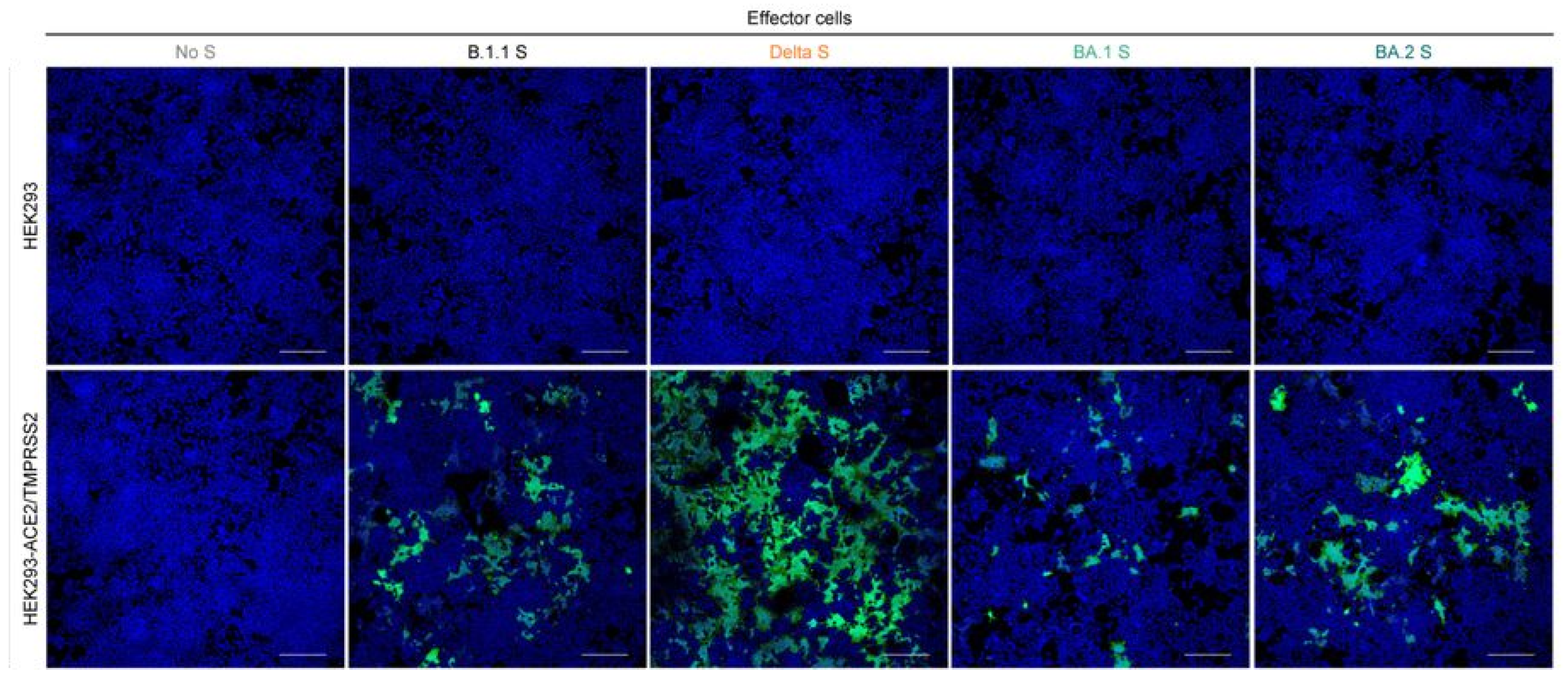
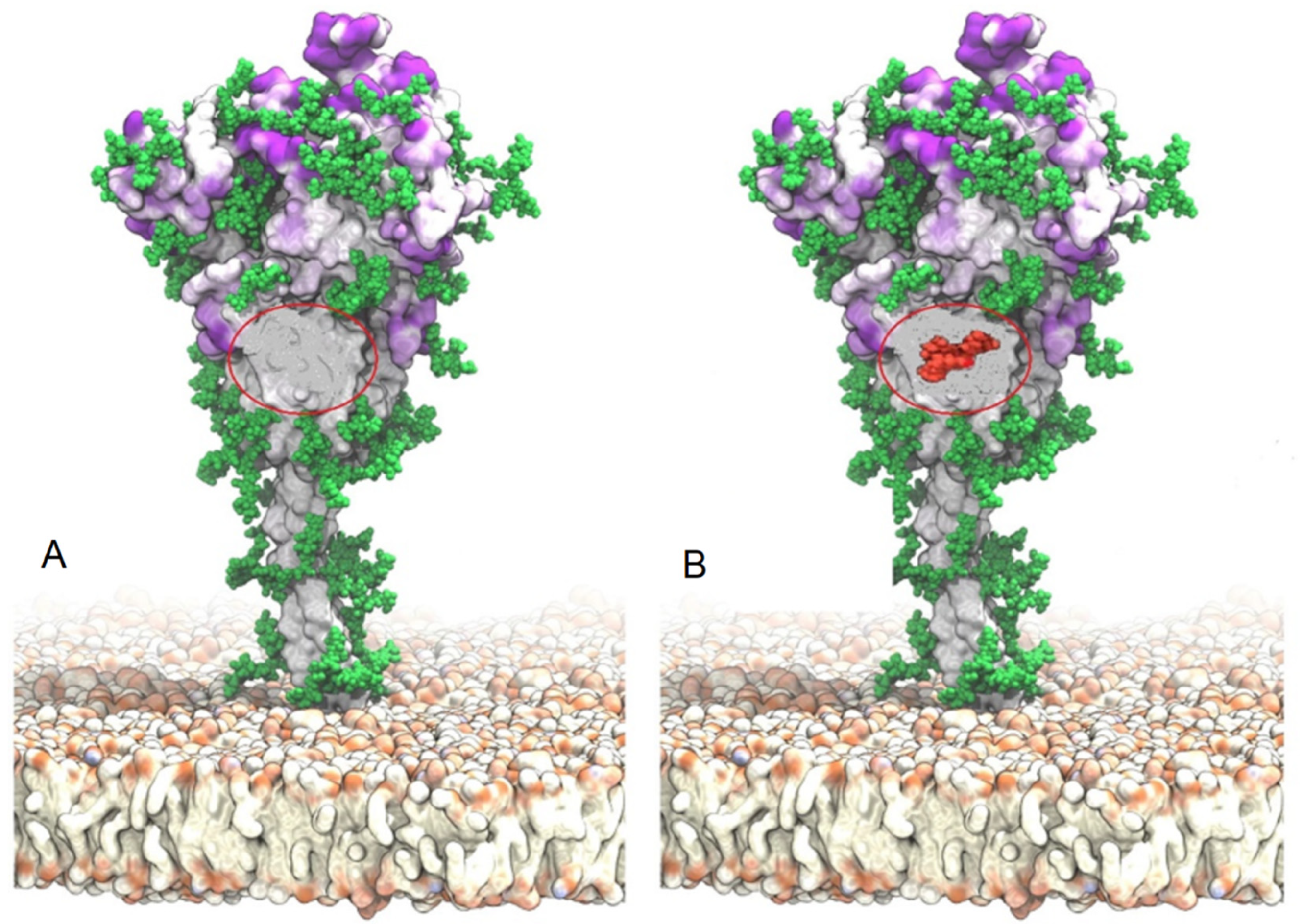
3. The Omicron Variant

In November 2021, Omicron was first discovered in South Africa [37]. Then, the BA.1 lineage of Omicron expanded quickly over the whole world, outcompeting the other variants, such as Delta. Another Omicron variant, the BA.2 lineage, was discovered in numerous nations, including Denmark and the United Kingdom, as of February 2022 [38]. BA.2 rapidly surpassed BA.1, showing that BA.2 had a higher transmission rate than BA.1. [19]. Omicron had the same P681H mutation as the Alpha and Delta strains, but it also harbors a new and unique glycosite, threonine (Thr376), that has only been found in Omicron. This was placed close to a proline amino acid that controls O-glycosylation [39]. When compared with the wild-type or Delta, researchers noticed a notable rise in the use of Core 2-type O-glycans for the Omicron variant, which is compatible with O-glycan insertion [39]. This discovery is groundbreaking, as it was formerly reported that, in vitro, the impairment of GALNT1 glycosyltransferase function (which regulates the insertion of O-glycans in the vicinity of the furin cleavage area) was caused by a mutation of proline 681 [27].
This means that Omicron’s new Thr376 mutation recovered the ability to insert O-glycans that cover the furin cleavage site. In conclusion, a mutation in p681 in the Delta variant resulted in glycans loss (Figure 1A), uncovering the furin cleavage site and making this variant more pathogenic due to enhanced syncytia formation [27]. On the contrary, a mutation in Thr376 in the Omicron variant induced the addition of O-glycans (Figure 1B), thus covering the furin cleavage site [39]. As a result, this mutation has increased immune evasion while decreasing pathogenicity. This phenomenon has also been documented by research on the Nipah virus, where numerous N-glycans on the fusion protein were removed, resulting in hyperfusogenic phenotypes but also showing an enhanced susceptibility to antibody neutralization [40]. Although glycans are known to perform an important role in immune evasion, they are also emerging as essential determinants of virulence [41,42].
In a recent study, it was found that coculturing spike-expressing cells with HEK293-ACE2/TMPRSS2 cells dramatically increased the amount of syncytia formation caused by BA.2 spike compared to BA.1 spike but less than Delta (Figure 2) [19]. Since the efficiency of S1/S2 cleavage has been related to cell fusion induced by SARS-CoV-2’s spike protein [26,44], it was proposed that BA.2 spike is cleaved in a more effective way than BA.1 spike. Nevertheless, a Western blot test revealed that BA.2 spike is cleaved less efficiently than BA.1 spike, implying that BA.2 spike produces more syncytia than BA.1 spike by using a S1/S2 cleavage-independent mechanism. To find out whether BA.2 S uses TMPRSS2, 293-ACE2 cells with or without TMPRSS2 expression were used in a cell-based fusion assay [19].
The findings demonstrated that BA.2’s relatively greater fusogenic potential depends on TMPRSS2’s expression in the target cells [19].
It is worth noting that BA.1 makes inefficient use of TMPRSS2 during infection [45]. Thus, the new mutations found in BA.2 apparently restored its capacity to use TMPRSS2, which has resulted in a higher fusogenicity and pathogenic potential compared with BA.1. [19].
Analyses of these genomic differences between the Delta and Omicron variants allow us to understand the relevance of glycans in virulence. Notably, Delta showed a greater fusogenicity due to the p681 mutation that eliminated an O-glycan molecule from the furin cleavage site [27], and also, the D614G mutation increased syncytia creation and the viral load through enhanced furin-induced spike cleavage [46]. Interestingly, a similar mutation occurred with the influenza virus. When the virulence of the H3N2 strains in mice was compared, it was discovered that viruses isolated after 1980 had high glycan numbers and caused mild disease in mice. An N-linked glycan from the hemagglutinin receptor of the influenza virus was lost due to a mutation in the gene codifying for such a receptor in the Beijing/89 strain, which was linked to enhanced virulence in mice [47]. The virulence of the 2009 H1N1 virus was also driven by a sudden glycan loss near the receptor-binding site [48]. A subsequent study in mice confirmed this discovery, providing evidence that a comparable mutation increased the virulence of the 1918 H1N1 pandemic virus [49]. The 1918 H1N1 virus contained fewer glycosylation sites on the hemagglutinin receptor than seasonal influenza viruses with lower virulence. Utilizing site-directed mutagenesis, it was discovered that, by incorporating two extra glycosylation sites (asparagine Asn71 and Asn286) in one flank of the hemagglutinin receptor, a highly virulent 1918 HA chimeric virus was considerably attenuated in mice, and removing the glycosylation sites enhanced the virulence in vivo [49].
Similarly, the Omicron variant BA.1 developed a mutation in Thr376, which is located near the FCS, and consisted of the addition of O-glycan molecules, thus covering the FCS (Figure 1B). That interference was traduced in a lower fusogenicity and pathogenicity [40,45]. This is also consistent with the clinical symptomatology being milder, as a significantly reduced Omicron reproduction velocity was recently detected in lung epithelial cells [50,51]. In vitro experiments showed that the Omicron variant BA.1 was less likely to disseminate by cell merging than the other variants, adding to the proof that the virus on its own caused less severe illness [45,50].
All these data may clarify why Omicron-BA.1-infected individuals suffered fewer serious complications [52,53]. Regarding the pathogenicity of BA.2 in humans, the risk of hospitalization in Germany after a BA.1 or BA.2 Omicron variant infection was approximately 80% lower than after a Delta variant infection, especially in people under 35 years old [54]. Both BA.1 and BA.2 showed a similar impact on hospitalization or intensive care unit (ICU) admission, implying that, despite evidence of the greater transmissibility of BA.2 [19], this variant is pathogenically equivalent to BA.1 [54]. Confirming these results, no clinical differences among individuals infected with BA.1 and BA.2 were detected in Denmark [55] and South Africa [56]. In a study carried out in France, the first 207 Omicron BA.2 cases were recorded, and it was discovered that severe Omicron BA.2 infections were exclusively seen in individuals over the age of 80. Three patients (1.5%) died at the ages of 80, 97 and 99, and two of them had diabetes. Two people (ages 80 and 97) received the COVID-19 vaccine (three doses). Individuals who passed away from Omicron BA.2 infection were much older than those who passed away from Omicron BA.1 infection [57].
The latest research findings showed that a genetic mutation in the E protein from Omicron BA.1 and BA.2 also contributed to its reduced pathogenic potential. The SARS-CoV-2 envelope protein (2-E) creates a homopentameric cation channel that is essential for virulence. SARS-CoV-2 does not travel through the traditional biosynthetic secretory channel; instead, it enters lysosomes and exits via lysosomal deacidification (alkalization). The lysosomes are where coronavirus envelope (E) protein channels are found. The E protein is therefore in charge of the calcium outflow to achieve an alkaline pH, since, otherwise, the virus would be destroyed by the lysosome’s acidic pH. While mutated T9I channels had less of an impact on the luminal pH, the expression of wild-type channels significantly alkalized the lysosomal pH. The viral load was decreased as a result of decreased lysosomal deacidification. T9I’s cytotoxic potential and cytokine production were both roughly 150-fold lower than those of the wild-type, which could further diminish the virulence of Omicron variants [58].

4. Discussion

Our proposal of an “intermittent virulence” reconciles two contrary hypotheses. The first claims that the Omicron variants (BA.1 and BA.2) were going to define the end of the current pandemic, whereas the other suggests that new virulent variants will arise in the future. We propose that, in order to survive, viruses may develop what we have termed “intermittent virulence”. This concept implies that, when the virus has become more contagious but less virulent, natural selection could create new variants with a higher pathogenic capacity; otherwise, the dominant variant could disappear or become endemic due to the community having reached herd immunity through natural immunity or due to global vaccination programs. In fact, during the ongoing pandemic, SARS-CoV-2-associated mortality has shown interesting fluctuations. For example, on 20 January 2021, the highest global mortality was reached with 18,144 deaths (Figure 3). Afterward, a significant decrease in mortality was observed worldwide. Such behavior has been repeated several times, giving rise to the so-called “waves”. By April 28, the third wave reached its peak level with 15,978 deaths. After a peak there is a sharp decrease in mortality; that could be because the population has reached a herd immunity.
However, a new rise in mortality numbers occurred by 23 August 2021, and again by 9 February 2022 (Figure 3), which contradicts the idea that the population reached a herd immunity. If it were true, the number of infections or deaths would have decreased, which was not the case. The Omicron variant was first discovered in South Africa in November 2021 [37]. This variant rapidly outcompeted Delta, and by February 2022, it was the dominant variant. Although it has been demonstrated that Omicron is less pathogenic than Delta, the mortality peak shown in Figure 3 by February 2022 could be due to the fact that, at that time, both variants were cocirculating or because the global population had no immunity against Omicron. After the February peak, there has been a consistent decrease in mortality worldwide, leading some experts to ask that “public health officials need to declare the end of the pandemic” [59].
Nevertheless, the recently reported virological characteristics of the new Omicron variant BA.2 suggested a return to a higher virulence than its previous antecessor, BA.1. [19]. This variant rapidly displaced Delta around the world, showing remarkable transmissibility but with lower pathogenicity. Compared to BA.1 and Delta, BA.2 was approximately 1.5 and 4.2 times more infectious, respectively [60,61]. In vitro studies have shown that BA.2 had a higher fusogenicity, and histological studies demonstrated that BA.2 replicated faster and more efficiently in hamster lungs and caused greater damage to this organ than BA.1 [19]. Interestingly, BA.2 showed a lower fusogenic capacity compared with Delta (Figure 2). This result suggests that BA.2 could produce a less severe clinical symptomatology in humans than the Delta variant but higher than BA.1. As of May 2022, Omicron subvariants harboring a mutation at the L452 residue of the spike (S) protein, including BA.4 and BA.5, have been frequently discovered [62,63].
Neutralization studies demonstrated that the immunity conferred by the BA.1 and BA.2 outbreaks is less effective against BA.4/5, according to recent findings [19]. Cell culture investigations demonstrated that BA.2.12.1 and BA.4/5 propagate more effectively in human alveolar epithelial cells than BA.2, and specifically, BA.4/5 produces more syncytia than BA.2. In addition, hamster infection tests revealed that BA.4/5 is more virulent than BA.2 “and could represent a greater threat to world health than the original BA.2 because SARS-CoV-2 does not necessarily evolve to attenuate its pathogenicity” [19]. However, as we mentioned earlier, there is an important difference between these in vitro studies, in vivo investigations using animal models, and humans. In contrast to animals, humans have received different COVID-19 vaccines, which have induced artificial immunity, and that could explain why mortality continues to decline. Epidemiological surveillance organizations like Worldometer (worldometers.info) or Our World in Data provide information in near-real time regarding the number of cases and deaths.
It is possible that the pandemic phase of COVID-19 is about to end but the virus is not going to disappear and an endemic phase will begin, where intermittent virulence could guarantee the survival of this pathogen. Recent findings showed that it is not a rule that viruses become less virulent over time [64]. A particularly aggressive variant of the human immunodeficiency virus (HIV) has been discovered in the Netherlands, where it has been circulating for some years. More than 100 individuals with HIV-1 subtype B infection experienced a two-fold decline in CD4+ cell numbers than predicted. These people were already at risk of contracting AIDS within two to three years of being diagnosed. This virus lineage, which appears to have emerged de novo around the 1990s, has undergone considerable changes in its genes, changing almost 300 amino acids, making it difficult to determine the reason for the increased virulence [64].
Since it is known that genetic recombination can result in the appearance of new, extremely pathogenic variants, the most concerning evolutionary aspect of SARS-CoV-2 is that it could experiment with widespread recombinations [65,66]. Simultaneous infections with various subtypes of the same virus might cause this outcome. The activity of the Nsp14 protein regulates this mechanism in SARS-CoV-2 [67]. In February 2022, for example, evidence of Deltacron XD recombinant SARS-CoV-2 transmission and circulation in Northwest France was reported. Following virological and epidemiological studies, 17 cases of this recombinant SARS-CoV-2 were validated by genotyping or inferred due to epidemiological relationships, indicating an extensive propagation incident and transmission of this virus but not showing evidence of severe clinical symptoms [68]. Another contemporary research discovered a Delta variant sub-lineage spreading throughout the United States, particularly in Colorado (CO), Texas (TX), and Wyoming (WY). This sub-lineage is characterized by a spike protein mutation at position 112 that has been found in low-prevalence lineages around the world, as well as in circulating U.S. isolates since the end of April 2021. Two unique mutations are found in this Delta S:S112L sub-lineage group: ORF1b:V2354F, which corresponds to nonstructural protein NSP15 at position 303 (NSP15:V303F), and a premature stop codon (Q94 *) that truncates ORF7a [69]. Unfortunately, the clinical characteristics of the people infected with this sub-lineage were not examined in this investigation.
To predict the epidemic’s future, researchers must accurately investigate how the virus’ tropism evolves. A respiratory tropism is prevalent at this time, but it is known from other coronaviruses that this can evolve very quickly [70]. The avian coronavirus spike protein, for example, had three amino acid modifications that enabled the virus to attach to kidney cells [71]. Coronaviruses also are neurotropic in mice [72] and SARS-CoV-2 also shows neurotropism [73,74,75]. The changing structure of the spike protein is significantly related to natural selection in cell ingress and fusion. SARS-CoV-2 was able to swap hosts and adapt to the human receptor ACE2 [76] after the acquisition of a furin cleavage site. At least in vitro, the virus has encountered another receptor for entry, specifically the CD147 receptor, a protein present in several tissues, which include epithelial and neural cells [77]. This is significant, because using a variety of receptors allows for switching between different cell types and, thus, different entry gateways [70]. Notably, SARS-CoV-2 can infect nonpermissive cells (which do not harbor ACE2 receptors) by using an innovative intracytoplasmic connection mechanism that could serve as an alternative viral transmission pathway, independent of the canonical extra-cytoplasmic ACE2-binding mechanism [78].
An increased virulence produced by new variants could guarantee the survival of this pathogen, thus confirming the validity of the term “intermittent virulence”. However, it is improbable that the virulence of SARS-CoV-2 would be the same as was seen during the alpha or delta waves, due to the fact that the human population has reached a sufficient level of herd immunity through natural infection or due to the employment of vaccination programs. The most recent global mortality data (Figure 3) makes us question whether this pandemic is really over, and it also makes uncertain when the endemic phase will begin. Darwin’s words “the survival of the fittest” are still correct. The virus will continue killing nonvaccinated old people, vaccinated old people, and those with comorbidities. For example, it is widely recognized that patients with COVID-19 may have a worse prognosis if their blood glucose levels are high, increasing the risk of multiple organ failure, shock, and the need for intensive care unit (ICU) placement [79]. People with type 2 diabetes mellitus exhibited a greater incidence, severity of symptoms, and mortality after COVID-19 infection [80]. According to a recent investigation, glycemic control significantly affects how well the patients would respond immunologically to the SARS-CoV-2 messenger ribonucleic acid (mRNA) vaccine [81]. The immune response is impaired by hyperglycemia during immunization: a recent study found that 21 days after the first dose of the vaccine, neutralizing antibody titers and CD4 cytokine responses involving type 1 helper T cells were lower in type 2 diabetic patients with glycosylated hemoglobin (HbA1c) levels > 7% than in diabetics with HbA1c levels ≤ 7% [81]. This aspect is very important, because according to the International Diabetes Atlas, it was estimated that, in 2021, there were 537 million diabetics in the world. Three out of four adults with diabetes live in low- and middle-income countries, and almost one in two (240 million) adults living with diabetes are undiagnosed [82]. An undiagnosed person will surely have elevated blood glucose levels, which will prevent a satisfactory immune response when vaccinated, as previously reported [81].
A recent study on the efficacy of the COVID-19 vaccination and the protection decline over time [83] reported that 8 months after receiving two doses of the COVID-19 vaccine, the immunological function was inferior in those who were older and had preexisting conditions. These findings suggest that booster doses for older people and people with known inadequate or declining vaccine-elicited immunogenicity should be prioritized because they also have the highest likelihood of experiencing severe COVID-19 symptoms if infected [83].
While this work was in peer review, a new preprint was posted on Biorxiv [84], which showed that Omicron BA.4 and BA.5 replication is linked to the lower activation of the epithelial innate immune responses. These subvariants have improved transmission and potentially reduce immune protection from severe disease by combining the evolution of antibody escape with the increased antagonism of interferon signaling. The study also discovered the increased expression of the innate immune antagonist proteins Orf6 and N, which are comparable to Alpha, implying that human adaptation processes are similar [84].
As a final comment, one should remember that the in vitro antibody neutralization tests performed to evaluate the efficacy of vaccines or monoclonal antibodies do not mirror the real situation within the host, where SARS-CoV-2 can hijack infected cells and release decoy targets to avoid being neutralized by vaccine-derived antibodies, or can infect non-permissive cells by using tunneling nanotubes or through the cell-to-cell infection, thus avoiding immune surveillance and causing syncytia-induced lymphopenia [85]. We have underestimated this master of immune escape, and have not yet seen the full adaptive potential this virus can develop through natural selection.

Author Contributions

Conceptualization, A.R.-C., E.M.R. and V.N.U.; Study design, A.R.-C., E.M.R. and V.N.U.; Literature Collection and Analysis, A.R.-C., E.M.R. and V.N.U.; Investigation, A.R.-C., E.M.R. and V.N.U.; Writing—Original Draft, A.R.-C., E.M.R. and V.N.U.; and Writing—Review and Editing, A.R.-C., E.M.R. and V.N.U. All authors have read and agreed to the published version of the manuscript.

Funding

This research received no external funding.

Institutional Review Board Statement

Not applicable.

Informed Consent Statement

Not applicable.

Data Availability Statement

Not applicable.

Conflicts of Interest

The authors declare no conflict of interest. The funders had no role in the design of the study; in the collection, analyses, or interpretation of the data; in the writing of the manuscript; or in the decision to publish the results.

References

  1. World Health Organization. Genomic Sequencing of SARS-CoV-2: A Guide to Implementation for Maximum Impact on Public Health; World Health Organization: Geneva, Switzerland, 2021; Available online: https://www.who.int/publications/i/item/9789240018440 (accessed on 5 June 2022).
  2. Park, M.; Loverdo, C.; Schreiber, S.J.; Lloyd-Smith, J.O. Multiple scales of selection influence the evolutionary emergence of novel pathogens. Philos. Trans. R. Soc. Lond B Biol. Sci. 2013, 368, 20120333. [Google Scholar] [CrossRef] [Green Version]
  3. Wasik, B. On the biological success of viruses. Annu. Rev. Microbiol. 2013, 67, 519–541. [Google Scholar] [CrossRef] [PubMed] [Green Version]
  4. van Dorp, L.; Richard, D.; Tan, C.C.S.; Shaw, L.P.; Acman, M.; Balloux, F. No evidence for increased transmissibility from recurrent mutations in SARS-CoV-2. Nat. Commun. 2020, 11, 5986. [Google Scholar] [CrossRef] [PubMed]
  5. Thomson, E.C.; Rosen, L.E.; Shepherd, J.G.; Spreafico, R.; da Silva Filipe, A.; Wojcechowskyj, J.A.; Davis, C.; Piccoli, L.; Pascall, D.J.; Dillen, J.; et al. Circulating SARS-CoV-2 spike N439K variants maintain fitness while evading antibody-mediated immunity. Cell 2021, 184, 1171–1187.e1120. [Google Scholar] [CrossRef] [PubMed]
  6. Piccoli, L.; Park, Y.J.; Tortorici, M.A.; Czudnochowski, N.; Walls, A.C.; Beltramello, M.; Silacci-Fregni, C.; Pinto, D.; Rosen, L.E.; Bowen, J.E.; et al. Mapping Neutralizing and Immunodominant Sites on the SARS-CoV-2 Spike Receptor-Binding Domain by Structure-Guided High-Resolution Serology. Cell 2020, 183, 1024–1042.e1021. [Google Scholar] [CrossRef]
  7. Korber, B.; Fischer, W.M.; Gnanakaran, S.; Yoon, H.; Theiler, J.; Abfalterer, W.; Hengartner, N.; Giorgi, E.E.; Bhattacharya, T.; Foley, B.; et al. Tracking Changes in SARS-CoV-2 Spike: Evidence that D614G Increases Infectivity of the COVID-19 Virus. Cell 2020, 182, 812–827.e819. [Google Scholar] [CrossRef]
  8. Yurkovetskiy, L.; Wang, X.; Pascal, K.E.; Tomkins-Tinch, C.; Nyalile, T.P.; Wang, Y.; Baum, A.; Diehl, W.E.; Dauphin, A.; Carbone, C.; et al. Structural and Functional Analysis of the D614G SARS-CoV-2 Spike Protein Variant. Cell 2020, 183, 739–751.e738. [Google Scholar] [CrossRef]
  9. Greaney, A.J.; Loes, A.N.; Crawford, K.H.D.; Starr, T.N.; Malone, K.D.; Chu, H.Y.; Bloom, J.D. Comprehensive mapping of mutations in the SARS-CoV-2 receptor-binding domain that affect recognition by polyclonal human plasma antibodies. Cell Host Microbe 2021, 29, 463–476.e466. [Google Scholar] [CrossRef]
  10. Wang, P.; Nair, M.S.; Liu, L.; Iketani, S.; Luo, Y.; Guo, Y.; Wang, M.; Yu, J.; Zhang, B.; Kwong, P.D. Antibody resistance of SARS-CoV-2 variants B. 1.351 and B. 1.1. 7. Nature 2021, 593, 130–135. [Google Scholar] [CrossRef]
  11. Burnet, F.M.; White, D. Natural History of Infectious Disease; Cambridge University Press: Cambridge, UK, 1972. [Google Scholar]
  12. Levin, S.; Pimentel, D. Selection of intermediate rates of increase in parasite-host systems. Am. Nat. 1981, 117, 308–315. [Google Scholar] [CrossRef]
  13. Anderson, R.M.; May, R.M. Coevolution of hosts and parasites. Parasitology 1982, 85, 411–426. [Google Scholar] [CrossRef] [PubMed]
  14. May, R.M.; Anderson, R.M. Epidemiology and genetics in the coevolution of parasites and hosts. Proc. R. Soc. London. Ser. B Biol. Sci. 1983, 219, 281–313. [Google Scholar]
  15. Frank, S.A. Models of parasite virulence. Q. Rev. Biol. 1996, 71, 37–78. [Google Scholar] [CrossRef]
  16. Galvani, A.P. Epidemiology meets evolutionary ecology. Trends Ecol. Evol. 2003, 18, 132–139. [Google Scholar] [CrossRef]
  17. Grubaugh, N.D.; Petrone, M.E.; Holmes, E.C. We shouldn’t worry when a virus mutates during disease outbreaks. Nat. Microbiol. 2020, 5, 529–530. [Google Scholar] [CrossRef] [PubMed] [Green Version]
  18. Markov, P.V.; Katzourakis, A.; Stilianakis, N.I. Antigenic evolution will lead to new SARS-CoV-2 variants with unpredictable severity. Nat. Rev. Microbiol. 2022, 20, 251–252. [Google Scholar] [CrossRef] [PubMed]
  19. Yamasoba, D.; Kimura, I.; Nasser, H.; Morioka, Y.; Nao, N.; Ito, J.; Uriu, K.; Tsuda, M.; Zahradnik, J.; Shirakawa, K. Virological characteristics of SARS-CoV-2 BA. 2 variant. Cell 2022, 185, 2103–2115. [Google Scholar] [CrossRef]
  20. Qu, P.; Evans, J.P.; Faraone, J.N.; Zou, X.; Zheng, Y.M.; Carlin, C.; Bednash, J.S.; Lozanski, G.; Mallampalli, R.K.; Saif, L.J. Differential Evasion of Delta and Omicron Immunity and Enhanced Fusogenicity of SARS-CoV-2 Omicron BA. 4/5 and BA. 2.12. 1 Subvariants. bioRxiv 2022. [Google Scholar] [CrossRef]
  21. European Centre for Disease Prevention and Control. Threat Assessment Brief: Emergence of SARS-CoV-2 B.1.617 Variants in India and Situation in the EU/EEA. Available online: https://www.ecdc.europa.eu/en/publications-data/threat-assessment-emergence-sars-cov-2-b1617-variants (accessed on 15 June 2022).
  22. Sheikh, A.; McMenamin, J.; Taylor, B.; Robertson, C. SARS-CoV-2 Delta VOC in Scotland: Demographics, risk of hospital admission, and vaccine effectiveness. Lancet 2021, 397, 2461–2462. [Google Scholar] [CrossRef]
  23. Twohig, K.A.; Nyberg, T.; Zaidi, A.; Thelwall, S.; Sinnathamby, M.A.; Aliabadi, S.; Seaman, S.R.; Harris, R.J.; Hope, R.; Lopez-Bernal, J. Hospital admission and emergency care attendance risk for SARS-CoV-2 delta (B. 1.617. 2) compared with alpha (B. 1.1. 7) variants of concern: A cohort study. Lancet Infect. Dis. 2022, 22, 35–42. [Google Scholar] [CrossRef]
  24. Kim, S.; Liu, Y.; Lei, Z.; Dicker, J.; Cao, Y.; Zhang, X.F.; Im, W. Differential interactions between human ACE2 and Spike RBD of SARS-CoV-2 variants of concern. J. Chem. Theory Comput. 2021, 17, 7972–7979. [Google Scholar] [CrossRef] [PubMed]
  25. Liu, Y.; Liu, J.; Johnson, B.A.; Xia, H.; Ku, Z.; Schindewolf, C.; Widen, S.G.; An, Z.; Weaver, S.C.; Menachery, V.D. Delta spike P681R mutation enhances SARS-CoV-2 fitness over Alpha variant. Cell Rep. 2022, 39, 110829. [Google Scholar] [CrossRef]
  26. Saito, A.; Irie, T.; Suzuki, R.; Maemura, T.; Nasser, H.; Uriu, K.; Kosugi, Y.; Shirakawa, K.; Sadamasu, K.; Kimura, I. Enhanced fusogenicity and pathogenicity of SARS-CoV-2 Delta P681R mutation. Nature 2022, 602, 300–306. [Google Scholar] [CrossRef]
  27. Zhang, L.; Mann, M.; Syed, Z.A.; Reynolds, H.M.; Tian, E.; Samara, N.L.; Zeldin, D.C.; Tabak, L.A.; Ten Hagen, K.G. Furin cleavage of the SARS-CoV-2 spike is modulated by O-glycosylation. Proc. Natl. Acad. Sci. USA 2021, 118, e2109905118. [Google Scholar] [CrossRef]
  28. Cai, Y.; Zhang, J.; Xiao, T.; Lavine, C.L.; Rawson, S.; Peng, H.; Zhu, H.; Anand, K.; Tong, P.; Gautam, A. Structural basis for enhanced infectivity and immune evasion of SARS-CoV-2 variants. Science 2021, 373, 642–648. [Google Scholar] [CrossRef]
  29. Berger, I.; Schaffitzel, C. The SARS-CoV-2 spike protein: Balancing stability and infectivity. Cell Res. 2020, 30, 1059–1060. [Google Scholar] [CrossRef] [PubMed]
  30. Phan, T. Genetic diversity and evolution of SARS-CoV-2. Infect. Genet. Evol. 2020, 81, 104260. [Google Scholar] [CrossRef]
  31. Zhang, L.; Jackson, C.B.; Mou, H.; Ojha, A.; Peng, H.; Quinlan, B.D.; Rangarajan, E.S.; Pan, A.; Vanderheiden, A.; Suthar, M.S. SARS-CoV-2 spike-protein D614G mutation increases virion spike density and infectivity. Nat. Commun. 2020, 11, 1–9. [Google Scholar] [CrossRef]
  32. Eaaswarkhanth, M.; Al Madhoun, A.; Al-Mulla, F. Could the D614G substitution in the SARS-CoV-2 spike (S) protein be associated with higher COVID-19 mortality? Int. J. Infect. Dis. 2020, 96, 459–460. [Google Scholar] [CrossRef]
  33. Jackson, C.B.; Zhang, L.; Farzan, M.; Choe, H. Functional importance of the D614G mutation in the SARS-CoV-2 spike protein. Biochem. Biophys. Res. Commun. 2021, 538, 108–115. [Google Scholar] [CrossRef]
  34. Mohammad, A.; Alshawaf, E.; Marafie, S.K.; Abu-Farha, M.; Abubaker, J.; Al-Mulla, F. Higher binding affinity of furin for SARS-CoV-2 spike (S) protein D614G mutant could be associated with higher SARS-CoV-2 infectivity. Int. J. Infect. Dis. 2021, 103, 611–616. [Google Scholar] [CrossRef] [PubMed]
  35. Becerra-Flores, M.; Cardozo, T. SARS-CoV-2 viral spike G614 mutation exhibits higher case fatality rate. Int. J. Clin. Pract. 2020, 74, e13525. [Google Scholar] [CrossRef] [PubMed]
  36. Fisman, D.N.; Tuite, A.R. Evaluation of the relative virulence of novel SARS-CoV-2 variants: A retrospective cohort study in Ontario, Canada. Cmaj 2021, 193, E1619–E1625. [Google Scholar] [CrossRef] [PubMed]
  37. WHO. Classification of Omicron (B.1.1.529): SARS-CoV-2 Variant of Concern (26 November 2021). 2021. Available online: https://www.who.int/news/item/26-11-2021-classification-of-omicron-(b.1.1.529)-sars-cov-2-variant-of-concern (accessed on 10 June 2022).
  38. UKHSA. SARS-CoV-2 Variants of Concern and Variants under Investigation in England. Technical Briefing 35 (28 January 2022). Available online: https://assets.publishing.service.gov.uk/government/uploads/system/uploads/attachment_data/file/1050999/Technical-Briefing-35-28January2022.pdf (accessed on 12 June 2022).
  39. Roberts, D.S.; Mann, M.; Li, B.H.; Kim, D.; Brasier, A.R.; Jin, S.; Ge, Y. Distinct Core Glycan and O-Glycoform Utilization of SARS-CoV-2 Omicron Variant Spike Protein RBD Revealed by Top-Down Mass Spectrometry. bioRxiv 2022. [Google Scholar] [CrossRef]
  40. Aguilar, H.C.; Matreyek, K.A.; Filone, C.M.; Hashimi, S.T.; Levroney, E.L.; Negrete, O.A.; Bertolotti-Ciarlet, A.; Choi, D.Y.; McHardy, I.; Fulcher, J.A. N-glycans on Nipah virus fusion protein protect against neutralization but reduce membrane fusion and viral entry. J. Virol. 2006, 80, 4878–4889. [Google Scholar] [CrossRef] [PubMed] [Green Version]
  41. Watanabe, Y.; Bowden, T.A.; Wilson, I.A.; Crispin, M. Exploitation of glycosylation in enveloped virus pathobiology. Biochim. Et Biophys. Acta (BBA)-Gen. Subj. 2019, 1863, 1480–1497. [Google Scholar] [CrossRef] [PubMed]
  42. Yang, Q.; Hughes, T.A.; Kelkar, A.; Yu, X.; Cheng, K.; Park, S.; Huang, W.C.; Lovell, J.F.; Neelamegham, S. Inhibition of SARS-CoV-2 viral entry upon blocking N-and O-glycan elaboration. eLife 2020, 9, e61552. [Google Scholar] [CrossRef]
  43. Sikora, M.; von Bülow, S.; Blanc, F.E.; Gecht, M.; Covino, R.; Hummer, G. Computational epitope map of SARS-CoV-2 spike protein. PLoS Comput. Biol. 2021, 17, e1008790. [Google Scholar] [CrossRef]
  44. Suzuki, R.; Yamasoba, D.; Kimura, I.; Wang, L.; Kishimoto, M.; Ito, J.; Morioka, Y.; Nao, N.; Nasser, H.; Uriu, K. Attenuated fusogenicity and pathogenicity of SARS-CoV-2 Omicron variant. Nature 2022, 603, 700–705. [Google Scholar] [CrossRef]
  45. Meng, B.; Abdullahi, A.; Ferreira, I.A.; Goonawardane, N.; Saito, A.; Kimura, I.; Yamasoba, D.; Gerber, P.P.; Fatihi, S.; Rathore, S. Altered TMPRSS2 usage by SARS-CoV-2 Omicron impacts infectivity and fusogenicity. Nature 2022, 603, 706–714. [Google Scholar] [CrossRef]
  46. Cheng, Y.W.; Chao, T.L.; Li, C.L.; Wang, S.H.; Kao, H.C.; Tsai, Y.M.; Wang, H.Y.; Hsieh, C.L.; Lin, Y.Y.; Chen, P.J. D614G substitution of SARS-CoV-2 spike protein increases syncytium formation and virus titer via enhanced furin-mediated spike cleavage. Mbio 2021, 12, e00587-21. [Google Scholar] [CrossRef]
  47. Reading, P.C.; Pickett, D.L.; Tate, M.D.; Whitney, P.G.; Job, E.R.; Brooks, A.G. Loss of a single N-linked glycan from the hemagglutinin of influenza virus is associated with resistance to collectins and increased virulence in mice. Respir. Res. 2009, 10, 1–11. [Google Scholar] [CrossRef] [PubMed] [Green Version]
  48. Wei, C.J.; Boyington, J.C.; Dai, K.; Houser, K.V.; Pearce, M.B.; Kong, W.P.; Yang, Z.y.; Tumpey, T.M.; Nabel, G.J. Cross-neutralization of 1918 and 2009 influenza viruses: Role of glycans in viral evolution and vaccine design. Sci. Transl. Med. 2010, 2, ra21–ra24. [Google Scholar] [CrossRef] [PubMed] [Green Version]
  49. Sun, X.; Jayaraman, A.; Maniprasad, P.; Raman, R.; Houser, K.V.; Pappas, C.; Zeng, H.; Sasisekharan, R.; Katz, J.M.; Tumpey, T.M. N-linked glycosylation of the hemagglutinin protein influences virulence and antigenicity of the 1918 pandemic and seasonal H1N1 influenza A viruses. J. Virol. 2013, 87, 8756–8766. [Google Scholar] [CrossRef] [Green Version]
  50. Bentley, E.G.; Kirby, A.; Sharma, P.; Kipar, A.; Mega, D.F.; Bramwell, C.; Penrice-Randal, R.; Prince, T.; Brown, J.C.; Zhou, J. SARS-CoV-2 Omicron-B. 1.1. 529 Variant leads to less severe disease than Pango B and Delta variants strains in a mouse model of severe COVID-19. bioRxiv 2021. [Google Scholar] [CrossRef]
  51. McMahan, K.; Giffin, V.; Tostanosky, L.; Chung, B.; Siamatu, M.; Suthar, M.S.; Halfmann, P.; Kawaoka, Y.; Piedra-Mora, C.; Jain, N.; et al. Reduced pathogenicity of the SARS-CoV-2 Omicron variant in hamsters. Med 2022, 3, 262–268.e4. [Google Scholar] [CrossRef] [PubMed]
  52. Shuai, H.; Chan, J.F.W.; Hu, B.; Chai, Y.; Yuen, T.T.T.; Yin, F.; Huang, X.; Yoon, C.; Hu, J.C.; Liu, H. Attenuated replication and pathogenicity of SARS-CoV-2 B. 1.1. 529 Omicron. Nature 2022, 603, 693–699. [Google Scholar] [CrossRef] [PubMed]
  53. Wolter, N.; Jassat, W.; Walaza, S.; Welch, R.; Moultrie, H.; Groome, M.; Amoako, D.G.; Everatt, J.; Bhiman, J.N.; Scheepers, C. Early assessment of the clinical severity of the SARS-CoV-2 omicron variant in South Africa: A data linkage study. Lancet 2022, 399, 437–446. [Google Scholar] [CrossRef]
  54. Sievers, C.; Zacher, B.; Ullrich, A.; Huska, M.; Fuchs, S.; Buda, S.; Haas, W.; Diercke, M.; an der Heiden, M.; Kröger, S. SARS-CoV-2 Omicron variants BA. 1 and BA. 2 both show similarly reduced disease severity of COVID-19 compared to Delta, Germany, 2021 to 2022. Eurosurveillance 2022, 27, 2200396. [Google Scholar] [CrossRef] [PubMed]
  55. Fonager, J.; Bennedbæk, M.; Bager, P.; Wohlfahrt, J.; Ellegaard, K.M.; Ingham, A.C.; Edslev, S.M.; Stegger, M.; Sieber, R.N.; Lassauniere, R. Molecular epidemiology of the SARS-CoV-2 variant Omicron BA. 2 sub-lineage in Denmark, 29 November 2021 to 2 January 2022. Eurosurveillance 2022, 27, 2200181. [Google Scholar] [CrossRef]
  56. Wolter, N.; Jassat, W.; von Gottberg, A.; Cohen, C. Clinical severity of Omicron sub-lineage BA. 2 compared to BA. 1 in South Africa. Lancet 2022, 400, 93–96. [Google Scholar] [CrossRef]
  57. Gautret, P.; Hoang, V.T.; Jimeno, M.T.; Lagier, J.C.; Rossi, P.; Fournier, P.E.; Colson, P.; Raoult, D. The severity of the first 207 infections with the SARS-CoV-2 Omicron BA. 2 variant, in Marseille, France, December 2021–February 2022. J. Med. Virol. 2022, 94, 3494–3497. [Google Scholar] [CrossRef]
  58. Xia, B.; Wang, Y.; Pan, X.; Cheng, X.; Ji, H.; Zuo, X.; Jiang, H.; Li, J.; Gao, Z. Why is the SARS-CoV-2 Omicron variant milder? Innovation 2022, 3, 100251. [Google Scholar] [CrossRef]
  59. Ioannidis, J.P.A. The end of the COVID-19 pandemic. Eur. J. Clin. Investig. 2022, 52, e13782. [Google Scholar] [CrossRef] [PubMed]
  60. Lyngse, F.P.; Kirkeby, C.T.; Denwood, M.; Christiansen, L.E.; Mølbak, K.; Møller, C.H.; Skov, R.L.; Krause, T.G.; Rasmussen, M.; Sieber, R.N. Transmission of SARS-CoV-2 Omicron VOC subvariants BA. 1 and BA. 2: Evidence from Danish Households. MedRxiv 2022. [Google Scholar]
  61. Chen, J.; Wei, G.W. Omicron ba. 2 (b. 1.1. 529.2): High potential for becoming the next dominant variant. J. Phys. Chem. Lett. 2022, 13, 3840–3849. [Google Scholar] [CrossRef]
  62. Tegally, H.; Moir, M.; Everatt, J.; Giovanetti, M.; Scheepers, C.; Wilkinson, E.; Subramoney, K.; Moyo, S.; Amoako, D.G.; Althaus, C.L. Continued emergence and evolution of Omicron in South Africa: New BA. 4 and BA. 5 lineages. medRxiv 2022. [Google Scholar]
  63. WHO. Tracking SARS-CoV-2 Variants. Available online: who.int (accessed on 18 June 2022).
  64. Wymant, C.; Bezemer, D.; Blanquart, F.; Ferretti, L.; Gall, A.; Hall, M.; Golubchik, T.; Bakker, M.; Ong, S.H.; Zhao, L. A highly virulent variant of HIV-1 circulating in the Netherlands. Science 2022, 375, 540–545. [Google Scholar] [CrossRef] [PubMed]
  65. Zhang, X.; Yap, Y.; Danchin, A. Testing the hypothesis of a recombinant origin of the SARS-associated coronavirus. Arch. Virol. 2005, 150, 1–20. [Google Scholar] [CrossRef] [Green Version]
  66. Goldstein, S.A.; Brown, J.; Pedersen, B.S.; Quinlan, A.R.; Elde, N.C. Extensive recombination-driven coronavirus diversification expands the pool of potential pandemic pathogens. BioRxiv 2021. [Google Scholar]
  67. Gribble, J.; Stevens, L.J.; Agostini, M.L.; Anderson-Daniels, J.; Chappell, J.D.; Lu, X.; Pruijssers, A.J.; Routh, A.L.; Denison, M.R. The coronavirus proofreading exoribonuclease mediates extensive viral recombination. PLoS Pathog. 2021, 17, e1009226. [Google Scholar] [CrossRef]
  68. Moisan, A.; Mastrovito, B.; De Oliveira, F.; Martel, M.; Hedin, H.; Leoz, M.; Nesi, N.; Schaeffer, J.; Ar Gouilh, M.; Plantier, J.C. Evidence of transmission and circulation of Deltacron XD recombinant SARS-CoV-2 in Northwest France. Clin. Infect. Dis. 2022, 10, ciac360. [Google Scholar] [CrossRef] [PubMed]
  69. Brinkac, L.; Diepold, S.; Mitchell, S.; Sarnese, S.; Kolakowski, L.F.; Nelson, W.M.; Jennings, K. SARS-CoV-2 Delta variant isolates from vaccinated individuals. BMC Genom. 2022, 23, 417. [Google Scholar] [CrossRef]
  70. Luo, R.; Delaunay-Moisan, A.; Timmis, K.; Danchin, A. SARS-CoV-2 biology and variants: Anticipation of viral evolution and what needs to be done. Environ. Microbiol. 2021, 23, 2339–2363. [Google Scholar] [CrossRef]
  71. Bouwman, K.M.; Parsons, L.M.; Berends, A.J.; De Vries, R.P.; Cipollo, J.F.; Verheije, M.H. Three amino acid changes in avian coronavirus spike protein allow binding to kidney tissue. J. Virol. 2020, 94, e01363-19. [Google Scholar] [CrossRef] [Green Version]
  72. Pasick, J.; Kalicharran, K.; Dales, S. Distribution and trafficking of JHM coronavirus structural proteins and virions in primary neurons and the OBL-21 neuronal cell line. J. Virol. 1994, 68, 2915–2928. [Google Scholar] [CrossRef] [PubMed] [Green Version]
  73. Wang, C.; Zhang, M.; Garcia Jr, G.; Tian, E.; Cui, Q.; Chen, X.; Sun, G.; Wang, J.; Arumugaswami, V.; Shi, Y. ApoE-isoform-dependent SARS-CoV-2 neurotropism and cellular response. Cell Stem Cell 2021, 28, 331–342.e5. [Google Scholar] [CrossRef] [PubMed]
  74. Cantuti-Castelvetri, L.; Ojha, R.; Pedro, L.D.; Djannatian, M.; Franz, J.; Kuivanen, S.; van der Meer, F.; Kallio, K.; Kaya, T.; Anastasina, M. Neuropilin-1 facilitates SARS-CoV-2 cell entry and infectivity. Science 2020, 370, 856–860. [Google Scholar] [CrossRef] [PubMed]
  75. Bullen, C.K.; Hogberg, H.T.; Bahadirli-Talbott, A.; Bishai, W.R.; Hartung, T.; Keuthan, C.; Looney, M.M.; Pekosz, A.; Romero, J.C.; Sillé, F.C. Infectability of human Brain Sphere neurons suggests neurotropism of SARS-CoV-2. ALTEX-Altern. Anim. Exp. 2020, 37, 665–671. [Google Scholar]
  76. Coutard, B.; Valle, C.; de Lamballerie, X.; Canard, B.; Seidah, N.G.; Decroly, E. The spike glycoprotein of the new coronavirus 2019-nCoV contains a furin-like cleavage site absent in CoV of the same clade. Antivir. Res. 2020, 176, 104742. [Google Scholar] [CrossRef] [PubMed]
  77. Wang, K.; Chen, W.; Zhang, Z.; Deng, Y.; Lian, J.Q.; Du, P.; Wei, D.; Zhang, Y.; Sun, X.X.; Gong, L. CD147-spike protein is a novel route for SARS-CoV-2 infection to host cells. Signal Transduct. Target. Ther. 2020, 5, 283. [Google Scholar] [CrossRef] [PubMed]
  78. Merolli, A.; Kasaei, L.; Ramasamy, S.; Kolloli, A.; Kumar, R.; Subbian, S.; Feldman, L.C. An intra-cytoplasmic route for SARS-CoV-2 transmission unveiled by Helium-ion microscopy. Sci. Rep. 2022, 12, 3794. [Google Scholar] [CrossRef] [PubMed]
  79. Sardu, C.; D’Onofrio, N.; Balestrieri, M.L.; Barbieri, M.; Rizzo, M.R.; Messina, V.; Maggi, P.; Coppola, N.; Paolisso, G.; Marfella, R. Outcomes in patients with hyperglycemia affected by COVID-19: Can we do more on glycemic control? Diabetes Care 2020, 43, 1408–1415. [Google Scholar] [CrossRef] [PubMed]
  80. Sardu, C.; Gargiulo, G.; Esposito, G.; Paolisso, G.; Marfella, R. Impact of diabetes mellitus on clinical outcomes in patients affected by Covid-19. Cardiovasc. Diabetol. 2020, 19, 76. [Google Scholar] [CrossRef]
  81. Marfella, R.; D’Onofrio, N.; Sardu, C.; Scisciola, L.; Maggi, P.; Coppola, N.; Romano, C.; Messina, V.; Turriziani, F.; Siniscalchi, M.; et al. Does poor glycaemic control affect the immunogenicity of the COVID-19 vaccination in patients with type 2 diabetes: The CAVEAT study. Diabetes Obes. Metab. 2022, 24, 160–165. [Google Scholar] [CrossRef] [PubMed]
  82. International Diabetes Atlas. Available online: https://diabetesatlas.org/atlas/tenth-edition/ (accessed on 20 July 2022).
  83. Nordström, P.; Ballin, M.; Nordström, A. Risk of infection, hospitalisation, and death up to 9 months after a second dose of COVID-19 vaccine: A retrospective, total population cohort study in Sweden. Lancet 2022, 399, 814–823. [Google Scholar] [CrossRef]
  84. Reuschl, A.K.; Thorne, L.G.; Whelan, M.V.X.; Mesner, D.; Ragazzini, R.; Dowgier, G.; Bogoda, N.; Turner, J.L.E.; Furnon, W.; Cowton, V.M.; et al. Enhanced innate immune suppression by SARS-CoV-2 Omicron subvariants BA.4 and BA.5. Biorxiv 2022. [Google Scholar] [CrossRef]
  85. Rubio-Casillas, A.; Redwan, E.M.; Uversky, V.N. SARS-CoV-2: A Master of Immune Evasion. Biomedicines 2022, 10, 1339. [Google Scholar] [CrossRef] [PubMed]
Figure 1. (A) Spike protein from the Delta variant showing the glycan hole (an area without glycans depicted with a red oval) where the furin cleavage site is located. In the original Wuhan strain, this site was covered with glycans. However, the P681 mutation in the Delta variant resulted in the loss of glycans, thus promoting a higher cell-to-cell fusion and syncytia formation, which was traduced in a higher pathogenicity. (B) In the Omicron variant, a mutation in Thr376 resulted in the addition of O-glycans (depicted in red) that obstruct the furin cleavage site and impede cell-to-cell fusion and syncytia formation. Modified with permission from Sikora, M.; von Bülow, S.; Blanc, F.E.; Gecht, M.; Covino, R.; Hummer, G. Computational epitope map of SARS-CoV-2 spike protein. PLoS Comput. Biol. 2021, 17, e1008790 [43]. Copyright 2021 Sikora et al.
Covid 02 00080 g001
Figure 2. BA.2 spike produced a greater (2.9-fold) syncytia formation than BA.1 but lesser than Delta. Since syncytia generation has long been related to pathogenesis, these findings propose that BA.2 could be more pathogenic than BA.1 but not as pathogenic as Delta. Reprinted from Cell, volume 185, Yamasoba, D.; Kimura, I.; Nasser, H.; Morioka, Y.; Nao, N.; Ito, J.; Uriu, K.; Tsuda, M.; Zahradnik, J.; Shirakawa, K. Virological characteristics of SARS-CoV-2 BA. 2 variant, pages 2103–2115, Copyright (2022) [19], with permission from Elsevier.
Covid 02 00080 g002
Figure 3. Graph showing global mortality numbers and the so-called “pandemic waves”. Latest data show a pattern of increased mortality, and it can be seen that a new pandemic wave is forming. During the month of July 2022, 57,179 people have died from COVID-19 worldwide, and such numbers might not be consistent with an endemic phase. Source: https://ourworldindata.org/coronavirus-data. Accessed on 30 July 2022.
Covid 02 00080 g003
Publisher’s Note: MDPI stays neutral with regard to jurisdictional claims in published maps and institutional affiliations.

