Article

Rapid Synthesis of Non-Toxic, Water-Stable Carbon Dots Using Microwave Irradiation

by
Olga Soledad-Flores
1,
Sonia J. Bailón-Ruiz
2,* and
Félix Román-Velázquez
3
1
Bioengineering Program, University of Puerto Rico, Mayaguez Campus, Mayagüez, PR 00682, USA
2
Department of Chemistry and Physics, University of Puerto Rico at Ponce, Ponce, PR 00716, USA
3
Department of Chemistry, University of Puerto Rico, Mayaguez Campus, Mayagüez, PR 00682, USA
*
Author to whom correspondence should be addressed.
Micro 2024, 4(4), 659-669; https://doi.org/10.3390/micro4040040
Submission received: 25 September 2024 / Revised: 5 November 2024 / Accepted: 7 November 2024 / Published: 9 November 2024
(This article belongs to the Special Issue Advances in Micro- and Nanomaterials: Synthesis and Applications)

Abstract

:
Carbon dots (C-Dots) have garnered significant attention in various fields, including biomedical applications, photocatalysis, sensing, and optoelectronics, due to their high luminescence, biocompatibility, and ease of functionalization. However, concerns regarding their potential toxicity persist. Conventional synthesis methods for C-Dots often require long reaction times, high pressures, expensive equipment, extreme temperatures, and toxic reagents. In contrast, microwave irradiation provides a rapid, cost-effective, and scalable alternative for the synthesis of high-quality C-Dots. In this study, we report the single-step, 3-min synthesis of water-stable carbon dots at 100 °C, 120 °C, and 140 °C using microwave irradiation. Particle stability was achieved through polyethyleneimine (PEI) functionalization. The toxicity of the synthesized carbon dots was evaluated in marine crustaceans, revealing that C-Dots with an estimated size below 10 nm did not exhibit toxicity after 24 and 48 h of exposure. These findings demonstrate the potential of microwave-synthesized carbon dots as non-toxic, water-stable nanomaterials for environmental and biomedical applications.

1. Introduction

Research on nanomaterials (1–100 nm) began to gain significant relevance in the 1980s due to their unique and highly controllable properties [1]. These materials have opened new possibilities in various fields, such as medicine, electronics, materials science, and environmental applications, allowing for the creation of innovative products with unprecedented characteristics. Due to their size (typically below 10 nm), these tiny nanostructures present unique properties such as high-intensity light emission, the ability to adjust their wavelength, and changes in the band gap depending on their size. These characteristics make them relevant in applications such as improving the efficiency of solar panels, creating brighter and longer-lasting displays in electronic devices, fluorescent imaging, targeted drug delivery, and precise detection of biomolecules. Among the best-known quantum dots for these applications are CdSe and InP due to their high fluorescence and adjustable optical properties. As for efficiency in solar panels, perovskite quantum dots stand out as they have high light absorption and emission. However, these quantum dots can be hazardous to health and the environment if not handled properly as they are highly toxic. An alternative to these quantum dots is carbon dots [2,3,4].
The carbon dots (C-Dots), discovered in 2004 [5], are particularly notable and have generated significant attention due to their exceptional properties and versatility, particularly because they are non-toxic, stemming from their carbon-based composition. Another key benefit of employing these quantum dots is the limitless options for precursor materials provided by their carbon structure, which facilitates various synthesis techniques and applications. With their high-intensity light emission and size-dependent optical properties, C-Dots are particularly relevant in the abovementioned applications. Their unique characteristics make them ideal for enhancing solar panel efficiency, creating advanced electronic displays, and serving as fluorescent probes in biomedical imaging. Furthermore, their biocompatibility and tunable surface functionalities make them suitable for targeted drug delivery and the precise detection of biomolecules; this is possible because their surface properties and chemical structure can be easily modified, allowing them to interact with specific ligands [6,7,8,9,10]. Although their applications are promising, there are growing concerns about these nanomaterials’ safety and toxicity due to controversy in the literature [11,12,13,14,15,16].
Methods for synthesizing carbon dots are categorized into two main approaches: bottom-up and top-down. In the bottom-up method, structures are constructed from individual atoms or molecules, allowing for precise control over the final product’s size, shape, and purity, qualities often superior to those achieved by the top-down method [5], which reduces more extensive materials to the nanoscale. The bottom-up approach finds hydrothermal, thermal decomposition, carbonization, pyrolysis, and solvothermal methods. These methods offer advantages like good dispersion, size control, and specific optical and electronic properties. However, they also have notable disadvantages, including difficulties in achieving uniform particle size, complexity, and high costs associated with conditions such as high temperatures, pressures, or specific solvents. Additionally, many methods can produce unwanted byproducts that require further purification and can be challenging to scale up for industrial production due to the need for specialized equipment and precise reaction conditions [17,18,19,20].
Various bottom-up synthesis techniques, including pyrolysis, ultrasonication, and sol–gel synthesis, are employed to create nanoparticles from simpler precursors; however, these methods can be quite time-consuming. In comparison, microwave synthesis enables the rapid production of C-Dots within minutes, allowing the entire process to be completed in a single step. This method shortens the reaction time and enhances uniformity and control over the properties of the resulting nanoparticles. This method is more efficient because it works thanks to the polar reagents’ direct molecular excitation (rotation and vibration) through electromagnetic waves (0.3 to 300 GHz), specifically in a range commonly used in synthesis around 2.45 GHz. This molecular excitation generates internal heat through molecular friction, accelerating chemical reactions and improving process efficiency. Microwave synthesis offers excellent control over synthesis parameters, preventing secondary reactions and improving yield and reproducibility. It allows for precise control over responses, enabling rapid temperature adjustments and quick cooling after synthesis, which is essential for achieving a uniform particle size distribution [21,22,23]. Because of the advantages listed above, carbon dots for this work were obtained in a single step via microwave synthesis using deionized water as a solvent.
Considering their potential applications in biomedical and environmental areas, the toxicity of these C-Dots was evaluated using brine shrimp. Brine shrimps are aquatic organisms with a short life cycle, and they have a high capacity to adapt to extreme conditions; however, it has been shown that they have a high sensitivity to toxic agents, especially during their nauplius (2–5 days from hatching) stage when they are most susceptible to harm. Due to these reasons and their ease of cultivation, the use of brine shrimp has become an advantageous method, allowing toxicity tests to be performed easily and under controlled conditions, with quickly reproducible results. These benefits make brine shrimp an excellent choice for initial toxicity evaluations, allowing for the efficient and effective assessment of toxicity [24,25,26].

2. Materials and Methods

2.1. Materials

D-(+)-Glucose, anhydrous (99%. Alfa Aesar, Ward Hill, MA, USA) and polyethyleneimine branched (M.W. 25,000, Sigma Aldrich, St. Louis, MO, USA) were used as precursor materials. Deionized water was the synthesis solvent. The reagents were used without further purification.

2.2. Synthesis/Preparation of C-Dots

In our preliminary study (data not published), C-Dots were synthesized at times of 3, 6, 12, and 24 min for different synthesis temperatures: 100, 120, 140, 160, and 180 °C. Since there was no significant difference in the optical properties with the variation in time, the shortest time (3 min) was chosen. Regarding the temperature variable, 100, 120, and 140 °C were selected because the temperatures of 160 and 180 °C did not show a significant difference compared to 140 °C.
Based on previous results, C-Dots were generated directly in the aqueous phase by using a microwave irradiation reactor (MARS 6, from CEM Corporation, Kawagoe-shi, Japan) at three different temperatures (100, 120, and 140 °C) with a fixed reaction time of 3 min. D-Glucose (C6H12O6, 99%) was utilized as the main source of carbon, and polyethyleneimine (PEI) (Mw: 25,000 g/mol) was the passivating agent. Specifically, 0.3 g of glucose and 350 µL of 25k PEI were dissolved in 15 mL of deionized water. The solution was sonicated for 15 min until a homogeneous, colorless mixture was obtained then placed in the microwave reactor. After synthesis, the solution turned dark brown, indicating the formation of C-dots. Figure 1 shows the proposed synthesis mechanisms for C-Dots, where glucose decomposes to form carbon nuclei. PEI acts as a passivating agent, binding to the surface of the C-Dots and providing amine groups. PEI is expected to enhance stability in aqueous suspension, facilitate surface functionalization, and improve the fluorescence of the C-Dots [27].

2.3. Characterization of C-Dots

The following methods were used to characterize the C-Dots: Transmission electron microscopy (HRTEM) examined the nanostructure morphology using the JEOL-ARM200F and JEOL-2011 operating at 200k (Figure 2). Fourier transform infrared spectroscopy (FT-IR) evaluated the nature of the chemical species on the nanoparticle’s surface (Figure 3). Optical properties were determined using room-temperature photoluminescence and UV–visible spectroscopy techniques (Figures 4 and 6).
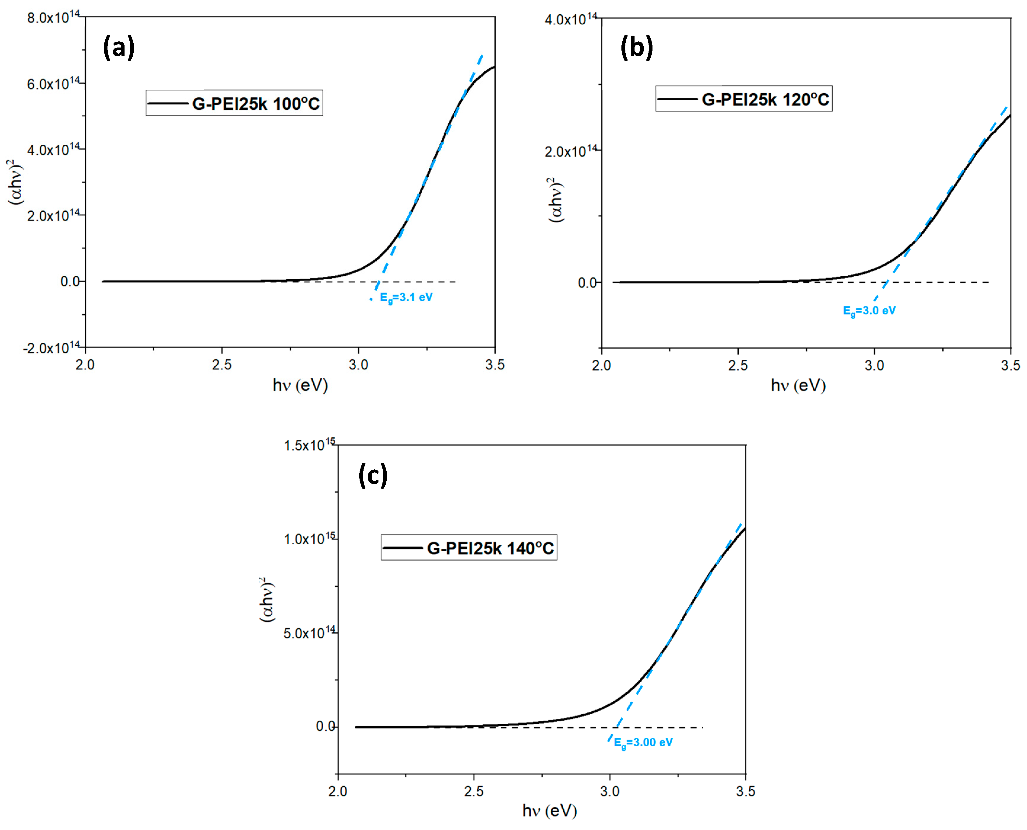
A Tauc plot was used to estimate band gap values. Since the band gap is the energy difference between the valence band and the conduction band of the material, this value can be obtained from the relationship between the photon energy (hν) and the absorption coefficient (α). In a Tauc plot, (αhν)² is plotted against hν, and extrapolation of the linear part of the curve to the energy axis (hν) gives the value of the optical band gap. The Tauc plot equation is as follows:
(αhυ)n = K(hυ-Eg),
where:
α: Absorption coefficient;
A: Absorbance;
h: Plank’s constant;
υ: Frequency (Hz);
K: Energy-independent constant;
Eg: Optical bandgap (eV);
n: Nature of transmission (n = 2 for direct band gap, and ½ for indirect bandgap).
It is plotted (αhυ)2 on the y-axis and hν on the x-axis. Extrapolating the linear part of the curve beyond the absorption edge, the value of the band gap is obtained at the point where the curve intercepts the x-axis.

2.4. Toxicity Test

Brine shrimp eggs were hatched at 30 °C in saline water (35 g/L) for 48 h. Following hatching, 10 brine shrimp nauplii were placed into each well. A total of 18 wells of a 24-well plate were prepared, with each well containing 3 mL of the test solution. The first well served as a blank control, while the remaining wells contained C-Dot concentrations of 10 ppm, 100 ppm, 300 ppm, 750 ppm, 1000 ppm, and 2000 ppm. All experiments were conducted in triplicate to ensure accuracy and reproducibility. The plates were then incubated, and the viability of the marine crustaceans was assessed at 24 and 48 h of exposure.

3. Results and Discussion

3.1. Morphological, Compositional, and Optical Characterization

The heating mechanism in microwave synthesis is due to two types of interactions generated by the electromagnetic field: dipolar polarization and ionic conduction. The constantly varying electric field causes polar molecules to rotate, generating frictional heating. Similarly, ionic movement causes ions to oscillate back and forth, colliding with neighboring molecules or atoms and generating heat [23,26]. We use deionized water as a polar solvent due to its rapid heating capacity and high energy efficiency. Glucose was the carbon source, which has high water solubility and low cost; glucose decomposes to form carbon clusters upon heating. PEI acts as a surfactant and passivating agent, preventing agglomeration during synthesis and maintaining a uniform distribution of C-Dots. It also provides amino functional groups to the surface of the C-Dots [27,28,29,30].
Figure 2a–d shows the morphology of the C-Dots generated at different temperatures. Images suggest that the particles were mainly spherical and had smaller sizes, below 10 nanometers. The microwave-assisted synthesis conditions likely enhanced nucleation while limiting particle growth. Microwave energy provides rapid, uniform heating, which can create localized “hot spots” that accelerate the nucleation phase. This quick energy input should have promoted the formation of a large number of nuclei within a short time, reducing the available material for each individual particle. As a result, the growth phase becomes restricted since the rapid heating does not allow enough time for extended particle growth.
The FT-IR results in Figure 3 illustrate the interaction between the precursors leading to the formation of C-Dots. Glucose exhibits peaks corresponding to alcohol and carbonyl groups, while PEI 25k shows characteristic amine peaks. The C-Dots exhibit combined characteristics of PEI 25k and a new peak around 1630 cm−1 (C=C), indicating the interaction between the precursors and the predominance of PEI 25k on the surface. In previous experiments, the zeta potential of the C-Dots was measured using a Malvern Zetasizer Nano ZS. Zeta potential is determined by placing the solution in a cell with two electrodes, where charged particles migrate toward the electrode with opposite charge at a speed proportional to their net charge. This particle mobility is analyzed via electrophoretic light scattering. Positive zeta potential values of 7.48, 5.67, and 9.81 mV were recorded for C-Dots synthesized at 100 °C, 120 °C, and 140 °C, respectively, suggesting a positively charged surface likely due to the presence of PEI molecules. Generally, zeta potential values above ±30 mV indicate nanoparticle stability with low aggregation risk, whereas values below ±10 mV suggest potential instability and a higher tendency for aggregation. In our results, zeta potential values were below 10 mV, implying a possible tendency for C-Dot agglomeration. These findings indicate that PEI contributes a positive charge to the C-Dots’ surface, helping maintain suspension stability in water through electrostatic repulsion, thus forming a colloidal suspension.
Furthermore, this positive charge is expected to facilitate the generation of energy traps, thereby enhancing their fluorescence [27,30,31]. The broad peak around 3390 cm−1 suggests significant interactions involving O-H and N-H bonds. Additionally, the peak around 1630 cm−1 becomes broader and decreases in intensity as the synthesis temperature increases, indicating changes in the structural organization and interaction of the functional groups involved.
Figure 4 shows the UV-Vis absorbance spectra for the three synthesis temperatures of the C-Dots; the samples’ absorbance range is from 338 to 358 nm.
All C-dots synthesized at different temperatures displayed broad absorption shoulders within the 320–360 nm range, indicating the presence of electronic transitions related to surface states and quantum confinement effects typical of carbon-based nanomaterials. Absorbance values around 320 nm can be attributed to π-π* electronic transitions of C=C bonds, while those near 360 nm can be attributed to n-π* electronic transitions for carbonyl bonds C=O [32]. The change in the peak width of the G-PEI 25k 100 °C sample has bonds in which n-π* electronic interactions predominate. These transitions are typical of C-Dots, which suggest confirmation of obtaining C-Dots as the only material [26,27,28].
Based on the absorbance data from Figure 4, the Tauc plot method was employed to estimate the bandgap values of the C-Dots [33,34,35,36]. These plots are illustrated in Figure 5, revealing a consistent bandgap value of approximately 3.00–3.10 eV for all samples. This value is within the band gap range reported for C-Dots [17,29,30].
The photoluminescence (PL) spectra of the C-Dots are presented in Figure 6. These spectra were measured under different excitation wavelengths to determine the wavelength at which each sample exhibits its maximum emission intensity. For both G-PEI 25k 100 °C and G-PEI 25k 120 °C, the maximum emission was observed around 520 nm with an excitation wavelength of 450 nm, suggesting that these samples share a similar electronic structure. In contrast, G-PEI 25k 140 °C exhibits a maximum emission at 571 nm with an excitation wavelength of 530 nm, likely due to the changes in the structural organization and interactions of the functional groups involved, as observed in the FTIR spectrum around the 1630 cm−1 peak. The redshift at 140 °C could be due to a change in the size of the C-Dots, as higher synthesis temperatures are expected to result in larger particles [21]. This occurs because, at higher temperatures in microwave synthesis, increased thermal energy speeds up particle growth by enhancing the mobility and collisions of reacting species, leading to larger nanoparticles [21,26]. Larger particles emit at longer wavelengths, and the changes in size can influence functional groups, affecting fluorescence efficiency.
The photoluminescence values obtained fall within the range reported for C-Dots. For example. Hinterberger et al. generated C-Dots from citric acid and DL-cysteine via the hydrothermal method, with emissions in the range of 420 to 530 nm [37]. He et al. obtained C-Dots from cow’s milk by the hydrothermal method, which by changing the excitation wavelength from 300 to 550 nm, the fluorescence peak shifted from 409 to 580 nm [38]. Wang et al. obtained c-Dots using the pyrolysis method and citric acid plus urea as precursors, the C-dots presented two fluorescence peaks, at 370 nm and 575 nm [39].
Figure 6d shows deionized water (0) and C-Dots synthesized at 100, 120, and 140 °C (1, 2, and 3, respectively) at concentrations of 4000 ppm, under both natural and ultraviolet light. Deionized water is used as a control to demonstrate the absence of fluorescence. An observable color change from yellow to orange-brown occurs as the synthesis temperature increases from 100 °C to 140 °C. Additionally, C-Dots synthesized at 140 °C exhibit photoluminescence in the yellow range, while those synthesized at 100 °C and 120 °C show fluorescence in the green range. All C-Dots were stable in water, as observed in Figure 6d, which is crucial for subsequent applications.

3.2. Toxicity Test in Brine Shrimp

Brine shrimps were exposed to varying concentrations of each type of C-Dot, and their survival was assessed after 24 and 48 h of exposure. Figure 7 illustrates the results. The experiments were independently repeated three times, with the results presented as mean ± standard deviation (SD). A one-way analysis of variance (ANOVA) was conducted to evaluate the toxicity effects of C-Dot at each concentration for both 24 and 48 h. The results yielded p-values of 0.564 and 0.744, respectively. These p-values indicate that there were no significant differences compared to the control group, suggesting that the C-Dots are not toxic under the evaluated conditions, even at elevated concentrations. Elevated concentrations were used due to reports of toxicity for C-Dots at high concentrations or with prolonged exposure [11,13,14].
C-Dots synthesized at different temperatures demonstrate inertness or non-reactivity towards biological organisms, as evidenced by these studies on brine shrimp. Although positively charged C-Dots might be toxic to brine shrimp due to their increased attraction to cell surfaces and potential generation of reactive oxygen species (ROS) [40], the C-Dots did not induce harmful effects. This suggests a mechanism of non-toxicity that could be attributed to their stable chemical structure. Previous studies on the stability of C-Dots in saline water over a 48 h period suggested that the fluorescence intensity remained constant for C-dots synthesized at 100, 120, and 140 °C (Figure S1). The stability and inertness of these particles significantly enhance their potential applications in various fields, including biomedical imaging, drug delivery, and environmental monitoring, where biocompatibility and safety are critical.
On the other hand, brine shrimp can activate antioxidant mechanisms, including the upregulation of endogenous enzymes such as superoxide dismutase (SOD), catalase (CAT), and glutathione peroxidase (GPx), which help neutralize reactive oxygen species (ROS) and protect the cells from damage [41]. It is known that brine shrimp have efficient excretion processes that prevent the accumulation of C-Dots. These cellular mechanisms enable brine shrimp to adapt to and manage relatively high levels of particles without experiencing significant toxicity [26,42].

4. Conclusions

C-Dots were rapidly synthesized in a single step using the microwave synthesis method, resulting in a spherical morphology with sizes below 10 nm. This innovative approach not only expedites the synthesis process but also facilitates the uniformity of the particles, which is crucial for their optical properties and potential applications. Moreover, the small size of the C-Dots enhances their surface-to-volume ratio, promoting effective interactions with target molecules in various applications, including biomedical imaging and drug delivery.
Furthermore, it can be observed that as the synthesis temperature increases, the size of the C-Dots also increases. This temperature-dependent size variation allows for controlled tuning of the particle dimensions, providing an additional degree of flexibility in the synthesis process. The C-dots also exhibit high stability in water, which can be attributed to the hydrophilicity and positive charge imparted by the PEI coating. This enhancement in dispersion prevents aggregation, ensuring that the C-Dots remain distributed in solution.
To assess their safety, the toxicity of the C-Dots was evaluated using marine crustaceans, specifically brine shrimp. The results confirmed that the C-Dots do not exhibit toxicity, even at elevated concentrations. This finding is crucial as it indicates their biocompatibility and safety, making them suitable for multiple biomedical and environmental applications. Collectively, these properties position the C-Dots as promising candidates for future research and development in nanotechnology.

Supplementary Materials

The following supporting information can be downloaded at https://www.mdpi.com/article/10.3390/micro4040040/s1: Figure S1: Fluorescence spectra of the C-Dots in saline and desionized water, Figure S2. CIE chromaticity coordinates derived from the corresponding maximum emission spectra for each C-Dots, Figure S3. Normalized Fluorescence Spectra of the C-Dots synthesized at different temperatures.

Author Contributions

Conceptualization, S.J.B.-R.; Funding acquisition, S.J.B.-R. and F.R.-V.; Methodology, O.S.-F.; Writing—original draft, S.J.B.-R. and O.S.-F.; Writing—review and editing, S.J.B.-R., O.S.-F., and F.R.-V. All authors have read and agreed to the published version of the manuscript.

Funding

This research was funded by Institutional Funds of the University of Puerto Rico in Ponce and the University of Puerto Rico at Mayaguez.

Institutional Review Board Statement

Not applicable.

Informed Consent Statement

Not applicable.

Data Availability Statement

The data related to this work are not publicly available but can be obtained from the corresponding author upon reasonable request.

Acknowledgments

T.E.M. analyses were performed at the National High Magnetic Field Laboratory, supported by National Science Foundation Cooperative Agreement No. DMR-2128556 and the State of Florida. Authors thank to the Chemistry and Physics Department at the UPRP for the facilities.

Conflicts of Interest

The authors declare no conflicts of interest.

References

  1. Khan, I.; Saeed, K.; Khan, I. Nanoparticles: Properties, Applications and Toxicities. Arab. J. Chem. 2019, 12, 908–931. [Google Scholar] [CrossRef]
  2. Bao, H.; Liu, Y.; Li, H.; Qi, W.; Sun, K. Luminescence of Carbon Quantum Dots and Their Application in Biochemistry. Heliyon 2023, 9, e20317. [Google Scholar] [CrossRef] [PubMed]
  3. Raut, N.S.; Umekar, M.J.; Dhoble, S.J. Quantum Dots: Novel Approach for Biological Imaging. In Quantum Dots; Elsevier: Amsterdam, The Netherlands, 2023; pp. 477–500. [Google Scholar] [CrossRef]
  4. Xu, Q.; Cai, W.; Li, W.; Sreeprasad, T.S.; He, Z.; Ong, W.-J.; Li, N. Two-Dimensional Quantum Dots: Fundamentals, Photoluminescence Mechanism and Their Energy and Environmental Applications. Mater. Today Energy 2018, 10, 222–240. [Google Scholar] [CrossRef]
  5. Gayen, B.; Palchoudhury, S.; Chowdhury, J. Carbon Dots: A Mystic Star in the World of Nanoscience. J. Nanomater. 2019, 2019, 3451307. [Google Scholar] [CrossRef]
  6. Vibhute, A.; Patil, T.; Gambhir, R.; Tiwari, A.P. Fluorescent Carbon Quantum Dots: Synthesis Methods, Functionalization and Biomedical Applications. Appl. Surf. Sci 2022, 11, 100311. [Google Scholar] [CrossRef]
  7. Sengar, P.; Chauhan, K.; Hirata, G.A. Progress on Carbon Dots and Hydroxyapatite Based Biocompatible Luminescent Nanomaterials for Cancer Theranostics. Transl. Oncol. 2022, 24, 101482. [Google Scholar] [CrossRef]
  8. Maruthupandi, M.; Varatharajan, P.; Shameem Banu, I.B.; Hafiz Mamat, M.; Vasimalai, N. White Light Emitting Diode and Anti-Counterfeiting Applications of Microwave Assisted Synthesized Green Fluorescent Carbon Dots Derived from Waste Curry Leaves. Results Opt. 2022, 8, 100249. [Google Scholar] [CrossRef]
  9. Khayal, A.; Dawane, V.; Yadav, V.K.; Yadav, K.K.; Khan, S.H. State of Art Technology: Synthesis of Carbon Dots and Their Potential Applications in Biomedical, Research and Environmental Remediation. Mater. Sci. 2021. preprint. [Google Scholar] [CrossRef]
  10. Ayodhya, D. Recent Progress on Detection of Bivalent, Trivalent, and Hexavalent Toxic Heavy Metal Ions in Water Using Metallic Nanoparticles: A Review. Results Chem. 2023, 5, 100874. [Google Scholar] [CrossRef]
  11. Liu, Y.-Y.; Li, J.-M.; Ji, R.; Zhang, H.; Zhang, W.; Miao, A.-J. Bioaccumulation Determines the Toxicity of Carbon Dots to Two Marine Dinoflagellates. Chemosphere 2023, 321, 138155. [Google Scholar] [CrossRef]
  12. Ghorbani, M.; Moradi, M.; Tajik, H.; Molaei, R.; Alizadeh, A. Carbon Dots Embedded Bacterial Cellulose Membrane as Active Packaging: Toxicity, in Vitro Release and Application in Minced Beef Packaging. Food Chem. 2024, 433, 137311. [Google Scholar] [CrossRef] [PubMed]
  13. Ma, Y.; Liu, Y.; Chen, W.; Li, F.; Guo, R.; Ji, R.; Chen, J. Carbon Quantum Dot-Induced Developmental Toxicity in Daphnia Magna Involves Disturbance of Symbiotic Microorganisms. Sci. Total Environ. 2023, 904, 166825. [Google Scholar] [CrossRef] [PubMed]
  14. Song, Y.; Wu, Y.; Wang, H.; Liu, S.; Song, L.; Li, S.; Tan, M. Carbon Quantum Dots from Roasted Atlantic Salmon (Salmo salar L.): Formation, Biodistribution and Cytotoxicity. Food Chem. 2019, 293, 387–395. [Google Scholar] [CrossRef] [PubMed]
  15. Yao, K.; Lv, X.; Zheng, G.; Chen, Z.; Jiang, Y.; Zhu, X.; Wang, Z.; Cai, Z. Effects of Carbon Quantum Dots on Aquatic Environments: Comparison of Toxicity to Organisms at Different Trophic Levels. Environ. Sci. Technol. 2018, 52, 14445–14451. [Google Scholar] [CrossRef]
  16. Wang, H.; Xie, Y.; Na, X.; Bi, J.; Liu, S.; Zhang, L.; Tan, M. Fluorescent Carbon Dots in Baked Lamb: Formation, Cytotoxicity and Scavenging Capability to Free Radicals. Food Chem. 2019, 286, 405–412. [Google Scholar] [CrossRef]
  17. Ozyurt, D.; Kobaisi, M.A.; Hocking, R.K.; Fox, B. Properties, Synthesis, and Applications of Carbon Dots: A Review. Carbon Trends 2023, 12, 100276. [Google Scholar] [CrossRef]
  18. Saputra, A.M.A.; Piliang, A.F.R.; Goei, R.; Ramadhan, H.R.; Gea, S. Synthesis, Properties, and Utilization of Carbon Quantum Dots as Photocatalysts on Degradation of Organic Dyes: A Mini Review. Catal. Commun. 2024, 187, 106914. [Google Scholar] [CrossRef]
  19. Pandey, V.K.; Tripathi, A.; Taufeeq, A.; Dar, A.H.; Samrot, A.V.; Rustagi, S.; Malik, S.; Bhattacharya, T.; Kovacs, B.; Shaikh, A.M. Significance and Applications of Carbon Dots in Anti Cancerous Nanodrug Conjugate Development: A Review. Appl. Surf. Sci. Adv. 2024, 19, 100550. [Google Scholar] [CrossRef]
  20. Khansili, N. Review on Synthesis and Properties of Carbon Dots with Emphasis to Combat Environmental Toxins. Environ. Adv. 2024, 16, 100542. [Google Scholar] [CrossRef]
  21. Krishnan, R.; Shibu, S.N.; Poelman, D.; Badyal, A.K.; Kunti, A.K.; Swart, H.C.; Menon, S.G. Recent Advances in Microwave Synthesis for Photoluminescence and Photocatalysis. Mater. Today Commun. 2022, 32, 103890. [Google Scholar] [CrossRef]
  22. Kheradmandfard, M.; Kashani-Bozorg, S.F.; Barati, M.R.; Sarfarazijami, S. A Novel Strategy for Fast and Facile Synthesis of Bioactive Bredigite Nanoparticles Using Microwave-Assisted Method. J. Mater. Res. Technol. 2023, 25, 1735–1747. [Google Scholar] [CrossRef]
  23. Gawande, M.B.; Shelke, S.N.; Zboril, R.; Varma, R.S. Microwave-Assisted Chemistry: Synthetic Applications for Rapid Assembly of Nanomaterials and Organics. Acc. Chem. Res. 2014, 47, 1338–1348. [Google Scholar] [CrossRef] [PubMed]
  24. Arulvasu, C.; Jennifer, S.M.; Prabhu, D.; Chandhirasekar, D. Toxicity Effect of Silver Nanoparticles in Brine Shrimp Artemia. Sci. World J. 2014, 2014, 256919. [Google Scholar] [CrossRef]
  25. Varó, I.; Perini, A.; Torreblanca, A.; Garcia, Y.; Bergami, E.; Vannuccini, M.L.; Corsi, I. Time-Dependent Effects of Polystyrene Nanoparticles in Brine Shrimp Artemia Franciscana at Physiological, Biochemical and Molecular Levels. Sci. Total Environ. 2019, 675, 570–580. [Google Scholar] [CrossRef] [PubMed]
  26. Rajabi, S.; Ramazani, A.; Hamidi, M.; Naji, T. Artemia Salina as a Model Organism in Toxicity Assessment of Nanoparticles. DARU J. Pharm. Sci. 2015, 23, 20. [Google Scholar] [CrossRef] [PubMed]
  27. Han, B.; Wang, W.; Wu, H.; Fang, F.; Wang, N.; Zhang, X.; Xu, S. Polyethyleneimine Modified Fluorescent Carbon Dots and Their Application in Cell Labeling. Colloids Surf. B Biointerfaces 2012, 100, 209–214. [Google Scholar] [CrossRef]
  28. Kappe, C.O.; Dallinger, D. Microwaves in Organic and Medicinal Chemistry; John Wiley & Sons: Hoboken, NJ, USA, 2012. [Google Scholar]
  29. Cao, Y.; Wang, X.; Bai, H.; Jia, P.; Zhao, Y.; Liu, Y.; Wang, L.; Zhuang, Y.; Yue, T. Fluorescent Detection of Tetracycline in Foods Based on Carbon Dots Derived from Natural Red Beet Pigment. LWT 2022, 157, 113100. [Google Scholar] [CrossRef]
  30. Guo, Y.; Zhang, J.; Zhang, W.; Hu, D. Green Fluorescent Carbon Quantum Dots Functionalized with Polyethyleneimine, and Their Application to Aptamer-Based Determination of Thrombin and ATP. Microchim Acta. 2019, 186, 717. [Google Scholar] [CrossRef]
  31. Li, R.; Wei, F.; Wu, X.; Zhou, P.; Chen, Q.; Cen, Y.; Xu, G.; Cheng, X.; Zhang, A.; Hu, Q. PEI Modified Orange Emissive Carbon Dots with Excitation-Independent Fluorescence Emission for Cellular Imaging and siRNA Delivery. Carbon 2021, 177, 403–411. [Google Scholar] [CrossRef]
  32. Ghanem, A.; Al-Qassar Bani Al-Marjeh, R.; Atassi, Y. Novel Nitrogen-Doped Carbon Dots Prepared under Microwave-Irradiation for Highly Sensitive Detection of Mercury Ions. Heliyon 2020, 6, e03750. [Google Scholar] [CrossRef]
  33. Pramudita, R.; Marpongahtun; Gea, S.; Daulay, A.; Harahap, M.; Tan, Y.Z.; Goei, R.; Tok, A.I.Y. Synthesis of Fluorescent Citric Acid Carbon Dots Composites Derived from Empty Fruit Bunches of Palm Oil Tree and Its Anti-Bacterial Property. Case Stud. Chem. Environ. Eng. 2022, 6, 100277. [Google Scholar] [CrossRef]
  34. Barrientos, K.; Gaviria, M.I.; Arango, J.P.; Placido, J.; Bustamante, S.; Londoño, M.E.; Jaramillo, M. Synthesis, Characterization and Ecotoxicity Evaluation of Biochar-Derived Carbon Dots from Spruce Tree, Purple Moor-Grass and African Oil Palm. Processes 2021, 9, 1095. [Google Scholar] [CrossRef]
  35. Jubu, P.R.; Yam, F.K.; Igba, V.M.; Beh, K.P. Tauc-Plot Scale and Extrapolation Effect on Bandgap Estimation from UV–Vis–NIR Data—A Case Study of β-Ga2O3. J. Solid State Chem. 2020, 290, 121576. [Google Scholar] [CrossRef]
  36. Shim, H.S.; Kim, J.M.; Jeong, S.; Ju, Y.; Won, S.J.; Choi, J.; Nam, S.; Molla, A.; Kim, J.; Song, J.K. Distinctive Optical Transitions of Tunable Multicolor Carbon Dots. Nanoscale Adv. 2022, 4, 1351–1358. [Google Scholar] [CrossRef] [PubMed]
  37. Hinterberger, V.; Damm, C.; Haines, P.; Guldi, D.M.; Peukert, W. Purification and Structural Elucidation of Carbon Dots by Column Chromatography. Nanoscale 2019, 11, 8464–8474. [Google Scholar] [CrossRef]
  38. He, S.; Turnbull, M.J.; Nie, Y.; Sun, X.; Ding, Z. Band Structures of Blue Luminescent Nitrogen-Doped Graphene Quantum Dots by Synchrotron-Based XPS. Surf. Sci. 2018, 676, 51–55. [Google Scholar] [CrossRef]
  39. Wang, C.; Yang, M.; Shi, H.; Yao, Z.; Liu, E.; Hu, X.; Guo, P.; Xue, W.; Fan, J. Carbon Quantum Dots Prepared by Pyrolysis: Investigation of the Luminescence Mechanism and Application as Fluorescent Probes. Dyes Pigment. 2022, 204, 110431. [Google Scholar] [CrossRef]
  40. Li, Q.; Shen, X.; Xing, D. Carbon Quantum Dots as ROS-Generator and -Scavenger: A Comprehensive Review. Dyes Pigment. 2023, 208, 110784. [Google Scholar] [CrossRef]
  41. Tapia-Salazar, M.; Diaz-Sosa, V.R.; Cárdenas-Chávez, D.L. Toxicological Effect and Enzymatic Disorder of Non-Studied Emerging Contaminants in Artemia Salina Model. Toxicol. Rep. 2022, 9, 210–218. [Google Scholar] [CrossRef]
  42. Browne, R.A.; Sorgeloos, P.; Trotman, C.N.A. (Eds.) Artemia Biology; CRC Press: Boca Raton, FL, USA, 1991. [Google Scholar]
Figure 1. Synthesis route of C-Dots.
Micro 04 00040 g001
Figure 2. High-resolution transmission electron microscopy (HRTEM) images of C-Dots synthesized at (a,b) 120 °C and (c,d) 140 °C. The white arrows point to the nanoparticles.
Micro 04 00040 g002
Figure 3. D-Glucose, polyethyleneimine, and C-Dots FTIR spectra.
Micro 04 00040 g003
Figure 4. UV-Vis spectra of C-Dots. The circle highlights electronic transitions.
Micro 04 00040 g004
Figure 5. Tauc plot of C-Dots: (a) G-PEI 25k 100 °C; (b) G-PEI 25k 120 °C; (c) G-PEI 25k 140 °C.
Micro 04 00040 g005
Figure 6. Fluorescence spectra of C-Dots synthesized at (a) 100 °C, (b) 120 °C, and (c) 140 °C. (d) Samples under both natural and ultraviolet light.
Micro 04 00040 g006
Figure 7. Viability of brine shrimp to the different C-Dots (G-PEI 25k 100 °C, G-PEI 25k 120 °C and G-PEI 25k 140 °C) at concentrations from 10 ppm to 2000 ppm, evaluated to times of 24 and 48 h.
Micro 04 00040 g007
Disclaimer/Publisher’s Note: The statements, opinions and data contained in all publications are solely those of the individual author(s) and contributor(s) and not of MDPI and/or the editor(s). MDPI and/or the editor(s) disclaim responsibility for any injury to people or property resulting from any ideas, methods, instructions or products referred to in the content.