
Article

Comparative Insecticidal Efficacy of Symbiotic Bacteria (Xenorhabdus and Photorhabdus) and Their Bacterial Fractions Against Fall Armyworm, Spodoptera frugiperda

by
Wandee Wattanachaiyingcharoen
1,*,
Aunchalee Thanwisai
2,
Apichat Vitta
2,
Patcharapun Wanitsumran
1,
Supawan Pansri
2 and
Det Wattanachaiyingcharoen
3,4
1
Department of Biology, Faculty of Science, Naresuan University, Phitsanulok 65000, Thailand
2
Department of Microbiology and Parasitology, Faculty of Medical Science, Naresuan University, Phitsanulok 65000, Thailand
3
Department of Agricultural Sciences, Faculty of Agriculture, Natural Resources and Environment, Naresuan University, Phitsanulok 65000, Thailand
4
Agriculture and Environmental Integration Research and Development Unit (AEI), Faculty of Agriculture, Natural Resources and Environment, Naresuan University, Phitsanulok 65000, Thailand
*
Author to whom correspondence should be addressed.
Appl. Microbiol. 2026, 6(8), 89; https://doi.org/10.3390/applmicrobiol6080089
Submission received: 19 June 2026 / Revised: 30 July 2026 / Accepted: 30 July 2026 / Published: 5 August 2026

Abstract

Background: The fall armyworm, S. frugiperda, is a globally threatening insect pest of agricultural crops, including in Thailand. Symbiotic bacteria in the genera Xenorhabdus and Photorhabdus, which are derived from entomopathogenic nematodes, can produce diverse bioactive compounds with insecticidal activities. However, no study on Thai isolates of these symbiotic bacteria against S. frugiperda has been reported, leading to a significant gap in local biopesticide development. Methods: Two symbiotic bacterial isolates, X. miraniensis (bMH16.1_TH) and P. akhurstii (bSBR11.1_TH), were isolated from Thai entomopathogenic nematodes and cultured in LB broth at 108 CFU/mL. Three bacterial fractions, i.e., whole-cell suspension, cell supernatant, and cell pellet, were prepared from each isolate. Insecticidal bioassays were conducted by topical application on second- and fifth-instar larvae of S. frugiperda (10 larvae × 5 replicates per treatment, CRD). Larval mortality was recorded daily for seven days, and median lethal time (LT50) was calculated. Data were analyzed by two-way ANOVA with Duncan’s Multiple Range Test (p < 0.05). Results: The results indicated that both bacterial isolates caused significantly higher larval mortality than controls across all treatments (p < 0.05). X. miraniensis (bMH16.1_TH) showed higher virulence, achieving ~80% mortality of second-instar larvae within 24 h and 100% by day three. Whole-cell suspensions consistently produced the fastest and highest mortality with the lowest LT50 values in both larval stages. Second-instar larvae were significantly more susceptible than fifth-instar larvae. The shortest LT50 was recorded in second-instar larvae treated with X. miraniensis supernatant at 0.616 days, while fifth-instar larvae generally showed higher LT50 values across all treatments. Conclusions: Our comparative study demonstrated that the insecticidal efficacy varied between the two symbiotic bacterial isolates and among their fractions. Whole-cell suspensions of X. miraniensis (bMH16.1_TH) produced the highest larval mortality and the smallest LT50 values, particularly against early-instar larvae. These findings demonstrated that local symbiotic bacteria are promising candidates to be developed as sources of biopesticides against this insect pest. In addition, characterizing active insecticidal compounds, evaluating field efficacy, and assessing safety to non-target organisms need to be further studied to support integration of these biopesticides in sustainable pest management programs.

1. Introduction

Fall armyworm, Spodoptera frugiperda (Smith) (Lepidoptera: Noctuidae), is one of the most destructive lepidopteran pests, affecting a wide range of agricultural crops worldwide. This insect is polyphagous; it can infest more than 350 plant species, including corn, rice, sorghum, sugarcane, cotton, soybean, wheat, and vegetables [1]. Damage on economic crops in many regions has threatened agricultural production and food security. Due to its high reproductive ability, capability to migrate, broad host range, and potential to develop resistance to chemical insecticides, S. frugiperda has become an invasive insect species throughout major agricultural regions worldwide [2,3]. This pest can cause substantial economic losses in corn production. Yield losses of up to 77% have been reported when crops are attacked by late-instar larvae, while damage to leaves, silks, and tassels ranging from 25% to 50% can reduce grain yield by as much as 58% [4,5]. Since its first detection in Thailand in 2018, infestations have rapidly spread across the country and it has become a major insect pest [6,7,8]. Chemical insecticide applications are the primary control strategy, but they have negative impacts on the environment and non-target organisms, and there is a growing problem of insecticide resistance. There have been reports of resistance in this insect species to organophosphate (OP) and pyrethroid (PYR) insecticides in several areas of America and Africa [9]. Hence, alternative approaches, such as the use of biological control agents, have received considerable attention as they are promising eco-friendly tools for insect management.
Entomopathogenic nematodes (EPNs) are among the effective biological control agents. The pathogenicity of EPNs is primarily determined by their symbiotic bacteria, principally Xenorhabdus and Photorhabdus spp. Once the EPNs enter the host body, these bacteria are released into the insect hemocoel, where they reproduce and release diverse bioactive compounds, including toxins, enzymes, antimicrobial metabolites, and immunosuppressive compounds, leading to host death within a short period [10,11]. Practical applications of whole EPNs in field conditions can reduce nematode survival and infectivity due to constraints by environmental factors such as temperature fluctuations, UV radiation, and soil moisture availability [12]. These limitations, combined with other challenges, such as maintaining viable nematode cultures and stability during storage and transportation, have led to interest in exploiting bacterially derived metabolites and cell preparations as standalone biopesticides [13]. Therefore, the utilization of symbiotic bacteria has been considered an alternative potential control strategy. These symbiotic bacteria are responsible for suppressing the host’s immune system, inducing digestive organ malfunctions, and causing host death [8]. Various species of Xenorhabdus and Photorhabdus have been explored from several regions, including Thailand. Refs. [14,15] reported several Thai EPN-associated symbiotic bacteria that have demonstrated insecticidal potential against Aedes aegypti larvae. The results suggested that native bacterial isolates may harbor distinct virulence traits effective against economically important insects adapted to tropical conditions.
The deployment of symbiotic bacteria depends on suspension preparations. Several investigations have demonstrated that the insecticidal activity of bacteria varies among fractions. The whole-cell suspension is a fraction that contains viable cells, which can multiply and continue to produce toxins after infection in the host body. Cell-free supernatant is a suspension without living cells and contains extracellular materials, secreted toxins, and other bioactive metabolites, whereas cell pellet suspension retains intracellular substances and membrane-associated virulence factors [16,17,18]. Hence, each fraction can perform different activities according to its composition. To use them as biological control pesticides, the effectiveness of each fraction should be examined to optimize bacterial preparations. Considering the suitable applications, the developmental stage of the insect host has a significant influence on susceptibility to microbial infection, particularly in early stages that are considerably susceptible to pathogen infections. Even though, at these stages, they consume less plant tissue, they are in the phase of dispersing to colonize host plants. In contrast, later instars cause significant damage due to higher food consumption capacity and can increase resistance to microbial infection as they approach full development [19,20]. Identifying the most susceptible larval stages is therefore critical for designing targeted biocontrol strategies that maximize pest mortality while minimizing the quantities of biopesticide required.
Despite the demonstrated potential of EPN-associated symbiotic bacteria as biocontrol agents against important insect pests, no study has evaluated Thai isolates against S. frugiperda. This gap limits our understanding of the insecticidal efficacy of native bacterial strains against one of the major agricultural pests in Thailand and other tropical regions. To clarify this gap, we evaluated the insecticidal efficacy of X. miraniensis (bMH16.1_TH) and P. akhurstii (bSBR11.1_TH), derived from Thai EPN populations, against S. frugiperda. We compared the pathogenicity of different bacterial fractions, including whole-cell, cell supernatant, and cell pellet fractions, against second- and fifth-instar larvae. The findings of this study may contribute to the development of locally sourced bacterial biopesticides as effective and sustainable alternatives for fall armyworm management in Thailand and other tropical agricultural regions.

2. Materials and Methods

2.1. Collection and Rearing of Fall Armyworm (S. frugiperda)

Larvae of fall armyworm, S. frugiperda, were collected from infested maize fields in northern Thailand. The species were verified according to the identification procedures of [21]. The larvae were maintained individually in 20 mL plastic containers and fed fresh pesticide-free maize leaves under laboratory conditions (25 ± 2 °C, 60 ± 10% RH, and a 14:10 h light:dark photoperiod). Pupae were placed in a plastic container and transferred into an adult emergence cage (30  ×  30  ×  30 cm), with young maize plants provided as an oviposition substrate. Upon emergence, the adults were provided with a 10% sugar solution as food. The insects were reared under laboratory conditions for at least two generations prior to the tests. This was to eliminate potential effects of previous environmental exposure, especially pesticide residues and/or infections by other natural enemies. Only healthy larvae and those with no visible signs of injury or deformity were selected for the experiments to ensure the homogeneity of experimental outcomes.

2.2. Isolation and Multiplication of Symbiotic Bacteria

The two symbiotic bacteria were isolated from their entomopathogenic nematode hosts according to [14,15]. Xenorhabdus miraniensis (bMH16.1_TH) (GenBank: KY404031) is associated with the entomopathogenic nematode Steinernema websteri, and Photorhabdus akhurstii (bSBR11.1_TH) (GenBank: MK478079) is associated with Heterorhabditis indica. Each nematode–bacterium complex suspension (50 μL) was cultured on Petri dishes with NBTA (Nutrient Agar (NA), Bromothymol blue, and 2,3,5-Triphenyl tetrazolium chloride, supplemented with Ampicillin (50 mg/mL)) [22]. They were incubated for 48 h at 25 ± 2 °C in the dark, before bacterial colonies were selected. After three to four days of room-temperature incubation, bacterial colonies were selected based on their characteristics. Xenorhabdus species were verified as rounded colonies with smooth margins and the ability to absorb colorants [23], whereas Photorhabdus was identified based on a light or dark green colony color with a convex or umbonated surface on NTBA [24].

2.3. Preparations of Bacterial Fractions (Whole-Cell, Cell Supernatant, and Cell Pellet Suspensions)

Each symbiotic bacterial isolate mentioned above was transferred from NBTA medium stock culture to Luria–Bertani (LB) medium and incubated at 25 °C in the dark and shaken at 150 rpm for 24 h. Subsequently, 100 mL of the bacterial suspension was transferred into 250 mL of fresh LB broth and further incubated under the same conditions for 48 h, following the methods described by [15].
The bacterial suspension was then serially diluted using a ten-fold dilution method, and the bacterial density was determined by the spread-plate technique, which resulted in an approximate concentration of 108 CFU/mL. The same stock bacterial suspension was divided into two equal volumes. The first volume was used as the whole-cell suspension and applied directly to this concentration. The second volume was transferred to prepare the cell supernatant and cell pellet suspensions. To separate these fractions, the suspension was centrifuged at 10,000× g for ten minutes. After centrifugation, the cell supernatant was collected and stored in 15 mL centrifuge tubes and used without filtration. The cell pellet fraction was prepared by adding the same volume of sterile distilled water to the remaining fraction, containing bacterial cells. Each fraction was tested using five technical replicates within a single experimental run.
The whole-cell suspension consisted of the bacterial culture in LB broth. After centrifugation, the supernatant was collected and used directly without further filtration. The bacterial pellet was resuspended in sterile distilled water to the original culture volume. All three bacterial preparations were kept at 4 °C until the bioassays.

2.4. Insecticidal Bioassay

Three treatments consisting of 0.5 mL of each bacterial suspension (whole-cell, cell supernatant, and cell pellet suspensions) and a control treatment were examined. Second- and fifth-instar larvae were tested separately. Each treatment consisted of five replicates with ten larvae per replicate in a completely randomized design (CRD).
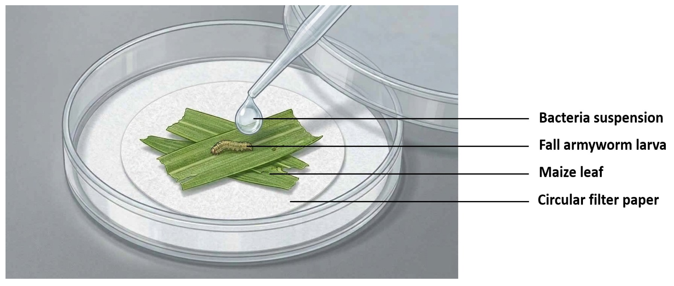
Each larva was individually placed in a Petri dish (5.5 cm in diameter) containing a detached maize leaf as food for the fall armyworm. For each treatment, 0.5 mL of the bacterial suspension was applied topically to larvae, to the leaf surface, and on the bottom of the Petri dish, to ensure that the tested larvae received an adequate volume of bacterial suspensions (Figure 1). A nominal bacterial dose of approximately 5 × 10⁷ CFU was used in each treatment. Sterile distilled water was used for control treatment. Treated larvae were maintained under laboratory conditions, and mortality was recorded daily for seven days.

2.5. Mortality Assessment

Larvae were considered dead when they showed no response to gentle stimulation with forceps. The observed mortality was calculated as
Observed   mortality = Total   number   of   dead   larvae × 100 ÷ Total   number   of   tested   larvae

2.6. Median Lethal Time (LT50)

The median lethal time (LT50) of each treatment was calculated based on days after infection, with the objective of comparing the period (days) that each bacterial isolate required to induce 50% death in the examined larvae. This value compared the pathogenicity among bacterial isolates and fractions.

2.7. Statistical Analysis

Mortality data from the control and treatment groups (each bacterial isolate) were analyzed using SPSS 17.0 at a significance level of p < 0.05. Two-way analysis of variance (ANOVA) was performed to evaluate the effect of bacterial treatment and infection status (infected vs. control) on larval mortality. When significant differences were detected, post hoc comparisons were conducted using Duncan’s Multiple Range Test (DMRT) to determine differences among treatments and observation times after infection. The lethal time (LT50) values were estimated using SPSS software following the method described by [25].

3. Results

3.1. Insecticidal Activity of Symbiotic Bacteria Against S. frugiperda

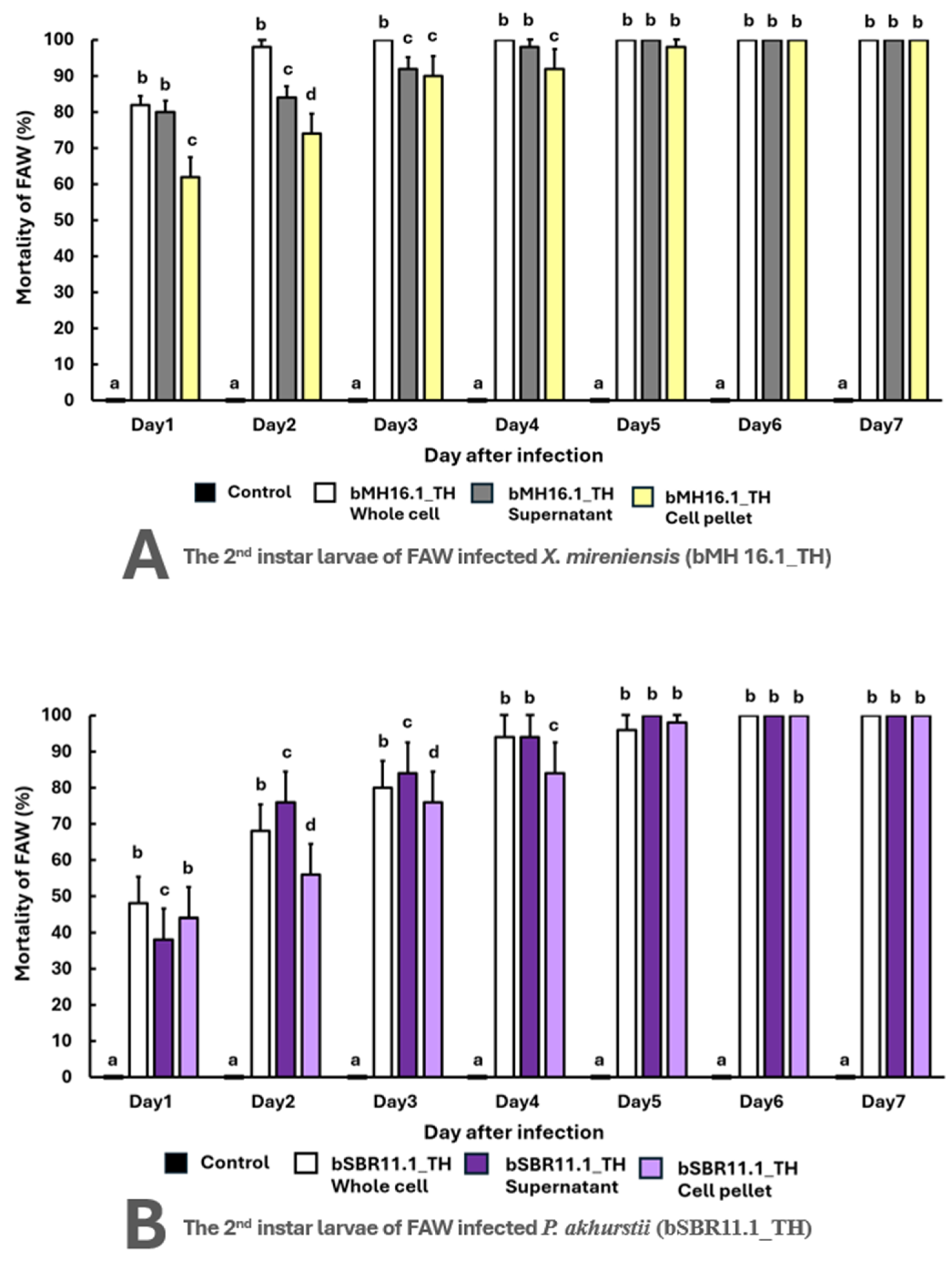
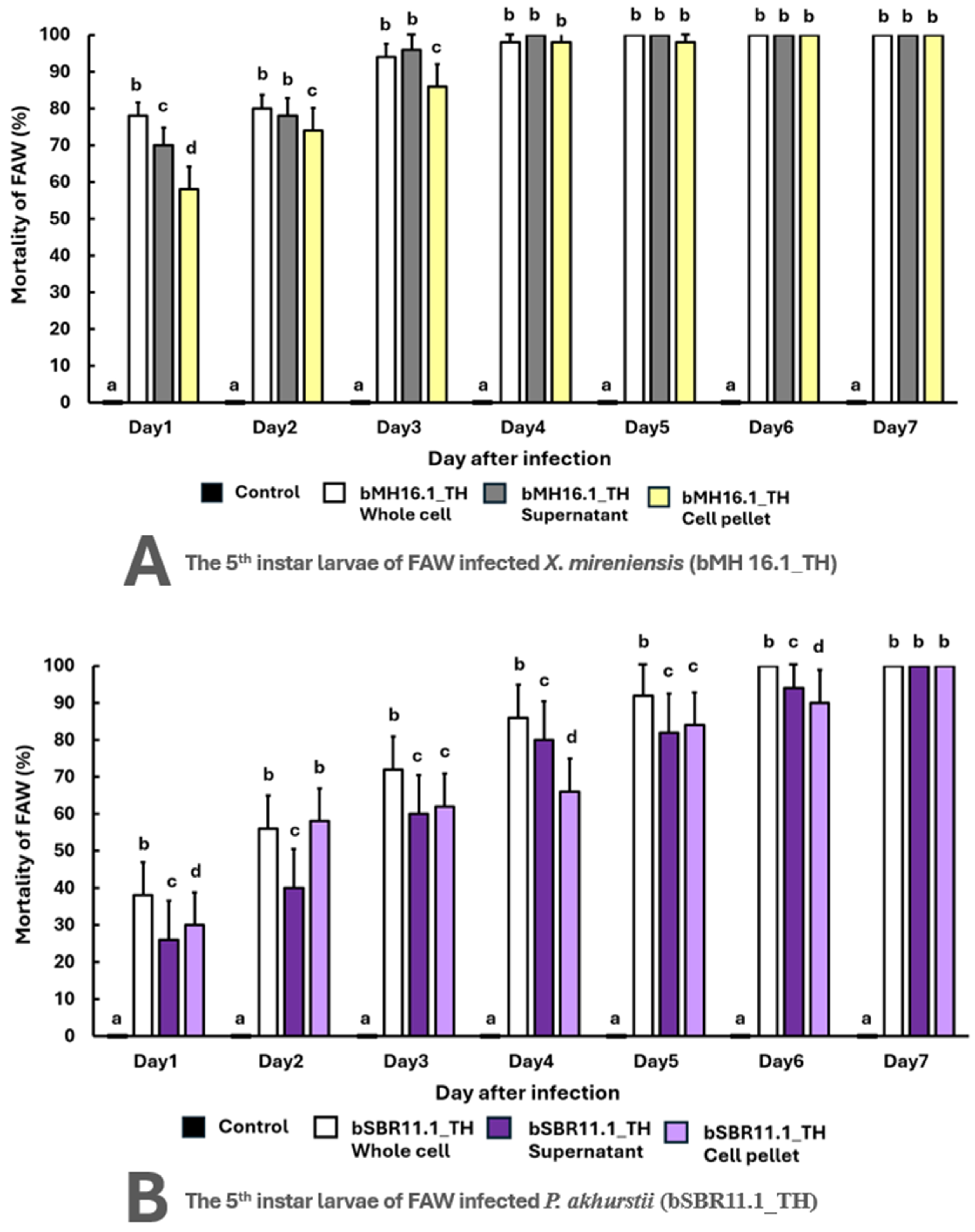
Both X. miraniensis (bMH16.1_TH) and P. akhurstii (bSBR11.1_TH) exhibited significant insecticidal activity against second- and fifth-instar larvae of S. frugiperda compared to the control (p < 0.05) (Figure 2 and Figure 3). No mortality was observed in the control treatments throughout the experimental period.
Among the tested bacterial isolates, X. miraniensis (bMH16.1_TH) consistently induced higher and more rapid larval mortality than P. akhurstii (bSBR11.1_TH). In second-instar larvae, X. miraniensis (bMH16.1_TH) caused considerable mortality within 24 h after infection and reached 100% on day three (Figure 2A). Mortality caused by P. akhurstii (bSBR11.1_TH) increased more gradually over time, with mortality generally occurring later, with around 30% on the first day after infection. The mortality increased at a slower rate and reached 100% later on day five or six (Figure 2B).
In both larval stages, whole-cell suspensions exhibited considerably greater and more rapid insecticidal activity than supernatant and cell pellet fractions among the examined bacterial fractions (p < 0.05) (Figure 2 and Figure 3). The whole-cell fraction of X. miraniensis (bMH16.1_TH) killed approximately 80% of the second-instar larvae in the first 24 h and 100% of the larvae in three days. While the cell pellet required up to six days to reach 100% mortality, the supernatant fraction treatment reached full mortality by day five (Figure 2A).
Infection by P. akhurstii (bSBR11.1_TH) resulted in a lower mortality rate (Figure 2B). The whole-cell fraction caused higher and faster mortality, with total mortality observed on day five and day six in the second and fifth instars, respectively. Day five of the second-instar treatment showed a 100% mortality rate, despite the supernatant fraction causing slower mortality. On the seventh day following infection, both fractions resulted in 100% death in the older stage (Figure 3A,B).

3.2. Efficacy of Different Bacterial Fractions on Larval Mortality

In both larval stages, whole-cell suspensions exhibited comparable higher insecticidal activity than supernatant and cell pellet fractions among the examined bacterial fractions (p < 0.05), particularly during the early period after infection (Figure 2 and Figure 3). The whole-cell fraction of X. miraniensis (bMH16.1_TH) killed approximately 80% of the second-instar larvae in the first 24 h and 100% of the larvae in three days. While the cell pellet and the supernatant fractions required up to five days to reach 100% mortality. Infection by P. akhurstii (bSBR11.1_TH) resulted in a lower mortality rate. The whole-cell fraction caused higher and faster mortality, with total 100% mortality observed on day five and day six in the second and fifth instars, respectively. Day five of the second-instar treatment showed a 100% mortality rate, despite the supernatant fraction causing slower mortality. On the seventh day following infection, both fractions resulted in 100% death in the older stage (Figure 3A,B).

3.3. Efficacy of Larval Instar Developmental Stages

Larval susceptibility to bacterial infection depends upon developmental stages. In all three bacterial fractions, the time required to reach complete mortality was consistently longer in older larvae. Second-instar larvae were significantly more susceptible, as evidenced by higher mortality and shorter time to death (p < 0.05) (Figure 1 and Figure 2). The results indicated a clear reduction in susceptibility with increasing larval age.

3.4. Lethal Time (LT50) Analysis

The median lethal time (LT50) values were computed to evaluate the virulence of bacterial fractions and isolates. Higher virulence occurred with the bacteria X. miraniensis (bMH16.1_TH), particularly in the second-instar larvae (Table 1). The supernatant fraction exhibited the shortest LT50 of 0.616 days (95% fiducial limit: 0.475–0.740 days), followed by the whole-cell suspension at 0.670 days (95% fiducial limit: 0.352–0.711 days), which was not significantly different (p > 0.05). The cell pellet fraction showed a substantially longer LT50 of 2.197 days (95% fiducial limit: 1.178–2.560 days) and differed significantly from the other two treatments (p < 0.05). On the other hand, the whole-cell and supernatant fractions of P. akhurstii (bSBR11.1_TH) required significantly longer time to reach LT50 values of 1.526 (95% fiducial limit: 1.102–1.707 days) and 1.720 days (95% fiducial limit: 1.220–2.401 days) (p > 0.05), respectively. The cell pellet fraction was the least effective in killing these earlier-instar insects, with an LT50 value of 2.170 days (95% fiducial limit: 1.765–2.357 days) and statistically significant differences from the other two fractions (p < 0.05).
As larval maturity increased to the fifth instar, the time required to achieve LT50 generally increased (Table 2), and the pattern of significant differences varied among bacterial fractions. The whole-cell treatment of X. miraniensis (bMH16.1_TH) remained the most effective, showing the lowest LT50 at 1.748 days (95% fiducial limit: 1.351–1.748 days), followed by the cell pellet fraction (1.891 days) (95% fiducial limit: 0.923–2.185 days) and supernatant fraction (1.915 days) (95% fiducial limit: 1.539–1.915 days) (Table 2). However, the overlapping fiducial limits and statistical comparisons indicated that the whole-cell suspension and cell supernatant were not significantly different (p > 0.05).
Similar to those in the younger larvae, P. akhurstii (bSBR11.1_TH) exhibited a higher LT50 value. Among the bacterial fractions, the cell pellet fraction showed the lowest LT50 value of 2.128 days (95% fiducial limit: 1.847–2.302 days), followed by the supernatant fraction, with a value of 2.182 days (95% fiducial limit: 1.974–2.249 days) (p > 0.05), whereas the whole-cell treatment showed the highest LT50 value at 2.551 days (95% fiducial limit: 2.326–2.703 days). The whole-cell suspension did not differ significantly from the cell pellet fraction (p > 0.05), but differed significantly from the supernatant treatment (p < 0.05). These results suggested that p. akhurstii (bSBR11.1_TH) was not the fastest-acting preparation, and numerical differences among fractions should be interpreted in conjunction with the statistical comparisons.

4. Discussion

The current study demonstrated that Xenorhabdus and Photorhabdus, two symbiotic bacteria derived from entomopathogenic nematodes, possess insecticidal efficacy against S. frugiperda. There were remarkable differences in pathogenicity among bacterial isolates, bacterial fractions, and larval developmental stages. This finding is consistent with previous studies indicating that Xenorhabdus and Photorhabdus possess bioactive potential and can produce toxins, enzymes, and immunosuppressive compounds that contribute to rapid host mortality [10,11]. Throughout all larval stages and bacterial preparations, X. miraniensis (bMH16.1_TH) continuously displayed greater virulence than P. akhurstii (bSBR11.1_TH), as indicated by higher mortality rates and lower LT50 values. Variations in virulence among bacterial isolates may be associated with differences in the composition and expression of these metabolites [26,27]. These results were consistent with previous studies showing that Xenorhabdus species can produce highly potent insecticidal substances that disrupt insect midgut function, suppress immune responses, induce septicemia, and eventually cause host death [11]. The production of toxin complexes (Tc toxins) and other virulence-associated compounds, which are common in Xenorhabdus and Photorhabdus species, may be responsible for the rapid insecticidal activity observed in this study [28,29]. Tc toxins specifically damage insect midgut epithelium and lumen, disrupting nutrient absorption and causing progressive tissue destruction [30]. In this study, differences in insecticidal activity between X. miraniensis (bMH16.1_TH) and P. akhurstii (bSBR11.1_TH) were observed, suggesting that pathogenicity among symbiotic bacteria of entomopathogenic nematodes is highly species- and isolate-specific. The superior virulence of X. miraniensis (bMH16.1_TH) likely reflects isolate-specific differences in the expression and diversity of these virulence-associated compounds, though the exact metabolite profiles of this Thai isolate remain to be fully characterized. Future metabolomic analyses would help elucidate which specific compounds drive the observed insecticidal potency. However, since this study emphasizes only two Thai local bacterial isolates, further investigations involving additional species of Xenorhabdus and Photorhabdus should be considered in order to expand the possibility of using symbiotic bacteria as biopesticides.
Whole-cell suspensions of both bacterial isolates showed the highest and fastest insecticidal activity across all bacterial isolates and larval developmental stages. These results coincide with previous research demonstrating that whole-cell fractions contain both extracellular and intracellular bioactive compounds, which may enhance pathogenicity by continuing the synthesis of toxins, enzymes, and immunosuppressive factors within the host body [16,17]. In addition, the whole-cell suspension used in this study was suspended in its original culture medium, which may have allowed continued bacterial metabolism and the production of additional extracellular metabolites during the bioassay. Consequently, the observed insecticidal activity likely reflects the combined effects of viable bacterial cells and their naturally occurring metabolites rather than bacterial cells alone. In addition, the whole-cell suspension used in the present study was standardized to 1 × 10⁸ CFU/mL prior to fractionation, corresponding to approximately 5 × 10⁷ CFU per treatment, which is within the range commonly applied for Xenorhabdus and Photorhabdus bioassays against insect pests [15,31]. Nevertheless, comparisons with previous studies on lepidopteran pests remain limited because bacterial inocula are frequently reported as culture broth or optical density rather than viable cell counts (CFU/mL) [32,33]. Standardized dose–response assays, standardized buffer-based preparations, and quantification of individual bioactive compounds are needed to improve experimental reproducibility and facilitate comparison among studies. Considering that the supernatant fraction contains mainly extracellular metabolites, whereas the cell pellet fraction contains intracellular and membrane-associated compounds, resulting in reduced and delayed insecticidal effects [34,35], the insecticidal activity observed in the cell supernatant is likely associated with a complex mixture of extracellular bioactive compounds rather than a single component. In addition to secreted protein toxins and secondary metabolites, Gram-negative bacteria (Xenorhabdus and Photorhabdus) also release lipopolysaccharides into the extracellular environment, which may contribute to biological activity [36]. However, the present study did not characterize or quantify individual extracellular components; therefore, the relative contribution of lipopolysaccharides and other secreted metabolites could not be determined.
Because the supernatant fraction used in this study was not membrane-filtrated before the bioassay, the possible presence of residual viable bacterial cells cannot be completely excluded. Hence, we might assume that part of the observed insecticidal activity may have resulted from both extracellular metabolites and a limited number of remaining bacterial cells. This would be a limitation of the study. So, we suggest that further studies on insecticidal activity of bacterial supernatant should involve filtration to distinguish the effects of secreted metabolites from those of live bacteria.
Larval developmental stage also played a critical role in determining susceptibility to bacterial infection. Compared to fifth-instar larvae, second-instar larvae were more vulnerable, as evidenced by higher mortality rate and lower LT50 values. According to previous research, younger insect stages are typically more susceptible to pathogen infections due to their thinner cuticles, less developed immune systems, and higher physiological sensitivity [37,38]. Additionally, early instars of S. frugiperda have been reported to be more susceptible to entomopathogenic nematode infections [39]. Tolerance in older larval instars is often attributed to more developed physiological and immunological defense, including stronger physical barriers such as thicker cuticles. Furthermore, they can enhance cellular encapsulation and increase antimicrobial peptide production, which collectively reduce pathogen efficacy and delay death [40,41,42]. In addition to mortality results, the median lethal time values (LT50) provided additional quantitative results of the speed of bacterial action. In X. miraniensis (bMH16.1_TH) treatment, lower LT50 values, particularly in second-instar larvae (0.616 days), indicated faster action against the insect host, which is a desired property for biological control agents. According to [29], rapid host mortality minimizes feeding damage and reduces the likelihood of resistance development. This plays an important role as a control strategy, especially for S. frugiperda, as they can cause severe yield losses in later feeding stages [2]. However, fiducial limits showed some considerable overlap, suggesting that differences between some treatments might not be statistically significant. This finding suggests that both whole-cell suspension and cell supernatant fractions may contain sufficient insecticidal factors to produce a similar rate of larval mortality. Additional studies under field conditions are required to confirm their relative performance against S. frugiperda.
Nevertheless, as our bioassays were conducted under controlled laboratory conditions, the results may not fully reflect bacterial performance in different conditions. In complex and variable environments of agricultural fields, factors such as temperature fluctuations, UV radiation, and competition with soil microorganisms can substantially affect bacterial viability and efficacy. Hence, field trials should be conducted to validate the efficacy of these isolates under realistic agricultural conditions. In addition, metabolomic profiling of active bioactive compounds should be pursued to identify and characterize the specific insecticidal compounds responsible for the observed pathogenicity. To implement this bacterial-based biopesticide, the stability of these bacterial preparations needs to be investigated.

5. Conclusions

This study demonstrated the significant insecticidal activity of two symbiotic bacteria isolated from entomopathogenic nematodes, X. miraniensis (bMH16.1_TH) and P. akhurstii (bSBR11.1_TH), against S. frugiperda larvae. X. miraniensis (bMH16.1_TH) showed higher pathogenicity across all larval instars and suspension fractions. Of the three bacterial fractions tested, whole-cell suspensions were the most effective, followed by cell pellet and cell supernatant fractions. The mortality rate of younger (second-instar) larvae increased rapidly after infection, and this larval stage was more susceptible to bacterial infection than older (fifth-instar) larvae. These results indicate the potential of X. miraniensis (bMH16.1_TH) as a promising biological control agent against S. frugiperda, with both whole-cell suspension and cell supernatant fractions demonstrating comparable insecticidal efficacy. Further investigations should focus on clarifying the active insecticidal compounds produced by this bacterium, evaluating its efficacy and stability under field conditions, and assessing its safety to non-target organisms, in order to integrate the bacteria-based biopesticide into sustainable biological control programs.

Author Contributions

Conceptualization, W.W.; Methodology, W.W., P.W. and S.P.; Validation, W.W., A.V., A.T. and D.W.; Formal Analysis, W.W. and P.W.; Investigation, W.W. and P.W.; Resources, W.W., A.T. and D.W.; Data Curation, W.W. and P.W.; Writing—Original Draft Preparation, W.W. and P.W.; Writing—Review and Editing, W.W., D.W., A.T. and A.V.; Visualization, W.W. and P.W.; Supervision, W.W. and D.W.; Project Administration, W.W.; Funding Acquisition, W.W. All authors have read and agreed to the published version of the manuscript.

Funding

Faculty of Science, Naresuan University, project no. R2568E084.

Institutional Review Board Statement

This research has been approved by and is aligned with the Ethics of Use of Animals for Scientific Work from Naresuan University (Approval No. 66-01-005).

Informed Consent Statement

Not applicable.

Data Availability Statement

Data are contained within the article material.

Acknowledgments

This research was financially supported by the Faculty of Science, Naresuan University, project no. R2568E084. All facilities used in this research were supported by the Department of Biology, Faculty of Science, the Department of Microbiology and Parasitology, Faculty of Medical Sciences, and the Faculty of Agriculture, Natural Resources and Environment, Naresuan University. The authors used QuillBot 4.100.0, Grammarly 3.1 and Gemini 3.1 Proto assist with English language editing, improvement and Figure 1 preparation during manuscript preparation. All scientific content, interpretation, and conclusions were developed and verified by the authors.

Conflicts of Interest

The authors declare no conflicts of interest.

References

  1. Montezano, D.G.; Specht, A.; Sosa-Gómez, D.R.; Roque-Specht, V.F.; Sousa-Silva, J.C.; Paula-Moraes, S.V.; Peterson, J.A.; Hunt, T.E. Host plants of Spodoptera frugiperda (Lepidoptera: Noctuidae) in the Americas. Afr. Entomol. 2018, 26, 286–300. [Google Scholar] [CrossRef]
  2. Yainna, S.; Nègre, N.; Silvie, P.J.; Brévault, T.; Tay, W.T.; Gordon, K.; Dalençon, E.; Walsh, T.; Nam, K. Geographic monitoring of insecticide resistance mutations in native and invasive populations of the fall armyworm. Insects 2021, 12, 468. [Google Scholar] [CrossRef] [PubMed]
  3. Chen, Y.; Long, H.; Jin, T.; Peng, Z.; Sun, Y.; Feng, T. Potential of entomopathogenic nematode HbSD as a candidate biocontrol agent against Spodoptera frugiperda. Insects 2022, 14, 2. [Google Scholar] [CrossRef] [PubMed]
  4. Lima, M.S.; Silva, P.S.L.; Oliveira, O.F.; Silva, K.M.B.; Freitas, F.C.L. Corn yield response to weed and fall armyworm controls. Planta Daninha 2010, 28, 103–111. [Google Scholar] [CrossRef]
  5. Chimweta, M.; Nyakudya, I.W.; Jimu, L.; Bray Mashingaidze, A. Fall armyworm [Spodoptera frugiperda (JE Smith)] damage in maize: Management options for flood-recession cropping smallholder farmers. Int. J. Pest Manag. 2020, 66, 142–154. [Google Scholar]
  6. IPPC. First detection of fall army worm on the border of Thailand. In IPPC Official Pest Report; IPPC: Rome, Italy, 2018; Volume 1, Available online: https://gd.eppo.int/media/data/reporting/rs-2019-01-en.pdf (accessed on 24 February 2026).
  7. Overton, K.; Maino, J.L.; Day, R.; Umina, P.A.; Bett, B.; Carnovale, D.; Ekesi, S.; Meagher, R.; Reynolds, O.L. Global crop impacts, yield losses and action thresholds for fall armyworm (Spodoptera frugiperda): A review. Crop Prot. 2021, 145, 105641. [Google Scholar] [CrossRef]
  8. Thirawut, S.; Sutjaritthammajariyangkun, W.; Rukkasikorn, A.; Punyawattoe, P.; Noonart, U.; Kobori, Y. Pesticide susceptibility monitoring of fall armyworms (Spodoptera frugiperda (JE Smith)): A simple methodology for information-sharing among Southeast Asian countries. CABI Agric. Biosci. 2023, 4, 19. [Google Scholar] [CrossRef]
  9. Rodriguez, G.I.D.; Omota, C. Inheritance of lambda-cyhalothrin resistance in Spodoptera frugiperda (J.E. Smith) (Lepidoptera: Noctuidae). Neotrop. Entomol. 2001, 30, 311–316. [Google Scholar] [CrossRef]
  10. Dreyer, J.; Malan, A.P.; Dicks, L.M. Bacteria of the genus Xenorhabdus, a novel source of bioactive compounds. Front. Microbiol. 2018, 9, 3177. [Google Scholar] [CrossRef] [PubMed]
  11. Cimen, H.; Touray, M.; Gulsen, S.H.; Hazir, S. Natural products from Photorhabdus and Xenorhabdus: Mechanisms and impacts. Appl. Microbiol. Biotechnol. 2022, 106, 4387–4399. [Google Scholar] [CrossRef] [PubMed]
  12. Shapiro-Ilan, D.I.; Gouge, D.H.; Piggott, S.J.; Fife, J.P. Application technology and environmental considerations for use of entomopathogenic nematodes in biological control. Biol. Control 2006, 38, 124–133. [Google Scholar] [CrossRef]
  13. Yüksel, E.; Lahlali, R.; Barış, A.; Sameeullah, M.; Ulaş, F.; Koca, A.S.; Barka, E.A.; İmren, M.; Dababat, A. Entomopathogenic nematodes and bioactive compounds of their bacterial endosymbionts act synergistically in combination with spinosad to kill Phthorimaea operculella (Zeller, 1873) (Lepidoptera: Gelechiidae), a serious threat to food security. Microorganisms 2025, 13, 2368. [Google Scholar] [CrossRef] [PubMed]
  14. Muangpat, P.; Suwannaroj, M.; Yimthin, T.; Fukruksa, C.; Sitthisak, S.; Chantratita, N.; Vitta, A.; Thanwisai, A. Antibacterial activity of Xenorhabdus and Photorhabdus isolated from entomopathogenic nematodes against antibiotic-resistant bacteria. PLoS ONE 2020, 5, 6. [Google Scholar]
  15. Fukruksa, C.; Yimthin, T.; Suwannaroj, M.; Muangpat, P.; Tandhavanant, S.; Thanwisai, A.; Vitta, A. Isolation and identification of Xenorhabdus and Photorhabdus bacteria associated with entomopathogenic nematodes and their larvicidal activity against Aedes aegypti. Parasites Vectors 2017, 10, 440. [Google Scholar] [CrossRef] [PubMed]
  16. Bussaman, P.; Sa-Uth, C.; Rattanasena, P.; Chandrapatya, A. Acaricidal activities of whole cell suspension, cell-free supernatant and crude cell extract of Xenorhabdus stokiae against mushroom mite, Luciaphorus sp. J. Zhejiang Univ. Sci. B. 2012, 13, 261–266. [Google Scholar] [CrossRef] [PubMed]
  17. Ünal, M.; Yüksel, E.; Canhilal, R. Biocontrol potential of cell suspensions and cell-free supernatants of different Xenorhabdus and Photorhabdus bacteria against the different larval instars of Agrotis ipsilon (Hufnagel) (Lepidoptera: Noctuidae). Exp. Parasitol. 2022, 242, 108394. [Google Scholar] [CrossRef] [PubMed]
  18. Nermut, J.; Hristov, Y.; Podrábská, V.; Kraml, A.; Skoková Habuštová, O.; Konopická, J.; Doležal, P.; Půža, V. Potential of cell-free supernatants of Xenorhabdus and Photorhabdus bacteria for the control of virus-transmitting aphids (Myzus persicae). J. Appl. Microbiol. 2025, 136, lxaf150. [Google Scholar] [CrossRef] [PubMed]
  19. Capinera, J.L. Fall Armyworm, Spodoptera frugiperda (JE Smith) (Insecta: Lepidoptera: Noctuidae); EENY098/IN255, rev. 7/2000, EDIS; University of Florida Cooperative Extension Service, Institute of Food and Agricultural Sciences: Gainesville, FL, USA, 2002; Volume 7. [Google Scholar]
  20. Heong, K.L.; Hardy, B. Planthoppers: New Threats to the Sustainability of Intensive Rice Production Systems in Asia; International Rice Research Institute: Los Baños, Philippines, 2009. [Google Scholar]
  21. Visser, D. Fall armyworm: An identification guide in relation to other common caterpillars, a South African perspective. In CABI Compendium, (Identification Guides); CABI: Oxfordshire, UK, 2018; Available online: https://www.grainsa.co.za/upload/Fall-Armyworm-Identification-Presentation.pdf (accessed on 15 June 2026).
  22. Wang, Y.; Fang, X.; An, F.; Wang, G.; Zhang, X. Improvement of antibiotic activity of Xenorhabdus bovienii by medium optimization using response surface methodology. Microb. Cell Factories 2011, 10, 98. [Google Scholar] [CrossRef] [PubMed]
  23. Boemare, N.E.; Akhurst, R.J. Biochemical and physiological characterization of colony form variants in Xenorhabdus spp. (Enterobacteriaceae). Microbiology 1988, 134, 751–761. [Google Scholar] [CrossRef]
  24. Thanwisai, A.; Tandhavanant, S.; Saiprom, N.; Waterfield, N.R.; Ke Long, P.; Bode, H.B.; Peacock, S.J.; Chantratita, N. Diversity of Xenorhabdus and Photorhabdus spp. and their symbiotic entomopathogenic nematodes from Thailand. PLoS ONE 2012, 7, e43835. [Google Scholar] [CrossRef] [PubMed]
  25. Stern Leonard, D. A Visual Approach to SPSS for Windows: A Guide to SPSS 17.0; Allyn & Bacon, Inc.: Boston, MA, USA, 2009. [Google Scholar]
  26. Dowds, B.C.; Peters, A. Virulence mechanisms. In Entomopathogenic Nematology; CABI Publishing: Wallingford, UK, 2002; pp. 79–98. [Google Scholar]
  27. Forst, S.; Clarke, D. Bacteria-nematode symbiosis. In Entomopathogenic Nematology; CABI Publishing: Wallingford, UK, 2002; pp. 57–77. [Google Scholar]
  28. Daborn, P.J.; Waterfield, N.; Silva, C.P.; Au, C.P.Y.; Sharma, S.; Ffrench-Constant, R.H. A single Photorhabdus gene, makes caterpillars floppy (mcf), allows Escherichia coli to persist within and kill insects. Proc. Natl. Acad. Sci. USA 2002, 99, 10742–10747. [Google Scholar] [CrossRef] [PubMed]
  29. Waterfield, N.R.; Daborn, P.J.; Dowling, A.J.; Yang, G.; Hares, M.; Ffrench-Constant, R.H. The insecticidal toxin makes caterpillars floppy 2 (Mcf2) shows similarity to HrmA, an avirulence protein from a plant pathogen. FEMS Microbiol. Lett. 2003, 229, 265–270. [Google Scholar] [CrossRef] [PubMed]
  30. Ffrench-Constant, R.H.; Bowen, D.J. Novel insecticidal toxins from nematode-symbiotic bacteria. CMLS 2000, 57, 828–833. [Google Scholar] [CrossRef] [PubMed]
  31. Thanwisai, A.; Muangpat, P.; Meesil, W.; Janthu, P.; Dumidae, A.; Subkrasae, C.; Ardpairin, J.; Tandhavanant, S.; Yoshino, T.P.; Vitta, A. Entomopathogenic nematodes and their symbiotic bacteria from the national parks of Thailand and larvicidal property of symbiotic bacteria against Aedes aegypti and Culex quinquefasciatus. Biology 2022, 11, 1658. [Google Scholar] [CrossRef] [PubMed]
  32. Kalia, V.; Sharma, G.; Shapiro-Ilan, D.; Ganguly, S. Biocontrol potential of Steinernema thermophilum and its symbiont Xenorhabdus indica against lepidopteran pests: Virulence to egg and larval stages. J. Nematol. 2014, 46, 18. [Google Scholar] [PubMed]
  33. Jihan, M.; Saheir, F.; Tarek, A.E. Toxicological effect of Photorhabdus luminescens and Xenorhabdus spp. on fall armyworm Spodoptera frugiperda (JE Smith) (Lepidoptera, Noctuidae) and its safety evaluation on Sprague Dawley albino rats. Egypt. J. Plant Prot. Res. Inst. 2025, 8, 58–67. [Google Scholar] [CrossRef]
  34. Webster, J.M.; Chen, G.; Hu, K.; Li, J. Bacterial metabolites. In Entomopathogenic Nematology; Gaugler, R., Ed.; CABI Publishing: Oxfordshire, UK, 2002; pp. 99–114. [Google Scholar]
  35. Lacey, L.A.; Grzywacz, D.; Shapiro-Ilan, D.I.; Frutos, R.; Brownbridge, M.; Goettel, M.S. Insect pathogens as biological control agents: Back to the future. J. Invertebr. Pathol. 2015, 132, 1–41. [Google Scholar] [CrossRef] [PubMed]
  36. Crawford, J.M.; Portman, C.; Zhang, X.; Roeffaers, M.B.; Clardy, J. Small molecule perimeter defense in entomopathogenic bacteria. Proc. Natl. Acad. Sci. USA 2012, 109, 10821–10826. [Google Scholar] [CrossRef] [PubMed]
  37. Moussian, B. Recent advances in understanding mechanisms of insect cuticle differentiation. Insect Biochem. Mol. Biol. 2010, 40, 363–375. [Google Scholar] [CrossRef] [PubMed]
  38. Hillyer, J.F. Insect immunology and hematopoiesis. Dev. Comp. Immunol. 2016, 58, 102–118. [Google Scholar] [CrossRef] [PubMed]
  39. Wattanachaiyingcharoen, W.; Lepcha, O.; Vitta, A.; Wattanachaiyingcharoen, D. Efficacy of Thai indigenous entomopathogenic nematodes for controlling fall armyworm (Spodoptera frugiperda) (JE Smith) (Lepidoptera; Noctuidae). Egypt. J. Biol. Pest Control 2021, 31, 149. [Google Scholar] [CrossRef]
  40. League, G.P.; Estévez-Lao, T.Y.; Yan, Y.; Garcia-Lopez, V.A.; Hillyer, J.F. Anopheles gambiae larvae mount stronger immune responses against bacterial infection than adults: Evidence of adaptive decoupling in mosquitoes. Parasites Vectors 2017, 10, 367. [Google Scholar] [CrossRef] [PubMed]
  41. Eleftherianos, I.; Zhang, W.; Heryanto, C.; Mohamed, A.; Contreras, G.; Tettamanti, G.; Wink, M.; Bassal, T. Diversity of insect antimicrobial peptides and proteins—A functional perspective: A review. Int. J. Biol. Macromol. 2021, 191, 277–287. [Google Scholar] [CrossRef] [PubMed]
  42. Manniello, M.D.; Moretta, A.; Salvia, R.; Scieuzo, C.; Lucchetti, D.; Vogel, H.; Sgambato, A.; Falabella, P. Insect antimicrobial peptides: Potential weapons to counteract the antibiotic resistance. Cell. Mol. Life Sci. 2021, 78, 4259–4282. [Google Scholar] [CrossRef] [PubMed]
Figure 1. Illustration of the bacterial suspension application procedure used for the insect bioassays.
Applmicrobiol 06 00089 g001
Figure 2. Mortality of the second-instar larvae of fall armyworm (S. frugiperda) after being infected by X. miraniensis (bMH16.1_TH) (A) and P. akhurstii (bSBR11.1_TH) (B) over seven days of observation. Error bars represent ± SD. Different lowercase letters indicate statistical significance between treatments within a bacterial isolate (p < 0.05).
Applmicrobiol 06 00089 g002
Figure 3. Mortality of the fifth-instar larvae of fall armyworm (S. frugiperda) after being infected by X. miraniensis (bMH16.1_TH) (A) and P. akhurstii (bSBR11.1_TH) (B) over seven days of observation. Error bars represent ± SD. Different lowercase letters indicate statistical significance between treatments within a bacterial isolate (p < 0.05).
Applmicrobiol 06 00089 g003
Table 1. LT50 values of second instar of fall armyworm (S. frugiperda) larvae exposed to two isolates of symbiotic bacteria of entomopathogenic nematodes, X. miraniensis (bMH16.1_TH) and P. akhurstii (bSBR11.1_TH).
Bacterial IsolatesBacterial FractionsT50 (Days)Fiducial Limit (95%), Lower–UpperSlope ± S.E.χ2Dfp
X. miraniensis (bMH16.1_TH)Whole-cell 0.670 a0.352–0.7110.56 ± 0.381.9040.219
Supernatant0.616 a0.475–0.7400.74 ± 0.132.2540.062
Cell pellet2.197 b1.178–2.5600.54 ± 0.275.4240.113
P. akhurstii
(bSBR11.1_TH)
Whole-cell 1.526 m1.102–1.7070.57 ± 0.774.9240.153
Supernatant1.720 m1.220–2.4010.52 ± 0.175.7240.170
Cell pellet2.170 n1.765–2.3570.80 ± 0.231.2840.261
Different lowercase letters indicate statistical significance (p < 0.05) between the bacterial suspensions within the bacterial isolate.
Table 2. LT50 values of fifth instar of fall armyworm (S. frugiperda) larvae exposed to two isolates of symbiotic bacteria of entomopathogenic nematodes, X. miraniensis (bMH16.1_TH) and P. akhurstii (bSBR11.1_TH).
Bacterial IsolatesBacterial FractionsT50 (Days)Fiducial Limit (95%), Lower–UpperSlope ± S.E.χ2Dfp
X. miraniensis (bMH16.1_TH)Whole-cell 1.748 a1.351–1.7480.47 ± 0.101.8040.090
Supernatant1.915 ab1.539–1.9150.69 ± 1.674.9240.059
Cell pellet1.891 b0.923–2.1850.93 ± 1.153.9340.088
P. akhurstii
(bSBR11.1_TH)
Whole-cell 2.551 m2.326–2.7030.42 ± 0.134.9740.006
Supernatant2.182 n1.974–2.2490.34 ± 0.511.2740.041
Cell pellet2.128 mn1.847–2.3020.31 ± 0.143.4240.294
Different lowercase letters indicate statistical significance (p < 0.05) between the bacterial suspensions within the bacterial isolate.
Disclaimer/Publisher’s Note: The statements, opinions and data contained in all publications are solely those of the individual author(s) and contributor(s) and not of MDPI and/or the editor(s). MDPI and/or the editor(s) disclaim responsibility for any injury to people or property resulting from any ideas, methods, instructions or products referred to in the content.