Article

Identification of Filamentous Fungi Present in Prolonged Fermentations of Coffea arabica L. var. Castillo

by
Camila Buitrago-Zuluaga
,
Carol Vanessa Osorio-Giraldo
and
Aida Esther Peñuela-Martínez
*
National Coffee Research Center, Cenicafé 170009, Colombia
*
Author to whom correspondence should be addressed.
Appl. Microbiol. 2025, 5(4), 114; https://doi.org/10.3390/applmicrobiol5040114
Submission received: 31 August 2025 / Revised: 30 September 2025 / Accepted: 8 October 2025 / Published: 15 October 2025

Abstract

In efforts to enhance the sensory profile of coffee, fermentation variations have been implemented, including extending the process for prolonged periods. Such practices create imbalances among the microbial groups involved and increase populations of filamentous fungi, compromising product safety. To identify the filamentous fungi present in fermentations for up to 192 h, coffee samples were collected from fermentations conducted under semi-anaerobic (SA) and self-induced anaerobic fermentation (SIAF) conditions. Microscopic, metabolic, and rDNA sequencing techniques were applied to identify the filamentous fungi. Relative abundance and taxonomic classification were obtained through High-Throughput Sequencing of the ITS region. In addition, the presence of Ochratoxin A (OTA) was evaluated through HPLC/FLD. The most abundant genera identified was Aspergillus in SA fermentations, and Fusarium in SIAFs both at 192 h. 3438 OTUs of filamentous fungi were obtained, distributed across 11 orders, 20 families, and 17 genera. The results suggest a greater presence of mycotoxin-producing genera in fermentations with longer processing times, particularly under semi-anaerobic conditions. However, OTA levels remained below 0.8 ppb (µg/kg). These findings provide essential information for microbiological control of fermentation, supporting the maintenance of product safety in subsequent stages and ensuring the safety of the final product.

1. Introduction

The wet processing method of coffee production—widely used in Central America, Colombia, and Hawaii [1,2]—involves placing depulped beans in fermentation tanks, where bacteria and yeasts metabolically remove the mucilage or mesocarp [3]. The epiphytic microbiota in coffee includes facultative microorganisms adapted to diverse environmental conditions [4], which proves advantageous during both aerobic and anaerobic phases. Oxygen availability supports the growth of aerobic microorganisms through cellular respiration, reducing fermentative capacity and the metabolization of secondary compounds [5]. In contrast, fully anaerobic conditions consume many more hexose molecules, producing additional metabolic compounds important to the sensory profile [6]. Currently, the self-induced anaerobic fermentation (SIAF) method is used, in which anaerobic condition is generated by the microbial activity that releases CO2 in bioreactors or sealed containers [7]. This process combines initial aerobic phases with later anaerobic ones, enhancing microbial performance by supporting yeast activity in the initial stages and improving the fermentative efficiency of lactic acid bacteria [8,9].
Fermentation is a key step in producing specialty coffees. Extending its duration promotes compound transformation and enhances the chemical contributions to coffee’s attributes [4]. A prolonged controlled process can create conditions for microbial succession that produce high levels of metabolites, such as lactic acid, ethanol, glycerol, and other volatile compounds, including esters and aldehydes, which are associated with superior sensory perception, characterized by a more floral and fruity flavor [2,4]. Yet, poor control can compromise coffee bean innocuousness, as the reduction in water activity inhibits bacterial and yeast growth while favoring the development of xerophilic fungi that produce toxins [10,11]. This microbial shift not only increases the risk of undesirable compounds such as butyric acid and mycotoxins, but can also lead to substantial economic losses by limiting marketability [5,12].
On the other hand, the study of phytopathogenic fungi is of interest in agriculture not only because of the toxic compounds they produce, which affect human health, but also because of their nutritional adaptation to carbohydrate-rich crops such as coffee, which supports their development and metabolic adjustment [13,14]. Carbon sources are not the only requirements for this group; factors such as water activity, pH, temperature, and oxygen availability also play a crucial role in their growth and proliferation [10,15]. Although specific descriptions of filamentous fungi during the wet processing of coffee remain limited, it is clear that their development largely results from inadequate post-harvest practices, particularly during drying and storage [16]. The fermentation stage in the wet process could evolve a source of proliferation for this microbial group; therefore, research carried out using this approach becomes necessary to prevent this type of contamination.
Mycotoxins are toxic secondary metabolites produced mainly by fungi of the genera Aspergillus, Penicillium, and Fusarium at certain stages of their life cycle as a defense mechanism in highly competitive environments [15,17]. Among these, ochratoxin A (OTA) is a structurally stable, low-molecular-weight mycotoxin found in foods such as nuts, cereals, alcoholic beverages, and both green and roasted coffee beans [13,18,19]. This toxin is produced primarily by species of the family Aspergillaceae, specifically the genera Aspergillus and Penicillium [20]. OTA is classified as a potent nephrotoxin in pigs and exhibits high carcinogenic activity in rodents, which led the International Agency for Research on Cancer [21] to designate it as a possible human carcinogen. Based on this, the European Food Safety Authority (EFSA) has established maximum concentration limits for various food products, including roasted coffee (both whole bean and ground) and instant coffee, at 3.0 and 5.0 μg/kg, respectively [22].
Several authors have noted that the low incidence of fungi in the wet processing method, such as those from the Nigri section, is linked to competition from lactic acid bacteria, which inhibit the growth of filamentous fungi through the production of metabolites acting as bacteriocins, including propionic acid, hydrogen peroxide, and diacetyl [23,24]. Inhibitory competition also occurs with yeasts such as Saccharomyces cerevisiae [18,25]. Aspergillus ochraceus is considered a moderate producer of OTA compared with A. westerdijkiae and A. steynii, classified as strong producers which also proliferate at aw values greater than 0.9 [26,27,28]. Other species, including A. carbonarius and A. niger, are also significant OTA producers, as are Penicillium verrucosum and P. nordicum in dried and ground coffee beans [15,17,27,29]. Other reports have also identified A. candidus, A. fumigatus, A. sydowii, A. flavus, A. versicolor, and A. parasiticus as contaminants during coffee processing [28].
Traditional fermentations for mucilage removal, with a time of 12 to 48 h, did not report higher filamentous fungi populations, probably due to the short processing time [30,31]. Furthermore, OTA production depends on the incubation time, fungal species, and medium composition [13]. Therefore, this study complements the investigation developed by Peñuela-Martínez et al. (2025) [32], in which mycelium growth was observed during prolonged fermentations. Given these types of coffee, and under the hypothesis that these fungi compromise coffee safety, the purpose of this exploratory study was to taxonomically identify the filamentous fungi present in prolonged fermentations, with a particular emphasis on those producing OTA. Understanding their presence and behavior is essential, as their proliferation during fermentation increases the risk of contamination in later stages, such as drying, storage, and transport, when inadequate practices are applied. Highlights the need to implement stricter control strategies during the prolonged fermentation stage to ensure the innocuousness and quality of the final product.

2. Materials and Methods

2.1. Fermentation Process Used in This Study

This research has an exploratory descriptive scope and was conducted in the Post-harvest Laboratory at the National Coffee Research Center (Cenicafé), in Manizales, Caldas, Colombia (4.59° N, 75.35° W). Coffee samples for the isolation and identification of filamentous fungi came from four types of fermentations of Coffea arabica L. var. Castillo, processed under wet processing, which included sorting and cleaning by flotation in clean water and mechanical removal of the exocarp (peel), as reported by Peñuela-Martínez et al. (2025) [32]. The variety used is resistant to the disease known as coffee rust, caused by the fungus Hemileia vastatrix Berk. & Broome, so it does not require the application of fungicides. The coffee production conditions were an altitude of 1381 m, with an average temperature of 21.4 °C, 82.6% humidity, 2854 mm of precipitation, and 1607 h of sunlight [33]. The four fermentations used corresponded to combinations of whole coffee fruit and depulped coffee under two fermentation conditions, semi-anaerobic (SA) and self-induced anaerobic fermentation (SIAF), each lasting 192 h. A control treatment was also included, consisting of a traditional fermentation that ended when determined by the Fermaestro® method, which ends according to the mucilage removal criterion greater than 95% [34] which in this case was 21.2 h [32]. All fermentations were conducted in rooms with controlled temperature at 20 ± 1 °C.

2.2. Sampling and Isolation in Pure Culture

A total of 33 samples, each with triplicate, were taken from the fermentation treatments every 24 h, along with the control. Serial dilutions of 10−3, 10−4, 10−5, and 10−6 were prepared and plated on Yeast Extract Glucose Chloramphenicol medium (YGC; Merck®, Darmstadt, Germany) and Plate Count Agar (PCA; Scharlab, Barcelona, Spain) for the enumeration of colony-forming units (CFU) of mesophilic microorganisms. From the media exhibiting mycelial growth (Table S1), morphotypes were characterized, and filamentous colonies were subsequently isolated onto selective fungal media, including Potato Dextrose Agar (PDA; Merck®, St. Louis, MO, USA), Czapek-Dox Agar (CDA; Merck®, Schaffhausen, Switzerland), and Malt Extract Agar (MEA; Merck®, Darmstadt, Germany). Spore germination and mycelial growth were recorded daily for each Petri dish. Furthermore, negative control Petri dishes were used to verify that the filamentous fungi colonies came from the samples.
The protocol proposed by Arias & Piñeros. (2008) [35] was used for colony purification, with the following modification. Two tubes containing 5 mL of sterile peptone water were prepared. A portion of the colony was transferred into the first tube using a needle loop. From this suspension, an aliquot was taken with a loop and inoculated into the second tube. Subsequently, 20 µL from the second tube were spread onto the surface of the culture media by extension. The procedure was performed in duplicate. Petri dishes were incubated at 26 °C for 17 days, with daily monitoring of spore germination and mycelial development.

2.3. Macroscopic and Microscopic Characterization

For each selective fungal medium, a detailed description of the macroscopic structures—referred to as morphotypes—was conducted, including colony coloration, shape, and hyphal growth pattern, following the protocol described by Patiño et al. (2023) [36]. The colony characteristics were then compared with the MacroPictures FF Database v 6.11 of the MicroStation Gen III System by BiologTM (Biolog Inc., Hayward, CA, USA).
Following the isolation and growth of each morphotype, microscopic mounts were prepared to visualize reproductive structures [35]. Adhesive tape was used to remove part of the mycelium during sporulation by pressing the sticky side against the colony surface. The tape was then placed on a slide with a drop of lactophenol blue and examined under a light microscope (Heated Phase, Led W/Cam—G395PC-LED, UNICO, Dayton, NJ, USA) at 40× and 100× magnification. To improve the descriptions, microcultures of some morphotypes were also prepared to allow continuous monitoring of different growth stages. This process consisted of placing a drop of MEA on a sterile glass slide, inoculating the spores from the original culture at one end of the medium using a small sample of the colony taken with a bacteriological loop, and then covering the agar—while still unsolidified—with a coverslip without pressing it down, to allow for air space, before incubation at 26 °C. Microscopic descriptions were compared with the “MicroPictures” FF Database (Biolog Inc., Hayward, CA, USA) and with the taxonomic keys reported by Barnett & Hunter (1986) and Pitt & Hocking (2022) [10,37].

2.4. Taxonomic Classification Based on Metabolic Fingerprinting

The MicroStation™ Gen III System (Biolog™ Inc., Hayward, CA, USA) was used with FF (Filamentous Fungi) identification microplates (Biolog™ Inc., Hayward, CA, USA). These plates provide 95 different carbon sources, and the metabolic activity of each isolated and purified organism was evaluated to allow identification at the genus and species level, as registered in the databases. The identification followed the preparation protocol recommended by Frąc et al. (2022) [38], with modifications involving the use of 2% MEA medium for strain inoculation and incubation at 26 °C for 168 h. Plates were analyzed daily during the incubation period, up to 168 h, by measuring well absorbance at 590 nm (substrate utilization) and 750 nm (fungal growth) with a microplate reader (Biolog Inc., Hayward, CA, USA). The values generated for each panel were compared against the databases available in the system, including Filamentous Fungi (FF), Food or Air (FA), and Penicillium (P). These comparisons generated probability values (PROB) based on similarity (SIM) or distance (DIS) indices according to the incubation time.

2.5. Metataxonomic Classification of Filamentous Fungi

Based on the sequences obtained by Peñuela-Martínez et al. (2025) [32], from ribosomal nuclear genes of the intergenic spacer (ITS) region of the 18S subunit, using the commercial DNeasy PowerLyzer PowerSoil Kit (QIAGEN, Hilden, Germany) for ADN extraction, the Illumina MiSeq platform (Illumina Inc., San Diego, CA, USA), the primer set ITS3F/ITS4R and the SILVA v138 database (published in PRJNA1254155), filamentous fungal sequences were identified in the following samples: depulped coffee under SA control at 48 h, 96 h, 144 h, and 192 h; SIAF at 48 h, 96 h, 144 h, and 192 h; whole coffee fruit under SA at 48 h, 144 h, and 192 h; and SIAF at 144 h only, since amplification was not achieved in some cases. The aim was to classify fungi showing mycelial development at any stage of their life cycle, assigning them to genus, family, order, and class according to the information available in MycoBank [39] and index Fungorum [40].

2.6. Determination of Ochratoxin A (OTA)

For this analysis, 100 g samples of green coffee beans from different fermentation processes were collected and sent to the chemical analysis laboratory at Almacafé (Bogotá). The presence of the compound was identified by high-performance liquid chromatography (HPLC) with fluorescence detection, using a wavelength range of 333–460 nm, following AOAC method 2004.10-2008 [41]. Quantification was performed with a detection range between 0.5 and 20 ppb (µg/kg), covering the limits permitted for green coffee beans according to EFSA [22].

3. Results

3.1. Macroscopic and Microscopic Filamentous Fungi Characterized

Mycelial growth was detected in 18 of the 33 samples analyzed for mesophilic counts (Table S1). Based on similarities in colony phenotypic traits, seven morphotypes were identified and designated m1 through m7. The isolates were incubated on each selective medium (CDA, PDA, and MEA), with monitoring conducted from 48 to 360 h to assess differences in spore germination, growth, and development (Table S2). In general, most morphotypes showed spore germination between 72 and 96 h of incubation across all media. However, morphotype m1 did not germinate on CDA and only showed development after 96 h on PDA and MEA. Similarly, morphotype m4 grew only on MEA, with development evident at 96 h of incubation.
Morphological differences were noted across the culture media, indicating that although the predominant sexual stage was anamorphic, most isolates exhibited pronounced phenotypic plasticity in response to the nutritional composition of the medium [42]. Morphotypes m5, m6, and m7 also showed high adaptability, evinced by early and continuous growth in the culture media used.
Microscopically, reproductive structures such as vegetative and reproductive mycelium, as well as conidiophores, macroconidia, and microconidia, were visualized and distinguished for initial identification, as described below. Figure 1, Figure 2, Figure 3, Figure 4, Figure 5, Figure 6 and Figure 7 present the microscopic features of each morphotype, which served to establish their taxonomic classification at the genus level, as described below:

3.1.1. Morphotype 1

The colony morphology on MEA showed complete colonization of the medium with aerial mycelium and a progressive color change from white to orange after 10 days of incubation (Figure 1a). In the microculture, conidia with 2 and 3 septa (Figure 1b) and septate hyphae (Figure 1c) were observed, along with fusiform macroconidia (Figure 1d), structures characteristic of the genus Fusarium.

3.1.2. Morphotype 2

Colony morphology on MEA after 10 days of incubation showed dark pigmentation and stromata elevation (Figure 2a). In the microculture, hyphal septations were observed (Figure 2b), as well as transversely septate macroconidia (3–4 septa) and distoseptate forms with hypertrophy of the intermediate cell (Figure 2b). Also, present were atypical bifurcated conidia (Figure 2c) and sympodial geniculate conidiophores (Figure 2d), structures characteristic of the genus Curvularia.

3.1.3. Morphotype 3

Colony morphology on MEA showed central elevation with a velvety, powdery appearance due to conidia production after 10 days of incubation (Figure 3a). In the microculture, dendritic conidiophores were distinguished (Figure 3b). Reproductive structures such as macroconidia and small, non-septate, ellipsoid conidia were observed (Figure 3c), along with branched acropetal chains (Figure 3d), structures characteristic of the genus Cladosporium.

3.1.4. Morphotype 4

Colony morphology on MEA after 10 days of incubation showed colony elevation with a wrinkled appearance and brown-olive coloration (Figure 4a). In the microculture, branched conidiophores with dendritic morphology were observed (Figure 4b). The predominant reproductive structures were small, lemon-shaped conidia (Figure 4c), along with sympodial conidiation forming acropetal chains of conidia (Figure 4d), structures characteristic of the genus Cladosporium.

3.1.5. Morphotype 5

Colony morphology on MEA after 10 days of incubation showed diffuse growth with a sandy texture in the central area and dark green coloration (Figure 5a). Reproductive structures observed included biverticillate conidiophores (Figure 5b) and extended chains of microconidia (Figure 5c), structures characteristic of the genus Penicillium.

3.1.6. Morphotype 6

Colony morphology on MEA after 10 days of incubation showed slight furrowing in the central area and olive-green coloration (Figure 6a). In the microculture, the development of terverticillate conidiophores was observed (Figure 6b), along with reproductive structures such as chains of microconidia (Figure 6c). The most representative parts of the conidiophore were also visible (Figure 6c): (1) branch, (2) metula, and (3) phialides, characteristic of the genus Penicillium.

3.1.7. Morphotype 7

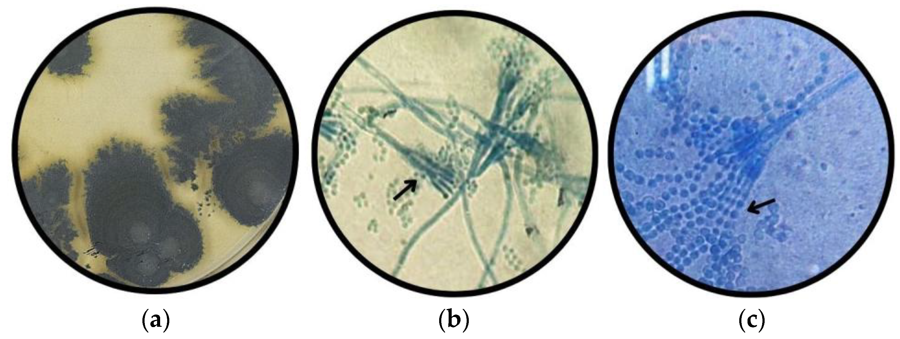
Colony morphology on MEA after 10 days of incubation showed diffuse growth with a sandy appearance at the periphery and blue-gray coloration (Figure 7a). In the microculture, the development of monoveriticillate conidiophores (Figure 7b) and chains of microconidia (Figure 7c) was observed, structures characteristic of the genus Penicillium.

3.2. Taxonomic Classification According to Metabolic Fingerprint

The results in Table 1 correspond to the data generated by the Biolog™ identification system software (FF database v 6.11) during daily microplate readings. These include the SIM index values, which represent the degree of similarity between the database values and the patterns generated by the strains: values close to zero indicate low similarity, values close to one indicate high similarity, and values equal to one indicate exact similarity. For the test panels used in the identification of filamentous fungi, the SIM index is defined by incubation time, with the higher SIM value at a lower time, as follows: >0.9 at 24 h, >0.7 at 48 h, >0.65 at 72 h, and >0.60 between 96 and 168 h. The DIS value reflects discrepancies between the database values and the patterns generated by the strains; lower values indicate greater similarity, while higher values indicate greater differences.
Taxonomic classifications were prioritized when SIM values were high and DIS values were low, in accordance with the FF-IF Biolog™ identification protocol. Presumptive strains of Cladosporium tenuissimum and Penicillium crustosum showed PROB values above 98%, with SIM > 0.60 and DIS < 5.78. Morphotype 4, although it did not yield a PROB value, was identified by the system as belonging to the genus Cladosporium, with SIM = 0.57 and DIS = 5.9. The remaining strains did not present index values sufficient for presumptive identification.
Additional databases provided by the system (FF, FA, and P) were also consulted to confirm or contrast the different systematic proposals. The results presented here correspond to the repositories and incubation times where classifications were consistent, showing higher SIM values and lower DIS values.

3.3. Taxonomic Classification According to ITS Sequencing

Sequencing of the ITS region provided valuable information on the fungal diversity present in the different fermentation treatments applied to whole fruit and depulped coffee. After sequencing and data cleaning, only sequences corresponding to filamentous fungi were selected, characterized by having mycelium as a stationary or permanent reproductive stage. In total, 472,477 sequences belonging to the kingdom Fungi were obtained, of which 3438 corresponded to filamentous fungi, representing 0.73% of the fungal community detected in the fermentations analyzed (Table S5).
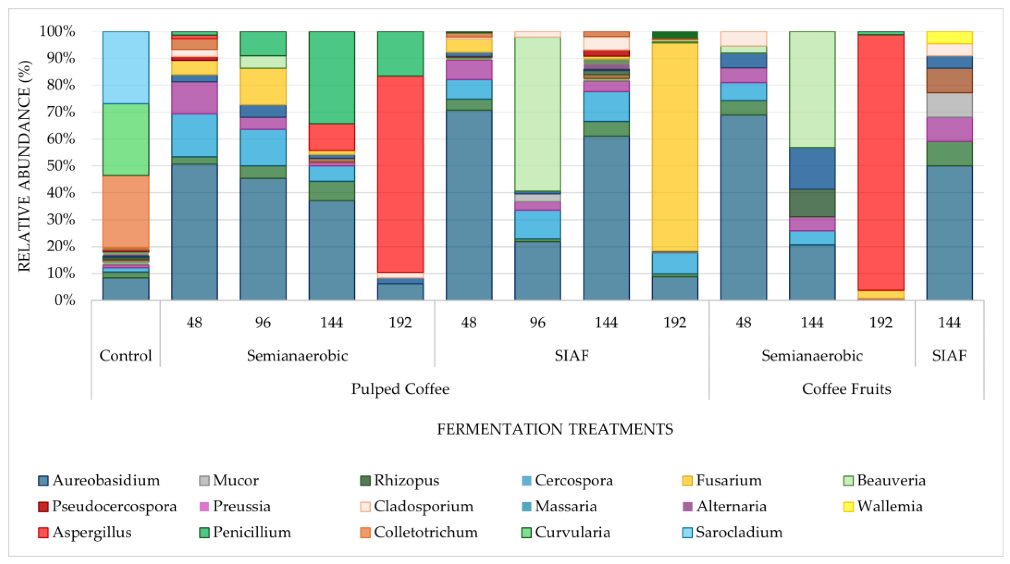
In most samples, the relative abundances of genera varied according to the type of fermentation (Figure 8), with Aureobasidium as the predominant genus. However, in SA fermentations lasting 192 h, Aspergillus showed the highest abundance, particularly in fermentations with whole coffee fruit. By contrast, Fusarium was more abundant in depulped coffee under SA at 192 h. Among all treatments, the control displayed the greatest number of fungal genera, a pattern not observed during the progression of the other fermentations. Another agronomically important genus, Beauveria, was also detected in most treatments under both SA and SIAF conditions, with considerable abundances in depulped coffee and whole fruit at 96 h and 144 h, respectively. The presence of this fungus in the fermentation process suggests that its incidence in the fruit may be either endophytic or epiphytic, as part of integrated coffee borer management strategies [43,44] persisting into later stages of production such as post-harvest.

3.4. Quantification of Ochratoxin A (OTA)

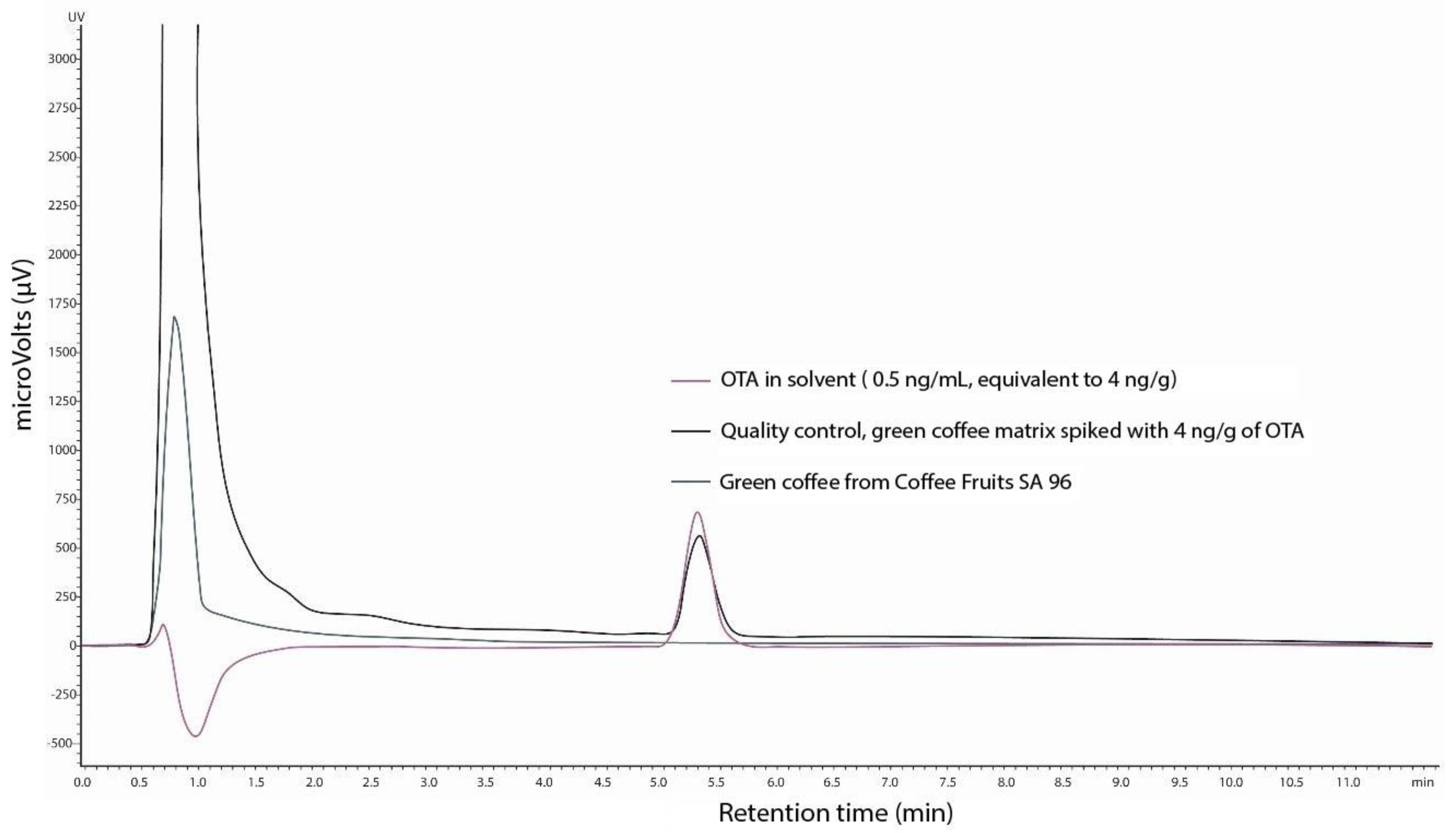
Based on the premise of the possible presence of filamentous fungi from the family Aspergillaceae capable of producing mycotoxins, chemical analysis was conducted to detect OTA in selected treatments, particularly in prolonged and less conventional fermentation times. The analyzed samples showed no chromatographic peaks indicating the presence of OTA in green coffee beans or their derivatives (Figure 9), as the levels were below the detection limits (Table S4).

4. Discussion

The characterization of the isolated fungi revealed differences in morphology and adaptation to different culture substrates. The seven morphotypes expressed across five genera of hyphomycetes are recognized as mycelial fungi that reproduce asexually through conidia formed by the maturation of hyphae [10]. A shared trait among these isolates is their opportunistic and saprophytic pathogenicity on plants such as coffee, affecting processes ranging from leaf development to the physiological ripening of the fruit [10,37]. Fusarium, a member of the family Nectriaceae, is responsible for wilt caused by radicle rot and is also an important producer of a wide range of toxins [45,46]. Dematiaceous fungi such as Curvularia and Cladosporium, both belonging to the class Dothideomycetes, can compromise crop viability at multiple stages by damaging seeds, reducing the plant’s photosynthetic capacity during leaf development, and affecting quality through progressive fruit degradation, which undermines both safety and commercial value [47,48]. In addition, Penicillium, a member of the family Aspergillaceae, is known to induce fruit rot and produce mycotoxins of public health concern such as OTA, posing a risk to food safety in products derived from coffee [49].
The results of spore germination (Table S2) in the culture media used revealed metabolic and morphological differences among the isolates studied. On CDA medium, the absence of growth in morphotypes 1 and 4 suggests an inability to metabolize substrates specific to this medium, such as sodium nitrate or sucrose, unlike the other isolates. By contrast, PDA medium supported early development in most morphotypes, except m4, indicating specific nutritional requirements not met by starch and dextrose. In comparison, MEA medium, selective for molds and yeasts, showed 100% spore germination, attributable to its composition with maltose, dextrin, glycerol, and peptone, which promote metabolic activation through multiple sources of carbon and organic nitrogen. These results highlight the importance of nutritional selectivity in taxonomic identification, where media such as CDA and PDA serve as complementary tools to differentiate microorganisms with contrasting metabolic profiles. Moreover, the use of different substrates revealed the adaptive capacity of the fungi, as the phenotypic plasticity [42] of some morphotypes (Table S3) produced distinctive patterns of mycelial coloration, growth, and senescence—patterns that are key to taxonomic approaches based on the macroscopic traits of each isolate.
For morphotype 1, microscopically classified as Fusarium, taxonomic discrepancies were found with the results obtained using the Biolog™ system (Table 1). The species assigned, Hemicarpenteles paradoux, belongs to the family Aspergillaceae and exhibits reproductive structures that differ markedly from those observed in the microscopic analysis (Figure 1). The low SIM percentage and the absence of a reliable PROB value rule out classification at the species level. This inconsistency suggests that the isolate may not be represented in the database of the software used. Combined with its instability in culture and rapid physiological deterioration, this further complicates precise identification. Mass transfer during replication also appears to contribute to its deterioration, adding to the complexity of classification [10].
For morphotype 2 identification, the Biolog™ system produced two species classifications from two different databases, with similar SIM indices and no PROB value (Table 1). These classifications corresponded presumptively to Alternaria alternata and Curvularia lunata var. lunata, the latter being consistent with the macro- and microscopic classifications (Figure 2). This relationship between species aligns with the phylogeny proposed by Manamgoda et al. (2012) [50], which describes a species complex comprising members of the genera Curvularia, Alternaria, and Bipolaris, due to their morphological similarities. Considering the reproductive morphology of the fungus isolated in this study, the presumptive species is most likely Curvularia lunata, characterized by ovate conidial apices and hypertrophy in the intermediate cells, as opposed to the more acuminate conidia of Alternaria [51].
For morphotypes 3 and 4, there was logical agreement between the Biolog™ system results and the microscopic descriptions (Figure 3 and Figure 4). In both cases, the initial classifications placed these morphotypes within the genus Cladosporium. For morphotype 3, the software produced a PROB value of 1.00, confirming the classification and presumptively assigning it to Cladosporium tenuissimum. For morphotype 4, although no consistent PROB index was obtained, the SIM and DIST values supported its classification at the genus and species level as Cladosporium sphaerospermum.
Morphotypes 5, 6, and 7 also showed taxonomic concordance between microscopic and metabolic analyses using the Biolog™ system databases (Table 1). This consistency placed them within the genus Penicillium. Morphotype 5 was classified reliably, being the only one with a PROB value of 0.98, corresponding to Penicillium crustosum, a species previously reported in both wet and dry coffee processing [14,52]. For morphotype 6, the system registered Penicillium vulpinum, with a considerable SIM value in the FA and P databases, while morphotype 7 was identified as Penicillium janczewskii.
The taxonomic support for these findings comes from ITS region sequencing, which showed that the relative abundance of filamentous fungi sequences accounted for approximately 0.73% of the entire fungal community. This indicates that the degradative contribution of these microorganisms during wet coffee processing is minimal, as also reported by Elhalis et al. (2023) [2], and Peñuela-Martínez et al. (2023b) [30]. These results suggest that fungal dominance is instead driven by other taxa, such as yeasts [14]. Furthermore, the presence of filamentous fungi in the fermentations appears to be entirely environmental, since most of the genera identified belong to the native microbiota of soil, air, water, and the crop itself, as described by de Melo Pereira et al. (2015) [53], and da Silva Vale et al. (2023) [9]. The surface of the coffee fruit is therefore the primary source of microorganisms, with fruit maturity and environmental conditions determining their initial behavior.
Within the taxonomic classification recorded (Table S3) and corroborated using data from Index Fungorum and MycoBank [39,40], the class Dothideomycetes emerged as the most representative among the genera of filamentous fungi. This class is characterized by pathogenic microorganisms, cellulose-degrading saprobes, and plant endophytes and epiphytes. Some members also display an anamorphic stage in which hyphomycetes develop sympodially through conidiogenous cells [54]. Highlighted in this study are fungi belonging to the family Pleosporaceae, such as the genera Alternaria and Curvularia, described earlier, and Mycosphaerellaceae, represented by Cercospora and Pseudocercospora.
This class also includes some of the most abundant genera identified in the study, such as Massaria, Preussia, Cladosporium, and Aureobasidium. The latter is a generalist and ubiquitous microorganism found in soil, air, and water [55]. Among the most abundant filamentous fungi throughout the fermentation process was Aureobasidium, which accounted for nearly 50% in the control—the treatment with the highest fungal incidence—as well as in SIAF treatments with depulped coffee at 48 and 144 h, and in coffee fruit at the same time points, compared with the other treatments. This suggests that the chemical composition of these treatments is similar, even as it changes over time, allowing the reestablishment of this microorganism and supporting its role as a pioneer species in fungal succession.
During fermentation, the genus Fusarium also exhibited growth patterns that varied according to oxygen availability. In depulped coffee under SIAF conditions, its population showed a steady upward trend, reaching predominance at 192 h. By contrast, under SA conditions the dynamics were different: although abundance increased up to 96 h, it dropped markedly over the following 48 h. These results are consistent with those reported by Cruz-O’Byrne et al. (2021) [45], in submerged fermentations up to 36 h, a phenomenon correlated with the simultaneous rise of Penicillium and Aspergillus [4,14]. Similar behavior was discussed by Silva et al. (2008a) [56], for drying and storage processes, noting that Aspergillus competes for substrate with Fusarium and Penicillium, with its incidence increasing only under high temperature and low water activity—conditions typically found in the final stages of processing [10]. This pattern coincides with the fermentation temperature behavior described for this process [32].
The increase in Aspergillus abundance can be explained not only by the chemical transformation of the fruit’s exocarp and mesocarp but also by the type of fermentation conducted. According to data published by Peñuela-Martínez et al. (2025) [32], fermentations with whole fruit under SA conditions raise the temperature to between 30 °C and 38 °C. This finding supports Lorenzoni et al.(2024) [57], who reported low incidence of Aspergillus up to 120 h in fermentations conducted at 18 °C, while fermentations conducted at 38 °C showed an increase in this microorganism’s population from 48 h to 120 h. This comparison suggests that prolonged fermentations (192 h) under SA conditions favor the proliferation and growth of Aspergillus not only due to substrate transformation but also because of the high temperatures reached 49. In contrast, the study by Lee et al. (2023) [58], showed that anaerobic fermentations (SIAF), like low-temperature conditions, inhibit the proliferation of contaminating fungi such as Aspergillus due to the dominant development of lactic acid bacteria (Leuconostoc) [32].
Unlike Aspergillus, the genus Penicillium does not exhibit thermotolerance to the previously described temperatures, which may explain its considerable abundance in depulped coffee subjected to open fermentation at 96 and 144 h [13,59]. However, these three microorganisms, along with Fusarium, can be observed coexisting within the same treatments, although their dominance shifts over time. For instance, in SA fermentations with depulped coffee, Penicillium predominated up to 144 h, while by 192 h Aspergillus became dominant. In contrast, SIAFs with depulped coffee showed a predominance of Fusarium. According to da Silva Vale et al. (2024) [60], environmental variations during fermentation influence population shifts in genera from families such as Nectriaceae (Fusarium) and Aspergillaceae (Aspergillus). Factors such as pH, temperature, and nutrients influence microbial selectivity, favoring opportunistic fungi and increasing competition [60]. However, the mechanisms of interaction at the molecular and metabolic levels remain poorly understood, highlighting the need for further research [9,61]. This suggests that as the fermentation environment transforms and oxygen availability becomes limited, fungal succession varies, reflecting the specialization of each genus in exploiting the substrate during prolonged fermentation, leading to competitive exclusion among them [62].
These three fungi are particularly important in agriculture due to their toxigenic nature across different compound families. Fusarium is a well-known producer of mycotoxins, especially trichothecenes and fumonisins, which are highly toxic to animals and interfere with cellular membrane functions in humans [63]. Similarly, the genera Aspergillus and Penicillium are of particular concern for their production of OTA in coffee [17]. Regarding the production of this mycotoxin, no peaks were detected at concentrations below 0.8 ppb. This suggests that despite the presence and abundance of these genera, prolonged fermentations of up to 192 h under wet conditions are not compromised by OTA production, at least within the limits established by the European Union [22]. However, it is essential to note that the time required to detect moderate levels of OTA from Aspergillus growing on a coffee-based medium was eight days [13], a timeframe that is remarkably close to the last time point of this study. Therefore, extensive fermentation for more than 192 h could generate a perceived concentration of OTA.
In fermentations with whole coffee fruit under SIAF conditions, ITS region DNA amplification was not achieved. This suggests the production of antifungal compounds by yeasts and bacteria during pulp decomposition [64], similar to how lactic acid bacteria generate compounds that inhibit fungal growth in wet processing [58].
The results of this study reveal fungal succession in the mucilage, where the presence of fungi varies according to the transformation stage of the coffee and the fermentation condition, particularly during extended fermentation periods. Although filamentous fungi generally represent low abundance in wet coffee processing—reported as less than 0.01% in samples from traditional fermentation methods [30,31]—this study found that prolonged fermentations increased their populations to nearly 1%. Thus, fermentation times longer than those evaluated here, or a lack of control in pre- or post-fermentation stages, pose a potential risk to coffee safety. Moreover, the contribution these microorganisms might make to changes in the chemical composition or physical quality of the bean remains unknown [65], underscoring the need to further explore this line of research.

5. Conclusions

Culture-dependent and culture-independent techniques were employed to identify filamentous fungi present in coffee fermentations conducted under various conditions. These techniques complemented each other to confirm taxonomic classification at the genus level and presumptive classification at the species level. For an approximate taxonomic identification of filamentous fungi through macroscopic and microscopic descriptions, it is essential to consider their physiological adaptations to diverse substrate sources, as these determine remarkable phenotypic plasticity in their colonies. In this context, the use of different culture media, such as PDA (Potato Dextrose Agar) and EMA (Malt Extract Agar), represents an adequate strategy to observe the morphological variations associated with these adaptations, thanks to their general nutrient compositions. For cases requiring greater specificity, such as the isolation of genera like Aspergillus and Penicillium, the use of selective media such as CDA (Czapek-Dox Agar) is recommended, as it allows for more precise characterization. The primary limitation of this approach is the requirement for optical microscopes equipped with integrated digital cameras to accurately quantify and measure reproductive structures, such as conidia, conidiophores, and hyphae. Failure to accurately measure these structures could lead to insufficient morphological identification. However, only DNA sequencing allows for precise identification of the participating genera. A thorough analysis of the phylogenetic relationship is necessary to determine the specific species accurately.
This study confirms that the presence of filamentous fungi in coffee post-harvest processing is inevitable, and that their incidence may vary during fermentation depending on the condition and duration of the process. Although higher abundances of mycotoxigenic genera were found at longer fermentation times, SIAF showed no OTA production and lower abundances of OTA-producing genera compared with the SA method. However, in the samples from the latest fermentation stages, there was a significant increase in Fusarium populations under SIAF and in Aspergillus under SA, underscoring the importance of both processing time and fermentation conditions in determining fungal dynamics. Finally, from the perspective of this study, it is necessary to confirm the participation of these fungi by improving molecular identification through the use of more specific primers, through explanatory research that considers different environmental conditions and other coffee varieties as sources of variation during fermentation, in addition to time.

Supplementary Materials

The following supporting information can be downloaded at: https://www.mdpi.com/article/10.3390/applmicrobiol5040114/s1, Table S1: Samples from fermentation treatments with filamentous fungi growth in culture medium; Table S2: Macroscopic descriptions of established morphotypes in different culture mediums; Table S3: Taxonomic classification of filamentous fungi found in the sequencing of the ITS region; Table S4: Ochratoxin A—OTA detection range in coffee samples from prolonged fermentation treatments; Table S5: Relative abundance of filamentous fungi, obtained through metataxonomic analysis of ITS region rRNA gene sequences, for coffee samples undergoing semi-anaerobic (SA) and self-induced anaerobic (SIAF) fermentation processes, and under different conditions, including pulped coffee and coffee fruit, from 24 to 192 h.

Author Contributions

Conceptualization, A.E.P.-M.; methodology, A.E.P.-M. and C.B.-Z.; formal analysis, C.B.-Z. and C.V.O.-G.; investigation, A.E.P.-M. and C.B.-Z.: data curation, C.B.-Z. and A.E.P.-M.; writing—original draft preparation, C.B.-Z.; writing—review and editing, A.E.P.-M. and C.V.O.-G.; supervision, A.E.P.-M.; project administration, A.E.P.-M.; funding acquisition, resources, A.E.P.-M. All authors have read and agreed to the published version of the manuscript.

Funding

This research was developed under the project: Research + Development Project to enhance the production of quality coffees, adapted to the agroecological zones of the department of Huila code 2021000100330 financed by the General Royalties System-Huila Government, in agreements signed with the National Colombian Coffee Growers Federation (Cenicafé—Crossref Funder ID 100019597).

Institutional Review Board Statement

Not applicable.

Informed Consent Statement

Not applicable.

Data Availability Statement

Data are contained within the article or Supplementary Materials. High-throughput sequencing (HTS) data are available in repository NCBI SRA BioProject, accession no: PRJNA1254155.

Acknowledgments

The authors thank Daniel Ricardo Toro Castaño for his support in the taxonomic description of the specimens. To National Center for Genomic Sequencing (CNSG) for processing the metagenomic samples, and the Agrochemicals and OTA Laboratory (ALAO) at the Coffee Quality Office, Almacafé S.A., for conducting the chemical analysis of OTA in green coffee The authors have reviewed and edited the output and take full responsibility for the content of this publication.

Conflicts of Interest

The authors declare no conflicts of interest.

References

  1. Oliveira Junqueira, A.C.; Melo Pereira, G.V.; Coral Medina, J.D.; Alvear, M.C.R.; Rosero, R.; Carvalho Neto, D.P.; Enríquez, H.G.; Soccol, C.R. First Description of Bacterial and Fungal Communities in Colombian Coffee Beans Fermentation Analysed Using Illumina-Based Amplicon Sequencing. Sci. Rep. 2019, 9, 8794. [Google Scholar] [CrossRef] [PubMed]
  2. Elhalis, H.; Cox, J.; Zhao, J. Coffee Fermentation: Expedition from Traditional to Controlled Process and Perspectives for Industrialization. Appl. Food Res. 2023, 3, 100253. [Google Scholar] [CrossRef]
  3. Peñuela-Martínez, A.E.; García-Duque, J.F.; Sanz-Uribe, J.R. Characterization of Fermentations with Controlled Temperature with Three Varieties of Coffee (Coffea arabica L.). Fermentation 2023, 9, 976. [Google Scholar] [CrossRef]
  4. Ferreira, L.J.C.; Souza Gomes, M.; Maciel Oliveira, L.; Diniz Santos, L. Coffee Fermentation Process: A Review. Food Res. Int. 2023, 169, 112793. [Google Scholar] [CrossRef]
  5. Pereira, L.L.; Junior, D.B.; Souza, L.H.; Gomes, W.S. Relationship Between Coffee Processing and Fermentation|Request PDF. In Quality Determinants in Coffee Production; Springer International Publishing: Cham, Switzerland, 2021. [Google Scholar] [CrossRef]
  6. Cardoso, W.S.; Agnoletti, B.Z.; Freitas, R.; Abreu Pinheiro, F.; Pereira, L.L. Biochemical Aspects of Coffee Fermentation. In Quality Determinants in Coffee Production; Louzada Pereira, L., Rizzo Moreira, T., Eds.; Springer International Publishing: Cham, Switzerland, 2021; pp. 149–208. [Google Scholar] [CrossRef]
  7. Mota, M.C.B.; Batista, N.N.; Rabelo, M.H.S.; Ribeiro, D.E.; Borém, F.M.; Schwan, R.F. Influence of Fermentation Conditions on the Sensorial Quality of Coffee Inoculated with Yeast. Food Res. Int. 2020, 136, 109482. [Google Scholar] [CrossRef]
  8. Pereira, T.S.; Batista, N.N.; Santos Pimenta, L.P.; Martinez, S.J.; Ribeiro, L.S.; Oliveira Naves, J.A.; Schwan, R.F. Self-Induced Anaerobiosis Coffee Fermentation: Impact on Microbial Communities, Chemical Composition and Sensory Quality of Coffee. Food Microbiol. 2022, 103, 103962. [Google Scholar] [CrossRef]
  9. Silva Vale, A.; Balla, G.; Rodrigues, L.R.S.; Carvalho Neto, D.P.; Soccol, C.R.; Melo Pereira, G.V. Understanding the Effects of Self-Induced Anaerobic Fermentation on Coffee Beans Quality: Microbiological, Metabolic, and Sensory Studies. Foods 2023, 12, 37. [Google Scholar] [CrossRef]
  10. Pitt, J.I.; Hocking, A. Fungi and Food Spoilage, 4th ed.; Springer: New York, NY, USA, 2022. [Google Scholar]
  11. López-Rodríguez, C.; Verheecke-Vaessen, C.; Strub, C.; Fontana, A.; Schorr-Galindo, S.; Medina, A. Reduction in Ochratoxin A Occurrence in Coffee: From Good Practices to Biocontrol Agents. J. Fungi 2024, 10, 590. [Google Scholar] [CrossRef] [PubMed]
  12. Carvalho Neto, D.P.; Melo Pereira, G.V.; Finco, A.M.O.; Letti, L.A.J.; Silva, B.J.G.; Vandenberghe, L.P.S.; Soccol, C.R. Efficient Coffee Beans Mucilage Layer Removal Using Lactic Acid Fermentation in a Stirred-Tank Bioreactor: Kinetic, Metabolic and Sensorial Studies. Food Biosci. 2018, 26, 80–87. [Google Scholar] [CrossRef]
  13. Muñoz, K.; Vega, M.; Rios, G.; Geisen, R.; Degen, G.H. Mycotoxin Production by Different Ochratoxigenic Aspergillus and Penicillium Species on Coffee- and Wheat-Based Media. Mycotoxin Res. 2011, 27, 239–247. [Google Scholar] [CrossRef]
  14. Shen, X.; Wang, Q.; Wang, H.; Fang, G.; Li, Y.; Zhang, J.; Liu, K. Microbial Characteristics and Functions in Coffee Fermentation: A Review. Fermentation 2024, 11, 5. [Google Scholar] [CrossRef]
  15. Adhikari, M.; Isaac, E.L.; Paterson, R.R.M.; Maslin, M.A. A Review of Potential Impacts of Climate Change on Coffee Cultivation and Mycotoxigenic Fungi. Microorganisms 2020, 8, 1625. [Google Scholar] [CrossRef]
  16. Kusumaningrum, H.D.; Rasyidah, M.M. Prevalence of Spoilage Mold in Coffee before and after Brewing. Food Res. 2019, 720–726. [Google Scholar] [CrossRef]
  17. Neves, T.T.; Brandão, R.M.; Barbosa, R.B.; Cardoso, M.G.; Batista, L.R.; Silva, C.F. Simulation of Coffee Beans Contamination by Aspergillus Species under Different Environmental Conditions and the Biocontrol Effect by Saccharomyces cerevisiae. LWT 2021, 148, 111610. [Google Scholar] [CrossRef]
  18. Neves, T.T.; Cassimiro, D.M.J.; Souza, J.G.L.; Castro, C.R.S.; Schwan, R.F.; Batista, L.R.; Silva, C.F. Inhibition of Aspergillus spp. Growth and Ochratoxin A Production in Conilon and Arabica Coffees Based-Medium by Saccharomyces cerevisiae. Int. J. Food Microbiol. 2024, 425, 110875. [Google Scholar] [CrossRef]
  19. Rojas-Pablo, M.; Toledo-Hernández, E.; Rodríguez-Barrera, M.A.; Toribio-Jiménez, J.; Torreblanca-Ramírez, C.; Rosas-Guerrero, V.M.; Salgado-Souto, S.A.; Álvarez-Fitz, P.; Bolaños-Dircio, A.; Romero-Ramírez, Y. Bacillus licheniformis M2-7 Decreases Ochratoxin A Concentrations in Coffee Beans During Storage. Curr. Microbiol. 2024, 81, 62. [Google Scholar] [CrossRef]
  20. Khaneghah, A.M.; Fakhri, Y.; Abdi, L.; Coppa, C.F.S.C.; Franco, L.T.; Oliveira, C.A.F. The Concentration and Prevalence of Ochratoxin A in Coffee and Coffee-Based Products: A Global Systematic Review, Meta-Analysis and Meta-Regression. Fungal Biol. 2019, 123, 611–617. [Google Scholar] [CrossRef]
  21. Humans, I.W.G. on the E. of C. R. to. IARC Monographs on the Evaluation of Carcinogenic Risks to Humans. In Human Immunodeficiency Viruses and Human T-Cell Lymphotropic Viruses; International Agency for Research on Cancer: Lyon, France, 1996. [Google Scholar]
  22. European Commission. Commission Regulation (EU) 2022/1370 of 5 August 2022 Amending Regulation (EC) No 1881/2006 as Regards Maximum Levels of Ochratoxin A in Certain Foodstuffs (Text with EEA Relevance); European Commission: Brussels, Belgium, 2022; Available online: https://eur-lex.europa.eu/eli/reg/2022/1370/oj (accessed on 5 June 2025).
  23. Lindgren, S.E.; Dobrogosz, W.J. Antagonistic Activities of Lactic Acid Bacteria in Food and Feed Fermentations. FEMS Microbiol. Rev. 1990, 7, 149–163. [Google Scholar] [CrossRef]
  24. Beugre, G.C.; Kadjo, A.C.; Yao, K.M.; Kone, K.M.; Piro-Metayer, I.; Poss, C.; Durand, N.; Fontana, A.; Guehi, T.S. Sensory Quality of Coffee Beverrage Produced Thereof Linked to the Inhibition of Molds Growth and Ochratoxin a Removal from Coffee Cherries Using Lactobacillus Plantarum Strains. Curr. J. Appl. Sci. Technol. 2023, 42, 10–20. [Google Scholar] [CrossRef]
  25. Velmourougane, K. Impact of Natural Fermentation on Physicochemical, Microbiological and Cup Quality Characteristics of Arabica and Robusta Coffee. Proc. Natl. Acad. Sci. India Sect. B Biol. Sci. 2013, 83, 233–239. [Google Scholar] [CrossRef]
  26. Abdel-Hadi, A.; Alshehri, B.; Waly, M.; Aboamer, M.; Banawas, S.; Alaidarous, M.; Palanisamy, M.; Awad, M.; Baazeem, A. Predictive Modeling and Validation on Growth, Production of Asexual Spores and Ochratoxin A of Aspergillus ochraceus Group under Abiotic Climatic Variables. Microorganisms 2021, 9, 1321. [Google Scholar] [CrossRef] [PubMed]
  27. Frisvad, J.C.; Frank, J.; Houbraken, J.; Kujipers, A.; Samson, R. New Ochratoxin A Producing Species of Aspergillus Section Circumdati. Stud. Mycol. 2004, 50, 23–43. Available online: https://orbit.dtu.dk/en/publications/new-ochratoxin-a-producing-species-of-aspergillus-section-circumd (accessed on 9 June 2025).
  28. Zakaria, L. An Overview of Aspergillus Species Associated with Plant Diseases. Pathogens 2024, 13, 813. [Google Scholar] [CrossRef] [PubMed]
  29. Viegas, C.; Pacífico, C.; Faria, T.; Oliveira, A.C.; Caetano, L.A.; Carolino, E.; Gomes, A.Q.; Viegas, S. Fungal Contamination in Green Coffee Beans Samples: A Public Health Concern. J. Toxicol. Environ. Health Part A 2017, 80, 719–728. [Google Scholar] [CrossRef]
  30. Peñuela-Martínez, A.E.; Velasquez-Emiliani, A.V.; Angel, C.A. Microbial Diversity Using a Metataxonomic Approach, Associated with Coffee Fermentation Processes in the Department of Quindío, Colombia. Fermentation 2023, 9, 343. [Google Scholar] [CrossRef]
  31. Góngora, C.E.; Holguín-Sterling, L.; Pedraza-Claros, B.; Pérez-Salinas, R.; Ortiz, A.; Navarro-Escalante, L. Metataxonomic Identification of Microorganisms during the Coffee Fermentation Process in Colombian Farms (Cesar Department). Foods 2024, 13, 839. [Google Scholar] [CrossRef]
  32. Peñuela-Martínez, A.E.; Osorio-Giraldo, C.V.; Buitrago-Zuluaga, C.; Medina-Rivera, R.D. Development of Fermentation Strategies for Quality Mild Coffee Production (Coffea arabica L.) Based on Oxygen Availability and Processing Time. Foods 2025, 14, 3001. [Google Scholar] [CrossRef]
  33. Federación Nacional de Cafeteros. Anuario 2023|Anuario Meteorológico Cafetero; Cenicafé: Chinchiná, Caldas, Colombia, 2023. [Google Scholar]
  34. Peñuela-Martínez, A.E.; Pabón, J.; Sanz-Uribe, J.R. Método fermaestro: Para determinar la finalización de la fermentación del mucílago de café. Av. Técnic. Cenicafé 2013, 431, 1–8. [Google Scholar] [CrossRef]
  35. Arias, E.L.; Piñeros, P.A. Aislamiento e Identificación de Hongos Filamentosos de Muestra de suelo de los Páramos de Guasca y Cruz Verde; Pontificia Universidad Javeriana: Bogotá, Colombia, 2008. [Google Scholar]
  36. Patiño Moscoso, M.A.; Osorio Guerrero, K.V.; Flórez Gómez, D.L.; Sarmiento Moreno, L.F.; Vargas Ramírez, D.N.; Mérida, M.J. Manual Ilustrado de Hongos Presentes en Semillas de Cultivos Semestrales: Arroz, Maíz, Soya y Sorgo; AGROSAVIA: Bogotá, Colombia, 2023. [Google Scholar]
  37. Barnett, H.L.; Hunter, B.B. Illustrated Genera of Imperfect Fungi; APS Press: St. Paul, MN, USA, 1986. [Google Scholar]
  38. Frąc, M.; Kaczmarek, J.; Jędryczka, M. Metabolic Capacity Differentiates Plenodomus lingam from P. biglobosus Subclade ‘Brassicae’, the Causal Agents of Phoma Leaf Spotting and Stem Canker of Oilseed Rape (Brassica napus) in Agricultural Ecosystems. Pathogens 2022, 11, 50. [Google Scholar] [CrossRef]
  39. Mycobank. Available online: https://www.mycobank.org/ (accessed on 25 August 2025).
  40. Index Fungorum-Search Page. Available online: https://www.indexfungorum.org/names/names.asp (accessed on 25 August 2025).
  41. AOAC 2004.10-2008; Ochratoxin A in Green Coffee. AOAC International: Rockville, MD, USA, 2004.
  42. Alster, C.J.; Allison, S.D.; Johnson, N.G.; Glassman, S.I.; Treseder, K.K. Phenotypic Plasticity of Fungal Traits in Response to Moisture and Temperature. ISME Commun. 2021, 1, 43. [Google Scholar] [CrossRef]
  43. Hollingsworth, R.G.; Aristizábal, L.F.; Shriner, S.; Mascarin, G.M.; Moral, R.A.; Arthurs, S.P. Incorporating Beauveria bassiana Into an Integrated Pest Management Plan for Coffee Berry Borer in Hawaii. Front. Sustain. Food Syst. 2020, 4, 22. [Google Scholar] [CrossRef]
  44. Ferrucho, R.L.; Marín-Ramírez, G.A.; Gaitan, A. Integrated Disease Management for the Sustainable Production of Colombian Coffee. Agronomy 2024, 14, 1286. [Google Scholar] [CrossRef]
  45. Cruz-O’Byrne, R.; Piraneque-Gambasica, N.; Aguirre-Forero, S. Microbial Diversity Associated with Spontaneous Coffee Bean Fermentation Process and Specialty Coffee Production in Northern Colombia. Int. J. Food Microbiol. 2021, 354, 109282. [Google Scholar] [CrossRef]
  46. Lu, L.; Tibpromma, S.; Karunarathna, S.C.; Jayawardena, R.S.; Lumyong, S.; Xu, J.; Hyde, K.D. Comprehensive Review of Fungi on Coffee. Pathogens 2022, 11, 411. [Google Scholar] [CrossRef] [PubMed]
  47. Manathunga, K.K.; Gunasekara, N.W.; Meegahakumbura, M.K.; Ratnaweera, P.B.; Faraj, T.K.; Wanasinghe, D.N. Exploring Endophytic Fungi as Natural Antagonists against Fungal Pathogens of Food Crops. J. Fungi 2024, 10, 606. [Google Scholar] [CrossRef]
  48. Priwiratama, H.; Wiyono, S.; Hidayat, S.H.; Wening, S.; Tondok, E.T. Identification and Characterization of Curvularia, the Causal Agent of Leaf Spot Disease of Oil Palm Seedlings in Indonesia. J. Saudi Soc. Agric. Sci. 2024, in press. [Google Scholar] [CrossRef]
  49. Akbar, A.; Medina, A.; Magan, N. Resilience of Aspergillus westerdijkiae Strains to Interacting Climate-Related Abiotic Factors: Effects on Growth and Ochratoxin A Production on Coffee-Based Medium and in Stored Coffee. Microorganisms 2020, 8, 1268. [Google Scholar] [CrossRef]
  50. Manamgoda, D.S.; Cai, L.; McKenzie, E.H.C.; Crous, P.W.; Madrid, H.; Chukeatirote, E.; Shivas, R.G.; Tan, Y.P.; Hyde, K.D. A Phylogenetic and Taxonomic Re-Evaluation of the Bipolaris-Cochliobolus-Curvularia Complex. Fungal Divers. 2012, 56, 131–144. [Google Scholar] [CrossRef]
  51. Iturrieta-González, I.; Gené, J.; Wiederhold, N.; García, D. Three New Curvularia Species from Clinical and Environmental Sources. Mycokeys 2020, 68, 1–21. [Google Scholar] [CrossRef] [PubMed]
  52. Khalil, N.M.; Rodríguez-Couto, S.; El-Ghany, M.N.A. Characterization of Penicillium Crustosum L-Asparaginase and Its Acrylamide Alleviation Efficiency in Roasted Coffee Beans at Non-Cytotoxic Levels. Arch. Microbiol. 2021, 203, 2625–2637. [Google Scholar] [CrossRef]
  53. Melo Pereira, G.V.; Neto, E.; Soccol, V.T.; Medeiros, A.B.P.; Woiciechowski, A.L.; Soccol, C.R. Conducting Starter Culture-Controlled Fermentations of Coffee Beans during on-Farm Wet Processing: Growth, Metabolic Analyses and Sensorial Effects. Food Res. Int. 2015, 75, 348–356. [Google Scholar] [CrossRef]
  54. Schoch, C.L.; Shoemaker, R.A.; Hambleton, S.; Spatafora, J.W.; Pedro, W. A Multigene Phylogeny of the Dothideomycetes Using Four Nuclear Loci. Mycologia 2006, 98, 1041–1052. [Google Scholar] [CrossRef]
  55. Di Francesco, A.; Zajc, J.; Stenberg, J.A. Aureobasidium spp.: Diversity, Versatility, and Agricultural Utility. Horticulturae 2023, 9, 59. [Google Scholar] [CrossRef]
  56. Silva, C.F.; Batista, L.R.; Schwan, R.F. Incidence and Distribution of Filamentous Fungi during Fermentation, Drying and Storage of Coffee (Coffea arabica L.) Beans. Braz. J. Microbiol. 2008, 39, 521–526. [Google Scholar] [CrossRef]
  57. Lorenzoni, T.L.; Luz, J.M.R.; Veloso, T.G.R.; Pereira, L.L.; Menezes, K.M.S.; Brioschi Júnior, D.; Kasuya, M.C.M.; Silva, M.D.C.S. Genetic Diversity of the Fungal Community That Contributes to the Sensory Quality of Coffee Beverage after Carbonic Maceration and Fermentation. 3 Biotech 2024, 14, 272. [Google Scholar] [CrossRef]
  58. Lee, B.-H.; Huang, C.-H.; Liu, T.-Y.; Liou, J.-S.; Hou, C.-Y.; Hsu, W.-H. Microbial Diversity of Anaerobic-Fermented Coffee and Potential for Inhibiting Ochratoxin-Produced Aspergillus Niger. Foods 2023, 12, 2967. [Google Scholar] [CrossRef]
  59. Mannaa, M.; Kim, K.D. Influence of Temperature and Water Activity on Deleterious Fungi and Mycotoxin Production during Grain Storage. Mycobiology 2017, 45, 240–254. [Google Scholar] [CrossRef] [PubMed]
  60. Silva Vale, A.; Pereira, C.M.T.; Dea Lindner, J.; Rodrigues, L.R.S.; Kadri, N.K.E.; Pagnoncelli, M.G.B.; Kaur Brar, S.; Soccol, C.R.; Pereira, G.V.M. Exploring Microbial Influence on Flavor Development during Coffee Processing in Humid Subtropical Climate through Metagenetic–Metabolomics Analysis. Foods 2024, 13, 1871. [Google Scholar] [CrossRef] [PubMed]
  61. Elhalis, H.; Cox, J.; Zhao, J. Ecological Diversity, Evolution and Metabolism of Microbial Communities in the Wet Fermentation of Australian Coffee Beans. Int. J. Food Microbiol. 2020, 321, 108544. [Google Scholar] [CrossRef]
  62. Katati, B.; van Diepeningen, A.D.; Njapau, H.; Kachapulula, P.W.; Zwaan, B.J.; Schoustra, S.E. Niche Partitioning Association of Fungal Genera Correlated with Lower Fusarium and Fumonisin-B1 Levels in Maize. BioControl 2024, 69, 185–197. [Google Scholar] [CrossRef]
  63. Pierzgalski, A.; Bryła, M.; Kanabus, J.; Modrzewska, M.; Podolska, G. Updated Review of the Toxicity of Selected Fusarium Toxins and Their Modified Forms. Toxins 2021, 13, 768. [Google Scholar] [CrossRef] [PubMed]
  64. Melo Pereira, G.V.; Mello Sampaio, V.; Wiele, N.; Silva Vale, A.; Carvalho Neto, D.P.; Souza, A.d.F.D.d.; Nogueira dos Santos, D.V.; Ruiz, I.R.; Rogez, H.; Soccol, C.R. How Yeast Has Transformed the Coffee Market by Creating New Flavors and Aromas through Modern Post-Harvest Fermentation Systems. Trends Food Sci. Technol. 2024, 151, 104641. [Google Scholar] [CrossRef]
  65. Silva, C.F.; Batista, L.R.; Abreu, L.M.; Dias, E.S.; Schwan, R.F. Succession of Bacterial and Fungal Communities during Natural Coffee (Coffea arabica) Fermentation. Food Microbiol. 2008, 25, 951–957. [Google Scholar] [CrossRef] [PubMed]
Figure 1. Morphological description of the reproductive structures of morphotype 1 (m1).
Applmicrobiol 05 00114 g001
Figure 2. Morphological description of the reproductive structures of morphotype 2 (m2).
Applmicrobiol 05 00114 g002
Figure 3. Morphological description of the reproductive structures of morphotype 3 (m3).
Applmicrobiol 05 00114 g003
Figure 4. Morphological description of the reproductive structures of morphotype 4 (m4).
Applmicrobiol 05 00114 g004
Figure 5. Morphological description of the reproductive structures of morphotype 5 (m5).
Applmicrobiol 05 00114 g005
Figure 6. Morphological description of the reproductive structures of morphotype 6 (m6).
Applmicrobiol 05 00114 g006
Figure 7. Morphological description of the reproductive structures of morphotype 7 (m7).
Applmicrobiol 05 00114 g007
Figure 8. Relative abundance of OTUs at the genus level across different fermentation treatments.
Applmicrobiol 05 00114 g008
Figure 9. Chromatogram of OTA detection in green coffee beans after 96 h of fermentation in whole fruit under semi-aerobic conditions.
Applmicrobiol 05 00114 g009
Table 1. Presumptive taxonomic classification of morphotypes based on the metabolic fingerprint of filamentous fungi.
MorphotypeDatabaseIncubation Time (h)PROBSIMDISSPECIES ID
1Filamentous Fungi24---0.1613.82Hemicarpenteles paradoux Sarbhoy & Elphick
Food or Air24---0.1613.82Hemicarpenteles paradoux Sarbhoy & Elphick
2Filamentous Fungi72---0.438.96Alternaria alternata (Fries) Keissl, BGA
Food or Air72---0.438.96Alternaria alternata (Fries) Keissl, BGA
168---0.419.50Curvularia lunata var lunata (Wakker) Boedijn BGA
3Filamentous Fungi720.990.734.03Cladosporium tenuissimum Cooke BGA
Food or Air721.000.655.36Cladosporium tenuissimum Cooke BGA
4Filamentous Fungi96---0.575.90Cladosporium sphaerospermum Penzing BGA
Food or Air96---0.575.90Cladosporium sphaerospermum Penzing BGA
5Filamentous Fungi960.980.625.78Penicillium crustosum Thom BGD
Food or Air960.980.625.78Penicillium crustosum Thom BGD
Penicillium960.980.625.78Penicillium crustosum Thom BGD
6Filamentous Fungi96---0.2413.91Penicillium roqueforti Thom BGE
Food or Air48---0.3810.21Penicillium vulpinum (Cooke & Massee) Seifert & Samson BGA
Penicillium48---0.3111.36Penicillium vulpinum (Cooke & Massee) Seifert & Samson BGA
7Filamentous Fungi48---0.3519,76Penicillium janczewskii Zaleski BGB
Food or Air96---0.398.03Penicillium crustosum Thom BGD
Penicillium48---0.3511.09Penicillium janczewskii Zaleski BGB
Disclaimer/Publisher’s Note: The statements, opinions and data contained in all publications are solely those of the individual author(s) and contributor(s) and not of MDPI and/or the editor(s). MDPI and/or the editor(s) disclaim responsibility for any injury to people or property resulting from any ideas, methods, instructions or products referred to in the content.