Article

Conversion of Komagataella phaffii Biomass Waste to Yeast Extract Supplement

by
Laura Murphy
1,2 and
David J. O’Connell
1,2,*
1
School of Biomolecular & Biomedical Science, University College Dublin, Belfield, D04V1W8 Dublin, Ireland
2
BiOrbic Bioeconomy Research Centre, University College Dublin, Belfield, D04V1W8 Dublin, Ireland
*
Author to whom correspondence should be addressed.
Appl. Microbiol. 2025, 5(3), 95; https://doi.org/10.3390/applmicrobiol5030095 (registering DOI)
Submission received: 5 August 2025 / Revised: 23 August 2025 / Accepted: 1 September 2025 / Published: 4 September 2025

Abstract

Valorisation of spent yeast biomass post-fermentation requires energy-intensive autolysis or enzymatic hydrolysis that reduces the net benefit. Here, we present a simple and reproducible method for generating functional yeast extract recycled from Komagataella phaffii biomass without a requirement of a pre-treatment process. Spent yeast pellets from fermentations were freeze-dried to produce a fine powder that can be used directly at low concentrations, 0.0015% (w/v), together with 2% peptone (w/v), to formulate complete media ready for secondary fermentations. This media formulation supported growth rates of yeast culture that were statistically indistinguishable (p-value > 0.05) from cultures grown in standard YPD media containing commercial yeast extract, and these cultures produced equivalent titres of recombinant β-glucosidase (0.998 Abs405nm commercial extract vs. 0.899 Abs405nm recycled extract). Additionally, nutrient analyses highlight equivalent levels of sugars (~23 g/L), total proteins, and cell yield per carbon source (~2.17 g) with this recycled yeast extract media formulation when compared to commercial media. This method reduces process complexity and cost and enables the circular reuse of yeast biomass. The protocol is technically straightforward to implement, using freeze drying that is commonly available in research laboratories, representing a broadly applicable and sustainable alternative to conventional media supplementation that achieves a circular approach within the same fermentation system.

1. Introduction

Yeasts play significant roles as a chassis in the biopharmaceutical, biotechnological, and brewing industries [1,2,3]. Yeast fermentation generates a large volume of waste biomass, with an estimated 5 million metric tonnes produced annually across the globe, that has the potential to be repurposed as recycled yeast extract [4,5,6,7,8].
To fulfil the ambitions of the circular bioeconomy, there is a growing requirement for simple, low-cost methods to repurpose spent microbial biomass. Currently, the valorisation of spent yeast biomass—particularly for use as yeast extract in culture media—requires multiple processing steps such as autolysis, hydrolysis, or chemical treatments. Previous studies have been performed to determine if spent yeast can serve as a feedstock for other microorganism cultures [9], with the successful production of optically pure L-lactic acid using Lactobacillus rhamnosus, for example [10]. However, to date, there is limited research on taking yeast biomass from completed fermentations and reusing it as a feedstock for secondary yeast fermentation, which is a central aim of circular bioeconomy practice. Predominantly studies have focused on using brewer’s spent grain (BSG) as a feedstock for simple yeast fermentations, where either growth [11,12] or lipid accumulation [13,14] is the primary goal. In addition, the pre-treatment of yeast and multi-step autolysis protocols [9,11,13] increase the energy requirements and labour intensity of valorising this waste, requiring dedicated infrastructure that limits adoption in small-to-mid-scale operations or sustainable workflows. Although the exact nutrient profile of spent biomass depends on upstream factors (strain, medium, growth phase) [15], we show here that terminal K. phaffii pellets consistently release enough proteins, vitamins, and peptides upon autolysis and freeze drying to serve as an effective circular media supplement. We hypothesize that nutrients (amino acids, short peptides, and water-soluble vitamins) originally in the culture medium are taken up and sequestered within the cells during primary fermentation, only to be uniformly liberated when the biomass is processed into yeast extract, yielding consistent protein content from each pellet batch [16,17].
This study presents a straightforward method for transforming K. phaffii biomass directly into a functional yeast extract in a one-step process, employing freeze drying of post-fermentation yeast pellets to a powder. This powder was used to supplement secondary fermentations for the de novo expression of β-glucosidase in secondary fermentation with K. phaffii. This approach was compared with the standard commercial yeast extract in YPD media to assess its suitability for supporting growth and recombinant protein expression. Our method eliminates the need for autolysis or enzymatic digestion, offering a reproducible workflow for media supplementation using recycled yeast extract.

2. Materials and Methods

2.1. Generation of K. phaffii Pellets for Recycled Yeast Extract

A β-glucosidase-overexpressing clone, IRA1, was provided as a gift and is described elsewhere [18]. K. phaffii biomass pellets were generated as a waste from primary fermentations producing β-glucosidase in YPD media (1% yeast extract, 2% peptone, 2% glucose). Single colonies of IRA1 K. phaffii were used to inoculate 5 mL of YPD media in a 50 mL falcon tube. These starter cultures were grown at 30 °C shaking at 225 RPM for 48 h and inoculated at a 1:120 (v/v) dilution into 50 mL YPD expression cultures in baffled shake flasks. Main cultures were grown at 30 °C shaking at 225 RPM for 96 h. The final cultures were spun at 4000× g for 5 min, and the cell supernatant and pellets were separated, and pellets were stored at −20 °C until future use. Prior to freeze drying, pellets were stored at −80 °C overnight. Parafilm was placed over the top of the open 50 mL falcon tubes containing the frozen pellets, and holes were punched through using a needle. Samples were freeze-dried at an ice condenser temperature of −85 °C under vacuum using the Christ Alpha 2–4 plus freeze dryer. Freeze drying was performed for 12 h under ~0.05 mbar of pressure until the yeast samples resembled a fine powder. Lids were added to the samples contained in falcon tubes, and they were stored in airtight conditions at −20 °C. Empirically, thawed material rapidly loses its physical integrity and becomes a fluffy, unusable mush; therefore, all handling and secondary culture work used material removed from frozen storage immediately prior to use.

2.2. Determination of Optimal Composition of Recycled Yeast Extract in YPD Media

Various compositions of YPD media were prepared to test the performance of recycled yeast extract. Peptone used was from Sigma–Aldrich (St. Louis, MO, USA)/Merck (Darmstadt, Germany), product Peptone, Mycological No. 2 (product code 07751)—lot BCCC2601. Due to the high concentration of the recycled yeast extract powder, several titrations of YPD were prepared until the optical density and turbidity of the media(s) were comparable to standard YPD media. Specifically, YPD media containing the following amount of recycled yeast extract were prepared: 10 g/L, 0.1 g/L, 0.05 g/L, and 15 mg/L, and were supplemented with either 1% or 2% (w/v) peptone (Table 1. All media were autoclaved at 121 °C for 15 min to ensure sterility and inactivation of yeast cells. Samples of the media were taken, and the optical density was read at OD600nm and compared to the density of standard YPD media in technical triplicate. A media containing only 2% peptone and 2% dextrose was prepared to serve as a negative control for the expression cultures. The media compositions are summarised in Table 1 below.

2.3. Culturing of K. phaffii β-Glucosidase-Expressing Clones

Single colonies of a K. phaffii β-glucosidase-expressing clone were used to inoculate 10 mL of YPD media in a 50 mL falcon tube. These starter cultures were grown at 30 °C shaking at 225 RPM for 48 h. Following this, a 1:120 dilution was performed in larger volumes of the previously described media types. Main cultures were grown at 30 °C by shaking at 225 RPM for 144 h. Every 24 h, 1 mL samples were taken for cell density measurements at OD600nm. Samples were spun at 4000× g for 5 min, and both the cell supernatant and cell pellets were stored at −20°. Aliquots of the supernatant were saved on ice prior to freezing for immediate β-glucosidase concentration quantification.

2.4. Determination of β-Glucosidase Secretion in IRA1 K. phaffii Clones in Varying Compositions of YPD Media

This assay was adapted from the protocol outlined by Offei et al. [18]. In summary, 2 μL of each sample supernatant (plus 2 μL of appropriate media type as a blank) was mixed with 98 μL of 5 mM 4-Nitrophenyl β-d-glucopyranoside (4-NPG; Sigma) (in 0.1 M sodium acetate buffer, pH 5.0) in a 96-well plate. This was incubated for 10 min at 40 °C. Reaction was stopped with the addition of 100 μL of 1 M Na2CO3. 4-NPG absorbance, which corresponds to β-glucosidase levels in the supernatant, was determined by reading plates at 405 nm. Final 4-NP values were determined by subtracting blank (appropriate media type) absorbance values.

2.5. Analysis of Reducing Sugars and Total Protein Concentrations

Samples prior to culture commencement (T0) and at the end of the fermentations (T144) were analysed for their reducing sugar, total protein, and total carbohydrate concentrations using three separate assays. Sugars were assessed using Levers assay, adapted from the protocol described by Iyer et al. [19]. In brief, all media samples were deproteinized through the addition of equal sample volume of cold 10% trichloroacetic acid (TCA). Samples were incubated for 10 min on ice, and centrifuged at 10,000× g for 5 min. The clarified supernatant was transferred to a new Eppendorf tube. Levers reagent containing 4-hydroxybenzoic acid hydrazide (PAHBAH, 0.76% w/v), potassium sodium tartrate (0.28% w/v), and NaOH (2% w/v) was prepared in ultrapure Milli-Q water. A 50 µL volume of media sample or glucose standard (0–1 mg/mL) was added to individual wells of a 96-well plate, and was incubated with 200 µL of Levers reagent at 70 °C for 30 min. Samples were cooled for 10 min and then read at 415 nm on a spectrophotometer. Reducing sugar concentrations were determined based upon the generated standard curve on Microsoft Excel. Samples were run in biological triplicates and technical duplicates, and standards were run in technical duplicates.
A Bradford assay was performed for total protein analysis. In summary, BSA standards (0–1 mg/mL) were prepared, and 200 µL of each was added to individual microcentrifuge tubes. Dilutions of 1:3 for YPD-C media samples and 1:2 for all other media types were performed in dH2O to ensure ranges within the standard curve. A 200 µL volume of each media type (further diluted 1:10 in dH2O) were also prepared, and 800 µL of Bradford reagent was added to all standard and sample tubes (1:5 dilution; for a total base dilution of 1:50 for each media and sample type), and were mixed and left to incubate at room temperature for 5 min. Samples were read at 595 nm, and protein concentration data were extrapolated based upon the generated standard curve using Microsoft Excel. Samples were run in biological triplicate and technical duplicate, and standards were run in technical duplicate.

2.6. Statistical Analysis

Following the acquisition of biological triplicate data, statistical analysis was performed using GraphPad Prism 10 software. For growth curve final densities and β-glucosidase secretion at T144, significance was determined using a two-way ANOVA with Tukey’s post-analysis test, where the p-value (α) of 0.05 was set as the significance threshold. Tukey’s post-analysis test compares the means of each group with each other. Growth curves were fitted per replicate to the Malthusian exponential model N(t) = N0ekt using nonlinear regression in GraphPad Prism; the fitted parameter k is the exponential rate constant (units h−1). Doubling time was calculated as td = ln(2)/k.

3. Results

3.1. Selection of Media Types for Expression of β-Glucosidase

Prior to testing the performance of the generated recycled yeast extract, it was necessary to determine the optimal formulation of YPD media for use. Therefore, several compositions of media using varying amounts of recycled yeast extract were prepared, as outlined in the left column of Table 2 below. Standard YPD media using commercial autolysed yeast extract was also prepared, along with a negative control media containing no yeast extract, just peptone (2% w/v) and dextrose (2% w/v). These formulations also allowed us to assess the impact of peptone concentration, as both 1% and 2% peptone media were tested. Column 2 of Table 2 indicates the corresponding optical density (600 nm) for each media type, with the goal of matching densities to YPD media using commercial yeast extract. As seen, only when recycled yeast extract is reduced to the milligram concentration is the density comparable to that of commercial YPD (OD600nm of 0.1482 and 0.1609 versus 0.1593, respectively). The increase in peptone from 1% to 2% among the in-house YPD media shows an expected increase in density, but this is still within comparable readings to commercial YPD. Standard deviations of the population were calculated, along with the coefficient of variation (CV), which measures the spread of values relative to the mean, indicating the precision and accuracy of the data. A CV below 15% reflects good precision and is common in biological experiments. As seen, when the amount of recycled yeast extract is reduced, the standard deviation and CV of the media are also reduced.

3.2. Growth of K. phaffii in Different YPD Media Compositions

Four media were tested for their capacity to support the growth of a β-glucosidase-expressing K. phaffii strain: YPD complete media (using commercial yeast extract; YPD-C), YPD media with 1% peptone (w/v) using 15 mg recycled yeast extract (YPD-1%P), YPD media with 2% peptone (w/v) using 15 mg recycled yeast extract (YPD-2%P), and PD media (2% peptone and 2% dextrose). Only the 15 mg recycled yeast extract media were utilized for growth and expression analysis as they most closely matched the densities of YPD-C media. Figure 1A illustrates the optical densities (OD600nm) of the cultures of each media type over the 144 h expression time-course. Here, an initial lag in density at 48 h is seen with the recycled yeast extract media; however, by the completion of the time-course, the YPD-2%P condition is at a comparable density to the YPD-C (Avg OD600nm of 53.5 YPD complete (black circles) versus Avg OD600nm of 52.1 YPD-2%P (green triangles)). The YPD-1%P condition produced the least-dense cultures at the end of the time-course. Figure 1B gives an overview of the growth kinetics of each culture. Y0 represents the initial biomass before exponential growth has occurred, and is used in calculating the doubling time of each culture. The k value indicates the growth constant for each culture, and the doubling time reflects the number of hours it takes for the density and cell count to double. Interestingly, YPD complete media has the highest doubling time at 66.17, with all other media being within a ~7 h range of this value.
Examining each condition at the completion of the 144 h time-course highlights some differences in the growth of K. phaffii in each media type. Figure 2A overviews the final optical density of each condition, and there is no statistically significant difference in density between the YPD complete cultures and the ‘Y’PD full-peptone cultures. Furthermore, there is a significant decrease in density observed with the ‘Y’PD half-peptone and PD media cultures when compared to the YPD complete values (p-value < 0.0001, ****; and p-value < 0.01, **, respectively). Figure 2B represents the average wet cell weights (WCW) of each condition, and the same pattern is reflected in this data. There is no significant difference in WCW between YPD complete cultures and the ‘Y’PD full-peptone cultures, whereas there is a statistically significant difference between pellet weights produced from the ‘Y’PD half-peptone and PD media cultures when compared to the YPD complete values (p-values < 0.0001, ****).

3.3. Expression of β-Glucosidase in Various Media Compositions

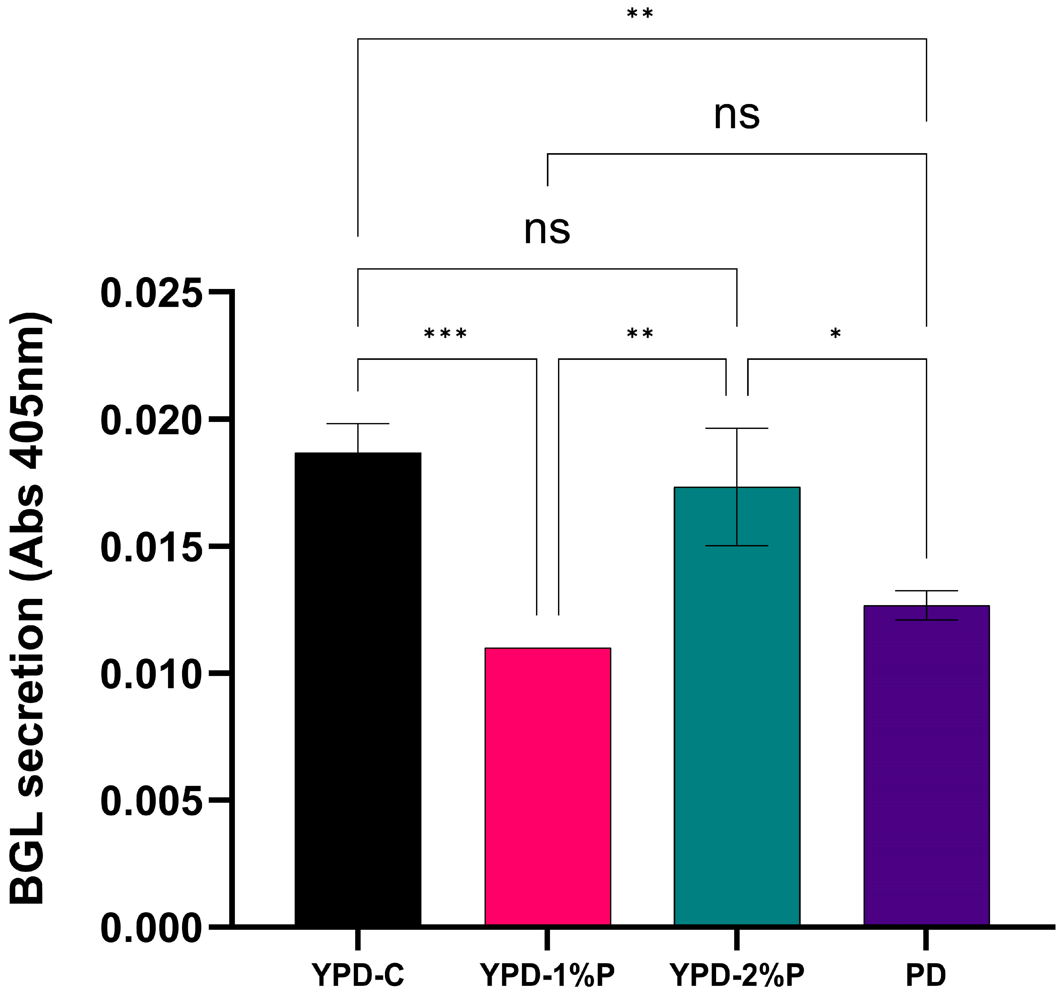
Secretion of β-glucosidase was monitored throughout a 144 h time-course to assess the capacity of the media types to support the expression of a recombinant protein. Figure 3A shows the expression of β-glucosidase at each 24 h mark. The YPD-1%P condition produced the least amount of enzyme at each timepoint, with the PD media cultures performing slightly better. The YPD-2%P condition matched closely to the YPD-C performance, with a similar pattern of expression observed across the time-course between the two conditions. Crucially, Figure 3B assesses any statistical differences between the secretion of β-glucosidase in the four conditions at the end of the time-course. As with the growth of the cultures, there is no statistical difference between the secretion levels of the YPD complete and the YPD-2%P cultures (p-value > 0.05, ns). There is a notable difference in secretion levels between the other two conditions and the YPD complete and YPD-2%P cultures, with the YPD-1%P condition producing significantly lower levels of β-glucosidase (p-value < 0.0001, ****), reaching only approximately 43% secretion compared to YPD complete, and 47% compared to YPD-2%P. PD media reached 64% secretion compared to YPD-2%P, but was still statistically significantly lower in output (p-value < 0.001, ***).
It is important to consider the varying optical densities of each condition when trying to ascertain which best support the production of β-glucosidase. Therefore, as outlined in Figure 4 below, secretion levels of each condition at the final 144 h timepoint were normalised to an OD600nm of 1 by dividing the secretion levels by the final OD. As seen, the same paradigm previously established with the growth rates and secretion levels is also observed. There is no statistical difference between the normalised secretion levels of the YPD complete and the YPD-2%P cultures (p-value > 0.05, ns). Importantly, there are still statistically significant differences in normalised secretion between both the YPD complete and YPD-2%P conditions and the other cultures.

3.4. Nutrient Uptake and Carbon Use Efficiency

To assess the functional equivalence of recycled extract media vs. commercial YPD, we quantified three key metrics: total soluble protein (Bradford assay), reducing sugar consumption (PAHBAH assay), and cell yield per glucose consumed.
Initial soluble protein in commercial YPD (YPD-C) was the highest among all four media types at 26.84 ± 0.25 g L−1, decreasing to 14.31 ± 0.15 g L−1 after 144 h. In recycled extract media, YPD + 1% peptone contained the lowest initial protein content at 13.22 ± 0.18 g L−1 initially, which dropped to 1.96 ± 0.12 g L−1, while YPD + 2% peptone started at 17.28 ± 0.16 g L−1 and fell to 6.86 ± 0.10 g L−1. PD minimal medium had a concentration of 15.44 ± 0.15 g L−1, which dropped to 6.26 ± 0.08 g L−1 residual protein at the end of the time-course. These assays confirm that recycled extract media deliver protein levels of the same order as commercial YPD and that cells utilize a substantial fraction of that pool during growth. A summary of this data is seen in Table 3 below.
Reducing sugar concentrations in culture supernatants were determined using the PAHBAH assay at the start (0 h) and the end (144 h) of fermentations (Table 4). Initial sugar concentrations were approximately 23 g/L−1 across all media formulations, consistent with the post-autoclaving addition of ~20 g·L−1 glucose (as 2% dextrose). After 144 h, residual sugars ranged from 2.14 ± 0.64 g/L−1 in YPD + 1% peptone to 3.52 ± 0.76 g/L−1 in PD minimal medium. Net sugar consumption was similar in all media (~20 g/L−1), confirming equivalent glucose utilization across formulations (Table 4). These results indicate that glucose was the primary carbon source metabolized during cultivation, and that the recycled yeast-extract-containing media supported hexose consumption comparably to the commercial YPD medium.
Cell yield per carbon source (g biomass/g glucose consumed), calculated based on wet cell biomass and glucose consumption (Table 4, above), reveals clear differences among the tested media formulations. As seen in Table 5 below, the YPD-C medium (commercial control) and the recycled YPD + 2% peptone medium showed comparable yields of 2.16 ± 0.10 and 2.18 ± 0.09 g wet biomass per gram glucose consumed, respectively. These yields align well with previously reported wet biomass yields (~2.0–2.5 g wet biomass per g glucose) for yeast cultivated in rich media [20,21], confirming efficient carbon utilization in both media types. The YPD + 1% peptone medium displayed a notably lower yield (1.47 ± 0.08 g wet biomass/g glucose), consistent with the reduced nitrogen and micronutrient availability at this lower peptone concentration. The PD minimal medium (peptone only, no yeast extract) also yielded lower biomass (1.74 ± 0.11 g wet biomass/g glucose), suggesting a link between yeast extract micronutrient availability for biomass productivity.

4. Discussion

4.1. Recycled Yeast Extract Is Soluble at Low Concentrations

The feasibility of reusing biomass waste as a feedstock for new fermentations is an important consideration. Here, it is demonstrated that simple freeze drying of frozen K. phaffii biomass following a primary fermentation is the only preparation step needed to create this recycled yeast extract. A study by Zarei [22] separated the yeast pellet into its soluble and insoluble components, requiring autoclaving and centrifugation as preparative steps. While effective, it is a more time-consuming and energy/material-intensive process. The method outlined in this study avoids these steps and produces a highly concentrated recycled yeast extract powder that is soluble only at very low concentrations. Based on the optical densities in Table 2, if equitable amounts of recycled yeast extract are added as in the standard YPD recipe, the resulting medium is extremely turbid, and after autoclaving, the yeast falls out of solution (i.e., YPD made with 10 g/L recycled yeast extract had an optical density OD600nm of 2.821). Therefore, through reduction in the amount of recycled yeast extract added to the YPD recipe, it was possible to determine the optimal amount for addition, i.e., 15 mg/L (OD600nm of 0.1482/0.1609, depending on the quantity of peptone added). This is a significant reduction than the usual 10 g/L amount of commercial yeast extract required, and the quantity of recycled yeast extract created from one biomass pellet would generate many litres of YPD media.
When considering the more common methods of preparing yeast extracts from biomass, there is a primary difference in the approach presented in this study. Typically, the methods of generating yeast extract are either hydrolysis or autolysis [23], involving specific incubation temperatures and often the addition of accelerators to assist in cell wall breakdown [24]. This study used pellets from a previous K. phaffii fermentation, where yeast cells had been grown for 96 h (plus an initial 48 h pre-culture prior to main culture inoculation). When culturing for an extended period such as this, autolysis can naturally occur, as is supported by the investigations of Zhang et al. [25] and Offei et al. [18]. Nutrient limitation, the accumulation of toxic metabolites, and cell growth plateau can lead to the autolysis of the cells within the culture. This is likely why no pre-treatment of the yeast biomass was required in this study, and a recycled bioavailable powder could be generated through simple freeze drying.

4.2. YPD Media Comprised of Recycled Yeast Extract and 2% Peptone Can Effectively Support the Growth of K. phaffii and the Secretion of β-Glucosidase

The YPD media formulation of 15 mg/L recycled yeast extract with 2% peptone (i.e., the usual amount found in YPD media) performed equally to YPD media made with commercial yeast extract both in terms of biomass generation and the secretion of the protein of interest β-glucosidase. This is reflected in Figure 2 and Figure 3, as there is no statistical difference (p-value > 0.05) between the final optical density of either media composition, nor in the final amount of secreted enzyme. It is also evident that K. phaffii have a minimum metabolic requirement of peptone for the growth and secretion of β-glucosidase, as the formulation of YPD media using recycled yeast extract but half the usual amount of peptone failed to effectively support these fermentations. This is observed through the less-dense cultures, reflected by the statistically significantly lower final optical density and smaller WCWs when compared to cultures containing recycled yeast extract and 2% peptone (Figure 1, Figure 2, Figure 3 and Figure 4). This has been seen previously where the addition of peptone to spent media can replenish the amino acid content sufficiently to enable waste media reuse [26]. Peptone is a valuable source of nutrients for many cell types, including mammalian and yeast cells, and indeed its addition has improved productivity of cultures in various scenarios [27,28].
Another crucial component of K. phaffii media is the yeast extract itself. The minimal medium used in this study, containing only 2% peptone (w/v) and 2% dextrose (v/v), provided a benchmark and a reference point for how a medium lacking yeast extract (regardless of source) performs. Yeast extract provides a source of nitrogen and other metabolically relevant elements and vitamins [29], and has been shown to demonstrate substantial variation in composition depending on the source of the yeast and the processing it undergoes [30]. Here, peptone and dextrose alone are not enough to match the performance of a complete YPD formulation, whether the yeast extract be commercial or generated in-house from recycled waste biomass pellets. The density of the PD cultures is significantly less than that of the YPD complete and the YPD-2%P cultures (Figure 2), and indeed, the final amount of secreted β-glucosidase is also significantly less in these PD fermentations (Figure 3).
Furthermore, when the level of β-glucosidase produced is normalised to an optical density of 1, removing the density of the culture as a potential confounding variable, there is still a significant difference observed in the YPD-1%P and PD media cultures compared to the commercial YPD and YPD-2%P cultures. Figure 4 illustrates that these cultures may be less dense at the end of the 144 h time-course, and they are also not as productive as the other media types.

4.3. YPD Media Comprised of Recycled Yeast Extract Has a Comparable Nutrient Profile to Commercial Yeast Extract Media

The comparative analysis of recycled yeast extract media formulations versus the conventional commercial YPD medium demonstrated sufficient nutrient profiles and performance in supporting yeast growth. Specifically, protein quantification by Bradford assay revealed non-limiting nutritional differences between the recycled extract media and the commercial control (Table 3). Initial protein concentrations (13.22–26.84 g·L−1) and their subsequent utilization (46–85%) during growth indicate the efficient uptake of available nitrogenous compounds, consistent with typical yeast fermentation profiles utilizing complex nitrogen sources [31,32]. Particularly notable was the YPD + 1% peptone formulation, which demonstrated the highest fractional protein consumption (85%), likely due to lower overall protein availability driving efficient nutrient uptake by yeast cells. The measured total protein concentrations indicate that the protein pool in the recycled YPD formulations and in PD is dominated by the peptone component and is consistent with the supplier COA (Sigma–Aldrich/Merck, product Peptone, Mycological No. 2 (product code 07751)—lot BCCC2601) and routine experimental variability. Assay values (recycled YPD-1%P ≈ 13.2 g·L−1, recycled YPD-2%P ≈ 17.3 g·L−1, PD ≈ 15.4 g·L−1) are of the same order as the crude protein expected from typical 2% (w/v) peptone (COA-derived estimate ≈ 15.9 g·L−1) and lie within the plausible lot-to-lot and preparation variation observed for peptone hydrolysates. The substantially higher protein observed in YPD-C (≈26.8 g·L−1) is attributable to the deliberate addition of 10 g·L−1 yeast extract: given typical yeast extract protein contents, a 10 g·L−1 supplement is expected to contribute several grams per litre of additional protein (order of magnitude ~5–9 g·L−1 depending on COA and conversion factor), which satisfactorily accounts for the elevation of YPD-C relative to the peptone-only media. It should be noted that the Bradford assay reports protein equivalents (peptides and free amino acids) and is subject to matrix effects; therefore, these values are presented as proximate indicators of the peptide/amino acid pool available to support fermentation rather than the exact measures of bioavailable free amino nitrogen (FAN), which would require targeted amino acid or FAN analysis for confirmation.
Reducing sugar analysis using the PAHBAH assay revealed that glucose utilization was consistent (~20 g·L−1 consumed) across all media formulations, indicating that glucose was the principal carbon source fuelling growth (Table 4). These findings align with typical hexose utilization patterns reported previously for yeast grown under similar aerobic conditions [32]. Yeast extract and peptone are primarily sources of peptides, free amino acids, and complex polysaccharides rather than simple, soluble reducing sugars; the carbohydrate fraction of yeast extracts is largely present as cell wall polysaccharides (mannans, β-glucans, glycogen) or insoluble material, and peptones are enzymatic protein hydrolysates whose simple sugar content is typically small and batch-dependent [29]. Consequently, the reducing sugar measurements reported here are dominated by the intentionally added dextrose; because the same dextrose stock and dispensing procedure were used for all media, near-identical initial reducing sugar concentrations (≈23 g·L−1) across formulations are expected and are fully consistent with routine preparation/assay uncertainty rather than substantial sugar contributions from yeast extract or peptone.
The cell yield per glucose consumed (Yxs) further validated the recycled yeast extract media (Table 5). Wet cell biomass yields were approximately 2.17 g wet biomass per gram of glucose consumed for the YPD-C and YPD + 2% peptone formulations, closely matching the literature benchmarks of around 2.0–2.5 g wet biomass per gram of glucose for yeast grown in rich media [33,34,35]. This confirms the metabolic efficiency and suitability of the recycled media as viable replacements for commercial formulations.
Growth kinetics analysis (specific growth rate, k, and doubling time, DT) also supported the conclusion that the recycled media were functionally equivalent to commercial YPD. Observed doubling times ranged between 59 and 66 h across all media conditions, consistent with typical slow-growing batch fermentations in minimal or moderately rich media [36]. The similarity in growth rates (k values ~0.011 h−1) further demonstrates that recycled yeast extract, even at significantly reduced concentrations (15 mg·L−1), provides adequate nutritional support, including critical micronutrients and vitamins, ensuring efficient biomass production [32].
Although total protein (Bradford) and reducing sugar measurements indicate similar bulk mass across PD, YPD-C, and recycled-YPD, these metrics do not quantify the fraction of nutrients that are immediately bioavailable to cells. Free amino nitrogen (FAN), small peptides, B vitamins, and trace metals are often present at low concentrations yet can be growth- or secretion-limiting [37]; recycled media and small yeast extract supplements are known to concentrate or retain such bioavailable species [29,38]. Thus, recycled-YPD (with 2% peptone plus a 15 mg·L−1 yeast extract spike) is hypothesised to provide an improved pool of utilizable amino acids, peptides, and micronutrients relative to PD and YPD-C even though gross protein and sugar are similar.
Collectively, these findings underscore the practical viability of recycled yeast extract media, presenting a sustainable and cost-effective alternative to commercial YPD formulations, with negligible differences in key growth parameters and nutrient utilization profiles. Furthermore, the recycled yeast extract approach described here offers several practical advantages beyond simplicity and reproducibility. It converts an on-site waste stream (spent K. phaffii biomass) into a useable supplementation, reducing raw material purchase and disposal costs and improving circularity with minimal additional processing (see Methods). Because only small additions (15 mg·L−1) were required to achieve the observed effect, the method imposes negligible additional upstream or downstream burden and requires no high-CAPEX equipment, facilitating rapid integration into existing pilot-scale workflows. The approach therefore lowers variable input demand and waste generation, while retaining the compositional benefits of yeast-derived peptides and micronutrients that can potentiate secondary cultures.

4.4. Method Limitations

While this method offers a simple and low-cost approach to reusing K. phaffii biomass, the several limitations that should be considered are as follows:
  • Organism-Specific Validation:
The current study was conducted exclusively using Komagataella phaffii as both the biomass source and production host. It remains to be tested whether the method is effective when applied to biomass from other yeast species, or used in heterologous fermentations (e.g., E. coli, Bacillus, etc.). However, it is believed that the principle is applicable to other strains of yeast.
  • Protein-Specific Results:
β-glucosidase was used as a model protein for validating fermentation performance. While it is a representative secreted enzyme, the generalizability of results to other recombinant proteins—especially intracellular or membrane-associated ones—has not yet been established.
  • Dependence on Spent Fermentation Conditions:
The method leverages biomass obtained from long (96+ hour) fermentations, which likely exhibit partial autolysis due to nutrient depletion and stress. Biomass harvested from shorter or different culture conditions may not provide the same bioavailability unless similar stress-induced degradation occurs.
  • Undetermined Quantitative Composition:
The specific biochemical composition of recycled yeast extract (e.g., amino acid profile, vitamin content, nucleotides) was not characterized. While performance metrics matched commercial media, and the reducing sugar and protein compositions were closely matched between the commercial yeast extract media and the recycled yeast extract media, differences in specific composition could influence outcomes in more nutrient-sensitive applications.
  • Sterility and Consistency Risks:
Since the process lacks defined sterilization or homogenization steps, batch-to-batch variation or microbial contamination risk could be higher in less controlled settings unless proper freeze drying and media handling practices are followed.

5. Conclusions

This study has provided a simple and effective method for valorising waste biomass from K. phaffii fermentations, through the generation of recycled yeast extract from the resulting pellets. Only milligram quantities of recycled yeast extract are required to efficiently and effectively support the growth and production of a recombinant protein, with densities and protein output showing no statistical difference between YPD media formulated with the commercially available yeast extract. Measurements demonstrating equivalent glucose consumption, total protein uptake, biomass yield per glucose, and matched growth kinetics in recycled yeast extract media compared to commercial YPD media demonstrates functional parity. This approach incorporates the core concepts of the circular bioeconomy and represents a simple and practical use for what is a considerable waste source.

Supplementary Materials

The following supporting information can be downloaded at: https://www.mdpi.com/article/10.3390/applmicrobiol5030095/s1.

Author Contributions

L.M. and D.J.O. conceived the study. L.M. performed the experiments and analysed the data. L.M. and D.J.O. wrote the manuscript. All authors have read and agreed to the published version of the manuscript.

Funding

This research was funded by the Atoms-2-Products centre for doctoral training, which is supported by the Science Foundation Ireland (SFI) and the Engineering and Physical Sciences Research Council (EPSRC) under Grant No. 18/EPSRC-CDT/3582.

Institutional Review Board Statement

Not applicable.

Informed Consent Statement

Not applicable.

Data Availability Statement

The data supporting this article have been included as part of the Supplementary Information.

Acknowledgments

Thanks to Ben Offei formerly of UCD and Ken Wolfe at UCD for providing the β-glucosidase-expressing clone.

Conflicts of Interest

The authors declare no conflicts of interest.

References

  1. Maicas, S. The Role of Yeasts in Fermentation Processes. Microorganisms 2020, 8, 1142. [Google Scholar] [CrossRef]
  2. Mattanovich, D.; Sauer, M.; Gasser, B. Yeast biotechnology: Teaching the old dog new tricks. Microb. Cell Factories 2014, 13, 34. [Google Scholar] [CrossRef]
  3. Demain, A.L.; Phaff, H.J.; Kurtzman, C.P. The industrial and agricultural significance of yeasts. In The Yeasts, 4th ed.; Kurtzman, C.P., Fell, J.W., Eds.; Elsevier: Amsterdam, The Netherlands, 1998; Chapter 4, pp. 13–19. [Google Scholar]
  4. Murphy, L.; O’Connell, D.J. The Role of Yeast in the Valorisation of Food Waste. Fermentation 2024, 10, 583. [Google Scholar] [CrossRef]
  5. Łukaszewicz, M.; Leszczyński, P.; Jabłoński, S.J.; Kawa-Rygielska, J. Potential Applications of Yeast Biomass Derived from Small-Scale Breweries. Appl. Sci. 2024, 14, 2529. [Google Scholar] [CrossRef]
  6. Nielsen, J. Production of biopharmaceutical proteins by yeast: Advances through metabolic engineering. Bioengineered 2013, 4, 207–211. [Google Scholar] [CrossRef]
  7. Barone, G.D.; Emmerstorfer-Augustin, A.; Biundo, A.; Pisano, I.; Coccetti, P.; Mapelli, V.; Camattari, A. Industrial Production of Proteins with Pichia pastoris-Komagataella phaffii. Biomolecules 2023, 13, 441. [Google Scholar] [CrossRef]
  8. Pichia.com. Pichia Produced Products on the Market. Available online: https://pichia.com/science-center/commercialized-products/ (accessed on 30 May 2024).
  9. Akermann, A.; Weiermüller, J.; Chodorski, J.N.; Nestriepke, M.J.; Baclig, M.T.; Ulber, R. Optimization of bioprocesses with Brewers’ spent grain and Cellulomonas uda. Eng. Life Sci. 2022, 22, 132–151. [Google Scholar] [CrossRef]
  10. Granget, C.; Manikandan, N.A.; Amulya, K.; Dabros, M.; Fahy, S.; Kelleher, S.M.; Rochfort, K.D.; Gaughran, J.; Freeland, B. Brewer’s spent grain as a self-sufficient feedstock for homofermentative production of optically pure L-lactic acid using Lactobacillus rhamnosus. Environ. Technol. Innov. 2024, 34, 103582. [Google Scholar] [CrossRef]
  11. Cooray, S.T.; Lee, J.J.L.; Chen, W.N. Evaluation of brewers’ spent grain as a novel media for yeast growth. AMB Express 2017, 7, 117. [Google Scholar] [CrossRef]
  12. Flores-Copa, V.; Romero-Soto, L.; Romero-Calle, D.; Alvarez-Aliaga, M.T.; Orozco-Gutierrez, F.; Vega-Baudrit, J.; Martín, C.; Carrasco, C. Residual Brewing Yeast as Substrate for Co-Production of Cell Biomass and Biofilm Using Candida maltosa SM4. Fermentation 2021, 7, 84. [Google Scholar] [CrossRef]
  13. Patel, A.; Mikes, F.; Buhler, S.; Matsakas, L. Valorization of Brewers' Spent Grain for the Production of Lipids by Oleaginous Yeast. Molecules 2018, 23, 3052. [Google Scholar] [CrossRef]
  14. Le, R.K.; Das, P.; Mahan, K.M.; Anderson, S.A.; Wells, T.; Yuan, J.S.; Ragauskas, A.J. Utilization of simultaneous saccharification and fermentation residues as feedstock for lipid accumulation in Rhodococcus opacus. AMB Express 2017, 7, 185. [Google Scholar] [CrossRef]
  15. Jacob, F.F.; Hutzler, M.; Methner, F.-J. Comparison of various industrially applicable disruption methods to produce yeast extract using spent yeast from top-fermenting beer production: Influence on amino acid and protein content. Eur. Food Res. Technol. 2019, 245, 95–109. [Google Scholar] [CrossRef]
  16. Sakarika, M.; Kerckhof, F.-M.; Van Peteghem, L.; Pereira, A.; Van Den Bossche, T.; Bouwmeester, R.; Gabriels, R.; Van Haver, D.; Ulčar, B.; Martens, L.; et al. The nutritional composition and cell size of microbial biomass for food applications are defined by the growth conditions. Microb. Cell Factories 2023, 22, 254. [Google Scholar] [CrossRef]
  17. Ahuja, V.; Arora, A.; Chauhan, S.; Thakur, S.; Jeyaseelan, C.; Paul, D. Yeast-Mediated Biomass Valorization for Biofuel Production: A Literature Review. Fermentation 2023, 9, 784. [Google Scholar] [CrossRef]
  18. Offei, B.; Braun-Galleani, S.; Venkatesh, A.; Casey, W.T.; O’Connor, K.E.; Byrne, K.P.; Wolfe, K.H. Identification of genetic variants of the industrial yeast Komagataella phaffii (Pichia pastoris) that contribute to increased yields of secreted heterologous proteins. PLoS Biol. 2022, 20, e3001877. [Google Scholar] [CrossRef]
  19. Iyer, A.; Bestwick, C.S.; Duncan, S.H.; Russell, W.R. Invasive Plants Are a Valuable Alternate Protein Source and Can Contribute to Meeting Climate Change Targets. Front. Sustain. Food Syst. 2021, 5, 575056. [Google Scholar] [CrossRef]
  20. Zarei, O.; Dastmalchi, S.; Hamzeh-Mivehroud, M. A Simple and Rapid Protocol for Producing Yeast Extract from Saccharomyces cerevisiae Suitable for Preparing Bacterial Culture Media. Iran. J. Pharm. Res. 2016, 15, 907–913. [Google Scholar]
  21. Inokuma, K.; Hasunuma, T.; Kondo, A. Efficient yeast cell-surface display of exo- and endo-cellulase using the SED1 anchoring region and its original promoter. Biotechnol Biofuels. 2014, 7, 8. [Google Scholar] [CrossRef]
  22. Vajdič, T.; Erjavec, M.S. Harnessing Environmental Yeasts—Pichia kudriavzevii Strain ZMUM_K002: The Quest for Isolates with Properties for Efficient Biotechnological Applications. Appl. Microbiol. 2025, 5, 30. [Google Scholar] [CrossRef]
  23. Jaeger, A.; Arendt, E.K.; Zannini, E.; Sahin, A.W. Brewer’s Spent Yeast (BSY), an Underutilized Brewing By-Product. Fermentation 2020, 6, 123. [Google Scholar] [CrossRef]
  24. Bayarjargal, M.; Munkhbat, E.; Ariunsaikhan, T.; Odonchimeg, M.; Uurzaikh, T.; Gan-Erdene, T.; Regdel, D. Utilization of spent brewer’s yeast Saccharomyces cerevisiae for the production of yeast enzymatic hydrolysate. Mong. J. Chem. 2014, 12, 88–91. [Google Scholar] [CrossRef]
  25. Zhang, C.; Ma, Y.; Miao, H.; Tang, X.; Xu, B.; Wu, Q.; Mu, Y.; Huang, Z. Transcriptomic Analysis of Pichia pastoris (Komagataella phaffii) GS115 During Heterologous Protein Production Using a High-Cell-Density Fed-Batch Cultivation Strategy. Front. Microbiol. 2020, 11, 463. [Google Scholar] [CrossRef]
  26. Murphy, L.; Lynch, C.D.; O’Connell, D.J. Valorisation of Spent Yeast Fermentation Media through Compositional-Analysis-Directed Supplementation. Appl. Microbiol. 2024, 4, 959–971. [Google Scholar] [CrossRef]
  27. Davami, F.; Eghbalpour, F.; Nematollahi, L.; Barkhordari, F.; Mahboudi, F. Effects of Peptone Supplementation in Different Culture Media on Growth, Metabolic Pathway and Productivity of CHO DG44 Cells; a New Insight into Amino Acid Profiles. Iran. Biomed. J. 2015, 19, 194. [Google Scholar]
  28. Krahulec, J.; Šafránek, M. Impact of media components from different suppliers on enterokinase productivity in Pichia pastoris. BMC Biotechnol. 2021, 21, 19. [Google Scholar] [CrossRef]
  29. Tao, Z.; Yuan, H.; Liu, M.; Liu, Q.; Zhang, S.; Liu, H.; Jiang, Y.; Huang, D.; Wang, T. Yeast Extract: Characteristics, Production, Applications and Future Perspectives. J. Microbiol. Biotechnol. 2023, 33, 151–166. [Google Scholar] [CrossRef]
  30. Tachibana, S.; Watanabe, K.; Konishi, M. Estimating effects of yeast extract compositions on Escherichia coli growth by a metabolomics approach. J. Biosci. Bioeng. 2019, 128, 468–474. [Google Scholar] [CrossRef]
  31. Broach, J.R. Nutritional control of growth and development in yeast. Genetics 2012, 192, 73–105. [Google Scholar] [CrossRef]
  32. Walker, G.M.; Stewart, G.G. Saccharomyces cerevisiae in the Production of Fermented Beverages. Beverages 2016, 2, 30. [Google Scholar] [CrossRef]
  33. Sonderegger, M.; Sauer, U. Evolutionary engineering of Saccharomyces cerevisiae for anaerobic growth on xylose. Appl. Env. Microbiol. 2003, 69, 1990–1998. [Google Scholar] [CrossRef]
  34. Vu, V.H.; Kim, K. High-cell-density fed-batch culture of Saccharomyces cerevisiae KV-25 using molasses and corn steep liquor. J. Microbiol. Biotechnol. 2009, 19, 1603–1611. [Google Scholar] [CrossRef]
  35. Ferndahl, C.; Bonander, N.; Logez, C.; Wagner, R.; Gustafsson, L.; Larsson, C.; Hedfalk, K.; Darby, R.A.; Bill, R.M. Increasing cell biomass in Saccharomyces cerevisiae increases recombinant protein yield: The use of a respiratory strain as a microbial cell factory. Microb. Cell Fact. 2010, 9, 47. [Google Scholar] [CrossRef]
  36. Sherman, F. Getting started with yeast. Methods Enzym. 2002, 350, 3–41. [Google Scholar] [CrossRef]
  37. Perli, T.; Wronska, A.K.; Ortiz-Merino, R.A.; Pronk, J.T.; Daran, J.M. Vitamin requirements and biosynthesis in Saccharomyces cerevisiae. Yeast 2020, 37, 283–304. [Google Scholar] [CrossRef]
  38. Wynne, E.; Yoon, J.; Park, D.; Cui, M.; Morris, C.; Lee, J.; Wang, Z.; Yoon, S.; Han, J. Regeneration of Spent Culture Media for Sustainable and Continuous mAb Production via Ion Concentration Polarization. Biotechnol. Bioeng. 2025, 122, 373–381. [Google Scholar] [CrossRef]
Figure 1. (A) Growth curve of K. phaffii β-glucosidase-expressing IRA1 clones in a variety of YPD media, and (B) Analysis of growth rate and doubling time for each culture. All culture conditions were grown in triplicate (n = 3) and were inoculated in a 1:120 ratio with a 48 h IRA1 pre-culture grown in fresh YPD media and allowed to express β-glucosidase for 144 h by shaking at 230 RPM at 30 °C. Error bars are representatives of the standard deviation of biological triplicates. Parameters from Malthusian exponential fits N(t) = Y0ekt the full 0–144 h time-course. Y0 = fitted intercept (predicted OD600 at t = 0; dimensionless), k = exponential rate constant (h−1), and doubling time = td = ln(2)/k (hours). Reported values are means of per-replicate fits. Comparison of per-replicate k and doubling times across media found no significant differences (one-way ANOVA/Tukey’s post hoc test, p > 0.05). All data analysed on GraphPad Prism 10 software.
Applmicrobiol 05 00095 g001
Figure 2. Final 144 h timepoint (A) Optical densities at 600 nm, and (B) wet cell weights (WCW) of K. phaffii β-glucosidase-expressing IRA1 clones in a variety of YPD media. (A) Samples were taken at the end of the 144 h time-course and read on a spectrophotometer at 600 nm. (B) Cultures were spun at 4000× g at the end of the 144 h time-course and weighed on a balance. (A,B) Error bars are representatives of the standard deviation of biological triplicates (n = 3). Significance is determined using a two-way ANOVA with Tukey’s post-analysis test. p-values are represented as p > 0.05, not significant (ns); p ≤ 0.05, *; p ≤0.01, **; p ≤0.001, ***; p ≤ 0.0001, ****.
Applmicrobiol 05 00095 g002
Figure 3. (A) Secretion of β-glucosidase by IRA1 clones in YPD media over the 144 h time-course, and (B) final β-glucosidase secretion levels at T144. (A) Samples were taken every 24 h, and β-glucosidase levels were measured through the colorimetric 4-NPG assay, where absorbance at 405 nm equals the quantity of β-glucosidase secreted. Error bars are representative of the standard deviation of biological triplicates, where each replicate was measured in technical triplicate. Key: YPD-C—black circle; YPD-2%P media—green triangle; ‘Y’PD media half peptone—pink square; PD media—purple inverted triangle. (B) Samples were taken at the end of the 144 h time-course, and β-glucosidase levels were measured through the colorimetric 4-NPG assay, where absorbance at 405 nm equals the quantity of β-glucosidase secreted. Error bars are representatives of the standard deviation of biological triplicates (n = 3), where each replicate was measured in technical triplicates. Significance is determined using a two-way ANOVA with Tukey’s post-analysis test. p-values are represented as p > 0.05, not significant (ns); p ≤ 0.001, ***; p ≤ 0.0001, ****.
Applmicrobiol 05 00095 g003
Figure 4. Optical density normalised β-glucosidase secretion at the end of the 144 h expression time-course. Samples were taken at the end of the 144 h time-course, and β-glucosidase levels were measured through the colorimetric 4-NPG assay, where absorbance at 405 nm equals the quantity of β-glucosidase secreted. Correspondingly, samples were taken at the end of the 144 h time-course and read on a spectrophotometer at 600 nm to determine optical density. Normalisation was performed by dividing the β-glucosidase secretion absorbance by the relevant calculated optical density. Error bars are representatives of the standard deviation of biological triplicates (n = 3), where each replicate was measured in technical triplicates. Significance is determined using a two-way ANOVA with Tukey’s post-analysis test. p-values are represented as p > 0.05, not significant (ns); p ≤ 0.05, *; p ≤ 0.01, **; p ≤ 0.001, ***.
Applmicrobiol 05 00095 g004
Table 1. Summary of components of various YPD media formulations.
Media TypeCommercial Yeast Extract (g)Recycled Yeast Extract (mg)Peptone (%)Dextrose (%)
YPD commercial yeast extract 1002%2%
YPD 10 g/L recycled yeast extract010,0002%2%
YPD 0.1 g/L recycled yeast extract01002%2%
YPD 0.05 g/L recycled yeast extract0502%2%
YPD 15 mg/L recycled yeast extract; 1% peptone0151%2%
YPD 15 mg/L recycled yeast extract; 2% peptone0152%2%
PD media (2% peptone; 2% dextrose)002%2%
Table 2. Composition of media types tested using recycled yeast extract with 1% or 2% peptone and their OD600nm values. Average optical density was determined by reading samples on a spectrophotometer at 600 nm in technical triplicates. Standard deviation of the population and coefficient of variation was calculated using Microsoft Excel formulae.
MediumAverage Optical
Density at 600 nm
Standard Deviation Coefficient of Variation
YPD commercial yeast extract 0.15930.0063.9%
YPD 10 g/L recycled yeast extract2.8210.35112.46%
YPD 0.1 g/L recycled yeast extract1.1220.13812.31%
YPD 0.05 g/L recycled yeast extract0.73350.0689.29%
YPD 15 mg/L recycled yeast extract; 1% peptone0.14820.0053.58%
YPD 15 mg/L recycled yeast extract; 2% peptone0.16090.0074.49%
PD media (2% peptone; 2% dextrose)0.13440.0075.15%
Table 3. Soluble protein concentrations (g/L) in the four media types from T0 to T144. Total protein content was assessed using a Bradford assay, and concentration of samples was calibrated against bovine serum albumin (BSA) standards (0–1 mg/mL), and absorbance of all samples were read at 595 nm. Values represent the mean ± standard deviation from biological triplicates (n = 3) run in technical duplicates.
MediumInitial
Prot. (g/L)
Residual Prot. (g/L)Consumed ΔProt. (g/L)% Consumed
YPD-C26.84 ± 0.2514.31 ± 0.1512.53 ± 0.2946.7 ± 2.6
YPD + 1%P13.22 ± 0.181.96 ± 0.1211.26 ± 0.2285.2 ± 1.9
YPD + 2%P17.28 ± 0.166.86 ± 0.1010.42 ± 0.1960.4 ± 2.1
PD15.44 ± 0.156.26 ± 0.089.18 ± 0.1759.5 ± 2.5
Table 4. Reducing sugar concentrations in culture media formulations measured by the PAHBAH assay at 0 h and after 144 h cultivation. Concentrations were determined spectrophotometrically at 410 nm following reaction with the PAHBAH reagent, calibrated against glucose standards (0–3 mg/mL). Samples were diluted appropriately prior to assay to fall within the linear range of the standard curve. Values represent mean ± standard deviation from biological triplicates that were run in technical duplicates (n = 3).
MediumInitial Sugar
T0 (g/L−1)
Residual Sugar
T144 (g/L−1)
Sugar
Consumed (g/L−1)
YPD-C23.64 ± 0.48 3.19 ± 0.2720.45 ± 0.55
YPD + 1%P23.80 ± 0.672.14 ± 0.6421.66 ± 0.92
YPD + 2%P23.07 ± 0.353.51 ± 0.8919.56 ± 0.96
PD23.73 ± 0.673.52 ± 0.7620.20 ± 1.02
Table 5. Cell yield per carbon source (g biomass/g glucose consumed) calculated from wet biomass (g/L−1) and glucose consumption (g/L−1) after 144 h of cultivation. Wet biomass was determined by harvesting 43 mL of culture, centrifuging, carefully removing the supernatant, allowing to drain and dry briefly, and weighing the resulting cell pellet. Reducing sugar (glucose) concentrations were measured using the PAHBAH assay, calibrated against glucose standards. All values represent mean ± standard deviation from biological triplicates (n = 3).
MediumWet Biomass (g/L−1)∆Sugar (g/L−1)Wet YX/S
(g Biomass/g Glucose)
YPD-C44.3 ± 1.020.45 ± 0.552.16 ± 0.06
YPD + 1% P31.9 ± 0.921.66 ± 0.921.47 ± 0.07
YPD + 2% P42.6 ± 1.019.56 ± 0.962.18 ± 0.11
PD35.1 ± 1.220.20 ± 1.021.74 ± 0.10
Disclaimer/Publisher’s Note: The statements, opinions and data contained in all publications are solely those of the individual author(s) and contributor(s) and not of MDPI and/or the editor(s). MDPI and/or the editor(s) disclaim responsibility for any injury to people or property resulting from any ideas, methods, instructions or products referred to in the content.