Article

Phyllosphere Antagonistic Bacteria Induce Growth Promotion and Effective Anthracnose Control in Cucumber

by
Mst. Habiba Kamrun Nahar
1,
Preangka Saha Briste
1,
Md. Rabiul Islam
2,
Touhidur Rahman Anik
3,
Md. Tanbir Rubayet
1,
Imran Khan
4,
Md. Motaher Hossain
1,* and
Mohammad Golam Mostofa
4,*
1
Department of Plant Pathology, Faculty of Agriculture, Gazipur Agricultural University, Gazipur 1706, Bangladesh
2
Pathology Division, Bangladesh Sugarcrop Research Institute, Pabna 6620, Bangladesh
3
Department of Plant and Soil Science, Institute of Genomics for Crop Abiotic Stress Tolerance, Texas Tech University, Lubbock, TX 79409, USA
4
Department of Chemistry, State University of New York College of Environmental Science and Forestry, Syracuse, NY 13210, USA
*
Authors to whom correspondence should be addressed.
Appl. Microbiol. 2025, 5(3), 94; https://doi.org/10.3390/applmicrobiol5030094
Submission received: 23 July 2025 / Revised: 30 August 2025 / Accepted: 31 August 2025 / Published: 4 September 2025

Abstract

The phyllosphere, the aerial part of plants, serves as a crucial habitat for diverse microorganisms. Phyllosphere bacteria can activate protective mechanisms that help plants resist disease. This study focuses on isolating and characterizing phyllosphere bacteria from cucurbits to evaluate their potential in controlling Colletotrichum orbiculare, a pathogen causing anthracnose in cucumbers. Among the 76 bacterial isolates collected, 11 exhibited strong antagonistic effects against C. orbiculare in vitro. Morphological and 16S rRNA analyses identified these isolates as different Bacillus species, including B. vallismortis, B. velezensis, B. amyloliquefaciens, and B. subtilis. These bacteria demonstrated essential plant-growth-promoting and biocontrol traits, such as motility, biofilm formation, phosphate solubilization, nitrogen fixation, and the production of indole acetic acid. Most of the bacterial strains also produced biocontrol compounds such as ammonia, acetoin, siderophores, hydrogen cyanide, chitinase, protease, lipase, and cellulase. The application of these bacteria significantly enhanced cucumber growth in both non-manured and organically manured soils, showing improvements in root and shoot length, chlorophyll content, and biomass accumulation. Additionally, bacterial treatments effectively reduced anthracnose severity, with isolates GL-10 and L-1 showing the highest disease suppression in both soil types. Colonization studies showed that phyllobacteria preferentially colonized healthy leaves over roots and diseased tissues, and they were more effective in manure-amended soils. These results suggest that Bacillus phyllobacteria have strong potential as sustainable bio-stimulants and biocontrol agents, offering an effective approach for enhancing cucumber growth and disease control under both fertilized and unfertilized soil conditions.

1. Introduction

Cucumber (Cucumis sativus L.), a member of the Cucurbitaceae family, is among the top 10 most widely cultivated vegetables worldwide and has a long history of cultivation in Asia [1]. Consumed year-round as both a salad ingredient and a vegetable, cucumber is valued for its refreshing, low-calorie nature and high-water content. It provides about 15 calories per cup and contains approximately 95% water [2]. In addition to its hydrating properties, cucumber is rich in a diverse array of bioactive compounds. Lignans, vitamin K, cucurbitacins and their derivatives (triterpenoids), and flavonoids (such as apigenin, luteolin, quercetin, and kaempferol), are abundant in cucumber. It is also rich in antioxidants such as beta-carotene, vitamin B and C, and various trace elements and minerals [3]. These constituents contribute a range of health-promoting effects. Cucumber triterpenes exhibit cytoprotective properties that support epidermal barrier function and cellular immunity [4]. Rutin and ascorbic acid oxidase act as free radical scavengers to protect against skin damage [5]. Cucumbers also have photoprotective properties, with a sun protection factor (SPF) of 0.2 [6]. Topical lotions that contain cucumber extract have been shown to reduce melanin and sebum production, offering benefits such as skin whitening and anti-acne effects [7]. These combined nutritional and therapeutic attributes contribute to the crop’s global popularity and economic value.
Cucumber is vulnerable to infections by various phytopathogenic fungi during cultivation. Anthracnose, caused by Colletotrichum orbiculare, is one of the most significant diseases in cucumbers [8]. This pathogen has a broad host range, infecting over 50 species within the Cucurbitaceae, Leguminosae, and Solanaceae families [9]. This fungus primarily targets plant leaves, stems, and fruits, which leads to yield and quality reductions [10]. Consequently, effective management of anthracnose is critical for stable cucumber production. Currently, chemical control remains the primary method employed by farmers [11]. However, a heavy reliance on chemical treatments contributes to the development of resistant microbes and raises concerns about environmental and health impacts. In response, biocontrol agents offer a promising, eco-friendly alternative. Recently, biocontrol agents have gained significant attention for disease management and crop improvement due to their non-toxic, environmentally safe attributes [12,13,14]. Among biocontrol agents, considerable research has focused on rhizosphere bacteria [8,15,16,17,18,19]. However, the application of phyllosphere bacteria to enhance plant growth and control diseases remains challenging due to several research gaps. The phyllosphere, which is the leaf surface environment, is an ecologically demanding niche subject to UV radiation, fluctuating humidity, and limited nutrient availability. These factors restrict the survival and consistency of introduced microbes [20]. Additionally, compared to root-applied plant-growth-promoting rhizobacteria (PGPR) techniques, there are fewer standardized and replicated field studies demonstrating effective and long-lasting disease suppression from foliar inoculants in cucumbers [21]. Furthermore, the community-level effects of foliar biocontrol on cucumber phyllosphere microbiomes under disease pressure remain poorly understood, which limits our mechanistic insights and strategies for optimization [22]. These challenges underscore the need for comprehensive research on the efficacy of phyllosphere bacteria as bio-stimulants and biocontrol agents [23].
The phyllosphere is the largest microbial habitat on Earth and is primarily inhabited by bacteria, with populations typically ranging from 106 to 107 cells per cm2 [24,25]. Although several studies suggest that phyllosphere microbiomes may play a crucial role in plant health and disease suppression under various environmental conditions [12,26], no studies have yet examined the potential of cucurbit phyllosphere bacteria in enhancing growth and controlling anthracnose in cucumbers. Moreover, the effects of these bacterial antagonists on plant growth and disease suppression, particularly in non-manured or organically managed soils, remain largely unexplored.
This study aimed to identify and characterize the key antagonistic bacteria found in the phyllosphere of cucurbits. It also assessed the impact of selected antagonistic bacteria on growth promotion and the suppression of anthracnose under both fertilized and non-fertilized conditions. By identifying effective bacterial candidates and evaluating how fertilization affects their efficacy, this research contributed to integrated disease management strategies that enhance cucumber resistance to anthracnose while promoting sustainable agriculture.

2. Materials and Methods

2.1. Sampling and Isolation of Phyllosphere Bacteria from Cucumber

Phyllosphere samples, including leaves, stems, and flowers from various cucurbit plants, were collected from the research fields of Gazipur Agricultural University, Gazipur, Bangladesh. Each type of sample was placed in a separate sterile polythene bag for transport to the laboratory. The samples were then stored at 4 °C until used for bacterial isolation (Figure 1).
To prepare for bacterial isolation, the plant samples were surface-sterilized by immersing them in 70% (v/v) ethanol for 5 min, followed by thorough rinsing with sterilized distilled water. Using a sterile mortar and pestle, the samples were macerated to prepare a homogenized suspension, which was then serially diluted. From each dilution, a 100 μL aliquot was plated on yeast extract potato dextrose agar (YPDA) and incubated at 28 ± 2 °C for 2 days. Individual bacterial colonies exhibiting diverse phenotypes were selected and re-streaked on YPDA plates to obtain pure isolates. The purified isolates were temporarily preserved in a 20% glycerol solution at –20 °C.

2.2. Host Plant and Pathogen

Cucumber variety Baromashi (Lal Teer Seed Ltd., Dhaka, Bangladesh) was used as the host throughout the experiment. An isolate of Colletotrichum orbiculare (104T) was used as the pathogen to induce cucumber anthracnose in this study. This isolate was obtained from the Laboratory of Plant Pathology at Gifu University, Gifu-shi, Japan [8].

2.3. Screening of the Bacterial Isolates Against C. orbiculare Using a Dual Plate Confrontation Assay

A dual plate confrontation assay was performed to evaluate the effectiveness of bacterial isolates against C. orbiculare [27]. A 1-week-old culture of C. orbiculare was used to prepare a 7 mm diameter mycelial plug, which was placed at one edge of a YPDA plate. The bacterial isolates were streaked at the center of the plate, with the fungal plug positioned on one side and the opposite edge left empty. Control plates were inoculated with C. orbiculare alone. All plates were incubated at 28 ± 2 °C until the fungal growth on the control plates reached completion. The assay was conducted with three independent biological replicates, each containing three technical replicates, to ensure statistical robustness. The diameter of the fungal colony was then recorded, and the percentage inhibition of radial growth was calculated using the following formula:
I n h i b i t i o n   o f   g r o w t h ( % ) = X Y X × 100
where X and Y indicate the mycelial growth of the fungus in the absence and presence of the bacterial isolates, respectively. Data from all replicates were subjected to statistical analysis (ANOVA) to determine significant differences among treatments.

2.4. Morphological, Biochemical, and Molecular Characterization of the Selected Bacterial Isolates

Conventional light microscopy was used to observe the morphological characteristics and Gram staining status of the bacterial isolates. The selected isolates were cultured on YPDA plates and incubated for 3 days. Afterwards, various colony features, including color, shape, elevation, and surface texture, were documented. Standard protocols were followed to assess physiological and biochemical properties, including the Gram reaction, KOH test, oxidase test, and the catalase test [28].
For molecular characterization, genomic DNA was extracted from the bacterial isolates using a genomic DNA extraction kit (Sangon Biotech, Shanghai, China) according to the manufacturer’s instructions. The 16S rRNA gene of each isolate was amplified using the primers 27F (5′AGAGTTTGATCCTGGCTCAG-3′) and 1492R (5′-GGTTACCTTGTTACGACTT-3′) [29]. The resulting PCR products were purified using the Wizard PCR Preps DNA Purification System (Promega, Madison, WI, USA). Purified, double-stranded PCR fragments were sequenced directly with the Big Dye Terminator Cycle Sequencing Kit (Applied Biosystems, Foster City, CA, USA), following the manufacturer’s protocol. Sequences were edited with Chromas Lite 2.0 and deposited in the GenBank database. For sequence analysis, the BLAST (BLAST+ 2.13.0) search program was used to identify nucleotide sequence homology for the 16S rRNA region of the isolates. Highly homologous sequences were aligned, and neighbor-joining phylogenetic trees were constructed using Clustal X version 2.0.11 and MEGA version 11 software, with statistical support for the tree nodes provided by 1000 bootstrap replications.

2.5. Qualitative Screening of Hydrolytic Enzyme Activity

The selected bacterial isolates were screened qualitatively for the production of hydrolytic enzymes, including cellulase, lipase, and protease. To assess cellulolytic activity, bacterial isolates were separately cultured in minimal medium (MM) supplemented with 0.1% (w/v) carboxymethyl cellulose (CMC) along with 0.1% NaNO3, 0.05% MgSO4, 0.1% K2HPO4, 0.1% KCl, 0.05% yeast extract, and 1.5% agar [30]. The plates were incubated at room temperature for 5 days. Following incubation, plates were flooded with Congo red solution and rinsed with 1 M NaCl. Clear halos around the colonies indicated positive cellulolytic activity.
Lipase activity was assessed by spotting bacterial isolates on LB medium containing 1% (v/v) Tween 20. The plates were incubated at room temperature for up to 5 days, and isolates that exhibited a clear halo around the colonies were considered positive for lipase production [31].
Protease production was determined by culturing bacterial isolates in LB medium at 28 °C on a rotary shaker at 180 rpm for 72 h. Subsequently, 2 µL of each 72 h old culture was inoculated onto skim milk agar plates and incubated for 48 h at 28 °C. The formation of a clear halo around the colony indicated positive protease production [32].

2.6. Biochemical and Metabolite Production Assays

Indole-3-acetic acid (IAA) production was evaluated using a colorimetric method [28]. Bacterial isolates were inoculated in Luria–Bertani (LB) broth supplemented with 5 mM tryptophan and incubated for 72 h at 29 ± 1 °C. The cultures were then centrifuged at 15,000 rpm for 1 min. One mL of the supernatant was mixed with 2 mL of Salkowski reagent, and absorbance was measured at 530 nm. The method had a detection limit of 0.5 μg mL−1 and a linear range of 0.5–50 μg mL−1 (R2 = 0.995). Specificity was ensured by using freshly prepared reagents, including reagent blanks, and confirming the characteristic 530 nm absorbance peak of IAA to minimize interference from structurally related metabolites. IAA concentration was calculated from a standard curve and expressed as µg mL−1.
Ammonia production was assessed by inoculating bacterial isolates in peptone water, incubating at 28 ± 2 °C for 48 h with continuous shaking. One mL of the culture supernatant was mixed with Nessler’s reagent, and a color change from yellow to brown indicated positive ammonia production [33].
Bacterial cultures (16 h old) were inoculated in 5 mL of yeast extract salt broth (per liter: 0.5 g NH4H2PO4, 0.2 g MgSO4·7H2O, 5.0 g NaCl, 5.0 g glucose), and incubated at 28 °C for 96 h. One mL of each culture was transferred to a sterile test tube, followed by the addition of 600 µL of 5% (w/v) alpha-naphthol in absolute alcohol. A crimson to ruby color within 4 h indicated positive acetoin production.
Siderophore production was assessed by spotting bacterial isolates on Chrome Azurol S (CAS) agar plates (pH 7), incubating at 28 ± 2 °C for 72 h. The formation of a yellow to orange halo around the colonies indicated positive siderophore production [34].
Hydrogen cyanide (HCN) production was tested by streaking bacterial isolates on nutrient agar supplemented with 4.4 g L−1 glycine. A Whatman No. 1 filter paper soaked in a 2% sodium carbonate and 0.5% picric acid solution was placed over the plate, which was then sealed with parafilm and incubated at 30 °C for 48 h. The development of an orange to red color indicated HCN production [35].
To test for indole production, bacterial isolates were inoculated into tryptone broth (10 g tryptone, 5 g NaCl per L) and incubated at 35 °C for 48 h. Five drops of Kovac’s reagent were added to each tube, and a cherry-red ring at the top of the medium indicated a positive indole test [36].

2.7. Motility Test

Bacterial motility was evaluated by incubating the isolates in nutrient broth for 36 h with shaking, then stabbing two-thirds of the semisolid motility agar medium with an inoculation needle containing the bacteria. After 48 h of incubation, a red turbid area extending from the line of inoculation indicated a positive motility test [34].

2.8. In Vitro Biofilm Formation

Isolates were grown in nutrient broth at 28 °C with shaking at 180 rpm overnight, then diluted to 1:100. Fifty µL of the diluted culture was added to borosilicate glass test tubes containing 5 mL of salt-optimized broth plus 2% glycerol. Tubes were incubated at 28 °C under static conditions for 72 h, after which the biofilm pellicle was collected and rinsed with sterile distilled water to measure absorbance at 600 nm. Biofilm formation (OD600) was quantified from three independent biological replicates per strain, each with three technical replicates. Data were expressed as mean ± standard error (SE) and analyzed using one-way ANOVA, followed by the least significant difference (LSD) test at p ≤ 0.05 to determine significant differences among strains.

2.9. Preparation of Bacterial Inoculum and Cucumber Seed Treatment

The selected bacterial isolates were cultured in 250 mL conical flasks containing 100 mL of nutrient broth. Cultures were incubated at 27 °C in an orbital shaker at 120 rpm for 72 h. After incubation, the bacterial cells were harvested by centrifugation at 15,000 rpm for 10 min at 4 °C. The cell pellets were then washed twice with sterilized distilled water (SDW) to remove residual medium. The washed bacterial pellets were resuspended in 1 mL SDW to achieve a concentration of approximately 106 colony-forming units (CFU) per seed. The bacterial suspension was vortexed thoroughly before being used as a seed treatment.
Approximately 5 g (around 150 seeds) of cucumber seeds were surface-sterilized and soaked in the bacterial suspension for 1 h. The bacterial concentration was adjusted to provide approximately 10,6106106 CFU per seed. Following treatment, the seeds were dried aseptically overnight at room temperature in a laminar flow hood. Control seeds, which underwent surface sterilization without bacterial inoculation, were prepared for comparison.

2.10. Effect of Phyllobacterial Treatment on the Growth of Cucumber Plants in Soil with and Without Organic Manure

Two pot experiments were conducted to evaluate the effect of phyllobacterial treatment on cucumber plant growth in soils with and without organic manure. In both setups, earthen pots measuring 14.5 cm × 8.5 cm were used, each filled with 3 kg of sterilized field soil, and three replicates were maintained for each treatment. For the non-manured treatment, field soil was prepared without adding cow manure, then autoclaved twice at 121 °C and 15 PSI for 20 min to ensure sterility. Pots were filled with sterilized soil, and six bacteria-treated cucumber seeds were sown in each pot for the phyllobacterial treatment, while control pots were sown with surface-sterilized seeds (no bacterial treatment). After germination, only three plants per pot were allowed to grow. After 2 weeks, a second application of phyllobacteria was performed by spraying a bacterial suspension on the foliage until runoff, while control plants received sterile distilled water (SDW) spray. Plants were then grown for 4 weeks.
For the manured treatment, field soil was mixed with cow manure at a rate of 10% of soil weight, then autoclaved as above. The same procedure was followed, of planting six bacteria-treated cucumber seeds per pot (control pots with surface-sterilized seeds) and allowing three plants to grow. After 2 weeks, the bacterial suspension was sprayed on phyllobacteria-treated plants, while control plants were sprayed with SDW. Plants were also allowed to grow for an additional 4 weeks.
At the end of the experiment, shoot height, root length, shoot and root weight, leaf number per plant, and chlorophylls content were measured for both soil conditions. For chlorophyll content analysis, one plant per replicate was used. Fresh leaf samples were cleaned, homogenized with 80% acetone, filtered, and brought to a final volume of 5 mL. The solution was centrifuged at 3000 rpm for 10 min. The absorbance of the supernatant was recorded at 663 and 645 nm for determining total chlorophyll levels using the following formula:
Total chlorophylls (a + b) (mg/g FW) = [20.2 × (A645) + 8.02 × (A663)] × 1

2.11. Effect of Phyllobacteria on the Suppression of Anthracnose in Cucumber Plants in Soil with and Without Organic Manure

Two sets of experiments were conducted: one using non-manured soil and the other using soil amended with organic manure. Each treatment consisted of three replicates. In the first set, field soil was autoclaved twice and transferred into earthen pots (14.5 cm × 8.5 cm) at a rate of 3 kg per pot. In the second set, decomposed cow dung was mixed into the potting soil at a rate of 10% of the soil weight, ensuring thorough integration. This soil mixture was also autoclaved twice, then poured into pots, maintaining a weight of 3 kg per pot. Both treated and non-treated control seeds were prepared and sown in the respective pots as previously described. At 2 weeks post-sowing, the foliage of the plants treated with phyllobacteria was sprayed with a bacterial suspension until runoff occurred. Control plants of the same age were treated with SDW. Three-week-old plants were then inoculated with C. orbiculare. For the preparation of the C. orbiculare inoculum, a fresh culture was grown on potato dextrose agar (PDA) and incubated at 25 °C for 10 days to promote sporulation. The spores were harvested by washing the culture with SDW, and the resulting suspension was filtered through eight layers of cheesecloth to remove mycelial fragments. The spore concentration was adjusted to 105 spores/mL using a hemocytometer for accurate quantification.
Finally, all 3-week-old plants were spray-inoculated with the C. orbiculare spore suspension. The inoculated plants were then incubated at 25 °C for 24 h in a dark, humid chamber (85–90% relative humidity) before being returned to the growth room for an additional 5 days to facilitate disease development. Seven days post-inoculation, plants were assessed for the number of diseased leaves and the total lesion area per leaf. Lesion diameters were measured using a digital caliper, and lesion area was calculated following the methodology of [8]. To ensure standardization and to reduce inter-sample variability, all measurements were performed by the same trained researcher. For each treatment, at least five leaves per replicate plant were measured. The level of protection conferred by each treatment was calculated according to the following formula:
Protection (%) = [1-(Total lesion area in treated plants/Total lesion area in control plants)] × 100

2.12. Bacterial Colonization Assay

Following disease scoring, phyllobacterial colonization in the roots, healthy leaves, and diseased leaves was evaluated. One plant from each treatment was randomly selected for this analysis. The roots and leaves were separated, washed under running tap water, and surface sterilized in 5% sodium hypochlorite for 1 min. After blotting dry, the samples were cut into small pieces, and 2 g of each was taken for the root colonization assay. Individual root and leaf samples were homogenized separately with sterilized distilled water using a mortar and pestle. The resulting stock suspension was serially diluted up to 105 times. An aliquot of 100 µL from each dilution was plated on YPDA plates. After incubation for 2 days at 28 ± 2 °C, bacterial colonies were counted, and (CFU) per gram of leaf and root tissues were calculated.

2.13. Statistical Analysis

All experiments followed a completely randomized design (CRD) with three biological replicates per treatment. The measured response variables included plant growth parameters (stem length, root length, leaf number, chlorophyll content, and dry biomass), enzymatic activity, disease incidence (percentage of diseased leaves, lesion area), protection percentage, and bacterial colonization (CFU counts). Data from representative experiments (repeated twice with similar results) were analyzed using Statistix 10 (Analytical Software, FL, USA). One-way analysis of variance (ANOVA) was performed for each response variable to test for differences among treatments. When significant differences were detected (p ≤ 0.05), Fisher’s Least Significant Difference (LSD) post hoc test was used to separate treatment means. This post hoc procedure was applied to all statistical comparisons. Results are presented as mean ± standard error (SE), and treatments sharing the same letter were not significantly different according to the LSD test at p ≤ 0.05.

3. Results

3.1. Isolation and Selection of Bacterial Isolates

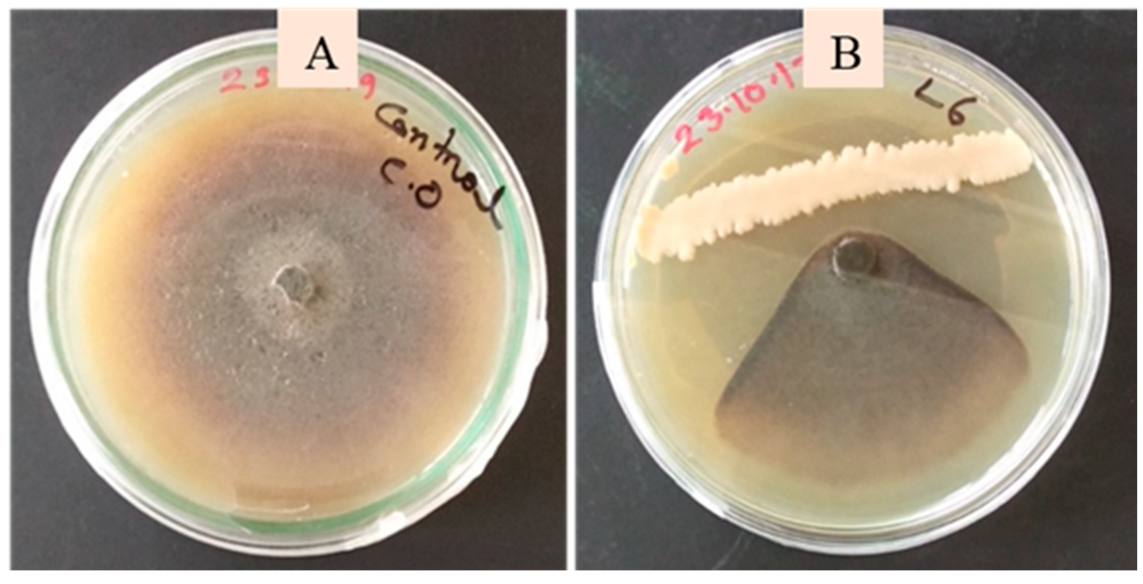
A total of 76 bacterial strains were isolated from the leaves, stems, and flowers (phyllosphere zone) of various cucurbits (Table 1). Preliminary screening against C. orbiculare resulted in the selection of 11 bacterial isolates: S-3, S-4, GL-9, GL-10, SG-6, SG-7, L-1, L-2, L-3, L-4, and L-6 (Figure 2). These isolates were obtained from cucumber, bottle gourd, and sweat gourd plants. Specifically, isolates S-3 and S-4 were sourced from stems, while the remaining isolates were obtained from leaves (Table 1).

3.2. Morphological, Biochemical, and Molecular Characterization of Phyllobacterial Isolates

The phyllobacterial isolates exhibited both variations and similarities in their colony characteristics (Table 2). All isolates were rod-shaped and demonstrated positive reactions in the citrate, catalase, and oxidase tests. In the KOH test, all isolates produced negative reactions, while Gram staining confirmed that all bacteria were Gram-positive. Molecular characterization, based on the 16S rRNA gene, revealed PCR products ranging from 1400 to 1500 bp (Figure 3A). The 16S rRNA gene sequence of isolate S-3 showed 100% similarity to Bacillus vallismortis and was submitted to GenBank under accession number PQ555272. Sequences of isolates S-4, GL-9, SG-6, L-2, and L-4 exhibited 99.40% to 99.93% similarity to B. velezensis and were submitted to GenBank under accession numbers PQ555282, PQ555273, PQ555281, PQ555275, and PQ555279, respectively. Isolates GL-10, SG-7, and L-1 showed 99.0% to 99.83% similarity to B. amyloliquefaciens, with sequences submitted to GenBank under accession numbers PQ555277, PQ555274, and PQ555276, respectively. Finally, sequences of isolates L-3 and L-6 displayed 99.85% to 100% similarity to B. subtilis and were submitted under accession numbers PQ555278 and PQ555280, respectively. The phylogenetic tree constructed from these sequences also supported these alignments (Figure 3B), indicating that all the antagonistic bacteria isolated from the cucurbit phyllosphere belong to various species of Bacillus.

3.3. Plant-Growth-Promoting Traits of the Phyllobacterial Strains

All isolates exhibited motility and demonstrated the ability to solubilize phosphate, indicating a consistent capability across these traits (Table 3). Biofilm formation, measured through optical density (OD600), varied among the isolates, with values ranging from 0.13 to 0.27. Isolate SG-7 displayed the highest biofilm-forming ability (0.27 ± 0.02), while isolate L-1 exhibited the lowest (0.13 ± 0.01). Additionally, all isolates were capable of growing in nitrogen-free media, suggesting potential nitrogen-fixing abilities. However, IAA production varied significantly, with levels ranging from 10.44 to 60.00 µg/mL; isolate S-4 was identified as the highest IAA producer, while isolate L-2 produced the least (Table 3).

3.4. Biocontrol Traits of the Phyllobacterial Strains

All isolates were qualitatively screened for the production of extracellular enzymes, including chitinase, protease, lipase, and cellulase. All isolates, except L2, tested positive for chitinase, protease, cellulase, and lipase activity (Table 4). Additionally, all 11 isolates were positive for ammonia, acetoin, siderophore, HCN, and indole production. In vitro screening against C. orbiculare demonstrated that all isolates significantly inhibited the mycelial growth of the fungus, with inhibition rates ranging from 83.33% to 94.44%. The isolates S-3, GL-9, GL-10, L-2, and L-6 exhibited the highest inhibition of C. orbiculare mycelial growth, while isolate L-3 showed the lowest inhibition (Table 4).

3.5. Effect of Phytobacterial Treatments on Cucumber Growth Parameters

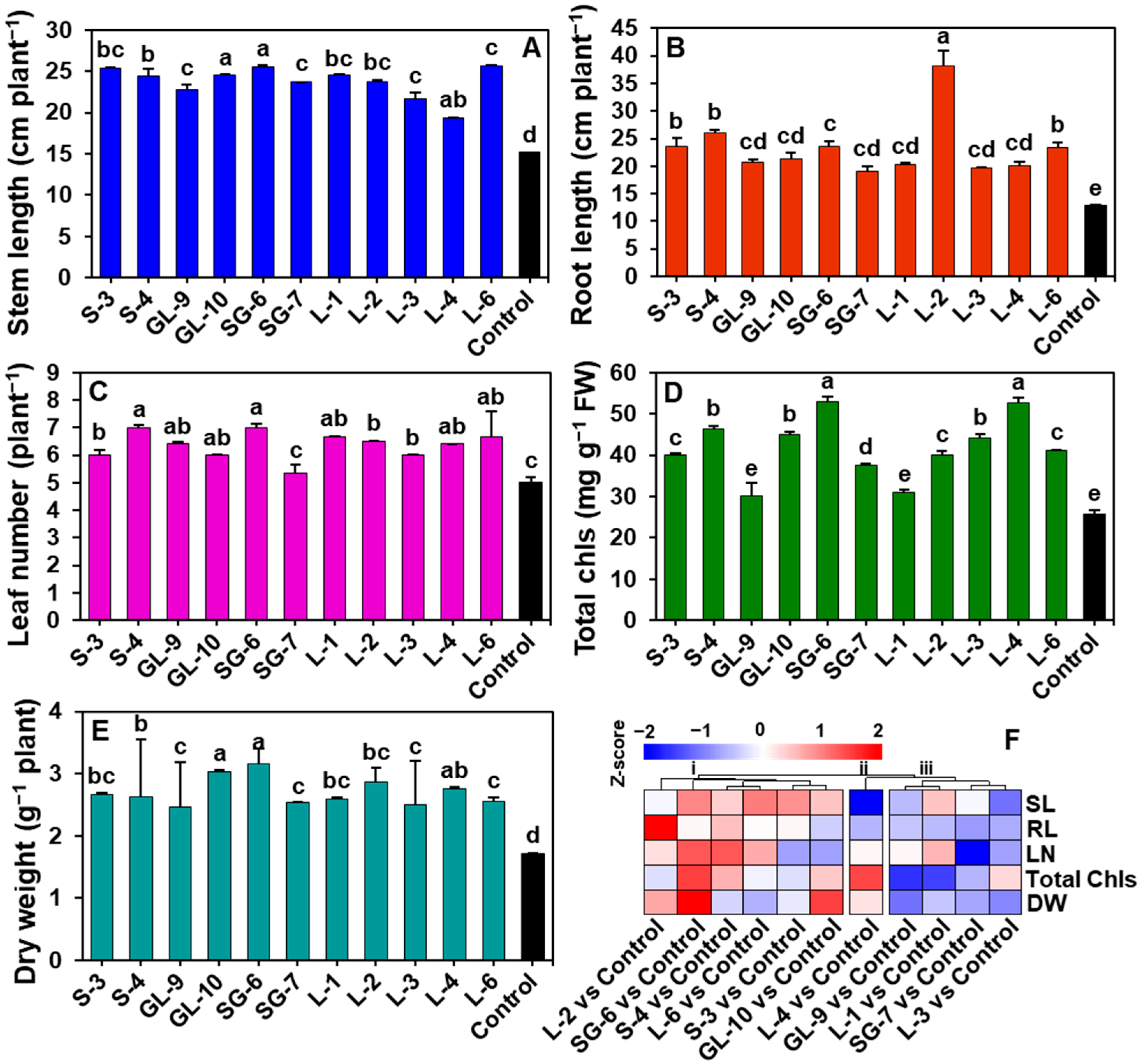
Phyllobacterial treatments significantly promoted cucumber plant growth in non-manured soil, with treated plants exhibiting substantial improvements across various growth parameters compared to the control (Figure 4A–F). The longest stem lengths were recorded in treatments SG-6 (25.50 cm) and L-6 (25.67 cm), representing increases of 68.21% and 69.33% over the control, respectively (Figure 4A). Root length showed the most notable enhancement in treatment L-2, reaching 38.16 cm, which was a 199.29% improvement (Figure 4B). The number of leaves was also significantly higher in most bacterial-treated plants, except those treated with SG-7, with the highest leaf counts observed in treatments S-4 and SG-6, amounting to a 40% increase over the control (Figure 4C). Chlorophyll content was markedly increased in bacteria-treated plants, with SG-6 exhibiting the highest boost of 105.15% (Figure 4d). Dry biomass was significantly enhanced by the bacterial treatments, with the highest dry weight increases in SG-6 (84.21%) and GL-10 (77.19%), indicating enhanced biomass accumulation (Figure 4E).

3.6. Effect of Phyllobacterial Treatment on the Growth of Cucumber Plants in Organically Manured Soil

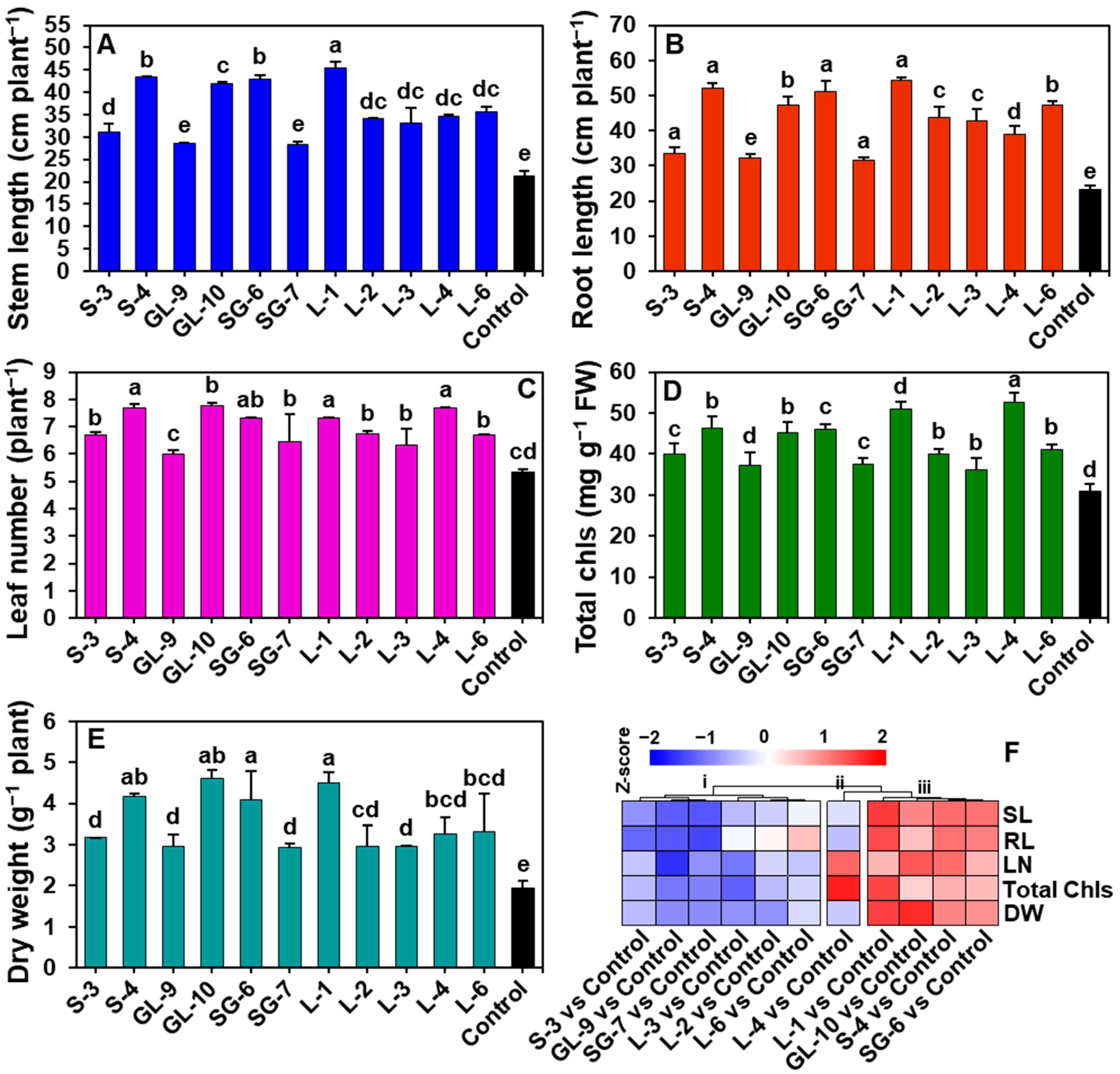
Phyllobacterial treatments significantly enhanced cucumber growth parameters in organically manured soil, with notable improvements across multiple metrics compared to the untreated control (Figure 5A–F). Treatment L-1 resulted in the highest shoot length, reaching 45.50 cm, a 112.12% increase over control values (Figure 5A). Treatments S-4, SG-6, and GL-10 followed closely with shoot lengths of 43.33 cm (102.00%), 43.00 cm (100.47%), and 42.00 cm (95.80%), respectively (Figure 5A). Additionally, treatment L-1 achieved the greatest root length at 54.33 cm, a 129.75% increase over control values (Figure 5B). Significant root length enhancements were also observed with treatments S-4, SG-6, and L-6, reaching 52.00 cm (119.13%), 51.00 cm (114.92%), and 47.33 cm (99.45%), respectively (Figure 5B). Leaf counts increased substantially, especially in treatments GL-10, S-4, L-4, and L-1, where leaf numbers rose by up to 46.14% over control, indicating more vigorous vegetative growth (Figure 5C). Chlorophyll content was notably higher, particularly in treatments L-4 and SG-6, with increases of 70.71% and 49.17% over control, suggesting enhanced photosynthetic efficiency (Figure 5D). Phyllobacterial treatments also significantly boosted biomass, with dry weights markedly higher than control plants (Figure 5E). Treatments GL-10, L-1, SG-6, and S-4 yielded the greatest dry biomass, ranging from 4.10 to 4.61 g, representing increases of 138.86% to 140.41% in dry weight (Figure 5D). These results indicate that phyllobacterial treatments, especially GL-10, S-4, L-4, SG-6, and L-1, effectively promote robust growth, increased biomass, and improved physiological traits in cucumber plants grown in nutrient-rich organic soil (Figure 5F, cluster-ii and -ii).

3.7. Effect of Phyllobacteria on Suppression of C. orbiculare in Cucumber Plants in Soils with and Without Organic Manure

Phyllobacterial treatments significantly reduced anthracnose disease incidence on cucumber leaves compared to untreated controls, with varying effectiveness depending on the treatment and soil condition (organic manure or non-manured) (Figure 6A–C). In both soil types, treatments GL-10 and L-1 were the most effective, achieving the lowest percentages of diseased leaves (9.99% and 14.29%, respectively, in non-manured soil) and minimal lesion areas (45.76 mm2 and 73.56 mm2, respectively, in non-manured soil), corresponding to high protection levels of 80.91% and 69.31% (Figure 6A–C). In manured soil, GL-10 continued to provide superior protection, achieving an 86.37% reduction in disease, with a corresponding decrease in diseased leaf area to 49.8 mm2 (Figure 6(B-ii),C). Treatments such as S-4, SG-6, and L-3 demonstrated moderate protection levels, ranging between 55% and 75% across both soil types, with slightly higher effectiveness in non-manured soil (Figure 6C). Control plants had the highest disease severity, with 55.72% and 75.25% diseased leaves and extensive lesion areas (239.72 mm2 and 365.25 mm2) in non-manured and manured soil, respectively (Figure 6A,B).

3.8. Leaf and Root Colonization by Phyllobacteria

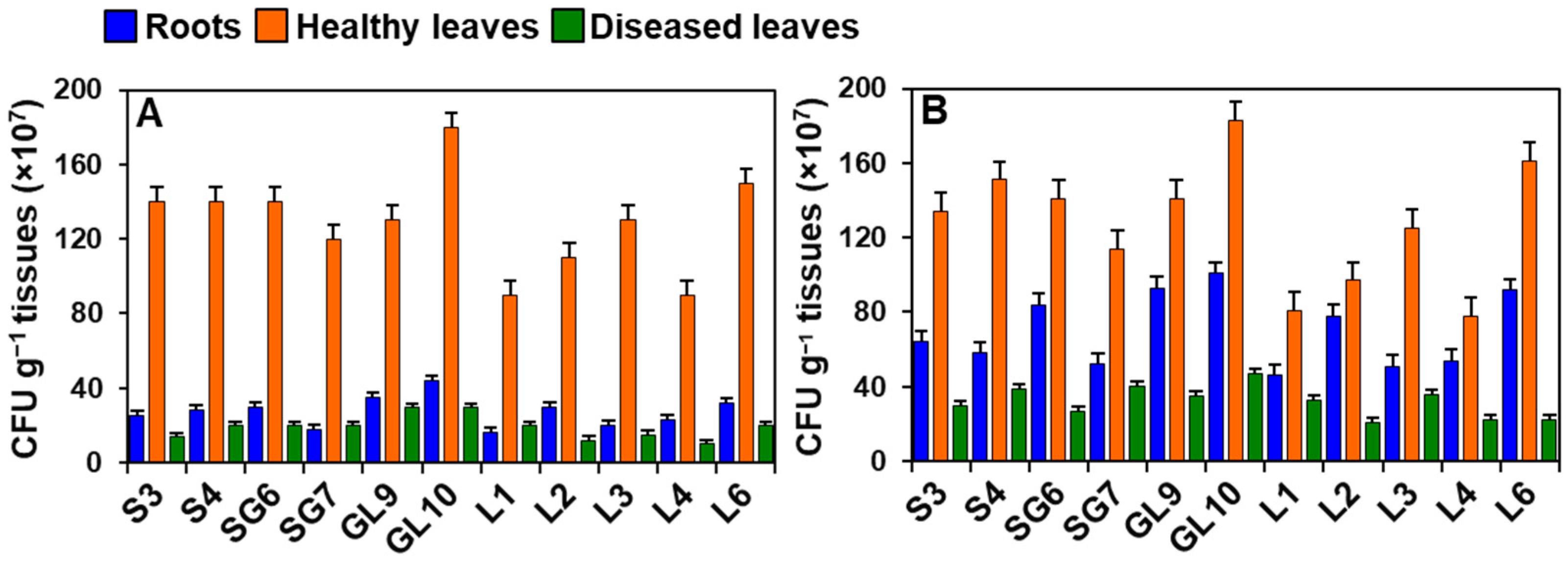
The figures demonstrate that all phyllobacterial isolates exhibit substantial colonization on cucumber roots, healthy leaves, and diseased leaves in both non-manured and manured soils. However, colonization levels vary by isolate and plant part (Figure 7A,B). In non-manured soil, healthy leaves exhibit the highest colonization, with CFU values ranging from approximately 120 to 180 × 107 CFU g−1, indicating a robust bacterial presence (Figure 7A). Isolate GL10 shows the highest colonization in healthy leaves, followed by GL9, S3, S4, SG6, and SG7. In the roots (represented by blue bars), colonization is lower, ranging from 16 to 44 × 107 CFU g−1, with GL10 leading in root colonization, followed by GL9, L3, and L6, which show values of around 20–35 × 107 CFU g−1. Diseased leaves (green bars) display the lowest colonization, with CFU counts between 10 and 30 × 107 CFU g−1.
In manured soil, overall colonization levels increase across all tissue types (Figure 7B). Healthy leaves still show the highest colonization, with isolates GL10 and GL9 reaching 183 × 107 and 141 × 107 CFU g−1, respectively. Root colonization also shows a notable increase, particularly with isolates GL10 and GL9, reaching 101 × 107 and 93 × 107 CFU g−1, respectively. Diseased leaves, although still exhibiting lower colonization than healthy leaves and roots, show a slight increase in CFU counts in manured soil, indicating that manure may enhance bacterial colonization across cucumber tissues. GL10 demonstrates the highest colonization in diseased leaves (47 × 107 CFU g−1), followed by SG-7 (40 × 107 CFU g−1).

4. Discussion

The current study successfully isolated, characterized, and demonstrated the potential of phyllobacterial isolates as effective bioagents for enhancing cucumber growth and resisting against C. orbiculare. Biochemical and molecular analyses revealed limited variation among the phyllosphere bacterial isolates with strong antagonistic activity against C. orbiculare, primarily identifying them as various species within the genus Bacillus. These results indicate that all the antagonistic bacteria isolated from the cucurbit phyllosphere belong to various species of Bacillus. This dominance of Bacillus in the cucurbit phyllosphere corroborates previous findings, which indicate that Bacillus is a prevalent genus in the phyllosphere across diverse agronomic crops due to its metabolic adaptability and resilience to varying environmental conditions [37,38,39,40,41].
In this study, the significant growth-promoting effects of phyllobacterial treatments on cucumber plants were observed in both non-manured and organically manured soils. The application of phyllobacteria consistently enhanced the lengths of cucumber stems and roots, as well as their fresh and dry weights. This promotion of plant growth is likely attributed to the key traits of these isolates, including phytohormone production, nutrient solubilization, and improved photosynthetic efficiency. The diversity in IAA production among the isolates highlights the role of IAA in influencing plant growth. IAA is a critical phytohormone that stimulates root elongation and branching, thereby improving nutrient and water uptake [42]. Moreover, the ability of these isolates to solubilize phosphate and fix nitrogen is another key mechanism that likely enhances root growth and nutrient uptake [43,44,45]. Additionally, phyllobacterial treatments led to increased leaf counts and chlorophyll content, indicating improved vegetative growth and photosynthetic capacity. Phyllobacteria can enhance chlorophyll content and photosynthetic efficiency, potentially by facilitating nutrient absorption or modulating plant hormone levels [46]. Enhanced chlorophyll levels not only contribute to better light capture but also support greater biomass production as evidenced by significant increases in dry weights [47]. Thus, the combined effects of IAA production, improved photosynthetic capacity, nitrogen fixation, and phosphate solubilization capabilities enable these phyllobacteria to enhance plant nutrient acquisition, particularly in low-nitrogen conditions such as non-manured soils. These findings support the potential of phyllobacterial treatments as powerful bio-stimulants in sustainable agriculture.
In organically manured soil, phyllobacterial treatments demonstrated strong growth-promoting effects, indicating a synergistic interaction between organic nutrients and the bio-stimulatory capabilities of the phyllobacteria [48]. The enriched organic matter in manured soils likely provides a favorable environment for these isolates to thrive, enhancing biofilm formation, root colonization, and overall growth-promoting activity. This synergy may facilitate more effective nutrient uptake and utilization by cucumber plants, leading to enhanced growth outcomes. As phyllobacteria capitalize on the organic substrates, their beneficial interactions with plant roots can further optimize plant health and productivity in sustainable agricultural practices.
While phyllobacterial strains like SG-7 showed strong biofilm formation and high extracellular enzyme activities, they demonstrated relatively lower plant growth promotion compared to other isolates. This may be due to several physiological or genetic factors. First, the strains’ metabolic investment in producing defensive or competitive metabolites (e.g., lytic enzymes, secondary metabolites) may divert resources away from synthesizing phytohormones such as IAA or other growth-stimulating compounds, as reflected in their moderate IAA production (40.22 µg/mL). Second, SG-7’s interaction with the plant host may favor defense priming over direct growth stimulation, resulting in less pronounced biomass gains under the tested conditions. Third, strain-specific differences in root colonization efficiency or rhizosphere competence may limit the benefits of nutrient acquisition, even with strong antagonistic potential. Finally, underlying genetic differences, such as the absence or reduced expression of key growth-promoting genes, could also contribute to this discrepancy. Whole-genome sequencing and the transcriptomic profiling of SG-7 in future work would help to elucidate these mechanisms in detail.
The results of this biocontrol study highlight the potential of phyllobacteria as effective biocontrol agents against C. orbiculare, the pathogen responsible for anthracnose in cucumber plants. Pot experiments demonstrated that specific phyllobacterial treatments significantly reduced both disease incidence and lesion area on cucumber leaves in manured and non-manured soil conditions. Notably, the isolate GL-10 provided the highest levels of disease suppression, achieving 86.37% protection in manured soil and 80.91% in non-manured soil. These findings indicate a strong antagonistic effect of the phyllobacteria against C. orbiculare. The biocontrol activity observed can be attributed to the production of key extracellular enzymes and metabolites by these strains. All isolates, except for L-2, produced significant levels of important enzymes such as chitinase, protease, cellulase, and lipase, which degrade pathogen cell walls, thereby weakening the fungal structures and limiting their ability to colonize plant tissues [49]. Notably, chitinase is well-documented for its role in breaking down fungal cell walls, which likely explains the observed strong inhibition of C. orbiculare [50].
The production of siderophores, ammonia, and HCN by the tested isolates likely contributed to pathogen suppression through competitive iron sequestration and antimicrobial effects, which supported the observed reductions in disease severity [44]. Siderophores not only deprive pathogens of iron but may also activate secondary metabolic pathways leading to the synthesis of antimicrobial compounds as a defense mechanism against competing microbes [49]. Furthermore, siderophore-producing bacteria significantly influence the availability of essential metals, such as iron (Fe), zinc (Zn), and copper (Cu), to plants [51]. On the other hand, HCN targets the electron transport chain of pathogens, leading to reduced ATP production and, consequently, inhibiting pathogen growth and development [52]. Ammonia not only possesses antimicrobial properties, but also serves as a vital nitrogen source for plants, thereby enhancing various cellular functions and contributing to increased crop yields [49]. The production of acetoin by beneficial bacteria further supports plant growth and resistance to pathogens [53,54]. Hence, the synergistic effects of enzyme production, sequestration of essential nutrients, and the production of bioactive compounds by these phyllobacterial isolates may play a crucial role in fortifying plant defenses against anthracnose.
The variability in biocontrol effectiveness across treatments and soil conditions underscores the importance of specific soil amendments in enhancing microbial biocontrol efficacy. Manured soil, enriched with organic matter and nutrients, likely fosters a more robust microbial community, creating a favorable environment for biocontrol agents [2]. This is reflected in the superior performance of most phyllobacterial isolates in manured conditions. Conversely, in non-manured soil, certain strains, such as GL-10 and L-1, demonstrated considerable effectiveness, albeit with slightly reduced overall protection levels. This highlights the resilience of these phyllobacterial strains across various soil conditions and emphasizes their adaptability as potential field-applied biocontrol agents, which would be particularly advantageous for organic and sustainable agricultural systems that minimize the use of synthetic chemicals.
The high efficacy of phyllobacteria in promoting plant growth and suppressing C. orbiculare may also be attributed to enhanced microbial colonization and the competitive exclusion of pathogens. The ability of phyllobacteria to colonize effectively is crucial for their fitness in the rhizosphere and the phyllosphere, which directly correlates with their beneficial effects on plants [28]. Consistent biofilm formation and motility observed across the isolates suggest robust colonization capabilities that enhance their roles as bio-stimulants and biocontrol agents. Specifically, biofilm formation facilitates microbial persistence on plant surfaces, allowing for sustained interactions with both roots and leaves [55]. In this study, all phyllobacteria produced indole, which serves as an intercellular signaling molecule that regulates various aspects of bacterial physiology, including biofilm formation [56]. Additionally, motility is crucial for initiating endophytic colonization by bacteria [57]. Therefore, phyllobacteria exhibiting motility, biofilm formation, and indole production may have a competitive edge in establishing stable associations with plants, thereby enhancing nutrient acquisition and providing effective pathogen defense.
Although colonization was observed across different cucumber tissues, a high affinity of these bacterial isolates for leaf tissues was found. Regarding the biological rationale, this tissue-specific affinity may be attributed to the fact that the isolates were originally obtained from the cucumber phyllosphere, where environmental conditions (e.g., nutrient exudates, microclimate) are more favorable for their growth and persistence compared to roots or diseased leaf tissues. Notably, these isolates demonstrated higher colonization in healthy leaves compared to diseased tissues, which could be influenced not only by nutrient availability but also by differences in plant exudate profiles, bacterial chemotaxis, and pathogen-induced alterations of host surface properties. Such factors may reduce the suitability of infected tissues while promoting the preferential colonization of healthy ones. This suggests that robust colonization in healthy tissues may enhance biocontrol efficacy by competitively excluding pathogens from leaf tissues. Additionally, the increased colonization levels in manured soil across all plant parts suggest that organic amendments enhance bacterial establishment and distribution. This effect may arise from the provision of extra nutrients or the modification of soil microbial communities to favor beneficial bacteria [58].

5. Conclusions

This study highlights the dual role of phyllobacterial isolates, particularly those from the Bacillus genus, as both biocontrol agents and bio-stimulants, providing a promising strategy for reducing chemical inputs while enhancing crop resilience and productivity. In particular, the isolates GL-10, SG-6, S-4, and L-1 showed consistent performance across parameters and soil conditions. These isolates increased cucumber stem length by 18–25%, root length by 20–27%, fresh weight by 22–30%, and dry weight by 19–28% compared to the untreated control. In addition, disease suppression against C. orbiculare reached up to 86.37% in manured soil and 80.91% in non-manured soil, surpassing previously reported biocontrol efficacies for similar phyllosphere Bacillus strains. These findings demonstrate that the tested isolates not only significantly enhance vegetative growth parameters, but also provide strong antagonistic activity against anthracnose under different soil conditions. Applying these beneficial microbes could reduce dependency on synthetic chemicals, fostering more environmentally sustainable agricultural practices that support long-term crop health and productivity. Future studies should focus on validating these results under field conditions, comparing the performance of these isolates with standard chemical treatments and developing stable formulations to ensure consistent efficacy across different environmental and soil conditions.

Author Contributions

Conceptualization: M.M.H. and M.G.M.; Investigation: M.H.K.N. and P.S.B.; Methodology: M.H.K.N.; Data curation: M.H.K.N. and M.T.R.; Formal analysis: P.S.B., M.R.I., T.R.A., M.T.R. and I.K.; Validation and software: M.R.I., T.R.A., I.K. and M.G.M.; Funding acquisition: M.M.H.; Resources: M.M.H.; Supervision: M.M.H. and M.G.M.; Project administration: M.M.H.; Writing—original draft: M.H.K.N. and M.M.H.; Writing—review & editing: M.G.M. and M.M.H. All authors have read and agreed to the published version of the manuscript.

Funding

This study was part of the project “Phyllosphere Bacteria for Plant Growth Promotion and Suppression of Anthracnose Disease in Cucumber”, funded by the Ministry of Science and Technology, Bangladesh.

Data Availability Statement

The data supporting the results of this article are included within the article.

Acknowledgments

The authors greatly acknowledge the support received from the staff and students of the Department of Plant Pathology, Gazipur Agricultural University, Gazipur, Bangladesh.

Conflicts of Interest

The authors declare no conflicts of interest.

References

  1. Zheng, L.; Gu, C.; Cao, J.; Li, S.-M.; Wang, G.; Luo, Y.-M.; Guo, J.-H. Selecting Bacterial Antagonists for Cucurbit Downy Mildew and Developing an Effective Application Method. Plant Dis. 2018, 102, 628–639. [Google Scholar] [CrossRef]
  2. Wang, S.; Heal, K.V.; Zhang, Q.; Yu, Y.; Tigabu, M.; Huang, S.; Zhou, C. Soil microbial community, dissolved organic matter and nutrient cycling interactions change along an elevation gradient in subtropical China. J. Environ. Manag. 2023, 345, 118793. [Google Scholar] [CrossRef]
  3. Mukherjee, P.K.; Nema, N.K.; Maity, N.; Sarkar, B.K. Phytochemical and therapeutic potential of cucumber. Fitoterapia 2013, 84, 227–236. [Google Scholar] [CrossRef]
  4. Ríos, J.L. Effects of Triterpenes on the Immune System. J. Ethnopharmacol. 2010, 128, 1–14. [Google Scholar] [CrossRef]
  5. Katiyar, S.; Saify, K.; Singh, S.K.; Rai, M. Botanical study of skin lightening agents. Int. J. Pharmacogn. 2014, 1, 534–544. [Google Scholar]
  6. Suryawanshi, R.K.; Patil, C.D.; Borase, H.P.; Narkhede, C.P.; Stevenson, A.; Hallsworth, J.E.; Patil, S.V. Towards an understanding of bacterial metabolites prodigiosin and violacein and their potential for use in commercial sunscreens. Int. J. Cosmet. Sci. 2015, 37, 98–107. [Google Scholar] [CrossRef]
  7. Akhtar, N.; Mehmood, A.; Khan, B.A.; Mahmood, T.; Khan, H.M.S.; Saeed, T. Exploring Cucumber Extract for Skin Rejuvenation. Afr. J. Biotechnol. 2011, 10, 1206–1216. [Google Scholar]
  8. Hossain, M.M.; Sultana, F.; Miyazawa, M.; Hyakumachi, M. The Plant Growth-promoting Fungus Penicillium spp. GP15-1 Enhances Growth and Confers Protection against Damping-off and Anthracnose in the Cucumber. J. Oleo Sci. 2014, 63, 391–400. [Google Scholar] [CrossRef] [PubMed]
  9. Hausbeck, M.K.; Lamour, K.H. Phytophthora capsici on Vegetable Crops: Research Progress and Management Challenges. Plant Dis. 2004, 88, 1292–1303. [Google Scholar] [CrossRef]
  10. Chai, C.H.; Hong, C.-F.; Huang, J.-W. Identification and Characterization of a Multifunctional Biocontrol Agent, Streptomyces griseorubiginosus LJS06, Against Cucumber Anthracnose. Front. Microbiol. 2022, 13, 923276. [Google Scholar] [CrossRef] [PubMed]
  11. Ko, E.J.; Shin, Y.H.; Hyun, H.N.; Song, H.S.; Hong, J.K.; Jeun, Y.C. Bio-Sulfur Pre-Treatment Suppresses Anthracnose on Cucumber Leaves Inoculated with Colletotrichum orbiculare. Mycobiology 2019, 47, 308–318. [Google Scholar] [CrossRef]
  12. Devarajan, A.K.; Muthukrishanan, G.; Truu, J.; Truu, M.; Ostonen, I.; Subramanian Kizhaeral, S.; Panneerselvam, P.; Gopalasubramanian, S.K. The Foliar Application of Rice Phyllosphere Bacteria induces Drought-Stress Tolerance in Oryza sativa (L.). Plants 2021, 10, 387. [Google Scholar] [CrossRef]
  13. Rubiales, D.; Fondevilla, S.; Chen, W.; Gentzbittel, L.; Higgins, T.J.V.; Castillejo, M.A.; Singh, K.B.; Rispail, N. Achievements and Challenges in Legume Breeding for Pest and Disease Resistance. Crit. Rev. Plant Sci. 2014, 34, 195–236. [Google Scholar] [CrossRef]
  14. Yue, C.; Du, C.; Wang, X.; Tan, Y.; Liu, X.; Fan, H. Powdery mildew-induced changes in phyllosphere microbial community dynamics of cucumber. FEMS Microbiol. Ecol. 2024, 100, fiae050. [Google Scholar] [CrossRef]
  15. Ni, L.; Punja, Z.K. Management of Fungal Diseases on Cucumber (Cucumis sativus L.) and Tomato (Solanum lycopersicum L.) Crops in Greenhouses Using Bacillus Subtilis. In Bacilli and Agrobiotechnology: Phytostimulation and Biocontrol. Bacilli in Climate Resilient Agriculture and Bioprospecting; Springer: Cham, Switzerland, 2019. [Google Scholar]
  16. Patil, N.; Raghu, S.; Mohanty, L.; Jeevan, B.; Basana-Gowda, G.; Adak, T.; Annamalai, M.; Rath, P.C.; Sengottayan, S.-N.; Govindharaj, G.-P. Rhizosphere Bacteria Isolated from Medicinal Plants Improve Rice Growth and Induce Systemic Resistance in Host Against Pathogenic Fungus. J. Plant Growth Regul. 2024, 43, 770–786. [Google Scholar] [CrossRef]
  17. Jain, A.; Chatterjee, A.; Das, S. Synergistic consortium of beneficial microorganisms in rice rhizosphere promotes host defense to blight-causing Xanthomonas oryzae pv. oryzae. Planta 2020, 252, 106. [Google Scholar] [CrossRef]
  18. Yang, F.; Jiang, H.; Chang, G.; Liang, S.; Ma, K.; Cai, Y.; Tian, B.; Shi, X. Effects of Rhizosphere Microbial Communities on Cucumber Fusarium wilt Disease Suppression. Microorganisms 2023, 11, 1576. [Google Scholar] [CrossRef]
  19. Wang, C.; Pan, G.; Lu, X.; Qi, W. Phosphorus solubilizing microorganisms: Potential promoters of agricultural and environmental engineering. Front. Bioeng. Biotechnol. 2023, 11, 1181078. [Google Scholar] [CrossRef]
  20. Rangel, L.I.; Leveau, J.H.J. Applied microbiology of the phyllosphere. Appl. Microbiol. Biotechnol. 2024, 108, 211. [Google Scholar] [CrossRef]
  21. Compant, S.; Duffy, B.; Nowak, J.; ClémEnt, C.; Barka, E.A. Use of Plant Growth-Promoting Bacteria for Biocontrol of Plant Diseases: Principles, Mechanisms of Action, and Future Prospects. Appl. Environ. Microbiol. 2005, 71, 4951–4959. [Google Scholar] [CrossRef]
  22. De Mandal, S.; Jeon, J. Phyllosphere Microbiome in Plant Health and Disease. Plants 2023, 12, 3481. [Google Scholar] [CrossRef]
  23. Bonaterra, A.; Cabrefiga, J.; Camps, J.; Montesinos, E. Increasing survival and efficacy of a bacterial biocontrol agent of fire blight of rosaceous plants by means of osmoadaptation. FEMS Microbiol. Ecol. 2007, 61, 185–195. [Google Scholar] [CrossRef]
  24. Vorholt, J.A. Microbial life in the phyllosphere. Nat. Rev. Microbiol. 2012, 10, 828–840. [Google Scholar] [CrossRef] [PubMed]
  25. Bulgarelli, D.; Schlaeppi, K.; Spaepen, S.; Van Themaat, E.V.L.; Schulze-Lefert, P. Structure and Functions of the Bacterial Mi-crobiota of Plants. Annu. Rev. Plant Biol. 2013, 64, 807–838. [Google Scholar] [CrossRef] [PubMed]
  26. Kembel, S.W.; O’cOnnor, T.K.; Arnold, H.K.; Hubbell, S.P.; Wright, S.J.; Green, J.L. Relationships between phyllosphere bacterial communities and plant functional traits in a neotropical forest. Proc. Natl. Acad. Sci. USA 2014, 111, 13715–13720. [Google Scholar] [CrossRef]
  27. Hari, S.B.; Briste, P.S.; Sumi, A.A.; Mosharaf, M.K.; Paul, S.I.; Masum, M.M.I.; Jannat, R. Endophytic bacteria isolated from medicinal plants induce plant growth promotion and southern blight disease suppression in tomato. J. Plant Pathol. 2022, 105, 197–210. [Google Scholar] [CrossRef]
  28. Kemal, S.; Koricha, A.D.; Parise, A. The Art, Microbial Quality, Safety, and Physicochemical Characteristics of Jikita: A Traditional Ethiopian Fermented Beverage. Int. J. Food Sci. 2024, 2024, 6698831. [Google Scholar] [CrossRef]
  29. Islam, S.; Akanda, A.M.; Prova, A.; Islam, M.T.; Hossain, M.M. Isolation and Identification of Plant Growth Promoting Rhizobacteria from Cucumber Rhizosphere and Their Effect on Plant Growth Promotion and Disease Suppression. Front. Microbiol. 2016, 6, 1360. [Google Scholar] [CrossRef]
  30. Setyaningsih, P.P.; Ningsih, F.; Rachmania, M.K.; Syafitri, W.A.; Sari, D.C.A.F.; Yabe, S.; Yokota, A.; Oetari, A.; Sjamsuridzal, W. Cellulolytic enzyme-producing thermophilic Actinobacteria isolated from the soil of Cisolok Geysers, West Java, Indonesia. Biodiversitas J. Biol. Divers. 2019, 20, 3134–3141. [Google Scholar] [CrossRef]
  31. Yahia, M.S.; Mohamed, M.K.; Othman, M.S.; Mostafa, D.N.; Gomaa, M.M.; Fahmy, M.A.; Shaaban, R.H.; Kamel, A.M.; Abdelhai, M.F.; Radwan, W.H.; et al. Isolation and identification of antibiotic producing Pseudomonas fluorescens NBRC-14160 from Delta Soil in Egypt. Arab. Univ. J. Agric. Sci. 2020, 28, 797–808. [Google Scholar] [CrossRef]
  32. Perwendha, R.; Oetari, A.; Sjamsuridzal, W. Skimmed Milk-Degrading Ability of Rhizopus Azygosporus UICC 539 at Various Temperatures. AIP Conf. Proc. 2020, 2242, 050006. [Google Scholar] [CrossRef]
  33. Cappuccino, J.G. Natalie Sherman Microbiology: A Laboratory Manual, 10th ed.; References—Scientific Research Publishing; Pearson Education Limited: London, UK, 2013; Available online: https://www.scirp.org/reference/referencespapers?referenceid=2887944 (accessed on 16 August 2025).
  34. Hossain, M.M.; Sultana, F. Methods for the Characterization of Plant-Growth Promoting Rhizobacteria. Methods Mol. Biol. 2018, 1734, 307–328. [Google Scholar] [CrossRef]
  35. El Habil-Addas, F.; Aarab, S.; Rfaki, A.; Laglaoui, A.; Bakkali, M.; Arakrak, A. Screening of Phosphate Solubilizing Bacterial Isolates for Improving Growth of Wheat. Available online: https://www.researchgate.net/publication/320988789_Screening_of_phosphate_solubilizing_bacterial_isolates_for_improving_growth_of_wheat (accessed on 16 August 2025).
  36. MacWilliams, M.P. Indole Test Protocol; American Society for Microbiology for Microbiology: Washington, DC, USA, 2009. [Google Scholar]
  37. Sameer, K.; Sobanbabu, G.; Sabarinathan, K.G.; Parthiban, V.K.; Ramamoorthy, V. Evaluation of phyllosphere antagonistic bacteria on the management of Fusarium ear rot of maize caused by Fusarium verticillioides. J. Biol. Control. 2018, 32, 257–263. [Google Scholar] [CrossRef]
  38. Janakiev, T.; Dimkić, I.; Bojić, S.; Fira, D.; Stanković, S.; Berić, T. Bacterial communities of plum phyllosphere and characterization of indigenous antagonistic Bacillus thuringiensis R3/3 isolate. J. Appl. Microbiol. 2019, 128, 528–543. [Google Scholar] [CrossRef]
  39. Shao, Z.; Schenk, P.M.; Dart, P. Phyllosphere bacterial strains Rhizobium b1 and Bacillus subtilis b2 control tomato leaf diseases caused by Pseudomonas syringae pv. tomato and Alternaria solani. J. Appl. Microbiol. 2023, 134, lxad139. [Google Scholar] [CrossRef]
  40. Sobanbabu, G.; Oviya, R.; Meena, B.; Vijayasamundeeswari, A.; Shanmugaiah, V.; Ramamoorthy, V. Evaluation of phyllosphere bacterial biocontrol agents for the suppression of rice foliar diseases. J. Phytopathol. 2024, 172, E3300. [Google Scholar] [CrossRef]
  41. Wiraswati, S.M.; Nawangsih, A.A.; Rusmana, I.; Wahyudi, A.T. Rice phyllosphere bacteria producing antifungal compounds as biological control agents of blast disease. Biodiversitas J. Biol. Divers. 2020, 21, 1223–1278. [Google Scholar] [CrossRef]
  42. Liu, M.; Zhang, Q.; Jin, R.; Zhao, P.; Zhu, X.; Wang, J.; Yu, Y.; Tang, Z. The Role of IAA in Regulating Root Architecture of Sweetpotato (Ipomoea batatas [L.] Lam) in Response to Potassium Deficiency Stress. Plants 2023, 12, 1779. [Google Scholar] [CrossRef] [PubMed]
  43. Eivy, F.Z.; Rubayet, T.; Sarkar, U.; Hossain, M. Suppression of Bipolaris Leaf Blotch and Improvement of Wheat Growth by Plant Growth Promoting Rhizobacteria Bacilli. Online J. Biol. Sci. 2024, 24, 412–426. [Google Scholar] [CrossRef]
  44. Hossain, M.M. Utilization of Stenotrophomonas koreensis and Bacillus amyloliquefaciens for Improving Growth, Reducing Nitrogen Fertilization and Controlling Bipolaris sorokiniana in Wheat. Caraka Tani: J. Sustain. Agric. 2024, 39, 81–93. [Google Scholar] [CrossRef]
  45. Timofeeva, A.M.; Galyamova, M.R.; Sedykh, S.E. Plant Growth-Promoting Soil Bacteria: Nitrogen Fixation, Phosphate Solubilization, Siderophore Production, and Other Biological Activities. Plants 2023, 12, 4074. [Google Scholar] [CrossRef]
  46. Su, F.; Zhao, B.; Dhondt-Cordelier, S.; Vaillant-Gaveau, N. Plant-Growth-Promoting Rhizobacteria Modulate Carbohydrate Metabolism in Connection with Host Plant Defense Mechanism. Int. J. Mol. Sci. 2024, 25, 1465. [Google Scholar] [CrossRef]
  47. Nazari, M.; Kordrostami, M.; Ghasemi-Soloklui, A.A.; Eaton-Rye, J.J.; Pashkovskiy, P.; Kuznetsov, V.; Allakhverdiev, S.I. Enhancing Photosynthesis and Plant Productivity through Genetic Modification. Cells 2024, 13, 1319. [Google Scholar] [CrossRef]
  48. Al Methyeb, M.; Ruppel, S.; Eichler-Löbermann, B.; Vassilev, N. The Combined Applications of Microbial Inoculants and Organic Fertilizer Improve Plant Growth under Unfavorable Soil Conditions. Microorganisms 2023, 11, 1721. [Google Scholar] [CrossRef]
  49. Chowdhury, S.K.; Roy, N.; Banerjee, M.; Basnett, D. Plant growth promotion and antifungal activities of the mango phyllosphere bacterial consortium for the management of Fusarium wilt disease in pea (Pisum sativum L.). Plant Sci. Today 2023, 10, 220–234. [Google Scholar] [CrossRef]
  50. Valdez, A.F.; de Souza, T.N.; Bonilla, J.J.A.; Zamith-Miranda, D.; Piffer, A.C.; Araujo, G.R.S.; Guimarães, A.J.; Frases, S.; Pereira, A.K.; Fill, T.P.; et al. Traversing the Cell Wall: The Chitinolytic Activity of Histoplasma capsulatum Extracellular Vesicles Facilitates Their Release. J. Fungi 2023, 9, 1052. [Google Scholar] [CrossRef]
  51. Khan, N.; Martínez-Hidalgo, P.; Ice, T.A.; Maymon, M.; Humm, E.A.; Nejat, N.; Sanders, E.R.; Kaplan, D.; Hirsch, A.M. Antifungal Activity of Bacillus Species against Fusarium and Analysis of the Potential Mechanisms Used in Biocontrol. Front. Microbiol. 2018, 9, 2363. [Google Scholar] [CrossRef]
  52. Kumari, S.; Khanna, V. Biological Management of Vascular Wilt of Chickpea (Cicer arietinum L.) Incited by Fusarium oxysporum f. sp. ciceris by Antagonistic Rhizobacteria Co-Inoculated with Native Mesorhizobium. Int. J. Curr. Microbiol. Appl. Sci. 2018, 7, 920–941. [Google Scholar] [CrossRef]
  53. Sharifi, R.; Kiani, H.; Ahmadzadeh, M.; Behboudi, K. Optimization of Acetoin Production by Biological Control Strain Bacillus Subtilis GB03 Using Statistical Experimental Design. J. Microb. Biol. 2019, 8, 47–57. [Google Scholar] [CrossRef]
  54. Balderas-Ruíz, K.A.; Bustos, P.; Santamaria, R.I.; González, V.; Cristiano-Fajardo, S.A.; Barrera-Ortíz, S.; Mezo-Villalobos, M.; Aranda-Ocampo, S.; Guevara-García, Á.A.; Galindo, E.; et al. Bacillus velezensis 83 a bacterial strain from mango phyllosphere, useful for biological control and plant growth promotion. AMB Express 2020, 10, 163. [Google Scholar] [CrossRef]
  55. Abdallah, M.; Benoliel, C.; Drider, D.; Dhulster, P.; Chihib, N.-E. Biofilm formation and persistence on abiotic surfaces in the context of food and medical environments. Arch. Microbiol. 2014, 196, 453–472. [Google Scholar] [CrossRef]
  56. Lee, J.H.; Lee, J. Indole as an Intercellular Signal in Microbial Communities. FEMS Microbiol. Rev. 2010, 34, 426–444. [Google Scholar] [CrossRef] [PubMed]
  57. Fernández-Llamosas, H.; Díaz, E.; Carmona, M. Motility, Adhesion and c-di-GMP Influence the Endophytic Colonization of Rice by Azoarcus sp. CIB. Microorganisms 2021, 9, 554. [Google Scholar] [CrossRef] [PubMed]
  58. Goswami, D.; Thakker, J.N.; Dhandhukia, P.C. Portraying mechanics of plant growth promoting rhizobacteria (PGPR): A review. Cogent Food Agric. 2016, 2, 1127500. [Google Scholar] [CrossRef]
Figure 1. Collection and preparation of cucumber phyllosphere samples for isolation of phyllosphere bacteria.
Applmicrobiol 05 00094 g001
Figure 2. In vitro antagonistic activity of a bacterial isolate against Colletotrichum orbicularae. (A) Control plate showing normal growth of C. orbicularae in the absence of the antagonistic bacteria. (B) Dual culture plate showing inhibited growth of C. orbicularae in the presence of the antagonistic bacteria.
Applmicrobiol 05 00094 g002
Figure 3. Polymerase Chain Reaction (PCR) profiles and the phylogenetic tree generated from the 16s rRNA gene of bacterial isolates. (A) PCR profiles of bacterial isolates using 16srRNA-specific primers 27F and 1492R primers, M denotes 1kb DNA ladder (Marker). (B) Neighbor-joining phylogenetic tree obtained from the 16S rRNA sequence data analysis of bacteria. The phylogeny reconstruction was based on 1000 bootstrap replications. Evolutionary analyses were conducted using MEGA 11 software, and all positions containing gaps and missing data were eliminated. Here, scale bar = 0.10 substitutions per nucleotide position.
Applmicrobiol 05 00094 g003
Figure 4. Effect of phyllobacterial treatment on the growth of cucumber plants cultivated in non-manured soil. Cucumber seeds were treated with 11 different phyllobacterial isolates and sown into pots containing autoclaved field soil. Control pots were sown with surface-sterilized seeds. Two-week-old plants received a foliar spraying with bacterial suspension (sterile distilled water in the case of control plants). Data were recorded from 4-week-old plants. (A) Stem length, (B) root length, (C) leaf number, (D), total chlorophylls (Chls) content, (E) plant dry weight of cucumber plants under different treatments. (F) Hierarchical clustering heatmap showing the normalized fold-change values of the parameters mentioned above. The fold-changes are indicated by the intensities of the colors with saturation at 2 and −2. The mean and standard error (n = 3) are displayed as bars. Significant changes (p ≤ 0.05) among different treatments are denoted by different letters (a–e) atop the bars and were calculated following the least significant difference test. SL, stem length; RL, root length; LN, leaf number; DW, plant dry weight.
Applmicrobiol 05 00094 g004
Figure 5. Effect of phyllobacterial treatment on the growth of cucumber plants cultivated in organically manured soil. Cucumber seeds were treated with 11 different phyllobacterial isolates and sown into pots containing autoclaved field soil mixed with cow manure (10% of soil). Control pots were sown with surface-sterilized seeds. Two-week-old plants received a foliar spraying with bacterial suspension (sterile distilled water in the case of control plants). Data were recorded from 4-week-old plants. (A) Stem length, (B) root length, (C) leaf number, (D), total chlorophylls (Chls) content, (E) plant dry weight of cucumber plants under different treatments. (F) Hierarchical clustering heatmap showing the normalized fold-change values of the parameters mentioned above. The fold-changes are indicated by the intensities of the colors with saturation at 2 and −2. The mean and standard error (n = 3) are displayed as bars. Significant changes (p ≤ 0.05) among different treatments are denoted by different letters (a–e) atop the bars and were calculated following the least significant difference test. SL, stem length; RL, root length; LN, leaf number; DW, plant dry weight.
Applmicrobiol 05 00094 g005
Figure 6. Effect of phyllobacteria on suppression of anthracnose disease caused by Colletotrichum orbiculare in cucumber plants grown in non-manured (A) and manured soil (B). Cucumber seeds were treated with 11 different phyllobacterial isolates and sown into pots containing autoclaved field soil with or without the addition of cow manure (10% of soil). Control pots were sown with surface-sterilized seeds. Two-week-old plants received a foliar spraying with bacterial suspension (sterile distilled water in the case of control plants). At 7 days post-spraying, all plants were inoculated with C. orbiculare spore suspension. Data were recorded 12 days after inoculation. (A-i,B-i) Diseased leaves (%), (A-ii,B-ii) total lesion length, (C) protection (%) of cucumber plants under different treatments. The phyllobacterial isolates were grouped into 3 distinct clusters based on the protection (%). The protection (%) is indicated by the intensities of the colors with saturation at 30 and 85. The mean and standard error (n = 3) are displayed as bars. Significant changes (p ≤ 0.05) among different treatments are denoted by different letters (a–i) atop the bars and were calculated following the least significant difference test.
Applmicrobiol 05 00094 g006
Figure 7. Colonization of cucumber roots, healthy leaves, and diseased leaves by different phyllobacteria isolates in non-manured soil and manured soil. (A) Colony-forming units (CFU) of different phyllobacteria colonizing cucumber roots, healthy leaves, and diseased leaves of cucumber plants grown in non-manured soil. (B) Colony-forming units (CFU) of different phyllobacteria colonizing cucumber roots, healthy leaves, and diseased leaves in manured soil. The mean and standard error (n = 3) are displayed as bars.
Applmicrobiol 05 00094 g007
Table 1. List of selected phyllobacteria identified through preliminary screening against Colletotrichum orbiculare.
Phyllobacterial Isolates Source Plant PartsHost Plants (Common Name)Host Plants (Botanical Name)Family
S-3StemCucumberCucumis sativusCucurbitaceae
S-4StemBottle GourdLagenaria sicerariaCucurbitaceae
GL-9LeafBottle GourdLagenaria sicerariaCucurbitaceae
GL-10LeafBottle GourdLagenaria sicerariaCucurbitaceae
SG-6 LeafSweat GourdCucurbita pepoCucurbitaceae
SG-7 LeafSweat GourdCucurbita pepoCucurbitaceae
L-1LeafCucumberCucumis sativusCucurbitaceae
L-2LeafCucumberCucumis sativusCucurbitaceae
L-3 LeafCucumberCucumis sativusCucurbitaceae
L-4 LeafCucumberCucumis sativusCucurbitaceae
L-6LeafBottle GourdLagenaria sicerariaCucurbitaceae
Table 2. Biochemical and molecular characterization (based on 16s rRNA sequencing) of isolated bacterial isolates from cucumber phyllosphere.
Bacterial Isolates Colony ColorColony MorphologyShapeCitrate TestCatalase TestKOH Test Gram StainingOxidase TestIdentificationSimilarity
S-3OpaqueSmoothRod+++ve+Bacillus vallismortis100%
S-4Creamy whiteRough with irregular marginsRod+++ve+B. velezensis99.40%
GL-9Creamy whiteRoughRod+++ve+B. velezensis99.93%
GL-10Pale yellowWrinkled and rough surfacesRod+++ve+B. amyloliquefaciens99.83%
SG-6 Creamy whiteRough with irregular marginsRod+++ve+B. velezensis99.83%
SG-7 Pale yellowWrinkled and rough surfacesRod+++ve+B. amyloliquefaciens99.00%
L-1Pale yellowWrinkled and rough surfacesRod++ +ve+B. amyloliquefaciens99.50%
L-2Creamy WhiteRough with irregular marginsRod+++ve+B. velezensis99.65%
L-3 Slightly yellowRoughRod+++ve+B. subtilis99.85%
L-4 Creamy whiteRough with irregular marginsRod+++ve+B. velezensis100%
L-6Slightly yellowRoughRod+++ve+B. subtilis100%
‘+’ or ‘+ve’, positive response; ‘−‘ or ‘−ve’, negative response.
Table 3. Characterization of phyllosphere bacterial isolates for plant-growth-promoting traits.
IsolateMotilityBiofilm Formation (OD600)Phosphate SolubilizationGrowth in N2-Free MediumIAA Production (µg/mL)
S-3+0.17 ± 0.01 *++35.32 ± 0.32
S-4+0.19 ± 0.01++60.00 ± 0.41
GL-9+0.20 ±0.01++33.55 ± 0.39
GL-10+0.14 ± 0.01++55.00 ± 0.46
SG-6+0.21 ± 0.01++56.44 ± 0.34
SG-7+0.27 ± 0.02++40.22 ± 0.23
L-1+0.13 ± 0.01++38.00 ± 0.40
L-2+0.26 ± 0.02++10.44 ± 0.11
L-3+0.21 ± 0.01++15.00 ± 0.14
L-4+0.23 ± 0.03++40.00 ± 0.27
L-6+0.18 ± 0.01++45.55 ± 0.38
‘+’, positive for the test; ‘−’, negative for the test; ‘*’, values are mean ± SE (n = 3).
Table 4. Characterization of phyllosphere bacterial isolates for biological control traits.
IsolateAmmoniaAcetoinSidero-PhoreHCNIndoleChitinaseProteaseLipaseCellulaseRadial Growth of C. orbiculare (cm)Growth Inhibition (%) of C. orbiculare
S-3+++++++++3.8 ± 0.02 d *94.44 ± 0.12
S-4+++++++++4.0 ± 0.02 c88.88 ± 0.24
GL-9+++++++++3.8 ± 0.01 d94.44 ± 0.04
GL-10+++++++++3.8 ± 0.02 d94.44 ± 0.33
SG-6+++++++++4.4 ± 0.01 b77.77 ± 0.31
SG-7+++++++++4.0 ± 0.01 c88.88 ± 0.54
L-1+++++++++4.4 ± 0.02 b77.77 ± 0.47
L-2++++++-++3.8 ± 0.01 d94.44 ± 0.62
L-3+++++++++4.2 ± 0.01 b83.33 ± 0.34
L-4+++++++++3.9 ± 0.01 c91.66 ± 0.62
L-6+++++++++3.8 ± 0.01 d94.44 ± 0.28
Control7.2 ± 0.03 a
‘+’, positive for the test; ‘−‘, negative for the test; ‘*’, values are mean ± SE (n = 3) followed by different letters within the same column indicating statistically significant differences (p ≤ 0.05).
Disclaimer/Publisher’s Note: The statements, opinions and data contained in all publications are solely those of the individual author(s) and contributor(s) and not of MDPI and/or the editor(s). MDPI and/or the editor(s) disclaim responsibility for any injury to people or property resulting from any ideas, methods, instructions or products referred to in the content.