Article

Applied Metagenomic Profiling of Domestic Cat Feces from Cali, Colombia: An Exploratory Approach

1
Grupo de Investigación en Ecología Animal, Departamento de Biología, Universidad del Valle, Cali 760031, Colombia
2
TAO-Lab, Centre for Bioinformatics and Photonics-CIBioFi, Universidad del Valle, Cali 760031, Colombia
3
Grupo de Investigación en Ecofisiología, Evolución y Biogeografía, Departamento de Biología, Universidad del Valle, Cali 760031, Colombia
*
Author to whom correspondence should be addressed.
Appl. Microbiol. 2025, 5(3), 67; https://doi.org/10.3390/applmicrobiol5030067
Submission received: 31 May 2025 / Revised: 27 June 2025 / Accepted: 28 June 2025 / Published: 8 July 2025

Abstract

This exploratory study presents the first metagenomic assessment of the gut microbiome in domestic cats from Cali, Colombia. Fecal samples were collected from 10 healthy, sterilized domestic cats, aged 8 months to over 2 years, with variation in sex (7 females, 3 males), diet (processed or raw), and outdoor access (5 with, 5 without). Using 16S rRNA gene metabarcoding and pooled shotgun metagenomic sequencing, the study characterized the taxonomic composition and functional potential of the feline gut microbiome. Dominant phyla included Bacillota and Bacteroidota, with substantial inter-individual variation. Peptoclostridium was the most consistently abundant genus, while Megamonas and Megasphaera showed higher variability. Shotgun analysis detected antibiotic resistance genes (ErmG, ErmQ) and virulence factors (pfoA, plc, colA, nanJ, nagI) in Clostridium perfringens, highlighting potential zoonotic risk. The composition of the gut microbiota was influenced primarily by diet and outdoor access, while age and gender had more moderate effects. The study concludes that lifestyle and environmental factors play a key role in shaping the gut microbiome of domestic cats. We recommend further longitudinal and larger-scale studies to better understand the dynamics of feline microbiota and their implications for animal and public health within a One Health framework.

1. Introduction

Domestic cats (Felis catus) are among the most common companion animals worldwide and share close living environments with humans, including homes, bedrooms, and even beds. This proximity creates numerous opportunities for bidirectional microbiota exchange, making the study of their gut microbiome particularly relevant from a One Health perspective. Understanding the composition and dynamics of the feline gut microbiota is crucial not only for improving animal health and welfare but also for assessing potential zoonotic risks. Cats may be exposed to environmental, dietary, and antimicrobial pressures that alter their microbiota, potentially leading to the carriage of antibiotic-resistant bacteria or virulence factors. Diagnosing these microbiota alterations can help identify conditions that negatively affect both the animals and their owners. Therefore, investigating the feline gut microbiome provides valuable insights into the health status of domestic cats and their role in the transmission of microbial traits that may impact public health. The gastrointestinal microbiome plays a pivotal role in maintaining host health through its involvement in digestion, nutrient absorption, immune modulation, and the production of bioactive metabolites [1]. In domestic animals such as cats and dogs, increasing evidence supports the notion that the gut microbiota is not only essential for gastrointestinal function but also impacts systemic physiology, including neurological, cardiovascular, and renal health [2]. Dysbiosis—defined as alterations in the composition or function of the gut microbiota—has been linked to a range of gastrointestinal and systemic diseases in companion animals [1,3].
Despite the rising interest in the cat gut microbiome, studies have historically lagged behind those focused on dogs. Recent advances in sequencing technologies and metagenomic approaches have begun to close this gap, offering novel insights into the microbial diversity and functional potential of the cat gut ecosystem [3,4]. Projects such as the Kitty Microbiome Project have provided valuable baseline data on the “core microbiome” of healthy cats, emphasizing how diet, environment, and age influence microbial community structure [5]. However, the extent to which these microbial communities respond to different household conditions and their interactions with diet and human contact remains underexplored.
A particularly challenging aspect in cat microbiome research is the variability introduced by dietary regimens. Cats, as obligatory carnivores, present a unique model for studying microbiome responses to non-traditional diets that include processed kibble, raw food, or commercial wet diets. Studies have shown that such dietary variations can significantly alter bacterial composition and function, with implications for both gut health and systemic outcomes [6,7]. Moreover, interventions like fecal microbiota transplantation (FMT) are being evaluated for their ability to restore microbial balance in cats with chronic digestive disorders, though their long-term efficacy and underlying mechanisms remain under investigation [8].
Controversies persist regarding the effectiveness and consistency of probiotic and dietary interventions in feline gastrointestinal health. While some studies suggest beneficial effects under specific conditions, others highlight methodological limitations, including insufficient controls, short study durations, and lack of mechanistic insight [2,6]. Additionally, the influence of non-dietary factors such as living conditions (e.g., shelters versus private homes) and human-animal interactions has only recently gained attention, yet may be equally important in shaping microbial ecology [5].
To date, no published studies have conducted metagenomic analyses of cat fecal samples in the city of Cali, Colombia. This represents a significant gap in the regional understanding of the gut microbiome in domestic cats, particularly in tropical urban environments where diet, climate, and human-animal interactions may shape unique microbial profiles. By addressing this gap, the present study provides the first exploratory insights into the taxonomic and functional composition of the cat gut microbiome in this region. The findings not only contribute to local microbial ecology knowledge but also establish a critical baseline for future research on antimicrobial resistance, zoonotic risk, and microbiome-based health interventions in companion animals in Latin America. This regional focus enhances the global relevance of microbiome science by incorporating underrepresented geographic and ecological contexts.
Given this context, this study presents a multifactorial exploration of the gut microbiome in domestic cats under varying household conditions, combining 16S rRNA gene metabarcoding and shotgun metagenomics to assess microbial diversity, as well as the distribution of antibiotic resistance genes (ARGs) and virulence factors (VFs).

2. Materials and Methods

2.1. Study Site, Sample Collection, and Processing

The study was conducted in Cali, Valle del Cauca, Colombia (3°43′72′′ N, 76°52′25′′ W), a region spanning 560.25 km2, of which 122 km2 corresponds to the urban area. A total of 14 fecal samples were initially collected; however, only 10 met the minimum DNA concentration required for sequencing and were included in the final analysis. These samples were labeled G1, G4, G5, G8, G9, G10, G11, G12, G13, and G14. The remaining samples (G2, G3, G6, and G7) were excluded due to insufficient DNA yield.
The final dataset consisted of 10 domestic cats: 7 females and 3 males, classified by age into two groups—young (8–24 months) and adult (>24 months). Diet was categorized as either processed (commercial dry or wet food) or raw (meat-based, home-prepared meals), based on owner-reported information. Outdoor access was determined through a structured survey, distinguishing cats with regular access to outdoor environments (n = 5) from those kept strictly indoors (n = 5).
All selected cats were healthy, sterilized, and had no history of illness or hospitalization. None had received antibiotics or deworming treatments in the 12 months prior to sampling. Additionally, none of the female cats had given birth in the three months preceding the study. Metadata collected for each individual included sex, age, diet type, and outdoor access.
Fecal samples were collected from July 2023 to August 2023 during home visits, without direct contact with the cats. Sample collection was conducted once per individual. The samples were preserved in a lysis buffer with proteinase K for subsequent DNA extraction in the laboratory. DNA extraction was performed using the DNeasy PowerSoil Pro commercial kit, following the manufacturer’s instructions with minor modifications. The extracted DNA was diluted in 50 μL of resuspension solution. The quality of the DNA was assessed using the Colibrí (Titertek Berthold) system with 2 μL of the sample, and DNA integrity was confirmed by visualization on a 1% agarose gel (w/v). The amplicon library preparation and shotgun metagenomic sequencing were performed by the technical team at Novogene (https://www.novogene.com/us-en/services/research-services/genome-sequencing/, accessed on 1 September 2023). For the metabarcoding analysis, all 10 DNA samples, each extracted individually from a different domestic cat, were processed independently, targeting the V3–V4 region of the 16S rRNA gene. In the case of shotgun metagenomic analysis, the 10 samples were pooled by combining 5 µL from each, resulting in a total volume of 50 µL, which was then submitted for Illumina sequencing.

2.2. Bacterial Composition

Metabarcoding analysis was performed using the QIIME2 v.2023.5 workflows [9], starting with the demultiplexed sequences provided by the sequencing company. We first used Flash v.1.2.11 [10] to merge the paired-end reads. The DADA2 v.1.26.0 algorithm was then applied for quality control and feature table construction. Finally, we identified the taxonomy using the SILVA v.138.2 database [11] and generated a phylogenetic tree. Metadata, taxonomy, rooted phylogeny, and processed sequence files were imported into R v.4.4.1 (R Core Team, 2024) to create a phyloseq object [12]. All taxonomic names by the modern nomenclature (accessed on 15 May 2024; https://lpsn.dsmz.de/). We analyzed the Bacterial counts, calculating the relative abundance of each Amplicon Variant Sequence (ASV). Pathogenic bacteria with the highest bacterial counts were identified across the different samples using the classification provided by the World Health Organization (WHO) Bacterial Priority Pathogens List, 2024 (accessed on 15 July 2024; Geneva: World Health Organization; 2024. ISBN: 978-92-4-009346-1, available at: https://iris.who.int/handle/10665/376776).

2.3. Statistical Distribution of Read Counts

To evaluate the distribution and variability of the most abundant taxa across samples and metadata categories, boxplots were constructed using log-transformed read counts, applying the transformation log10(read count + 1) to reduce the influence of extreme values and normalize the data scale. For each of the top 10 taxa, individual read counts from all fecal samples were plotted. The boxplot displays the median (central line), interquartile range (IQR; the box spanning from the 25th to the 75th percentile), and whiskers (extending to 1.5 × IQR beyond the box). Outliers beyond this range are represented as individual points.

2.4. Shotgun Metagenomic Analysis

The quality of the raw metagenomic readings was assessed using FastQC v.0.11.8. We removed the first five low-quality nucleotides using Trimmomatic v.0.39. The high-quality reads, each 145 nucleotides in length, were subsequently assembled using SPAdes v.3.15.5 [13]. The metaSPAdes parameter for metagenomic assembly was utilized [14] with k-mer lengths of 23, 33, 55, 77, 99, 119, and 127 chosen. The resulting scaffolds were filtered using Seqmagick v.0.8.6 to eliminate contigs smaller than 500 base pairs.

2.5. Pathogenic Bacteria Genome Reconstruction

Genome mapping was conducted using minimap2 v.2.14 (r883) [15] with the -xa asm5 (assembly to assembly/ref alignment) settings for Clostridium perfringens (GCF_016027375.1). The genome assembly scaffolds derived from the mapping process were extracted and organized according to their genomic position using Samtools [16] with the markdup option. The completeness and N50 values of the reconstructed genome were assessed using QUAST v.5.0.2, compared to the respective reference genome. Only completeness values exceeding 40% were considered for further analysis. Following this, genome annotation was performed using Bakta v.1.9.4 [17] with the full database v.5.1. The search for resistance genes and virulence factors was again conducted using ABRicate v.1.0.1 (https://github.com/tseemann/abricate, accessed on 16 July 2024) along with the CARD [18] and VFDB [19] databases (updated August 2024). We extracted contigs that displayed the presence of resistance genes with an identity percentage greater than 80% using a custom Python 3.12 script (abricate_scaffolds.py). These contigs were then assigned taxonomic classifications using Kraken 2 v.2.1.3 [20] with the PlusPFP database (updated June 2024). The results were visualized in R v.4.4.1 using the Pavian v.1.2.1 package [21]. Furthermore, we utilized the KrakenTools suite [20] to extract contigs containing ARGs and VFs through the KrakenTools script (extract_kraken_reads.py) based on the identified genera. The results were imported and reviewed in (tsv) format before being visualized in R v.4.4.1 for graphical representation.

3. Results

A total of 10 fecal samples from domestic cats were successfully sequenced and analyzed. Sequencing depth per sample ranged from 389,070 to 1,040,130 total reads after quality filtering (Table 1). Among the cats sampled, five had outdoor access, while the other five were strictly indoor. In terms of gender, the sample set included seven females and three males. Age distribution showed that three cats were classified as young and seven as adults. Regarding dietary patterns, four cats were fed a raw diet, whereas six consumed processed food.

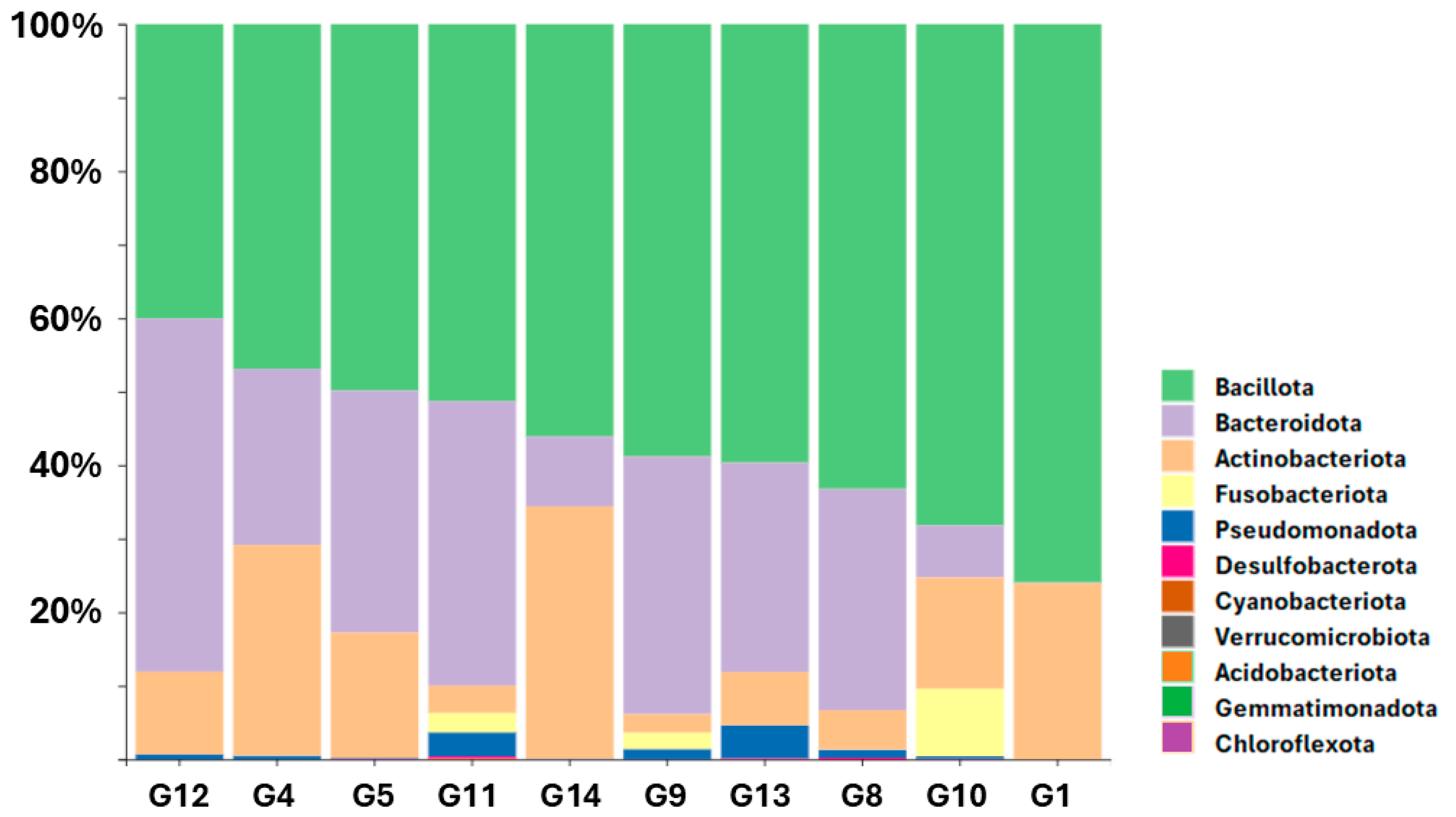
3.1. Relative Abundance of Bacterial Phyla Across Samples

Figure 1 illustrates the relative abundance of bacterial phyla detected in 10 fecal samples. The phylum Bacillota (light green) is consistently the most dominant across all samples, representing approximately 40% to 70% of the total bacterial community. Its prevalence is particularly pronounced in samples G8, G10, and G1. The second most abundant phylum is Bacteroidota (purple), ranging from 20% to 50% among the samples, with a notably high proportion in G12, where its abundance nearly matches that of Bacillota. Actinobacteriota (orange) are substantially represented in samples G14 and G4, contributing up to 30% in some cases, but their abundance decreases markedly in samples such as G11 and G9. Pseudomonadota (blue) and Fusobacteriota (yellow) are present in low yet detectable levels, particularly in samples G13, G11, and G9. Other minor phyla, including Desulfobacterota, Verrucomicrobiota, Cyanobacteriota, Acidobacteriota, Chloroflexota, and Gemmatimonadota, appear sporadically at very low relative abundances, typically below 5%.

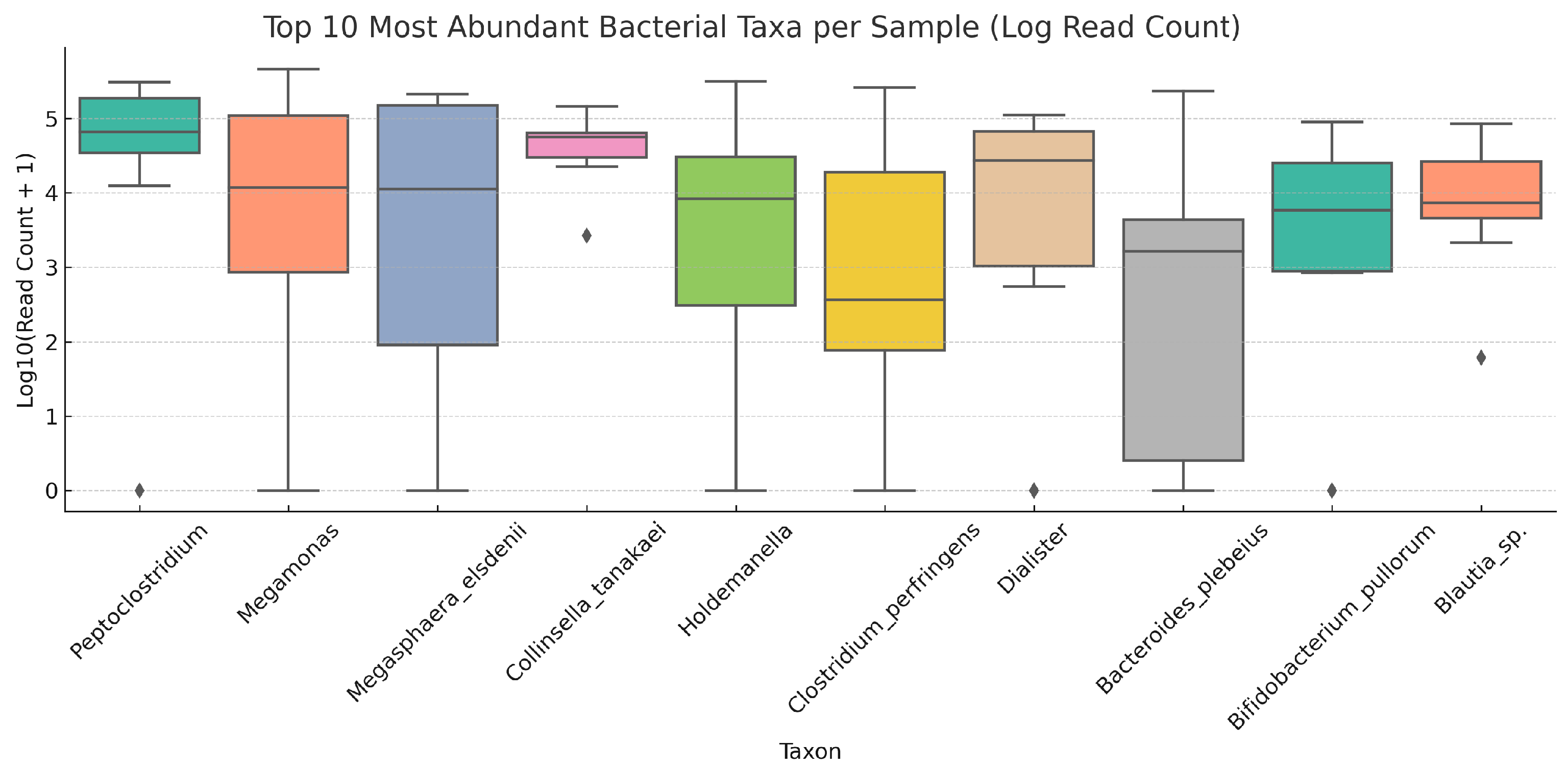
3.1.1. Distribution and Variability of the Most Abundant Taxa Across Samples

Figure 2 illustrates the distribution of the top 10 most abundant bacterial taxa in the domestic cat gut microbiome across individual samples using boxplots of log-transformed read counts (log10[read count + 1]). Peptoclostridium stands out as the most consistently dominant taxon across all samples. Its high median and compact interquartile range (IQR), combined with the near absence of outliers, suggests that this genus is stable and uniformly present in most individuals. This consistency across samples implies that Peptoclostridium may function as a core member of the feline gut microbiome, potentially playing essential roles in host digestion or mucosal health. In contrast, Megamonas exhibited much higher variability in read counts across samples. While it was highly abundant in certain individuals, it was nearly absent in others, as indicated by a wide IQR and multiple extreme values. This broad distribution suggests significant ecological plasticity or dependence on host-associated factors, such as diet composition, microbial interactions, or intestinal transit time. The presence of both high and low extremes suggests a taxon that may bloom under certain conditions while remaining suppressed under others. Megasphaera elsdenii also displayed substantial inter-individual variation. It was one of the most abundant taxa in a subset of cats, but completely undetectable or rare in others. The sharp contrasts in its abundance profile, reflected in a wide IQR and extended whiskers, may point to specific host-microbe or microbe-microbe interactions that modulate its presence, potentially including sensitivity to antimicrobials or competition for substrate with functionally similar taxa. Collinsella tanakaei and Holdemanella showed moderately high medians, but with greater dispersion compared to Peptoclostridium, suggesting an intermediate consistency. Holdemanella, in particular, had some extremely high values, indicating dominance in a few samples, while its near-zero levels in others reflect patchy colonization. This might be due to differences in host physiology, immune response, or environmental exposure. C. perfringens revealed a distinctive bimodal distribution: it was highly abundant in a few samples but completely absent in others. Known for its pathogenic potential, this pattern may be indicative of transient colonization events, opportunistic overgrowth, or host-specific susceptibility. The sudden spikes in abundance in certain individuals suggest that C. perfringens may act more as a facultative member of the microbiome, potentially linked to dysbiosis or dietary disturbances. Bacteroides plebeius and Dialister were notable for their low to moderate medians and broad variability. Both taxa were entirely undetectable in some samples, suggesting that their presence is inconsistent and possibly conditional on certain dietary inputs (e.g., polysaccharides for Bacteroides) or host factors. Their fluctuating detection patterns imply limited ubiquity within this sample set.
Finally, Bifidobacterium pullorum and Blautia sp. had the lowest overall abundances, as shown by their tightly compressed boxplots near the bottom of the transformed scale. Detected only in a fraction of the cats, these taxa likely represent conditionally rare bacteria, whose ecological roles may emerge under specific conditions or in response to host developmental stages or metabolic states.

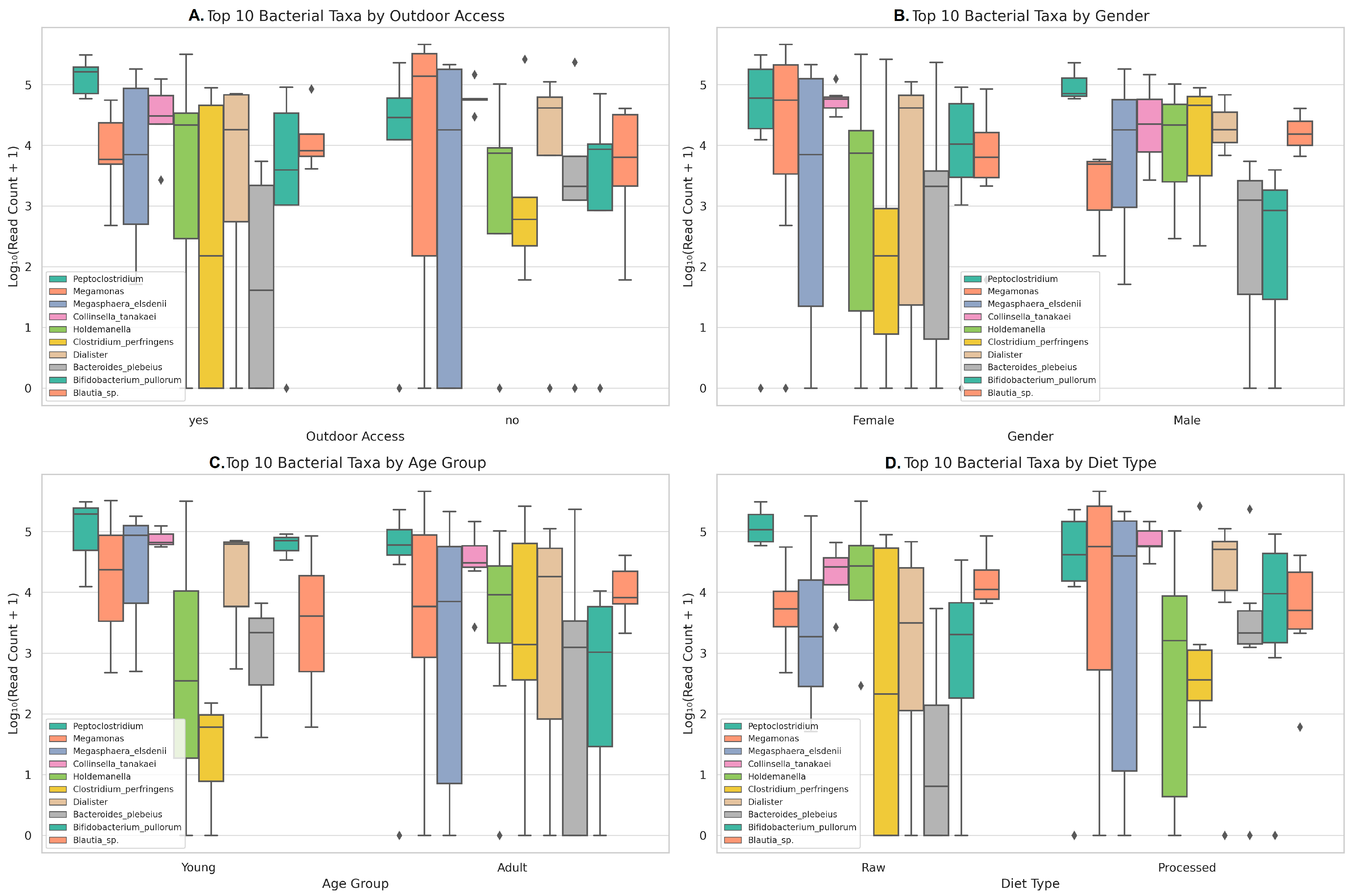
3.1.2. Distribution and Variability of the Most Abundant Taxa According to Metadata Variables

Figure 3 presents four boxplots for the top 10 most abundant bacterial taxa, stratified by four key metadata variables: outdoor access, gender, age, and diet type.
For Outdoor Access (A panel), Peptoclostridium and Holdemanella showed higher median abundances in cats without outdoor access, suggesting a more stable or enriched presence in indoor environments. This may relate to differences in environmental exposure, stress levels, or microbial acquisition routes. In contrast, Megamonas and Megasphaera elsdenii exhibited higher abundance in outdoor-access cats, but with wide dispersion, indicating that while exposure to external microbiota may promote their presence, individual factors modulate their colonization success. Notably, Collinsella tanakaei and Dialister showed more balanced distributions, suggesting a more consistent presence across both environments.
For Gender (B panel), Megamonas and Bacteroides plebeius were more abundant in females, as seen in the elevated median values and consistent IQRs. This might reflect differences in hormonal regulation, grooming behavior, or dietary patterns influenced by owners. In contrast, Megasphaera elsdenii and C. perfringens showed greater abundance in males, although Clostridium’s distribution was bimodal, suggesting sporadic but potentially dominant colonization in select individuals. Peptoclostridium maintained relatively high abundance in both genders but with reduced variability in males, reinforcing its status as a core taxon.
For the Age Group (C panel), Collinsella tanakaei, Megamonas, and Blautia sp. were notably enriched in younger cats, potentially reflecting a developmental stage of the gut microbiome characterized by fermentative taxa associated with energy metabolism and mucosal maturation. Conversely, Peptoclostridium, Holdemanella, and C. perfringens were more prevalent in adult individuals, possibly indicative of increased microbial diversification with age or adaptations to dietary shifts. Dialister and Bifidobacterium pullorum remained relatively stable across age groups, suggesting they are not tightly age-dependent.
For Diet Type (D Panel), Cats on a raw diet had significantly elevated levels of Megasphaera elsdenii and Megamonas, both known for metabolizing simple carbohydrates and protein fermentation by-products. This suggests a microbial adaptation to high-protein, unprocessed diets. Conversely, Peptoclostridium and Holdemanella were more abundant in cats on processed diets, indicating possible reliance on complex carbohydrates or additives present in commercial food. Interestingly, Bifidobacterium pullorum was detected mainly in the processed diet group, which could reflect exposure to dietary prebiotics or thermal-stable microbial components.

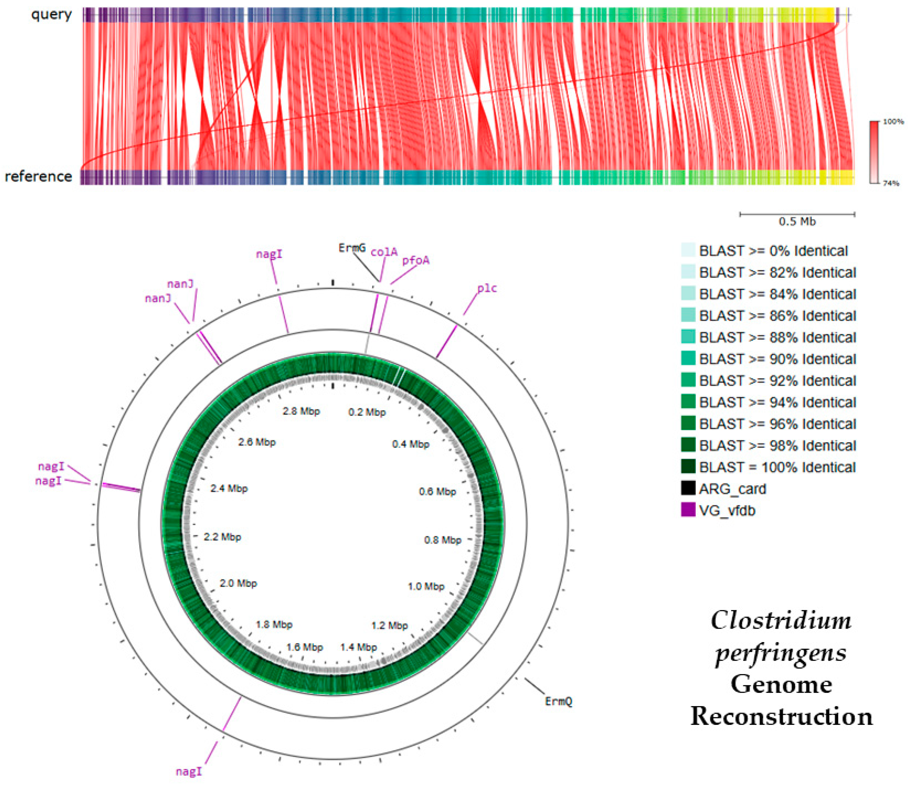
3.1.3. Antibiotic Resistance and Virulence Genes Identified in C. perfringens

The shotgun metagenomic analysis of C. perfringens revealed the presence of multiple genes associated with antibiotic resistance and virulence, as shown in Figure 4 and Table 1. Figure 4 provides a comprehensive comparative genomic analysis and reconstruction of the C. perfringens genome, combining a synteny alignment with a circular genome map to visualize both sequence conservation and the spatial distribution of key genetic determinants. In the upper panel, a synteny plot compares the genome of the analyzed C. perfringens strain (query) against a reference genome. Red ribbons connecting the two rows indicate homologous regions with conserved gene order and orientation. The high density and continuity of these red blocks suggest strong structural conservation between the two genomes. Next to this alignment, a heatmap gradient depicts the percentage identity of BLAST v2.14.1 alignments, ranging from 74% to 100%. Regions with darker green and black shades reflect higher sequence similarity, indicating that most of the query genome shares at least 96% identity with the reference. This result demonstrates a high degree of genomic conservation and suggests a close phylogenetic relationship between the strains.
The lower panel features a circular representation of the query genome, providing a spatial overview of annotated features. These genes are distributed throughout the genome, implying that they are not confined to a single pathogenic island but rather dispersed across the chromosome. Two inner concentric rings provide further detail on the genome’s structure. The first inner ring, shown in green, represents a coverage histogram that illustrates the relative depth of sequence similarity along the genome compared to the reference. The second inner ring visualizes BLAST identity, using a gradient that shifts from light green to black to indicate increasing levels of nucleotide identity (from approximately 82% to 100%). The outermost ring displays the locations of genes associated with antibiotic resistance and virulence. Antibiotic resistance genes identified via the ARG card database, such as ErmG and ErmQ, are labeled in black. Meanwhile, virulence factors identified using the Virulence Factor Database (VFDB), including pfoA, plc, colA, nanJ, and nagI, are labeled in purple. Together, these representations emphasize the high level of genomic identity between the query and reference strains and highlight the integrated presence of both resistance and virulence determinants, reinforcing the pathogenic and adaptive potential of the studied C. perfringens isolate.
Table 2 shows that antibiotic resistance and virulence genes were annotated based on sequence coverage, identity percentage, functional classification, and database references. Two antibiotic resistance genes (ARGs) were identified using the ARG card database. The first, ErmG, exhibited 100% sequence coverage and 99.05% identity. This gene encodes an rRNA methyltransferase that modifies the 23S rRNA, preventing the binding of antibiotics to the ribosome and conferring resistance. The associated resistance phenotype includes lincosamides, macrolides, and streptogramins. The second resistance gene, ErmQ, also showed 100% coverage and 100% identity. It encodes a methyltransferase responsible for conferring the MLSb phenotype through ribosomal methylation, leading to resistance against the same antibiotic classes as ErmG. These findings indicate the presence of highly conserved and functionally relevant resistance mechanisms, which may impair the efficacy of common protein synthesis-inhibiting antibiotics used in clinical and veterinary contexts. In addition to ARGs, five virulence-associated genes (VGs) were identified through the VFDB. The gene pfoA, encoding perfringolysin O (theta-toxin), showed 100% coverage and 98.74% identity. This toxin is a pore-forming cytolytic that contributes to host cell lysis and immune evasion. Similarly, plc was detected with full coverage and 99.33% identity; it encodes phospholipase C (alpha-toxin), a major virulence factor that degrades phospholipid membranes, leading to host tissue damage. The nagI gene, identified with 94.5% coverage and 98.72% identity, encodes hyaluronidase (mu-toxin), an enzyme that breaks down hyaluronic acid in connective tissue, facilitating bacterial invasion and dissemination. Another enzyme, nanJ, showed 97.56% coverage and 97.64% identity. It encodes an exo-alpha-sialidase that cleaves sialic acids from host glycoconjugates, supporting nutrient acquisition and colonization. Lastly, the colA gene, with 100% coverage and 98.7% identity, encodes collagenase (kappa-toxin), which degrades collagen, a key component of the extracellular matrix, thus enhancing tissue invasion.

4. Discussion

This study presents the first metagenomic analysis of cat fecal samples conducted in Cali, Colombia, offering initial insights into the taxonomic and functional composition of the feline gut microbiome in this region. The findings highlight the relevance of an exploratory applied metagenomic profiling approach as a foundational step toward understanding bacterial diversity, antimicrobial resistance, and the virulence potential within the domestic cat gut microbiota. By integrating 16S rRNA metabarcoding and shotgun metagenomic sequencing, this approach enabled the identification of key bacterial taxa and functional genes across cats living under different household conditions. Although the limited sample size and the absence of gene expression validation constrain the generalizability of the results, the applied nature of this study offers valuable baseline data that can guide future research within the frameworks of One Health, microbial surveillance, and veterinary microbiology. Thus, our results not only support previous observations about the dominance of Bacillota and Bacteroidota in domestic cat gut microbiota but also highlight taxonomic and functional shifts associated with environmental, biological, and dietary metadata. Moreover, the detection of ARGs and pathogenicity-associated genes in C. perfringens underscores the potential public and veterinary health implications of microbial reservoirs in companion animals.
The metagenomic analysis of the relative abundance of bacterial phyla in our study in domestic cat fecal samples revealed that Bacillota is the predominant phylum, representing approximately 40–70% of the total bacterial community across all samples. This finding is consistent with multiple studies that have identified Bacillota as a principal phylum within the gut microbiota of mammals, including cats [22]. Bacillota encompasses a wide array of obligatory anaerobes that specialize in the fermentation of complex carbohydrates and the production of short-chain fatty acids (SCFAs), such as butyrate and acetate, which are critical for host gut health [23]. In the feline gut, Bacillota likely contribute to energy salvage from the diet and to maintaining the integrity of the intestinal mucosa [24]. Notably, the second most abundant phylum, Bacteroidota, exhibited significant variation, ranging from 20 to 50% and peaking in the sample G12. Bacteroidota members are renowned for their ability to degrade a wide range of polysaccharides, thereby complementing the metabolic functions of Bacillota [25]. The dynamic fluctuations of Bacteroidota observed in the feline samples may reflect dietary influences, as high-fiber diets tend to enrich Bacteroidota populations, consistent with findings in other carnivorous and omnivorous mammals [26]. Interestingly, Actinobacteriota were well represented in the samples G14 and G4 (up to 30%) but declined in the others. Members of this phylum, such as Bifidobacterium species, are typically considered beneficial, often associated with gut health and mucosal homeostasis [27]. Their initial abundance suggests a potential role in early gut colonization, but the subsequent decrease might indicate a shift toward a more adult-like gut community structure as the animal matures, like patterns observed in human and murine microbiome studies [28]. The detection of Pseudomonadota and Fusobacteriota in low but consistent amounts, particularly in G13, G11 and G9 samples, is also notable. Pseudomonadota, including members of Enterobacteriaceae, are frequently observed as minor constituents in healthy gut communities but can bloom during dysbiosis or inflammatory states [29]. Fusobacteriota, while typically low in abundance, have been associated with protein-rich diets and mucosal interactions [30]. Their presence in these feline samples may hint at dietary transitions or transient microbial colonization from environmental sources. Finally, minor phyla such as Cyanobacteriota, Verrucomicrobiota, Acidobacteriota, and others were detected at <5% relative abundance. These phyla are considered part of the “rare biosphere” and are generally transient or environmentally influenced, possibly reflecting exposure to environmental microbes through diet, outdoor access, or contact with soil and water [31]. Taken together, these findings underscore the consistency of feline gut microbiota with general mammalian gut ecology and highlight the dynamic interactions among dominant and minor phyla in shaping a functional microbial community. This detailed characterization sets the foundation for further exploration of how diet, environment, and host factors modulate the feline gut ecosystem.
The Top 10 bacterial taxa by average abundance in our analysis of the cat gut microbiota revealed that Peptoclostridium was the most dominant genus, with an average relative abundance of approximately 24%. Peptoclostridium belongs to the family Peptostreptococcaceae within the phylum Bacillota and is recognized for its role in anaerobic fermentation processes, particularly the production of short-chain fatty acids (SCFAs) such as butyrate, which are crucial for maintaining colonic health and mucosal integrity [23]. Its spore-forming capacity likely contributes to its resilience within the gut environment and its ability to persist despite fluctuations in dietary intake or antibiotic exposure [32]. The dominance of Peptoclostridium underscores its central ecological role in the cat gastrointestinal tract, possibly linked to its ability to utilize dietary fibers and host-derived mucins as substrates. Following Peptoclostridium, Megamonas was identified with a relative abundance of ~15%. This genus has been described as a common component of the gut microbiota in various mammals, including humans and animals. Megamonas ferments carbohydrates to produce SCFAs, primarily acetate and propionate, which not only serve as energy sources for colonocytes but also modulate systemic metabolism and immune function [33]. The relatively high abundance of Megamonas in these feline samples suggests that diets rich in fermentable carbohydrates, either through processed pet foods or raw diets, may support its proliferation and metabolic activity. Megasphaera elsdenii, comprising ~13% of the average relative abundance, is another key SCFA producer. In ruminants, Megasphaera elsdenii is recognized for its role in lactic acid utilization and butyrate production during high carbohydrate feeding [34]. While its role in feline gut ecology is less well characterized, the prevalence of Megasphaera in these samples suggests a similar contribution to lactate metabolism and maintenance of colonic pH balance. This metabolic capacity can be particularly important in carnivorous species that may ingest high-protein diets but also receive plant-based carbohydrates in commercial feeds. Collinsella tanakaei (~11% abundance) represents another saccharolytic bacterium within the Actinobacteriota phylum. Collinsella species have been linked to bile acid metabolism, including bile salt hydrolase (BSH) activity. Its prominence in feline gut samples may reflect similar ecological roles [35], although specific feline-focused studies are warranted. C. perfringens was present at ~8%, an important finding given its dual nature as both a commensal and an opportunistic pathogen. In healthy animals, C. perfringens may be part of the normal gut microbiota, but it is also known to produce toxins such as alpha-toxin and perfringolysin O, which can cause enteric disease in susceptible hosts [36]. Its presence at relatively high abundance in these feline samples raises questions about its role in gut homeostasis versus its potential pathogenicity, which may be influenced by environmental triggers, antibiotic use, or dietary imbalances. Other genera identified at lower relative abundances (<7%) include Bacteroides plebeius, Holdemanella, Dialister, Blautia sp., and Bifidobacterium pullorum. Blautia and Bifidobacterium are well-established beneficial genera in mammalian gut microbiota [37,38]. Overall, this diverse community structure reflects a complex interplay of carbohydrate fermenters, bile acid metabolizers, opportunistic pathogens, and commensals in the feline gut. Such diversity supports the notion that the gut microbiota of domestic cats is both metabolically versatile and ecologically dynamic, shaped by factors like diet, environment, and host physiology [39].
The influence of environment on the gut microbiota composition of cats in this study reveals complex but coherent ecological patterns that align with broader findings in microbiome research. The observation that outdoor access modulated the bacterial community structure is particularly significant. Peptoclostridium and Megamonas were enriched in indoor-only cats, while Holdemanella and C. perfringens were more abundant in cats with outdoor access. This pattern suggests that environmental microbial exposure and behavioral factors associated with outdoor access shape gut microbial communities. Environmental seeding from soil, vegetation, and wildlife can introduce transient or persistent microbes into the gut ecosystem, as reported by Song et al. (2013), who demonstrated that exposure to diverse environmental microbiota significantly modifies gut microbial diversity and structure [40]. The presence of C. perfringens in outdoor-access cats underscores the zoonotic and ecological implications of environmental contact. C. perfringens is a known enteric pathogen with environmental reservoirs, capable of colonizing multiple hosts [41]. Its detection in these outdoor cats may be linked to exposure to contaminated water, soil, or prey animals, a hypothesis supported by studies showing that environmental and dietary exposure can increase the gut abundance of pathogenic clostridia [42]. Gender-related differences in the gut microbiota, such as the higher prevalence of Peptoclostridium in males, have been documented in other mammalian hosts and may reflect hormonal influences on immune responses and gut physiology. Markle et al. (2013) found that sex hormones like testosterone and estrogen can influence microbial community assembly through immune modulation, resulting in sex-specific microbiome profiles [43]. The slightly higher relative abundance of Megamonas and Megasphaera in females observed here may similarly reflect subtle interactions between sex hormones and microbial colonization dynamics. Age-related differences were minimal in this dataset, with Peptoclostridium showing a slight enrichment in adults compared to juveniles. This limited age effect aligns with the findings of Roswall et al. (2021), who observed that although microbiota matures during early life, adult microbiota remains relatively stable under consistent environmental conditions [44]. However, the modest increase of Peptoclostridium in adults might also be linked to changes in host physiology and dietary patterns with age. Perhaps most strikingly, diet emerged as a dominant driver of microbiota composition, as has been widely documented in mammalian gut ecology [45]. In this study, processed food refers to commercial cat food (e.g., kibble or canned), while raw food includes uncooked animal-based ingredients such as raw meat or organs. The raw diet was associated with an increase in Peptoclostridium, Megasphaera elsdenii, and Dialister. These taxa are known to ferment dietary fibers and resistant starches, producing beneficial short-chain fatty acids (SCFAs) that support gut and immune health [46]. Peptoclostridium is a spore-forming Firmicute that thrives in fiber-rich, anaerobic environments, reinforcing its role in raw diet contexts [23]. Conversely, processed food diets, typically higher in refined carbohydrates and lower in fiber, supported an increased abundance of Megamonas, Collinsella, and Bacteroides plebeius. These taxa have been linked to diets rich in simple sugars and processed components, consistent with observations by Bermingham et al. (2017) in domestic cats fed commercial diets [47]. Collinsella has been associated with dietary emulsifiers and processed food additives that can disrupt gut barrier integrity and promote inflammation [48]. These findings collectively highlight the profound impact of diet and environmental exposure on feline gut microbiota composition, mirroring broader insights in mammalian gut ecology. They underscore the importance of considering host behavior, diet, and environmental contact when evaluating the gut microbiota, as these factors interact dynamically to shape community structure and function. Furthermore, understanding these associations in companion animals not only informs cats’ health but also has implications for zoonotic risk and the environmental microbiome interface.
The identification of antibiotic resistance and virulence genes in C. perfringens, specifically of the ErmG and ErmQ genes in the metagenomic dataset of cat gut microbiota, highlights a critical concern regarding antibiotic resistance. These genes encode rRNA methyltransferases that methylate the 23S rRNA component of the bacterial ribosome, thereby preventing the binding of macrolides, lincosamides, and streptogramins (MLSb phenotype) [49]. Such ribosomal protection confers high-level resistance and can render these antibiotics ineffective, posing significant therapeutic challenges in both human and veterinary medicine. Notably, the presence of Erm genes in C. perfringens has been previously documented in livestock and poultry, suggesting that these resistance determinants are widespread in animal reservoirs [50]. Their detection in the cat gut environment underscores the potential for horizontal gene transfer of these resistance genes across different animal hosts, including companion animals like cats, which may serve as reservoirs for further dissemination to humans and other animals. Equally concerning is the identification of five highly conserved virulence-associated genes: pfoA, plc, colA, nanJ, and nagI. These genes encode critical toxins and enzymes that enhance the pathogenicity of C. perfringens in the host gut environment [51]. For instance, pfoA encodes perfringolysin O (theta-toxin), a cholesterol-dependent pore-forming cytolysin that disrupts eukaryotic cell membranes, leading to tissue necrosis and immune evasion [52]. Similarly, plc encodes phospholipase C (alpha-toxin), which hydrolyzes phospholipids in host cell membranes and has been identified as a primary virulent factor in gas gangrene and necrotic enteritis [53]. The collagenase gene colA facilitates tissue invasion by degrading collagen, a key structural protein in the extracellular matrix. NanJ encodes an exo-alpha-sialidase, which cleaves sialic acid residues from host glycoconjugates, aiding in nutrient acquisition and colonization [54]. Lastly, nagI encodes hyaluronidase, an enzyme that breaks down hyaluronic acid in connective tissues, facilitating bacterial dissemination [55]. The high conservation of these virulence genes (coverage ≥94%, identity ≥97%) in the analyzed C. perfringens strain indicates that these pathogenic traits are stably maintained within the bacterial genome. Such conservation suggests strong selective pressures for retaining these factors, potentially due to their essential roles in gut colonization and survival in competitive microbial communities [56]. Taken together, the concurrent presence of antibiotic resistance and virulence determinants in the cat gut metagenome underscores the zoonotic potential of C. perfringens. Companion animals like cats may act as asymptomatic carriers of these strains, posing a risk for transmission to humans and other animals through close contact or environmental contamination [57]. Therefore, the findings highlight the importance of vigilant antibiotic stewardship in veterinary settings and the need for routine surveillance of antimicrobial resistance and virulence gene reservoirs in domestic pets [58]. The identification of antibiotic resistance genes (ErmG, ErmQ) and virulence factors (pfoA, plc, colA, nanJ, nagI) in C. perfringens from domestic cats suggests a potential public health concern, particularly in urban settings where close human–pet interaction is frequent. These findings can be applied in veterinary and public health surveillance programs aimed at monitoring the emergence and spread of antimicrobial resistance (AMR) and pathogenic strains within companion animal populations. Furthermore, the results underscore the importance of promoting responsible antibiotic use in veterinary practice and encourage the development of preventive strategies, such as dietary interventions or probiotics, to reduce colonization by pathogenic strains. This information is also valuable for future One Health risk assessments, where domestic animals are considered potential reservoirs or vectors of clinically relevant AMR genes and virulence determinants that could affect human health.
The findings of this exploratory study generate several testable hypotheses regarding the ecological and epidemiological dynamics of C. perfringens and its associated genetic determinants within the feline gut microbiome. First, given the observed association between higher C. perfringens abundance and raw feeding, one hypothesis is that raw diets in domestic cats increase the risk of colonization by toxigenic and antimicrobial-resistant strains of C. perfringens. This could be due to direct bacterial contamination of raw animal products or the absence of processing steps that would normally reduce microbial loads. Second, the presence of antimicrobial resistance genes (ErmG, ErmQ) and virulence factors (pfoA, plc, colA, nanJ, nagI) in cats with outdoor access suggests the hypothesis that environmental exposure may act as a significant vector for the acquisition of resistance and virulence traits. This may occur through contact with contaminated soil, water, or other animals carrying resistant or pathogenic microorganisms. A third hypothesis is that cats living under combined conditions of raw feeding and outdoor access represent a higher-risk reservoir for zoonotic transmission of clinically relevant bacteria and genes, which could have implications for both veterinary and public health. Additionally, the absence of clear patterns related to age and sex may suggest that diet and environment are more influential factors than host biology in shaping the gut resistome and virulome, though larger-scale studies would be needed to confirm this. These hypotheses warrant further investigation through longitudinal studies with larger sample sizes and individual-level metagenomic sequencing to assess causality and transmission dynamics.
Several limitations inherent to the dataset should be considered when interpreting the results. The relatively small sample size reduces the statistical power of the analyses and limits the generalizability of the findings to the wider feline population. Although the use of shotgun metagenomic sequencing and 16S rRNA metabarcoding enabled detailed taxonomic and functional profiling, these approaches do not provide information on gene expression or confirm the functional activity of the detected resistance and virulence factors within the host environment. Moreover, while reference-based databases are valuable for gene annotation, they may fail to detect novel or feline-specific genes, highlighting the need for complementary strategies such as genome-resolved metagenomics and culturomics. Additionally, the broad categorization of dietary and environmental variables may obscure subtle but important interactions between host factors and microbiota composition.
A notable methodological decision in this study was the pooling of DNA samples for shotgun metagenomic sequencing. While individual DNA samples were processed separately for 16S rRNA gene amplicon sequencing, equal volumes (5 µL) from each of the ten extracted DNA samples were combined to form a single pooled sample for shotgun metagenomics. This approach was motivated by financial constraints and the exploration of the research, which aimed to provide a preliminary functional profile of the feline gut microbiome, particularly focusing on genes related to antimicrobial resistance and virulence in C. perfringens. Pooling allowed us to maximize the sequencing output and generate a consensus metagenome that reflects the collective functional potential of the study population. However, we acknowledge that pooling can obscure important inter-individual differences, especially in light of the variability in microbial community composition observed across individuals in our study. Such variation may influence the presence or abundance of specific genes, potentially biasing the results toward dominant community members. Consequently, while the pooled metagenome provides valuable insights into shared functional traits, it may underestimate rare or individual-specific features. This methodological limitation underscores the importance of interpreting the metagenomic findings as representative of the group rather than of individual hosts. Considering these limitations, future studies should pursue larger and more diverse cohorts, integrate functional approaches such as metatranscriptomics or metaproteomics, and implement more detailed metadata collection protocols to advance our understanding of the cat gut microbiome and its implications for health and zoonotic risk.

5. Conclusions

This study presents the first metagenomic characterization of the gut microbiome in domestic cats from Cali, Colombia, offering novel insights into both the taxonomic composition and functional potential of these microbial communities. Using a combination of 16S rRNA gene metabarcoding and pooled shotgun metagenomic sequencing, we identified Peptoclostridium, Megamonas, and Megasphaera as dominant genera, with their abundance patterns influenced primarily by diet and outdoor access. Additionally, we detected clinically relevant antibiotic resistance genes (ErmG, ErmQ) and virulence factors (pfoA, plc, colA, nanJ, nagI) associated with C. perfringens, indicating potential zoonotic and public health implications.
Our results demonstrate that dietary composition and environmental exposure play key roles in shaping the gut microbiota of domestic cats, while age and gender have a more moderate impact. Despite limitations such as small sample size and pooled metagenomic data, this study provides a valuable baseline for future microbiome research in urban companion animals within Latin America. It also highlights the relevance of domestic cats as potential reservoirs of antimicrobial resistance and virulence determinants.
These findings reinforce the importance of microbiome monitoring under the One Health framework, emphasizing the need to include underrepresented geographic regions in global microbial surveillance and risk assessment strategies.

Author Contributions

Conceptualization, A.C. and R.S.; methodology, M.P., H.F.-R. and A.F.; software, H.F.-R. and A.F.; validation, A.P.-M., L.M., A.C. and R.S.; formal analysis, M.P., H.F.-R. and A.F.; investigation, M.P. and A.P.-M.; resources, L.M.; data curation, M.P.; writing—original draft preparation, M.P., A.P.-M. and A.C.; writing—review and editing, A.C. and R.S.; visualization, H.F.-R. and A.F.; supervision, A.C. and R.S.; project administration, A.C. and L.M.; funding acquisition, A.C. and L.M. All authors have read and agreed to the published version of the manuscript.

Funding

This research was funded by the Vice-Rector at Universidad del Valle, grant numbers CI 71335 and CI 71356.

Institutional Review Board Statement

This study did not involve direct animal interventions; thus, no specific ethical approval was required. Owners provided informed consent for participation and sample collection.

Data Availability Statement

The data generated in this study are available from the corresponding author upon request.

Acknowledgments

We gratefully acknowledge all the cat owners and the submitting laboratories for providing genetic sequences and metadata. We also thank Nelson Rivera and Diana Lopez for their valuable contributions to this work.

Conflicts of Interest

The authors declare no conflicts of interest.

Abbreviations

The following abbreviations are used in this manuscript:
SCFAShort-Chain Fatty Acid
ARGAntibiotic Resistance Gene
VFVirulence Factor
rRNARibosomal Ribonucleic Acid
MLSbMacrolide–Lincosamide–Streptogramin B
BSHBile Salt Hydrolase
DNADeoxyribonucleic Acid
PCRPolymerase Chain Reaction
NCBINational Center for Biotechnology Information
AMRAntimicrobial Resistance
NGSNext-Generation Sequencing
OTUOperational Taxonomic Unit
LCALowest Common Ancestor
QCQuality Control
IQRInterquartile Range

References

  1. Suchodolski, J.S. Analysis of the gut microbiome in dogs and cats. Vet. Clin. Pathol. 2022, 50 (Suppl. S1), 6–17. [Google Scholar] [CrossRef] [PubMed]
  2. Schmitz, S.S. Value of Probiotics in Canine and Feline Gastroenterology. Vet. Clin. Small Anim. Pract. 2021, 51, 171–217. [Google Scholar] [CrossRef] [PubMed]
  3. Rojas, C.A.; Gardy, J.; Eisen, J.A.; Ganz, H.H.; Rasko, D. Recovery of 52 bacterial genomes from the fecal microbiome of the domestic cat (Felis catus) using Hi-C proximity ligation and shotgun metagenomics. Microbiol. Resour. Announc. 2023, 12, e0060123. [Google Scholar] [CrossRef] [PubMed]
  4. Ing, N.H.; Steiner, J.M. The Use of Diets in the Diagnosis and Treatment of Common Gastrointestinal Diseases in Dogs and Cats. Adv. Exp. Med. Biol. 2024, 1446, 39–53. [Google Scholar] [CrossRef]
  5. Ganz, H.H.; Jospin, G.; Rojas, C.A.; Martin, A.L.; Dahlhausen, K.; Kingsbury, D.D.; Osborne, C.X.; Entrolezo, Z.; Redner, S.; Ramirez, B.; et al. The Kitty Microbiome Project: Defining the Healthy Fecal “Core Microbiome” in Pet Domestic Cats. Vet. Sci. 2022, 9, 635. [Google Scholar] [CrossRef]
  6. Butowski, C.F.; Thomas, D.G.; Cave, N.J.; Bermingham, E.N.; Rosendale, D.I.; Hea, S.-Y.; Stoklosinski, H.M.; Young, W. In Vitro Assessment of Hydrolysed Collagen Fermentation Using Domestic Cat (Felis catus) Faecal Inocula. Animals 2022, 12, 498. [Google Scholar] [CrossRef]
  7. Summers, S.; Quimby, J.; Gagné, J.; Lappin, M. The Effect of Dietary Protein Concentration on the Fecal Microbiome and Serum Concentrations of Gut-Derived Uremic Toxins in Healthy Adult Cats. Vet. Sci. 2023, 10, 497. [Google Scholar] [CrossRef]
  8. Rojas, C.A.; Entrolezo, Z.; Jarett, J.K.; Jospin, G.; Kingsbury, D.D.; Martin, A.; Eisen, J.A.; Ganz, H.H. Microbiome Responses to Fecal Microbiota Transplantation in Cats with Chronic Digestive Issues. Vet. Sci. 2023, 10, 561. [Google Scholar] [CrossRef]
  9. Bolyen, E.; Rideout, J.R.; Dillon, M.R.; Bokulich, N.A.; Abnet, C.C.; Al-Ghalith, G.A.; Alexander, H.; Alm, E.J.; Arumugam, M.; Asnicar, F.; et al. Reproducible, interactive, scalable and extensible microbiome data science using QIIME 2. Nat. Biotechnol. 2019, 37, 1091. [Google Scholar] [CrossRef]
  10. Magoč, T.; Salzberg, S.L. FLASH: Fast length adjustment of short reads to improve genome assemblies. Bioinformatics 2011, 27, 2957–2963. [Google Scholar] [CrossRef]
  11. Quast, C.; Pruesse, E.; Yilmaz, P.; Gerken, J.; Schweer, T.; Yarza, P.; Peplies, J.; Glöckner, F.O. The SILVA ribosomal RNA gene database project: Improved data processing and web-based tools. Nucleic Acids Res. 2013, 41, D590–D596. [Google Scholar] [CrossRef] [PubMed]
  12. McMurdie, P.J.; Holmes, S. phyloseq: An R package for reproducible interactive analysis and graphics of microbiome census data. PLoS ONE 2013, 8, e61217. [Google Scholar] [CrossRef] [PubMed]
  13. Prjibelski, A.; Antipov, D.; Meleshko, D.; Lapidus, A.; Korobeynikov, A. Using SPAdes De Novo Assembler. Curr. Protoc. Bioinform. 2020, 70, e102. [Google Scholar] [CrossRef]
  14. Nurk, S.; Meleshko, D.; Korobeynikov, A.; Pevzner, P.A. metaSPAdes: A new versatile metagenomic assembler. Genome Res. 2017, 27, 824–834. [Google Scholar] [CrossRef] [PubMed]
  15. Li, H. Minimap2: Pairwise alignment for nucleotide sequences. Bioinformatics 2018, 34, 3094–3100. [Google Scholar] [CrossRef]
  16. Danecek, P.; Bonfield, J.K.; Liddle, J.; Marshall, J.; Ohan, V.; Pollard, M.O.; Whitwham, A.; Keane, T.; McCarthy, S.A.; Davies, R.M.; et al. Twelve years of SAMtools and BCFtools. GigaScience 2021, 10, giab008. [Google Scholar] [CrossRef]
  17. Schwengers, O.; Jelonek, L.; Dieckmann, M.A.; Beyvers, S.; Blom, J.; Goesmann, A. Bakta: Rapid and standardized annotation of bacterial genomes via alignment-free sequence identification. Microb. Genom. 2021, 7, 000685. [Google Scholar] [CrossRef]
  18. Jia, B.; Raphenya, A.R.; Alcock, B.; Waglechner, N.; Guo, P.; Tsang, K.K.; Lago, B.A.; Dave, B.M.; Pereira, S.; Sharma, A.N.; et al. CARD 2017: Expansion and model-centric curation of the comprehensive antibiotic resistance database. Nucleic Acids Res. 2017, 45, D566–D573. [Google Scholar] [CrossRef]
  19. Chen, L.; Zheng, D.; Liu, B.; Yang, J.; Jin, Q. VFDB 2016: Hierarchical and refined dataset for big data analysis—10 years on. Nucleic Acids Res. 2016, 44, D694–D697. [Google Scholar] [CrossRef]
  20. Lu, J.; Rincon, N.; Wood, D.E.; Breitwieser, F.P.; Pockrandt, C.; Langmead, B.; Salzberg, S.L.; Steinegger, M. Metagenome analysis using the Kraken software suite. Nat. Protoc. 2024, 17, 2815–2839. [Google Scholar] [CrossRef]
  21. Breitwieser, F.P.; Salzberg, S.L.; Schwartz, R. Pavian: Interactive analysis of metagenomics data for microbiome studies and pathogen identification. Bioinformatics 2020, 36, 1303–1304. [Google Scholar] [CrossRef] [PubMed]
  22. Tun, H.M.; Brar, M.S.; Khin, N.; Jun, L.; Hui, R.K.-H.; Dowd, S.E.; Leung, F.C.-C. Gene-centric metagenomics analysis of feline intestinal microbiome using 454 junior pyrosequencing. J. Microbiol. Methods 2012, 88, 369–376. [Google Scholar] [CrossRef]
  23. Louis, P.; Flint, H.J. Formation of propionate and butyrate by the human colonic microbiota. Environ. Microbiol. 2017, 19, 29–41. [Google Scholar] [CrossRef] [PubMed]
  24. Hooda, S.; Minamoto, Y.; Suchodolski, J.S.; Swanson, K.S. Current state of knowledge: The canine gastrointestinal microbiome. Anim. Health Res. Rev. 2012, 13, 78–88. [Google Scholar] [CrossRef]
  25. Thomas, F.; Hehemann, J.-H.; Rebuffet, E.; Czjzek, M.; Michel, G. Environmental and gut bacteroidetes: The food connection. Front. Microbiol. 2011, 2, 93. [Google Scholar] [CrossRef] [PubMed]
  26. Bermingham, E.N.; Maclean, P.; Thomas, D.G.; Cave, N.J.; Young, W. Key bacterial families (Clostridiaceae, Erysipelotrichaceae and Bacteroidaceae) are related to the digestion of protein and energy in dogs. PeerJ 2017, 5, e3019. [Google Scholar] [CrossRef]
  27. Milani, C.; Duranti, S.; Bottacini, F.; Casey, E.; Turroni, F.; Mahony, J.; Belzer, C.; Delgado Palacio, S.; Arboleya Montes, S.; Mancabelli, L.; et al. The First Microbial Colonizers of the Human Gut: Composition, Activities, and Health Implications of the Infant Gut Microbiota. Microbiol. Mol. Biol. Rev. 2017, 81, e00036-17. [Google Scholar] [CrossRef]
  28. Yatsunenko, T.; Rey, F.E.; Manary, M.J.; Trehan, I.; Dominguez-Bello, M.G.; Contreras, M.; Magris, M.; Hidalgo, G.; Baldassano, R.N.; Anokhin, A.P.; et al. Human gut microbiome viewed across age and geography. Nature 2012, 486, 222–227. [Google Scholar] [CrossRef]
  29. Shin, N.-R.; Whon, T.W.; Bae, J.-W. Pseudomonadota: Microbial signature of dysbiosis in gut microbiota. Trends Biotechnol. 2015, 33, 496–503. [Google Scholar] [CrossRef]
  30. Tetz, G.; Tetz, V. Bacteriophage infections of microbiota can lead to leaky gut in an experimental rodent model. Gut Pathog. 2016, 8, 33. [Google Scholar] [CrossRef]
  31. Shade, A.; Peter, H.; Allison, S.D.; Baho, D.L.; Berga, M.; Bürgmann, H.; Huber, D.H.; Langenheder, S.; Lennon, J.T.; Martiny, J.B.H.; et al. Fundamentals of microbial community resistance and resilience. Front. Microbiol. 2012, 3, 417. [Google Scholar] [CrossRef]
  32. Paredes-Sabja, D.; Shen, A.; Sorg, J.A. Clostridioides difficile spore biology: Sporulation, germination, and spore structural proteins. Trends Microbiol. 2014, 22, 406–416. [Google Scholar] [CrossRef]
  33. Morrison, D.J.; Preston, T. Formation of short chain fatty acids by the gut microbiota and their impact on human metabolism. Gut Microbes 2016, 7, 189–200. [Google Scholar] [CrossRef]
  34. Counotte, G.H.M.; Prins, R.A.; Janssen, R.H.A.M.; Debie, M.J.A. Role of Megasphaera elsdenii in the Fermentation of dl-[2-C]lactate in the Rumen of Dairy Cattle. Appl. Environ. Microbiol. 1981, 42, 649–655. [Google Scholar] [CrossRef] [PubMed]
  35. Krautkramer, K.A.; Kreznar, J.H.; Romano, K.A.; Vivas, E.I.; Barrett-Wilt, G.A.; Rabaglia, M.E.; Keller, M.P.; Attie, A.D.; Rey, F.E.; Denu, J.M. Diet-Microbiota Interactions Mediate Global Epigenetic Programming in Multiple Host Tissues. Mol. Cell 2016, 64, 982–992. [Google Scholar] [CrossRef]
  36. Cook, A.K. Feline Infectious Diarrhea. Top. Companion Anim. Med. 2008, 23, 169–176. [Google Scholar] [CrossRef] [PubMed]
  37. Liu, X.; Mao, B.; Gu, J.; Wu, J.; Cui, S.; Wang, G.; Zhao, J.; Zhang, H.; Chen, W. Blautia—A new functional genus with potential probiotic properties? Gut Microbes 2021, 13, 1875796. [Google Scholar] [CrossRef] [PubMed]
  38. Turroni, F.; Milani, C.; Duranti, S.; Ferrario, C.; Lugli, G.A.; Mancabelli, L.; van Sinderen, D.; Ventura, M. Bifidobacteria and the infant gut: An example of co-evolution and natural selection. Cell. Mol. Life Sci. 2018, 75, 103–118. [Google Scholar] [CrossRef] [PubMed]
  39. Wernimont, S.M.; Radosevich, J.; Jackson, M.I.; Ephraim, E.; Badri, D.V.; MacLeay, J.M.; Jewell, D.E.; Suchodolski, J.S. The Effects of Nutrition on the Gastrointestinal Microbiome of Cats and Dogs: Impact on Health and Disease. Front. Microbiol. 2020, 11, 1266. [Google Scholar] [CrossRef]
  40. Song, S.J.; Lauber, C.; Costello, E.K.; Lozupone, C.A.; Humphrey, G.; Berg-Lyons, D.; Caporaso, J.G.; Knights, D.; Clemente, J.C.; Nakielny, S.; et al. Cohabiting family members share microbiota with one another and with their dogs. eLife 2013, 2, e00458. [Google Scholar] [CrossRef]
  41. Rood, J.I.; Adams, V.; Lacey, J.; Lyras, D.; McClane, B.A.; Melville, S.B.; Moore, R.J.; Popoff, M.R.; Sarker, M.R.; Songer, J.G.; et al. Expansion of the Clostridium perfringens toxin-based typing scheme. Anaerobe 2018, 53, 5–10. [Google Scholar] [CrossRef] [PubMed]
  42. Uzal, F.A.; Vidal, J.E.; McClane, B.A.; Gurjar, A.A.C. perfringens Toxins Involved in Mammalian Veterinary Diseases. Open Toxinology J. 2010, 2, 24–42. [Google Scholar] [CrossRef]
  43. Markle, J.G.M.; Frank, D.N.; Mortin-Toth, S.; Robertson, C.E.; Feazel, L.M.; Rolle-Kampczyk, U.; von Bergen, M.; McCoy, K.D.; Macpherson, A.J.; Danska, J.S. Sex differences in the gut microbiome drive hormone-dependent regulation of autoimmunity. Science 2013, 339, 1084–1088. [Google Scholar] [CrossRef] [PubMed]
  44. Roswall, J.; Olsson, L.M.; Kovatcheva-Datchary, P.; Nilsson, S.; Tremaroli, V.; Simon, M.-C.; Kiilerich, P.; Akrami, R.; Krämer, M.; Uhlén, M.; et al. Developmental trajectory of the healthy human gut microbiota during the first 5 years of life. Cell Host Microbe 2021, 29, 765–776.e3. [Google Scholar] [CrossRef]
  45. David, L.A.; Maurice, C.F.; Carmody, R.N.; Gootenberg, D.B.; Button, J.E.; Wolfe, B.E.; Ling, A.V.; Devlin, A.S.; Varma, Y.; Fischbach, M.A.; et al. Diet rapidly and reproducibly alters the human gut microbiome. Nature 2014, 505, 559–563. [Google Scholar] [CrossRef]
  46. Flint, H.J.; Scott, K.P.; Duncan, S.H.; Louis, P.; Forano, E. Microbial degradation of complex carbohydrates in the gut. Gut Microbes 2012, 3, 289–306. [Google Scholar] [CrossRef]
  47. Bermingham, E.N.; Young, W.; Kittelmann, S.; Kerr, K.R.; Swanson, K.S.; Roy, N.C.; Thomas, D.G. Dietary format alters fecal bacterial populations in the domestic cat (Felis catus). Microbiologyopen 2013, 2, 173–181. [Google Scholar] [CrossRef] [PubMed]
  48. Koh, A.; De Vadder, F.; Kovatcheva-Datchary, P.; Bäckhed, F. From Dietary Fiber to Host Physiology: Short-Chain Fatty Acids as Key Bacterial Metabolites. Cell 2016, 165, 1332–1345. [Google Scholar] [CrossRef]
  49. Leclercq, R. Mechanisms of resistance to macrolides and lincosamides: Nature of the resistance elements and their clinical implications. Clin. Infect. Dis. 2002, 34, 482–492. [Google Scholar] [CrossRef]
  50. Brouwer, M.S.; Roberts, A.P.; Hussain, H.; Williams, R.J.; Allan, E.; Mullany, P. Horizontal gene transfer converts non-toxigenic Clostridioides difficile strains into toxin producers. Nat. Commun. 2013, 4, 2601. [Google Scholar] [CrossRef]
  51. Rood, J.I. Virulence genes of C. perfringens. Annu. Rev. Microbiol. 1998, 52, 333–360. [Google Scholar] [CrossRef]
  52. Verherstraeten, S.; Goossens, E.; Valgaeren, B.; Pardon, B.; Timbermont, L.; Haesebrouck, F.; Ducatelle, R.; Deprez, P.; Wade, K.R.; Tweten, R.; et al. Perfringolysin O: The Underrated Clostridium perfringens Toxin? Toxins 2015, 7, 1702–1721. [Google Scholar] [CrossRef] [PubMed]
  53. Sakurai, J.; Nagahama, M.; Oda, M.C. Clostridium perfringens alpha-toxin: Characterization and mode of action. J. Biochem. 2004, 136, 569–574. [Google Scholar] [CrossRef] [PubMed]
  54. Li, J.; Uzal, F.A.; McClane, B.A. Clostridium perfringens Sialidases: Potential Contributors to Intestinal Pathogenesis and Therapeutic Targets. Toxins 2016, 8, 341. [Google Scholar] [CrossRef]
  55. Camargo, A.; Ramírez, J.D.; Kiu, R.; Hall, L.J.; Muñoz, M. Unveiling the pathogenic mechanisms of Clostridium perfringens toxins and virulence factors. Emerg. Microbes Infect. 2024, 13, 2341968. [Google Scholar] [CrossRef] [PubMed]
  56. Kiu, R.; Hall, L.J. An update on the human and animal enteric pathogen Clostridium perfringens. Emerg. Microbes Infect. 2018, 7, 141. [Google Scholar] [CrossRef]
  57. Camargo, A.; Páez-Triana, L.; Camargo, D.; García-Corredor, D.; Pulido-Medellín, M.; Camargo, M.; Ramírez, J.D.; Muñoz, M. Carriage of Clostridium perfringens in domestic and farm animals across the central highlands of Colombia: Implications for gut health and zoonotic transmission. Vet. Res. Commun. 2024, 48, 2857–2862. [Google Scholar] [CrossRef]
  58. Djordjevic, S.P.; Jarocki, V.M.; Seemann, T.; Cummins, M.L.; Watt, A.E.; Drigo, B.; Wyrsch, E.R.; Reid, C.J.; Donner, E.; Howden, B.P. Genomic surveillance for antimicrobial resistance—A One Health perspective. Nat. Rev. Genet. 2024, 25, 142–157. [Google Scholar] [CrossRef]
Figure 1. Relative abundance of bacterial phyla across 10 individual fecal samples. Stacked bar plot showing the composition of dominant bacterial phyla in each sample. Bacillota and Bacteroidota are the most prevalent groups, with notable inter-sample variation in Actinobacteriota, Pseudomonadota, and minor phyla. Each color represents a distinct phylum, with relative frequency expressed as a percentage of total classified reads.
Applmicrobiol 05 00067 g001
Figure 2. Distribution of Read Counts for the Top 10 Most Abundant Bacterial Taxa in the Domestic Cat Gut Microbiome Across 10 Samples. Boxplots show log-transformed read counts (log10[read count + 1]) for the ten most abundant bacterial taxa detected in fecal samples from domestic cats.
Applmicrobiol 05 00067 g002
Figure 3. Distribution of Log10-Transformed Read Counts for the Top 10 Most Abundant Bacterial Taxa Across Metadata Categories. Each subplot shows how the abundance of bacterial taxa varies according to: (A). Outdoor access; (B). Gender; (C). Age group; and (D). Diet type.
Applmicrobiol 05 00067 g003
Figure 4. Genome reconstruction of C. perfringens. Top: Synteny plot comparing the query and reference genomes, showing conserved regions (red links) and BLAST identity levels (color gradient). Bottom: Circular genome map of the query strain with annotated antibiotic resistance (black) and virulence genes (purple). Inner rings show coverage and sequence identity (BLAST ≥ 82% to 100%).
Applmicrobiol 05 00067 g004
Table 1. Summary of metadata and sequencing data for fecal samples collected from 10 domestic cats included in the study. Index: Sample identifier; Total Reads: The number of sequencing reads obtained from each sample after quality filtering; Outdoor Access: Indicates whether the cat had access to outdoor environments (“yes”) or was kept strictly indoors (“no”); Gender: Biological sex of the cat (Male or Female); Age: Age category of the cat (Young or Adult); and Diet Type: Type of diet the cat was fed, categorized as “Raw” (uncooked animal-based food) or “Processed” (commercial or cooked food).
IndexTotal ReadsOutdoor AccessGenderAgeDiet Type
G11040130YesFemaleYoungRaw
G10953230NoMaleAdultProcessed
G11644640YesFemaleAdultRaw
G12764420NoFemaleYoungProcessed
G13744580YesMaleAdultRaw
G14714650YesFemaleYoungProcessed
G4751440NoFemaleAdultProcessed
G5607410NoFemaleAdultProcessed
G8993870NoFemaleAdultProcessed
G9389070YesMaleAdultRaw
Table 2. Antibiotic resistance and virulence genes identified in C. perfringens. Summary of genes detected with ≥94% identity, including coverage, functional annotation, and resistance profile. ARGs (from ARG card) and virulence factors (from VFDB) are listed separately.
GeneCoverage (%)Identity (%)DatabaseFunctionResistance
C. perfringens Antibiotic Resistance Genes
ErmG10099.05ARG cardrRNA methyltransferase protects the ribosome from antibiotic bindingLincosamides, Macrolides, Streptogramins
ErmQ100100ARG cardConfers MLSb phenotype via ribosomal methylationLincosamides, Macrolides, Streptogramins
C. perfringens Virulence Genes
pfoA10098.74VG vfdbPerfringolysin O (theta-toxin); pore-forming cytolysin
Plc10099.33VG vfdbPhospholipase C (alpha-toxin) disrupts host membranes
nagI94.598.72VG vfdbHyaluronidase (mu-toxin) degrades connective tissue
nanJ97.5697.64VG vfdbExo-alpha-sialidase; cleaves sialic acids for colonization
colA10098.7VG vfdbCollagenase (kappa-toxin) breaks down collagen
Disclaimer/Publisher’s Note: The statements, opinions and data contained in all publications are solely those of the individual author(s) and contributor(s) and not of MDPI and/or the editor(s). MDPI and/or the editor(s) disclaim responsibility for any injury to people or property resulting from any ideas, methods, instructions or products referred to in the content.

