Article

Application of Trichoderma spp. to Control Colletotrichum sp. and Pseudopestalotiopsis spp., Causing Agents of Fruit Rot in Pomelo (Citrus maxima (Burm.) Merr.)

1
Faculty of Crop Science, College of Agriculture, Can Tho University, Can Tho 94000, Vietnam
2
Institute of Food and Biotechnology, Can Tho University, Can Tho 94000, Vietnam
3
Experiment and Practice Section, An Giang University, Vietnam National University, Ho Chi Minh City 90000, Vietnam
4
Department of Biology, College of Natural Sciences, Can Tho University, Can Tho 94000, Vietnam
*
Author to whom correspondence should be addressed.
Appl. Microbiol. 2025, 5(3), 66; https://doi.org/10.3390/applmicrobiol5030066
Submission received: 18 June 2025 / Revised: 2 July 2025 / Accepted: 4 July 2025 / Published: 6 July 2025

Abstract

Fruit rot seriously damages pomelo production. Given concerns regarding the safety of chemical agents, biological alternatives are becoming more preferable. Therefore, the experiment aimed to (i) identify the pathogens causing pomelo fruit rot disease and (ii) select Trichoderma spp. strains controlling the determined pathogens in Ben Tre, Vietnam. Three pathogenic fungal strains isolated from diseased pomelo fruits were selected. The three pathogenic fungal strains were randomly injected into 9 healthy pomelo fruits. The strain PCP-B02-A2 led to a completely rotten fruit on day 17 after infection, while strains PCP-B02-B2 and PCP-B03-A1 had infected spots whose lengths were 17.5 and 28.1 mm, became larger, and eventually led to the whole fruit rot. The pathogens were identified by the internal transcribed spacer (ITS) technique as Colletotrichum gloeosporioides PCP-B02-A2, Pseudopestalotiopsis camelliae sinensis PCP-B03-A1, and P. chinensis PCP-B02-B2. Twenty-five Trichoderma spp. strains were isolated. The ITS technique identified four strains, including Trichoderma asperellum TP-B01, T. harzianum TP-B08, T. harzianum TP-B09, and T. asperellum TP-C25. The PCP-B02-A2 strain had antagonism at 66.7–68.7%, while those of PCP-B02-B2 and PCP-B03-A1 were 64.2–71.1% and 55.7–57.4%, respectively.

1. Introduction

Pomelo (Citrus maxima (Burm.) Merr.) is native to Southeast Asia and is most widely distributed in Thailand and Malaysia [1]. In Vietnam, pomelo has high economic value, occupies a large planting area, and is popular in the consuming market [2]. In addition to its nutritional value with a high vitamin C content, pomelo also contains sugars such as sucrose, glucose, fructose, and fiber, while its by-product is pomelo peel, which is rich in ascorbic acid, essential oils, pectin, flavonoids, polyphenols, sugars, fiber, biomass, and some trace elements [3]. However, fungal diseases are now widely distributed in many citrus-growing areas where temperature and humidity conditions are suitable [4]. Postharvest fungal pathogens are one of the causes of significant reduction in citrus quality and marketable yield, and have serious economic impacts [5].
The fungus Pestalotiopsis sensu lato includes three genera, Neopestalotiopsis, Pseudopestalotiopsis, and Pestalotiopsis. The fungus is the causing agent of diseases including fruit rot, leaf blight, leaf spot, stem rot, scab, and postharvest rot [6,7,8]. Pseudopestalotiopsis spp. are a broad host species and have been reported to cause the rot of tropical fruits in Thailand such as strawberries, avocados, mangosteens, and plums [8]. In addition to causing fruit rot, the fungus also causes leaf blight of jackfruit and rambutan [8]. In China, Pseudopestalotiopsis spp. have been reported to cause leaf spot disease on Euonymus japonicus [9]. Initial symptoms appear as yellow spots ranging from 1.2 to 4.9 mm in diameter, which then enlarge into large, irregular lesions with white centers surrounded by brown halos. Under humid conditions, black dots appear in the center of the lesions [9]. Pseudopestalotiopsis spp. are the cause of leaf blight on tea (Camellia sinensis) in Malaysia and China; symptoms include the formation of small, circular to irregular brown spots that gradually enlarge, turn darker, and eventually impede plant growth [7,8,9,10].
Colletotrichum gloeosporioides causes anthracnose on citrus, affecting leaves and branches, causing fruit drop after fruit set, and fruit rot after harvest [11,12]. Colletotrichum spp. survive in the field on alternative hosts, wild plants, and plant residues on diseased fruit tissues. Transmission is mainly by rain in small areas or by wind [13]. Because the pathogen exists as spores in soil and residues during cultivation, the use of chemical pesticides is not effective in the long term and also leads to negative impacts on the environment. Integrated disease control is the most effective management method for fruit rot in crops due to the combination of basic factors for disease management, such as cultivation techniques, breeding resistant varieties, and chemical control, in which biological control is considered [14]. Using biofertilizers containing antagonistic fungal strains can control some pathogens in the soil and can be environmentally friendly [15]. On the other hand, antagonistic fungi also bring many benefits when existing in the soil by increasing crop quality and yield [15]. Trichoderma spp. can increase biomass, root length, height, branches, and number of fruits [16]. Trichoderma spp. can also stimulate plant resistance against pests and diseases, decompose organic matter, and help plants withstand abiotic stress [17].
In addition, Trichoderma spp. fungi are used to control plant diseases in sustainable disease management systems [18], because of their antibiotic, parasitic, and competitive mechanisms [19]. The ability to antagonize Trichoderma is because they can survive in various unfavorable conditions, be reproductive, and use nutrients efficiently [20]. Species of Trichoderma are widely distributed in most climates in the world and can grow and develop on a variety of substrates under different environmental conditions, which is another reason why Trichoderma spp. are widely used in the world [21,22].
Trichoderma spp. can mobilize and absorb nutrients from the soil better than other rhizosphere microorganisms. Therefore, the control of some pathogens using Trichoderma spp. involves the coordination of many mechanisms of which nutrient competition is one of the most important [23]. Siderophores secreted by Trichoderma spp. can be up to 12 to 14 types and can convert insoluble iron into soluble iron, helping plants uptake more iron, reducing the amount of iron in the environment, and thus reducing the amount of iron available for pathogenic fungi to use [24]. Besides the ability to compete for nutrients, parasitism is also one of the important antagonistic mechanisms of Trichoderma [20]. According to Kumar et al. [25], T. harzianum can parasitize via forming adhesions on the pathogenic hyphae of F. solani by gently rolling around the hyphae within 95 h. After 6 days of infection, the pathogenic fungus was completely inhibited, while T. harzianum multiplied via asexual conidiogenesis. At the same time, Trichoderma spp. grew along the host hyphae and secreted enzymes that degraded the cell wall mechanically and enzymatically during penetration [26]. Most fungal cell walls are composed of chitin and β-1,3-glucan [27]. Chitin is covalently cross-linked to the β-1,3-glucan network and contributes to the stiffness and physical strength of the fungal cell wall [28]. Trichoderma spp. secrete a variety of chitinase and β-1,3-glucanase that degrade glycosidic bonds in β-glucan and chitin polymers of fungal cell walls, thus inhibiting the growth of pathogenic fungi [28,29].
Trichoderma spp. are used to antagonize and control anthracnose caused by C. gloeosporioides on grape leaves [30]. T. koningiopsis PSU3-2 strain effectively prevents anthracnose on postharvest chili peppers [31]. Under in vitro, in silico, and in planta experimental conditions, two fungal strains, T. harzianum MC2 and NBG can antagonize three fungal pathogens, Fusarium oxysporum f. sp. lycopersicum, C. gloeosporioides, and Rhizoctonia solani, which cause diseases such as wilt and fruit rot in solanaceous plants, causing huge yield losses in the field as well as in storage [32]. However, the antagonistic ability of Trichoderma against Pseudopestalotiopsis sp. has not been recorded to date.
Trichoderma spp. have been reported to be resistant to a wide range of citrus pathogens. T. harzianum, T. guizhouense, T. atroviride, and T. koningiopsis have been reported to be effective against Alternaria alternata, C. gloeosporioides, and Penicillium digitatum causing postharvest diseases on oranges [12]. Trichoderma has been used to antagonize the pathogen causing postharvest rot of pomelo caused by the fungus Lasiodiplodia theobromae with an efficiency of up to 84% [33]. Fusarium and Phytophthora species are also pathogens of citrus, associated with root rot and fruit rot. T. asperellum has been reported to be effective against both Fusarium and Phytophthora pathogens [34]. In Indonesia, the fungus Botryodiplodia theobromae seriously affects citrus orchards and T. asperellum Tc-Pjn-02 is proposed to replace chemical fungicides [35]. Based on the above results, a study was conducted to use Trichoderma spp. to antagonize Colletotrichum sp. and Pseudopestalotiopsis spp. causing fruit rot and to demonstrate disease control measures to improve the yield of pomelo in Ben Tre.

2. Materials and Methods

2.1. Materials

Soil samples for isolation of pathogenic fungi and Trichoderma spp. were collected from pomelo-growing soil in Ben Tre. The experiment was conducted from October 2022 to May 2023 at the Laboratory of Edible and Medicinal Mushrooms, Faculty of Crop Science, College of Agriculture, Can Tho University.

2.2. Methods

2.2.1. Isolation of Fungi Causing Pomelo Rot in Ben Tre

Sample collection and processing: A total of 12 samples with fruit showing fruit rot disease were collected in 3 districts of Giong Trom, Chau Thanh, and Binh Dai in Ben Tre province. After collection, the diseased fruit samples were brought to the laboratory and washed, the surface was disinfected with 700 alcohol, then washed again with sterilized distilled water, dried on sterilized absorbent paper, placed in a cool place, and observed for diseased samples for 7 to 10 days.
Isolation and identification of fungi causing fruit rot: Isolation method according to Burgess et al. [36] and Roger and Beasley [37]. A knife was used to cut a V-shape on the diseased sample at the junction between diseased and healthy tissue. The cut sample was placed in a Petri dish containing PDA medium. Next, the sample dishes were incubated for about 3–5 days in an incubator at 25–28 °C. The fungi were purified on a PDA medium and observed for the uniformity of color, the surface of fungal colonies, and the shape of mycelia. After the fungal strains had been purified, observed, and recorded for the morphological characteristics of the colonies, the fungal morphology was determined according to Klich [38] and identified.
Inoculate the fruit (Koch’s postulates) to observe the possibility of causing disease: The fruit sample was washed with sterilized distilled water, then dried with a paper towel to dry, then sterilized with 70° alcohol for 30 s, then a sharp needle was used to puncture the pomelo peel to create a wound (6 wounds on the same fruit corresponding to 3 replications). After 10 days of culturing the fungus, sterilized distilled water was added and a glass slide was used to scrape the fungus out. The fungal suspension diluted the spore density to 107 spores/mL. A micropipette was used to aspirate 200 µL of the diluted fungal suspension and inject it into the previously created wound. At the same time, sterilized distilled water was added to the control fruit. Then, the pomelo fruits were placed on a Petri dish and wrapped in a nylon bag. Notably, wet cotton must be added to provide moisture and create favorable conditions for the fungus to grow. The pomelo fruits were observed daily to determine when the disease appeared on the fruit and compare the infected fruit with the control fruit.

2.2.2. Isolation and Selection of Trichoderma spp. Fungi with the Ability to Antagonize Colletotrichum sp. and Pseudopestalotiopsis spp. Causing Fruit Rot Disease on Pomelos in Ben Tre

Sample collection and processing method: Experimental soil was collected at the surface layer of 0–20 cm from 30 households growing pomelo in 3 districts of Giong Trom, Chau Thanh, and Binh Dai, Ben Tre province. Soil samples were collected in wet areas near the roots of the trees. After collection, plant residues were removed from the soil samples and stored in cold storage at 4 °C.
Isolation of pathogenic fungi: Fungal strains were collected according to the method of Kumar et al. [39] in a specialized TSM (Trichoderma Selective Medium). Soil samples were mixed with sterilized distilled water at a ratio of 1.0 g soil to 99.0 mL sterilized distilled water, shaken well for 30 min, and left to settle for 24 h at 28–32 °C. Then, 200.0 µL of soil sample were spread evenly on the TSM medium that had been sterilized at 121 °C for 20 min. After 48 h, the samples were observed and selected for colonies. Fungal samples were separated and purified on the PDA medium. The morphological characteristics of the mycelia (color and shape of the mycelia) were observed. The fungal spores were observed under a 40× optical microscope to identify Trichoderma fungi. After determining the pure sample, the strains were stored in a refrigerator at 4 °C (In Eppendorf tubes and Petri plates containing PDA medium). Finally, the isolated fungal strains were named according to the genus Trichoderma (T) and the name of Pomelo (P) followed by the serial number of the sampling location in order from 01 to 25.
Evaluation of growth of Trichoderma spp. strains: Growth of Trichoderma strains was tested after being purified on the PDA medium. The experiment was completely randomized with 3 replications for each strain and incubated at 28 ± 2 °C. Trichoderma spp. strains were cultured on the PDA medium to observe and record colony diameter after 24, 48, and 72 h. Observations recorded the growth of mycelia and colony color, and the average yield for each strain corresponding to the growth indicators.
DNA extraction and ITS region gene sequencing: DNA of fruit rot fungi PCP-B02-A2, PCP-B02-B2, PCP-B03-A1, and Trichoderma spp. culture after 7 days on PDA medium was extracted according to Doyle and Doyle [40]. Fungal spores on PDA medium were put into 2.2 mL Eppendorf, shaken well, incubated at room temperature for 10 min, centrifuged at 13,000 rpm for 5 min to collect the supernatant, transferred to a new Eppendorf, washed with 500.0 µL of 70% ethanol, centrifuged at 13,000 rpm for 5 min, and vacuum dried. Next, the DNA was dissolved in 100 uL of TE 0.1X. Then, the PCR reaction was carried out with primer pair ITS 1: 5′-TCCGTAGGTGAACCTGCGG-3′; ITS 4: 5′-TCCTCCGCTTATTGATATGC-3′ [41]. The PCR reaction was carried out in a total volume of 50 µL through the following reaction stages: Initial denaturation (95 °C for 5 min), a 30-cycle procedure of denaturation (95 °C for 90 s), annealing (52 °C for 60 s), and extension (72 °C for 90 s), final extension (72 °C for 5 s), and termination at room temperature.
Sequence analysis and identification of Trichoderma spp. PCR products were purified and sequenced using an automated sequencing system. Based on the sequences, they were compared with the database in the Genbank using BLAST version 2.15.0 at NCBI. The fungal sequences were used to construct a phylogenetic tree using MEGA 6.0 software.

2.2.3. Antagonistic Ability of Trichoderma spp. Against Pathogenic Fungi

According to Bell et al. [42] to evaluate the antagonistic activity of Trichoderma strains against Colletotrichum sp. and Pseudopestalotiopsis spp., the double culture method should be used. The experiment was arranged in a completely randomized design, with 3 replications. Trichoderma spp. and Colletotrichum spp. strains or Pseudopestalotiopsis spp. were cultured in the same Petri dish, with a center distance of 3 cm between two colonies at opposite ends of the PDA Petri dish (90 mm in diameter) for 7 days. The Petri dishes were placed under uniform light conditions, with a temperature of 28 ± 2 °C. The antagonistic ability of Trichoderma spp. strains was monitored through the colony radius of Colletotrichum spp. and Pseudopestalotiopsis spp. and the antagonistic efficiency was determined at 24, 48, 72, and 96 h after incubation according to the formula of Kakraliya et al. [43].
A E % = ( C T ) C × 100
in which: C is the radius of the pathogenic fungal colony in the control; T is the radius of the pathogenic fungal colony corresponding to the treatment with Trichoderma spp.

2.3. Data Processing

Data were processed using SPSS 13.0 software. Analysis of variance (ANOVA) was used to determine the difference between treatments using Duncan’s test at a significance level of 5%.

3. Results

3.1. Isolation of Fungi Causing Pomelo Fruit Rot Disease in Ben Tre

3.1.1. Morphological Characteristics of Fungal Strains Causing Pomelo Fruit Rot

Figure S1 shows the results of isolation and purification of 19 fungal strains causing fruit rot on PDA medium from 12 fruit samples showing signs of fruit rot collected from Chau Thanh, Binh Dai, and Giong Trom districts, Ben Tre province. The morphology of the fungal strains and the percentage of infected fruit samples at different locations were very diverse. That is, the mycelium, color, ability to develop fungal colonies, and ability to cause disease of the selected strains were very distinct (Table 1).

3.1.2. Growth of Fungal Strains Causing Pomelo Fruit Rot on PDA Medium

Table 2 shows that the colony growth diameter significantly differed by 5% at 24, 48, and 72 h after inoculation on the PDA medium. At 72 h after inoculation, the PCP-B17-B2 strain had the fastest growth (5.25 cm), followed by the PCP-B17-C1 strain (5.23 cm). In addition, the PCP-B15-A2 strain (0.64 cm) was assessed to have slower growth than the other strains examined.

3.1.3. Morphological Classification of Fungal Strains Causing Pomelo Fruit Rot

Based on the morphological and color characteristics of the fungal strains after isolation, it was found that nearly 85% of the total 19 isolated fungal strains had morphological characteristics as well as color and growth rates that matched the Lasiodiplodia spp. strains causing post-harvest fruit stem rot on pomelos. In addition, only three fungal strains, PCP-B02-A1, PCP-B02-B2, and PCP-B03-A1, after isolation and observation of morphology and growth rate, were found to be different from the remaining strains. These strains were used to perform the following experiments (Table 1 and Table 2).

3.1.4. Evaluation of the Pathogenicity of Selected Fungal Strains

The results in Table 3 show that on day 7 after inoculation, the PCP-B02-A2, PCP-B02-B2, and PCP-B03-A1 strains were all toxic with lesion diameters ≥ 10 mm, of which the largest diameter was the PCP-B02-B2 strain reaching 12.8 mm. On day 9 after inoculation, the diameter of the rapidly developing lesion was PCP-B03-A1 (16.7 mm). On day 14 after inoculation, the pomelo inoculated with C. gloeosporioides completely rotted. When cutting the pomelo, it was seen that the fungus had penetrated deep into the fruit, causing the fruit to rot (Figure S2a). At this time, the pomelo inoculated with Ps. camelliae-sinensis had lesions that were deep into the fruit peel, and yellow spots appeared on the surface of the fruit. For the pomelo inoculated with Ps. chinensis around the lesion, a pale-yellow hernia appeared surrounding the lesion.
On day 17 after inoculation, the fruit inoculated with Ps. camelliae-sinensis completely rotted, white fungal filaments appeared on the pomelo peel, and the fruit flesh was severely rotten and began to ooze water (Figure S2b). The fruit inoculated with Ps. chinensis had no significant changes around the lesion with a surrounding yellowish border (Figure S2c).

3.1.5. Identification of Fungi Causing Pomelo Fruit Rot

Sequencing of the ITS region gene showed that the PCP-B02-A2 strain was identified as Colletotrichum gloeosporioides with 100% identity (GenBank database accession number OQ824978). Meanwhile, the PCP-B03-A1 strain was identified as Pseudopestalotiopsis camelliae-sinensis and the PCP-B02-B2 strain was Pseudopestalotiopsis chinensis with 100% identity, with accession numbers OQ824977 and OQ824976, respectively (Figure 1).

3.2. Isolation of Trichoderma spp. from Pomelo Farms in Ben Tre

3.2.1. Isolation of Trichoderma spp. from Pomelo Cultivation Soil

Twenty-five strains of Trichoderma spp. were isolated from 30 samples of soil from the rhizosphere of pomelo trees collected in 30 pomelo farms in 3 districts of Giong Trom, Chau Thanh, Binh Dai, in Ben Tre province. Results in Table 4 after isolating and purifying the Trichoderma spp. strains showed that the initial colonies were white and filamentous, then gradually turned green or brown. After 48 h of inoculation, the isolated strains had fast-growing, slightly porous mycelia, spreading in concentric circles on the surface of the plate. The strains formed long sporangiophores and had short and thick lateral branches. The top of the branches had a short bottle-shaped fruiting body. The appearance of conidia was egg-shaped (oval) in small numbers (Figure S3).

3.2.2. Growth of Trichoderma spp. on PDA Medium

The growth of Trichoderma spp. was significantly different by 5% after 24, 48, and 72 h of inoculation. At hour 48 of inoculation, TP-B09, TP-G18, and TP-C22 strains had diameters ranging from 4.20 to 4.46 (cm) which were significantly different by 5% compared to the other strains. In addition, the strains with slow growth rates were TP-C20 and TP-B04 with diameters of only 2.70 (cm). At hour 48 after inoculation, TP-G14, TP-G19, and TP-C24 strains had large fungal colony diameters ranging from 7.70 to 7.83 (cm). In addition, strains with small fungal colony diameters included strains TP-B04 and TP-C20 with values of 5.20 and 5.36 (cm). At hour 72 after inoculation, the observed Trichoderma strains had covered the surface of the plate, except for strains with slow growth rates such as TP-B04, TP-G16, and TP-C20 with fungal colony diameters of 7.90–7.60–7.30 (cm), respectively, significantly different by 5% compared to strains TP-B01, TP-B03, TP-B09, TP-B10, and TP-C25 (Table 5).

3.3. Antagonistic Ability of Trichoderma spp. Against Fruit Rot Fungi

The results of Table 6 show that at hour 48 after inoculation, the antagonistic efficiency of Trichoderma spp. with the fungal strains C. gloeosporioides PCP-B02-A2, Ps. camelliae-sinensis PCP-B03-A1, and Ps. chinensis PCP-B02-B2 were not statistically different. At hour 72 after inoculation, dead mycelia appeared, the antagonistic efficiency was at an average level, and the difference was statistically significant at 5%. At hour 96 after inoculation, the pathogenic fungi were completely inhibited, and the antagonistic efficiency of Trichoderma spp. reached a high level of resistance with 3 pathogenic fungal strains.

3.3.1. Antagonistic Ability of Trichoderma spp. Against Colletotrichum gloeosporioides PCP-B02-A2 on PDA Medium

Table 6 shows the antagonistic performance at hour 48 after inoculation with the equivalent antagonistic ability of 44% including TP-B08 and TP-B09 strains. At hour 72 after inoculation, the strains with the highest antagonistic performance were TP-B08 (60.6%) with a statistically significant difference of 5% compared to the remaining strains. At hour 96 after inoculation, the highest antagonistic performance was TP-C25 (68%). Next, the TP-B01, TP-B08, and T-B09 strains achieved an efficiency of 66.7–68%. In addition, low antagonistic performance was recorded in TP-B05 (55.2%) and TP-G13 (59.3%). The best antagonistic strains selected were TP-C25, TP-B09, TP-B01, and TP-B08 (Figure S4).

3.3.2. Antagonistic Ability of Trichoderma spp. Against Pseudopestalotiopsis chinensis PCP-B02-B2 on PDA Medium

All Trichoderma spp. strains had antagonistic efficiency against P. chinensis. Therein, TP-B08 (49.2%) and TP-G14 (63.2%) strains had a statistically significant difference of 5% compared to the remaining strains at hours 48 and 72 after inoculation. At hour 96 after inoculation, Trichoderma spp. strains had high antagonistic efficiency against pathogenic fungi, reaching 50%, especially TP-B08 with an antagonistic efficiency of up to 71.1% (Table 7). In summary, the strains with good antagonistic efficiency against pathogenic fungi were TP-B08, TP-B09, TP-B01, and TP-C25 (Figure S5).

3.3.3. Antagonistic Ability of Trichoderma spp. Against Pseudopestalotiopsis camelliae-sinensis PCP-B03-A1 on PDA Medium

In the result in Table 8 at hour 96 after inoculation, strains TP-B01, TP-B08, TP-B09, and TP-C25 had the highest antagonism level ranging from 55.7% to 57.4% compared to strains TP-B05, TP-G13, and TP-G14 (34.9–39.2%). Thus, for the antagonism between Trichoderma spp. and Ps. camelliae-sinensis, strains with high antagonism efficiency against pathogenic fungi were selected, such as TP-B01, TP-B08, TP-B09, and TP-C25 (Figure S6).

3.4. Identification of Trichoderma spp. Fungi that can Antagonize Fungi Causing Pomelo Fruit Rot

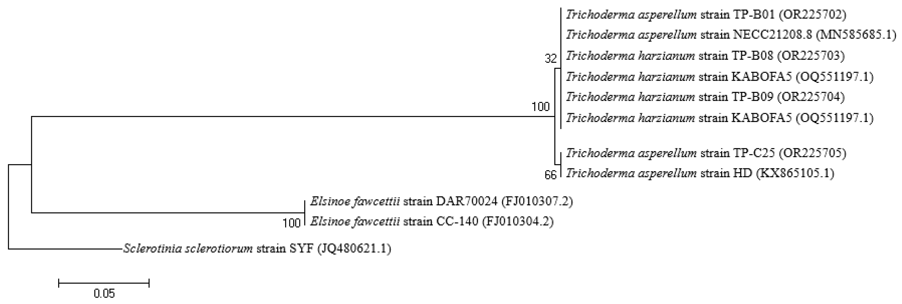
The TP-B01 and TP-C25 strains were identified as Trichoderma asperellum with accession numbers in the Genbank database of OR225702 and OR225705, respectively. Meanwhile, the TP-B08 and TP-B09 strains were identified as Trichoderma harzianum with accession numbers of OR225703 and OR225704, respectively, with 100% similarity (Figure 2).

3.5. Ability to Produce Enzymes

TP-B01, TP-B08, TP-B09, and TP-C25 strains produced chitinase (0.217–0.301 UI mL−1), endo-β-1,3-glucanase (0.019–0.064 UI mL−1), and exo-β-1,3 glucanase (0.007–0.020 UI mL−1). These enzymes are considered as a mechanism to prevent pathogen growth (Table 9).

4. Discussion

Postharvest diseases are one of the problems affecting both the fresh citrus and citrus juice industries, mainly caused by fungal pathogens [11]. Fungi can infect fruit before, during, or after harvest, but the disease develops after the fruit is harvested, causing serious economic losses to the citrus industry [44]. In this study, three highly virulent fungal strains—Colletotrichum gloeosporioides PCP-B02-A2, Pseudopestalotiopsis chinensis PCP-B02-B2, and P. camelliae-sinensis PCP-B03-A1—were identified as causal agents of fruit rot in pomelo. Importantly, all three pathogens showed rapid lesion development, indicating high aggressiveness and their potential to cause significant postharvest losses. Postharvest anthracnose of citrus fruits is caused by different Colletotrichum species, especially C. gloeosporioides [11]. Anthracnose of citrus can also be a field disease, affecting leaves and branches and causing fruit drop after flowering. However, C. gloeosporioides is a weak pathogen of citrus fruits. Asexual spores are spread by rain or overhead irrigation to developing fruits [11]. In contrast, conidia are less numerous but are airborne, participating in long-distance dispersal [11]. Lasiodiplodia sp. is a species that has been reported to cause fruit rot on many citrus trees such as lemon, pomelo, and orange [33,45,46]. The primary cell wall of the fruit is composed of approximately 10% protein and 90% polysaccharides, which can be divided into three groups: cellulose, hemicellulose, and pectin [47]. Many cell wall-degrading enzymes can be secreted by invading pathogens and use the plant cell wall as a source of nutrients, reducing postharvest shelf life and ultimately leading to undesirable quality loss, softness, and decay. Some of the notable polysaccharide-degrading enzymes include xylanase, polygalacturonase, cellulase, and α-amylase [48]. Cell wall polysaccharides in fruits play an important role in resistance to invasion by L. theobromae and some other pathogens [49]. Spore-forming fungi invade plant tissue by secreting enzymes that degrade cell wall polysaccharides (cellulases, β-galactosidases, polygalacturonases, and pectinesterases). Enzymatic cleavage of cell wall polysaccharides is the essential pathway for L. theobromae to infect harvested fruit and thus lead to disease onset and fruit softening [49]. In addition to Colletotrichum sp. and Lasiodiplodia sp., other agents responsible for fruit rot have been reported, including Phomopsis, Phytophthora, Alternaria, and Penicillium [12,50,51].
The results in Table 6 and Figures S4–S6 show that the antagonistic efficiency of Trichoderma spp. reached a high level of resistance against 3 pathogenic fungi. Table 6 shows that Trichoderma spp. strains had good antagonistic rates and inhibitory efficiency against C. gloeosporioides PCP-B02-A2 (68%), P. chinensis PCP-B02-B2 (71.1%), and Ps. camelliae-sinensis PCP-B03-A1 (55.7–57.4%) at hour 96 after inoculation. These strains were selected and identified as T. asperellum TP-B01, T. harzianum TP-B08, T. harzianum TP-B09, and T. asperellum TP-C25 (Figure 2). This result is consistent with the study by Nasreen Musheer and Shabbir Ashraf [52] where the antagonistic efficiency of the Trichoderma viride strain against pathogenic fungus C. gleosporiodes under laboratory conditions was 69.79%. Furthermore, T. koningiopsis PSU3-2 was found to inhibit the growth of C. gloeosporioides isolated from chili pepper up to 79.57% [31]. To control Pseudopestalotiopsis spp., T. asperellum also showed remarkable results [53]. On mango, T. asperellum KUFA 0042 was used to control both L. theobromae and C. gloeosporioides [54].
In addition, Trichoderma strains have a mechanism directly related to the antagonistic efficiency against pathogenic fungi with fast growth rate, good nutritional competition, living space with pathogenic fungi [55,56] and secrete chitinase and glucanase that destroy the cell wall of pathogenic fungi [57]. The study by Kucuk and Sharma [58] applied biological control measures on the antagonistic ability of T. harzianum against Aspergillus ustus (71.90%). According to Rajendiran et al. [59] the antagonism rates against A. niger, A. flavus, and A. fumigatus were 55%, 51%, and 52%, respectively. In addition, Padder and Sharma [60] showed that the antagonism rate against C. lindemuthianum causing anthracnose on beans was 59.48%. Apart from microbial antagonists, plant extracts are also candidates to control Pseudopestalotiopsis spp. [8]. Bacteria, such as Burkholderia theae [61] and B. subtilis [54], also exhibit antagonism against Colletotrichum spp. However, compared to other biological control agents, Trichoderma spp. are used the most because of their diverse traits as antagonists [62]. Notably, in the study by Widyaningsih and Triasih [63], a combined application of both T. asperellum and endophytic bacteria increased the resistance of strawberry against a disease.
Compared to chemical fungicides, which can lead to resistance and environmental hazards, the use of Trichoderma offers a sustainable, eco-friendly alternative for disease management. This supports the growing need for efficient, low-cost, and eco-friendly biocontrol options in agriculture [62], aligning with integrated pest management (IPM) principles. Field application of such strains—either as seed treatments, soil amendments, or postharvest dips—could significantly reduce the incidence of fruit rot without harmful residues [64]. Future research should evaluate the efficacy of these promising Trichoderma strains under field and storage conditions to optimize delivery methods and formulations. Integration into IPM programs could also include their combination with other biocontrol agents, such as Bacillus spp. or plant extracts, for synergistic effects which would produce promising results [65]. In summary, this study contributes novel insights into the biological control of Colletotrichum and Pseudopestalotiopsis spp. using Trichoderma spp., expanding the range of effective microbial agents and supporting the transition to sustainable disease management in citrus production systems.

5. Conclusions

The 3 selected strains of fungi causing fruit rot of pomelos included Colletotrichum gloeosporioides PCP-B02-A2, Pseudopestalotiopsis camelliae-sinensis PCP-B03-A1, and Pseudopestalotiopsis chinensis PCP-B02-A2. Twenty-five strains of Trichoderma spp. were isolated from 30 soil samples collected in 3 districts of Chau Thanh, Giong Trom, and Binh Dai, in Ben Tre province. Four strains of antagonists were selected, including Trichoderma asperellum TP-B01, Trichoderma harzianum TP-B08, Trichoderma harzianum TP-B09, and Trichoderma asperellum TP-C25 to antagonize 3 strains of pathogenic fungi C. gloeosporioides PCP-B02-A2, Ps. camelliae-sinensis PCP-B03-A1, and Ps. chinensis PCP-B02-B2. The achieved antagonistic efficiencies were 66.7–68.7%, 55.7–57.4%, and 64.2–71.1%, respectively.

Supplementary Materials

The following supporting information can be downloaded at: https://www.mdpi.com/article/10.3390/applmicrobiol5030066/s1; Figure S1: Fruit and mycelia of the fungal pathogens on the PDA medium; Figure S2: Disease symptoms caused by (a) Colletotrichum gloeosporioides PCP-B02-A2, (b) Pseudopestalotiopsis camelliae-sinensis PCP-B03-A1, and (c) Pseudopestalotiopsis chinensis PCP-B02-B2 on pomelo fruits; Figure S3: Morphology of 25 Trichoderma spp. strains; Figure S4: Antagonism of Trichoderma spp. (a) TP-B01, (b) TP-B08, (c) TP-B09, and (d) TP-C25 against Colletotrichum gloeosporioides PCP-B02-A2 on the PDA medium; Figure S5: Antagonism of Trichoderma spp. (a) TP-B01, (b) TP-B08, (c) TP-B09, and (d) TP-C25 against Pseudopestalotiopsis chinensis PCP-B02-B2 on PDA medium; Figure S6: Antagonism of Trichoderma spp. (a) TP-B01, (b) TP-B08, (c) TP-B09, and (d) TP-C25 against Pseudopestalotiopsis camelliae-sinensis PCP-B03-A1 on the PDA medium.

Author Contributions

Conceptualization, L.B.D. and N.Q.K.; methodology, L.B.D., V.M.T., N.T.N. and N.Q.K.; formal analysis, P.C.H., N.D.T., H.N.T., L.T.M.T., T.T.K.N. and L.N.T.X.; investigation, L.B.D., V.M.T., N.T.N., P.C.H., N.D.T., H.N.T., L.T.M.T., T.T.K.N. and L.N.T.X.; writing—original draft preparation, N.Q.K. and L.B.D.; writing—review and editing, N.T.P., D.T.X. and L.T.Q. All authors have read and agreed to the published version of the manuscript.

Funding

This research was funded by Ben Tre Department of Science and Technology, grant number 752/HĐ-SKHCN.

Data Availability Statement

The raw data supporting the conclusions of this article will be made available by the authors on request.

Conflicts of Interest

The authors declare no conflicts of interest. The funders had no role in the design of the study; in the collection, analyses, or interpretation of data; in the writing of the manuscript; or in the decision to publish the results.

Abbreviations

The following abbreviations are used in this manuscript:
ITSInternal Transcribed Spacer
PCRPolymerase Chain Reaction
PDAPotato Dextrose Agar
TSMTrichoderma Selective Medium

References

  1. Methacanon, P.; Krongsin, J.; Gamonpilas, C. Pomelo (Citrus Maxima) pectin: Effects of extraction parameters and its properties. Food Hydrocoll. 2013, 35, 383–391. [Google Scholar] [CrossRef]
  2. Long, T.B.; Muoi, N.V.; Tuoi, N.T.K.; Toan, H.T. Determination of physico-chemical characteristics of da xanh (Citrus maxima) and nam roi (Citrus grandis L.) pomelo by weight. Can Tho Univ. J. Sci. 2023, 59, 98–106. [Google Scholar] [CrossRef]
  3. Swamy, C.T.; Sivadurga, K.; Reddy, M.P.; Marimuthu, G.; Prashantkumar, C.S.; Premkumar, C.; Purewal, S.S. Citrus waste: A treasure of promised phytochemicals and its nutritional-nutraceutical value in health promotion and industrial applications. In Recent Advances in Citrus Fruits; Singh Purewal, S., Punia Bangar, S., Kaur, P., Eds.; Springer International Publishing: Cham, Switzerland, 2023; pp. 395–426. [Google Scholar] [CrossRef]
  4. Narasimha Rao, S.; Bhattiprolu, S.L.; Prasanna Kumari, V.; Vijaya Gopal, A. Cultural and morphological variability in Colletotrichum lindemuthianum (Sacc. and Magn.) causing anthracnose of field bean. Pharma. Innov. J. 2022, 11, 212–222. [Google Scholar]
  5. Wang, Z.; Sui, Y.; Li, J.; Tian, X.; Wang, Q. Biological control of postharvest fungal decays in citrus: A review. Crit. Rev. Food Sci. Nutr. 2020, 62, 861–870. [Google Scholar] [CrossRef]
  6. Maharachchikumbura, S.S.N.; Hyde, K.D.; Groenewald, J.Z.; Xu, J.; Crous, P.W. Pestalotiopsis revisited. Stud. Mycol. 2014, 79, 121–186. [Google Scholar] [CrossRef]
  7. Wang, S.; Mi, X.; Wu, Z.; Zhang, L.; Wei, C. Characterization and pathogenicity of Pestalotiopsis-like species associated with gray blight disease on Camellia sinensis in Anhui Province, China. Plant Dis. 2019, 103, 2786–2797. [Google Scholar] [CrossRef] [PubMed]
  8. Darapanit, A.; Boonyuen, N.; Leesutthiphonchai, W.; Nuankaew, S.; Piasai, O. Identification, Pathogenicity and effects of plant extracts on Neopestalotiopsis and Pseudopestalotiopsis causing fruit diseases. Sci. Rep. 2021, 11, 22606. [Google Scholar] [CrossRef]
  9. Ma, W.; Liu, M.; Liu, L.; Tang, Z.; Dan, Y.; Cui, X.; Yin, F. First report of Pseudopestalotiopsis theae causing leaf spot on Euonymus japonicus in China. Plant Dis. 2022, 107, 1238. [Google Scholar] [CrossRef]
  10. Shahriar, S.A.; Nur-Shakirah, A.O.; Mohd, M.H. Neopestalotiopsis clavispora and Pseudopestalotiopsis camelliae-sinensis causing grey blight disease of tea (Camellia sinensis) in Malaysia. Eur. J. Plant Pathol. 2021, 162, 709–724. [Google Scholar] [CrossRef]
  11. Martínez-Blay, V.; Pérez-Gago, M.B.; De La Fuente, B.; Carbó, R.; Palou, L. Edible coatings formulated with antifungal GRAS salts to control citrus anthracnose caused by Colletotrichum gloeosporioides and preserve postharvest fruit quality. Coatings 2020, 10, 730. [Google Scholar] [CrossRef]
  12. Ferreira, F.V.; Herrmann-Andrade, A.M.; Calabrese, C.D.; Bello, F.; Vázquez, D.; Musumeci, M.A. Effectiveness of Trichoderma strains isolated from the rhizosphere of citrus tree to control Alternaria alternata, Colletotrichum gloeosporioides and Penicillium digitatum A21 resistant to Pyrimethanil in post-harvest oranges (Citrus sinensis L. (Osbeck)). J. Appl. Microbiol. 2020, 129, 712–727. [Google Scholar] [CrossRef] [PubMed]
  13. Penet, L.; Guyader, S.; Pétro, D.; Salles, M.; Bussière, F. Direct splash dispersal prevails over indirect and subsequent spread during rains in Colletotrichum ghloeosporioides infecting yams. PLoS ONE 2014, 9, e115757. [Google Scholar] [CrossRef]
  14. Drenth, A.; Guest, D.I. 7.1 Principles of phytophthora disease management. In Diversity and Management of Phytophthora in Southeast Asia; Drenth, A., Guest, D.I., Eds.; ACIAR Monograph: Canberra, Australia, 2004; pp. 154–160. [Google Scholar]
  15. Ayaz, M.; Li, C.-H.; Ali, Q.; Zhao, W.; Chi, Y.-K.; Shafiq, M.; Ali, F.; Yu, X.-Y.; Yu, Q.; Zhao, J.-T.; et al. Bacterial and fungal biocontrol agents for plant disease protection: Journey from lab to field, current status, challenges, and global perspectives. Molecules 2023, 28, 6735. [Google Scholar] [CrossRef] [PubMed]
  16. Contreras-Cornejo, H.A.; Macías-RodríGuez, L.; Cortés-Penagos, C.; López-Bucio, J. Trichoderma virens, a plant beneficial fungus, enhances biomass production and promotes lateral root growth through an auxin-dependent mechanism in Arabidopsis. Plant Physiol. 2009, 149, 1579–1592. [Google Scholar] [CrossRef]
  17. Sood, M.; Kapoor, D.; Kumar, V.; Sheteiwy, M.S.; Ramakrishnan, M.; Landi, M.; Araniti, F.; Sharma, A. Trichoderma: The “Secrets” of a multitalented biocontrol agent. Plants 2020, 9, 762. [Google Scholar] [CrossRef]
  18. Rodrigues, A.O.; De Mio, L.L.M.; Soccol, C.R. Trichoderma as a powerful fungal disease control agent for a more sustainable and healthy agriculture: Recent studies and molecular insights. Planta 2023, 257, 31. [Google Scholar] [CrossRef]
  19. Woo, S.L.; Hermosa, R.; Lorito, M.; Monte, E. Trichoderma: A multipurpose, plant-beneficial microorganism for eco-sustainable agriculture. Nat. Rev. Microbiol. 2022, 21, 312–326. [Google Scholar] [CrossRef]
  20. Martinez, Y.; Ribera, J.; Schwarze, F.W.M.R.; De France, K. Biotechnological development of Trichoderma-based formulations for biological control. Appl. Microbiol. Biotechnol. 2023, 107, 5595–5612. [Google Scholar] [CrossRef] [PubMed]
  21. Zin, N.A.; Badaluddin, N.A. Biological functions of Trichoderma spp. for agriculture applications. Ann. Agric. Sci. 2020, 65, 168–178. [Google Scholar] [CrossRef]
  22. Madbouly, A.K. Biodiversity of genus Trichoderma and their potential applications. In Industrially Important Fungi for Sustainable Development. Fungal Biology; Abdel-Azeem, A.M., Yadav, A.N., Yadav, N., Usmani, Z., Eds.; Springer International Publishing: Cham, Switzerland, 2021; pp. 429–460. [Google Scholar] [CrossRef]
  23. Bargaz, A.; Lyamlouli, K.; Chtouki, M.; Zeroual, Y.; Dhiba, D. Soil microbial resources for improving fertilizers efficiency in an integrated plant nutrient management system. Front. Microbiol. 2018, 9, 1606. [Google Scholar] [CrossRef]
  24. Vinale, F.; Nigro, M.; Sivasithamparam, K.; Flematti, G.; Ghisalberti, E.L.; Ruocco, M.; Varlese, R.; Marra, R.; Lanzuise, S.; Eid, A.; et al. Harzianic acid: A novel siderophore from Trichoderma harzianum. FEMS Microbiol. Lett. 2013, 347, 123–129. [Google Scholar] [CrossRef]
  25. Kumar, V.; Sharma, D.D.; Babu, A.M.; Datta, R.K. SEM studies on the hyphal interaction between a biocontrol agent Tharzianum and mycopathogen Fusarium causing root rot disease in mulberry. Ind. J. Seric. 1998, 37, 17–20. [Google Scholar]
  26. Brotman, Y.; Kapuganti, J.G.; Viterbo, A.  Trichoderma. Curr. Biol. 2010, 20, R390–R391. [Google Scholar] [CrossRef] [PubMed]
  27. Gow, N.A.R.; Lenardon, M.D. Architecture of the dynamic fungal cell wall. Nat. Rev. Microbiol. 2022, 21, 248–259. [Google Scholar] [CrossRef]
  28. Gow, N.A.R.; Latge, J.-P.; Munro, C.A. The fungal cell wall: Structure, biosynthesis, and function. Microbiol. Spectr. 2017, 5, 267–292. [Google Scholar] [CrossRef] [PubMed]
  29. Kaur, R.; Kalia, A.; Lore, J.S.; Kaur, A.; Yadav, I.; Sharma, P.; Sandhu, J.S. Trichoderma sp. Endochitinase and Β-1,3-glucanase impede Rhizoctonia solani growth independently, and their combined use does not enhance impediment. Plant Pathol. 2021, 70, 1388–1396. [Google Scholar] [CrossRef]
  30. Holkar, S.K.; Ghotgalkar, P.S.; Lodha, T.D.; Bhanbhane, V.C.; Shewale, S.A.; Markad, H.; Shabeer, A.T.P.; Saha, S. Biocontrol potential of endophytic fungi originated from grapevine leaves for management of anthracnose disease caused by Colletotrichum gloeosporioides. 3 Biotech 2023, 13, 258. [Google Scholar] [CrossRef]
  31. Ruangwong, O.-U.; Pornsuriya, C.; Pitija, K.; Sunpapao, A. Biocontrol mechanisms of Trichoderma koningiopsis PSU3-2 against postharvest anthracnose of chili pepper. J. Fungi 2021, 7, 276. [Google Scholar] [CrossRef]
  32. Rahman, M.; Borah, S.M.; Borah, P.K.; Bora, P.; Sarmah, B.K.; Lal, M.K.; Tiwari, R.K.; Kumar, R. Deciphering the antimicrobial activity of multifaceted rhizospheric biocontrol agents of Solanaceous crops Viz., Trichoderma harzianum MC2, and Trichoderma harzianum NBG. Front. Plant Sci. 2023, 14, 1141506. [Google Scholar] [CrossRef]
  33. Khuong, N.Q.; Nhien, D.B.; Thu, L.T.M.; Trong, N.D.; Hiep, P.C.; Thuan, V.M.; Quang, L.T.; Thuc, L.V.; Xuan, D.T. Using Trichoderma asperellum to antagonize Lasiodiplodia theobromae causing stem-end rot disease on pomelo (Citrus maxima). J. Fungi 2023, 9, 981. [Google Scholar] [CrossRef]
  34. Vu, X.T.; Tran, V.T. Isolation and characterization of Trichoderma strains antagonistic against pathogenic fungi on orange crops. Vietnam Natl. Univ. J. Sci. Nat. Sci. Technol. 2020, 36, 98–104. [Google Scholar] [CrossRef]
  35. Silvia, M. Application of Trichoderma as an alternative to the use of sulfuric acid pesticides in the control of Diplodia disease on pomelo citrus. IOP Confer. Ser. Earth Environ. Sci. 2021, 819, 012007. [Google Scholar] [CrossRef]
  36. Burgess, L.W.; Knight, T.E.; Tesoriero, L.; Phan, H.T. Diagnostic Manual for Plant Diseases in Vietnam; ACIAR Monograph: Canberra, Australia, 2008. [Google Scholar]
  37. Roger, S.; Beasley, D. Management of Plant Pathogen Collections; Department of Agriculture, Fisheries and Forestry: Queensland, Australia, 2005.
  38. Klich, M.A. Identification of Common Aspergillus Species; Centraalbureau voor Schimmelcultures: Utrecht, The Netherlands, 2002. [Google Scholar]
  39. Kumar, K.; Amaresan, N.; Bhagat, S.; Madhuri, K.; Srivastava, R.C. Isolation and characterization of Trichoderma spp. for Antagonistic activity against root rot and foliar pathogens. Indian J. Microbiol. 2011, 52, 137–144. [Google Scholar] [CrossRef] [PubMed]
  40. Doyle, J.J.; Doyle, J.L. A rapid DNA isolation procedure for small quantities of fresh leaf tissue. Phytochem. Bull. 1987, 19, 11–15. [Google Scholar]
  41. White, T.J.; Bruns, T.; Lee, S.; Taylor, J. Amplification and direct sequencing of fungal ribosomal RNA genes for phylogenetics. In PCR Protocols: A Guide to Methods and Applications; Innis, M.A., Gelfand, D.H., Sninsky, J.J., White, T.J., Eds.; Academic Press: San Diego, CA, USA, 1990; pp. 315–322. [Google Scholar]
  42. Bell, D.K.; Wells, H.D.; Markham, C.R. In vitro antagonism of Trichoderma species against six fungal plant pathogens. Phytopathology 1982, 72, 379–382. [Google Scholar] [CrossRef]
  43. Kakraliya, S.S.; Zacharia, S.; Bajiya, M.R.; Sheshma, M. Management of leaf blight of wheat (Triticum aestivum L.) with bio-agents, neem leaf extract and fungicides. Int. J. Curr. Microbiol. Appl. Sci. 2017, 6, 296–303. [Google Scholar] [CrossRef]
  44. Zhang, J. Lasiodiplodia theobromae in citrus fruit (Diplodia stem-end rot). In Postharvest Decay; Bautista-Baños, S., Ed.; Academic Press: Cambridge, MA, USA, 2014; pp. 309–335. [Google Scholar] [CrossRef]
  45. Awan, Q.N.; Akgül, D.S.; Unal, G. First report of Lasiodiplodia pseudotheobromae causing postharvest fruit rot of lemon in Turkey. Plant Dis. 2016, 100, 2327. [Google Scholar] [CrossRef]
  46. Chen, J.; Zhu, Z.; Fu, Y.; Cheng, J.; Xie, J.; Lin, Y. Identification of Lasiodiplodia pseudotheobromae causing fruit rot of citrus in China. Plants 2021, 10, 202. [Google Scholar] [CrossRef]
  47. Juge, N. Plant protein inhibitors of cell wall degrading enzymes. Trends Plant Sci. 2006, 11, 359–367. [Google Scholar] [CrossRef]
  48. Terefe, N.S.; Gamage, M.; Vilkhu, K.; Simons, L.; Mawson, R.; Versteeg, C. The kinetics of inactivation of pectin methylesterase and polygalacturonase in tomato juice by thermosonication. Food Chem. 2009, 117, 20–27. [Google Scholar] [CrossRef]
  49. Chen, Y.; Zhang, S.; Lin, H.; Lu, W.; Wang, H.; Chen, Y.; Lin, Y.; Fan, Z. The role of cell wall polysaccharides disassembly in Lasiodiplodia theobromae-induced disease occurrence and softening of fresh longan fruit. Food Chem. 2021, 351, 129294. [Google Scholar] [CrossRef] [PubMed]
  50. Graham, J.; Feichtenberger, E. Citrus Phytophthora diseases: Management challenges and successes. J. Citrus Pathol. 2015, 2, 27203. [Google Scholar] [CrossRef]
  51. Cerioni, L.; Bennasar, P.B.; Lazarte, D.; Sepulveda, M.; Smilanick, J.L.; Ramallo, J.; Rapisarda, V.A. Conventional and reduced-risk fungicides to control postharvest Diplodia and Phomopsis stem-end rot on lemons. Sci. Hortic. 2017, 225, 783–787. [Google Scholar] [CrossRef]
  52. Musheer, N.; Ashraf, S. Effect of Trichoderma spp. on Colletotrichum gleosporiodes Penz and Sacc. causal organism of Turmeric leaf spot disease. Trends Biosci. 2017, 10, 9605–9608. [Google Scholar]
  53. Ahmmed, M.S.; Sikder, M.M.; Sarker, S.R.; Hossain, M.S.; Shubhra, R.D.; Alam, N. First Report of Pseudopestalotiopsis theae Causing Aloe Vera Leaf Spot Disease in Bangladesh. Am. J. Plant Sci. 2022, 13, 193–204. [Google Scholar] [CrossRef]
  54. Nujthet, Y.; Kaewkrajay, C.; Kijjoa, A.; Dethoup, T. Biocontrol Efficacy of Antagonists Trichoderma and Bacillus against Post-Harvest Diseases in Mangos. Eur. J. Plant Pathol. 2024, 168, 315–327. [Google Scholar] [CrossRef]
  55. De Los Santos-Villalobos, S.; Guzmán-Ortiz, D.A.; Gómez-Lim, M.A.; Délano-Frier, J.P.; De-Folter, S.; Sánchez-García, P.; Peña-Cabriales, J.J. Potential use of Trichoderma asperellum (Samuels, Liechfeldt et Nirenberg) T8a as a biological control agent against Anthracnose in mango (Mangifera indica L.). Biol. Control 2012, 64, 37–44. [Google Scholar] [CrossRef]
  56. Chaverri, P.; Samuels, G.J. Hypocrea/Trichoderma (Ascomycota, Hypocreales, Hypocreaceae): Species with Green Ascospores; Centraalbureau voor Schimmelcultures: Utrecht, The Netherlands, 2003. [Google Scholar]
  57. Wu, Q.; Sun, R.; Ni, M.; Yu, J.; Li, Y.; Yu, C.; Dou, K.; Ren, J.; Chen, J. Identification of a novel fungus, Trichoderma asperellum GDFS1009, and comprehensive evaluation of its biocontrol efficacy. PLoS ONE 2017, 12, e0179957. [Google Scholar] [CrossRef]
  58. Kucuk, C.; Kivanc, M. Mycoparasitism in the biological control of Gibberella zeae and Aspergillus ustus by Trichoderma harzianum strains. J. Agric. Technol. 2008, 4, 49–55. [Google Scholar]
  59. Rajendiran, R.; Jegadeeshkumar, D.; Sureshkumar, B.T.; Nisha, T. In vitro assessment of antagonistic activity of Trichoderma viride against post harvest pathogens. J. Agric. Technol. 2010, 6, 31–35. [Google Scholar]
  60. Padder, B.A.; Sharma, P.N. In vitro and in vivo antagonism of biocontrol agents against Colletotrichum lindemuthianum causing bean anthracnose. Arch. Phytopathol. Plant Prot. 2011, 44, 961–969. [Google Scholar] [CrossRef]
  61. Dong, Y.; Wang, X.; Feng, G.-D.; Yao, Q.; Zhu, H. A Novel Strain Burkholderia theae GS2Y Exhibits Strong Biocontrol Potential against Fungal Diseases in Tea Plants (Camellia sinensis). Cells 2024, 13, 1768. [Google Scholar] [CrossRef] [PubMed]
  62. Guzmán-Guzmán, P.; Kumar, A.; de los Santos-Villalobos, S.; Parra-Cota, F.I.; Orozco-Mosqueda, M.D.C.; Fadiji, A.E.; Hyder, S.; Babalola, O.O.; Santoyo, G. Trichoderma Species: Our Best Fungal Allies in the Biocontrol of Plant Diseases—A Review. Plants 2023, 12, 432. [Google Scholar] [CrossRef] [PubMed]
  63. Widyaningsih, S.; Triasih, U. Biological Control of Strawberry Crown Rot Disease (Pestalotiopsis sp.) Using Trichoderma harzianum and Endophytic Bacteria. IOP Conf. Ser. Earth Environ. Sci. 2021, 752, 012052. [Google Scholar] [CrossRef]
  64. Sharma, N.; Bhandari, A. (Eds.) Bio-management of Postharvest Diseases and Mycotoxigenic Fungi, 1st ed.; CRC Press: Boca Raton, FL, USA, 2020; ISBN 978-1003089223. [Google Scholar] [CrossRef]
  65. Kredics, L.; Büchner, R.; Balázs, D.; Allaga, H.; Kedves, O.; Racić, G.; Varga, A.; Nagy, V.D.; Vágvölgyi, C.; Sipos, G. Recent Advances in the Use of Trichoderma-Containing Multicomponent Microbial Inoculants for Pathogen Control and Plant Growth Promotion. World J. Microbiol. Biotechnol. 2024, 40, 162. [Google Scholar] [CrossRef]
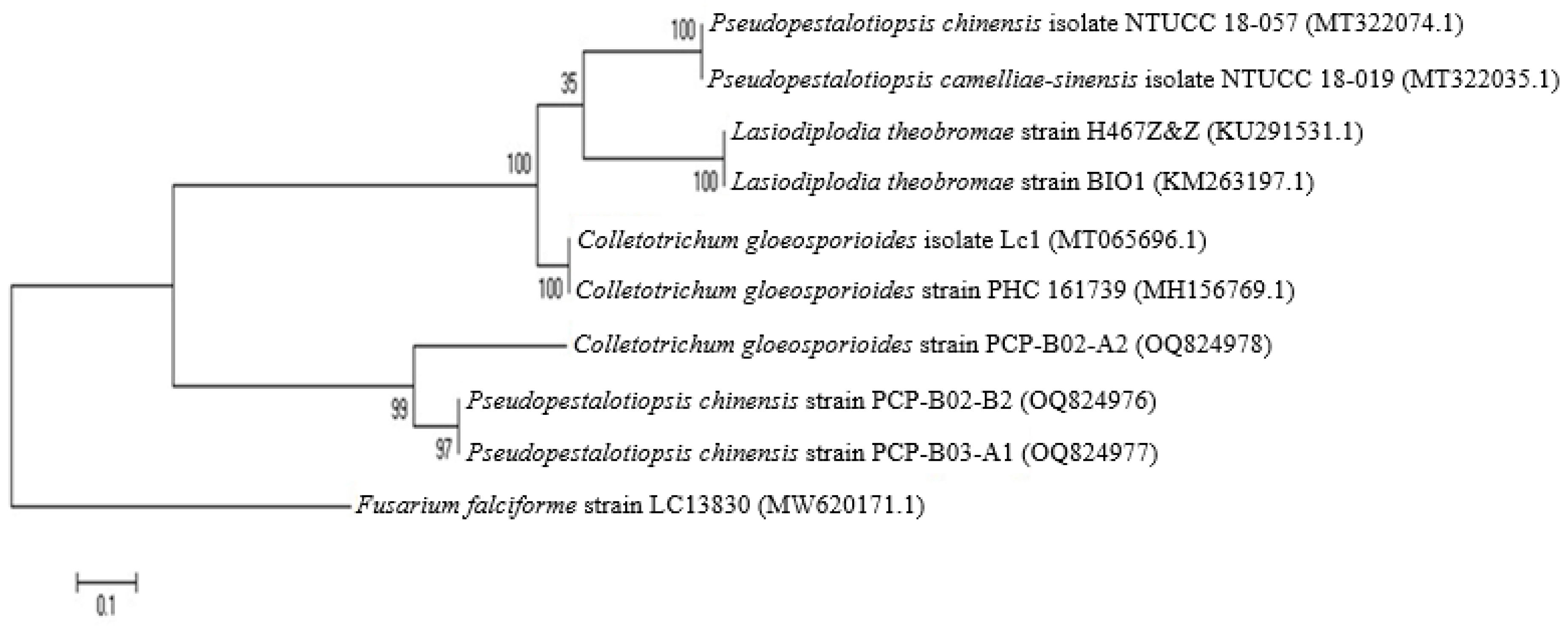
Figure 1. The phylogenetic tree based on ITS sequences of the 3 species compared to closely related strains in the GenBank database. The data are the names of the strains Lasiodiplodia pseudotheobromae PL-M01-A4-B, L. theobromae PL-M01-A6-B, L. pseudotheobromae PL-B01-A7-B along with the accession numbers of each species. The bootstrap analysis percentage level of 1000 iterations is specified at each node. Bar 0.05 substitutions for each nucleotide position. The strain Colletotrichum gloeosporioides A677 (KX463049.1) is used as an outgroup strain. The accession numbers of the GenBank sequences are specified in parentheses.
Applmicrobiol 05 00066 g001
Figure 2. The phylogenetic tree based on ITS sequences of the 2 Trichoderma spp. compared to closely related strains in the GenBank database. The bold data are the names of the strains Trichoderma asperellum TP-B01, Trichoderma harzianum TP-B08, Trichoderma harzianum TP-B09, and Trichoderma asperellum TP-C25 along with the accession numbers of each species. The bootstrap analysis percentage level of 1000 iterations is specified at each node. Bar 0.05 substitutions for each nucleotide position. The strain Sclerotinia sclerotiorum strain SYF (JQ80621.1) is used as an outgroup strain. The accession numbers of the GenBank sequences are specified in parentheses.
Applmicrobiol 05 00066 g002
Table 1. Colony characteristics of pathogenic fungal strains at 8 days of culture in PDA medium.
StrainShapeSurface ShapeSurface ColorDiameter (cm)
PCP-B02-A2Evenly radiatedThe center of the fungus was pinkish white, raised, and winding in a wavy shapeWhite-gray, initially pinkish4.35
PCP-B02-B1Almost roundThe black and gray center then wander out evenlyBlackish gray8.80
PCP-B02-B2Almost round to roundHyphae grew thick and raisedCreamy white4.57
PCP-B02-C1Evenly roundedIn the form of cotton foam, hyphae protrude from the mediumGray8.90
PCP-B02-C2RadianceThe center of the fungus was initially black, then spread evenlyDarkish gray90
PCP-B02-D1Almost roundThe hyphae grew thick with blackish hyphaeBlackish gray8.70
PCP-B03-A1Spread evenly to almost roundThe white hyphae were as porous as cotton, rising and then radiatingMilky white, light yellow in the center6.72
PCP-B03-A2Evenly roundedWhite hyphae grew thickly, gradually turned grayLight gray90
PCP-B15-A2Almost roundMycelia thickened and raised to form circlesCreamy white6.25
PCP-B17-A1RadianceThick hyphae grew close to the mediumGray8.95
PCP-B17-A2Almost roundMycelia grew thickly and were sticking to the mediumBlackish brown8.54
PCP-B17-B2RadianceThe hyphae grew thickly, protruding from the mediumGrayish white8.90
PCP-B17-C1Evenly roundedWhite hyphae spreads evenlyGray8.95
PCP-B17-C2Evenly roundedThick and silky myceliaBlack interspersed with gray90
PCP-B17-D1RadianceHyphae grew tightly togetherLight gray90
PCP-B17-D2Almost roundThe center of the fungus was initially black, then spread outward.Darkish gray8.75
PCP-C02-A1Evenly roundedMycelia grew in a circle, beyond the edge of the colony protrudingBlack interspersed with gray8.90
PCP-C02-A2Evenly radiatedBlackish brown to black from the center to the edge of the plate, fungal foci appearedBlackish gray8.85
PCP-C08-A1Almost roundWhite alternating with irregular blackish brownBlackish brown8.70
Notes: P: Pomelo; C: Colletotrichum; P: Pseudopestalotiopsis; C: Chau Thanh; B: Binh Dai; A, B, C, and D: Fruit code.
Table 2. Growth rate of fungal strains causing fruit rot on pomelo on PDA medium 3 days after transplantation.
Fungus StrainGrowth Rate of Fungal Strain (cm)
24 h48 h72 h
PCP-B02-A21.55 g2.75 j3.65 g
PCP-B02-B14.60 b7.40 d–f8.50 e
PCP-B02-B21.13 h2.73 k3.55 g
PCP-B02-C14.05 cd7.45 c–f8.80 ab
PCP-B02-C24.05 cd7.66 bc8.55 de
PCP-B02-D13.67 e7.65 bc8.65 b–e
PCP-B03-A11.15 h2.65 l3.25 i
PCP-B03-A24.55 b7.50 b–e8.80 ab
PCP-B15-A20.64 i2.70 l5.35 f
PCP-B17-A15.20 a7.60 b–d8.75 a–c
PCP-B17-A23.57 e7.70 ab8.85 a
PCP-B17-B25.25 a7.25 fg8.50 e
PCP-B17-C12.67 f7.90 a8.55 de
PCP-B17-C22.46 f7.05 gh8.65 b–e
PCP-B17-D13.80 de7.50 b–e8.60 c–e
PCP-B17-D24.15 c7.30 ef8.50 e
PCP-C02-A13.65 e7.50 b–e8.70 a–d
PCP-C02-A24.70 b6.80 i8.30 f
PCP-C08-A14.50 b6.95 hi8.65 b–e
F***
CV (%)8.205.103.00
Note: The mean numbers in the same column followed by one or more identical letters do not differ statistically at 5% on the Duncan test. (*) a 5% difference with statistical significance.
Table 3. Length of lesions (mm) on pomelo caused by Colletotrichum gloeosporioides PCP-B02-A2, Pseudopestalotiopsis camelliae-sinensis PCP-B02-B2, and Pseudopestalotiopsis chinensis PCP-B03-A1.
Fungus StrainLength of Lesions (mm) on the Fruit in Days After Infection
Day 7Day 9Day 12Day 14Day 17
PCP-B02-A212.314.2 b18.2 b21.8 bnd
PCP-B02-B212.814.1 b15.5 c16.6 c17.5 b
PCP-B03-A111.816.7 a19.9 a23.9 a28.1 a
Fns****
CV (%)10.410.27.139.273.83
Note: The mean numbers in the same column followed by one or more identical letters do not differ statistically at 5% on the Duncan test. nd: not determined, * difference by significance of 5%, ns: no significance.
Table 4. General characteristics of Trichoderma spp. on PDA after 8 days of culture.
Strain NameShapeSurface ShapeSurface ColorDiameter (cm)
TP-B01RadianceLight green granules, forming concentric circlesGreen, white8.90
TP-B02Almost roundGreen mixed with yellow granulesGreen, light yellow9.0
TP-B03Almost round to roundIn the form of green granules, the center of the fungus forms a silky white circleGreen, white9.0
TP-B04Evenly roundedIn the form of green granules, radiating evenly to form concentric circlesDark green8.70
TP-B05RadianceWhite, thickly grown, alternating green hyphaeWhite, green9.0
TP-B06Uneven roundnessFine green granules, the center of the mycelia was whiteDark green, white8.85
TP-B07Evenly roundedGreen granules, alternating whiteGreen, White8.90
TP-B08Evenly radiatedGranules were green, white and light yellowLight yellow, white9.0
TP-B09Round to near-roundIn the form of granules, the center of the colony was green, alternating in the middle of the hyphae with whiteGreen, White9.0
TP-B10Evenly radiatedSilky, thickly grown mycelia with a light yellowish color mixed with greenGreen, white, light yellow8.80
TP-B11Uneven roundnessGreen granular formDark green, white9.0
TP-B12Uneven roundnessIn the form of green hyphae, the hyphae radiated unevenlyGreen8.90
TP-G13RadianceWhite hyphae with green granules in the middleGreen, White8.95
TP-G14Almost roundWhite granules mixed with green granulesWhite, green center8.86
TP-G15Evenly radiatedLight yellow hyphae mixed with white hyphae, scattered fungus grew thickly adhering to the mediumBrownish yellow, white9.0
TP-G16Uneven roundnessFine granular form with a dark green colorDark green9.0
TP-G17RadianceIn the form of green granules, around the center of the fungus there were protruding white hyphaeGreen alters white, green in the center8.85
TP-G18Almost round to roundWhite mixed with yellowish green hyphaeLight green, yellow-brown, white8.90
TP-G19Uneven roundnessWhite hyphae thickly grew around the border with green granulesWhite, green, and green center9.0
TP-C20Almost roundThe hyphae were green mixed with whiteGreen, white8.70
TP-C21Almost round to roundGreen granules, with raised colony marginsGreen, white8.90
TP-C22Round to roundIn the form of green granules, the center of the fungus had white granulesDark green mixed with white9.0
TP-C23Evenly roundedLight yellow hyphae, the center of the fungi had green granulesYellow-brown, green center9.0
TP-C24Evenly radiatedWhite hyphae densely grew around the border with green granulesWhite, green8.90
TP-C25RadianceDense white hyphae gradually turned greenWhite mixed with green9.0
Table 5. Average growth rate of Trichoderma spp. on PDA medium.
Fungal StrainGrowth Rate of Fungal Strain (cm)
24 h48 h72 h
TP-B013.56 a–c7.76 a8.70 ab
TP-B023.06 e–g7.36 d–f8.63 ab
TP-B032.80 hi6.90 hi8.76 ab
TP-B042.36 j5.20 n7.90 e
TP-B053.63 a–c7.63 a–c8.80 ab
TP-B063.50 bc7.60 a–d8.36 c
TP-B072.43 j5.56 m8.20 cd
TP-B083.20 ef7.10 gh8.66 ab
TP-B092.66 i7.30 e–g8.80 ab
TP-B102.90 gh7.50 b–e8.66 ab
TP-B113.13 ef6.56 k8.30 cd
TP-B123.76 a7.80 a8.70 ab
TP-G133.70 ab7.60 a–d8.76 ab
TP-G143.50 bc7.70 ab8.70 ab
TP-G153.20 ef7.20 fg8.33 cd
TP-G162.16 k5.86 l7.60 f
TP-G173.03 fg6.80 ij8.26 cd
TP-G183.26 de7.40 c–f8.60 b
TP-G193.56 a–c7.83 a8.60 b
TP-C202.80 hi5.36 mn7.30 g
TP-C213.10 ef6.63 jk8.36 c
TP-C222.66 i6.86 i8.16 d
TP-C233.43 cd7.20 fg8.36 c
TP-C243.66 ab7.76 a8.66 ab
TP-C253.73 a7.66 ab8.83 a
F***
CV (%) 5.053.61
Note: The mean numbers in the same column followed by one or more identical letters do not differ statistically at 5% on the Duncan test. * difference by significance of 5%.
Table 6. Antagonistic performance of Trichoderma spp. with Colletotrichum gloeosporioides PCP-B02-A2 causing pomelo fruit rot.
Fungal StrainAntagonistic Efficiency (%)Diameter of Colletotrichum gloeosporioides PCP-B02-A2 (cm)
48 h72 h96 h48 h72 h96 h
TP-B0135.7 bc49.1 d–f67.7 a1.73 ef ± 0.061.85 b–d ± 0.051.55 d ± 0.05
TP-B0233.3 cd53.2 b–d61.4 bc1.80 de ± 0.101.70 d–f ± 0.101.85 b ± 0.05
TP-B0319.7 ef39.2 g61.1 bc2.16 bc ± 0.062.21 a ± 0.081.85 bc ± 0.05
TP-B0512.9 g36.8 g55.2 d2.35 a ± 0.052.30 a ± 0.102.15 a ± 0.05
TP-B0844.4 a60.6 a66.7 a1.50 g ± 0.101.43 g ± 0.061.60 d ± 0.10
TP-B0944.4 a52.6 b–d68.0 a1.50 g ± 0.101.72 d–f ± 0.071.53 d ± 0.12
TP-B1020.9 ef46.4 ef62.5 b2.13 bc ± 0.061.95 bc ± 0.151.80 c ± 0.10
TP-B1229.6 d51.1 cd62.5 b1.90 d ± 0.101.77 de ± 0.111.80 c ± 0.10
TP-G1322.2 e56.3 ab59.3 c2.10 c ± 0.101.59 f ± 0.081.95 b ± 0.05
TP-G1438.8 b50.5 c–e63.5 b1.65 f ± 0.051.80 c–e ± 0.101.75 c ± 0.05
TP-G1832.0 cd50.5 c–e62.5 b1.83 de ± 0.121.80 c–e ± 0.101.80 c ± 0.10
TP-G1916.6 fg45.0 f63.9 b2.25 ab ± 0.052.00 b ± 0.101.73 c ± 0.06
TP-C2432.0 cd56.0 b61.8 bc1.83 de ± 0.061.60 f ± 0.101.83 bc ± 0.06
TP-C2533.3 cd55.1 bc68.7 a1.80 de ± 0.101.63 ef ± 0.061.50 d ± 0.10
F******
CV (%)55.736.220.46.087.055.83
Note: The mean numbers in the same column followed by one or more identical letters do not differ statistically at 5% on the Duncan test. * difference by significance of 5%.
Table 7. Antagonistic performance of Trichoderma spp. with the fungal strain Pseudopestalotiopsis chinensis PCP-B02-B2 causing pomelo fruit rot.
Fungal StrainAntagonistic Efficiency (%)Diameter of Pseudopestalotiopsis chinensis PCP-B02-B2 (cm)
48 h72 h96 h48 h72 h96 h
TP-B0119.9 f38.6 g64.2 b2.05 a ± 0.052.0 b ± 0.101.30 f ± 0.10
TP-B0242.7 b–d52.4 b–d54.6 d1.46 cef ± 0.061.55 e–g ± 0.051.65 d ± 0.05
TP-B0337.5 de54.0 b53.2 d1.60 bc ± 0.101.50 g ± 0.101.70 cd ± 0.10
TP-B0518.0 f24.8 h40.9 f2.10 a ± 0.102.45 a ± 0.052.15 a ± 0.05
TP-B0849.2 a55.0 b71.1 a1.30 f ± 0.101.46 g ± 0.061.05 g ± 0.05
TP-B0933.6 e47.8 d–f66.1 b1.70 b ± 0.101.70 c–e ± 0.101.23 f ± 0.06
TP-B1045.3 ab48.9 c–f64.2 b1.40 ef ± 0.101.66 c–f ± 0.061.30 f ± 0.10
TP-B1243.3 bc46.3 ef58.7 c1.45 de ± 0.051.75 cd ± 0.051.50 e ± 0.10
TP-G1316.0 f35.6 g42.3 f2.15 a ± 0.052.10 b ± 0.102.10 a ± 0.10
TP-G1443.3 bc63.2 a67.0 b1.45 de ± 0.051.20 h ± 0.101.20 f ± 0.10
TP-G1838.3 c–e44.8 f49.1 e1.57 b–d ± 0.041.80 c ± 0.101.85 b ± 0.05
TP-G1941.4 b–d50.2 b–e60.1 c1.50 cef ± 0.101.62 d–g ± 0.071.45 e ± 0.05
TP-C2446.6 ab50.9 b–e49.6 e1.36 ef ± 0.061.60 d–g ± 0.101.83 bc ± 0.06
TP-C2535.5 e52.9 bc64.2 b1.65 b ± 0.051.53 fg ± 0.121.30 f ± 0.10
F******
CV (%)49.038.128.86.086.406.23
Note: The mean numbers in the same column followed by one or more identical letters do not differ statistically at 5% on the Duncan test. * difference by significance of 5%.
Table 8. Antagonistic performance of Trichoderma spp. with Psedopestalotiopsis camelliae-sinensis PCP-B03-A1 causing pomelo fruit rot.
Fungal StrainAntagonistic Efficiency (%)Diameter of Pseudopestalotiopsis camelliae-sinensis PCP-B03-A1 (cm)
48 h72 h96 h48 h72 h96 h
TP-B0128.0 bc50.6 a57.4 a1.90 de ± 0.101.50 f ± 0.101.63 e ± 0.06
TP-B0219.2 de24.3 f47.9 b2.13 bc ± 0.062.30 a ± 0.102.00 d ± 0.10
TP-B0318.6 de38.2 bc42.7 c2.15 bc ± 0.051.87 de ± 0.072.20 c ± 0.10
TP-B0524.2 cd32.5 c–e34.9 e2.00 cd ± 0.102.05 b–d ± 0.052.50 a ± 0.10
TP-B0817.9 e41.9 b56.5 a2.16 b ± 0.151.76 e ± 0.061.66 e ± 0.06
TP-B0926.1 bc43.0 b55.7 a1.95 de ± 0.051.76 e ± 0.15170 e ± 0.10
TP-B1031.8 ab27.6 ef40.1 cd1.80 ef ± 0.102.20 ab ± 0.102.30 bc ± 0.10
TP-B1211.0 f24.3 f37.5 de2.35 a ± 0.052.30 a ± 0.102.40 ab ± 0.10
TP-G1330.5 ab29.2 d–f34.9 e1.83 ef ± 0.062.15 a–c ± 0.052.50 a ± 0.10
TP-G1426.1 bc27.6 ef39.2 cd1.95 de ± 0.052.20 ab ± 0.102.33 bc ± 0.06
TP-G1831.8 ab34.2 cd49.9 b1.80 ef ± 0.102.0 cd ± 0.101.92 d ± 0.07
TP-G1916.7 e33.1 c–e40.1 cd2.20 b ± 0.102.03 b–d ± 0.122.30 bc ± 0.10
TP-C2435.6 a30.9 de40.1 cd1.70 f ± 0.102.10 bc ± 0.102.30 bc ± 0.10
TP-C2535.6 a40.8 b56.5 a1.70 f ± 0.101.80 e ± 0.101.66 e ± 0.06
F******
CV (%)66.954.234.16.376.716.17
Note: The mean numbers in the same column followed by one or more identical letters do not differ statistically at 5% on the Duncan test. * difference by significance of 5%.
Table 9. Ability to produce enzymes of selected Trichoderma spp.
StrainsChitinaseEndo-β-1,3-GlucanaseExo-β-1,3 Glucanase
(UI mL−1)
TP-B010.301 a0.064 a0.020 a
TP-B080.217 c0.032 c0.007 c
TP-B090.251 b0.049 b0.017 b
TP-B250.223 c0.019 d0.007 c
F***
CV (%)4.1013.38.60
Note: The mean numbers in the same column followed by one or more identical letters do not differ statistically at 5% on the Duncan test. * difference by significance of 5%.
Disclaimer/Publisher’s Note: The statements, opinions and data contained in all publications are solely those of the individual author(s) and contributor(s) and not of MDPI and/or the editor(s). MDPI and/or the editor(s) disclaim responsibility for any injury to people or property resulting from any ideas, methods, instructions or products referred to in the content.