Article

Wickerhamomyces pijperi: An Up-And-Coming Yeast with Pectinolytic Activity Suitable for Cocoa Bean Fermentation

by
Susette Freimüller Leischtfeld
1,
Alexander Hämmerli
2,
Armin Lehmann
1,
Andrea Tönz
1,
Barbara Maria Beck
1,
Jessica Wild
1,
Stefanie Weis
1,
Lukas Neutsch
2 and
Susanne Miescher Schwenninger
1,*
1
ZHAW Zurich University of Applied Sciences, Institute of Food and Beverage Innovation, Food Biotechnology Research Group, 8820 Wädenswil, Switzerland
2
ZHAW Zurich University of Applied Sciences, Institute of Chemistry and Biotechnology, Bioprocess Technology Research Group, 8820 Wädenswil, Switzerland
*
Author to whom correspondence should be addressed.
Appl. Microbiol. 2025, 5(2), 43; https://doi.org/10.3390/applmicrobiol5020043
Submission received: 4 April 2025 / Revised: 22 April 2025 / Accepted: 23 April 2025 / Published: 1 May 2025

Abstract

:
During cocoa bean fermentation, yeasts, particularly those with pectinolytic activity, contribute to pulp degradation, facilitating and accelerating fermentation. This study aimed to identify and evaluate pectinolytic yeast strains for their suitability as starter cultures in cocoa bean fermentation. A high-throughput screening of 1139 yeasts previously isolated from cocoa bean fermentations identified three strains of Wickerhamomyces pijperi with strong pectinolytic activity. These strains also reduced the viscosity of a pectin-enriched cocoa pulp simulation medium (mCPSMpc) from 23.06 ± 0.11 mPa·s (uninoculated sample) to 4.40 ± 0.14 mPa·s, 4.22 ± 0.13 mPa·s, and 4.77 ± 0.17 mPa·s after 24 h for samples inoculated with strains H312, H403, and H404, respectively. W. pijperi H403 and H404, applied in co-culture with Limosilactobacillus fermentum 223 and Saccharomyces cerevisiae H290 in 1 kg lab-scale fermentations, significantly enhanced pulp degradation, with runoff after 23.8 h reaching 12.6–13.3%, compared to 4.7% in uninoculated controls. In 20 kg fermentations in Costa Rica, the effect was less pronounced, likely due to lower inoculation rates and environmental factors. Quantitative PCR confirmed the persistence of W. pijperi H403 in fermentations. Additionally, trial cultivations in 15 L stirred-tank bioreactors successfully demonstrated the ability to produce larger biomass quantities for upscaled applications. These findings highlight W. pijperi H403 as a promising candidate for controlled cocoa fermentation, potentially accelerating biochemical changes and improving process stability.

1. Introduction

As part of the cocoa post-harvest process, cocoa beans undergo a fermentation process that results in a series of transformations, including pulp degradation, pH decrease, cotyledon inactivation, and the accumulation of flavour precursors. Successful cocoa fermentation involves yeast, lactic acid bacteria (LAB), and acetic acid bacteria (AAB). Under unfavourable conditions (e.g., insufficient aeration and lack of homogenization of the fermentation mass, poor temperature development during fermentation, prolonged fermentation, extended drying due to rain, or storage and transport of beans with high residual moisture), toxigenic fungal strains and aerobic spore forming bacteria may be present [1,2,3].
Yeasts and LAB are the most common microorganisms at the beginning of the fermentation process. Different yeast species are known for their pectinolytic activity, which causes the pulp to degrade, liquefy, and drain, creating an aerobic environment that promotes the growth of strictly aerobic AAB. Additionally, the exothermic conversion of ethanol to acetic acid by AAB in the cocoa pulp increases the temperature, further reducing the pulp’s viscosity. Pulp reduction at the beginning of the fermentation and the resulting aeration is a crucial step in the post-harvest process to ensure high bean quality. Furthermore, too high levels of pulp on the cocoa bean shells reduce the efficiency of further processing of beans into chocolate [3,4,5,6,7].
In addition to the main components of 82–86% water and 10–13% fermentable sugars, cocoa pulp contains 0.6–0.7% protein, 0.3–1.3% citrate, and 1.5–2.8% plant cell wall polymers, consisting of 0.5–1.5% pectin, 0.5–2.0% hemicellulose, 0.7–0.9% cellulose, and 0.1–0.3% lignin, depending on the origin and cocoa cultivar [8]. Plant cell wall polymers, such as pectins, contribute to cell growth, plant tissue rigidity, ion transport, and the water-holding capacity of the cell wall [9].
Pectins are a group of galacturonic acid rich polysaccharides, consisting of a backbone with partially methyl esterified α-(1,4)-linked homogalacturonic acid and branched neutral sugar side chains. They can be divided in homogalacturonan, rhamnogalacturonan I, and the substituted galacturonans rhamnogalacturonan II and xylogalacturonan [10,11].
Enzymes capable of cleaving pectins are known as pectinolytic enzymes or pectinases, which can be produced by bacteria, filamentous fungi, and yeasts [12,13,14]. They can be divided into two main groups, namely pectinesterases and depolymerases. Pectinesterases remove the methoxyl residues from pectin, whereas depolymerases cleave the main chain. Depolymerases are further subdivided into lyases, which break the glycosidic linkages by β-elimination, and polygalacturonases, which then hydrolyze the glycosidic linkages [10,15,16].
Meersman et al. [10,15,16] showed the ability of endopolygalacturonase to modify the linear pectin backbone from cocoa pulp during fermentation and a significant effect of yeast endopolygalacturonase on decreasing the viscosity of cocoa pulp. In contrast, further trials with hemicellulose-degrading enzymes did not lead to a significant reduction in the viscosity of cocoa pulp. The main pectinolytic enzyme produced from yeast is endopolygalacturonase, which hydrolyzes the α-(1,4) glycosidic linkage between non-methylated galacturonic acid residues in a random manner. In contrast, exo-polygalacturonase cleaves galacturonic acid monomers from the non-reducing end of the pectin [10,15,16].
During the fermentation of cocoa, various yeasts have been shown to secrete endopolygalacturonases, including strains of Kluyvermyces fragilis, Kluyveromyces marxianus, Kluyveromyces thermotolerans, Saccharomyces chevalieri, Saccharomyces cidri, Saccharomyces cerevisiae, Zygosaccharomyces cidro and Zygosacchoromyces fermentati, Pichua guilliermondii, Pichi kudrazevii, and Candida norwegensis, Candida nitrativorans, and Candida rugopelliculosa [6,17,18,19].
Currently, cocoa bean fermentations are primarily spontaneous processes driven by the natural microbiota present on the bean pulp mass and the surrounding environment. However, in recent years, the application of defined microbial cultures has gained significant attention as a means to stabilize this spontaneous process and introduce specific functionalities [4,20,21,22,23]. One such functionality is pectinolytic activity, which may have the potential to accelerate fermentation by enhancing pulp degradation and improving aeration [6].
An important prerequisite for the development and later commercialization of microbial cultures for food applications is the production of the corresponding microorganisms on an industrial scale and a method to stabilize the obtained biomass for transport and storage until final usage. For an optimal expression of the functional properties in the food, the starter cultures should be applied in a metabolically active state (living) and in sufficient quantities [24]. Usually, 105 to 107 active cells/g of product are used to initiate the fermentation process.
Optimizing the biotechnological process for quality-compliant and cost-effective biomass production is, therefore, essential. This optimization task is multi-factorial and comprises the following important parameters: nutrient medium (nutrients, minerals, and vitamins), possible inhibitors or unknown limiting factors in the nutrient medium, feed type and profiles (time course and addition rate of important nutrients during fermentation), overall fermentation time, oxygen supply, pH, temperature, shear stress, and foam formation [25,26,27,28]. Post-process stabilization of the biomass is usually achieved by freeze-drying, since (1) the freeze-dried product is easy to transport, store, and apply, and (2) the microbial cells have a comparatively high cell survival rate (viability) over long periods of time [29].
The aim of this study was to select pectinolytic yeast strains that are well-adapted to the cocoa bean environment, proliferate the selected culture in a pre-pilot-scale bioreactor to obtain sufficient biomass for application trials, and verify the overall impact of the culture addition on the cocoa bean fermentation process. A strain of the species Wickerhamomyces pijperi showing high pectinolytic activity was selected and tested in laboratory-scale cocoa fermentations (1 kg) and in small-scale fermentations on-farm (20 kg) in Costa Rica. Additionally, a qPCR method was developed to monitor the growth of W. pijperi. To the best of our knowledge, this study represents the first investigation of W. pijperi as a potential starter culture for cocoa bean fermentation, as well as the first description of a qPCR method for this species.

2. Materials and Methods

2.1. Yeast Strains and Maintenance of Strains

The 1139 presumptive yeast strains used in the present study are part of the ZHAW Food Biotechnology culture collection (Wädenswil, Switzerland) and were isolated from cocoa beans during fermentation and/or drying processes in Honduras [7], Bolivia and Brazil [30], Ecuador [31], and Switzerland (unpublished data) on yeast glucose chloramphenicol medium (YGC medium; Biolife Italiana S.r.l., Milan, Italy Italiana S.r.l., Milan, Italy) after 3 days incubation at 25–30 °C. For proliferation and maintenance, strains were cultivated on dichloran rose-bengal chloramphenicol (DRBC, Oxoid, Basingstoke, UK), malt extract broth (ME, Biolife Italiana S.r.l., Milan, Italy), and modified cocoa pulp simulation medium (mCPSM). The mCPSM broth was produced as described by Romanens et al. [32]. The medium contained 2.5% (w/v) fructose (Carl Roth, Karlsruhe, Germany), 2.5% (w/v) glucose (Carl Roth, Karlsruhe, Germany), 1% (w/v) citric acid (Carl Roth, Karlsruhe, Germany), 0.5% (w/v) yeast extract (Carl Roth, Karlsruhe, Germany), 0.5% (w/v) peptone bacteriological (Biolife Italiana S.r.l., Milan, Italy), 0.05% (w/v) magnesium sulphate heptahydrate (Carl Roth, Karlsruhe, Germany), 0.02% (w/v) manganese sulphate monohydrate (Carl Roth, Karlsruhe, Germany), and 0.1% (v/v) tween 80 (Carl Roth, Karlsruhe, Germany). To prevent a Maillard reaction, fructose, glucose, and citric acid were dissolved in 0.6 L dH2O (solution 1), while all other ingredients were dissolved in another 0.4 L dH2O (solution 2). Solution 1 was adjusted to pH 3.8 with 10 M NaOH before sterilization (121 °C; 15 min) aiming at pH 4.0 after sterilization. The pH was measured with a pH-Meter (pH-Meter 766, Knick Elektronische Messgeräte GmbH & Co. KG, Berlin, Germany). Both solutions were combined after being sterilized separately.

2.2. High-Throughput Screening of Yeasts for Pectinolytic Activity

The pectinolytic activity was determined as described by Silva et al. [33] on mineral medium containing polygalacturonic acid (MP5) without modifications, and as described by Martos et al. [34] on yeast nitrogen base agar (YNB) with the following modifications: pectin A with 50–75% esterification (YNBpa) and pectin C with ≥69% esterification (YNBpc) were used instead of polygalacturonic acid as the pectic substances. MP5 contained 5 g/L D(+)-glucose (Carl Roth, Karlsruhe, Germany), 5 g/L polygalacturonic acid (Carl Roth, Karlsruhe, Germany), 1 g/L yeast extract (Carl Roth, Karlsruhe, Germany), 6 g/L potassium dihydrogen phosphate (Carl Roth, Karlsruhe, Germany), 2 g/L ammonium sulfate (Fluka, Buchs, Switzerland), 0.1 mL/L mineral solution [composed of 0.0001% FeSO4 (Sigma-Aldrich, St. Louis, MO, USA); 0.02% MgSO4 (Carl Roth, Karlsruhe, Germany); 0.0001% CaCl2 (Carl Roth, Karlsruhe, Germany); 0.0002% H3BO3 (Merck, Darmstadt, Germany); 0.0002% MnSO4 (Fluka, Buchs, Switzerland); 0.0014% ZnSO4 7 H2O (Carl Roth, Karlsruhe, Germany); 0.001% CuSO4 5 H2O (Merck, Darmstadt, Germany); 0.0002% MoO3 (Sigma-Aldrich, St. Louis, MO, USA)], and 18 g/L agar bios special LL (Biolife Italiana S.r.l., Milan, Italy). YNBpa contained 6.7 g/L yeast nitrogen base (Sigma-Aldrich, St. Louis, MO, USA), 5 g/L D(+)-glucose (Carl Roth, Karlsruhe, Germany), 5 g/L pectin A (Sigma-Aldrich, St. Louis, MO, USA), and 18 g/L agar bios special LL (Biolife Italiana S.r.l., Milan, Italy). For YNBpc, 5 g/L pectin C (Carl Roth, Karlsruhe, Germany) was used.
Prior to screening, strains were revitalized using malt yeast glucose peptone (MYGP) agar plates composed of 3 g/L yeast extract (Carl Roth, Karlsruhe, Germany), 3 g/L malt extract (Biolife Italiana S.r.l., Milan, Italy), 5 g/L peptone bacteriological (Biolife Italiana S.r.l., Milan, Italy), 10 g/L D(+)-glucose (Carl Roth, Karlsruhe, Germany), and 18 g/L agar bios special LL (Biolife Italiana S.r.l., Milan, Italy), and incubated at 30 °C for 48 h. After growth on MYGP, nine strains per plate were spotted on MP5 for polygalacturonase (PG) and on YNBpa and YNBpc for unspecific pectinolytic activity using sterile wooden toothpicks. Inoculated plates were incubated at 30 °C for 48 h.
After incubation, the MP5 plates were flooded with approximately 15 mL of 5 M hydrochloric acid (HCl, Sigma-Aldrich, St. Louis, MO, USA), and YNBpa and YNBpc plates were flooded with approximately 15 mL of 1% cetyl trimethyl ammonium bromide (CTAB, Carl Roth, Karlsruhe, Germany) solution and all plates were further incubated for one hour at room temperature (around 25 °C). The enzymatic degradation of PG, pectin A, and pectin C was assessed by measuring the clear zones formed around the colonies twice along the diagonal with a caliper and calculating the radius from the average diameter. In addition, based on the observed extent of enzymatic degradation of PG and pectin, scores from 0.0 (absent) to 3.5 (strong) were assigned (Table 1). Finally, for the evaluation of pectinolytic activity, a total score for each isolate was calculated by adding the individual scores obtained for each tested medium.
The strain Hanseniaspora opuntiae 168b was used as a negative control and the strain Kluyveromyces marxianus DSM 70292 (Leibniz Institute, DSMZ-German Collection of Microorganisms and Cell Cultures GmbH, Braunschweig, Germany) was used as a positive control (both strains were tested and identified in preliminary works, unpublished data).

2.3. Identification of Selected Strains

Forty-three selected yeast strains which showed pectinolytic activity scores ≥ 1.0) were identified by MALDI-TOF MS (Bruker Daltonics, Bremen, Germany) as described by Miescher Schwenninger et al. [30]. In the present work, strains with a MALDI-TOF MS identification score of 1.700–1.999 were listed according to their identification at genus level and strains with a score of ≥2.000 with their identification at species level. Strains with score < 1.700 were additionally identified by sequencing the rRNA gene internal transcribed spacer (ITS) region. The ITS region was amplified with ITS1 and ITS4 primers according to Glass and Donaldson [35]. PCR amplicons were purified using the DNA Clean & Concentrator-5 kit (Zymo Research, Irvine, CA, USA) and subjected to Sanger sequencing (Microsynth, Balgach, Switzerland). The sequences were compared with sequences available in the NCBI nucleotide collection (nr/nt) database using BLAST (version 2.9.0).

2.4. Verification of the Ability of Pectinolytic Yeasts to Reduce Viscosity in a Pectin-Rich Cocoa Simulation Medium

Three Wicherhamomyces pijperi strains (H312, H403, H404), five S. cerevisiae strains (Y170, Y171, Y175, Y177, Y179a) and two unidentified yeast strains (Y196, Y197), as well as the strains L. fermentum 223 and S. cerevisiae H290 (previously selected for cocoa bean fermentations by Romanens et al. [21]), were cultivated in mCPSM broth overnight for approximately 15 h at 30 °C. After propagation, the total cell count of each strain was microscopically determined using a hemacytometer (Bioswisstec Ltd., Schaffhausen, Switzerland) and adjusted to 6.8 log cells/mL with mCPSM broth.
Subsequently, two bottles per strain of 60 mL mCPSM broth suppl. with 6% pectin 6 (mCPSMpc) were inoculated with 0.1 mL of the adjusted culture, aiming at a final concentration of 4.0 log cells/mL mCPSMpc). Additionally, the yeast strains H312, H403, H404, and Y170 were cultivated in co-culture with L. fermentum 223 (H312 + 223; H403 + 223; H404 + 223; Y170 + 223) and L. fermentum 223 + S. cerevisiae H290 (H312 + 223 + H290; H403 + 223 + H290; H404 + 223 + H290; Y170 + 223 + H290), whereby for 223 and H290 the cell count was similarly adjusted to 6.8 log cells/mL and 0.1 mL was used to inoculate the mCPSMpc. L. fermentum 223 and S. cerevisiae H290 were not evaluated individually, only in co-culture (223 + H290). The inoculated mCPSMpc broth was cultivated for 24 and 72 h at 150 rpm and 30 °C.
After 24 and 72 h, the dynamic viscosity (η) was determined as described in Section 2.6. The viscosity of inoculated samples was compared with non-inoculated mCPSMpc broth, which was used as a blank. Hanseniaspora opuntiae Y168a was used as a negative control, and Kluyveromyces marxianus DSM 70292, the pectinolytic activities of which were known from previous work (unpublished data), was used as a positive control (n = 1 to n = 4).

2.5. Investigation of Different Growth Conditions for W. pijperi Strains

W. pijperi strains H312 and H403 were chosen as representatives to evaluate the growth behavior of the species W. pijperi in mCPSM and yeast broth (YB), at 25 and 30 °C each at pH 4.5 and 5.0. The mCPSM was prepared as described in Chapter 2.1. The YB contained 2% (w/v) glucose (Carl Roth, Karlsruhe, Germany), 1% (w/v) yeast extract (Carl Roth, Karlsruhe, Germany), 0.8% (w/v) malt extract (Biolife Italiana S.r.l., Milan, Italy), and 0.1% (v/v) Tween 80 (Carl Roth, Karlsruhe, Germany). The pH was adjusted with 5 M HCl before sterilization. After preparation, 150 mL of the different media with pH 4.5 and pH 5.0 were transferred into sterile Schott flasks (n = 3 for each strain and condition).
Prior to inoculation, the strains were revitalized on DRBC for 48 h at 30 °C, followed by overnight propagation in ME at 30 °C. The total cell count was determined microscopically using a hemacytometer (Bioswisstec Ltd., Schaffhausen, Switzerland). Subsequently, 150 mL of broth was inoculated with 1 mL of a cell suspension containing 7.2 log cells/mL. After inoculation, the media were fermented for 48 h at 30 °C.
Samples were taken at 0, 24, and 48 h to determine (1) pH, (2) optical density at 600 nm (OD600), (3) viable cell count and (4) the ability of the cell-free supernatant, after cultivation in mCPSM at 30 °C (pH 4.5 and 5.0), to reduce the viscosity of mCPSMpc. The pH was determined using a pH-meter (pH-Meter 766, Knick Elektronische Messgeräte GmbH & Co. KG, Berlin, Germany) and the OD600 using an UV–Vis spectrophotometer (Genesys 10, Thermo Fisher Scientific Inc., Waltham, MA, USA). Viable cell counts were analyzed by performing a decimal dilution series with sterile dilution solution and dropping 20 µL of up to three suitable dilutions onto one DRBC plate, which was then tilted to allow the drops to run down it slantwise. The plates were incubated at 30 °C for 48 h. To evaluate the ability of the cell-free supernatant to reduce the viscosity of mCPSMpc, 15 mL cultivated mCPSM was centrifuged (5 min, 10,000× g, RT). Then, 10 mL of the supernatant was mixed with 20 mL of mCPSMpc and incubated for 24 h at 50 °C, followed by viscosity determination of the medium, as described in Section 2.6.
Additionally, growth curves were performed in 96-well plates using a plate reader (Synergy HTX, BioTek Instruments Inc., Winooski, VT, USA), whereby 200 µL of inoculated medium was pipetted into the wells of the plates. The OD was measured at 600 nm every 30 min over 72 h at 30 °C using the genesis 5 software (version 02.06.2010). The lag time, the maximal specific growth μmax, and the time at the maximal specific growth for each growth curve were directly calculated by the same software.

2.6. Determination of Dynamic Viscosity (η)

The dynamic viscosity (η) was determined using 50 ± 0.5 g of sample and a rotational viscometer (Haake Viscotester 550, Thermo Fisher Scientific Inc., Waltham, MA, USA), employing the MV-DIN 222-1251 cup and the MV-DIN 222-1252 rotor for medium viscous liquids at a temperature of 20 ± 1 °C (F12-MA Refrigerated/Heating Circulator, Julabo F12-CH). The Haake RheoWin Job Manager software (version 4.91.0000) was used to conduct the measurement at a shear rate ( γ ˙ ) of 200 1/s and a recording duration of 2 min with one measurement point per second. The acquired data were analyzed using the Haake RheoWin Data Manager software (version 4.91.0000).

2.7. Biomass Production in a 15 L Pre-Pilot-Scale Bioreactor

2.7.1. Inoculum Preparation, Reactor Preparation, Inoculation and Biomass Production

W. pijperi H403 was previously cultivated on a DRBC agar plate (48 h, 30 °C). Colony material was used to inoculate four 250 mL shake flasks with baffles containing 50 mL YB under sterile conditions that were cultivated overnight in a Multitron shaking incubator (Infors AG, Bottmingen, Swizerland) at 30 °C and 150 rpm. In the next step, four 500 mL shake flasks with baffles, containing 180 mL YB broth, were inoculated with the previous 250-mL culture at a ratio of 1:10 (200 mL total culture volume) and incubated under the same conditions. After purity control of the cultures under the microscope, the contents of the four flasks were combined in a 2.0 L Schott flask with a sterile connection set. This setup served as inoculum for the 15 L pre-pilot-scale bioreactor (Bilfinger Life Science GmbH, Puch bei Hallein, Austria) that was equipped with a standard pH sensor, a pO2 sensor, and biomass sensors (Incytee and Densitee; Hamilton Bonaduz AG, Bonaduz, Switzerland), as well as off-gas analytics by means of a mass spectrometer (InProcessInstruments GmbH, Bremen, Germany). Following the calibration of all sensors, the reactor was steam-sterilized in place (SIP) in accordance with the automated standard protocol (15 min, 121 °C, 2 bar). Subsequently, 12 L of YB broth was transferred into the bioreactor under sterile conditions, followed by conditioning of the optimal cultivation parameters (800 rpm stirring speed, 30 °C cultivation temperature, gassing rate of 3 vvm sterile air at 0.5 bar overpressure, and automated pH correction at 4.5). After reaching the stable state, 0.6 L of inoculum aiming at 7 log CFU/mL was transferred into the bioreactor using sterile air (t0).

2.7.2. Measurements During and After Cultivation

During the cultivation, basic and advanced parameters, such as stirring speed (in rpm), temperature (in °C), pH, pO2 (in %), CO2 off-gas (in %), air flow (in L/min), pressure (in bar), weight of reactor (in g), and biomass formation (via in-line monitoring of OD860 and permittivity/conductivity), were continuously measured and recorded using the process control system Lucullus® (Securecell AG, Schlieren, Switzerland).
The feed phase was carried out gravimetrically and dosed using a peristaltic pump. The exponential feed rate was controlled by means of a process control system (Lucullus).
The maximum specific growth rate (μmax) was calculated from the data obtained during the batch phase via the natural logarithmization of the initial biomass formed at t0 (x0) minus the biomass formed at time t [x(t)] in relation to the time (t−t0), as in the following equation):
μ m a x = d x d t = 1 x = ln x t l n ( x 0 ) t t 0
where the μmax is expressed in h−1. This physiological constant is characteristic for a strain, provided environmental conditions remain constant. The formulation accurately conveys the concept of maximum specific growth rate as a physiological parameter that remains consistent for a particular strain under unchanging conditions. t0 is the start of the batch phase in h; t is the determined time during the batch phase in h; x0 is the initial biomass formed at t0; x(t) is the biomass formed at time t.
The observed yield (Yx/s) expressed in g/g was calculated from the data obtained during the batch phase using the produced biomass (x) per used substrate concentration (s) over time by applying the following where the):
Y x / s = d x d s = x s = x t x 0 s 0 s ( t )
where the yield coefficient (Yx/s), defined as the ratio of biomass produced [x(t)] to substrate consumed [s(t)], is expressed in grams of biomass formed per gram of substrate utilized. x0 is the initial biomass formed at t0 (dimensionless); x(t) is the biomass formed at time t (dimensionless); t is the time at the end of the batch phase.
The initial feed rate (F0) with 300 g/L D(+)-glucose (Carl Roth, Karlsruhe, Germany) was calculated based on parameters determined during the batch phase (μmax and Yx/s) using the where). The biomass concentration was estimated using an empirically derived conversion factor, where the biomass concentration (g/L) = OD600 divided by two. The maintenance substrate consumption (ms) was neglected in this context.
F 0 = μ s e t Y x / s + m s · V 0 · x 0 w i n
where the initial feed rate (F0), expressed in g/h for achieving a defined specific growth rate (μset) during the feed phase, was calculated based on estimated initial parameters. It is crucial to note that the initial volume (V0) and produced biomass (x0) are specifically references to the starting point of the feed phase, not the initial inoculation conditions. Yx/s is the observed yield in g/g; ms is the substrate consumption for maintenance metabolism (neglected); win is the g substrate/g feed solution in feed 1 (300 g/L).
Automated sampling was realized using the Numera system (Securecell AG, Schlieren, Switzerland) with a sterile sampling port at the bioreactor for subsequent in-line and offline analytics. At the respective time points (listed below for each method), unfiltered biomass or filtered supernatant was filled into 1.8 mL vials and stored in a temperature-controlled rack (4 °C) for further automated analyses.
The total cell count was analyzed in-line using a Cedex HiRes cell count analyzer (Roche Diagnostics International Ltd., Rotkreuz, Switzerland) at 0, 1.2, 5.2, 6.5, 24.1 (the end of the batch phase), 31.9, 47.4, and 49.8 h of process time. The viable cell counts were determined at 0, 24.1, and 70.9 h on DRBC agar (30 °C, 48 h).
The glucose content in the medium was determined in-line every 1 to 2 h using HPLC (Agilent 1100/1200, equipped with DAD and RID detectors, Agilent Technologies, Santa Clara, CA, USA). The separation was performed on an Aminex HPX-87H ion column (BioRad, Cressier, Switzerland) with 5 mM H2SO4 (Carl Roth, Karlsruhe, Germany) as the eluent, at a flow rate of 0.6 mL/min and a column temperature of 40 °C.
Offline samples for OD600 and cell dry weight (CDW) were taken with a sterile syringe after 0, 1.2, 3.3, 5.2, 6.5, 12.7, 24.1, 26.1 28.2, 30.1, 30.9, 33.9, 37.3, 47.4, 49.8, 52.0, 54.1, 55.6, and 70.9 h. The OD600 was determined using a photometer (Thermo Fisher Scientific Genesys 10S UV–Vis, Reinach, Switzerland). To analyze CDW, a pre-dried 2 mL tube was weighed and filled with 2 mL of fermentate, which was then centrifuged for 5 min at 18,407 g and 4 °C (Eppendorf 5424 R). After discarding the supernatant, the pellet was dried for 3 days at 104 °C and weighed again.

2.7.3. Freeze-Drying of Produced Biomass

After fermentation, the biomass was harvested by centrifugation at 4 °C and 7268 g for 20 min using a Sorvall® RC 3C Plus centrifuge (Thermo Fisher Scientific Inc., Waltham, MA, USA). The biomass was then prepared for freeze-drying by adding a mixture of protective substances (confidential recipe) provided by MOGUNTIA Schweiz AG (Gossau, Switzerland). This mixture was then poured into sterile petri dishes and stored at −20 °C until freeze-drying.
The freeze-drying process was performed in two phases using a Gama 2-16 LSCplus freeze dryer (Martin Christ Gefriertrocknungsanlagen GmbH, Osterode am Harz, Germany). During phase 1, without vacuum, the sample was frozen at −25 °C for 51 min. In phase 2, with a vacuum of 0.63 mbar during all steps, the following temperature and time profile was applied: −25 °C for 5 h and 10 min, −20 °C for 20 h, −15 °C for 20 h, −10 °C for 20 h, 0 °C for 8 h, and 15 °C for 9 h. The freeze-dried yeast was stored at −20 °C until further use.
The viable cell counts of W. pijperi H403 before and after freeze-drying were determined by suspending 1 g of fresh yeast mixed with protective substances or freeze-dried yeast into 9 mL of dilution solution. Before the decimal dilution series was performed, the 1:10 diluted sample was incubated at room temperature (RT) for 1 h. Finally, 0.1 mL of adequate dilutions were pipetted onto DRBC agar plates. The plates were incubated at 30 °C for 48 h. In addition, the ability of the freeze-dried cells to reduce the viscosity of mCPSMpc was confirmed using two different approaches, using a pooled sample from three batches. Only one batch is presented exemplarily in the publication, while data from the other two batches are not included. First, 200 mL of mCPSMpc was inoculated with the powder (freeze-dried culture of W. pijperi H403) to achieve a final concentration of 5 log cells/mL. This mixture was incubated at 30 °C and 150 rpm for 48 h (n = 3) (Minitron HT, Infors AG, Switzerland). As a control, 200 mL of mCPSMpc without inoculation was incubated under the same conditions (n = 3). Viscosity measurements of the mCPSMpc were carried out at 0, 24, and 48 h, following the protocol described in Chapter 2.6. In a parallel experiment, mCPSM (without pectin) was also inoculated with 5 log cells/mL of the powder (freeze-dried culture of W. pijperi H403) and incubated for 24 h at 30 °C and 150 rpm (n = 3). As a control, uninoculated mCPSM was incubated under the same conditions (n = 3). Samples of 15 mL were taken at 0, 15, and 24 h, centrifuged (10,000× g, 5 min, RT), and sterile filtered (0.45 µm). Then, 10 mL of the resulting cell-free supernatant (CFS) was mixed with 20 mL of mCPSMpc and incubated at 50 °C for 24 h. Viscosity measurements were then conducted as described in Section 2.6.

2.8. Growth of Pectinolytic Yeast Strains on Cocoa Beans in a Micro-Scale Fermentation (20 g)

The strong pectinolytic W. pijperi strains H312, H403, and H404 were cultivated on cocoa beans in a micro-scale fermentation as single cultures (H312; H403; H404), as well as in co-culture with L. fermentum 223 (H312 + 223; H403 + 223; H404 + 223), and in co-culture with both, L. fermentum 223 and Saccharomyces cerevisiae H290 (H312 + 223 + H290; H403 + 223 + H290; H404 + 223 + H290).
First, all four yeast strains (S. cerevisiae H290 and W. pijperi strains H312, H403, and H404) and L. fermentum 223 were cultivated on mCPSM agar plates, prepared as described by Romanens et al. [32] for 2 days at 25 °C (aerobic) and 37 °C (anaerobic), respectively. Then, all strains were inoculated into 10 mL mCPSM broth and incubated for 24 h at the same conditions as in the step before. After 24 h, 100 µL culture was added to 10 mL fresh mCPSM broth and incubated overnight for ca. 15 h under the same conditions.
After the two propagation steps in mCPSM broth, the total cell count was microscopically determined using a hemacytometer (Bioswisstec Ltd., Schaffhausen, Switzerland). The targeted cell concentration was adjusted in a sterile dilution solution containing 8.5 g/L NaCl (Carl Roth, Karlsruhe, Germany) and 1 g/L peptone bacteriological (Biolife Italiana S.r.l., Milan, Italy). Growth on cocoa beans was determined in a micro-scale fermentation, previously described by Romanens et al. [33]. Therefore, 20 g of fresh cocoa beans were inoculated under sterile conditions with 400 μL of cell suspension, aiming at 6 log cells/g for L. fermentum 223, 3 log cells/g for yeasts in single culture tests, and 2 log cells/g for yeasts in LAB-yeast co-culture tests. The inoculated beans were placed into 10 mL petri dishes and incubated for 4 days at 30 °C (n = 1). The fresh cocoa beans used in the micro-scale fermentation were previously extracted from ripe cocoa fruits (Blumenbörse Schweiz Genossenschaft, Wangen bei Dübendorf, Switzerland) under sterile conditions in the laboratory and stored at −20 °C until use. Before opening, the surface of the cocoa pod was disinfected with a 70% EtOH solution. The yeast and LAB cell counts were determined at the beginning and at the end of the incubation period of 96 h on DRBC (48 h, 25 °C, aerobic) and MRS (48 h, 37 °C, anaerobic) agar plates, respectively. Trials with non-inoculated fresh cocoa beans and with cocoa beans inoculated with L. fermentum 223 or with the co-culture L. fermentum 223 + S. cerevisiae H290 were conducted as control fermentations.

2.9. Cocoa Bean Fermentation at the Lab-Scale (1 kg) with Pectinolytic Yeast Strains

To evaluate the effect of the pectinolytic activity on pulp degradation during fermentation, fermentation trials (n = 2) at a 1 kg lab-scale were carried out in flowerpots, as described by Romanens et al. [7], with the W. pijperi strains H403 and H404, each in co-culture with L. fermentum 223 and S. cerevisiae H290 (H403 + 223 + H290; H404 + 223 + H290). Additionally, a fermentation trial with the co-culture L. fermentum 223 + S. cerevisiae H290 and a control fermentation trial without inoculation (Control) were conducted.
First, as described in Section 2.8, the strains 223, H290, H403, and H404 were cultivated on mCPSM agar plates, followed by two propagations in mCPSM broth. After this, yeast and LAB cell counts were microscopically determined using a hemacytometer (Bioswisstec Ltd., Schaffhausen, Switzerland), and the targeted cell concentration was adjusted in a sterile dilution solution, as described above. For the lab-scale fermentation trials, 1 kg of fresh cocoa beans were inoculated with 2 mL of cell suspension, aiming at 6 log cells/g of each strain.
During fermentation, the pulp drainage (runoff), pulp content, pH of pulp and cotyledon, and yeast and LAB cell count were accessed daily. The analyses are described in Section 2.11.1, Section 2.11.2 and Section 2.11.3.

2.10. Cocoa Bean Fermentation at a Small-Scale (20 kg) in Costa Rica with One Selected Pectinolytic Yeast

Cocoa bean fermentations with the pectinolytic strain W. pijperi H403 in co-culture with L. fermentum 223 and S. cerevisiae H290 (H403 + 223 + H290) were conducted at the Edelkakaoinstitut and Cacao Farm Rausch < Tres Equis–Finca de Cacao > in Tres Equis, Cartago, Costa Rica during the second crop of 2019 (November–December). The performance of the co-culture H403 + 223 + H290 was compared to fermentations conducted with the co-culture composed of L. fermentum 223 and S. cerevisiae H290 (223 + H290) and with non-inoculated fermentations (control). Prior to fermentation, the strains L. fermentum 223 and S. cerevisiae H290 were individually propagated in mCPSM at industrial scale, and the biomass was harvested by centrifugation and freeze-dried by an external partner (MOGUNTIA Schweiz AG, Gossau, Switzerland). The strain W. pijperi H403 was produced at a pilot scale and freeze-dried as described in Section 2.7. After freeze-drying, the three cultures were grinded for ca. 20 s using a hand-blender coupled to a processor mill (Bamix, Mettlen, Switzerland).
After the transportation of the freeze-dried microorganisms to Costa Rica at RT, which took around 24 h, the viable cell counts were determined. For this purpose, 1 g of the frozen microorganisms was diluted 10-fold with sterile 0.9% physiological salt solution (Laboratorios Biogalenic, Soyapango, El Salvador) and incubated for 1 h at room temperature (25–30 °C). After incubation, the dilution series was continued using a sterile physiological salt solution. Then, 1 mL of the required dilutions were plated on rapid yeast and mold (RYM) PetrifilmsTM (3M Company, Maplewood, MN, USA) for W. pijperi H403 and S. cerevisiae H290 and on lactic acid bacteria (LAB) PetrifilmsTM (3M Company, Maplewood, MN, USA) for L. fermentum 223. The PetrifilmsTM were incubated at RT for 48 and 24 h, respectively.
The small-scale fermentation experiments of 20 kg were conducted in wooden boxes, which were split into two parts, divided by a removable piece of wood to facilitate mixing, with a volume of 0.021 m3 and holes at the bottom to ensure pulp drainage. The wooden boxes were surrounded by Styrofoam to prevent the cocoa from cooling down too much overnight (Figure 1).
For the fermentations, the local cocoa varieties Catie-R4, Catie-R6, Catie-R1, PMCT-58, and ICS-95 were used. The harvested cocoa pods were collected and opened manually, variety by variety, with a metal blade placed on a wooden block. A determined amount of each variety (in total 14 kg of Catie-R4 and R6; 2 kg each of Catie-R1, PMCT-58, and ICS-95) was transferred to three plastic buckets for fermentations with (223 + H290 and H403 + 223 + H290) and without (control) the addition of culture, reaching a total weight of 20 kg per bucket. The three fermentation variations (223 + H290; H403 + 223 + H290; and control) were conducted simultaneously in 3 independent runs (n = 3) and lasted 134 ± 14 h (run 1: 145 h; run 2: 138.5 h; run 3: 118.5 h).
For each strain, S. cerevisiae H290 and L. fermentum 223, 6 log CFU/g, while for W. pijperi H403, 5.8 log CFU/g were applied. The inoculum for fermentation was prepared by suspending the required number of freeze-dried microorganisms in 10 mL of sterile physiological salt solution, pouring it over the beans and mixing well with a plastic ladle. The cocoa beans were incubated in the plastic buckets (Figure 1a) for 1 h prior to being transferred to their respective fermentation boxes (Figure 1b). The cocoa beans were covered with banana leaves (Figure 1c) and transferred to the other half of the box every 24 h to ensure good homogenization of the beans.
After 5 to 6 days, when fermentation was stopped, the beans were spread out on a grid for sun drying, covered by a roof made from plastic foil to protect the cocoa from rain. The beans were left to dry to a moisture content below 7% (310 h for run 1, 270 h for run 2, and 264 h for run 3). The moisture content was measured with a grain moisture meter (HE50, Pfeuffer, Kitzingen, Germany).
Fermentation and environmental temperature were measured online every 10 min during fermentation using a datalogger (Testo 176t4) with 4 Teflon-coated temperature probes (TE Type K (NiCr-Ni), Testo AG, Mönchaltorf, Switzerland). The surface temperature of the cocoa beans during drying was measured 10 times daily with an infrared thermometer (FoodPro Infrared Food Thermometer, Fluke). Samples for bean weight, pulp content, pH of pulp and cotyledon, and cell counts were taken every 24 h during the turning/transfer of the beans during fermentation (day 0, 1, 2, 3, 4, 5, and 6), in the middle of drying (dmid), and at the end of drying (dend), using sterile plastic bags. Sampling and analysis were based on Romanens et al. [7] with minor modifications (see Section 2.11.1, Section 2.11.2, Section 2.11.3 and Section 2.11.4). The total yeast count, as well as specific quantifications of S. cerevisiae and W. pijperi, were determined by a real-time quantitative polymerase chain reaction (qPCR) at days 0, 1, 4, and fend (5 days for run 3 and 6 days for runs 1 and 2), as described in Section 2.11.5, to verify if W. pijperi could prevail during fermentation.

2.11. Analyses Carried out During Cocoa Bean Fermentations

2.11.1. Determination of Pulp Drainage (Runoff), Bean Weight, and Pulp Content During Fermentation

During lab-scale fermentations (1 kg), the pulp drainage was determined gravimetrically by weighing the accumulated pulp in the saucer of the flowerpot. Before starting, each flowerpot, saucer, and covering was weighed. The decreasing mass of the cocoa beans due to sampling was considered in the calculations.
To calculate the pulp drainage percentage, the weight of the accumulated pulp in the saucer was recorded. This weight was then divided by the weight of the remaining cocoa bean pulp mass from the previous sampling point (after sampling and removing the drained pulp), with the total weight of the empty flowerpot, saucer, and covering subtracted before division. The resulting value was multiplied by 100 to obtain the percentage (%).
The determination of the runoff was only feasible during the lab-scale trials and not during the small-scale (20 kg) fermentations. During the latter, 100 beans were weighed at each sampling timepoint to evaluate the pulp loss. To determine the pulp content during the first 3 days of fermentation, 10 beans were weighed before and after removing the pulp manually for lab-scale trials (1 kg), while 20 beans were weighed before and after for small-scale trials (20 kg). The difference in weight was calculated as the pulp content in percentage terms.

2.11.2. Determination of the pH of the Cocoa Pulp and Cotyledon

The pH of the pulp and cotyledon of the samples was measured indirectly.
To determine the pH of the pulp during the lab-scale fermentations (1 kg), 10 g of fresh beans (5 g from each replicate) were mixed with the same weight of dilution solution in a plastic bag and manually homogenized for 1 min, and then a pH probe (pH-Meter 761 Calimatic, Merck, Darmstadt, Germany) was inserted to measure the pH. To measure the pH of the cotyledon, 10 g (5 g from each replicate) of peeled beans were used and processed in the same way.
During the small-scale fermentations in Costa Rica (20 kg), 20 g fresh beans for the pulp pH and 20 g of peeled beans for the cotyledon pH were taken. Furthermore, deionized battery water (TRIDEXTILEX, Salazar y Ulloa S.A., Turrialba, Costa Rica) instead of dilution solution and the pH meter pH110 (VWR) were used. Before measuring the pH of the cotyledon, the mixture was blended with a blender for 1 min (ESGE Zauberstab M100 D, Berlin, Germany).

2.11.3. Microbiological Analysis

During the lab-scale fermentations (1 kg), viable cell counts were analyzed using the dropping method. First, 10 g of cocoa beans were weighed into a sterile stomacher bag with a filter, diluted 10-fold with dilution solution, and manually kneaded for 1 min. Following a decimal dilution series with sterile dilution solution was performed. After that, 20 µL of up to three suitable dilutions were dropped onto DRBC plates for the yeasts and MRS plates for LAB. The plates were then tilted to allow the drops to run down slantwise. The DRBC plates were incubated aerobically at 30 °C for 48 h, while the MRS plates were incubated anaerobically under the same conditions.
For small-scale fermentations (20 kg), sterile physiological salt solution was used instead of dilution solution. After the decimal dilution, 1 mL of suitable dilutions was plated on LAB Petrifilm (3 M) for the enumeration of lactic acid bacteria, while RYM (3 M) was used for the enumeration of yeasts and molds. The LAB and RYM Petrifilms were incubated for 24 and 60 h, respectively, at room temperature, which was between 25–30 °C.

2.11.4. Cut Test

At the end of the small-scale fermentation (20 kg), a cut test was performed for every fermentation by cutting 50 beans in half to determine their fermentation status. After drying, six times fifty beans per fermentation were cut in half (MAGRA 12 Cutting unit, teserba, Rüti ZH, Switzerland). The cut beans were placed in one of the following nine categories: violet, slightly fermented, well-fermented, moldy, slaty, insect-infested, germinated, over fermented, and white/pale.

2.11.5. Quantification of Yeasts by qPCR

During the small-scale fermentations (20 kg), samples were taken for the quantification of yeasts (total yeast count) and the specific quantification of S. cerevisiae and W. pijperi by qPCR. qPCR was performed using a LightCycler 480 II (Roche Diagnostics International Ltd., Rotkreuz, Switzerland) and the LightCycler® 480 Software release 1.5.0 (Roche Diagnostics International Ltd., Rotkreuz, Switzerland) for each target microorganism.
After transport to Switzerland in a frozen state using a thermal bag with ice, microbial DNA from cocoa bean samples was isolated as described by Schwendimann et al. [36] using the DNeasy PowerFood Microbial Kit (Qiagen, Hilden, Germany) with the modifications proposed in their study [36].
For one reaction, 0.75 µL of forward primer (10 µM), 0.75 µL of reverse primer (10 µM), 10 of µL of Fast Evagreen® qPCR Master Mix (2×) (Biotium, Fremont, CA, USA), 3.5 µL of double distilled water (ddH2O), and 5 µL of DNA template were mixed. All primers were purchased from Microsynth AG (Balgach, Switzerland). Non-template controls (NTCs) were carried out by replacing the DNA template with ddH2O.
Cycle conditions were applied as follows: initial preincubation at 95 °C for 10 min (ramp speed 4.4 °C/s), followed by 45 cycles with a sequence of 95 °C for 15 s (ramp speed 4.4 °C/s), with a melting temperature according to Table 2. for 60 s (ramp speed 2.2 °C/s), and 72 °C for 30 s (ramp speed 2.2 °C/s). The final cooling step was performed at 40 °C for 10 s (ramp speed 2.2 °C/s). Each qPCR reaction was technically replicated twice in the same reaction plate.
For standard curves, DNA extraction from yeasts cultivated in ME (30 °C, 15 h) was performed using the Dneasy® Blood & Tissue Kits (Qiagen, Hilden, Germany) according to the manufacturer’s instructions. For the quantification of the total yeast count and for S. cerevisiae, the strain S. cerevisiae DSM 1334 (Leibniz Institute, DSMZ-German Collection of Microorganisms and Cell Cultures GmbH, Braunschweig, Germany) was used to perform the standard curves, while for W. pijperi, the strain W. pijperi H312 was used to perform the standard curves. Standard curves were generated by plotting the cycle threshold (CT) values from qPCR assays conducted on a DNA dilution series (0 to 7 log cells/mL) against the logarithmic concentration of cells per milliliter (log cell/mL) (n = 6). First, the detection limit was determined using the Kruskal–Wallis test followed by a pairwise Wilcoxon test with Holm correction using R (version 4.2.2) and RStudio (version 2022.07.1). The detection limit established if significant differences between concentration groups were detected (Figure S1a–c in the Supplementary Materials). Subsequently, a linear regression was performed from 1 to 7 log cells/g (Figure S1d–f in the Supplementary Materials).
The slopes and intercepts from the linear regression, necessary for the calculation of cell concentration (x) in the samples based on the CT values (y), R2 and the efficiency of the assays, are summarized in Table 3.

2.12. Statistical Analyses

Statistical analyses were performed using R Studio (version 2022.07.1), with a significance level set at 0.05. The normal distribution of the data was assessed using the Q–Q plot and/or the Kolmogorov–Smirnov test. If the data followed a normal distribution, an ANOVA was conducted, followed by Tukey’s HSD post hoc test. If the data did not follow a normal distribution, the Kruskal–Wallis test was used, followed by a Wilcoxon post hoc test for variance analysis. For repeated trials, the mean and standard deviation were calculated.

3. Results

3.1. Selection of Yeasts with Pectinolytic Activity

3.1.1. In Vitro Determination of the Pectinolytic Activity of Yeasts on Nutrient Media Containing Pectic Substances

A total of 1139 presumptive yeast strains previously isolated during cocoa bean fermentations and/or drying processes in Honduras [7], Bolivia and Brazil [30], Ecuador [31], and Switzerland (unpublished data) were screened for pectinolytic activity on MP5, YNBpc, and YNBpa. A total of 973 (85.4%) strains showed no pectinolytic activity on these media (total score 0.0).
For 159 strains (14.0%), no clear zone around the colony was observed; however, a noticeable clearing in the medium around the colony was observed (minimal activity). This minimal activity was seen on at least 1 of the 3 media (total score 0.5) for 123 of the strains (10.8%), on 2 media (total score 1.0) for 12 of the strains, or on all 3 media (total score 1.5) for 24 of the strains. Among the 123 strains that showed activity on only 1 of the 2 media (total score 0.5), 121 strains showed activity on MP5, while 2 showed activity on YNBpc. Of the strains with a total score of 1.0, all showed pectinolytic activity on MP5 and then either on YNBpc (7 strains) or YNBpa (5 strains). Strains with a total score ≥ 1.0 were identified using MALDI-TOF MS. All strains scoring 1.0 and 1.5 were identified as Saccharomyces sp. or S. cerevisiae, except for strain Y114, which was identified as H. opuntiae (Table 4).
Additionally, two strains (0.2%) showed a clear degradation zone (1.0–4.9 mm) around the colony on MP5 and YNBpc (total score 3.0). These two strains could not be identified using MALDI-TOF MS; sequencing of the ITS region identified them at the genus level as Ustilago sp. (Table 4). Another two strains (0.2%) showed a moderate zone (5.0–9.9 mm) on MP5 and a weak zone on at least one other medium (total score 4.0 and 5.5). These strains could not be completely purified, and identification using both MALDI-TOF MS and sequencing did not yield reliable results (Table 4).
Finally, three strains (0.3%), i.e., H312, H403, and H404, identified as W. pijperi, showed strong pectinolytic activity on all three media (total score 10.5) (Table 4).
The negative control remained negative on all media, and the positive control showed moderate activity on all media (total score 7.5).
A total of 10 strains (the 3 W. pijperi strains H312, H403, and H404 with a total score of 10.5, a total of 7 S. cerevisiae strains, 5 strains with a total score of 1.5, and 2 strains with a total score of 1.0), labeled in Table 4 with a star (*), were selected for the in vitro verification of their ability to reduce the viscosity of a pectin-rich medium.

3.1.2. In Vitro Determination of the Pectinolytic Activity of Yeasts Based on Their Ability to Reduce the Viscosity of a Pectin-Rich Medium

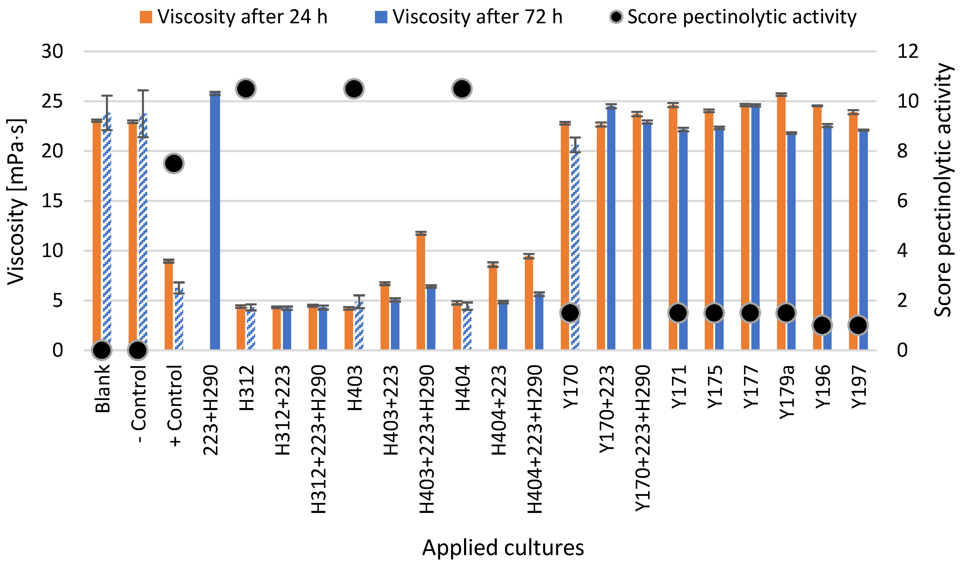
It was observed that the viscosity of the uninoculated pectin-rich medium (blank) and the medium inoculated with H. opuntiae Y168a (the negative control) were similar, with values of 23.06 ± 0.11 mPa s after 24 h and 22.95 ± 0.14 mPa s after 24 h, respectively. The viscosity remained at the same level after 72 h (23.84 ± 1.74 mPa s for the blank and 23.75 ± 2.35 mPa s for the negative control) without significant differences (p = 0.95) (Figure 2). The viscosity of the sample fermented with the co-culture L. fermentum 223 + S. cerevisiae H290, both of which were not known to have pectinolytic properties, was at the same level as the blank (25.78 ± 0.16 mPa s). In contrast, the strain K. marxianus DSM 70292 (the positive control) was able to significantly reduce the viscosity to 8.96 ± 0.14 mPa s after 24 h and 6.27 ± 0.56 mPa s after 72 h, compared to the blank (p < 0.05).
The three W. pijperi strains (H312, H403, and H404) which showed a high total score for pectinolytic activity (Table 4) were able to reduce the viscosity of mCPSMpc to 4.40 ± 0.14 mPa s, 4.22 ± 0.13 mPa s, and 4.77 ±0.17 mPa s, respectively, after 24 h when fermented in single cultures (Figure 2). After 72 h, the viscosity remained within the same range (4.31 ± 0.30 mPa s, 4.88 ± 0.65 mPa s, and 4.44 ± 0.37 mPa s for H312, H403, and H404, respectively), and was significantly lower than the viscosity of the blank (23.84 ± 1.74 mPa s) and the negative control (23.75 ± 2.35 mPa s) after 72 h (p < 0.05). In co-culture with strain L. fermentum 223 and with the combination of strains L. fermentum 223 + S. cerevisiae H290, the viscosity for the co-cultures H312 + 223 and H312 + 223 + H290 remained in a similar range after 24 h (4.32 ± 0.09 mPa s and 4.49 ± 0.09 mPa s, respectively) and 72 h (4.22 ± 0.17 mPa s and 4.30 ± 0.020 mPa s, respectively). As can be seen in Figure 2, the samples with W. pijperi strains H403 and H404, in co-culture with L. fermentum 223 (H403 + 223; H404 + 223) and L. fermentum 223 + S. cerevisiae H290 (H403 + 223 + H290; H404 + 223 + H290), showed higher viscosity after 24 h compared to samples with single cultures, which had values of 6.70 ± 0.14 mPa s for H403 and 8.61 ± 0.22 mPa s for H404 in co-culture with strain 223, and 11.75 ± 0.14 mPa s and 9.46 ± 0.22 mPa s in co-culture with strains 223 + H290. The viscosity after 72 h was lower than after 24 h but tendentially higher than the viscosity of samples fermented with the single cultures (i.e., H403 and H404 alone).
Single culture fermentations with S. cerevisiae strains with a total score for pectinolytic activity of 1.5 (Y170, Y171, Y175, Y177, and Y179a) and 1.0 (Y196 and Y197) were not able to reduce the viscosity of mCPSM to levels comparable to the positive control or to fermentations with W. pijperi strains H312, H403, and H404 (Figure 2). The only S. cerevisisae strain able to significantly reduce viscosity when compared to the blank control was strain Y170. When S. cerevisiae Y170 was applied as a single culture, the viscosity was tendentially lower after 24 h (22.81 ± 0.13 mPa s) and significantly lower after 72 h (20.62 ± 0.74 mPa s) (p = 0.008). In co-culture with L. fermentum 223 (Y170 + 223) and L. fermentum 223 + S. cerevisiae H290 (Y170 + 223 + H290), the viscosity was at the same level as the blank or other samples fermented with single S. cerevisiae strains. For samples fermented with S. cerevisiae strains, the viscosity ranged between 22.67 and 25.67 mPa s after 24 h and between 21.81 and 24.59 mPa s after 72 h (Figure 2).

3.2. Growth of W. pijperi Under Different Conditions

Growth curve tests for W. pijperi strains H312 and H403 were conducted in both YB and mCPSM media at 25 °C and 30 °C, and at pH values of 4.5 and 5.0. The results for the cell count, OD600, pH, and ability of the supernatant to reduce the viscosity in mCPSMpc are displayed in Table 5.
During cultivation, the cell counts of the strains increased from 3.4–4.5 log CFU/mL initially to 6.5–7.6 log CFU/mL after 24 h and to 6.6–8.0 log CFU/mL after 48 h. After 24 h, rather higher values were observed for trials at 30 °C (7.4 ± 0.3 log CFU/mL) compared to 25 °C (7.0 ± 2.4 log CFU/mL), except for the trial in mCPSM at 25 °C and pH 4.5 for strain H312 (7.5 ± 0.1 log CFU/mL), which showed equally high cell counts as samples fermented at 30 °C.
However, the OD600 after 24 h was higher for samples cultivated in YB at 25 °C (1.66 ± 0.06) compared to 30 °C (1.46 ± 0.11). In mCPSM, no clear tendency could be observed between fermentations at 25 °C and 30 °C, regardless of the strain. After 48 h, the OD600 was tendentially higher for all samples at 25 °C (2.03 ± 0.31) than at 30 °C (1.76 ± 0.18) regardless of strain, medium, or pH. Significant differences between trials carried out at 25 and 30 °C were only found for experiments in mCPSM (2.32 ± 0.11 at 25 °C and 1.86 ± 0.20 at 30 °C).
The pH after 24 h appeared to be influenced by the initial pH, with samples that started at pH 4.5 showing significantly lower pH levels than those that started with a pH of 5.0. After 48 h, the pH was higher in samples cultivated in YB at 25 °C regardless of strain and in general rather higher in experiments that started with pH 5.0.
The viscosity of mCPSMpc (pectin-rich medium) was significantly lower for supernatants from experiments carried out at 30 °C after 24 and 48 h compared with experiments prepared with the supernatant of non-cultivated media (0 h) and with experiments conducted at 25 °C. The viscosity of supernatants generated at 25 °C had high variability for strain H312 and less variability for H403. At 25 °C, a small difference in viscosity can be observed after 48 h for the trials conducted in mCPSM.
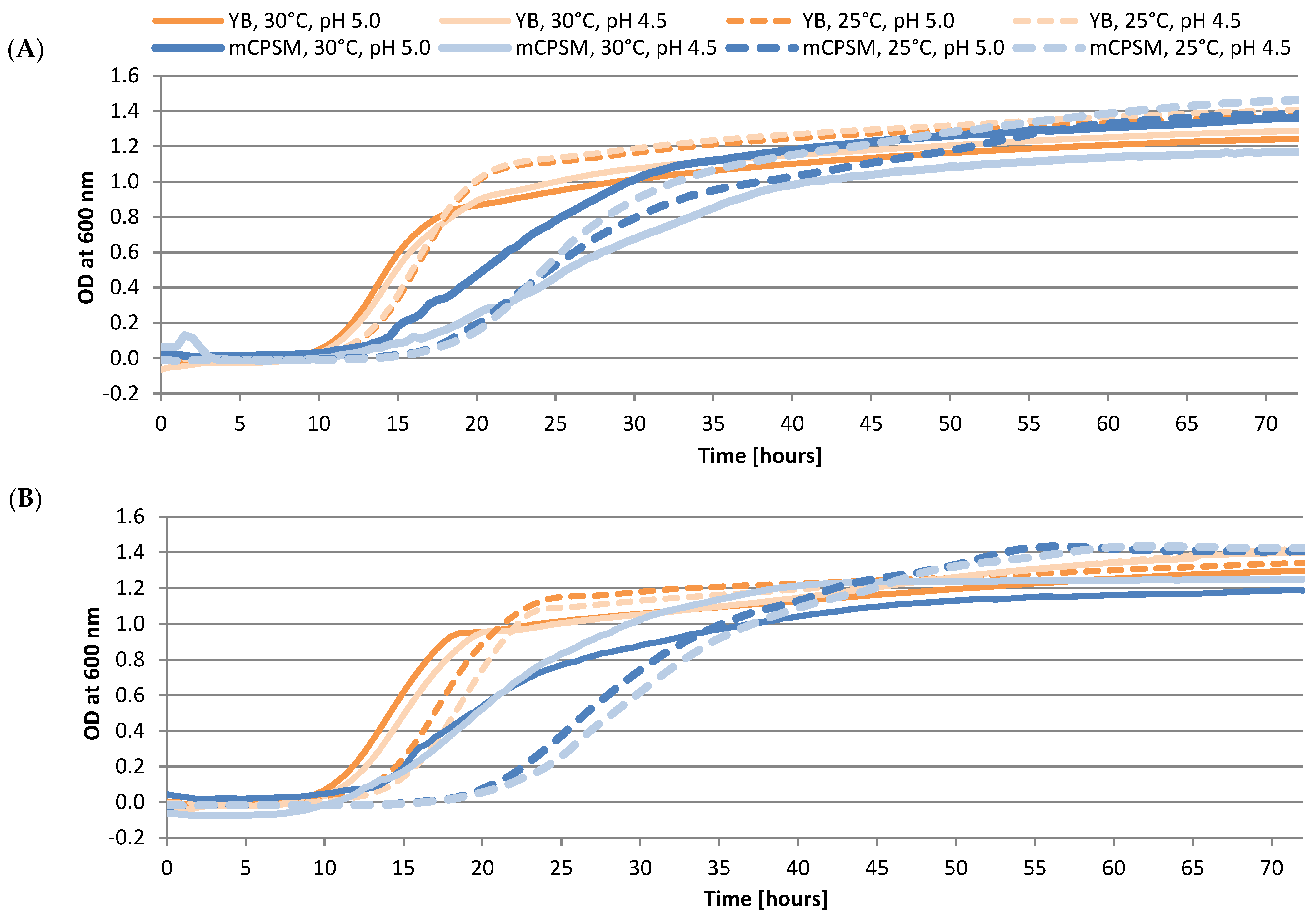
As can be observed in Figure 3 and in Table S1 in the Supplementary Materials, for both W. pijperi strains H312 and H403, the lag phase was significantly shorter in trials conducted in YB at 30 °C (varying from 10.52 ± 0.12 and 11.17 ± 0.27 h) than in YB at 25 °C or in mCPSM at 25 or 30 °C. In contrast, the longest lag phases were observed in experiments carried out in mCPSM at 25 °C (varying from 18.26 ± 0.34 and 22.09 ± 0.84 h) and in mCPSM at 30 °C at pH 4.5 for strain H312 (20.45 ± 4.23 h) (p < 0.05).
Similar tendencies were observed for OD600 measured during the experiments in microplate reader and during cultivation in shake flasks, where external OD determination was performed, although absolute values differed between methods (Figure 3 and Table S1 in the Supplementary Materials). Higher OD600 values after 24 h were measured in YB, independent of strain, temperature, or pH (1.11 ± 0.02 at 25 °C and 0.98 ± 0.03 at 30 °C), followed by experiments in mCPSM at 30 °C (0.75 ± 0.03), except for H312 at pH 4.5 (0.4 ± 0.17). The OD600 values after 24 h for the experiment with strain H312 in mCPSM at 30 °C and pH 4.5, as well as for all experiments in mCPSM at 25 °C (0.47 ± 0.01 for strain H312 and 0.25 ± 0.08 for strain H403), were significantly lower compared to other samples (p < 0.05). After 48 h, significant differences in OD600 were observed only between the experiment in YB at 25 °C for strain H312 (1.28 ± 0.03) and the experiment in mCPSM at 25 °C for strain H403 (1.29 ± 0.00), which were both significantly higher than the OD observed for strain H312 in mCPSM at 30 °C and pH 4.5 (1.07 ± 0.10) (p < 0.05). All other samples showed no significant differences (1.21 ± 0.05) (p > 0.05). At the end of the cultivation of 72 h, a tendency towards higher OD600 values for experiments carried out at 25 °C could be observed. Experiments in mCPSM at 30 °C and pH 4.5 for strain H312 showed the lowest OD600 values (1.17 ± 0.13) and experiments in mCPSM at 25 °C and pH 4.5 for H312 showed the highest values (1.46 ± 0.04).
Trials carried out in YB showed higher maximal specific growth (µmax) (2.48 ± 0.17) and a shorter time until µmax (15.68 ± 1.77 h) than trials in mCPSM (µmax of 1.64 ± 0.18 and a time until µmax of 23.40 ± 3.03 h). This was true except for trials in mCPSM at 30 °C and pH 5.0 (µmax of 2.50 ± 0.29 and a time until µmax of 15.68 ± 0.35 h).

3.3. Production of a Freeze-Dried Ready-to-Use Culture of W. pijperi H403

In order to produce a ready-to-use freeze-dried culture of the pectinolytic strain W. pijperi H403 for application trials in field, cells were grown in a 15 l pre-pilot bioreactor. An intensified fed-batch process strategy was used to increase the biomass quantity of W. pijperi H403 to an optimal extent, in the controlled environment of a fully automated STR bioreactor. The fed-batch process was carried out using YB medium at pH 4.0, with constant temperature set at 30 °C, 300 rpm stirring speed, and 3 vvm aeration.
During the batch phase, a maximum specific growth rate (μmax) of 0.46 h−1 and a biomass/substrate yield coefficient (Yx/s) of 0.62 were observed. The overall feeding phase lasted from 24 to 71.3 h of process duration, with six different subphases, each with identical initial feed rates, to achieve lower growth rates (at higher biomass levels) and thereby account for a deceleration in cell proliferation over time, as visualized in Table 6.
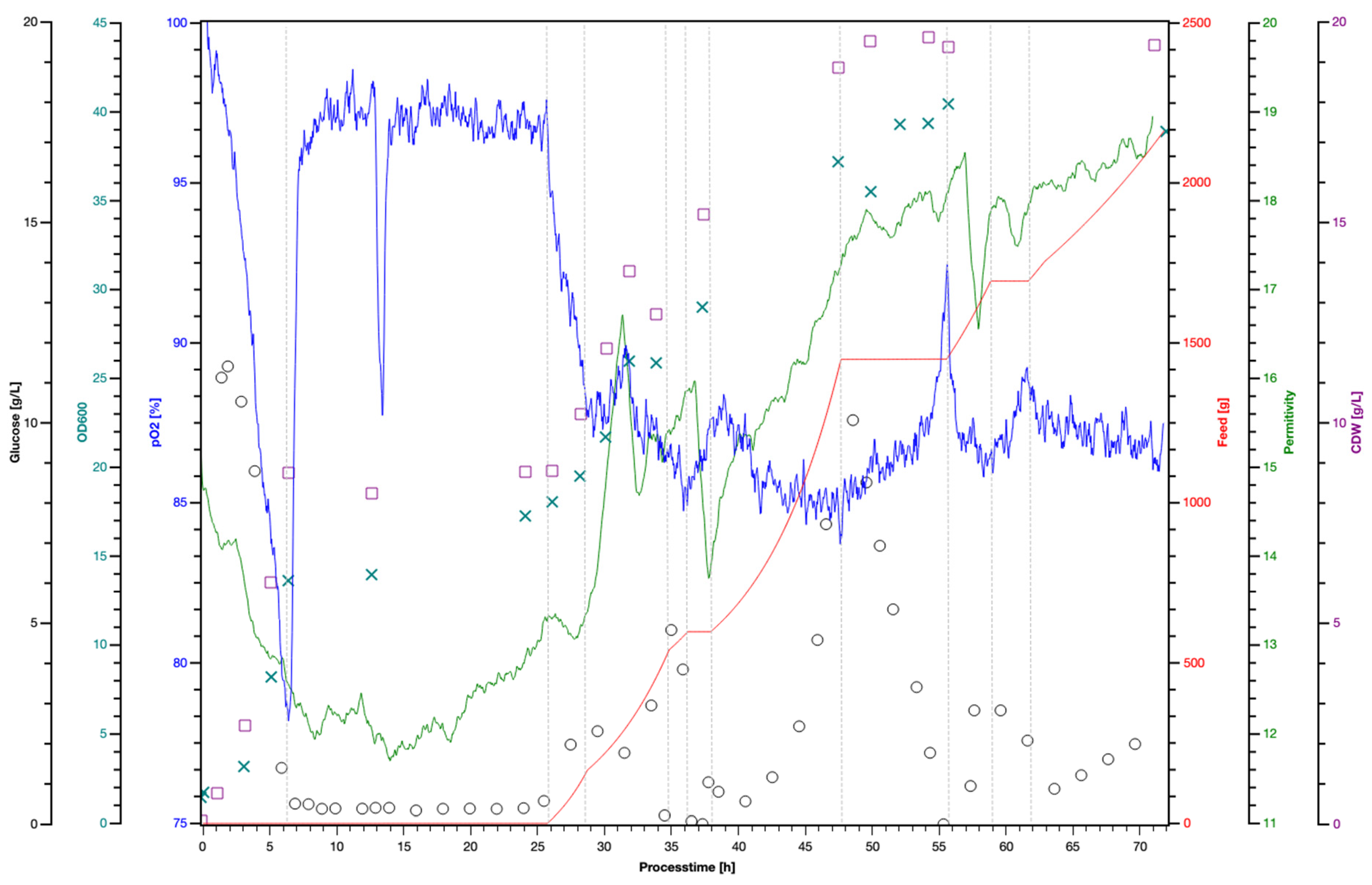
As visualized in Figure 4, glucose levels declined rapidly during the batch phase, reaching 0.4 g/L after 7 h, which coincided with a sharp increase in dissolved oxygen (pO2) to over 95% at the batch end. Biomass formation, as measured by cell dry weight (CDW) and visualized in crosses, increased from 0.1 g/L initially to 19.4 g/L at the batch end. A negative deflection of the pO2 signal after 14 h cultivation time was due to the installation of the feed inlet on the bioreactor.
Glucose accumulation was observed relatively soon upon the initiation of feeding after 24 h with a defined initial feed rate (F0) and growth rate (μset), as shown in Table 6. A feed pause (after 37 h process time) was implemented, aimed at the consumption of excessive amounts of glucose (overfeeding).
The initial feed rate (F0) set in feed-phase 1 was periodically reset to the start value of 36.36 g glucose/h for each new feed phase (2–6) to avoid overfeeding, which resulted in a new, reduced growth rate (μset). The biomass concentration used to calculate the growth rate was determined using the OD600 measurements, which were approximately double the CDW values (we confirmed that the typical method of extrapolation is based on empirical values).
The yeast cell concentration after inoculation measured with both an automated cell counter (Cedex) and by plating the sample on DRBC was 7.1 log CFU/mL measured with the Cedex and 7.0 ± 0.4 log CFU/mL by plate count. At the beginning of the feed (24.1 h process time) 8.3 log CFU/mL and 7.7 ± 0.1 log CFU/mL were detected, respectively. At the end of the feed phase (71.3 h process time) 8.5 log CFU/mL and 8.3 ± 0.5 log CFU/mL, respectively, were determined.
A strong fruity/banana odor was noted during pre-culture and inoculum preparation, as well as during the first 24 h of fermentation. This olfactory characteristic was not detected after the 24 h timepoint.
After the feed phase (from 24 to 71.3 h), the biomass was harvested, centrifuged, and freeze-dried. The viable yeast count values in the biomass after centrifugation and after drying were 9.2 ± 0.1 log CFU/g and 8.2 ± 0.2 log CFU/g, respectively.
The confirmation of the pectinolytic activity of the freeze-dried W. pijperi H403 was assessed in a pooled sample of three batches by measuring viscosity reduction in mCPSMpc under different experimental conditions (Table 7). When using freeze-dried powder (5 log cells/mL), viscosity significantly decreased from 11.2 ± 1.2 mPa.s to 4.6 ± 1.2 mPa.s after 24 h and to 3.3 ± 0.1 after 48 h, whereas the control showed only minor fluctuations (10.6 ± 0.4 to 12.8 ± 2.6 mPa.s). In the experiment using CFS, the viscosity dropped from 6.9 ± 0.9 mPa.s to 3.2 ± 0.2 mPa.s in the H403 treatment after 15 h and further declined to 2.4 ± 0.2 mPa.s after 24 h. A decrease in viscosity was also observed in control samples, although it was less pronounced (from 6.9 ± 0.9 to 4.6 ± 0.3 after 15 h).

3.4. In Vitro Growth of Pectinolytic Yeast on Cocoa Beans

During the 4-day cultivation of the pectinolytic strains W. pijperi H312, H403, H404, and S. cerevisiae Y170 on cocoa beans, both in single cultures and in co-cultures with L. fermentum 223 and the strain combination L. fermentum 223 + S. cerevisiae H290, it was observed that the cell count of the pectinolytic yeasts increased in all samples. In single cultures, the cell count rose from 3 log CFU/g to 5.6–6.9 log CFU/g, in co-culture with L. fermentum 223 from 2 log CFU/g to 4.3–7.7 log CFU/g, and in co-culture with L. fermentum 223 + S. cerevisiae H290 from 2 log CFU/g to 6.4–7.7 log CFU/g. (Table 8).
In samples inoculated with the single culture H290 and with the co-cultures 223 + H290, H312 + 223 + H290, H403 + 223 + H290, and H404 + 223 + H290, the strain S. cerevisiae H290 could also be detected on DRBC in all samples in a range between 7.7 and 8.1 CFU/g (Table 8). Due to differences in colony patterns, it was possible to differentiate the W. pijperi strains from the S. cerevisiae strain H290. In samples inoculated with the co-culture Y170 + 223 + H290, it was not possible to differentiate both S. cerevisiae strains. In this case the total yeast cell count was determined (7.8 log CFU/g). The strain L. fermentum 223 was also detected in a range between 7.7–8.1 log CFU/g in all samples inoculated with co-cultures containing strain 223 (Table 8).
Non-inoculated samples showed values below the method’s detection limit of 2.7 log CFU/g for all microorganisms tested (Table 8).

3.5. Cocoa Bean Fermentation with Pectinolytic Yeasts in Lab-Scale (1 kg) and Small-Scale (20 kg) Fermentation

3.5.1. Impact of Pectinolytic Yeasts on Pulp Degradation During Cocoa Bean Fermentation

Figure 5A graphically presents the results of the gravimetric determination of pulp runoff during the lab-scale fermentation for the uninoculated control and the samples inoculated with L. fermentum 223 + S. cerevisiae H290 alone or in co-culture with the pectinolytic W. pijperi strains H403 or H404. The percentages of pulp drainage in relation to the total cocoa bean mass of each variation are shown as colored bars at different measurement times. It can be observed that pulp drainage was similar in the first 7.8 h for all samples but showed significant differences after 18.8 and 23.8 h (p < 0.05). In the control sample, 4.7 ± 0.0 and 1.4 ± 0.0% pulp runoff was detectable, in sample H290 + 223, 9.1 ± 1.0 and 2.6 ± 0.1% pulp runoff was detected, in sample 403 + 223 + H290, 13.3 ± 0.0% and 2.1 ± 0.6 pulp runoff was detected, and in sample H404 + 223 + H290, 12.6 ± 0.4% and 3.7 ± 0.6% pulp runoff was detected after 13.8 and 23.8 h, respectively.
Figure 5B shows the results of the gravimetric determination of pulp content of the samples fermented in lab-scale (1 kg, patterned bars) and small-scale (20 kg, solid bars) systems. The pulp content is expressed as a percentage of the total bean pulp sample at different measurement times. At 0 h, a gravimetric triplicate determination of the total bean pulp mass showed a pulp content of 44.1 ± 4.2%. It was also apparent that within the first 24 h, the pulp content of all samples was reduced to between 22.7 and 30.9%. Figure 5B further shows that pulp degradation was particularly active during the first three days, during which the pulp content in all samples decreased by at least half of the initial level. After 5 days, the pulp content in all samples ranged between 9.3% and 12.8%.
In lab-scale fermentations, the samples inoculated with the pectinolytic W. pijperi strains H403 or H404 in co-culture with L. fermentum 223 + S. cerevisiae H290 (H403 + 223 + H290 and H404 + 223 + H290) always showed lower pulp content than the samples fermented without pectinolytic strains (the control fermentation and 223 + H290). For small-scale fermentations, the pulp content of the cocoa beans was measured in triplicate during the first two days of the fermentation process and in duplicate on day 3 of the fermentation. In Figure 5B, it can be seen that the control fermentation and the fermentation with the co-culture of 223 + H290 showed an almost linear decrease in pulp content, decreasing by 66% and 69%, respectively, from the beginning to day 3 of the fermentation. Fermentation with H403 + 223 + H290 showed a decrease of 55% during the first three days of fermentation.
The weight of 100 cocoa beans was measured in triplicate during the fermentation and drying process. The daily mean values of the four different fermentations are depicted in Figure 5C. In all fermentations, the weight decreases strongly from the initial weight between 331 ± 36 and 384 ± 15 g/100 beans to a value between 249 ± 15 and 273 ± 15 g/100 beans on the second day of fermentation. A second strong decrease of 90–126 g/100 beans was observed during the first half of drying (after 9 days) in all fermentations.

3.5.2. pH Development During the Fermentation of Cocoa Beans with Pectinolytic Yeast

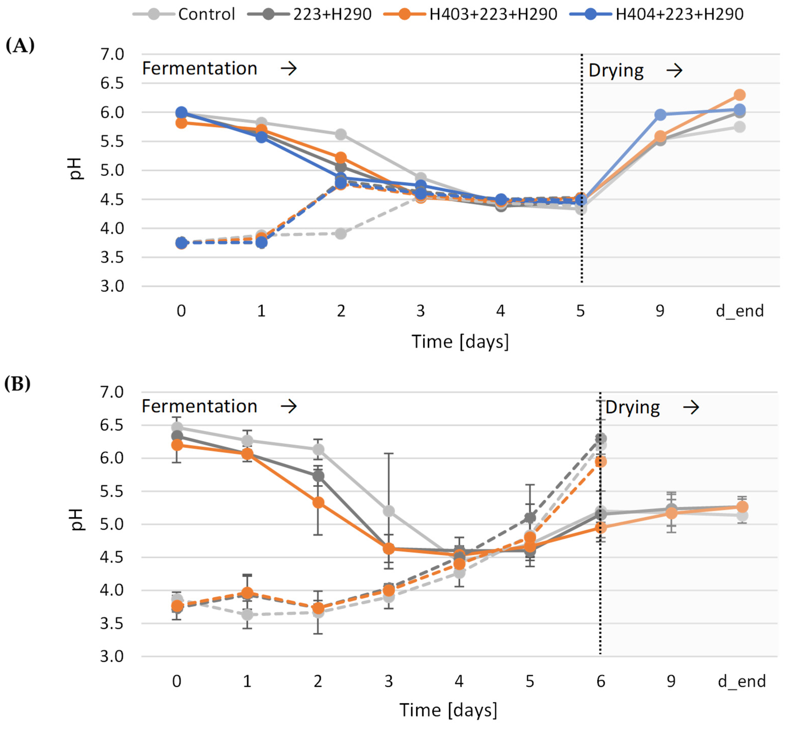
The pH values of the cotyledon (pHcot) and of the pulp (pHpulp) of the cocoa beans were measured daily during fermentation and are displayed in Figure 6A for lab-scale fermentations and Figure 6B for the small-scale fermentations (20 kg). The pH of the cotyledon was measured in the mid- and end drying phase.
As can be seen in Figure 6, a faster decrease in pHcot was observed in the inoculated samples and a delayed pH decrease was observed in the non-inoculated control samples in both the lab-scale and small-scale systems. Inoculated samples fermented at lab-scale showed a decrease in pHcot from initially 5.82–6.00 to below 5.22, after 2 days while the pH of non-inoculated samples was around 5.62 after 2 days. The same pH development was observed in the small-scale fermentation. The pHcot was initially around 6.28 ± 0.322 and decreased to 6.13 ± 0.15 and 5.20 ± 0.87 for the control, to 5.73 ± 0.15 and 4.6 ± 0.06 for the co-culture 223 + H290, and to 5.33 ± 0.49 and 4.63 ± 0.21 for the co-culture W. pijperi H403 + L. fermentum 223 + S. cerevisiae H290 after 2 and 3 days, respectively. A significantly lower pHcot could only be seen between the control sample and the sample fermented with the co-culture H403 + 223 + H290 after 2 days (p < 0.05). In both 1- and 20-kg scale fermentations, the pHcot reached similar values in all samples after 4 days (4.45 ± 0.08). In lab-scale systems, the pHcot reached the lowest values of 4.33 to 4.5 in all samples after 94 to 118 h (4 and 5 days, respectively). In small-scale systems, the lowest pH values were detected after 4 days, with a slight increase towards the end of fermentation. During the drying process, pHcot increased to values between 5.75 and 6.30 for the lab-scale fermentations and between 5.1 and 5.3 for the small-scale fermentations.
In contrast to pHcot, the initial pHpulp ranged between 3.74 and 3.76 for lab-scale fermentations and between 3.73 and 3.87 for small-scale fermentations. During lab-scale fermentations, the pHpulp increased drastically to 4.63–4.82 after 2 days for the inoculated samples, regardless of the applied culture, but remained at 3.91 for the control sample. From day 3 until the end of the fermentation, the pH remained stable across all samples. (4.52 ± 0.07). During small-scale fermentations, the pHpulp increased continuously with progressing fermentation time, as can be seen in Figure 6B. The control showed a decrease of 0.2 units in pHpulp from day 0 to day 1. Both fermentations 223 + H290 and H403 + 223 + H290 followed similar trends, with a first tendential pHpulp peak of 3.9 ± 0.3 on day 1. In all three small-scale fermentations, an increase from day 2 to a final pHpulp between 5.95 ± 0.92 and 6.2 ± 0.14 at the end of fermentation was observed.

3.5.3. Temperature Profile During Small-Scale Fermentation (20 kg) Inoculated with Pectinolytic Yeasts

The three small-scale fermentations were always terminated at the same time due to logistical reasons and lasted on average 134 ± 14 h. During this time, the average temperature (Tav) during fermentation varied between 37.0 ± 2.1 °C and 38.7 ± 0.8 °C, and maximum temperatures (Tmax) of 48.6 ± 0.8 to 48.9 ± 1.3 °C were reached (Table 9). Although no significant differences in Tav and Tmax were observed, there was a significant effect of the fermentation variation on the time to reach Tmax (p < 0.01). For trials in which beans were inoculated, the time until Tmax was significantly shorter, varying from 59.4 ± 7.5 (for W. pijperi H403 in co-culture with L. fermentum 223 + S. cerevisiae H290) and 59.6 ± 8.0 h (for 223 + H290 without additional yeast strain) compared to the control fermentation (83.1 ± 11.8 h). The drying of the beans took 281 ± 20 h at an average temperature of 31.5 ± 1.5 °C. During drying, no significant differences regarding average temperature, highest temperature (Tmax), and lowest (Tmin) temperature were identified (Table 9).

3.5.4. Microbial Development of Inoculated Microorganisms During Cocoa Bean Fermentation

Microbial analyses of the four lab-scale fermentations and of the three small-scale fermentations were performed daily on days 0 to 5, and at end of the fermentation (lab-scale: 5 days; small-scale runs 1 and 2: 6 days; small-scale run 3: 5 days). For lab-scale fermentations, samples of two replicates were mixed, and measurements were conducted once. For small-scale fermentations, measurements were conducted in triplicate with the exception of on fermentation day 4, on which only a duplicate analysis was possible due to a shortage of consumables. The mean value of CFU/g was calculated for every analysis timepoint for yeasts and LAB in every fermentation and is displayed in Table 10.
The initial cell count for LAB was, as expected, higher in inoculated samples than in control fermentations. In lab-scale systems, the peak was reached at day 2 for the variations with the pectinolytic strains W. pijperi H403 and H404 both in co-culture with L. fermentum 223 + S. cerevisiae H290 (9.1 log CFU/g) and at day 3 for the control fermentation and the variation 223 + H290 alone (9.0 log CFU/g). For the small-scale fermentations, the peak was reached at day 2 for all samples, although higher cell counts were achieved in the inoculated variations.
The initial yeast cell counts for the inoculated fermentations were between 5.4 ± 0.9 and 6.2 ± 0.6 log CFU/g. In the control fermentation, values of 2.7 log CFU/g for the lab-scale fermentation and 2.9 ± 0.7 log CFU/g for the small-scale fermentation were detected. In lab-scale systems, maximal yeast cell counts were reached on day 3 and were between 8.2 and 8.4 log CFU/g. In the three small-scale fermentations, the highest yeast concentrations were detected on day 2 of the fermentation and were between 6.9 ± 0.3 and 7.7 ± 0.2 log CFU/g. The yeast concentration in the control fermentation decreased to 4.6 ± 0.6 log CFU/g on day 4, in the fermentation with 223 + H290 to 4.3 ± 0.5 log CFU/g on day 3, and in the fermentation with H403 + 223 + H290 to 3.4 ± 0.6 log CFU/g on day 4. At the end of the fermentation, the yeast levels were around 5.3 to 5.5 log CFU/g for all samples.

3.5.5. Culture-Independent Monitoring of Yeasts During Fermentation in Small-Scale Systems (20 kg)

The total yeast count determined by qPCR (Figure 7) showed the same overall behavior as the total yeast count determined by cultural methods (Table 10).
The initial total yeast count in control fermentations (4.1 log CFU/g) was lower than in inoculated fermentations (4.7 log CFU/g and 6.1 log CFU/g in samples inoculated with L. fermentum 223 + S. cerevisiae H290 alone and co-cultured with W. pijperi H403, respectively). In control samples, the yeast count reached 5.2 log CFU/g after one day of fermentation and stayed at this level until the end of fermentation (Figure 7A). In samples fermented with the co-culture 223 + H290, the total yeast count increased from 4.7 to 5.3 log CFU/g from days 0 to 1 and then decreased to 4.5 log CFU/g after 4 days (Figure 7B). In samples fermented with the co-culture H403 + 223 + H290, the total yeast count reached a maximum of 7.2 log CFU/g after one day of fermentation and 5.7 log CFU/g after four days. In fermentations inoculated with H403 + 223 + H290 higher total yeast counts (1 to 2 log units) were recorded from day 0 to 4 (Figure 7C). The total yeast counts at the end of the fermentation were very similar across all variations (control, 223 + H290, and H403 + 223 + H403), ranging from 4.7 to 5.6 log CFU/g.
In control samples, the total yeast count was always higher than the concentration of S. cerevisiae or W. pijperi. At the beginning of fermentation, 1 log CFU/g S. cerevisiae and 1.3 log CFU/g W. pijperi were found (Figure 7A). The concentration of S. cerevisiae increased continuously to 3.70 log CFU/g at the end of fermentation, while the concentration of W. pijperi remained at the same level (Figure 7A). In samples inoculated with L. fermentum 223 + S. cerevisiae H290, the counts for S. cerevisiae started at 3.5 log CFU/g at the beginning of fermentation, increased to 4.7 log CFU/g after 1 day, and decreased to 3.2 log CFU/g after 4 days, following a similar trend as the total yeast count. The counts for W. pijperi remained below 1 CFU/g on all analyzed days (Figure 7B). Finally, in samples inoculated with W. pijepri H403 in co-culture with L. fermentum 223 + S. cerevisiae H290, the values for S. cerevisiae and W. pijperi remained consistently at the same level but were at least 1 log unit below the total yeast count. S. cerevisiae and W. pijperi started at 4.7 log CFU/g at the beginning of fermentation, increased to 5.2 and 5.5 log CFU/g, respectively, after one day, and then decreased to 3.8 and 4.0 log CFU/g, respectively, after four days of fermentation (Figure 7C).
As for the total yeast counts, the S. cerevisiae concentration at the end of the fermentation reached similar levels in all fermentation variations, varying from 3.1 to 3.7 log CFU/g. The W. pijperi concentration at the end of fermentation was lower in the control samples and in samples fermented with 223 + H290 (<1.1 log CFU/g) compared to samples fermented with W. pijepri H403 co-cultured with L. fermentum 223 + S. cerevisiae H290 (3.7 log CFU/g).

3.5.6. Cut Test of Beans After Fermentation and Drying of Small-Scale Fermentations (20 kg)

The fermentation status of the cocoa beans was determined by performing a cut test with 50 fermented beans at the end of each fermentation. A second cut test with 300 beans was performed at the end of drying to determine the quality of the dried cocoa beans. Of all the examined beans, no internal mold, insect infestation, germination, overfermentation, slaty beans, or white/pale beans were detected. At the end of the fermentation no significant effect of fermentation on the amount of well-fermented cocoa beans (p = 0.49), violet beans (p = 0.48), and slightly fermented beans (p = 0.42) was observed. After fermentation, 57–90% of the analyzed cocoa beans were slightly fermented, 1–2% were well-fermented (Table 11), and 7–13% were violet.
The cut test performed at the end of drying revealed no significant influence of the fermentation variation on the amount of violet (p = 0.39), slightly fermented (p = 0.99), and well-fermented (p = 0.86) cocoa beans. The amount of well-fermented beans varied from 9 to 13.2%, the amount of slightly fermented beans varied from 83.1 to 83.6%, and the amount of violet beans varied from 3.7 to 7.6%. Nevertheless, a tendency towards more well-fermented and fewer violet beans for the variation that was inoculated with W. pijperi H403 in co-culture with L. fermentum 223 + S. cerevisiae H290 than for the non-inoculated control and the variation inoculated with 223 + H290 alone could be detected (Table 11).

4. Discussion

4.1. Relevance of Pectinolytic Yeasts in Pulp Degradation During Cocoa Bean Fermentation

Among the 1139 yeast strains screened for pectinolytic activity, 159 strains exhibited minimal activity, while 2 Ustilago sp. strains showed weak activity, 2 unidentified strains showed moderate activity, and 3 W. pijperi strains exhibited strong pectinolytic activity. W. pijperi (formerly Pichia pijperi) has previously been identified in cocoa bean fermentations through metagenomic approaches [39], but, to the best of our knowledge, its pectinolytic activity has not yet been reported in the literature. Among the 159 strains showing minimal pectinolytic activity, 36 strains showing a total score for pectinolytic activity of ≥1.0 were identified. Except for one strain that was identified as H. opuntiae, all other strains were identified as Saccharomyces sp. or S. cerevisiae. The pectinolytic activity of S. cerevisiae strains isolated during cocoa bean fermentations has already been described [6,19,40,41]. Other yeast species, including Pichia kudriavzevii, Candida nitrativorans, Kluyveromyces marxianus, C. rugopelliculosa, K. thermotolerans, C. ethanolica, H. guilliermondii, and S. pombe, have been recognized for their role in pectin degradation during cocoa bean fermentation [18,19,40] but were not found among the strains showing pectinolytic activity in the present study.
Of the 159 strains with minimal pectinolytic activity, 123 were active on only 1 of the tested media, with the majority (121 strains) showing activity on MP5, while only 2 were active on YNBpc. Among the strains with a total score of 1.0 (indicating minimal activity on at least two out of three media), all displayed pectinolytic activity on MP5 and additionally on either YNBpc (seven strains) or YNBpa (five strains). The higher number of strains showing pectinolytic activity on MP5 compared to YNBpc and YNBpa can be attributed to its specific composition. MP5 contained polygalacturonic acid, a less complex form of pectin that is more readily accessible to polygalacturonase, a pectinolytic enzyme produced by yeasts, such as S. cerevisiae. In S. cerevisiae, polygalacturonase activity is primarily driven by the PGU1 gene, which encodes the (endo)polygalacturonase responsible for hydrolyzing the α-1,4-glycosidic bonds in the pectin chain [6,42,43]. Given that polygalacturonic acid is the substrate for polygalacturonase, its presence in MP5 likely facilitated the detection of the pectinolytic activity by the tested yeasts. In contrast, citrus pectin and apple pectin in YNBpc and YNBpa have more complex structures and higher degrees of esterification, requiring the combined activity of other enzymes, such as pectin methylesterases and pectin lyases, in addition to polygalacturonase [44,45].
Notably, among the 1139 yeast strains previous isolated from cocoa beans during fermentation and/or drying processes [7,30,31] and analyzed in the present study, only 166 (14.6%) exhibited pectinolytic activity, highlighting the relatively low prevalence of pectinolytic activity among yeast strains. Comparable incidences were reported in related studies. For instance, a screening of 205 yeast isolates from cocoa bean fermentation on a pectin-containing medium identified 36 pectinolytic strains (17.5%), with H. uvarum and P. kudriavzevii being the most stress-tolerant and enzymatically active species [18]. A further screening of 28 yeast strains isolated from coffee beans on a citric pectin-containing medium revealed that 8 strains had pectinolytic activity (28.6%) [46].
Furthermore, the evaluation of selected pectinolytic strains, including those with strong and minimal pectinolytic activity (W. pijperi strains with a total score of 10.5, and seven S. cerevisiae strains, with five strains with a total score of 1.5 and two strains with a total score of 1.0), revealed that only the three W. pijperi strains H312, H403, and H404 significantly reduced the viscosity of mCPSMpc. In contrast, the viscosity of mCPSMpc inoculated with further pectinolytic strains remained similar to that of the blank. These results suggest that pectinolytic yeasts with weak polygalacturonase activity are not sufficient for viscosity reduction in a pectin-containing medium. Either very strong enzymatic activity or a consortium of microorganisms with diverse enzymatic functions may be needed, allowing complementary interactions that could at the end also enhance polygalacturonase activity. Additionally, interactions between different yeast species could further contribute to pectin degradation through cooperative metabolism. These findings align with metagenomic and metatranscriptomic studies showing that pectin degradation in cocoa bean fermentation involves a variety of microorganisms, including Acetobacter sp., Cellvibrio sp., Pectobacterium sp., lactic acid bacteria (Ligilactobacillus sp., Leuconostoc sp.), S. cerevisiae, and H. opuntiae, which contribute through glycoside hydrolases (GH28, GH43, and GH53) and pectate lyases (PL1, PL3, PL4, and PL9) [44]. Additionally, Meersman et al. [6] found that the reduction in cocoa pulp viscosity caused by S. cerevisiae is primarily driven by endopolygalacturonase activity, a process further enhanced by increasing fermentation temperatures.
The results of the lab- and small-scale cocoa bean fermentations lead to the assumption that the presence of pectinolytic yeasts (W. pijepri strains H403 and H404) influenced pulp degradation, particularly in the early stages of fermentation. In lab-scale fermentations, the impact of pectinolytic yeasts was more pronounced, with inoculated samples consistently showing lower pulp content than the control and non-pectinolytic fermentations. After 23.8 h, pulp runoff in fermentations with pectinolytic yeasts was significantly higher (13.3–12.6%) compared to the control (4.7%) and non-pectinolytic fermentations (9.1%), suggesting that pectinolytic activity contributed to faster pulp degradation. These findings align with previous studies indicating that pectin degradation occurs primarily in the first 48 h of fermentation and is facilitated by microbial pectinolytic activity [3,6,18,47]. The higher pulp runoff observed in lab-scale fermentations supports the idea that W. pijepri H403 and H404 contributed to this process. The initial pulp content (~40%) observed in this study is comparable to that reported in the literature [48,49].
In contrast, in small-scale fermentations, based on the results for pulp content and weight of 100 beans, the effect of pectinolytic strains was less pronounced. This could indicate that the pectinolytic strains were less effective in the larger-scale fermentation, potentially due to a lower initial inoculation concentration but also due to environmental differences, such as temperature, aeration, or microbial competition. The literature suggests that fermentation parameters (e.g., temperature, oxygen availability, and microbial interactions) affect pectin degradation efficiency by yeasts [50,51], which could explain why the pectinolytic activity in 20 kg fermentations was less pronounced than in the 1 kg setup. However, since the measurement of pulp runoff was not feasible during small-scale fermentation and the determination of pulp content, and bean weight are highly sensitive to fluctuations and only an indirect indication of pectinolytic activity was investigated, they alone may not be sufficient to confirm the extent of the pectinolytic activity of the applied strains during fermentation. Additional analytical methods, such as the detection of galacturonic acid, a key degradation product of pectin, would provide stronger evidence of pectin hydrolysis and should be considered in future studies. Furthermore, a faster decline in citric acid and the formation of specific metabolites, such as ethanol, may serve as additional indicators of an accelerated fermentation process [20,45,51,52] and should be considered in further trials.
The presence of pectinolytic yeast species in cocoa fermentation is crucial for the degradation of the mucilaginous pulp surrounding the beans, facilitating aeration and enhancing microbial succession. Future research should investigate whether a microbial consortium, rather than a single pectinolytic yeast species, is necessary for effective pulp degradation during cocoa fermentation. In addition, the impact of other microorganisms, such as Bacillus spp. and filamentous fungi, in pectin degradation has been investigated in a few studies [53,54]. However, further research is needed to clarify their specific contributions to cocoa pulp degradation.
The unexpected detection of strong pectinolytic activity in W. pijperi and its ability to reduce viscosity in a pectin-rich medium, even after freeze-drying, underscores its potential relevance in cocoa bean fermentation. Future research should focus on characterizing the specific enzymes responsible for this activity, assessing their stability, and identifying optimal conditions for their function. Additionally, evaluating how these enzymatic properties influence fermentation dynamics on a larger scale is important, as environmental factors may affect their efficacy, as discussed above. Furthermore, potential synergies with other beneficial microorganisms should be investigated to better understand their roles and possible advantages in controlled cocoa bean fermentations. Further research should also explore the impact of W. pijperi H403 on the sensory attributes of cocoa and chocolate. The perception of a strong banana flavor during biomass production of W. pijperi H403 also indicates its potential for a positive impact on aroma formation, as previously observed during the fermentation of whey with W. pijperi [55].

4.2. Growth of W. pijperi Strains on Cocoa Beans and Overall Influence on Fermentation and Quality of Fermented and Dried Cocoa Beans

The in vitro experiments with pectinolytic yeast strains (W. pijperi strains H312, H403, H404, and S. cerevisiae Y170) demonstrated their ability to colonize and grow on cocoa beans under fermentation conditions, both in single cultures and in co-cultures with L. fermentum 223 and S. cerevisiae H290. The strains L. fermentum 223 and S. cerevisiae H290 were developed specifically for cocoa bean fermentations [21] and were chosen to verify the growth behaviour of the W. pijperi strains in the presence of other microorganisms typically found during cocoa bean fermentation. The pectinolytic S. cerevisiae Y170 reached the highest cell count (7.8 log CFU/g), followed by W. pijperi H312 (6.9 log CFU/g) and H403 (6.6 log CFU/g), whereas H404 displayed the lowest growth (5.6 log CFU/g). No strong inhibitory effects were observed in co-cultures, except for the reduced growth of W. pijperi H404 when combined with L. fermentum 223 alone. Similar trends were observed in fermentations conducted in lab- and small-scale systems, with significantly higher initial yeast and LAB counts in inoculated variants compared to the control, as has already been observed in other studies [21,56].
Yeast populations increased in the first 2–3 days but then declined as fermentation progressed, which is expected due to increasing temperatures [48]. The qPCR analysis provided additional insights into yeast dynamics. In 20 kg fermentations, S. cerevisiae was detected in the control at only 1.0 log CFU/g on day 0, while inoculated samples (L. fermentum 223 + S. cerevsiae H290 alone or in co-culture with W. pijepri H403) showed significantly higher initial values (4.7 and 3.5 log CFU/g, respectively). Despite a decline after day 3, S. cerevisiae remained present at higher levels than in the control throughout fermentation. Similarly, W. pijperi was barely detectable in the control (<1.5 log CFU/g on day 0 and <1.2 log CFU/g on day 6), whereas in the variation inoculated with L. fermentum 223 + S. cerevsiae H290 in co-culture with W. pijepri H403, W. pijperi reached 5.5 log CFU/g on day 1 and persisted at 3.7 log CFU/g by day 6. These findings indicate that the introduced strains successfully established themselves, even though yeast counts declined over time, probably due to thermal inactivation or a shortness in substrate [21,51].
Overall, both qPCR and cultural methods confirmed that the introduced yeasts proliferated in the early stages of fermentation and remained detectable until the end. The control fermentations (without inoculation) exhibited much lower initial yeast counts, further indicating that the added strains actively contributed to the fermentation process. The growth, prevalence, and benefits of the strains L. fermentum 223 and S. cerevisiae H290 was already investigated during fermentation trials conducted in Honduras [21]. The persistence of higher yeast, S. cerevisiae, and W. pijperi counts in inoculated fermentations suggests that the inoculated strains were not just present but also capable of active growth and metabolic activity in the system, making them suitable candidates for controlled fermentation applications [21,47,51].
The inoculated fermentations showed a more rapid drop in cotyledon pH compared to the non-inoculated control after 2 days in small-scale fermentations. Similarly, pH changes in the pulp occurred faster in inoculated fermentations. The faster decrease in cotyledon pH and the more pronounced increase in pulp pH in samples inoculated with L. fermentum 223, S. cerevisiae H290, and W. pijperi H403 may indicate increased microbial activity, potentially leading to enhanced sugar metabolism and organic acid production, which in turn could influence fermentation kinetics and bean quality [21,51]. However, as all samples reached similar pH values after 4–5 days, the impact of inoculation appears to be most pronounced in the early phase of fermentation.
The inoculated fermentations in small-scale systems reached the maximum temperature (Tmax ~49 °C) significantly faster than the non-inoculated control fermentation. In the control fermentation without inoculation, Tmax was reached after 83.1 ± 11.8 h, whereas in the inoculated fermentations (L. fermentum 223 + S. cerevsiae H290 alone or in co-culture with W. pijepri H403), Tmax was reached in ~59.4–59.6 h (p < 0.01). This faster temperature increase suggests once more that the inoculated cultures enhanced metabolic activity, likely due to increased sugar metabolism and acid production by yeasts and LAB [31,57]. The higher Tmax compared to the 45 °C reported by Sandhya et al. [57] could be attributed to the further oxidation of ethanol to acetic acid by acetic acid bacteria (AAB), an additional exothermic process. Consequently, higher ethanol production by yeasts may contribute to increased fermentation temperatures. The shorter fermentation time observed with co-culture inoculation aligns with the findings of Chadha et al. [58], who reported accelerated fermentation through S. cerevisiae inoculation. Since most yeast species present in cocoa bean fermentations are thermolabile, their activity is expected to decline as fermentation temperatures rise, which may explain the observed decrease in yeast counts in later fermentation stages. The faster temperature rise in inoculated fermentations could also indicate a more efficient fermentation process, potentially allowing for a shorter overall fermentation time while maintaining microbial succession.
There was no big difference in drying time of the cocoa beans from the different fermentation variations at small-scale. At the end of fermentation, a cut test was performed with 50 beans to determine the fermentation status of the beans. A trend towards fewer violet beans in the fermentations with co-cultures was observed. This suggests that the addition of co-cultures can reduce the duration of the fermentation process. These findings are in accordance with those of Sandhya et al. [57] and Ramos et al. [59]. After drying, a second cut test with 300 beans was performed, showing similar results for all fermentations. There were fewer violet and more well-fermented beans, indicating that the fermentation process continues during drying. As in the cut test after fermentation, a trend towards faster fermentation with co-culture addition was observed.

4.3. First Insights and Challenges in the Cultivation of W. pijperi

The growth experiments of W. pijperi strains H312 and H403 under different conditions in flasks and microplates revealed that their lag phase was significantly influenced by the cultivation medium, temperature, and pH. The shortest lag phases were observed in YB medium at 30 °C, whereas the longest occurred in mCPSM at 25 °C and 30 °C with pH 4.5, particularly for strain H312. This suggests that W. pijperi adapts more efficiently to nutrient-rich conditions and elevated temperatures, whereas environments with lower pH might impose greater stress, delaying growth onset. The pH level significantly affects yeast growth, with a pH of 4.5 already presenting a stress condition [32]. Despite variations in lag phase duration and pH, both strains reached their stationary phase within approximately 8–10 h in YB and 15 h in mCPSM, with higher values for OD600 at 25 °C, particularly in YB. This discrepancy between the shorter lag-time at 30 °C but higher OD600 at 25 °C could indicate morphological changes, such as increased cell size rather than cell proliferation, a phenomenon previously described for yeast adaptation under suboptimal growth conditions [58].
The enzymatic activity of W. pijperi appears to be closely linked to its growth dynamics. The viscosity reduction in mCPSMpc was most pronounced using cell-free supernatant collected after 24 h of cultivation in mCPSM at 30 °C, with only marginal changes between 24 and 48 h. This suggests that pectinolytic enzyme secretion is highest during the exponential growth phase and plateaus as cells transition to the stationary phase. Similar observations have been reported for other pectinolytic yeasts, where enzyme production is tightly regulated and peaks during active growth [60,61]. Consequently, W. pijperi might contribute most effectively to pectin degradation in cocoa bean fermentations during the early stages when it is still proliferating, whereas its role in later phases may be less significant or less accentuated.
Although experiments in microtiter plates and bioreactors are not directly comparable, both provided valuable insights into the growth characteristics of W. pijperi H403. In microtiter plate experiments, a prolonged lag phase was observed, whereas in the controlled bioreactor conditions, the yeast entered exponential growth immediately. This was likely due to optimized aeration, controlled pH, and the elimination of nutrient depletion during inoculation. However, the duration of the growth phase for trials using YB medium was approximately the same in both microtiter plates (ca. 8–10 h) and the pilot-scale bioreactor (ca. 7 h). In the pilot-scale bioreactor, a suboptimal feeding strategy during fed-batch cultivation appeared to limit biomass yield. As can be seen from the pO2 signal (low value at 79% dissolved oxygen), the substrate was depleted from the medium after 7 h, leading the yeast to enter the starvation phase. However, the feed phase was only initiated after 24 h, whereas an earlier feeding, ideally at the onset of the pO2 drop, could have prevented nutrient depletion and maintained exponential growth [62]. The lack of further cell proliferation as concluded from the viable cell counts despite an increase in OD600 suggests that nutrient availability or metabolic constraints prevented additional cell division, leading to an increase in cell volume rather than cell number, as already observed by other authors [58]. This observation may be further supported by the increase in the permittivity signal, which indicates a rise in polarizable biomass, typically associated with cell enlargement rather than proliferation. The observation of sustained metabolic activity in cells despite the absence of growth is further verified by the continuous oxygen demand, which manifests as a reduced pO2 signal during the feeding phase. This correlation between metabolic activity and oxygen consumption in the absence of cell proliferation suggests the maintenance of cellular basic functions under suboptimal growth conditions. Furthermore, the persistence of oxygen demand implies that the cells, notwithstanding growth stagnation, continue to maintain active biochemical processes, indicating a potential metabolic adaptation to the given environmental conditions [63].
Several factors may have contributed to the gradual stagnation in cell growth rate over the prolonged fed-batch duration. Firstly, the continuous addition of a total of 38.15 g of antifoaming agent (polypropylene glycol) due to the presence of tween in the YB medium formulation might have inhibited oxygen transfer, affecting aerobic respiration [64]. Secondly, the initial feed rate setpoint was likely too high, as the correct value was difficult to determine without ample prior knowledge (using only the preceding reactor batch phase as reference). The overfeeding resulted in intermittent glucose accumulation (the process ended with 1.8 g/L remaining glucose) and potentially induced anaerobic metabolism due to the Crabtree effect, which is known to manifest at glucose concentrations exceeding approximately 100 mg/L [65,66,67,68]. This phenomenon results in reduced biomass yield while increasing ethanol production, as glucose is preferentially metabolized to ethanol and carbon dioxide, rather than carbon dioxide and water as under aerobic conditions. Based on these findings, it was decided to continue with feeding on a sequential exploratory basis. Additionally, the depletion and limitation of essential nutrients, such as nitrogen, phosphate, trace elements, vitamins, and amino acids, could have further restricted biomass formation [69,70]. The sole sources of these components in the batch medium YB were 2% glucose, 1% yeast extract, and 0.8% malt extract, which may have been insufficient for sustained fermentation. However, in the absence of quantitative analysis of the aforementioned media components, this hypothesis remains unconfirmed and warrants further investigation. The mitigation of this potential limitation could have been achieved through the administration of a bolus shot at the initiation of the feed phase or through the use of a specifically supplemented feed.
A comprehensive analysis of trace element and nutrient requirements regarding growth as well as aromatic behaviour and enzymatic activity is crucial for enhancing fermentation performance. Based on the results obtained, a short-batch fermentation or an earlier start of feeding with a feed medium optimized for growth and enriched with trace elements and nutrients (as described above) will probably represent an efficient strategy to maximize the active yeast population during its exponential growth phase. Future process optimizations should focus on (1) elucidating the optimal conditions for maximal biomass production and (2) refining feeding strategies, possibly by implementing an adaptive feeding regime based on real-time biomass including permittivity analysis regarding cell physiology and morphology and metabolite measurements. Moreover, the role of environmental parameters, such as aeration rate and oxygen transfer, should be further investigated to maximize yeast cell viability and pectinolytic activity. From an application perspective, these findings indicate that the cultivation of W. pijperi H403 in bioreactors is feasible, but further optimizations are required to enhance biomass yields and viable cell counts for use as pectinolytic starter cultures in cocoa bean fermentation.

5. Conclusions

This study provides the first evidence of pectinolytic activity in W. pijperi (formerly Pichia pijperi). The strong pectin-degrading activity observed in vitro was confirmed in lab-scale fermentations, where the W. pijperi strains H403 and H404 significantly enhanced pulp degradation, potentially improving aeration and fermentation kinetics. During small-scale fermentation in Costa Rica, the ability of the strain H403 to persist during fermentation was verified through qPCR, demonstrating its adaptability to the cocoa fermentation environment. Moreover, the development of a pre-pilot biomass production process, including freeze-drying, ensures its practical application in cocoa bean fermentation at an industrial scale on field. While the use of defined starter cultures remains an emerging strategy, W. pijperi H403 presents a viable approach to standardizing fermentation processes, optimizing microbial dynamics, and enhancing cocoa bean quality. Further studies should investigate the pectinolytic enzymes of W. pijperi strains and their mechanisms of action. Additionally, culture’s impact on chocolate’s sensory properties should be assessed, along with potential synergies with other beneficial microorganisms to enhance its pectinolytic activity in scaled-up fermentations. These findings open new perspectives on the potential enzymatic roles of W. pijperi in cocoa fermentation.

Supplementary Materials

The following supporting information can be downloaded at: https://www.mdpi.com/article/10.3390/applmicrobiol5020043/s1, Figure S1: Determination of detection limit using a Kruskal–Wallis test [p values are indicated] followed by a Wilcoxon test [different letters indicate significant differences between groups] for total yeast count (a), S. cerevisiae (b), and W. pijperi (c). Standard curves for total yeast count (d), S. cerevisiae (e), and W. pijperi (f).; Table S1: Lag time, optical density (OD600nm) at 24, 48, and 72 h, maximal specific growth (µmax), and time at µmax for growth curves conducted in microtiter plates with the strains Wickerhamomyces pijperi H312 and H403 using yeast broth (YB) and modified cocoa pulp simulation medium (mCPSM) at 25 and 30 °C, respectively, at pH 4.5 and 5.0; Table S2: Cell counts during lab-scale (1 kg) and small-scale (20 kg) fermentations determined by cultural methods (P) and by molecular biological methods (qPCR) at day 0 until day 6 of fermentation given in log CFU/g. CFU is colony forming unit; LAB is lactic acid bacteria; 223 is Limosilactobacillus fermentum 223; H290 is Saccharomyces cerevisiae H290; H403 and H404 is Wickerhamomyces pijperi H403 and H404. nd, not determined.

Author Contributions

Conceptualization, S.M.S. and S.F.L.; methodology, S.F.L., A.H., A.T., B.M.B., S.W. and J.W.; formal analysis, S.F.L. and A.H.; investigation, S.F.L., A.H., A.T., B.M.B., S.W. and J.W.; writing—original draft preparation, S.F.L.; writing—review and editing, A.H., A.L., L.N. and S.M.S.; visualization, S.F.L. and A.H.; supervision, S.M.S.; project administration, S.M.S.; funding acquisition, S.F.L. and S.M.S. All authors have read and agreed to the published version of the manuscript.

Funding

This research was funded by the internal start-up funding of ZHAW (InnoYeast), Call 2/2020. The development of the qPCR method for the detection of W. pijperi was funded by the internal start-up funding of ZHAW (MicroDyn), Call 1/2019. Open access funding was provided by the ZHAW Zurich University of Applied Sciences.

Data Availability Statement

The data presented in this study are available in the article and supplementary material.

Acknowledgments

The authors gratefully thank the project partners MOGUNTIA Schweiz AG for their collaboration in the production of the strains L. fermentum 223 and S. cerevisiae H290, as well as for providing the mixture of protective substances for the freeze-drying of the W. pijperi H403 culture for field applications. Grateful thanks also go to Rausch GmbH and Rausch PCE Costa Rica Ltd. for their support and valuable exchange during the cocoa bean fermentations with functional cultures at the Rausch Edelkakaoinstitut and Cacao Farm Rausch < Tres Equis–Finca de Cacao > in Tres Equis, Cartago, Costa Rica. Furthermore, our thanks go to Giverny Ganz for the English proof-reading.

Conflicts of Interest

The authors declare no conflicts of interest.

References

  1. Akinfala, T.O.; Houbraken, J.; Sulyok, M.; Adedeji, A.R.; Odebode, A.C.; Krska, R.; Ezekiel, C.N. Moulds and Their Secondary Metabolites Associated with the Fermentation and Storage of Two Cocoa Bean Hybrids in Nigeria. Int. J. Food Microbiol. 2020, 316, 108490. [Google Scholar] [CrossRef] [PubMed]
  2. Sarbu, I.; Csutak, O. The Microbiology of Cocoa Fermentation. In Caffeinated and Cocoa Based Beverages; Grumezescu, A.M., Holban, A.M., Eds.; Woodhead Publishing: Cambridge, UK, 2019; pp. 423–446. ISBN 978-0-12-815864-7. [Google Scholar]
  3. Schwan, R.F.; Wheals, A.E. The Microbiology of Cocoa Fermentation and Its Role in Chocolate Quality. Crit. Rev. Food Sci. Nutr. 2004, 44, 205–221. [Google Scholar] [CrossRef]
  4. Leal, G.A.; Gomes, L.H.; Efraim, P.; De Almeida Tavares, F.C.; Figueira, A. Fermentation of Cacao (Theobroma cacao L.) seeds with a Hybrid Kluyveromyces Marxianus Strain Improved Product Quality Attributes. FEMS Yeast Res. 2008, 8, 788–798. [Google Scholar] [CrossRef] [PubMed]
  5. Lopez, A.S.; Dimick, P.S. Cocoa Fermentation. In Biotechnology Set; Rehm, H.-J., Reed, G., Eds.; Wiley: Hoboken, NJ, USA, 2001; pp. 561–577. ISBN 978-3-527-25762-1. [Google Scholar]
  6. Meersman, E.; Struyf, N.; Kyomugasho, C.; Jamsazzadeh Kermani, Z.; Santiago, J.S.; Baert, E.; Hemdane, S.; Vrancken, G.; Verstrepen, K.J.; Courtin, C.M.; et al. Characterization and Degradation of Pectic Polysaccharides in Cocoa Pulp. J. Agric. Food Chem. 2017, 65, 9726–9734. [Google Scholar] [CrossRef] [PubMed]
  7. Romanens, E.; Näf, R.; Lobmaier, T.; Pedan, V.; Leischtfeld, S.F.; Meile, L.; Schwenninger, S.M. A Lab-Scale Model System for Cocoa Bean Fermentation. Appl. Microbiol. Biotechnol. 2018, 102, 3349–3362. [Google Scholar] [CrossRef]
  8. Pettipher, G.L. Analysis of Cocoa Pulp and the Formulation of a Standardised Artificial Cocoa Pulp Medium. J. Sci. Food Agric. 1986, 37, 297–309. [Google Scholar] [CrossRef]
  9. Schols, H.A.; Voragen, A.G.J. Pectic Polysaccharides. In Handbook of Food Enzymology; Whitaker, J.R., Voragen, A.G.J., Wong, D.W.S., Eds.; Marcel Dekker: New York, NY, USA; Basel, Switzerland, 2003; pp. 811–825. ISBN 978-0-8247-0686-9. [Google Scholar]
  10. Chaudhri, A. Microbially Derived Pectinases: A Review. IOSR J. Pharm Biol. Sci. 2012, 2, 1–5. [Google Scholar] [CrossRef]
  11. Mohnen, D. Pectin Structure and Biosynthesis. Curr. Opin. Plant Biol. 2008, 11, 266–277. [Google Scholar] [CrossRef]
  12. Chakraborty, S.; Fernandes, V.O.; Dias, F.M.V.; Prates, J.A.M.; Ferreira, L.M.A.; Fontes, C.M.G.A.; Goyal, A.; Centeno, M.S.J. Role of Pectinolytic Enzymes Identified in Clostridium Thermocellum Cellulosome. PLoS ONE 2015, 10, e0116787. [Google Scholar] [CrossRef]
  13. Pedrolli, D.B.; Carmona, E.C. Purification and Characterization of the Exopolygalacturonase Produced by Aspergillus Giganteus in Submerged Cultures. J. Ind. Microbiol. Biotechnol. 2010, 37, 567–573. [Google Scholar] [CrossRef]
  14. Swain, M.R.; Ray, R.C. Production, Characterization and Application of a Thermostable Exo-Polygalacturonase by Bacillus Subtilis CM5. Food Biotechnol. 2010, 24, 37–50. [Google Scholar] [CrossRef]
  15. Blanco, P.; Sieiro, C.; Villa, T.G. Production of Pectic Enzymes in Yeasts. FEMS Microbiol. Lett. 1999, 175, 1–9. [Google Scholar] [CrossRef] [PubMed]
  16. Benen, J.A.E.; Visser, J. Polygalacturonases. In Handbook of Food Enzymology; Whitaker, J.R., Voragen, A.G.J., Wong, D.W.S., Eds.; Marcel Dekker: New York, NY, USA; Basel, Switzerland, 2003; pp. 838–847. ISBN 978-0-8247-0686-9. [Google Scholar]
  17. Dasilva, E.; Borges, M.; Medina, C.; Piccoli, R.; Schwan, R. Pectinolytic Enzymes Secreted by Yeasts from Tropical Fruits. FEMS Yeast Res. 2005, 5, 859–865. [Google Scholar] [CrossRef] [PubMed]
  18. Samagaci, L.; Ouattara, H.; Niamké, S.; Lemaire, M. Pichia Kudrazevii and Candida Nitrativorans Are the Most Well-Adapted and Relevant Yeast Species Fermenting Cocoa in Agneby-Tiassa, a Local Ivorian Cocoa Producing Region. Food Res. Int. 2016, 89, 773–780. [Google Scholar] [CrossRef]
  19. Schwan, R.F.; Cooper, R.M.; Wheals, A.E. Endopolygalacturonase Secretion by Kluyveromyces Marxianus and Other Cocoa Pulp-Degrading Yeasts. Enzym. Microb. Technol. 1997, 21, 234–244. [Google Scholar] [CrossRef]
  20. Lima, C.-C.; Vaz, A.-M.; De Castro, G.M.; Lobo, F.; Solar, R.; Rodrigues, C.; Martins Pinto, L.R.; Vandenberghe, L.; Pereira, G.; Miúra Da Costa, A.; et al. Integrating Microbial Metagenomics and Physicochemical Parameters and a New Perspective on Starter Culture for Fine Cocoa Fermentation. Food Microbiol. 2021, 93, 103608. [Google Scholar] [CrossRef]
  21. Romanens, E.; Pedan, V.; Meile, L.; Miescher Schwenninger, S. Influence of Two Anti-Fungal Lactobacillus Fermentum-Saccharomyces Cerevisiae Co-Cultures on Cocoa Bean Fermentation and Final Bean Quality. PLoS ONE 2020, 15, e0239365. [Google Scholar] [CrossRef]
  22. Figueroa-Hernández, C.; Mota-Gutierrez, J.; Ferrocino, I.; Hernández-Estrada, Z.J.; González-Ríos, O.; Cocolin, L.; Suárez-Quiroz, M.L. The Challenges and Perspectives of the Selection of Starter Cultures for Fermented Cocoa Beans. Int. J. Food Microbiol. 2019, 301, 41–50. [Google Scholar] [CrossRef]
  23. Díaz-Muñoz, C.; De Vuyst, L. Functional Yeast Starter Cultures for Cocoa Fermentation. J. Appl. Microbiol. 2022, 133, 39–66. [Google Scholar] [CrossRef]
  24. Taskila, S. Industrial Production of Starter Cultures. In Starter Cultures in Food Production; John Wiley & Sons: Hoboken, NJ, USA, 2017; pp. 79–100. ISBN 978-1-118-93379-4. [Google Scholar]
  25. Liu, C.-F.; Zhang, X.-F.; Yu, T.-L.; Lee, C.-L. Utilizing Deep Ocean Water in Yeast Fermentation for Enhanced Mineral-Rich Biomass Production and Fermentative Regulation by Proteomics Modulation. Heliyon 2024, 10, e31031. [Google Scholar] [CrossRef]
  26. Yüzgeç, U.; Türker, M.; Hocalar, A. On-Line Evolutionary Optimization of an Industrial Fed-Batch Yeast Fermentation Process. ISA Trans. 2009, 48, 79–92. [Google Scholar] [CrossRef] [PubMed]
  27. Kasperski, A.; Miśkiewicz, T. Optimization of Pulsed Feeding in a Baker’s Yeast Process with Dissolved Oxygen Concentration as a Control Parameter. Biochem. Eng. J. 2008, 40, 321–327. [Google Scholar] [CrossRef]
  28. Guo, Y.-F.; Wang, M.-Q.; Wang, Y.-L.; Wang, H.-T.; Xu, J.-Z. Controlling the Formation of Foams in Broth to Promote the Co-Production of Microbial Oil and Exopolysaccharide in Fed-Batch Fermentation. Fermentation 2022, 8, 68. [Google Scholar] [CrossRef]
  29. Polo, L.; Mañes-Lázaro, R.; Olmeda, I.; Cruz-Pio, L.E.; Medina, Á.; Ferrer, S.; Pardo, I. Influence of Freezing Temperatures Prior to Freeze-Drying on Viability of Yeasts and Lactic Acid Bacteria Isolated from Wine. J. Appl. Microbiol. 2017, 122, 1603–1614. [Google Scholar] [CrossRef] [PubMed]
  30. Miescher Schwenninger, S.; Freimüller Leischtfeld, S.; Gantenbein-Demarchi, C. High-throughput Identification of the Microbial Biodiversity of Cocoa Bean Fermentation by MALDI-TOF MS. Lett. Appl. Microbiol. 2016, 63, 347–355. [Google Scholar] [CrossRef]
  31. Streule, S.; Freimüller Leischtfeld, S.; Galler, M.; Miescher Schwenninger, S. Monitoring of Cocoa Post-Harvest Process Practices on a Small-Farm Level at Five Locations in Ecuador. Heliyon 2022, 8, e09628. [Google Scholar] [CrossRef]
  32. Romanens, E.; Freimüller Leischtfeld, S.; Volland, A.; Stevens, M.J.A.; Krähenmann, U.; Isele, D.; Fischer, B.; Meile, L.; Miescher Schwenninger, S. Screening of Lactic Acid Bacteria and Yeast Strains to Select Adapted Anti-Fungal Co-Cultures for Cocoa Bean Fermentation. Int. J. Food Microbiol. 2019, 290, 262–272. [Google Scholar] [CrossRef]
  33. Silva, C.F.; Vilela, D.M.; De Souza Cordeiro, C.; Duarte, W.F.; Dias, D.R.; Schwan, R.F. Evaluation of a Potential Starter Culture for Enhance Quality of Coffee Fermentation. World J. Microbiol. Biotechnol. 2013, 29, 235–247. [Google Scholar] [CrossRef] [PubMed]
  34. Martos, M.A.; Zubreski, E.R.; Combina, M.; Garro, O.A.; Hours, R.A. Isolation of a Yeast Strain Able to Produce a Polygalacturonase with Maceration Activity of Cassava Roots. Food Sci. Technol. 2013, 33, 332–338. [Google Scholar] [CrossRef]
  35. Glass, N.L.; Donaldson, G.C. Development of Primer Sets Designed for Use with the PCR to Amplify Conserved Genes from Filamentous Ascomycetes. Appl. Environ. Microbiol. 1995, 61, 1323–1330. [Google Scholar] [CrossRef]
  36. Schwendimann, L.; Kauf, P.; Fieseler, L.; Gantenbein-Demarchi, C.; Miescher Schwenninger, S. Development of a Quantitative PCR Assay for Rapid Detection of Lactobacillus Plantarum and Lactobacillus Fermentum in Cocoa Bean Fermentation. J. Microbiol. Methods 2015, 115, 94–99. [Google Scholar] [CrossRef] [PubMed]
  37. Hierro, N.; Esteve-Zarzoso, B.; González, Á.; Mas, A.; Guillamón, J.M. Real-Time Quantitative PCR (QPCR) and Reverse Transcription-QPCR for Detection and Enumeration of Total Yeasts in Wine. Appl. Environ. Microbiol. 2006, 72, 7148–7155. [Google Scholar] [CrossRef] [PubMed]
  38. Díaz, C.; Molina, A.M.; Nähring, J.; Fischer, R. Characterization and Dynamic Behavior of Wild Yeast during Spontaneous Wine Fermentation in Steel Tanks and Amphorae. BioMed Res. Int. 2013, 2013, 540465. [Google Scholar] [CrossRef]
  39. Serra, J.L.; Moura, F.G.; Pereira, G.-M.; Soccol, C.R.; Rogez, H.; Darnet, S. Determination of the Microbial Community in Amazonian Cocoa Bean Fermentation by Illumina-Based Metagenomic Sequencing. LWT 2019, 106, 229–239. [Google Scholar] [CrossRef]
  40. Visintin, S.; Alessandria, V.; Valente, A.; Dolci, P.; Cocolin, L. Molecular Identification and Physiological Characterization of Yeasts, Lactic Acid Bacteria and Acetic Acid Bacteria Isolated from Heap and Box Cocoa Bean Fermentations in West Africa. Int. J. Food Microbiol. 2016, 216, 69–78. [Google Scholar] [CrossRef]
  41. Sanchez, J.; Guiraud, J.P.; Galzy, P. A Study of the Polygalacturonase Activity of Several Yeast Strains Isolated from Cocoa. Appl. Microbiol. Biotechnol. 1984, 20, 262–267. [Google Scholar] [CrossRef]
  42. Naumova, E.S.; Borovkova, A.N.; Shalamitskiy, M.Y.; Naumov, G.I. Natural Polymorphism of Pectinase PGU Genes in the Saccharomyces Yeasts. Microbiology 2021, 90, 349–360. [Google Scholar] [CrossRef]
  43. van Wyk, H.; Divol, B. Recovery of Endo-Polygalacturonase Activity in Wine Yeast and Its Effect on Wine Aroma. FEMS Yeast Res. 2009, 10, 58–71. [Google Scholar] [CrossRef]
  44. Li, J.; Peng, C.; Mao, A.; Zhong, M.; Hu, Z. An Overview of Microbial Enzymatic Approaches for Pectin Degradation. Int. J. Biol. Macromol. 2024, 254, 127804. [Google Scholar] [CrossRef]
  45. Ouattara, H.G.; Elias, R.J.; Dudley, E.G. Microbial Synergy between Pichia Kudriazevii YS201 and Bacillus Subtilis BS38 Improves Pulp Degradation and Aroma Production in Cocoa Pulp Simulation Medium. Heliyon 2020, 6, e03269. [Google Scholar] [CrossRef]
  46. Haile, M.; Kang, W.H. Isolation, Identification, and Characterization of Pectinolytic Yeasts for Starter Culture in Coffee Fermentation. Microorganisms 2019, 7, 401. [Google Scholar] [CrossRef]
  47. De Vuyst, L.; Weckx, S. The Cocoa Bean Fermentation Process: From Ecosystem Analysis to Starter Culture Development. J. Appl. Microbiol. 2016, 121, 5–17. [Google Scholar] [CrossRef] [PubMed]
  48. Streule, S.; Freimüller Leischtfeld, S.; Chatelain, K.; Miescher Schwenninger, S. Effect of Pod Storage and Drying Temperature on Fermentation Dynamics and Final Bean Quality of Cacao Nacional in Ecuador. Foods 2024, 13, 1536. [Google Scholar] [CrossRef] [PubMed]
  49. Streule, S.; André, A.; Freimüller Leischtfeld, S.; Chatelain, K.; Gillich, E.; Chetschik, I.; Miescher Schwenninger, S. Influences of Depulping, Pod Storage and Fermentation Time on Fermentation Dynamics and Quality of Ghanaian Cocoa. Foods 2024, 13, 2590. [Google Scholar] [CrossRef]
  50. Samagaci, L.; Ouattara, H.G.; Goualie, B.G.; Niamke, S.L. Polyphasic Analysis of Pectinolytic and Stress-Resistant Yeast Strains Isolated From Ivorian Cocoa Fermentation. J. Food Res. 2014, 4, 124. [Google Scholar] [CrossRef]
  51. Lefeber, T.; Papalexandratou, Z.; Gobert, W.; Camu, N.; De Vuyst, L. On-Farm Implementation of a Starter Culture for Improved Cocoa Bean Fermentation and Its Influence on the Flavour of Chocolates Produced Thereof. Food Microbiol. 2012, 30, 379–392. [Google Scholar] [CrossRef]
  52. Díaz-Muñoz, C.; Van de Voorde, D.; Comasio, A.; Verce, M.; Hernandez, C.E.; Weckx, S.; De Vuyst, L. Curing of Cocoa Beans: Fine-Scale Monitoring of the Starter Cultures Applied and Metabolomics of the Fermentation and Drying Steps. Front. Microbiol. 2020, 11, 616875. [Google Scholar] [CrossRef]
  53. Ouattara, H.G.; Koffi, B.L.; Karou, G.T.; Sangaré, A.; Niamke, S.L.; Diopoh, J.K. Implication of Bacillus Sp. in the Production of Pectinolytic Enzymes during Cocoa Fermentation. World J. Microbiol. Biotechnol. 2008, 24, 1753–1760. [Google Scholar] [CrossRef]
  54. Ardhana, M.M.; Fleet, G.H. The Microbial Ecology of Cocoa Bean Fermentations in Indonesia. Int. J. Food Microbiol. 2003, 86, 87–99. [Google Scholar] [CrossRef]
  55. Izawa, N.; Kudo, M.; Nakamura, Y.; Mizukoshi, H.; Kitada, T.; Sone, T. Production of Aroma Compounds from Whey Using Wickerhamomyces Pijperi. AMB Express 2015, 5, 23. [Google Scholar] [CrossRef]
  56. Falconí, C.E.; Yánez-Mendizábal, V.; Haro, R.J.; Claudio, D.R. Inoculum of a Native Microbial Starter Cocktail to Optimize Fine-Aroma Cocoa (Theobroma cacao) Bean Fermentation. Agronomy 2023, 13, 2572. [Google Scholar] [CrossRef]
  57. Sandhya, M.V.S.; Yallappa, B.S.; Varadaraj, M.C.; Puranaik, J.; Rao, L.J.; Janardhan, P.; Murthy, P.S. Inoculum of the Starter Consortia and Interactive Metabolic Process in Enhancing Quality of Cocoa Bean (Theobroma cacao) Fermentation. LWT 2016, 65, 731–738. [Google Scholar] [CrossRef]
  58. Chadha, Y.; Kukhtevich, I.; Padovani, F.; Schneider, R.; Schmoller, K. Single-Cell Imaging Reveals a Key Role of Bck2 in Budding Yeast Cell Size Adaptation to Nutrient Challenges. bioRxiv 2024. [Google Scholar] [CrossRef]
  59. Ramos, C.L.; Dias, D.R.; Miguel, M.G.D.C.P.; Schwan, R.F. Impact of Different Cocoa Hybrids (Theobroma cacao L.) and S. Cerevisiae UFLA CA11 Inoculation on Microbial Communities and Volatile Compounds of Cocoa Fermentation. Food Res. Int. 2014, 64, 908–918. [Google Scholar] [CrossRef] [PubMed]
  60. Zerva, I.; Remmas, N.; Melidis, P.; Tsiamis, G.; Ntougias, S. Microbial Succession and Identification of Effective Indigenous Pectinolytic Yeasts From Orange Juice Processing Wastewater. Waste Biomass Valorization 2021, 12, 4885–4899. [Google Scholar] [CrossRef]
  61. Gutierrez-Cano, A.R.; Jones, B.; Macario, J.; Martin, S.; Cardenas, D.; Simpson, H.; Boundy-Mills, K.; Edwards, M.C. Characterization of Pectinase-Producing Saccharomyces Cerevisiae UCDFST 09-448 and Its Effects on Cull Peach Fermentations. J. Ind. Microbiol. Biotechnol. 2024, 51, kuae037. [Google Scholar] [CrossRef]
  62. Vanags, J.; Hrynko, V.; Viesturs, U. Development and Application of a Flexible Controller in Yeast Fermentations Using pO2 Cascade Control. Eng. Life Sci. 2010, 10, 321–332. [Google Scholar] [CrossRef]
  63. Rosenfeld, E.; Beauvoit, B.; Blondin, B.; Salmon, J.-M. Oxygen Consumption by Anaerobic Saccharomyces Cerevisiae under Enological Conditions: Effect on Fermentation Kinetics. Appl. Environ. Microbiol. 2003, 69, 113–121. [Google Scholar] [CrossRef]
  64. Tiso, T.; Demling, P.; Karmainski, T.; Oraby, A.; Eiken, J.; Liu, L.; Bongartz, P.; Wessling, M.; Desmond, P.; Schmitz, S.; et al. Foam Control in Biotechnological Processes—Challenges and Opportunities. Discov. Chem. Eng. 2024, 4, 2. [Google Scholar] [CrossRef]
  65. De Deken, R.H. The Crabtree Effect: A Regulatory System in Yeast. J. Gen. Microbiol. 1966, 44, 149–156. [Google Scholar] [CrossRef]
  66. Elsemman, I.E.; Rodriguez Prado, A.; Grigaitis, P.; Garcia Albornoz, M.; Harman, V.; Holman, S.W.; Van Heerden, J.; Bruggeman, F.J.; Bisschops, M.M.M.; Sonnenschein, N.; et al. Whole-Cell Modeling in Yeast Predicts Compartment-Specific Proteome Constraints That Drive Metabolic Strategies. Nat. Commun. 2022, 13, 801. [Google Scholar] [CrossRef] [PubMed]
  67. Vahalová, P.; Červinková, K.; Cifra, M. Biological Autoluminescence for Assessing Oxidative Processes in Yeast Cell Cultures. Sci. Rep. 2021, 11, 10852. [Google Scholar] [CrossRef] [PubMed]
  68. Crabtree, H.G. The Carbohydrate Metabolism of Certain Pathological Overgrowths. Biochem. J. 1928, 22, 1289–1298. [Google Scholar] [CrossRef] [PubMed]
  69. Broach, J.R. Nutritional Control of Growth and Development in Yeast. Genetics 2012, 192, 73–105. [Google Scholar] [CrossRef]
  70. Duc, C.; Pradal, M.; Sanchez, I.; Noble, J.; Tesnière, C.; Blondin, B. A Set of Nutrient Limitations Trigger Yeast Cell Death in a Nitrogen-Dependent Manner during Wine Alcoholic Fermentation. PLoS ONE 2017, 12, e0184838. [Google Scholar] [CrossRef]
Figure 1. Setup of the small-scale fermentation in Costa Rica. (a) Inoculation of cocoa beans in plastic buckets. (b) Wooden boxes containing fresh cocoa beans. (c) Cocoa beans covered with banana leaves.
Applmicrobiol 05 00043 g001
Figure 2. Viscosity of mCPSMpc broth after 24 and 72 h fermentation at 30 °C with selected pectinolytic strains in single cultures and in co-cultures with strains previously suggested for cocoa bean fermentation (L. fermentum 223 and S. cerevisiae H290 [21]). Blank, uninoculated mCPSMpc broth; −Control, negative control Hanseniaspora opuntiae Y168a; +Control, positive control Kluyveromyces marxianus DSM 70292; H123, H403, and H404 are W. pijperi strains; Y170, Y171, Y175, Y177, Y179a, Y196, and Y197 are S. cerevisiae strains. The solid bars represent mean values of 120 measurements from 1 replicate; the patterned bars represent mean values of 120 measurements from 2–4 replicates. Error bars indicate standard deviations of all measurements. Score pectinolytic activity according to Table 4.
Applmicrobiol 05 00043 g002
Figure 3. Growth curves for the strains W. pijperi H312 (A) and H403 (B) in YB (orange lines) and mCPSM (blue lines) carried out in a plate reader during 72 h at 25 °C (dashed lines) and 30 °C (solid lines), respectively, at pH 4.5 (light color) and 5.0 (dark color). The lines (solid and dashed) represent the average of three repetitions. Non-inoculated media served as blanks, and their values were subtracted from those of the inoculated samples. OD, optical density.
Applmicrobiol 05 00043 g003
Figure 4. Process overview with the most relevant data sets for characterizing the cultivation of W. pijperi H403 (n = 1). On- and in-line recorded signals (pO2 (blue), feed (red), and permittivity(green) are shown in continuous lines, with off- and in-line measured values (glucose as ◯, OD600 as ×, and CDW as □) in individual data points. The overall duration of the process was 71.3 h, of which 24 h corresponds to the batch phase and 48 h to the feed phase. The feed phase contained six different feed profiles. The reactor setup and all measured results can be seen in the Section 2, while the process parameters used for the feed phases can be seen in Table 6.
Applmicrobiol 05 00043 g004
Figure 5. (A) Pulp runoff (%) during the first 24 h fermentation of cocoa beans in lab-scale fermentations (1 kg, n = 2). (B) Pulp content (%) of cocoa beans at the first 3 days during small-scale fermentations (20 kg, solid bars, n = 3) and until end of fermentation for lab-scale fermentations (1 kg, patterned bars, n = 1). (C) Weight of 100 beans (g) of beans during fermentation and drying of cocoa beans at a small scale (20 kg, n = 3). Control is the trial without inoculation; 223 is Limosilactobacillus fermentum 223; H290 is Saccharomyces cerevisiae H290; H403 is Wickerhamomyces pijperi H403; H404 is W. pijperi H404. d_end is the end of drying, after 310, 270, and 264 h drying for runs 1, 2, and 3, respectively. Error bars indicate the standard deviation, if n > 1.
Applmicrobiol 05 00043 g005
Figure 6. pH value of cotyledon (solid lines) and pulp (dashed lines) for the (A) lab-scale (1 kg, n = 1) and (B) small-scale fermentation carried out in Costa Rica (20 kg, n = 3). Fermentation in a lab-scale system lasted 117 h (5 days) and fermentation lasted 145 h (6 days), 138.5 h (6 days), and 118.5 h (5 days) for runs 1, 2, and 3, respectively, in the small-scale system. d_end is the end of drying for the lab-scale system after 306 h drying; d_end is the end of drying for the small-scale system after 310 h, 270 h, and 264 h drying for runs 1, 2, and 3, respectively. Control is the trial without inoculation; 223 is Limosilactobacillus fermentum 223; H290 is Saccharomyces cerevisiae H290; H403 is Wickerhamomyces pijperi H403; H404 is W. pijperi H404. Error bars indicate the standard deviation, if n = 3.
Applmicrobiol 05 00043 g006
Figure 7. Specific determination of total yeasts, S. cerevisiae, and W. pijperi by quantitative polymerase chain reaction (qPCR) after 0, 1, and 4 days of fermentation as well as at the end of fermentation (fend) during the small-scale fermentation (20 kg) of the control sample (A), inoculated with L. fermentum 223 + S. cerevisiae H290 (B), and W. pijperi H403 co-cultured with 223 + H290 (C). For comparison, the total yeast cell count detected by culture-dependent methods (cultural method) presented in Table 10 is displayed as patterned bars. Fermentations in small-scale systems lasted 6 (runs 1 and 2) and 5 days (run 3). CFU is colony forming unit. The bars represent the average of three independent fermentation runs and two technical replications (n = 6). Error bars indicate the standard deviation.
Applmicrobiol 05 00043 g007
Table 1. Description of the extent of enzymatic degradation during the high-throughput assay and definition of scores for the evaluation of the pectinolytic activity.
Pectinolytic ActivityDescription of the Extent of the Pectinolytic ActivityRadius of Clear Zone (mm)Score Based on Extent of Pectinolytic Activity
AbsentNo visible degradation zone00.0
+/−MinimalNo clear degradation zone around the colony, but a clearing of medium00.5
+WeakSmall, but clear degradation zone around the colony1.0–4.91.5
++MiddleMedium-sized degradation zone around the colony5.0–9.92.5
+++StrongLarge degradation zone around the colony≥10.03.5
Table 2. Primers for qPCR.
Target
Organism/s
Target GeneName of PrimersSequence 5′–3′Melting Temperature [°C]Amplicon Length [bp]Reference
Total yeast count26S rRNAYeastFGAGTCGAGTTGTTTGGGAATGC55124Hierro et al. [37]
YeastRTCTCTTTCCAAAGTTCTTTTCATCTTT
S. cerevisiae26S rRNASC-5fwAGGAGTGCGGTTCTTTCTAAAG55 *215Díaz et al. [38]
SC-3bwTGAAATGCGAGATTCCCCCA
W. pijperiLSU rDNAWp2FGGCGATATTCAGTCTCTCGTAGACTG60154This study
Wp2RGCAGAAGCCGCAGTCCTCGGTC
* Melting temperature was modified for this study from 59 °C originally to 55 °C.
Table 3. Slope, intercept, R2, and efficiency of qPCR assays performed with DNA dilutions of S. cerevisiae DSM 1334 for total yeast count and S. cerevisiae and W. pijperi H312 for W. pijperi.
ParameterAssays for the Quantification of
Total Yeast CountS. cerevisiaeW. pijperi
Slope−3.51 [−3.61, −3.41]−3.50 [−3.66, −3.34]−3.56 [−3.62, −3.50]
Intercept *35.69 [35.24, 36.13]36.95 [36.23, 37.67]36.92 [36.67, 37.17]
R20.98920.97240.9976
Efficiency **92.71%93.07%90.94%
* Confidence intervals for the slopes and intercepts were calculated using the function “confint”, part of base R (version 4.2.2) using RStudio (version 2022.07.1). ** Efficiency = −1 + 10(−1/slope).
Table 4. Pectinolytic activity and identification of yeast strains showing a total score for a pectinolytic activity of ≥1.0. The total score is the sum of the scores for the pectinolytic activity on MP5 containing polygalacturonic acid and on YNB containing pectin C (YNBpc) or pectic A (YNBpa). Note: 0.0, no activity (−); 0.5, minimal activity (+/−); 1.5, weak activity (+); 2.5, middling activity (++); and 3.5, strong activity (+++). H. opuntiae Y168a was used as a negative control and K. marxianus DSM 70292 was used as a positive control. Na, not analyzed.
SpeciesStrainIdentification of Selected Yeast SpeciesPectinolytic Activity
MALDI-TOF MSScoreITS Gene Sequence % Similarity
(Accession Number)
MP5YNBpcYNBpaTotal Score for
Pectinolytic Activity
ActivityScoreActivityScoreActivityScore
fH. opuntiaeY168anana0.00.00.00.0
K. marxianusDSM 70292nana++2.5++2.5++2.57.5
W. pijperi *H3122.24na+++3.5+++3.5+++3.510.5
W. pijperi *H4032.15na+++3.5+++3.5+++3.510.5
W. pijperi *H4042.17na+++3.5+++3.5+++3.510.5
Not identified **Y166a<1.7No reliable identification++2.5+1.54.0
Ustilago sp. ***H405<1.799 (KY284846.1)+1.5+1.50.03.0
Ustilago sp. ***H406<1.799 (KY284846.1)+1.5+1.50.03.0
S. cerevisiaeY091<1.799 (KT64941.1)+/−0.5+/−0.5+/−0.51.5
S. cerevisiaeY081<1.7100 (KX434760.1)+/−0.5+/−0.5+/−0.51.5
S. cerevisiae *Y1752.15na+/−0.5+/−0.5+/−0.51.5
S. cerevisiae *Y179a2.15na+/−0.5+/−0.5+/−0.51.5
S. cerevisiaeY1632.14na+/−0.5+/−0.5+/−0.51.5
S. cerevisiaeY167a2.11na+/−0.5+/−0.5+/−0.51.5
S. cerevisiae *Y1712.10na+/−0.5+/−0.5+/−0.51.5
S. cerevisiaeY0762.09na+/−0.5+/−0.5+/− 0.51.5
S. cerevisiae *Y1702.08na+/−0.5+/−0.5+/−0.51.5
S. cerevisiaeY168b2.08na+/−0.5+/−0.5+/−0.51.5
S. cerevisiae *Y1772.05na+/−0.5+/−0.5+/−0.51.5
S. cerevisiaeY1352.02na+/−0.5+/−0.5+/−0.51.5
S. cerevisiaeY0822.01na+/−0.5+/−0.5+/−0.51.5
Saccharomyces sp.Y180a1.95na+/−0.5+/−0.5+/−0.51.5
Saccharomyces sp.Y0931.94na+/−0.5+/−0.5+/−0.51.5
Saccharomyces sp.Y0891.93na+/−0.5+/−0.5+/−0.51.5
Saccharomyces sp.Y169b1.91na+/−0.5+/−0.5+/−0.51.5
Saccharomyces sp.Y1741.9na+/−0.5+/−0.5+/−0.51.5
Saccharomyces sp.Y2391.88na+/−0.5+/−0.5+/−0.51.5
Saccharomyces sp.Y1581.87na+/−0.5+/−0.5+/−0.51.5
Saccharomyces sp.Y1721.85na+/−0.5+/−0.5+/−0.51.5
Saccharomyces sp.Y0951.73na+/−0.5+/−0.5+/−0.51.5
Saccharomyces sp.Y0941.72na+/−0.5+/−0.5+/−0.51.5
S. cerevisiaeY212b2.00na+/−0.5+/−0.5+/−0.51.5
S. cerevisiaeY156<1.799 (JX497730.1)+/−0.5+/−0.50.01.0
H. opuntiaeY114a2.38na+/−0.50.0+/−0.51.0
S. cerevisiaeY1522.20na+/−0.5+/−0.50.01.0
S. cerevisiae *Y1972.12na+/−0.50.0+/−0.51.0
S. cerevisiaeY2342.07na+/−0.5+/−0.50.01.0
S. cerevisiae *Y1962.03na+/−0.50.0+/−0.51.0
Saccharomyces sp.Y2401.96na+/−0.5+/−0.50.01.0
Saccharomyces sp.Y0921.95na+/−0.50.0+/−0.51.0
Saccharomyces sp.Y2321.95na+/−0.5+/−0.50.01.0
Saccharomyces sp.Y275b1.93na+/−0.50.0+/−0.51.0
Saccharomyces sp.Y1301.92na+/−0.5+/−0.50.01.0
Saccharomyces sp.Y2311.90na+/−0.5+/−0.50.01.0
* Strains selected for determination of their ability to reduce viscosity in a pectin-rich medium (mCPSMpc). ** The culture could not be fully purified, making the identification unreliable. These strains were not further pursued. *** The genus Ustilago is associated with smut. Therefore, the strains H405 and H406 were not further pursued.
Table 5. Yeast cell count, pH value, and optical density at 600 nm (OD600) measured after 0, 24, and 48 h of fermentation with strains W. pijperi H312 and H403 in YB and mCPSM at 25 and 30 °C, respectively, at pH 4.5 and 5.0. For trials in mCPSM, the ability of the cell-free supernatant after 0, 24, and 48 h of fermentation to reduce the viscosity of mCPSM enriched with pectin C (24 h at 50 °C) is additionally displayed.
Yeast StrainMediumTemperaturepHCell Count (log CFU/mL)OD600pHViscosity η (mPa.s)
0 h24 h48 h24 h48 h0 h24 h48 h0 h24 h48 h
H312YB25 °C4.53.56.5 ± 0.3 b7.3 ± 0.2 ab1.59 ± 0.0 abc1.62 ± 0.0 acde4.45 ± 0.04.33 ± 0.0 c4.45 ± 0.0 aendndnd
5.03.47.1 ± 0.3 ab7.2 ± 0.4 ab1.58 ± 0.0 abc1.74 ± 0.0 abcd5.03 ± 0.04.53 ± 0.0 a4.73 ± 0.1 andndnd
30 °C4.53.87.4 ± 0.0 a6.9 ± 0.1 ab1.24 ± 0.0 abde1.50 ± 0.0 ae4.52 ± 0.14.11 ± 0.0 b3.66 ± 0.1 bcndndnd
5.03.97.4 ± 0.1 a6.6 ± 0.4 a1.31 ± 0.0 abcde1.55 ± 0.1 ae5.07 ± 0.04.35 ± 0.0 c3.81 ± 0.0 bcndndnd
mCPSM25 °C4.53.67.5 ± 0.1 a7.0 ± 0.1 ab1.56 ± 0.1 abc1.99 ± 0.1 bc4.53 ± 0.0ndndndndnd
5.03.77.2 ± 0.2 ab7.2 ± 0.1 ab1.34 ± 0.1 abcde1.96 ± 0.1 bcd5.03 ± 0.0ndndndndnd
30 °C4.54.17.5 ± 0.1 a7.1 ± 0.9 ab1.18 ± 0.3 bde1.53 ± 0.3 ae4.53 ± 0.14.09 ± 0.0 b3.66 ± 0.0 bc20.86 ± 5.6 b4.57 ± 0.5 bc2.87 ± 0.4 b
5.03.97.4 ± 0.0 a6.8 ± 0.4 ab0.89 ± 0.1 d1.33 ± 0.1 e5.074.68 ± 0.0 d3.38 ± 0.6 b18.37 ± 1.3 ab5.71 ± 0.5 bc3.14 ± 0.3 b
H403YB25 °C4.53.47.0 ± 0.1 ab7.6 ± 0.4 ab1.68 ± 0.0 ac1.73 ± 0.0 abcd4.45 ± 0.04.21 ± 0.0 e4.35 ± 0.0 adendndnd
5.03.47.1 ± 0.2 ab7.4 ± 0.2 ab1.71 ± 0.0 c1.76 ± 0.1 abcd5.03 ± 0.04.47 ± 0.0 af4.61 ± 0.0 andndnd
30 °C4.54.47.5 ± 0.0 a7.3 ± 0.2 ab1.38 ± 0.2 abce1.59 ± 0.2 ade4.52 ± 0.14.17 ± 0.0 be3.70 ± 0.0 bcndndnd
5.04.47.6 ± 0.0 a6.6 ± 0.4 a1.45 ± 0.1 abce1.59 ± 0.1 ade5.07 ± 0.04.41 ± 0.0 cf3.89 ± 0.0 bcdndndnd
mCPSM25 °C4.53.77.3 ± 0.0 a6.9 ± 0.6 ab0.93 ± 0.1 d2.04 ± 0.1 b4.53 ± 0.0ndndndndnd
5.03.77.1 ± 0.1 ab6.8 ± 0.9 a1.02 ± 0.2 de2.04 ± 0.1 b5.03 ± 0.0ndndndndnd
30 °C4.54.57.0 ± 0.9 ab6.8 ± 0.2 ab1.04 ± 0.0 de1.43 ± 0.0 ae4.53 ± 0.14.12 ± 0.0 b3.62 ± 0.0 bc16.27 ± 2.9 ab3.00 ± 0.2 c2.59 ± 0.3 b
5.04.3 7.6 ± 0.0 a8.0 ± 0.1 b1.02 ± 0.3 de1.25 ± 0.2 e5.074.66 ± 0.1 d3.99 ± 0.0 cde21.00 ± 5.7 b9.55 ± 0.9 ad3.25 ± 0.5 b
Values in the same column with the same superscript letters are statistically equal. Values represent the mean (n = 3) ± standard deviation.
Table 6. Defined specific growth rate (μset) feed-parameters calculated for the initial feed rate (F0) across all feed phases (1–6) and duration of each feed phase (n = 1).
Feed ParametersFeed Phase
123456
μset (h−1)0.40.260.160.10.050.01
F(0) (g/h)36.3636.3636.3636.3636.3636.36
Duration (h)246110310
Table 7. Viscosity changes in mCPSMpc over time after inoculation with W. pijperi H403 in different experimental setups. Viscosity (η) was measured at different incubation times for the following two conditions: (1) using freeze-dried powder, whereby 200 mL of mCPSMpc was inoculated with freeze-dried powder to achieve a final concentration of 5 log cells/mL. This mixture was incubated at 30 °C and 150 rpm for 48 h. The control was not inoculated. Viscosity measurements of the mCPSMpc were carried out at 0, 24, and 48 h. (2) Using cell-free supernatant (CFS), whereby mCPSM (without pectin) was inoculated with 5 log cells/mL of the freeze-dried powder and incubated for 24 h at 30 °C and 150 rpm. As a control, uninoculated mCPSM was incubated under the same conditions. Then, 10 mL of CFS taken after 0, 15, and 24 h incubation was mixed with 20 mL of mCPSMpc and incubated at 50 °C for 24 h. Viscosity measurements were then conducted. Control samples were not inoculated.
Experimental Condition to Evaluate Viscosity Reduction in mCPSMpcIncubation TimeViscosity η (mPa.s)
ControlH403
Using freeze-dried powder
(5 log cells/mL)
0 h11.2 ± 1.2-
24 h10.6 ± 0.44.6 ± 1.2
48 h12.8 ± 2.63.3 ± 0.1
Using CFS (1:3) after 0, 15, and 24 h incubation of freeze-dried powder in mCPSM0 h6.9 ± 0.9-
15 h4.6 ± 0.33.2 ± 0.2
24 h4.7 ± 0.22.4 ± 0.2
Values represent the mean (n = 3) ± standard deviation.
Table 8. Growth of pectinolytic yeasts in single cultures and in combination with L. fermentum 223 and S. cerevisiae H290 on cocoa beans in a micro-scale system (20 g) after 96 h at 30 °C (n = 1). The beans were inoculated with 6 log CFU/g for L. fermentum 223, 3 log CFU/g for yeast in single culture tests, and 2 log CFU/g for yeast in LAB-yeast co-culture trials. na, not analyzed.
Applied MicroorganismsCell Count after 96 h [log CFU/g]
Pectinolytic Yeast (H312, H403, H404, Y170)S. cerevisiae H290Total Yeast *L. fermentum 223
Non-inoculated beans<2.7<2.7<2.7<2.7
H290na7.8nana
223 + H290na7.9na9.4
H3126.9nanana
H312 + 2236.6nana9.2
H312 + 223 + H2906.48.1na9.3
H4036.6nanana
H403 + 2236.5nana9.4
H403 + 223 + H2907.57.9na10.0
H4045.6nanana
H404 + 2234.3nana9.4
H404 + 223 + H2906.47.7na9.2
Y1707.8nanana
Y170 + 2237.7nana9.2
Y170 + 223 + H290na **na **7.89.2
* Total yeast was determined when the pectinolytic yeast and S. cerevisiae H290 could not be differentiated morphologically. ** In the co-culture Y170 + 223 + H290, the S. cerevisiae strains Y170 and H290 could not be differentiated. Therefore, the total yeast count was determined.
Table 9. Average (Tav) and highest (Tmax) temperature (°C) recorded during small-scale fermentation in Costa Rica (20 kg). Tav, average temperature during fermentation and drying; Tmax, maximal temperature reached during fermentation and drying; Time until Tmax is the time at which Tmax was recorded; Tmin, lowest temperature measured during drying. Control is the trial without inoculation; 223 is Limosilactobacillus fermentum 223; H290 is Saccharomyces cerevisiae H290; H403 is Wickerhamomyces pijperi H403.
VariationFermentationDrying
Tav (°C)Tmax (°C)Time until Tmax (h)Tav (°C)Tmax (°C)Tmin (°C)
Control37.0 ± 2.148.7 ± 1.283.1 ± 11.831.6 ± 1.849.9 ± 2.317.7 ± 1.2
223 + H29038.7 ± 0.848.6 ± 0.859.6 ± 8.031.6 ± 1.949.7 ± 0.717.7 ± 1.1
H403 + 223 + H29038.6 ± 1.548.9 ± 1.359.4 ± 7.531.4 ± 1.850.2 ± 1.817.7 ± 1.2
Values represent the means of three independent fermentation runs (n = 3) ± standard deviation.
Table 10. Cell counts for lactic acid bacteria (LAB) and yeasts during the fermentation of cocoa beans at lab-scale in Switzerland (1 kg, n = 1) and at a small scale in Costa Rica (20 kg, n = 3). Fermentations at the lab-scale lasted 5 days, while they lasted 6 (runs 1 and 2) and 5 days (run 3) at the small-scale level. Control is the trial without inoculation; 223 is Limosilactobacillus fermentum 223; H290 is Saccharomyces cerevisiae H290; H403 is Wickerhamomyces pijperi H403; H404 is W. pijperi H404. CFU is colony forming unit.
Microbial GroupScaleVariationFermentation Duration (Day)
0123456
LAB
(log CFU/g)
1 kgControl2.75.27.99.08.38.3-
223 + H2906.07.7nd9.08.08.0-
H403 + 223 + H2906.07.79.18.97.67.6-
H404 + 223 + H2906.07.89.19.07.97.9-
20 kgControl2.0 ± 0.05.4 ± 1.47.7 ± 1.16.6 ± 0.67.4 ± 0.07.0 ± 0.98.4 ± 0.2
223 + H2905.3 ± 1.18.7 ± 0.38.9 ± 0.06.5 ± 0.56.5 ± 1.07.1 ± 1.37.7 ± 0.3
H403 + 223 + H2905.8 ± 0.68.7 ± 0.28.6 ± 0.26.5 ± 0.65.8 ± 0.76.6 ± 1.08.2 ± 0.5
Yeasts
(log CFU/g)
1 kgControl2.73.36.78.27.17.1-
223 + H2906.08.17.78.47.87.8-
H403 + 223 + H2906.08.27.88.47.37.3-
H404 + 223 + H2906.08.27.78.47.57.5-
20 kgControl2.9 ± 0.76.5 ± 0.17.7 ± 0.26.7 ± 0.94.6 ± 0.65.1 ± 0.55.4 ± 0.2
223 + H2905.4 ± 0.96.8 ± 0.37.1 ± 0.44.3 ± 0.54.4 ± 0.04.8 ± 0.95.3 ± 0.3
H403 + 223 + H2906.2 ± 0.66.6 ± 0.46.9 ± 0.43.9 ± 0.63.4 ± 0.64.1 ± 0.95.5 ± 0.6
Values represent the means, if n = 3 ± standard deviation.
Table 11. Cut test evaluation of 50 cocoa beans after fermentation and of 300 after drying during small-scale fermentations (20 kg) of control samples (not inoculated) and samples inoculated with 223 + H290 and H403 + 223 + H290. 223 is Limosilactobacillus fermentum 223; H290 is Saccharomyces cerevisiae H290; H403 is Wickerhamomyces pijperi H403; Control is without inoculation.
StepVariationWell
Fermented (%)
Slightly
Fermented (%)
Violet Beans (%)
FermentationControl2.0 ± 2.085.3 ± 8.112.7 ± 7.0
223 + H2900.7 ± 1.292.7 ± 2.36.7 ± 3.1
H403 + 223 + H2900.7 ± 1.290.0 ± 7.28.0 ± 6.9
DryingControl9.0 ± 9.283.5 ± 10.37.6 ± 4.6
223 + H29011.9 ± 7.283.6 ± 7.94.6 ± 2.5
H403 + 223 + H29013.2 ± 11.483.1 ± 9.33.7 ± 2.5
Values represent the means of three independent fermentation runs (n = 3) ± standard deviation.
Disclaimer/Publisher’s Note: The statements, opinions and data contained in all publications are solely those of the individual author(s) and contributor(s) and not of MDPI and/or the editor(s). MDPI and/or the editor(s) disclaim responsibility for any injury to people or property resulting from any ideas, methods, instructions or products referred to in the content.