Article

Novel Epigallocatechin Gallate (EGCG) Analogs with Improved Biochemical Properties for Targeting Extracellular and Intracellular Staphylococcus aureus

Alfred E. Mann School of Pharmacy and Pharmaceutical Sciences, University of Southern Los Angeles, Los Angeles, CA 90033, USA
*
Author to whom correspondence should be addressed.
Appl. Microbiol. 2024, 4(4), 1568-1581; https://doi.org/10.3390/applmicrobiol4040107
Submission received: 26 October 2024 / Revised: 26 November 2024 / Accepted: 27 November 2024 / Published: 29 November 2024

Abstract

Staphylococcus aureus is a leading cause of bloodstream infection (SAB), with up to 30% mortality. Despite treatment with standard antibiotics, one in three patients develops a persistent infection, which portends a five-fold increase in the risk of death. Persistent SAB has been attributed in part to the inability of antistaphylococcal antibiotics to eradicate intracellular S. aureus surviving inside macrophages. (-)- Epigallocatechin gallate (EGCG) is a catechin found in green tea that has been widely studied for its broad biological activities, ranging from anticancer to antibacterial activity. However, EGCG is greatly limited by its poor drug-like properties in terms of stability, membrane permeability, and bioavailability. In this study, we established through a series of in vitro experiments that structural modifications of EGCG enhanced drug-like properties while maintaining or improving its antistaphylococcal activity. Our lead EGCG analogs (MCC-1 and MCC-2) showed improved biochemical properties along with increased potency against extracellular S. aureus and restored susceptibility of β-lactam agents to methicillin-resistant S. aureus (MRSA). Importantly, the lead analogs but not EGCG potentiated macrophage- and antibiotic-mediated clearance of intracellular bacteria. Overall, EGCG analogs showed promise for further development as adjunctive therapy candidates for the treatment of SAB.

1. Introduction

Staphylococcus aureus (SA) is a Gram-positive bacterium that causes a wide range of infectious syndromes from skin soft tissue infection to pneumonia and bacteremia (SAB). S. aureus was reported to be a leading cause of death in 135 countries and the only bacterial pathogen to cause more than 1 million associated deaths worldwide in 2017 [1,2]. Of concern, SAB-associated mortality remains high at 30% over the past decades despite treatment with available antibiotics [3,4]. We and others have shown that one in three patients with SAB develops persistent bacteremia despite antibiotic therapy. Compared to patients with prompt resolution of bacteremia, those with persistence (defined as the growth of S. aureus from blood cultures for 3 or more days) had a five-fold higher risk of death [5,6]. Importantly, each additional day of persistence increases the risk of mortality by 16% [5].
The liver has been recognized as the sentinel immune organ for clearing pathogens from the bloodstream. Studies using murine models of SAB have shown that liver-resident macrophages, Kupffer cells (KCs), efficiently sequester and clear 90% of the S. aureus bacterial load, owed in part to their proximity to liver sinusoids, giving them ready access to bloodstream pathogens [7,8]. However, in 10% of Kupffer cells, S. aureus appears to survive and grow intracellularly, leading to persistence and dissemination [7]. This intracellular niche shields SA from the action of poor cell membrane penetrating antibiotics, rendering antibiotic treatment ineffective at clearing intracellular SA [9]. The inability of Kupffer cells to effectively clear all intracellular SA, combined with the suboptimal clinical efficacy of antibiotic treatment of SAB as shown by high persistence and mortality rates, underscores the need for adjunctive therapy to facilitate Kupffer cell and antibiotic-mediated clearance of intracellular SA.
EGCG ((-)- epigallocatechin gallate) is a green tea catechin that has been widely studied for its broad biological activities, which include anticancer, antioxidative, antiviral, neuroprotective, and cholesterol-lowering effects [10]. Importantly, EGCG demonstrates promising multipronged antistaphylococcal properties, which include direct antibacterial activity against extracellular bacteria and antivirulence activity, making EGCG an attractive agent for adjunctive therapy for SAB [11,12,13]. However, a major limitation to the clinical application of EGCG lies in its suboptimal drug-like properties, exhibiting poor stability and poor membrane permeability. EGCG has a bioavailability of 0.1%, with the maximum recommended oral dose of 800 mg only reaching a peak plasma concentration (Cmax) of ≤1.5 µg/mL [14,15,16,17]. Therefore, to maximize the therapeutic potential of EGCG, structural modifications are needed to achieve the desired physiological and biochemical properties.
Various structural modifications have been made by other investigators in an attempt to overcome the poor biochemical properties of EGCG. Esterification of EGCG with fatty acid (EGCG-S) to improve lipid solubility was shown to enhance biofilm reduction and antibacterial activity against extracellular S. aureus [18]. Complexing EGCG with zinc acetate and subsequent encapsulation in chitosan nanoparticles to improve the antioxidative property and stability of EGCG was shown to increase its antibacterial activity by lowering the MIC (minimum inhibitory concentration) of EGCG from 93.7 µg/mL to 15.625 µg/mL against extracellular MRSA strain ATCC 43300 [19]. Furthermore, complexing EGCG with FeCl3 led to an improvement in EGCG’s ability to reduce biofilm production and viability of extracellular S. aureus [20]. Altogether, these modifications demonstrate that structural modifications to EGCG can improve its biochemical properties while enhancing its antibacterial activity. However, these methods have not evaluated EGCG’s activity against intracellular S. aureus in the context of persistent bloodstream infection and are generally limited to improving one or two of EGCG’s unfavorable biochemical features. In this study, we aim to improve the activity of EGCG against both extracellular and intracellular S. aureus and its biochemical properties through structural modifications.
Through computational modeling and structure–activity relationship (SAR) studies, we designed, synthesized, and tested 10 novel EGCG analogs with improved biochemical properties and evaluated their effectiveness in clearing intracellular SA from infected Kupffer cells. Our findings serve as an initial step towards identifying lead compounds for the future development of an adjunct therapy candidate for use in combination with antistaphylococcal antibiotics to treat SAB. From our 10 synthesized analogs, we identified two lead compounds (MCC-1 and MCC-2) that demonstrate promising antistaphylococcal activity against both intracellular and extracellular MRSA and MSSA (Methicillin Susceptible S. aureus) in combination with guideline-recommended antistaphylococcal antibiotics [3].

2. Materials and Methods

2.1. Compounds and Antibiotics

The following agents were purchased from commercial sources: vancomycin hydrochloride purified from Streptomyces orientalis (Sigma, St. Louis, MO, USA), daptomycin (Selleck, Radnor, PA, USA), cefazolin (Selleck, Radnor, PA, USA), oxacillin sodium salt (TargetMol, Boston, MA, USA), and EGCG (APExBio, Houston, TX, USA).

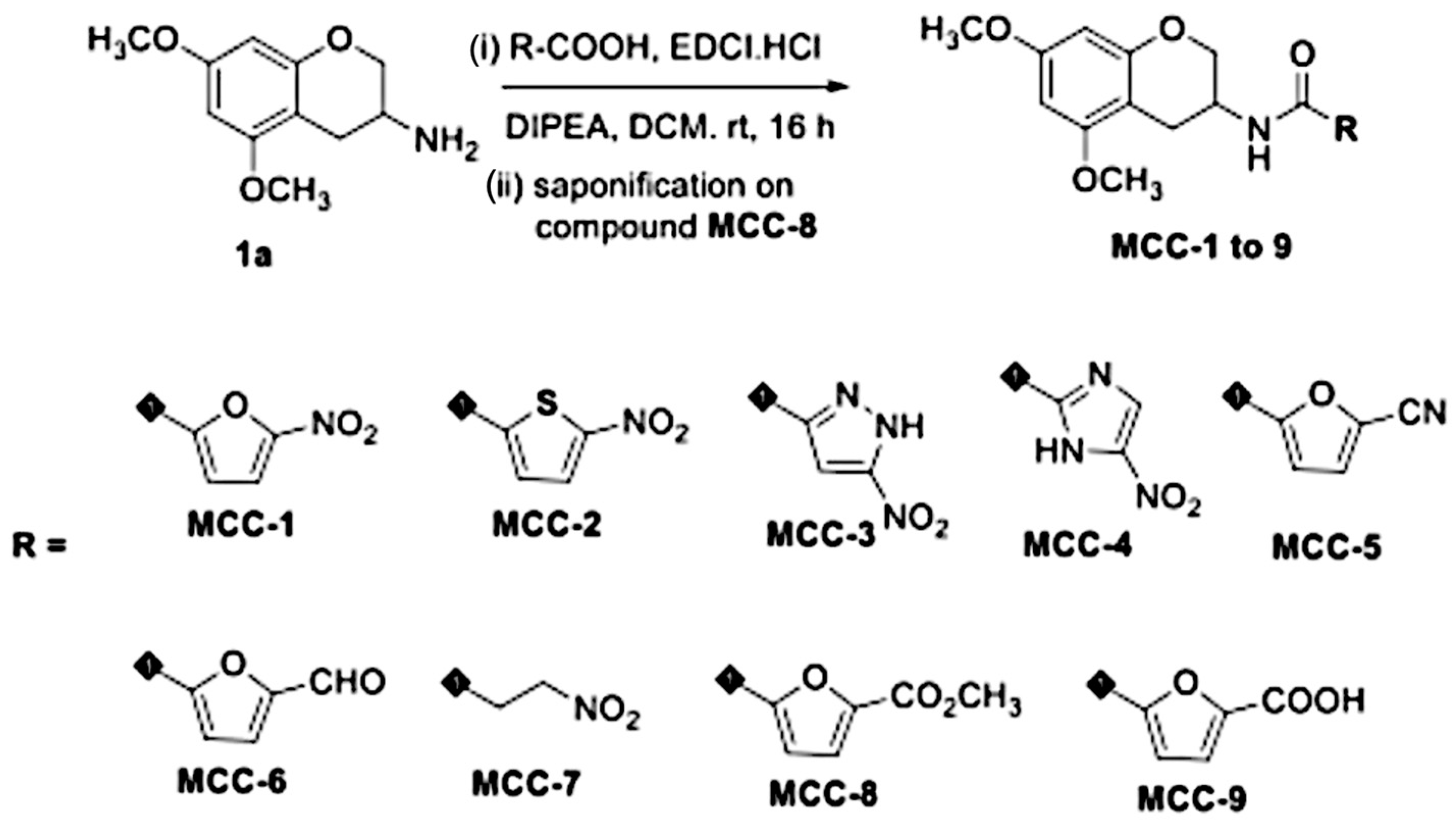
2.2. Synthesis and Evaluation of the Chemical Properties of EGCG Analogs

EGCG was selected as an initial hit compound for a ligand-based drug-design approach to optimize physicochemical properties. The major issue with polyphenolic EGCG’s chemical structure is low cellular permeability, which is potentially attributed to its low cLogD values (Table 1). The ideal cLogD values and other drug-like properties, as indicated in Table 1, were evaluated using the StarDrop/ADMET-predictor software [21]. The molecular structure of EGCG consists of ring-A, B, C, and D with two stereocenters at C-2 and C-3 positions. Maintaining the configuration of these two stereocenters is crucial for biological activity. The major culprit involved in changing the configuration of these stereocenters (epimerization) is the B-ring of EGCG (Figure 1), leading to reduced biological activity of EGCG. Based on this finding, we designed 10 compounds (MCC-1 to 10) in which the B-ring of EGCG was removed, thereby reducing one chiral center. This change additionally led to decreased polar surface area and increased the lipophilic character of the molecule. To further increase the metabolic stability of our novel EGCG analogs, the ester linkage between the C and D rings of EGCG was replaced with a more stable amide functionality. All EGCG analogs were synthesized from a key intermediate compound 1a, which was made in bulk using a published method [22]. Intermediate compound 1a was coupled with corresponding carboxylic acids mediated by EDC HCl to obtain target compounds MCC-1 to 9 in modest to good amounts of yield, as depicted in Scheme 1 [23]. Finally, amide functionality was also replaced by one of its bioisosteres, such as that in the triazole compound (MCC-10). This compound was synthesized using azide intermediate 1b, which was further reacted with 5-ethynyl-1,2,3-trimethoxybenzene under standard click chemistry conditions as depicted in Scheme 2. All synthesized compounds were well characterized by NMR and LC-MS/MS methods, and the purity of these compounds was determined by analytical HPLC methods. Compounds with >95% purity were used for biological assays. Synthetic procedures and analytical data of these new analogs are included in the Supplementary Materials.
All assays to evaluate the absorption, distribution, metabolism, and excretion (ADME) properties of our lead analogs were performed following published methods. Solubility testing was performed as described in [24]. Membrane permeability and absorption of EGCG analogs were determined in Caco-2 cells (ATCC) [25,26]. Permeability was expressed as the apparent permeability coefficient (Papp), which was calculated using the following equation: Papp = (dQ/dt/C0 × A), where dQ/dt is the rate of permeation of EGCG analogs across the cells, C0 is the concentration in the donor compartment at time zero, and A is the area of the cell monolayer. The efflux ratio (ER) was calculated as the ratio of basolateral-to-apical apparent permeability (Papp, B-A) to apical-to-basolateral apparent permeability (Papp, A-B). The distribution coefficient (LogD) of EGCG analogs in the 1-octanol/buffer pH 7.4 system was determined by the shake flask method. The plasma protein binding (PPB) of EGCG analogs in mice (CD-1; Pooled: Male and Female, BioIVt, New York, NY, USA) was determined using the equilibrium dialysis method [27]. Liver microsomal stability was determined using human microsomes (Pooled, Male and Female, Cat. No. 452117, Corning, New York, NY, USA) and mouse microsomes (Pooled, Male CD-1; Cat. No. M1000). The in vitro half-life (in vitro t1/2) was determined from the slope value, in vitro t1/2 = −(0.693/k), using the reference methods [28,29].

2.3. Bacterial Strains

S. aureus strains used in this study included one reference methicillin-resistant strain (MRSA LAC USA300) with a GFP reporter (a gift from Dr. Bas Surewaard) and four clinical strains representing different resistance phenotypes and associated patient outcomes (persistent infection and death vs. prompt resolution) [7]. Clinical strains were obtained from adult patients hospitalized for S. aureus bacteremia who were enrolled in a multi-center prospective observational study approved by the Institutional Review Board of the University of Southern California [5,6]. All strains were stored at −80 °C until use and once thawed were grown on Tryptic Soy Agar plates (TSA, Sigma, St. Louis, MO, USA). Additional information on the clinical isolates tested can be found in the Supplementary Materials, Table S1.

2.4. Antimicrobial Susceptibility Testing

Direct antimicrobial activity of EGCG and EGCG analogs was assessed by broth microdilution assays to determine minimum inhibitory concentrations (MICs) against the study strains, following the CLSI standardized methodology [30]. EGCG or EGCG analogs were tested over a range of doubling concentrations, from 1.56 µg/mL to 200 µg/mL, in triplicate. EGCG was dissolved in Hypure cell culture water (Cytiva, Marlborough, MA), while MCC-1 and MCC-2 required dissolution in 70% DMSO for every mg of compound (Sigma, St. Louis, MO, USA). Following incubation, growth was visually inspected, with the MIC defined as the lowest concentration where no growth of S. aureus was observed. MIC testing was performed in biological duplicates. Additionally, S. aureus growth curves were performed in the absence or presence of EGCG, MCC-1, or MCC-2 at ⅟16 ×, ⅟8 ×, ⅟4 ×, ⅟2 ×, and 1 × MICs. The LAC USA300 strain was incubated in TSB overnight at 37 °C. Broth cultures were adjusted to 0.5 McFarland, then further diluted 1:100 in Cation-Adjusted Mueller Hinton Broth (CAMHB) (BD, Sparks, MD, USA) with or without the addition of EGCG, MCC-1, or MCC-2. OD600 was taken every 15 min using a kinetic spectrophotometer plate reader (Accuris, Denver, CO, USA, 96-T) for 24 h. To rule out the effect of DMSO on bacterial growth, both MIC and growth curve assays included DMSO control treatments at equivalent DMSO concentrations found within the MCC-1 and MCC-2 treatments; no effects on growth were found at relevant concentrations of DMSO.
To evaluate the extent to which EGCG, MCC-1, and MCC-2 may potentiate the direct antimicrobial activity of antistaphylococcal beta-lactam antibiotics (oxacillin and cefazolin) against MRSA (USA300), checkerboard synergy assays were performed following published protocol in biological duplicates [31]. Combinations where no visible growth was observed, following overnight incubation at below the MICs of the respective antibiotic agent and EGCG, MCC-1, or MCC-2 when tested alone, were recorded. Additional broth microdilution testing was performed at doubling serial dilutions of antibiotics combined with EGCG, MCC-1, or MCC-2 at the fixed concentration of 6.25 µg/mL. Synergy was determined by calculating the fractional inhibitory concentration (FIC) following the published method and interpreted as follows for each combination: synergistic at FIC ≤ 0.5, additive at 0.5–1, indifferent at 1–4, and antagonistic at >4 [32]. To rule out the effect of DMSO on antibiotic efficacy, DMSO control treatments with equivalent DMSO concentrations as found within MCC-1 and MCC-2 treatments were used, and no effect on bacterial growth was observed.

2.5. Intracellular Killing Assay

Intracellular killing assays were performed following previously published protocols with minor variations and in biological duplicates [9,33,34,35]. Immortalized murine Kupffer cells (Kup5) were seeded into 48-well tissue culture-treated plates at 1.5 × 10⁵ cells/well in prepared DMEM as previously described and incubated overnight at 37 °C with 5% CO2. Seeded cells were washed with PBS and then infected with study strains at an MOI of 5 for 1.5 h at 37 °C with 5% CO2. Following infection, KCs were washed with PBS and then incubated with gentamicin at 100 µg/mL for 2 h to eliminate remaining extracellular SA. After the gentamicin incubation, the cells were washed with PBS and treated in duplicate with EGCG (100 µg/mL), MCC-1, and MCC-2 (6.25 µg/mL) alone or in combination with antibiotics at Cmax for up to 24 h at 37 °C with 5% CO2. The ¼ MIC concentration of 6.25 µg/mL (MCC-1 and MCC-2) dissolved in DMSO was chosen as the highest concentration with no effect on Kupffer cell viability to evaluate their potential as an adjunct therapy to augment Kupffer cell and antibiotic-mediated intracellular clearance of S. aureus. At 0, 10, and 24 h after treatment, KCs were washed with PBS and lysed with 100 µL of 0.1% TritonX-100 in water for 5 min at room temperature. KC lysates were serially diluted in PBS and plated on TSA for enumeration of intracellular CFU counts. To rule out the effect of DMSO on bacterial growth, DMSO control treatments with equivalent DMSO concentrations as found within MCC-1 and MCC-2 treatments were tested, and no effect on intracellular bacterial growth or on Kup5 cell viability was observed.

3. Results

3.1. Preliminary Screening and Selection of EGCG Analogs

Ten EGCG analogs were synthesized overall (Scheme 1 and Scheme 2). Analogs were screened initially for biological activity by performing MIC testing against the reference strain, MRSA LAC USA300, over the range of 0.39–200 µg/mL. Analogs that showed direct antibacterial activity with an MIC equal to or less than that of EGCG (50 µg/mL) were further evaluated. All analogs except MCC-1, MCC-2, and MCC-3 exhibited MICs > 200 µg/mL. MCC-1 and MCC-2 both exhibited MICs of 25 µg/mL, whereas MCC-3 exhibited an MIC of 100 µg/mL. Thus, only MCC-1 and MCC-2 were selected as our lead compounds and were the only compounds chosen for further evaluation.

3.2. EGCG-Lead Analog MCC-1 Exhibited More Favorable ADME (Absorption, Distribution, Metabolism, and Excretion) Properties than That of EGCG In Vitro

Lead discovery and optimization through medicinal chemistry efforts (Scheme 1 and Scheme 2) were performed to generate SAR studies on EGCG analogs to improve potency and efficacy as well as drug-like properties, such as lipophilicity (LogD) plasma protein binding (PPB) and permeability (Papp). MCC-1 was our first lead compound and therefore we investigated its in vitro absorption, distribution, metabolism, and excretion (ADME) properties. We found that the majority of the ADME properties of our first lead compound are within the targeted range (Table 2) except for microsomal stability, which is outside of the targeted range.

3.3. EGCG Analogs Demonstrate Activity Alone Against Both Clinical and Reference Strains of MRSA and MSSA

The results for EGCG, MCC-1, and MCC-2 antibacterial susceptibility testing against the study strains are shown in Table 3. MCC-1 and MCC-2 showed similar activity and are more potent than EGCG, consistently exhibiting a 2-fold lower MIC regardless of methicillin-resistant phenotype. Notably, MRSA strain HH35 demonstrated relative resistance to EGCG, MCC-1, and MCC-2 with a 2-fold higher MIC when compared to the other S. aureus strains tested.

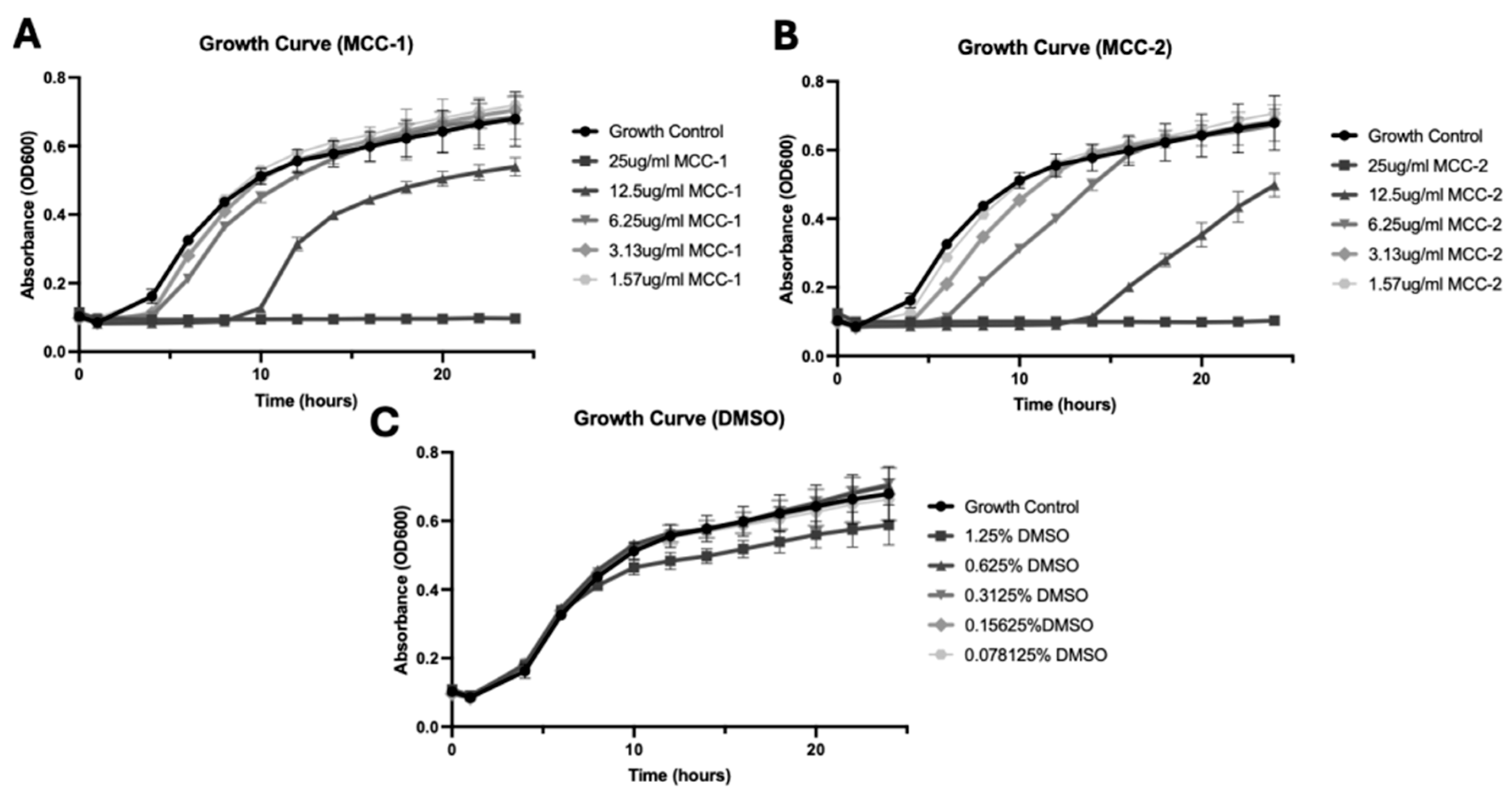
3.4. MCC-1 and MCC-2 Exhibit Dose-Dependent Growth Inhibition of S. aureus

MCC-1 and MCC-2 were tested at ⅟16 × to 1 × MIC and were shown to delay or inhibit growth of USA300 MRSA in a dose-dependent manner over 24 h, with growth delay at ½ × MIC (12.5 µg/mL) and complete growth inhibition at 1 × MIC (25 µg/mL). As MCC-1 and MCC-2 analogs are dissolved in 70% DMSO, we performed growth curve assays in the presence of DMSO alone to rule out the contribution of DMSO to growth inhibition (Figure 2). DMSO was tested at a range of concentrations from 0.078% to 1.25%, which corresponds to concentrations equivalent to that found in 1.57 to 25 µg/mL of MCC-1 and MCC-2. Similar growth patterns to the control (CAMHB alone) were observed amongst all DMSO concentrations tested, exhibiting that DMSO toxicity against SA did not contribute to EGCG analog antibacterial activity.

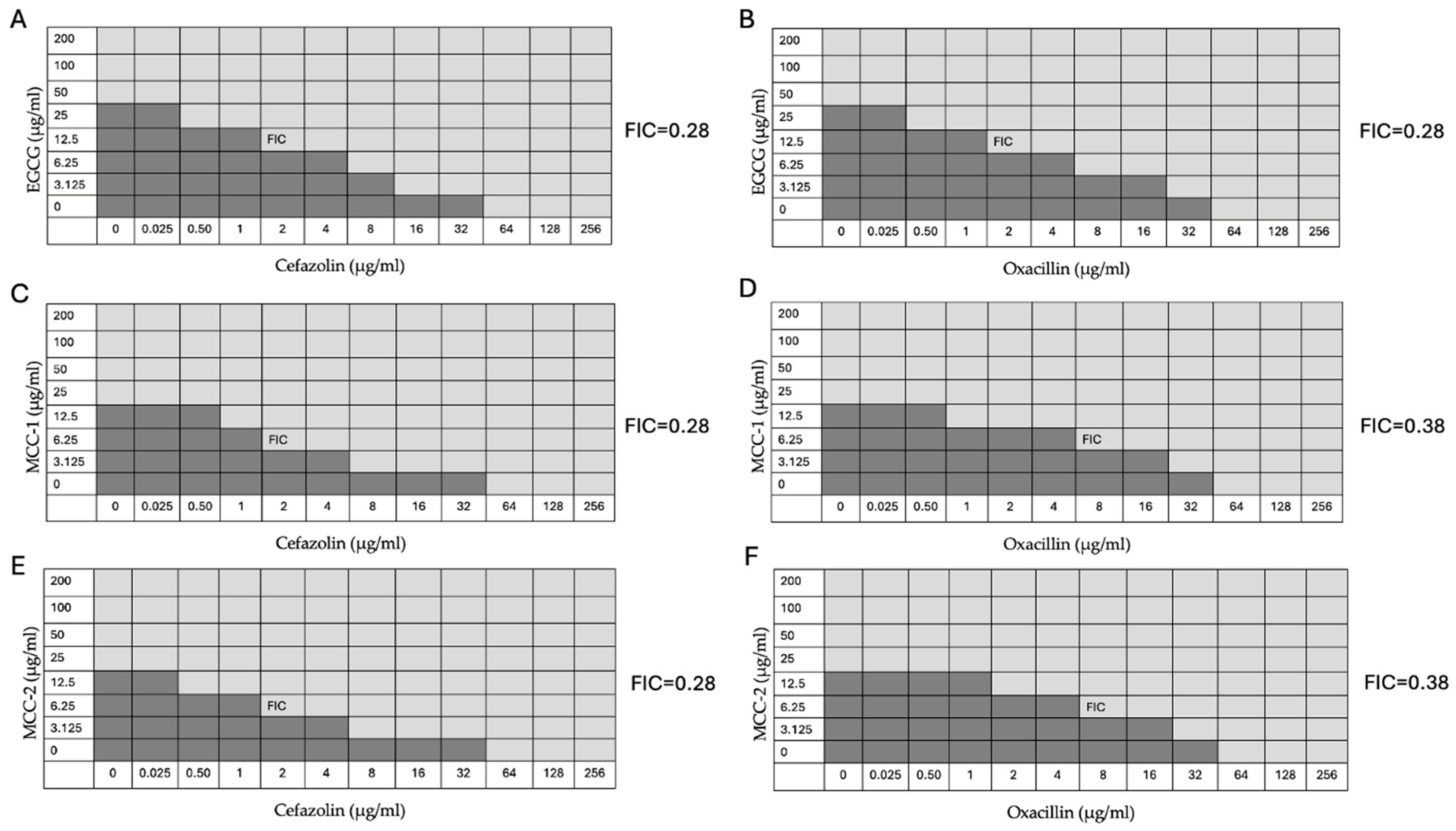
3.5. EGCG, MCC-1, and MCC-2 Restore Susceptibility of MRSA to β-lactam Antibiotics

We have shown here that EGCG, MCC-1, and MCC-2 restored the susceptibility of MRSA USA300 to oxacillin and cefazolin (Figure 3). Both cefazolin and oxacillin MICs were 64 µg/mL when tested alone against our reference MRSA strain. When combined with subinhibitory concentrations of EGCG (12.5 µg/mL), the MIC of cefazolin and oxacillin decreased from 64 µg/mL to 2 µg/mL, achieving a synergistic FIC value of 0.28 (Figure 3A,B). MCC-1 and MCC-2 at subinhibitory concentrations (6.25 µg/mL) also effectively decreased the MIC for both cefazolin and oxacillin from 64 µg/mL to 2 µg/mL and 64 µg/mL to 8 µg/mL, respectively, leading to synergistic FIC values of 0.28 and 0.38 (Figure 3C–F). As a control, DMSO at concentrations equal to that found in MCC-1 and MCC-2 solutions (0.625%) were also combined with cefazolin and oxacillin but no difference in growth inhibition was observed, demonstrating that the DMSO in which the analogs were dissolved did not contribute to the synergy observed.

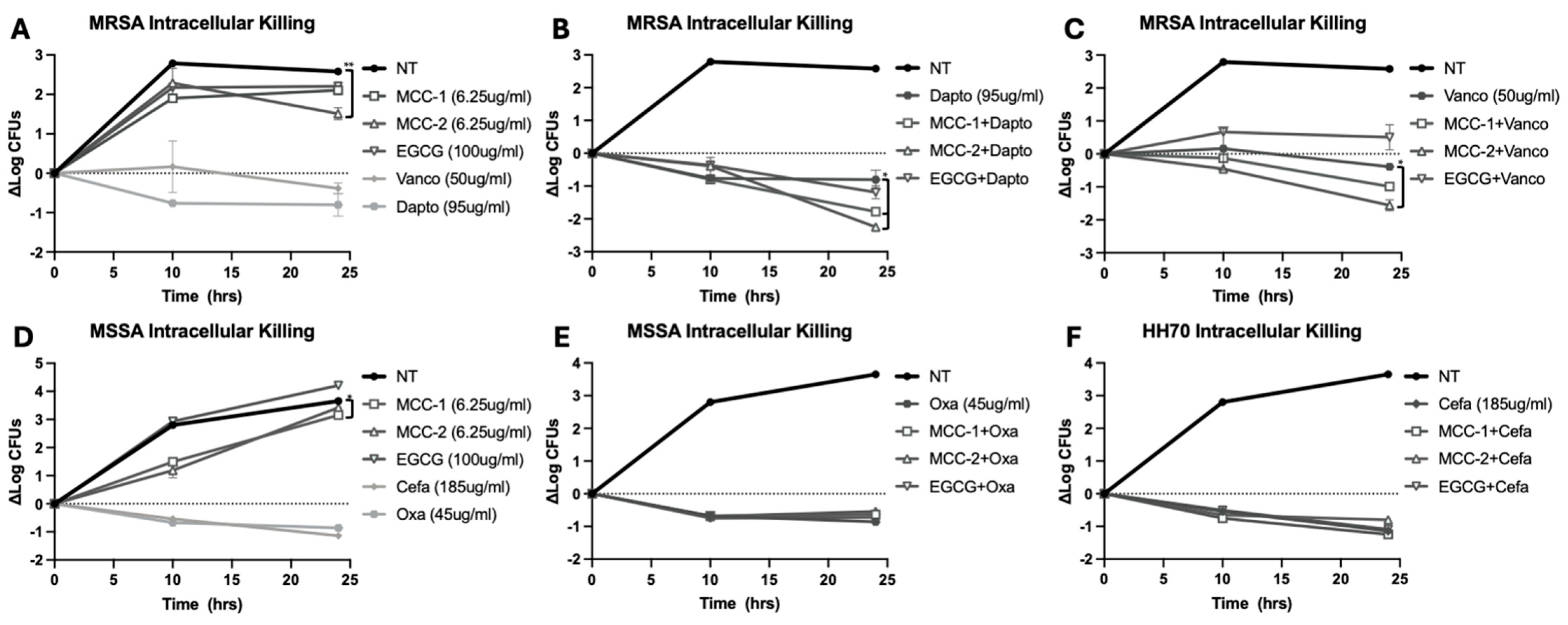
3.6. MCC-1 and MCC-2 at Subinhibitory Concentrations Decrease Intracellular S. aureus Alone and in Combination with Antibiotics

To evaluate the potential for MCC-1 and MCC-2 to augment Kupffer cell and antibiotic-mediated killing of intracellular MRSA and MSSA, Kup5 cells infected with reference MRSA strain USA300 or clinical MSSA strain HH70 were treated with either analog at subinhibitory concentration (6.25 µg/mL) and at 2 × MIC of EGCG (100 µg/mL) with or without the addition of antistaphylococcal antibiotics at human Cmax.
When used alone against MRSA and MSSA at subinhibitory concentrations, EGCG analogs were not effective at reducing intracellular CFU counts when compared to baseline (T0). However, when MCC-1 and MCC-2 were combined with the antistaphylococcal antibiotics vancomycin and daptomycin, both analogs effectively enhanced the killing of intracellular MRSA when compared to antibiotic treatment alone (Figure 4A–C). In contrast, an antagonistic effect was observed when EGCG was combined with the same antibiotics, leading to increased intracellular growth of MRSA, except in the case of daptomycin where EGCG treatment had no effect. On the other hand, when used alone or combined with cefazolin and oxacillin against MSSA, neither EGCG nor EGCG analogs were effective at enhancing intracellular clearance of the bacteria (Figure 4D–F).

4. Discussion

Epigallocatechin gallate (EGCG) has been shown in previous studies to possess therapeutic potential in a multitude of disease areas including the treatment of bacterial infections. The direct antistaphylococcal activity and antivirulence properties of EGCG support its consideration as a promising adjunct therapy for S. aureus bacteremia. However, its therapeutic potential is limited by its eight hydroxyl groups, which confer EGCG with high polarity, high hydrophilicity, and high molecular weight, rendering it a poorly bioavailable compound with low cellular penetration. Furthermore, these hydroxyl groups are prone to oxidation, limiting the duration of pharmacologic activity. Given that the oxidant scavenging ability of EGCG—a key desirable property for non-antimicrobial use—is attributed to the hydroxyl groups, prior attempts to improve its biochemical properties have avoided modifications of the hydroxyl groups. However, for the use of EGCG as an adjunct treatment to potentiate antibiotic-mediated intracellular killing of S. aureus, such antioxidative effects are not considered essential. Thus, we explored a novel structural design of EGCG analogs that included the removal of the hydroxyl groups. Through computer modeling and in vitro experiments, we demonstrated that the EGCG analogs synthesized in this study exhibit improved drug-like properties without loss of antibacterial activity despite the removal of such hydroxyl groups. Importantly, the EGCG analogs at subinhibitory concentrations potentiated the antibacterial activity of available antistaphylococcal antibiotics against intracellular S. aureus.
Our first set of structure–activity relationship (SAR) studies on EGCG yielded promising results, in particular, the membrane permeability of our synthesized analogs increased dramatically in comparison to EGCG. During this first set of SAR studies, our major focus was to remove ring-B of EGCG and replace ring-D with heterocyclic, alkyl moieties. We found positive results with our lead compounds MCC-1 and MCC-2 with respect to antibacterial activity against extracellular and intracellular S. aureus in tandem with promising drug-like properties, with the exception of low MLM t1/2 and high clearance rates. The future chemical analog design will focus on the improvement in microsomal stability, decreasing the rate of intrinsic clearance and maintaining or improving its antimicrobial potency. During the next set of SAR studies, we will target the amide bond and the ring-D and ring-A structures of the current EGCG analogs representing the sites of vulnerabilities to increase the stability of future analogs, thus optimizing lead compounds for in vivo proof-of-concept studies.
EGCG and our novel analogs, MCC-1 and MCC-2, demonstrated direct antimicrobial activity against both reference and clinical strains of MRSA and MSSA. In vitro experiments showed that the parent compound, EGCG, was successful at inhibiting the growth of clinical SA isolates at a concentration of 50 µg/mL with the exception of one strain. This observation is consistent with previously published studies, which showed that EGCG has an MIC ≤ 100 µg/mL against a variety of MRSA and MSSA isolates [11]. In comparison to EGCG, our novel EGCG analogs, MCC-1 and MCC-2, demonstrated improvements in biochemical properties as well as direct antibacterial activity, with both analogs inhibiting growth of the tested strains at 2-fold lower concentrations. Following a similar pattern as EGCG, the single strain that showed relative resistance to EGCG also demonstrated a parallel increase in MIC to MCC-1 and MCC-2. Additionally, when combined at subinhibitory concentrations (¼ × MIC) with β-lactam antibiotics such as oxacillin and cefazolin, EGCG and our lead analogs demonstrated synergy (FIC 0.28–0.38) in a checkerboard assay, restoring MRSA susceptibility to the β-lactam agents. However, neither EGCG nor the lead analogs showed synergy when combined with vancomycin and daptomycin against MRSA, which is consistent with previously published literature [11].
The therapeutic target of EGCG and our EGCG analogs to inhibit growth or potentiate β-lactam-mediated killing of S. aureus is currently unknown. Previous literature suggests a variety of different mechanisms of action, which are generally related to the damage of the S. aureus cell membrane or cell wall. Kitichalermkiat et al. reported that treatment of S. aureus with EGCG decreased SA membrane potential and upregulated genes related to SA cell membrane repair [12]. Others found that the antimicrobial activity of EGCG against SA was inhibited by the addition of peptidoglycan in a dose-dependent manner, suggesting that peptidoglycan on the SA cell wall may be a target of EGCG and that adding exogenous peptidoglycan competes for EGCG binding [36]. Furthermore, EGCG appeared to exert more potent antimicrobial activity against Gram-positive bacteria than Gram-negative bacteria, as the former contains a thicker peptidoglycan matrix. Zhao et al. further support the purported peptidoglycan-related mechanism and not a more general cell wall-based mechanism, as only β-lactams and not other non-PBP-based cell wall damaging antibiotics, such as DL-cycloserine, have increased efficacy when combined with EGCG [11]. Additionally, EGCG does not show synergy with antibiotics that act through bacterial protein or nucleic acid synthesis, such as fluoroquinolones [11]. Since the primary goal in designing our novel analogs is to retain the biological activity of EGCG while improving its biochemical properties, the EGCG analogs are hypothesized to act on the same target(s) as that of EGCG. We speculated that the improved activity observed with MCC-1 and MCC-2 over EGCG may be attributed to their enhanced lipophilicity and lower molecular weight, thereby allowing them to reach higher concentrations at the site of action where the target is present or bind more efficiently to the target in question rather than an altered target. Further studies are planned to elucidate the molecular targets of EGCG analogs in the context of intracellular antistaphylococcal action.
Alternatively, it is also possible that EGCG and its analogs may act on the biosynthesis of teichoic acid (TA), which, like peptidoglycan, is a cell wall polymer contained in the cell walls of SA. Brown et al. have shown that the glycosyltransferase encoded by the gene, tarS, is responsible for decorating teichoic acids with the substituent β-O-N-acetyl-D-glucosamine (β-O-GlcNac), which is required to maintain MRSA resistance to β-lactams, likely through its influence on cell wall milieu related to the packing density of the peptidoglycan matrix and/or its ability to act as a scaffold for cell envelope associated proteins, including PBP2a [37]. Deletion of the tarS gene restores MRSA sensitivity to β-lactam agents but not to other cell wall-active agents such as vancomycin. Considering that EGCG and its analogs were found to restore the susceptibility of cefazolin and oxacillin to MRSA but to not potentiate vancomycin- and daptomycin-mediated killing against extracellular MRSA, tarS may be a putative target for EGCG and its analogs. Not surprisingly, as the native transpeptidases that function to cross-link peptidoglycan strands are susceptible to β-lactam agents, the addition of EGCG or its analogs is not expected to alter MSSA sensitivity as the putative tarS target is only required for the MRSA phenotype. Importantly, we have shown here that the hydroxyl groups on EGCG are not essential for its cell wall or cell membrane interference, as demonstrated by the efficacy of our novel analogs. Notably, the observed increase in potency of MCC-1 and MCC-2 when used alone, compared to EGCG, suggests that the structural changes we made may lead to increased target binding. Interestingly, MCC-1 and MCC-2 were able to restore sensitivity to oxacillin-resistant SA at subinhibitory concentrations, which suggests that different mechanisms of action likely exist for their direct antibacterial effects when used alone, compared to their antibiotic-potentiating effects. Further studies will need to be performed to confirm the main target of EGCG and its analogs against S. aureus and the active moieties that best correspond to an increase in antimicrobial potency.
Despite EGCG analogs maintaining direct antibacterial activity against extracellular pathogens, the clinical challenge for improving the treatment outcome of SAB lies in the clearance of persistent S. aureus within intracellular niches. To overcome the inability of EGCG to achieve adequate intracellular penetration, we have demonstrated in this study that structural modifications to EGCG, which included the removal of hydroxyl groups, led to improved drug-like properties and effective killing of extracellular as well as intracellular S. aureus using an in vitro macrophage infection model. Even at subinhibitory concentrations that did not inhibit or delay extracellular growth of extracellular MRSA, our lead EGCG analogs were able to potentiate the clearance of intracellular MRSA from infected Kupffer cells when combined with antistaphylococcal antibiotics (vancomycin, daptomycin, cefazolin, and oxacillin). In contrast, even at concentrations 16x higher than that of MCC-1 and MCC-2, EGCG showed no potentiation when combined with antibiotics in clearing intracellular MRSA.
Previous studies show that EGCG treatment not only increases MRSA susceptibility to cell wall-active antibiotics but also decreases SA tolerance to osmotic pressure and oxidative stress [11]. Thus, at sub-MIC concentrations, our EGCG analogs may exert immunoactivating effects that increase the efficacy of Kupffer cell-mediated reactive oxidant species-based killing or antivirulence effects that target strain-specific virulence factors. Intriguingly, the beneficial effects of our EGCG analogs were observed only with the intracellular clearance of MRSA and not MSSA. One possible explanation is that MRSA and MSSA may utilize different virulence or host evasion strategies for intracellular survival, which our EGCG analogs may be selectively targeting. Indeed, previous studies have demonstrated that EGCG may modulate macrophage 67LR/p38/JNK signaling, offering a potential immunomodulating effect to more efficiently control bacterial growth, while also exhibiting antivirulence potential by decreasing transcription of a variety of virulence factors [12,13,38]. Further evaluation of the immunoactivating and antivirulence potential of our EGCG analogs is planned.
We note several limitations when considering the overall positive antibacterial efficacy of our EGCG-lead analogs, MCC-1 and MCC-2. First, both analogs require high DMSO concentrations for dissolution. While this does not interfere with the antibacterial assays as the study strains had an MIC to DMSO much higher than the concentrations present in the EGCG analog solutions, Kup5 cell viability was affected by DMSO, thereby limiting our highest EGCG analog concentration to 6.25 µg/mL in order to maintain 100% cell viability. It is important to note that the observed decrease in cell viability corresponded to that seen in cells treated with DMSO alone, suggesting that cell toxicity is due to DMSO and not the EGCG analogs. Additionally, murine macrophages, including Kupffer cells, have been shown to have altered bacteria–cell interactions with human-adapted strains of S. aureus with murine cells not being able to effectively clear human-adapted S. aureus, regardless of inoculum amount, and with S. aureus virulence factors not being as effective against murine cells as well [39]. It is possible that testing with a human cell line may yield greater or reduced efficacy than observed in this study. Nonetheless, positive results from this study are encouraging and serve as the basis to undertake further design, synthesis, and testing of additional EGCG analogs with the goal of increasing the aqueous solubility of the compounds to overcome solvent limitation with DMSO. Furthermore, lead compounds will undergo assays performed not only with murine Kupffer cells but also with primary human Kupffer cells and THP-1 differentiated macrophages, in order to more accurately investigate clinically relevant interactions between human cells and SA.
Taken together, EGCG analogs show promise as an adjunct therapy for SAB, with the potential to not only increase the clearance of intracellular SA through direct antibacterial effects but also through immunoactivating and antivirulence activity. We herein present a proof-of-concept study, demonstrating that the structural modification of EGCG is an effective method for improving the biochemical properties of EGCG as well as its antistaphylococcal activity, which supports the further design, synthesis, and testing of additional EGCG analogs with the ultimate goal of treating intracellular S. aureus to effect prompt clearance of SAB.

5. Patents

The authors declare the following competing financial interests: A.W.-B., S.K.A., R.G. and Vy-N. submitted the US provisional patent application 63/639,449, dated 26 April 2024, and are listed as inventors.

Supplementary Materials

The following supporting information can be downloaded at https://www.mdpi.com/article/10.3390/applmicrobiol4040107/s1: Table S1. Synthesis instructions for EGCG analogs.

Author Contributions

Conceptualization, A.W.-B.; methodology, R.G., V.N., S.K.A. and A.W.-B.; formal analysis, S.K.A. and A.W.-B.; investigation, R.G. and V.N.; resources, V.N.; writing—original draft preparation, R.G. and V.N.; writing—review and editing, S.K.A. and A.W.-B.; supervision, A.W.-B.; funding acquisition, A.W.-B. All authors have read and agreed to the published version of the manuscript.

Funding

This research received no external funding.

Data Availability Statement

The original contributions presented in this study are included in the article and the Supplementary Materials. Further inquiries can be directed to the corresponding author.

Conflicts of Interest

The authors declare no conflicts of interest.

References

  1. Naber, C.K. Staphylococcus aureus Bacteremia: Epidemiology, Pathophysiology, and Management Strategies. Clin. Infect. Dis. 2009, 48, S231–S237. [Google Scholar] [CrossRef] [PubMed]
  2. Ikuta, K.S.; Swetschinski, L.R.; Robles Aguilar, G.; Sharara, F.; Mestrovic, T.; Gray, A.P.; Davis Weaver, N.; Wool, E.E.; Han, C.; Gershberg Hayoon, A.; et al. Global Mortality Associated with 33 Bacterial Pathogens in 2019: A Systematic Analysis for the Global Burden of Disease Study 2019. Lancet 2022, 400, 2221–2248. [Google Scholar] [CrossRef] [PubMed]
  3. Holland, T.L.; Bayer, A.S.; Fowler, V.G., Jr. Persistent Methicillin-Resistant Staphylococcus aureus Bacteremia: Resetting the Clock for Optimal Management. Clin. Infect. Dis. 2022, 75, 1668–1674. [Google Scholar] [CrossRef]
  4. van Hal, S.J.; Jensen, S.O.; Vaska, V.L.; Espedido, B.A.; Paterson, D.L.; Gosbell, I.B. Predictors of Mortality in Staphylococcus aureus Bacteremia. Clin. Microbiol. Rev. 2012, 25, 362–386. [Google Scholar] [CrossRef]
  5. Minejima, E.; Mai, N.; Bui, N.; Mert, M.; Mack, W.J.; She, R.C.; Nieberg, P.; Spellberg, B.; Wong-Beringer, A. Defining the Breakpoint Duration of Staphylococcus aureus Bacteremia Predictive of Poor Outcomes. Clin. Infect. Dis. 2020, 70, 566–573. [Google Scholar] [CrossRef] [PubMed]
  6. Minejima, E.; Bensman, J.; She, R.C.; Mack, W.J.; Tuan Tran, M.; Ny, P.; Lou, M.; Yamaki, J.; Nieberg, P.; Ho, J.; et al. A Dysregulated Balance of Proinflammatory and Anti-Inflammatory Host Cytokine Response Early During Therapy Predicts Persistence and Mortality in Staphylococcus aureus Bacteremia. Crit. Care Med. 2016, 44, 671–679. [Google Scholar] [CrossRef] [PubMed]
  7. Surewaard, B.G.J.; Deniset, J.F.; Zemp, F.J.; Amrein, M.; Otto, M.; Conly, J.; Omri, A.; Yates, R.M.; Kubes, P. Identification and Treatment of the Staphylococcus aureus Reservoir in Vivo. J. Exp. Med. 2016, 213, 1141–1151. [Google Scholar] [CrossRef]
  8. Pidwill, G.R.; Gibson, J.F.; Cole, J.; Renshaw, S.A.; Foster, S.J. The Role of Macrophages in Staphylococcus aureus Infection. Front. Immunol. 2021, 11, 620339. [Google Scholar] [CrossRef]
  9. Beadell, B.; Yamauchi, J.; Wong-Beringer, A. Comparative in Vitro Efficacy of Antibiotics against the Intracellular Reservoir of Staphylococcus aureus. J. Antimicrob. Chemother. 2024, 79, dkae241. [Google Scholar] [CrossRef]
  10. Chacko, S.M.; Thambi, P.T.; Kuttan, R.; Nishigaki, I. Beneficial Effects of Green Tea: A Literature Review. Chin. Med. 2010, 5, 13. [Google Scholar] [CrossRef]
  11. Zhao, W.-H.; Hu, Z.-Q.; Okubo, S.; Hara, Y.; Shimamura, T. Mechanism of Synergy between Epigallocatechin Gallate and β-Lactams against Methicillin-Resistant Staphylococcus aureus. Antimicrob. Agents Chemother. 2001, 45, 1737–1742. [Google Scholar] [CrossRef]
  12. Kitichalermkiat, A.; Katsuki, M.; Sato, J.; Sonoda, T.; Masuda, Y.; Honjoh, K.-I.; Miyamoto, T. Effect of Epigallocatechin Gallate on Gene Expression of Staphylococcus aureus. J. Glob. Antimicrob. Resist. 2020, 22, 854–859. [Google Scholar] [CrossRef]
  13. Liu, C.; Hao, K.; Liu, Z.; Liu, Z.; Guo, N. Epigallocatechin Gallate (EGCG) Attenuates Staphylococcal Alpha-Hemolysin (Hla)-Induced NLRP3 Inflammasome Activation via ROS-MAPK Pathways and EGCG-Hla Interactions. Int. Immunopharmacol. 2021, 100, 108170. [Google Scholar] [CrossRef]
  14. Warden, B.A.; Smith, L.S.; Beecher, G.R.; Balentine, D.A.; Clevidence, B.A. Catechins Are Bioavailable in Men and Women Drinking Black Tea throughout the Day. J. Nutr. 2001, 131, 1731–1737. [Google Scholar] [CrossRef]
  15. Chow, H.H.; Cai, Y.; Alberts, D.S.; Hakim, I.; Dorr, R.; Shahi, F.; Crowell, J.A.; Yang, C.S.; Hara, Y. Phase I Pharmacokinetic Study of Tea Polyphenols Following Single-Dose Administration of Epigallocatechin Gallate and Polyphenon E. Cancer Epidemiol. Biomark. Prev. 2001, 10, 53–58. [Google Scholar]
  16. Chow, H.-H.S.; Cai, Y.; Hakim, I.A.; Crowell, J.A.; Shahi, F.; Brooks, C.A.; Dorr, R.T.; Hara, Y.; Alberts, D.S. Pharmacokinetics and Safety of Green Tea Polyphenols after Multiple-Dose Administration of Epigallocatechin Gallate and Polyphenon E in Healthy Individuals. Clin. Cancer Res. 2003, 9, 3312–3319. [Google Scholar]
  17. Ullmann, U.; Haller, J.; Decourt, J.; Girault, N.; Girault, J.; Richard-Caudron, A.; Pineau, B.; Weber, P. A Single Ascending Dose Study of Epigallocatechin Gallate in Healthy Volunteers. J. Int. Med. Res. 2003, 31, 88–101. [Google Scholar] [CrossRef]
  18. Shinde, S.; Lee, L.H.; Chu, T. Inhibition of Biofilm Formation by the Synergistic Action of EGCG-S and Antibiotics. Antibiotics 2021, 10, 102. [Google Scholar] [CrossRef]
  19. Zhao, J.; Qian, D.; Zhang, L.; Wang, X.; Zhang, J. Improved Antioxidative and Antibacterial Activity of Epigallocatechin Gallate Derivative Complexed by Zinc Cations and Chitosan. RSC Adv. 2024, 14, 10410–10415. [Google Scholar] [CrossRef]
  20. Wang, C.; Xiao, R.; Yang, Q.; Pan, J.; Cui, P.; Zhou, S.; Qiu, L.; Zhang, Y.; Wang, J. Green Synthesis of Epigallocatechin Gallate-Ferric Complex Nanoparticles for Photothermal Enhanced Antibacterial and Wound Healing. Biomed. Pharmacother. 2024, 171, 116175. [Google Scholar] [CrossRef] [PubMed]
  21. Tyzack, J.D.; Hunt, P.A.; Segall, M.D. Predicting Regioselectivity and Lability of Cytochrome P450 Metabolism Using Quantum Mechanical Simulations. J. Chem. Inf. Model. 2016, 56, 2180–2193. [Google Scholar] [CrossRef] [PubMed]
  22. Khandelwal, A.; Hall, J.; Blagg, B.S.J. Synthesis and Structure Activity Relationships of EGCG Analogues, A Recently Identified Hsp90 Inhibitor. J. Org. Chem. 2013, 78, 7859–7884. [Google Scholar] [CrossRef] [PubMed]
  23. Li, Q.; Zhang, S.-Y.; He, G.; Ai, Z.; Nack, W.A.; Chen, G. Copper-Catalyzed Carboxamide-Directed Ortho Amination of Anilines with Alkylamines at Room Temperature. Org. Lett. 2014, 16, 1764–1767. [Google Scholar] [CrossRef] [PubMed]
  24. Mondal, S.K.; Mondal, N.B.; Banerjee, S.; Mazumder, U.K. Determination of Drug-like Properties of a Novel Antileishmanial Compound: In Vitro Absorption, Distribution, Metabolism, and Excretion Studies. Indian. J. Pharmacol. 2009, 41, 176–181. [Google Scholar] [CrossRef] [PubMed]
  25. Pasha, M.K.; Jayaraman, R.; Reddy, V.P.; Yeo, P.; Goh, E.; Williams, A.; Goh, K.C.; Kantharaj, E. Preclinical Metabolism and Pharmacokinetics of SB1317 (TG02), a Potent CDK/JAK2/FLT3 Inhibitor. Drug Metab. Lett. 2012, 6, 33–42. [Google Scholar] [CrossRef] [PubMed]
  26. Hubatsch, I.; Ragnarsson, E.G.E.; Artursson, P. Determination of Drug Permeability and Prediction of Drug Absorption in Caco-2 Monolayers. Nat. Protoc. 2007, 2, 2111–2119. [Google Scholar] [CrossRef] [PubMed]
  27. Banker, M.J.; Clark, T.H.; Williams, J.A. Development and Validation of a 96-Well Equilibrium Dialysis Apparatus for Measuring Plasma Protein Binding. J. Pharm. Sci. 2003, 92, 967–974. [Google Scholar] [CrossRef] [PubMed]
  28. Davies, B.; Morris, T. Physiological Parameters in Laboratory Animals and Humans. Pharm. Res 1993, 10, 1093–1095. [Google Scholar] [CrossRef] [PubMed]
  29. Barter, Z.E.; Bayliss, M.K.; Beaune, P.H.; Boobis, A.R.; Carlile, D.J.; Edwards, R.J.; Houston, J.B.; Lake, B.G.; Lipscomb, J.C.; Pelkonen, O.R.; et al. Scaling Factors for the Extrapolation of in Vivo Metabolic Drug Clearance from in Vitro Data: Reaching a Consensus on Values of Human Microsomal Protein and Hepatocellularity per Gram of Liver. Curr. Drug Metab. 2007, 8, 33–45. [Google Scholar] [CrossRef]
  30. EM100 Connect—CLSI M100 ED34:2024. Available online: https://em100.edaptivedocs.net/GetDoc.aspx?doc=CLSI%20M100%20ED34:2024&scope=user (accessed on 10 September 2024).
  31. Bellio, P.; Fagnani, L.; Nazzicone, L.; Celenza, G. New and Simplified Method for Drug Combination Studies by Checkerboard Assay. MethodsX 2021, 8, 101543. [Google Scholar] [CrossRef]
  32. Meletiadis, J.; Pournaras, S.; Roilides, E.; Walsh, T.J. Defining Fractional Inhibitory Concentration Index Cutoffs for Additive Interactions Based on Self-Drug Additive Combinations, Monte Carlo Simulation Analysis, and In Vitro-In Vivo Correlation Data for Antifungal Drug Combinations against Aspergillus fumigatus. Antimicrob. Agents Chemother. 2010, 54, 602–609. [Google Scholar] [CrossRef] [PubMed]
  33. Peyrusson, F.; Nguyen, T.K.; Buyck, J.M.; Lemaire, S.; Wang, G.; Seral, C.; Tulkens, P.M.; Van Bambeke, F. In Vitro Models for the Study of the Intracellular Activity of Antibiotics. Methods Mol. Biol. 2021, 2357, 239–251. [Google Scholar] [CrossRef] [PubMed]
  34. Backert, S.; Hofreuter, D. Molecular Methods to Investigate Adhesion, Transmigration, Invasion and Intracellular Survival of the Foodborne Pathogen Campylobacter Jejuni. J. Microbiol. Methods 2013, 95, 8–23. [Google Scholar] [CrossRef]
  35. Sharma, A.; Puhar, A. Gentamicin Protection Assay to Determine the Number of Intracellular Bacteria during Infection of Human TC7 Intestinal Epithelial Cells by Shigella Flexneri. Bio Protoc. 2019, 9, e3292. [Google Scholar] [CrossRef]
  36. Yoda, Y.; Hu, Z.-Q.; Shimamura, T.; Zhao, W.-H. Different Susceptibilities of Staphylococcus and Gram-Negative Rods to Epigallocatechin Gallate. J. Infect. Chemother. 2004, 10, 55–58. [Google Scholar] [CrossRef]
  37. Brown, S.; Xia, G.; Luhachack, L.G.; Campbell, J.; Meredith, T.C.; Chen, C.; Winstel, V.; Gekeler, C.; Irazoqui, J.E.; Peschel, A.; et al. Methicillin Resistance in Staphylococcus aureus Requires Glycosylated Wall Teichoic Acids. Proc. Natl. Acad. Sci. USA 2012, 109, 18909–18914. [Google Scholar] [CrossRef]
  38. Yang, Y.; Han, X.; Chen, Y.; Wu, J.; Li, M.; Yang, H.; Xu, W.; Wei, L. EGCG Induces Pro-Inflammatory Response in Macrophages to Prevent Bacterial Infection through the 67LR/P38/JNK Signaling Pathway. J. Agric. Food Chem. 2021, 69, 5638–5651. [Google Scholar] [CrossRef]
  39. Trübe, P.; Hertlein, T.; Mrochen, D.M.; Schulz, D.; Jorde, I.; Krause, B.; Zeun, J.; Fischer, S.; Wolf, S.A.; Walther, B.; et al. Bringing Together What Belongs Together: Optimizing Murine Infection Models by Using Mouse-Adapted Staphylococcus aureus Strains. Int. J. Med. Microbiol. 2019, 309, 26–38. [Google Scholar] [CrossRef]
Figure 1. Molecular structure of (-)- Epigallocatechin gallate (EGCG).
Applmicrobiol 04 00107 g001
Scheme 1. Synthesis of N-(Chroman-3-yl) benzamide analogs (1–9).
Applmicrobiol 04 00107 sch001
Scheme 2. Synthesis of 1-(5,7-methylchromen-3-yl)-4-(3,4,5-trimethoxyphenyl-analog (10).
Applmicrobiol 04 00107 sch002
Figure 2. EGCG, MCC-1, and MCC-2 dose-dependently inhibit growth of USA300 MRSA. USA300 MRSA was incubated in CAMHB supplemented with ⅟16, ⅟8, ⅟4, ⅟2, and 1 × MIC concentrations of EGCG, MCC-1, and MCC-2. Bacterial growth was measured through OD600 every 15 min for 24 h utilizing a kinetic spectrophotometric plate reader to assess growth over time. (A) Growth curve of USA300 S. aureus treated with MCC-1. (B) Growth curve of USA300 S. aureus treated with MCC-2. (C) Growth curve of USA300 S. aureus treated with EGCG.
Applmicrobiol 04 00107 g002
Figure 3. MIC of cefazolin and oxacillin against USA300 when used alone or in combination with EGCG, MCC-1, and MCC-2. USA300 MRSA was incubated for 20 h with CAMHB supplemented with 2× serial dilutions of cefazolin (A,C,E) or oxacillin (B,D,F) at a concentration range of 0.5 µg/mL to 256 µg/mL, in combination with EGCG (A,B), MCC-1 (C,D), and MCC-2 (E,F) at a concentration range of 3.125 µg/mL to 200 µg/mL. The MIC was determined by visual inspection with lowest concentration of antibiotic where no growth was observed defined as the MIC. Fractional inhibitory concentration (FIC) values ≤ 0.5 are considered synergistic, >0.5–1 additive, 1–4 indifferent, and >4 antagonistic.
Applmicrobiol 04 00107 g003
Figure 4. EGCG analogs potentiate intracellular killing of S. aureus when used alone or in combination with clinically relevant antistaphylococcal antibiotics. Immortalized murine Kupffer cells (Kup5) infected with USA300 MRSA (AC) were left untreated (NT) or treated with MCC-1 (6.25 µg/mL), MCC-2 (6.25 µg/mL), EGCG (100 µg/mL) +/− Cmax concentrations of vancomycin (Vanco: 50 µg/mL) (C) and daptomycin (Dapto: 95 µg/mL) (B). Kup5 cells were also infected with HH70 MSSA (DF) and were left untreated or treated with the same concentrations of MCC-1, MCC-2, EGCG +/− cefazolin (Cefa: 85 µg/mL) and oxacillin (Oxa: 45 µg/mL) (E). ** indicates p < 0.01; * indicates p < 0.05 in one-way ANOVA with Tukey’s post hoc.
Applmicrobiol 04 00107 g004
Table 1. Predicted lipophilicity (cLogD values) of EGCG and EGCG analogs (MCC-1 through MCC-10).
EGCG0.65
MCC-12.93
MCC-23.18
MCC-32.56
MCC-42.57
MCC-52.57
MCC-62.50
MCC-71.94
MCC-82.69
MCC-91.95
MCC-103.58
Table 2. In vitro absorption, distribution, metabolism, and excretion (ADME) properties of MCC-1.
In Vitro ADME AssayTarget ValuesEGCGMCC-1MCC-2
LogD2–4>42.673.64
PPB/Bound<95%92.4 ± 3.186.90ND
Papp (Caco2) 10−6 cm/s≤10.130.910.89
Sol. (µM)>10 µM>30016.892.88
HLM t½min>30 minND17.4314.54
MLM t½min>30 minND<4.51<4.51
Note: LogD: measurement of lipophilicity. PPB: plasma protein binding. Cell permeability was performed in Caco-2 cells. Papp: apparent permeability coefficient. Sol: solubility results of the test compound in PBS pH 7.4. HLM: human liver microsomes. MLM: mouse liver microsomes. ND = not determined.
Table 3. Minimum inhibitory concentrations (MICs, µg/mL) of EGCG and EGCG analogs against S. aureus strains.
USA300 (MRSA)HH35 (MRSA)HH37 (MRSA)HH70 (MSSA)LAC164 (MSSA)
EGCG50100505050
MCC-12550252525
MCC-22550252525
Note: Bacteria strains were incubated in CAMHB supplemented with 2× serial dilutions of EGCG or EGCG analogs (MCC-1 and MCC-2) at a concentration range between 1.56 µg/mL and 200 µg/mL for 20 h, following CLSI methodology. Growth was inspected visually and the lowest concentration where no growth was observed was defined as the MIC.
Disclaimer/Publisher’s Note: The statements, opinions and data contained in all publications are solely those of the individual author(s) and contributor(s) and not of MDPI and/or the editor(s). MDPI and/or the editor(s) disclaim responsibility for any injury to people or property resulting from any ideas, methods, instructions or products referred to in the content.

