Article

Bacillus velezensis and Paenibacillus peoriae Strains Effective as Biocontrol Agents against Xanthomonas Bacterial Spot

1
Centre Armand-Frappier Santé Biotechnologie, Institut National de la Recherche Scientifique (INRS), Laval, QC H7V 1B7, Canada
2
Center for Food Safety and Applied Nutrition, US Food and Drug Administration, College Park, MD 20740, USA
*
Author to whom correspondence should be addressed.
These authors contributed equally to this work.
Appl. Microbiol. 2023, 3(3), 1101-1119; https://doi.org/10.3390/applmicrobiol3030076
Submission received: 29 June 2023 / Revised: 16 August 2023 / Accepted: 8 September 2023 / Published: 14 September 2023

Abstract

:
Gram-negative bacteria belonging to the Xanthomonas genus include plant pathogens representing a major challenge in the field of agriculture for a wide variety of economically important crops, such as tomato, pepper, and lettuce. Due to the massive usage of agrochemicals, Xanthomonas spp. are developing resistance to copper pesticides typically used to control microbial infections. An interesting alternative approach to control bacterial phytopathogens consists of using eco-friendly biocontrol agents, often beneficial microorganisms. Here, following the targeted, broad-spectrum screening of thousands of microorganisms isolated from different environmental locations, we isolated Bacillus velezensis strain 71 and Paenibacillus peoriae strain To99 displaying potent antagonistic activity against Xanthomonas spp. We found that oxydifficidin and polymyxin A secreted by B. velezensis 71 and P. peoriae To99, respectively, are mainly responsible for the anti-Xanthomonas activity. We further evaluated the performance of cell suspensions and cell-free supernatants of these isolates in controlling tomato bacterial spot disease in growth chamber and greenhouse conditions to validate the in vitro results. The overall results demonstrate the potential of treatments based on the secondary metabolites from both isolates and their cells as an alternative to copper-based chemicals to control leaf spot diseases caused by Xanthomonas spp. phytopathogens.

1. Introduction

Bacterial spot is one of the most serious infectious diseases of tomato, pepper, lettuce, and strawberry plants, caused by four closely related species of XanthomonasX. gardneri, X. euvesicatoria, X. vesicatoria, and X. perforans [1,2,3,4]. Bacterial spot can cause dark lesions on the foliage, stems, and fruits. On leaves, with the progression of infection, the lesions coalesce to form large dead spots. Once more than 50% of the leaf is affected, it rapidly dies, which entails reduced photosynthetic capacity and consequently crop yields [4,5]. For more than 60 years, bacterial spot has been economically devastating to growers and its control has proved to be challenging, with limited efficient available measures [6]. For several decades, copper-based chemicals and their combinations, along with dithiocarbamate and ethylene bis-dithiocarbamate classes of fungicides such as mancozeb, have been used to chemically control bacterial spot disease, with only partial effectiveness [7]. Furthermore, the efficacy of these formulations is falling due to the inherent emergence of copper-tolerant plant pathogens, due to decades of use [5,8,9,10,11]. Moreover, the reiterated application of copper-based formulations may not only result in the accumulation of toxic levels of Cu in the soil but also can cause plant stress and affect the diversity of beneficial microbial communities [12].
Overall, the emergence of pathogen resistance, non-target environmental impacts, along with the consumer demand for organic food has driven research efforts into the development of effective and sustainable disease management strategies. In this regard, biological control products based on microbes have become attractive alternatives to chemical control when managing plant diseases. This has led to numerous studies exploring the possibility of using plant-associated microbiota or their metabolites to control the most important and devastating bacterial and fungal crop diseases [13,14,15].
Given the importance of managing bacterial diseases caused by Xanthomonas species in a variety of economically important crops, several studies have reported the isolation of potential biocontrol agents from phyllosphere, rhizosphere, leaf, or endosphere samples with in vitro antagonistic activity against Xanthomonas species [16,17,18,19,20,21]. While effective in vitro, these studies have not yet led to the development of acceptable commercial biological control product against Xanthomonas pathogens. Indeed, reports of successful disease control in planta with such biocontrol agents are rare [22], as successful in vitro results do not always translate into in planta conditions [23,24,25].
In this study, by adopting a screening strategy based on direct antibiosis, we performed targeted, broad-spectrum screening of thousands of microorganisms isolated from different environmental locations to found strains possessing an antagonistic activity against Xanthomonas species. The performance of the two most promising isolates in controlling tomato bacterial spot disease was further evaluated in growth chamber and greenhouse trials to validate the in vitro results and help to select the best isolate for the development of a potential biocontrol product. Moreover, genome mining and an analysis of secondary metabolite biosynthesis gene clusters, along with the activity-guided fractionation of the crude culture extract from the selected isolates, led to the identification of bioactive secondary metabolites involved in antimicrobial activity against Xanthomonas species.

2. Materials and Methods

2.1. Bacterial Strains and Growth Conditions

Unless otherwise specified, the bacteria were stored at −80 °C in tryptic soy broth (TSB) (BD) amended with 20% glycerol and were routinely grown from frozen stocks by culturing them at 30 °C in TSB at 240 rpm in a TC-7 roller drum (New Brunswick Scientific
Co., Inc., Edison, NJ, USA), or on tryptic soy agar (TSA) (BD) plates. For growth chamber and greenhouse assays, TSB and Landy medium were used as the cultivation media for the preparation of cell-free supernatants from P. peoriae and B. velezensis isolates, respectively. The Landy medium contained the following ingredients: 20 g/L glucose, 5.0 g/L L-glutamic acid, 1.0 g/L yeast extract, 1.0 g/L K2HPO4, 0.5 g/L MgSO4·7H2O, 0.5 g/L KCl, 1.6 mg/L CuSO4, 1.2 mg/L Fe2(SO4)3, 0.4 mg/L MnSO4. For the identification of secondary metabolites, cultures of P. peoriae To99 and B. velezensis 71 were carried out in 2000 mL Erlenmeyer flasks containing 250 mL of the medium for 3 days in incubator shakers. Glucose starch calcium carbonate (GSC) and ANBV003 media were used for P. peoriae To99 and B. velezensis 71, respectively. The GSC medium contained the following ingredients: 20 g/L glucose, 20 g/L starch, 20 g/L (NH4)2SO4, 10 g/L yeast extract, 2.6 g/L K2HPO4, 9 g/L CaCO3, 0.5 g/L MgSO4, 0.5 g/L KCl and 2 mL/L of trace element solution (TES: 5 g/L FeSO4·7H2O, 2.5 g/L MnSO4·H2O, 2.5 g/L (NH4)6Mo7O24. 4H2O, 2.5 g/L ZnSO4·7H2O, 0.25 g/L CoCl2·6H2O, 2 g/L Sodium citrate·2H2O). The ANBV003 medium contained the following ingredients: 34 g/L glycerol, 3 g/L KNO3, 4.5 g/L KH2PO4 1 g/L Yeast extract, 0.5 g/L MgSO4, 0.5 g/L KCl and 2 mL/L TES.
The strains P. polymyxa ATCC 7070, P. polymyxa CR1, P. peoriae LMG 16104, and P. peoriae LMG 16111, as well as the three type strains P. polymyxa LMG 13294, P. peoriae LMG 14832, and P. jamilae LMG 21667 received from the Belgian Coordinated Collections of Microorganisms (BCCM), were used as references. The pathogenic strains used as indicators for evaluating the antagonistic and antimicrobial activity of bacterial isolates were as follows. Xanthomonas campestris ED1985 was isolated from Sherrington lettuce leaves. Xanthomonas campestris 901 was received from MAPAQ-Agri-Réseau-Phytoprotection. Xanthomonas perforans T4 and Xanthomonas euvesicatoria R4 were provided by Gary Vallad, University of Florida, FL, USA. Xanthomonas gardneri DC00T7A was provided by Agriculture and Agri-Food Canada (London, ON, Canada).

2.2. Environmental Samples for the Isolation of Microorganisms

Different plant organs (leaves, stems, and fruits) and seeds of tomatoes, peppers, onions, corns, eggplants, strawberries, and raspberries, as well as soil samples from agricultural fields, were collected in 2011–2013 from various locations (Table 1).

2.3. Isolation of Microorganisms from Environmental Samples

Three solid non-selective media (Reasoner’s 2A Agar (R2A), TSA, and Plate Count Agar (PCA)) and three selective media (Benedict medium, for the isolation of Streptomyces sp. [26]; BCSA, for the isolation of Burkholderia sp. [27]; and Gould medium, for the isolation of Pseudomonas sp. [28]) with cycloheximide (50 mg/mL) were used for the isolation of bacteria from environmental samples. To isolate microorganisms from different plant organs and seeds, ten seeds or three randomly cut segments (0.5 cm2) of each plant organ were vortexed in 5 mL of 0.85% NaCl. Aliquots (100 µL) of each suspension were spread on non-selective and selective medium plates. The plates used for the isolation of bacteria were incubated in the dark for 2 days at room temperature (approximately 21 °C). To isolate microorganisms from soil and water, suspensions were prepared with 1 g of soil or 1 mL of water added to 9 mL of phosphate-buffered saline (PBS) under agitation for 30 min. For the isolation of sporulating bacteria, soil suspensions were heated at 80 °C for 30 min. The sample suspensions were submitted to serial dilutions. Here, 100 µL of each dilution (10−2, 10−3, and 10−4) was spread on non-selective and selective medium plates for the isolation of bacteria. The plates were incubated under the same conditions described above. The isolation of microorganisms was performed in triplicate. For the preservation of isolated bacteria, colonies of bacteria with different morphological characteristics were transferred to tubes containing 3 mL of TSB and were cultivated overnight at 30 °C. The bacteria were preserved in TSB amended with 20% of glycerol at −80 °C for further manipulations.

2.4. Screening the In Vitro Antagonism of Isolates against X. campestris and X. perforans T4

The antagonistic activity of bacterial isolates against X. campestris was determined using Spot-Check and Top Agar assays. In the Spot-Check assay, individual bacterial colonies were picked for each bacterial strain and incubated overnight in 3 mL of TSB at 30 °C. Next, 5 µL of each bacterial suspension was dropped on a lawn of the target bacterium, X. campestris ED1985 or X. campestris 901, on TSA plates. The plates were then incubated at room temperature (approximately 21 °C) for 2 days. Bacterial colonies with a clear growth inhibition zone around them on the lawn of X. campestris were selected to determine their antimicrobial activity against X. perforans T4 in the second step of screening. In the Top Agar assay, bacterial colonies that appeared on Petri dishes with non-selective and selective media after 2 days of incubation at room temperature (approximately 21 °C) were covered by a layer of 5 mL of Top Agar containing 100 µL of X. campestris ED1985 (OD600 = 0.2). Bacterial colonies with a clear zone of growth inhibition of X. campestris were selected to determine the antimicrobial activity against X. perforans T4 in the second step of the screening. To evaluate the extracellular antimicrobial activity, bacterial isolates were cultivated in 3 mL of TSB at 30 °C and 150 rpm for 2 days. Then, the cultures of bacteria were centrifuged at 18,000× g for 10 min at 20 °C. To obtain a cell-free supernatant filtrate, the supernatants of bacterial isolates were separately collected and filtered (0.22 µm pore diameter) [29,30]. The antimicrobial activity against X. perforans T4 was assessed using a well diffusion inhibition assay [31,32]. First, a lawn of indicator bacteria was produced on the surface of the agar plates. Next, 50 µL of X. perforans T4 (OD600nm = 0.2) grown overnight in TSB at 30 °C and resuspended in sterile water was spread on TSA plates. Wells were bored into the agar layer with a sterile glass tube (10 mm diameter) and filled with 200 µL of cell-free supernatant filtrate. The plates were then incubated at room temperature (approximately 21 °C) and the growth inhibition diameter of X. perforans T4 around the wells was measured after 2 days. To establish the controls, 200 µL of TSB was added to the wells on the lawn of X. perforans T4. Plates were incubated under the same conditions described above. Three replicates were performed for each treatment.

2.5. 16S rRNA Gene Sequence Analyses

To identify the isolates that showed the strongest anti-Xanthomonas activity, PCR amplification of the gene coding for 16S rRNA was performed using the primers pA-27f-YM (5′-AGAGTTTGATYMTGGCTCAG-3′) and pH (5′-AAGGAGGTGATCCARCCGCA-3′) [33,34]. The PCR amplifications were carried out in a 50 µL reaction mixture with 1XTaq buffer, 200 µM dNTPs mix, 0.4 µM pA-27f-YM (forward primer) and 0.4 µM pH (reverse primer), 1-unit Feldan Taq DNA Polymerase (Bio Basic Canada Inc., Markham, ON, Canada), and 50 ng of extracted DNA. The amplifications were performed in a C1000 Touch TM Thermal Cycler (Bio-Rad Laboratories Inc., Hercules, CA, USA) with an initial DNA denaturation step of 5 min at 95 °C, followed by 29 cycles of 30 s at 95 °C, primer annealing for 40 s at 55 °C, primer elongation for 1.5 min at 72 °C, and the final extension step for 10 min at 72 °C. After DNA amplification, the PCR products were analyzed using agarose gel electrophoresis (1.0% of agarose, 100 V, 60 min), and the DNA was stained using ethidium bromide (0.5 µg/mL) and visualized under UV illumination.

2.6. Whole-Genome Sequencing and In Silico Analyses of Secondary Metabolite Biosynthetic Gene Clusters

Genomic DNA was isolated from an overnight culture of each strain using a Qiagen DNeasy blood and tissue kit (Qiagen Inc., Valencia, CA, USA). Genome sequencing was performed using an Illumina MiSeq sequencing system (Illumina, San Diego, CA, USA), achieving > 50X average genome coverage. A de novo assembly was created for each genome using SPAdes 3.0.0 (St. Petersburg genome assembler) and annotated with the NCBI Prokaryotic Genomes Automatic Annotation Pipeline (http://www.ncbi.nlm.nih.gov/genomes/static/Pipeline.html (accessed on 23 May 2016)). The taxonomy of each isolate was assigned using Kraken [35]. ABACAS was used to scaffold the contigs [36]. The two-way ANI calculator (http://enve-omics.ce.gatech.edu/ani/index (accessed on 5 June 2016)) was used to estimate the average nucleotide identity between genomic datasets of the best anti-Xanthomonas bacterial isolates and type strains of Paenibacillus and Bacillus species from the NCBI database (http://www.ncbi.nlm.nih.gov/nuccore (accessed on 25 May 2016)). The recommended species cut-off of 95% was used [37]. The genome mining of biosynthetic gene clusters including non-ribosomal peptide synthetases (NRPSs) and other secondary metabolites was achieved with antibiotics and the Secondary Metabolite Analysis Shell (antiSMASH) [38,39] web server (http://antismash.secondarymetabolites.org/ (accessed on 6 May 2019)).

2.7. Growth Chamber Trials

2.7.1. Plant Materials

Solanum lycopersicum L. var. cerasiforme (Dunal) cv. Bonny Best (OSC Seeds, Waterloo, ON, Canada) or Florida 47 (Harris seeds, Rochester, NY, USA) tomato seeds were planted in Pro-mix BX Mycorrhizae (Premier horticulture Inc., Quakertown, Pennsylvania, USA) with all-purpose NPK (20-20-20) fertilizer (Plant Products Co., LTD., Brampton, ON, Canada). The tomato seedlings were grown in plastic pots (6.0 × 6.0 cm) in a growth chamber (23 °C, relative humidity (RH) of 45%), with a photoperiod of 16 h (light intensity of about 200.0 lum/sqf) for two weeks (the four-true-leaf stage) prior to inoculation by X. perforans T4 or X. gardneri DC00T7A. The temperature, relative humidity (RH), and light were monitored hourly during the research using a Hobo™ digital system (Onset Computer Corporation, Bourne, MA, USA) that was placed in the center of the shelf with the tomatoes.

2.7.2. Plant Pathogens

To prepare cell suspensions of pathogens, X. perforans T4 or X. gardneri DC00T7A was incubated in TSB at 30 °C for 16 h (overnight). The culture medium was centrifuged at 10,000× g for 5 min. The supernatant was discarded and bacterial cells were resuspended in sterile distilled water at a final concentration of OD600 = 0.2, which was empirically determined to represent 2 × 10⁸ CFU/mL by plating serial dilutions of the suspension and counting colonies. Prior to spraying tomato seedlings, a cell suspension of the pathogen was mixed with the surfactant Silwet L-77 at 0.025% (v/v) to help the penetration and infection of the plants.

2.7.3. Preparation of Bacterial Cell-Free Supernatants and Cell Suspensions

The production of metabolites by bacterial isolates was carried out in 2 L conical flasks with 200 mL of medium and shaken at 250 rpm at 30 °C for 48 h. To obtain cell-free supernatants, the culture medium was centrifuged at 17,700× g for 1 h. The pellets were discarded and the supernatants were filtered using a Stericup vacuum filtration system (0.2 µm). Fresh cell-free supernatants were used in the present research. To prepare suspensions of bacterial cells, isolates were cultivated in 3 mL of TSB at 30 °C for 16 h (overnight). The cell culture medium was centrifuged at 5000× g for 5 min and washed twice with 0.85% NaCl. The supernatant was discarded and bacterial cells were resuspended in sterile 0.85% NaCl, adjusted to the final concentration of 3 × 10⁸ CFU/mL (OD600 = 0.3). Cell suspension of the bacterial isolates was achieved on tomato leaves immediately after preparation.

2.7.4. Curative and Preventive Effects of Bacterial Cell-Free Supernatants and Cell Suspensions

To evaluate the curative effect of cell-free bacterial supernatants, tomato seedlings were sprayed on their abaxial and adaxial leaf surfaces to wetness with a hand sprayer, applying approximately 2 mL of X. perforans T4 suspension per plant. After 30 min of inoculation, the seedlings were treated by spraying cell-free supernatants, water, or TSB using a hand sprayer until run-off. A combined solution of Kocide™ 3000 (copper hydroxide) at 2 g/L and mancozeb (a mixture of zinc and manganese ethylene bis-dithiocarbamate) at 1 g/L was sprayed as the industrial standard. Each treatment was replicated 4 times (1 plant per replicate). The tomato seedlings were maintained for 48 h under plastic bags and then placed in a growth chamber (28 °C, RH 60% with a photoperiod of 16 h) for 10 days. The plants were assessed for disease severity via the visual estimation of the percentage of leaf tissue with spots and lesions 10 days after infestation. Symptoms on the leaves were recorded for each plant by two people independently. Disease severity assessments were made based on leaf ratings compiled from four plants.
To evaluate the preventive effect of bacterial cells and their supernatants, tomato seedlings were sprayed on their abaxial and adaxial leaf surfaces to leaf wetness with a hand sprayer, applying approximately 2 mL of cell-free supernatants or cell suspension per plant. Water and TSB were used as the controls. Each treatment was replicated 4 times (1 plant per replicate). After treatment, the plants were placed in a growth chamber (28 °C, RH 60%, with a photoperiod of 16 h) for one week. Then, tomato seedlings were inoculated by X. gardneri DC00T7A in the same way using a hand sprayer until run-off and placed in a growth chamber. After 10 days, the effectiveness of the cell-free supernatants and cell suspensions was evaluated as the reduction in spot numbers per plant compared to the pathogen-only control.
Non-inoculated, water-treated plants and non-inoculated, TSB-treated plants served as the negative controls. Tomato seedlings that had been inoculated and treated by water, inoculated only, and treated by TSB only served as positive controls. Bacterial cell-free supernatants from each strain were applied to test their phytotoxicity in plants.

2.7.5. Microbial Viability of Bacterial Isolates on Tomato Leaves

To evaluate the microbial viability of selected bacterial isolates on tomato leaves, a foliar treatment of the tomato seedlings was performed by suspending bacterial cells (3 × 10⁸ CFU/mL) of various Bacillus and Paenibacillus isolates and respective control solutions using a hand sprayer with application on abaxial and adaxial leaf surfaces until run-off. To confirm the presence of bacterial isolates on tomato leaves, a serial dilution method was performed 1 h and 6 days after treatment.
One leaf from each plant was cut onto segments (0.5 cm2), placed in tubes containing 5 mL of 0.85% NaCl and 0.01% Triton-100, and resuspended by vortexing for 5 min. Then, 100 µL samples of each dilution (10−2, 10−3, and 10−4) were spread on TSA plates to isolate the bacteria. The plates were incubated in the dark at 30 °C for 2 days. The number of grown colonies was counted and the CFU/leaf rate was calculated. Assessments of microbial viability were performed in quadruplicate.

2.8. Greenhouse Trial Assay

The greenhouse trial was conducted by FarmForest Research (Almonte, Ont., Canada). The production of cell-free supernatants by bacterial isolates was carried out in 2 L conical flasks with 500 mL of corresponding culture medium and shaken at 250 rpm at 30 °C for 48 h. Then, the cultures were centrifuged at 17,700× g for 1 h. The pellets were discarded and the supernatants were filtered using a Stericup vacuum filtration system (0.2 µm). To prepare suspensions of bacterial cells, isolates were cultivated in 25 mL of TSB at 30 °C for 16 h (overnight). The cell culture medium was centrifuged at 5000× g for 10 min and washed twice with 0.85% NaCl. The supernatant was discarded and the bacterial cells were resuspended in sterile 0.85% NaCl, adjusted to the final concentration of 3×10⁸ CFU/mL (OD600 = 0.3). The trial was designed as a randomized complete block (RCBD) with a minimum of 4 replications. Each plot consisted of at least 6 tomato plants (Bonny Best cultivar) raised from seeds in the greenhouse (a total of 24 plants per treatment). The plants were maintained in similar conditions to those of commercial greenhouse practice, with the appropriate use of fertilizers and insecticides. The plants were inoculated with X. gardneri DC00T7A two days after the initial application of the treatments. Kocide™ 3000 at 1.58 kg/ha was used as the industry standard. The application rate was 200 L/ha of water volume. The initial treatments were applied 2 DBI (days before infection) as foliar sprays, followed by foliar spray applications every 7 days ± 1 day for 3 additional weeks for a total of 4 applications. Efficacy assessments were carried out every 7 days after the first application for a total of 4 assessments. Significance differences between treatments were tested using an ANOVA followed by Tukey’s multiple comparisons post hoc test using GraphPad Prism (Significance level = 10%).

2.9. Bioassay-Guided Fractionation and Isolation of Active Metabolites

To extract the metabolites from the cultures, the biomass was removed from 700 mL cultures by centrifugation at 17,700× g for 20 min. Thereafter, pre-equilibrated Amberlite XAD-16 resin at 4% (w/v) was added to the cell-free culture supernatant and shaken overnight at 120 rpm and 4 °C. The resin was then removed by filtration, washed with 1 L dH2O, and eluted with 1 L of 100% methanol. The methanolic elution was evaporated to near dryness in a vacuum rotary evaporator and the crude extract was further lyophilized after being redissolved in a small volume of ultrapure water (Milli-Q system; Millipore, Bedford, MA, USA). Thereafter, the lyophilized extract (20–120 mg) was redissolved in 50:50 MeOH/H2O and subjected to the semi-preparative HPLC for fractionation using a Thermo Fisher Scientific Ultimate 3000 high-performance liquid chromatography (HPLC) system equipped with a reverse-phase Hypersil Gold (250 × 10 mm) column, a charged aerosol detector (CAD) from Corona Veo, and a fraction collector. The separation was performed using a mobile phase consisting of a MeCN/H2O gradient containing 0.1% formic acid over 60 min at 4 mL/min. Fractions containing the same chromatographic peaks of interest were pooled and evaporated to dryness using a vacuum rotary evaporator. To identify the fractions containing anti-Xanthomonas activity, each fraction was redissolved in 2 mL of 50:50 H2O/MeOH solution and its activity was tested using an agar disk diffusion assay. Briefly, 40 microliters of each fraction solution was applied to a blank filter paper disk (D = 6 mm) and then air-dried in a biosafety cabinet for 30 min. The disk was then placed on a TSA plate freshly inoculated with a lawn of X. perforans T4 and incubated at room temperature (~21 °C). The growth inhibition area was measured after 48 h.

2.10. LC-ESI-MS Analysis

The active fractions were further analyzed using a HPLC system (Waters 2795, Milford, MA, USA) equipped with a 250 × 4.6 mm i.d. Luna Omega Polar C18 reverse-phase column (particle size 3 μm), using a MeCN/H2O gradient containing 1% acetic acid at a flow rate of 400 µL/min. The detector was a quadrupole mass spectrometer (Quattro Premier XE, Waters, Milford, MA, USA). Analyses were carried out in the positive electrospray ionization (ESI) mode with mass-to-charge ratio (m/z) windows ranging from 130 to 1930. The capillary voltage was set at 3.5 kV and the cone voltage at 30 V. The source temperature was kept at 120 °C and the desolvating gas at 200 °C.

3. Results

3.1. Screening of Isolated Microorganisms with Antimicrobial Activity against Xanthomonas Species

In this study, about 5000 bacterial isolates were recovered from 123 environmental samples (seeds, different vegetable tissues, and soil) from different geographic locations. In the first step of the screening, these bacterial isolates were tested against X. campestris 901 and X. campestris ED1985 using antagonistic activity assays. A subset of 612 bacterial isolates exhibited inhibitory activity with diverse clear zones for different strains (Figure 1A,B). In the second step of the screening, the antimicrobial activity levels of cell-free supernatant filtrates of these strains were further tested against both phytopathogenic indicators. From 612 bacterial isolates, 108 isolates produced growth inhibition activities in their culture supernatants (Figure 1C).

3.2. Identification of Isolates

Out of 108 isolates that showed extracellular inhibitory activity against Xanthomonas, 22 isolates with the strongest anti-X. perforans activity were selected to be further identified via PCR amplification of the gene coding for 16S rRNA. The results of 16S rRNA analysis along with the sequencing of other functional gene markers such as gyrA, gyrB, and rpoB (data not shown) revealed that strains exhibiting the strongest cell-free supernatant antimicrobial activity against X. perforans T4, no matter where these strains were isolated, all belonged to the Paenibacillus and Bacillus species (Table S1). Isolates 71 and To99, with the strongest antimicrobial activity from Bacillus and Paenibacillus species, respectively, were selected for a further analysis. Cell-free supernatants from these strains showed almost the same antimicrobial activity levels against X. campestris 901 and three other Xanthomonas species (Table S2).
To confirm our identification, Bacillus strain 71 and Paenibacillus strain To99 were subjected to whole-genome sequencing. Genome annotations for these bacteria are summarized in Table 2. The high genome coverage rates (88–110%) and very low (2.06–4.53%) percentages of unclassified reads reflect the accuracy of the affiliation.
The average nucleotide identity (ANI)-based analysis of bacterial isolate 71 showed an ANI value of 98.22% when compared with Bacillus velezensis FZB42 (Table 3), confirming our previous identification based on the sequencing of the gyrA, gyrB and rpoB genes (data not shown) specific for the Bacillus subtilis group. Respectively, by comparing the genome of Paenibacillus isolate To99 with two closely related and well-known strains, Paenibacillus peoriae HS311 and Paenibacillus polymyxa SC2, it was found that the ANI value of isolate To99 with P. peoriae strain HS311 reached 99% (Table 3). However, To99 showed only 89.24% nucleotide identity similarity with P. polymyxa SC2. Thus, Paenibacillus isolates To99 was identified as P. peoriae.
Following the screening for antimicrobial activity-producing bacteria against X. perforans T4, the inhibitory effects of cell-free supernatants of the To99 and 71 isolates were tested against each other. B. velezensis 71 inhibited the growth of P. peoriae To99, showing a clear inhibition zone with a diameter of 26 mm, while P. peoriae To99 showed the antimicrobial activity against B. velezensis 71, with a 25 mm growth inhibitory zone (Table S3). Interestingly, peoriae To99 produces metabolites inhibiting its own growth (Table S3).

3.3. Curative Effect of B. velezensis 71 and P. peoriae To99 Cell-Free Supernatants in Controlling Tomato Bacterial Spot Disease Caused by X. perforans T4

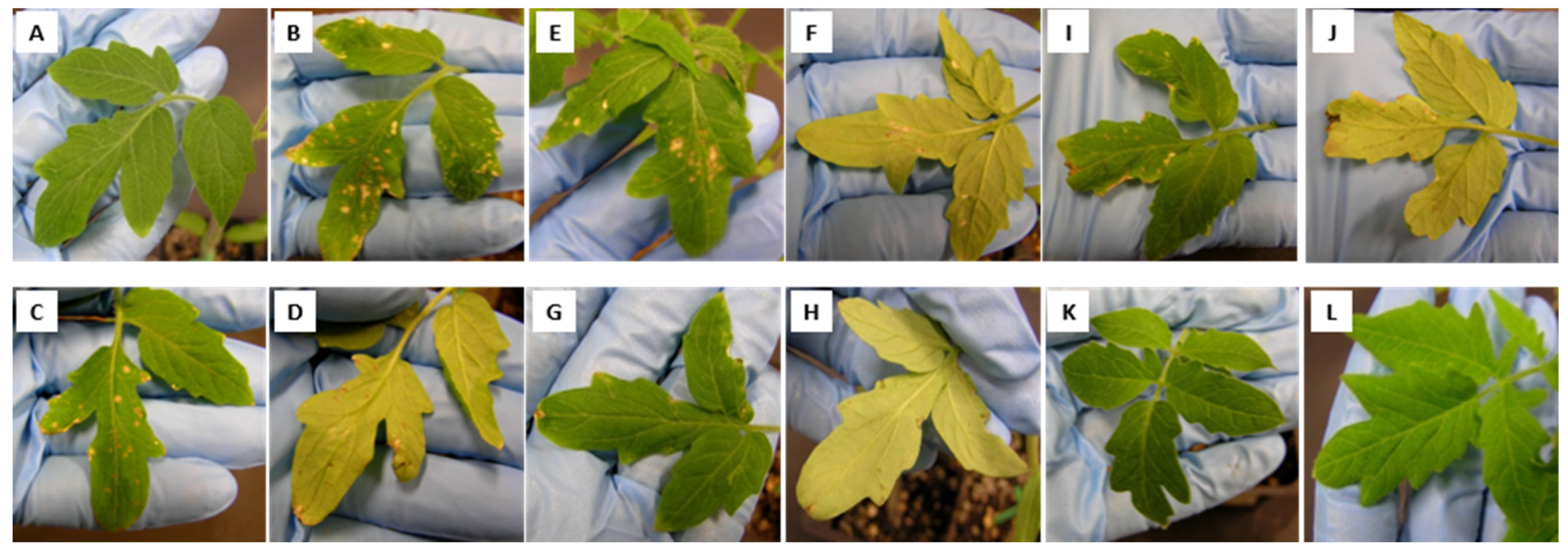
The potential of inhibitory culture supernatants for controlling an infection in tomato seedlings was then investigated. The disease severity was evaluated as the percentage of leaf tissue with spots and lesions 10 days after infestation. The disease severity was 70% among plants infected by X. perforans T4 then treated with water and 90% among plants infected by X. perforans T4 then treated with TSB, as compared to non-infected tomato seedlings (Figure 2A–D). Susceptible reactions manifested 7 days after infestation as small, greasy, water-soaked spots (about 3 mm) on leaflets. The older spots became dry and brown, often surrounded by yellow halos. The spots increased in size to form large, irregular dead spots. The lesions were frequently surrounded by large chlorotic haloes and perforations, referring to the holes in the leaf following infection by the bacterium.
Treatment with the cell-free supernatant of B. velezensis 71 (Figure 2G,H) reduced the disease severity by almost four times compared to the control (Figure 2C,D). Thus, only 20% of the leaf surfaces showed signs of infection by X. perforans T4 after treatment with cell-free supernatants of strain 71 cultures. The cell-free supernatant of P. peoriae To99 cultures reduced the disease severity by half compared to the positive control (Figure 2I,J). Thus, about 40% of leaf surfaces of tomato seedlings were infected by X. perforans T4, which is comparable with treatment using a combination of Kocide and Mancozeb (Figure 2E,F). Of note, spots and lesions appeared on only one of six leaves per plant infected by X. perforans T4 and treated by bacterial cell-free supernatants, while infected and non-treated plants had four of six leaves with these disease symptoms. The bacterial cell-free culture supernatant of the two isolates did not cause any phytotoxic effect on the tomato plants, as shown by the examples with B. velezensis 71 and P. peoriae To99 (Figure 2K,L).

3.4. Preventive Effects of Bacillus and Paenibacillus Cells and Their Cell-Free Supernatants in Controlling Bacterial Spot Disease Caused by X. gardneri DC00T7A

We then asked whether the two bacterial strains or the culture supernatants could protect the plants from an infection. For this assay, we tested a different tomato pathogen. In contrast with the yellow-brownish lesions caused by X. perforans T4 on tomato leaves in the curative assay, a susceptible reaction caused by X. gardneri DC00T7A manifested 10 days after infestation as well-defined brown spots, appearing on leaves and stems (Figure S1). Thus, the preventive effects of cell-free supernatants and cell suspensions of B. velezensis 71 and P. peoriae To99 were assessed as reductions in the numbers of spots per plant.
The treatment of tomato leaves by cell suspensions of Bacillus and Paenibacillus isolates or by their cell-free culture supernatants 6 days before X. gardneri DC00T7A infection reduced the bacterial spot disease rates by 2–5 times compared to untreated plants (Figure 3). The cell-free supernatant of B. velezensis 71 was twice as effective in controlling this disease than the treatment with the bacteria themselves, while the cell-free supernatant of P. peoriae To99 was 1.6-fold less effective than its cell suspension. Interestingly, both B. velezensis 71 and P. peoriae To99 isolates (cell suspensions and cell-free supernatants) applied before pathogen infection showed 2-fold suppression of bacterial spot disease caused by X. gardneri DC00T7A compared to the pretreatment with combined Kocide and Mancozeb, which is frequently used by growers in controlling bacterial spot disease caused by Xanthomonas species.

3.5. Microbial Viability of Bacillus and Paenibacillus Isolates on Tomato Leaves

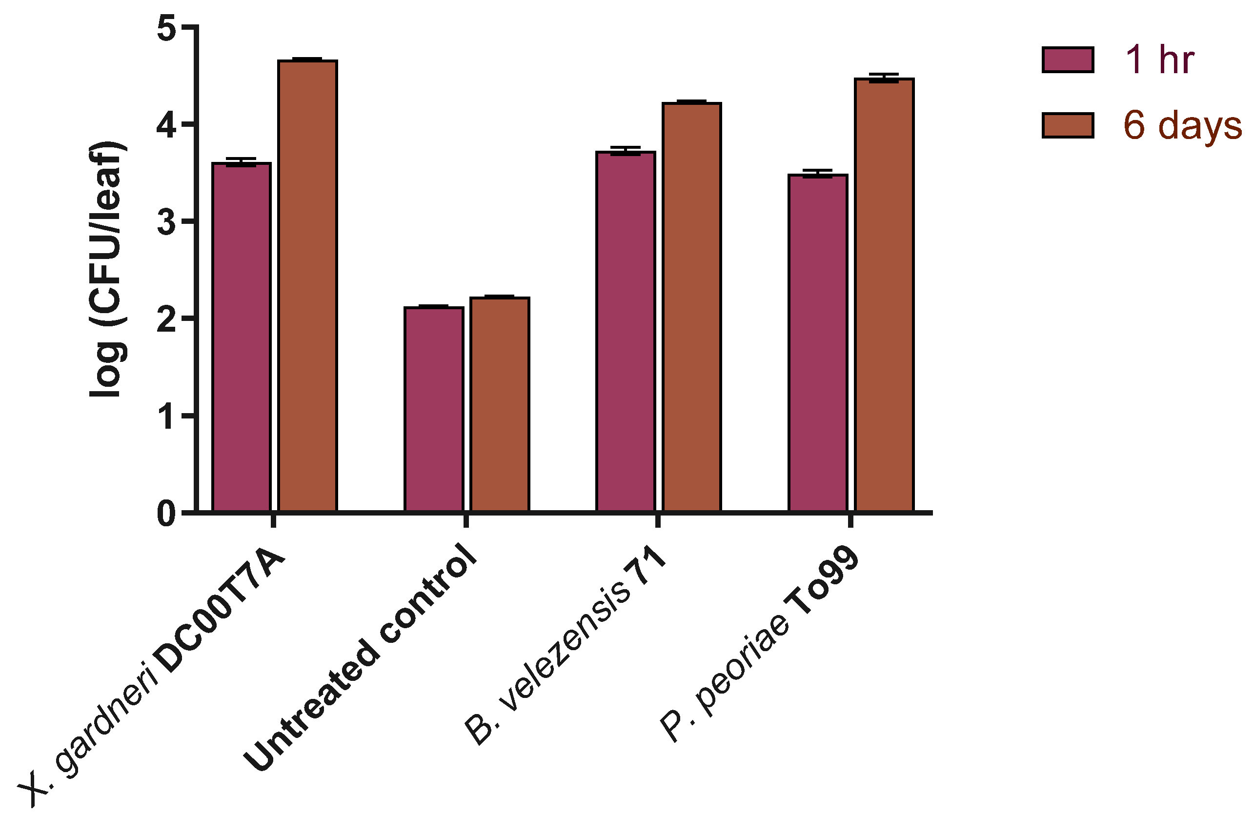
Six days after treatment of tomato leaves by B. velezensis 71 and P. peoriae To99, bacterial cells of both strains were still viable, and the CFU/leaf rate increased by almost 1.5 log from the initial measurement 1 h after treatment. As expected, there was no significant difference in the quantities of viable bacterial cells (the natural phyllosphere) on untreated tomato leaves 1 h and 6 days after treatment (Figure 4). Interestingly, using the serial dilution method, 2 fungal and 4 bacterial morphologically different colonies were isolated from untreated tomato leaves, while only B. velezensis 71 and P. peoriae To99 colonies appeared on TSA after their isolation from respectively treated tomato leaves.

3.6. Greenhouse Trials

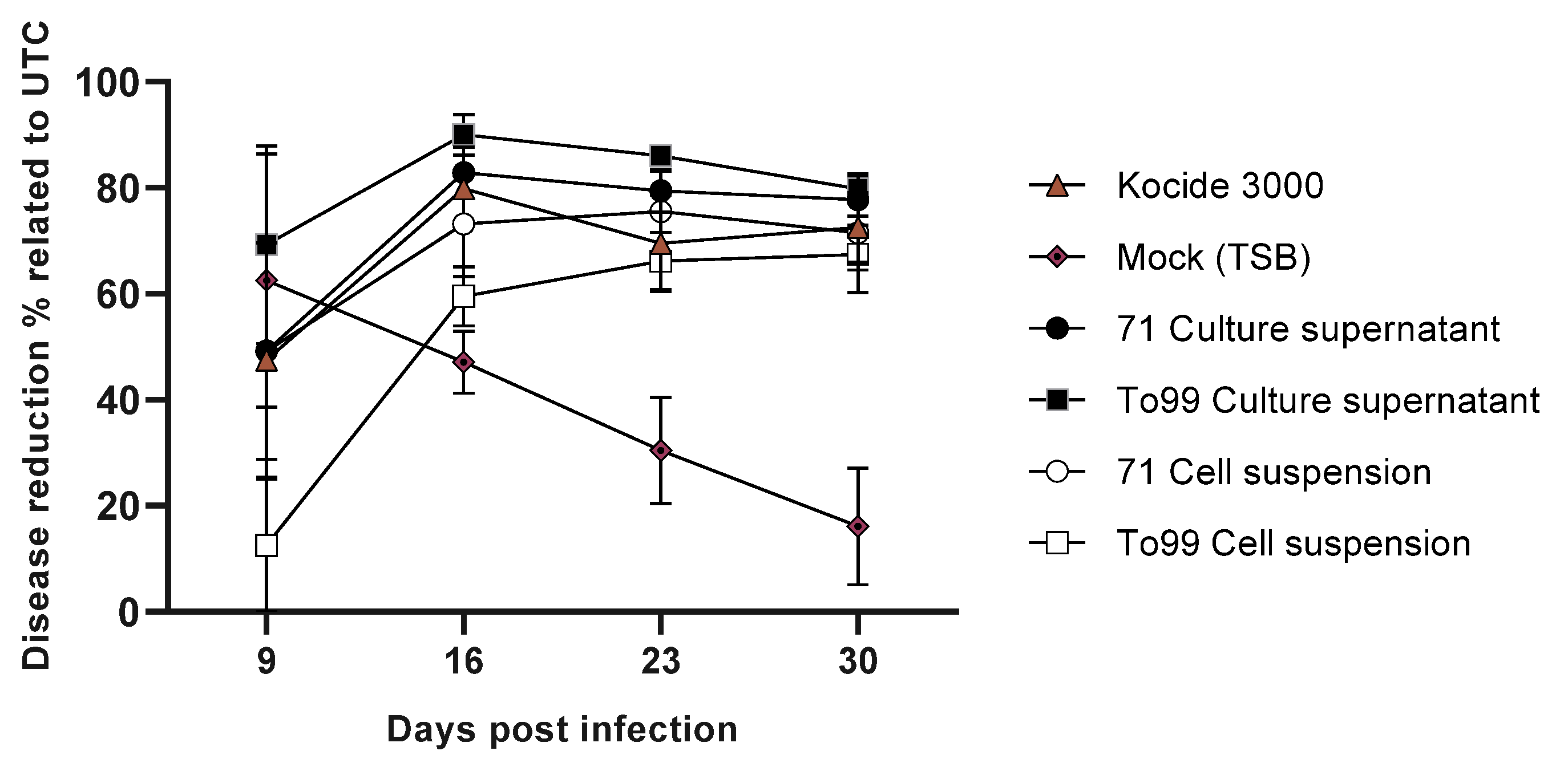
The objective of the trials was to evaluate the efficacy of treatments based on cell-free supernatants and cell suspensions of B. velezensis 71 and P. peoriae To99 isolates in the suppression of the leaf spot disease caused by X. gardneri DC00T7A under greenhouse conditions. The greenhouse trial results in terms of rates of disease control as compared to the untreated control are shown in Figure 5. In general, the results show that 5 weeks after the first application, both the cell-free supernatants and cell suspensions from strains 71 and To99 demonstrated more than 60% efficacy in controlling the leaf spot disease, which is not significantly different than the industrial standard (Kocide 3000). In all cases, the treatments were also significantly better than the untreated or Mock (TSB).

3.7. Gene Clusters for Secondary Metabolites

To predict biosynthetic pathways that are responsible for the production of metabolites with anti-Xanthomonas activity in B. velezensis 71 and P. peoriae To99 isolates, antiSMASH was used. Hence, for B. velezensis 71, the antiSMASH results suggested the presence of biosynthetic gene clusters responsible for three polyketide synthases (PKs) with known antibacterial activity: Bacillaene, Difficidin, and Macrolactin [40,41]. Furthermore, biosynthetic gene clusters were detected coding for non-ribosomal peptides synthetases (NRPSs) responsible for the production of the cyclic lipopeptides surfactin, bacillomycin D, and fengycins, along with a gene cluster for the biosynthesis of the dipeptide Bacilysin (Table S4).
Similarly, performing the antiSMASH analysis on P. peoriae To99 resulted in the detection of one polyketide synthase with a yet unknown product, which shares 28% similarity with Bacillaene. Moreover, two gene clusters responsible for the synthesis of NRPSs such as polymyxin and fusaricidins were also detected (Table S5).

3.8. Bioassay-Guided Fractionation and Identification of Active Metabolites

To identify the produced metabolites exhibiting anti-Xanthomonas activity, bioassay-guided fractionations of active crude extracts of B. velezensis 71 and P. peoriae To99 cultures were carried out using reverse-phase chromatography. The fractions that showed activity in a disk diffusion assay were subjected to LC-ESI-MS runs (Figure S2). The mass spectral data obtained from the active fractions and their comparison with mass spectral data available for the products of antiSMASH-predicted gene clusters led to the identification of inhibitory molecules active against Xanthomonas. Thus, analyzing the active chromatographic fraction of the B. velezensis 71 crude extract led to the identification of ion peaks corresponding to the polyketide oxydifficidin as the major active molecules (Figure 6), which are also produced by B. velezensis FZB42.
Similarly, performing the activity-guided chromatography on crude extracts of P. peoriae To99 cultures led to the identification of ion peaks at m/z = 1157.7 and m/z = 1143.8, corresponding to polymyxin A1 and A2 as the main inhibitory metabolites against X. perforans [42] (Figure 7).

4. Discussion

In pursuit of identifying potential biocontrol agents effective against leaf spot disease in tomatoes, we conducted an extensive screening campaign focusing on direct antibiosis. Our goal was to identify bacteria that produce antagonistic metabolites against phytopathogenic species of Xanthomonas.
Interestingly, despite using multiple selective and non-selective culture media to ensure a broad diversity among the isolates, most bacteria that exhibited the highest activity against Xanthomonas species were predominantly from the Paenibacillus (16 isolates) and Bacillus (6 isolates) genera. This was consistent regardless of the sample origin or the nutrient medium used for isolation (Table S1). A significant proportion of these isolates were derived from soil in the rhizosphere or various plant organs collected from agricultural fields previously affected by leaf bacterial spot disease. This observation aligns with the hypothesis that natural competitors might exist in environments previously afflicted by the disease (selection based on ecological niche) (Table S1).
It is noteworthy that the Bacillus and Paenibacillus genera, both members of the phylum Bacillota, are renowned for their extensive biosynthetic gene clusters. These clusters often encode for non-ribosomal peptide synthetases (NRPSs) and polyketide synthases (PKSs), leading to the production of a diverse array of antimicrobial compounds such as lipopeptides (LPs) and polyketides (PKs) [43]. The roles of LPs and PKs in antibiosis and biocontrol are well-documented [43,44,45]. However, the distribution of NRPS and PKS gene clusters is not uniform across the different orders of Bacillota. Notably, the Bacillus and Paenibacillus genera, from the order Bacillales, are predominant contributors to this secondary metabolite gene cluster count [42]. In fact, known metabolites from Bacillus amyloliquefaciens and Bacillus subtilis account for a significant portion of the LP and PK diversity within the Bacillus genus [43].
The combined results of the whole-genome sequencing and ANI analysis revealed that Bacillus isolate 71 and Paenibacillus isolate To99, possessing the highest anti-Xanthomonas activity, belong to the B. velezensis and P. peoriae species, respectively. Both species are well known for their vast capacity to promote plant growth and suppress plant pathogens through diverse mechanisms [46,47]. Regarding direct antibiosis via the production of antibacterial metabolites, few studies have identified the secondary metabolites implicated in anti-Xanthomonas activity secreted from B. velezensis and P. peoriae species. In B. velezensis FZB42, difficidin and bacilysin display in vitro and in vivo antibacterial activity against Xanthomonas oryzae pv. oryzae and X. oryzae pv. oryzicola [21]. In another study, in vitro and in vivo results demonstrated that Bacillus subtilis mutants deficient in the production of bacillomycin or iturin A, similar compounds that are also produced by B. velezensis, completely lost the ability to control bacterial leaf spot and soft rot pathogen of cucurbits, X. campestris pv. cucurbitae [48]. Similarly, in a study with a Bacillus isolate, an iturin molecule was found to have in vitro inhibitory activity against X. oryzae pv. oryzae [49].
In the case of Paenibacillus species, in a study similar to ours, da Silva et al. [50] reported a Paenibacillus sp. isolate with strong in vitro and in vivo inhibitory activity of cell-free filtrates against X. campestris pv. campestris. However, the active metabolite was not identified. In another study by Ghazalibiglar et al. [51], several Paenibacillus spp. isolates were reported to display inhibitory activity against X. campestris pv. campestris. However, no correlation between the in vitro antagonist activity of isolates and their in vivo biocontrol of black rot disease suppression in cabbage was found [51].
In the present study, the antiSMASH results along with the bioassay-guided fractionation of culture extracts demonstrated that oxydifficidin and polymyxin A secreted by B. velezensis 71 and P. peoriae To99, respectively, are mainly if not solely responsible for the in vitro anti-Xanthomonas activity under in vitro conditions.
Oxydifficidin, the oxidized form of polyketide difficidin, is a broad-spectrum macrolide antibiotic containing a highly unsaturated, 22-membered lactone with a hydroxyl substituent at C-5 and a phosphate group at C-15 [52].
Chen et al. (2006) highlighted the detection of difficidin and oxydifficidin exclusively in their deprotonated forms ([M-H]– = 543.4 and 559.3) when using the negative ionization mode [40]. Conversely, while utilizing the positive ionization mode in our research, we primarily identified oxydifficidin in its dephosphorylated state (m/z 463.4) and as a dimer (m/z 1121.3), aligning with the findings by Wilson et al. (1987) and Dagher et al. (2021) [52,53]. A further analysis in our study revealed other oxydifficidin species, with potential annotations presented in Figure 6. The discrepancies in m/z values between our study and previous ones could be attributed to variations in experimental conditions or differences in instrument calibration. Only a few studies have previously reported the antibacterial activity of difficidin produced by B. velezensis against phytopathogens such as X. oryzae [21] and Erwinia amylovora [53,54]. Here, we extended the potential application of oxydifficidin to suppressing Xanthomonas-caused tomato leaf spot diseases. Indeed, while the antibacterial activity of these metabolites and their analogues is well established, their functions with regards to plant associations and triggering plant defense mechanisms remain to be established [55,56]. Overall, difficidin- and oxydifficidin-producing species such as B. velezensis are recognized as potent biocontrol agents that play a crucial role in plant health [47,57]. Difficidin is also able to cause the downregulation of the expression of genes involved in Xanthomonas virulence, cell division, and protein and cell wall synthesis [21]. However, additional studies are required to further understand the exact mechanisms involved in their antibiosis action on the leaf surface.
Polymyxin A, a less-studied member of the polymyxin family with potent antimicrobial activity against almost all Gram-negative bacteria [43], is a cyclic cationic decapeptide possessing an unusual α,γ-diaminobutyric acid (Dab) residue with a d-configuration at position 3. Polymyxin A, as reported previously and identified here, consists of two species, polymyxin A1 and A2, which are N-terminally acylated by (S)-6-methyloctanoic acid and (S)-6-methylheptanoic acid, respectively [42].
In spite of displaying excellent antibacterial activity and while being considered the last resort option in fight against multi-drug-resistant Gram-negative bacteria, the clinical utility of polymyxins is limited by their toxicity [58]. In protection against phytopathogens, reports of studying the activity of polymyxins against phytopathogenic Xanthomonas spp. are rare, and such reports date back to 70 years ago when polymyxins were newly discovered [59]. Similarly, there are only a few studies that have demonstrated the role of polymyxins produced from Paenibacillus spp. against other phytopathogenic bacteria. Niu et al. [60] reported on the in vitro antagonistic activity of polymyxin P from P. polymyxa M-1 as the main component of biocontrol in the suppression of E. amylovora Ea273 and Erwinia carotovora.
In the present study, direct antibiosis was used as the screening strategy and the screening was designed to discover isolates that secrete the antagonist metabolites, it should be noted that achieving consistent and efficient field performance in suppressing bacterial pathogens will also depend on other capabilities of the isolate, such as competition for nutrients and space, indirect antagonism, and host plant immunization [61]. Indeed, one of the advantages of microbial biopesticides compared to most other phytosanitary products is their multiple mechanism of action. Hence, in our growth chamber and greenhouse assays, apart from testing the cell-free supernatants of spent cultures, we also evaluated the capacity of the bacterial cells for leaf surface colonization and pathogen suppression.
In the leaf surface colonization assays, B. velezensis 71 and P. peoriae To99 remained viable after 6 days, and even the CFU/leaf rates of both isolates increased over that period. Furthermore, contrary to the tomato leaves treated with either B. velezensis 71 or P. peoriae To99, several morphologically different colonies were isolated from untreated tomato leaves, which suggests the role of B. velezensis 71 or P. peoriae To99 in ecological niche exclusion. The ability of Bacillus isolates to colonize plant leaves, thereby protecting them from different phytopathogens, was shown in other studies. Bacillus sp. strain 5B6, isolated from the leaves of cherry trees, exhibited leaf-colonizing capacity and promoted the plant growth of Prunus avium L [62]. In another research, bacilli spore preparation significantly improved leaf growth parameters such as the leaf thickness and photosynthesis capacity, indicating that bacilli treatment directly promotes leaf growth [63]. In addition, foliar treatment by bacilli improved fruit quality indicators including the water, glucose, and sucrose contents [63].
Interestingly, in this study, the preventive application of cell suspensions of both B. velezensis 71 and P. peoriae To99 alone demonstrated better or comparable efficiency in disease suppression than the combined use of Kocide and Mancozeb when tested in growth chamber and greenhouse conditions. These results suggest that the biocontrol mechanisms of both isolates against leaf spot disease in tomato plants is not limited to the direct antibiosis and is likely attributed to other well-reported plant-growth-promoting mechanisms of these two strains [46,47]. Moreover, the curative and preventive growth chamber results, along with the greenhouse trial results, validated our screening strategy based on the secretion of anti-Xanthomonas metabolites as cell-free spent culture supernatants of both B. velezensis 71 and P. peoriae To99, which demonstrated comparable efficacy levels of leaf spot disease suppression when compared with combined Kocide and Mancozeb, which are widely being used by growers.

5. Conclusions

Collectively, the results of this study demonstrate that B. velezensis 71 and P. peoriae To99, at least partially thanks to the production of their metabolites (including oxydifficidin and polymyxin A), have potent antagonistic activity against Xanthomonas spp., the causal agents of leaf spot in tomato plants, under both laboratory and greenhouse conditions. Therefore, in future work, a potential biocontrol product based on cells of B. velezensis 71 and P. peoriae To99, together with their metabolites, could be a potential suitable replacement for copper- and dithiocarbamate-based products in controlling bacterial spot disease caused by Xanthomonas species.

6. Patents

Dagher, Fadi, Eric Déziel, and Snizhana Olishevska. “Bacterial And Fungal Metabolites Possessing Anti-Microbial Activity Against Xanthomonas Species, Compositions, Methods, Kits, And Uses Relating To the Same”. Patent No. US 11,089,785.

Supplementary Materials

The following are available online at https://www.mdpi.com/article/10.3390/applmicrobiol3030076/s1: Table S1. Characteristics of the most active bacterial isolates against Xanthomonas perforans T4. Table S2. Activity of bacterial supernatants against plant pathogens. Table S3. Inter-species and inter-strain antibacterial activity of cell-free supernatants. Table S4. AntiSMASH results for B. velezensis 71. Table S5. AntiSMASH results for P. peoriae To99. Figure S1. Symptoms caused by X. gardneri DC00T7A on tomato leaf surfaces. Figure S2. Testing the activity of crude extract fractions of B. velezensis 71 and P. peoriae To99 cultures obtained from semi-preparative HPLC fractionation against X. perforans T4 by disk diffusion assay.

Author Contributions

Conceptualization, E.D., S.O. and F.D.; methodology, S.O., C.R. and A.N.; validation, S.O., A.N. and E.D.; formal analysis, Y.L. and J.Z.; investigation, S.O., A.N. and E.D.; resources, E.D.; data curation, A.N.; writing—original draft preparation, S.O. and A.N.; writing—review and editing, A.N. and E.D.; supervision, E.D.; project administration, E.D.; funding acquisition, E.D. All authors have read and agreed to the published version of the manuscript.

Funding

This research was funded by the Natural Science and Engineering Research Council of Canada (NSERC) Engage and Collaborative Research and Development (CRD) grants EGP 417883 and CRDPJ488896, as well as a CRIBIQ grant in partnership with Agri-Neo and a CRD grant CRDPJ537051 in partnership with Axter Agrosciences.

Data Availability Statement

Purified cultures of each of the bacterial strains B. velezensis 71 and P. peoriae To99 were deposited at the Agricultural Research Service Culture Collection (NRRL) (USDA, ARS, 1815 North University Street, Peoria, Ill., 61064, USA) on March 9, 2015. The deposits were made under the terms of the Budapest Treaty. “Bacillus velezensis 71” has been assigned accession number NRRL B-67021, and the corresponding NCBI GenBank accession number is NZ_LYNA00000000. “Paenibacillus peoriae To99” has been assigned accession number NRRL B-67020, and the corresponding NCBI GenBank accession number is NZ_LYMY00000000. All relevant data are included in the manuscript. Further inquiries can be directed to the corresponding author.

Acknowledgments

This has been a long-term project and many people have been involved over the years. We gratefully acknowledge the experimental contributions of Louise Courtemanche during the initial screening efforts.

Conflicts of Interest

The authors declare no conflict of interest.

References

  1. Jones, J.B.; Lacy, G.H.; Bouzar, H.; Stall, R.E.; Schaad, N.W. Reclassification of the xanthomonads associated with bacterial spot disease of tomato and pepper. Syst. Appl. Microbiol. 2004, 27, 755–762. [Google Scholar] [CrossRef] [PubMed]
  2. Jones, J.B.; Lacy, G.H.; Bouzar, H.; Minsavage, G.V.; Stall, R.E.; Schaad, N.W. Bacterial Spot-Worldwide Distribution, Importance and Review. In I International Symposium on Tomato Diseases; ISHS: Leuven, Belgium, 2005; pp. 27–34. [Google Scholar]
  3. Potnis, N.; Krasileva, K.; Chow, V.; Almeida, N.F.; Patil, P.B.; Ryan, R.P.; Sharlach, M.; Behlau, F.; Dow, J.M.; Momol, M.; et al. Comparative genomics reveals diversity among xanthomonads infecting tomato and pepper. BMC Genom. 2011, 12, 146. [Google Scholar] [CrossRef]
  4. European and Mediterranean Plant Protection Organization. Xanthomonas spp. (Xanthomonas euvesicatoria, Xanthomonas gardneri, Xanthomonas perforans, Xanthomonas vesicatoria) causing bacterial spot of tomato and sweet pepper. EPPO Bull. 2013, 43, 7–20. [Google Scholar] [CrossRef]
  5. Toussaint, V. Bacterial leaf spot, a new disease of lettuce in Quebec caused by Xanthomonas campestris pv. vitians. Phytoprotection 1999, 80, 121–125. [Google Scholar] [CrossRef]
  6. Conover, R.A.; Gerhold, N.R. Mixtures of Copper and Maneb or Mancozeb for Control of Bacterial Spot of Tomato and Their Compatibility for Control of Fungus Diseases. Fla. State Hortic. Soc. Proc. 1981, 94, 154–156. [Google Scholar]
  7. Strayer-Scherer, A.; Liao, Y.Y.; Young, M.; Ritchie, L.; Vallad, G.E.; Santra, S.; Freeman, J.H.; Clark, D.; Jones, J.B.; Paret, M.L. Advanced Copper Composites Against Copper-Tolerant Xanthomonas perforans and Tomato Bacterial Spot. Phytopathology 2018, 108, 196–205. [Google Scholar] [CrossRef]
  8. Basim, H.; Minsavage, G.V.; Stall, R.E.; Wang, J.F.; Shanker, S.; Jones, J.B. Characterization of a unique chromosomal copper resistance gene cluster from Xanthomonas campestris pv. vesicatoria. Appl. Environ. Microbiol. 2005, 71, 8284–8291. [Google Scholar] [CrossRef]
  9. Lee, Y.A.; Hendson, M.; Panopoulos, N.J.; Schroth, M.N. Molecular cloning, chromosomal mapping, and sequence analysis of copper resistance genes from Xanthomonas campestris pv. juglandis: Homology with small blue copper proteins and multicopper oxidase. J. Bacteriol. 1994, 176, 173–188. [Google Scholar] [CrossRef]
  10. Voloudakis, A.E.; Reignier, T.M.; Cooksey, D.A. Regulation of resistance to copper in Xanthomonas axonopodis pv. vesicatoria. Appl. Environ. Microbiol. 2005, 71, 782–789. [Google Scholar] [CrossRef]
  11. Bull, C.T.; Koike, S.T. Evaluating the efficacy of commercial products for management of bacterial leaf spot on lettuce. Plant Health Prog. 2005, 6, 3. [Google Scholar] [CrossRef]
  12. Baldi, E.; Miotto, A.; Ceretta, C.A.; Quartieri, M.; Sorrenti, G.; Brunetto, G.; Toselli, M. Soil-applied phosphorous is an effective tool to mitigate the toxicity of copper excess on grapevine grown in rhizobox. Sci. Hortic. 2018, 227, 102–111. [Google Scholar] [CrossRef]
  13. Weller, D.M. Biological-Control of Soilborne Plant-Pathogens in the Rhizosphere with Bacteria. Annu. Rev. Phytopathol. 1988, 26, 379–407. [Google Scholar] [CrossRef]
  14. Compant, S.; Duffy, B.; Nowak, J.; Clement, C.; Barka, E.A. Use of plant growth-promoting bacteria for biocontrol of plant diseases: Principles, mechanisms of action, and future prospects. Appl. Environ. Microbiol. 2005, 71, 4951–4959. [Google Scholar] [CrossRef]
  15. Eljounaidi, K.; Lee, S.K.; Bae, H. Bacterial endophytes as potential biocontrol agents of vascular wilt diseases—Review and future prospects. Biol. Control 2016, 103, 62–68. [Google Scholar] [CrossRef]
  16. Chavan, N.P.; Pandey, R.; Nawani, N.; Nanda, R.K.; Tandon, G.D.; Khetmalas, M.B. Biocontrol potential of actinomycetes against Xanthomonas axonopodis pv. punicae, a causative agent for oily spot disease of pomegranate. Biocontrol. Sci. Technol. 2016, 26, 351–372. [Google Scholar] [CrossRef]
  17. El-Hendawy, H.H.; Osman, M.E.; Sorour, N.M. Biological control of bacterial spot of tomato caused by Xanthomonas campestris pv. vesicatoria by Rahnella aquatilis. Microbiol. Res. 2005, 160, 343–352. [Google Scholar] [CrossRef] [PubMed]
  18. Massomo, S.M.S.; Mortensen, C.N.; Mabagala, R.B.; Newman, M.A.; Hockenhull, J. Biological control of black rot (Xanthomonas campestris pv. campestris) of cabbage in Tanzania with Bacillus strains. J. Phytopathol. 2004, 152, 98–105. [Google Scholar] [CrossRef]
  19. Monteiro, L.; Mariano, R.D.R.; Souto-Maior, A.M. Antagonism of Bacillus spp. against Xanthomonas campestris pv. campestris. Braz. Arch. Biol. Technol. 2005, 48, 23–29. [Google Scholar] [CrossRef]
  20. Lee, G.; Lee, S.H.; Kim, K.M.; Ryu, C.M. Foliar application of the leaf-colonizing yeast Pseudozyma churashimaensis elicits systemic defense of pepper against bacterial and viral pathogens. Sci. Rep. 2017, 7, 39432. [Google Scholar] [CrossRef]
  21. Wu, L.; Wu, H.; Chen, L.; Yu, X.; Borriss, R.; Gao, X. Difficidin and bacilysin from Bacillus amyloliquefaciens FZB42 have antibacterial activity against Xanthomonas oryzae rice pathogens. Sci. Rep. 2015, 5, 12975. [Google Scholar] [CrossRef]
  22. Marin, V.R.; Ferrarezi, J.H.; Vieira, G.; Sass, D.C. Recent advances in the biocontrol of Xanthomonas spp. World J. Microbiol. Biotechnol. 2019, 35, 72. [Google Scholar] [CrossRef] [PubMed]
  23. Besset-Manzoni, Y.; Joly, P.; Brutel, A.; Gerin, F.; Soudière, O.; Langin, T.; Prigent-Combaret, C. Does in vitro selection of biocontrol agents guarantee success in planta? A study case of wheat protection against Fusarium seedling blight by soil bacteria. PLoS ONE 2019, 14, e0225655. [Google Scholar] [CrossRef] [PubMed]
  24. Collinge, D.B.; Jensen, D.F.; Rabiey, M.; Sarrocco, S.; Shaw, M.W.; Shaw, R.H. Biological control of plant diseases—What has been achieved and what is the direction? Plant Pathol. 2022, 71, 1024–1047. [Google Scholar] [CrossRef]
  25. Clough, S.E.; Jousset, A.; Elphinstone, J.G.; Friman, V.-P. Combining in vitro and in vivo screening to identify efficient Pseudomonas biocontrol strains against the phytopathogenic bacterium Ralstonia solanacearum. Microbiologyopen 2022, 11, e1283. [Google Scholar] [CrossRef] [PubMed]
  26. Porter, J.N.; Wilhelm, J.J.; Tresner, H.D. Method for the preferential isolation of Actinomycetes from soil. Appl. Microbiol. 1960, 8, 174–178. [Google Scholar] [CrossRef] [PubMed]
  27. Henry, D.A.; Campbell, M.E.; LiPuma, J.J.; Speert, D.P. Identification of Burkholderia cepacia isolates from patients with cystic fibrosis and use of a simple new selective medium. J. Clin. Microbiol. 1997, 35, 614–619. [Google Scholar] [CrossRef]
  28. Fromin, N.; Achouak, W.; Thiéry, J.M.; Heulin, T. The genotypic diversity of Pseudomonas brassicacearum populations isolated from roots of Arabidopsis thaliana: Influence of plant genotype. FEMS Microbiol. Ecol. 2001, 37, 21–29. [Google Scholar] [CrossRef]
  29. Rashid, A.; Khan, M.A. Evaluation of antagonistic organisms against Xanthomonas campestris pv. malvacearum in vitro and on the inoculated cotton plant for the control of bacterial blight disease. Pak. J. Agric. Sci. 2000, 37, 135–139. [Google Scholar]
  30. Velmurugan, N.; Choi, M.S.; Han, S.S.; Lee, Y.S. Evaluation of antagonistic activities of Bacillus subtilis and Bacillus licheniformis against wood-staining fungi: In vitro and in vivo experiments. J. Microbiol. 2009, 47, 385–392. [Google Scholar] [CrossRef]
  31. Obradovic, A.; Jones, J.B.; Momol, M.T.; Balogh, B.; Olson, S.M. Management of Tomato Bacterial Spot in the Field by Foliar Applications of Bacteriophages and SAR Inducers. Plant Dis. 2004, 88, 736–740. [Google Scholar] [CrossRef]
  32. Lindow, S.E.; Brandl, M.T. Microbiology of the Phyllosphere. Appl. Environ. Microbiol. 2003, 69, 1875–1883. [Google Scholar] [CrossRef] [PubMed]
  33. Priest, F.G.; Goodfellow, M.; Shute, L.A.; Berkeley, R.C.W. Bacillus amyloliquefaciens sp. nov., nom. rev. Int. J. Syst. Evol. Microbiol. 1987, 37, 69–71. [Google Scholar] [CrossRef]
  34. Frank, J.A.; Reich, C.I.; Sharma, S.; Weisbaum, J.S.; Wilson, B.A.; Olsen, G.J. Critical evaluation of two primers commonly used for amplification of bacterial 16S rRNA genes. Appl. Environ. Microbiol. 2008, 74, 2461–2470. [Google Scholar] [CrossRef]
  35. Wood, D.E.; Salzberg, S.L. Kraken: Ultrafast metagenomic sequence classification using exact alignments. Genome Biol. 2014, 15, R46. [Google Scholar] [CrossRef]
  36. Assefa, S.; Keane, T.M.; Otto, T.D.; Newbold, C.; Berriman, M. ABACAS: Algorithm-based automatic contiguation of assembled sequences. Bioinformatics 2009, 25, 1968–1969. [Google Scholar] [CrossRef] [PubMed]
  37. Goris, J.; Konstantinidis, K.T.; Klappenbach, J.A.; Coenye, T.; Vandamme, P.; Tiedje, J.M. DNA-DNA hybridization values and their relationship to whole-genome sequence similarities. Int. J. Syst. Evol. Microbiol. 2007, 57, 81–91. [Google Scholar] [CrossRef] [PubMed]
  38. Weber, T.; Blin, K.; Duddela, S.; Krug, D.; Kim, H.U.; Bruccoleri, R.; Lee, S.Y.; Fischbach, M.A.; Müller, R.; Wohlleben, W.; et al. antiSMASH 3.0—A comprehensive resource for the genome mining of biosynthetic gene clusters. Nucleic Acids Res. 2015, 43, W237–W243. [Google Scholar] [CrossRef]
  39. Blin, K.; Shaw, S.; Steinke, K.; Villebro, R.; Ziemert, N.; Lee, S.Y.; Medema, M.H.; Weber, T. antiSMASH 5.0: Updates to the secondary metabolite genome mining pipeline. Nucleic Acids Res. 2019, 47, W81–W87. [Google Scholar] [CrossRef]
  40. Chen, X.H.; Vater, J.; Piel, J.; Franke, P.; Scholz, R.; Schneider, K.; Koumoutsi, A.; Hitzeroth, G.; Grammel, N.; Strittmatter, A.W.; et al. Structural and Functional Characterization of Three Polyketide Synthase Gene Clusters in Bacillus amyloliquefaciens FZB 42. J. Bacteriol. 2006, 188, 4024–4036. [Google Scholar] [CrossRef]
  41. Schneider, K.; Chen, X.H.; Vater, J.; Franke, P.; Nicholson, G.; Borriss, R.; Sussmuth, R.D. Macrolactin is the polyketide biosynthesis product of the pks2 cluster of Bacillus amyloliquefaciens FZB42. J. Nat. Prod. 2007, 70, 1417–1423. [Google Scholar] [CrossRef]
  42. Jangra, M.; Randhawa, H.K.; Kaur, M.; Srivastava, A.; Maurya, N.; Patil, P.P.; Jaswal, P.; Arora, A.; Patil, P.B.; Raje, M.; et al. Purification, Characterization and in vitro Evaluation of Polymyxin A From Paenibacillus dendritiformis: An Underexplored Member of the Polymyxin Family. Front. Microbiol. 2018, 9, 2864. [Google Scholar] [CrossRef] [PubMed]
  43. Olishevska, S.; Nickzad, A.; Déziel, E. Bacillus and Paenibacillus secreted polyketides and peptides involved in controlling human and plant pathogens. Appl. Microbiol. Biotechnol. 2019, 103, 1189–1215. [Google Scholar] [CrossRef] [PubMed]
  44. Ongena, M.; Jacques, P. Bacillus lipopeptides: Versatile weapons for plant disease biocontrol. Trends Microbiol. 2008, 16, 115–125. [Google Scholar] [CrossRef] [PubMed]
  45. Fazle Rabbee, M.; Baek, K.-H. Antimicrobial Activities of Lipopeptides and Polyketides of Bacillus velezensis for Agricultural Applications. Molecules 2020, 25, 4973. [Google Scholar] [CrossRef] [PubMed]
  46. Jeong, H.; Choi, S.-K.; Ryu, C.-M.; Park, S.-H. Chronicle of a Soil Bacterium: Paenibacillus polymyxa E681 as a Tiny Guardian of Plant and Human Health. Front. Microbiol. 2019, 10, 467. [Google Scholar] [CrossRef]
  47. Fan, B.; Wang, C.; Song, X.; Ding, X.; Wu, L.; Wu, H.; Gao, X.; Borriss, R. Bacillus velezensis FZB42 in 2018: The Gram-Positive Model Strain for Plant Growth Promotion and Biocontrol. Front. Microbiol. 2018, 9, 2491. [Google Scholar] [CrossRef]
  48. Zeriouh, H.; Romero, D.; Garcia-Gutierrez, L.; Cazorla, F.M.; de Vicente, A.; Perez-Garcia, A. The iturin-like lipopeptides are essential components in the biological control arsenal of Bacillus subtilis against bacterial diseases of cucurbits. Mol. Plant-Microbe Interact. 2011, 24, 1540–1552. [Google Scholar] [CrossRef]
  49. Beric, T.; Kojic, M.; Stankovic, S.; Topisirovic, L.; Degrassi, G.; Myers, M.; Venturi, V.; Fira, D. Antimicrobial Activity of Bacillus sp Natural Isolates and Their Potential Use in the Biocontrol of Phytopathogenic Bacteria. Food Technol. Biotechnol. 2012, 50, 25–31. [Google Scholar]
  50. da Silva, R.S.; Moutinho, B.L.; dos Santos, D.R.; Vasconcelo-Rodrigues, I.S.; Talamini, V.; Fernandes, M.F.; Fernandes, R.P.M. Using antagonistic soil bacteria and their cell-free filtrates to control the black rot pathogen Xanthomonas campestris pv. campestris. J. Phytopathol. 2018, 166, 494–501. [Google Scholar] [CrossRef]
  51. Ghazalibiglar, H.; Hampton, J.G.; van ZijlldeJong, E.; Holyoake, A. Evaluation of Paenibacillus spp. isolates for the biological control of black rot in Brassica oleracea var. capitata (cabbage). Biocontrol. Sci. Techn. 2016, 26, 504–515. [Google Scholar] [CrossRef]
  52. Wilson, K.E.; Flor, J.E.; Schwartz, R.E.; Joshua, H.; Smith, J.L.; Pelak, B.A.; Liesch, J.M.; Hensens, O.D. Difficidin and oxydifficidin: Novel broad spectrum antibacterial antibiotics produced by Bacillus subtilis. II. Isolation and physico-chemical characterization. J. Antibiot. 1987, 40, 1682–1691. [Google Scholar] [CrossRef] [PubMed]
  53. Dagher, F.; Nickzad, A.; Zheng, J.; Hoffmann, M.; Déziel, E. Characterization of the biocontrol activity of three bacterial isolates against the phytopathogen Erwinia amylovora. Microbiologyopen 2021, 10, e1202. [Google Scholar] [CrossRef] [PubMed]
  54. Chen, X.H.; Scholz, R.; Borriss, M.; Junge, H.; Mogel, G.; Kunz, S.; Borriss, R. Difficidin and bacilysin produced by plant-associated Bacillus amyloliquefaciens are efficient in controlling fire blight disease. J. Biotechnol. 2009, 140, 38–44. [Google Scholar] [CrossRef] [PubMed]
  55. Aleti, G.; Sessitsch, A.; Brader, G. Genome mining: Prediction of lipopeptides and polyketides from Bacillus and related Firmicutes. Comput. Struct. Biotechnol. J. 2015, 13, 192–203. [Google Scholar] [CrossRef]
  56. Chakraborty, K.; Kizhakkekalam, V.K.; Joy, M.; Dhara, S. Difficidin class of polyketide antibiotics from marine macroalga-associated Bacillus as promising antibacterial agents. Appl. Microbiol. Biotechnol. 2021, 105, 6395–6408. [Google Scholar] [CrossRef]
  57. Rabbee, M.F.; Hwang, B.-S.; Baek, K.-H. Bacillus velezensis: A Beneficial Biocontrol Agent or Facultative Phytopathogen for Sustainable Agriculture. Agronomy 2023, 13, 840. [Google Scholar] [CrossRef]
  58. Brown, P.; Dawson, M.J. Development of new polymyxin derivatives for multi-drug resistant Gram-negative infections. J. Antibiot. 2017, 70, 386–394. [Google Scholar] [CrossRef]
  59. Katznelson, H.; Sutton, M.D. Inhibition of plant pathogenic bacteria in vitro by antibiotics and quaternary ammonium compounds. Can. J. Bot. 1951, 29, 270–278. [Google Scholar] [CrossRef]
  60. Niu, B.; Vater, J.; Rueckert, C.; Blom, J.; Lehmann, M.; Ru, J.-J.; Chen, X.-H.; Wang, Q.; Borriss, R. Polymyxin P is the active principle in suppressing phytopathogenic Erwinia spp. by the biocontrol rhizobacterium Paenibacillus polymyxa M-1. BMC Microbiol. 2013, 13, 137. [Google Scholar] [CrossRef]
  61. Backer, R.; Rokem, J.S.; Ilangumaran, G.; Lamont, J.; Praslickova, D.; Ricci, E.; Subramanian, S.; Smith, D.L. Plant Growth-Promoting Rhizobacteria: Context, Mechanisms of Action, and Roadmap to Commercialization of Biostimulants for Sustainable Agriculture. Front. Plant Sci. 2018, 9, 1473. [Google Scholar] [CrossRef]
  62. Kim, B.K.; Chung, J.-h.; Kim, S.-Y.; Jeong, H.; Kang, S.G.; Kwon, S.-K.; Lee, C.H.; Song, J.Y.; Yu, D.S.; Ryu, C.-M.; et al. Genome Sequence of the Leaf-Colonizing Bacterium Bacillus sp. Strain 5B6, Isolated from a Cherry Tree. J. Bacteriol. 2012, 194, 3758–3759. [Google Scholar] [CrossRef] [PubMed]
  63. Ryu, C.-M.; Shin, J.-N.; Qi, W.; Ruhong, M.; Kim, E.-J.; Pan, J.-G. Potential for augmentation of fruit quality by foliar application of bacilli spores on apple tree. Plant Pathol. J. 2011, 27, 164–169. [Google Scholar] [CrossRef]
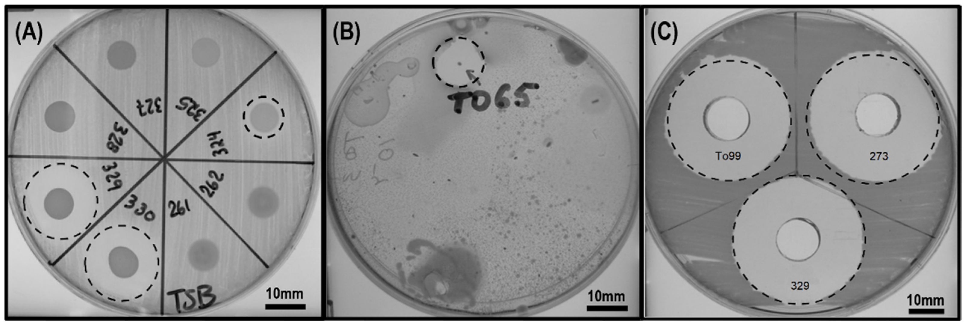
Figure 1. Screening and antagonistic activity of environmental bacterial isolates against X. campestris: (A) example of a spot check method against X. campestris 901; (B) example of antagonistic activity of a bacterial colony against X. campestris ED1985 using Top Agar assay screening method; (C) example of the antimicrobial activity of cell-free supernatants of three bacterial isolates against X. perforans T4. Inhibition zone boundaries are delineated by dotted circles.
Applmicrobiol 03 00076 g001
Figure 2. Curative effect of bacterial cell-free supernatant treatment on the severity of leaf spot caused by X. perforans T4 on tomato seedlings. Representative samples: (A) non-infected and treated by water; (B) infected by X. perforans T4 and treated by TSB. Infected by X. perforans T4 and treated by water: (C) adaxial and (D) abaxial leaf surfaces. Infected by X. perforans T4 and treated by combined Kocide and Mancozeb: (E) adaxial and (F) abaxial leaf surfaces. Infected by X. perforans T4 and treated by cell-free supernatant of B. velezensis 71: (G) adaxial and (H) abaxial leaf surfaces. Infected by X. perforans T4 and treated by cell-free supernatant of P. peoriae To99: (I) adaxial and (J) abaxial leaf surfaces. Not infected and treated by cell-free supernatants of (K) B. velezensis 71 and (L) P. peoriae To99.
Applmicrobiol 03 00076 g002
Figure 3. Preventive effects of B. velezensis 71 and P. peoriae To99 cell suspensions or their metabolites on the severity of leaf spot disease caused by X. gardneri DC00T7A on tomato seedlings.
Applmicrobiol 03 00076 g003
Figure 4. Counting of the viable microbial colonies on the tomato leaves at different time points (1 h and 6 days) post-treatment with the B. velezensis 71 and P. peoriae To99 isolates.
Applmicrobiol 03 00076 g004
Figure 5. Effects of B. velezensis 71 and P. peoriae To99 based treatments in controlling the bacterial leaf spot disease on tomato plants caused by X. gardneri DC00T7A in a greenhouse. The disease reduction % values related to the untreated control (UTC) 5 weeks after the first application of the treatments were measured.
Applmicrobiol 03 00076 g005
Figure 6. Positive-mode ESI-MS spectra of the chromatographic peak of oxydifficidin in the active fraction of the B. velezensis 71 extract and the proposed annotations of the related oxydifficidin species for the detected pseudomolecular ions (the blue dashed arrows indicate proposed fragmentation point across oxydifficidin structure).
Applmicrobiol 03 00076 g006
Figure 7. Positive-mode ESI-MS spectra of chromatographic peaks of (A) polymyxin A1 and (B) polymyxin A2 in the inhibitory fractions of the P. peoriae To99 extract.
Applmicrobiol 03 00076 g007
Table 1. Characteristics of environmental samples collected for the isolation of microorganisms.
SampleLocation and Date of SamplingQuantity
Soil from agricultural fieldsLaval (Québec, Canada), November 201260
Soil from agricultural fieldsSherrington (Québec, Canada), November 201111
Soil from vegetable garden of INRSLaval (Québec, Canada), November 20122
Soil from a strawberry fieldFlorida Area (USA), April 20132
Dead plant leaves, stems, and roots from agricultural fieldsLaval (Québec, Canada), November 201212
Dead plant leaves, stems, and roots from agricultural fieldsSherrington (Québec, Canada), November 20114
Fruits and leaves of fresh plants from vegetable garden of INRSLaval (Québec, Canada), June 20126
Fruits and leaves of fresh plantsSherrington (Québec, Canada), November 20114
Fruits and leaves of tomatoesFlorida Area (USA), June and July 20124
Fruits and leaves of citrusFlorida Area (USA), June 20122
Leaves of garden strawberriesFlorida Area (USA), July 20122
Vegetables seeds (tomatoes, peppers, onions)Provided by farmers from Laval (Québec, Canada), November 20128
Ditch waterSherrington (Québec, Canada), November 20112
Mud from a riverRivière des Prairies, Laval (Québec, Canada), June 20122
Soil from rhizosphere from vegetable garden of INRSLaval (Québec, Canada), June 20122
Total123
Table 2. Genome sequencing and annotation details of the most active bacterial isolates against X. perforans.
Isolates71To99
Genome coverage (%)97110
N50 (bp)93,76292,187
Number of contigs105203
Unclassified reads (%)2.064.53
Table 3. The average nucleotide identity (%) values based on complete genome sequences of Bacillus and Paenibacillus isolates possessing anti-Xanthomonas activity vs. type strains.
Type StrainsBacillus amyloliquefaciens subsp. amyloliquefaciens DSM7Bacillus velezensis FZB42Paenibacillus peoriae HS311Paenibacillus polymyxa SC2
Isolate
7194.1798.22--
To99--99.0389.24
Disclaimer/Publisher’s Note: The statements, opinions and data contained in all publications are solely those of the individual author(s) and contributor(s) and not of MDPI and/or the editor(s). MDPI and/or the editor(s) disclaim responsibility for any injury to people or property resulting from any ideas, methods, instructions or products referred to in the content.