Article

Safety and Effects of Intravaginal Administration of Lacticaseibacillus rhamnosus CRL1332 Immobilized on Nanofibers in a Murine Experimental Model

by
Jessica Alejandra Silva
,
Priscilla Romina De Gregorio
and
María Elena Fátima Nader-Macías
*
Pharmabiotics Department, Centro de Referencia para Lactobacilos (CERELA-CONICET), Chacabuco 145, San Miguel de Tucumán 4000, Argentina
*
Author to whom correspondence should be addressed.
Appl. Microbiol. 2023, 3(3), 1013-1026; https://doi.org/10.3390/applmicrobiol3030069
Submission received: 25 June 2023 / Revised: 21 August 2023 / Accepted: 21 August 2023 / Published: 24 August 2023

Abstract

:
The design of probiotic hygiene products for daily use is considered an adequate alternative for the restoration of the vaginal microbiome, maintaining health, and/or preventing infections of the female urogenital tract. Most of these probiotic products are available on the world market, but their efficacy and safety are not sufficiently documented. One of the requirements to transfer novel probiotic formulas/products to the productive sector is to demonstrate their innocuity and the absence of adverse or collateral effects on the host, mainly assayed in experimental models. The inclusion of beneficial lactobacilli in nanofibers by electrospinning technique has shown promising application possibilities, and the immobilization of Lacticaseibacillus rhamnosus CRL1332 in nanofibers with and without bioprotective substances and their characterization were previously performed by our research group. In this work, the safety of the intravaginal (i.va.) administration of these functional nanofibers in a murine experimental model was evaluated. L. rhamnosus CRL1332 immobilized in different nanofibers was intravaginally inoculated into mice (seven daily doses). Vaginal washes were taken for microbiological (cultivable lactobacilli) and cytological techniques, and the vagina was used for histological and morphological-ultrastructural evaluation. Our results demonstrated that the intravaginal administration of L. rhamnosus CRL1332 immobilized in nanofibers is safe in murine models, given the absence of an inflammatory response at the cytological and histological levels, with minor modifications at the ultrastructural level, and also related to the normal cultivable vaginal microbiota. On the other hand, the number of cultivable lactobacilli increased in the vagina of mice receiving L. rhamnosus CRL1332 nanofibers. The results indicate the safety of lactobacilli-functional nanofibers and support their inclusion in the design of vaginal probiotic products to prevent/treat urogenital infections and reconstitute the women’s vaginal microbiota.

1. Introduction

The design of probiotic hygiene products for daily use is considered an adequate alternative for the restoration of the vaginal microbiome, maintaining the health, and/or preventing infections of the female urogenital tract. Different probiotic products or formulas with beneficial lactobacilli for vaginal application are available on the market, but their efficacy and safety are not sufficiently documented [1,2]. Lately, the inclusion of probiotics in hygiene products for daily use with lactobacilli immobilized by electrospinning techniques in nanofibers has shown promising application possibilities [3,4,5]. However, at present, the evaluation of the safety and effects of intravaginal (i.va.) administration of lactobacilli immobilized on nanofibers has not been published. The potential health benefits of probiotic use in women around the world strongly support the need for further studies to complement the current knowledge and further clinical applications of probiotics in the urogenital tract. The multiple and potential effects must be demonstrated, mainly in terms of its safety, restoration of the vaginal microbiota, and preventive/therapeutic effect against urogenital infections. Then, to advance in the development of probiotic formulas, different criteria and requirements must be taken into account, which are not only supported by the individual beneficial properties of the probiotic strains that will be incorporated into the formula but also by their security aspects [6,7]. In this way, the temporal persistence and permanence/colonization capability of beneficial microorganisms and the cellular and molecular effects they cause on the integrity of the host mucosa must be evaluated [2,8,9,10]. Although probiotics are generally recognized as safe (GRAS) by international organizations for their inclusion in food, their efficacy and safety are directly related to each strain, which cannot be assumed without their specific assays [11]. It is not possible to expect that a new strain will share the same documented safety record as pre-existing ones. Experts recommend different efficacy and safety evaluation protocols for a potential probiotic candidate prior to its confirmation and acceptance for human use. Therefore, before carrying out clinical studies on patients, protocols applied in experimental animal models in the preliminary stages are required that support the transfer of microorganisms and/or probiotic formulas to the productive sector in such a way as to demonstrate they do not produce adverse or collateral effects in the host [2,7,12,13].
The safety of probiotic formulas can be assessed in experimental mouse models. Vaginal lactobacilli were shown to be innocuous in our research group, according to the requirements established for the design of probiotic formulas for human beings, administered either intraurethal or i.va. to female mice, evidencing their persistence in the urogenital tract, absence of adverse effects, protection against different urogenital pathogens (Staphylococcus aureus, Streptococcus agalactiae, Escherichia coli, and Candida albicans), and immune system modulation [8,14,15,16,17,18,19]. Also, the safety of vaginal lactobacilli was demonstrated in a phase I trial in healthy women [7]. Recently, the successful immobilization of Lacticaseibacillus rhamnosus CRL1332 in nanofibers with excellent shelf life and antimicrobial activity and an adequate technology system to incorporate these functional nanofibers in feminine hygiene products were demonstrated [4,20]. Based on the previous results, we aimed to use the experimental murine model (BALB/c mice) set up at CERELA to evaluate the safety and effects of i.va. administration of L. rhamnosus CRL1332 immobilized on nanofibers on the murine vaginal tract.

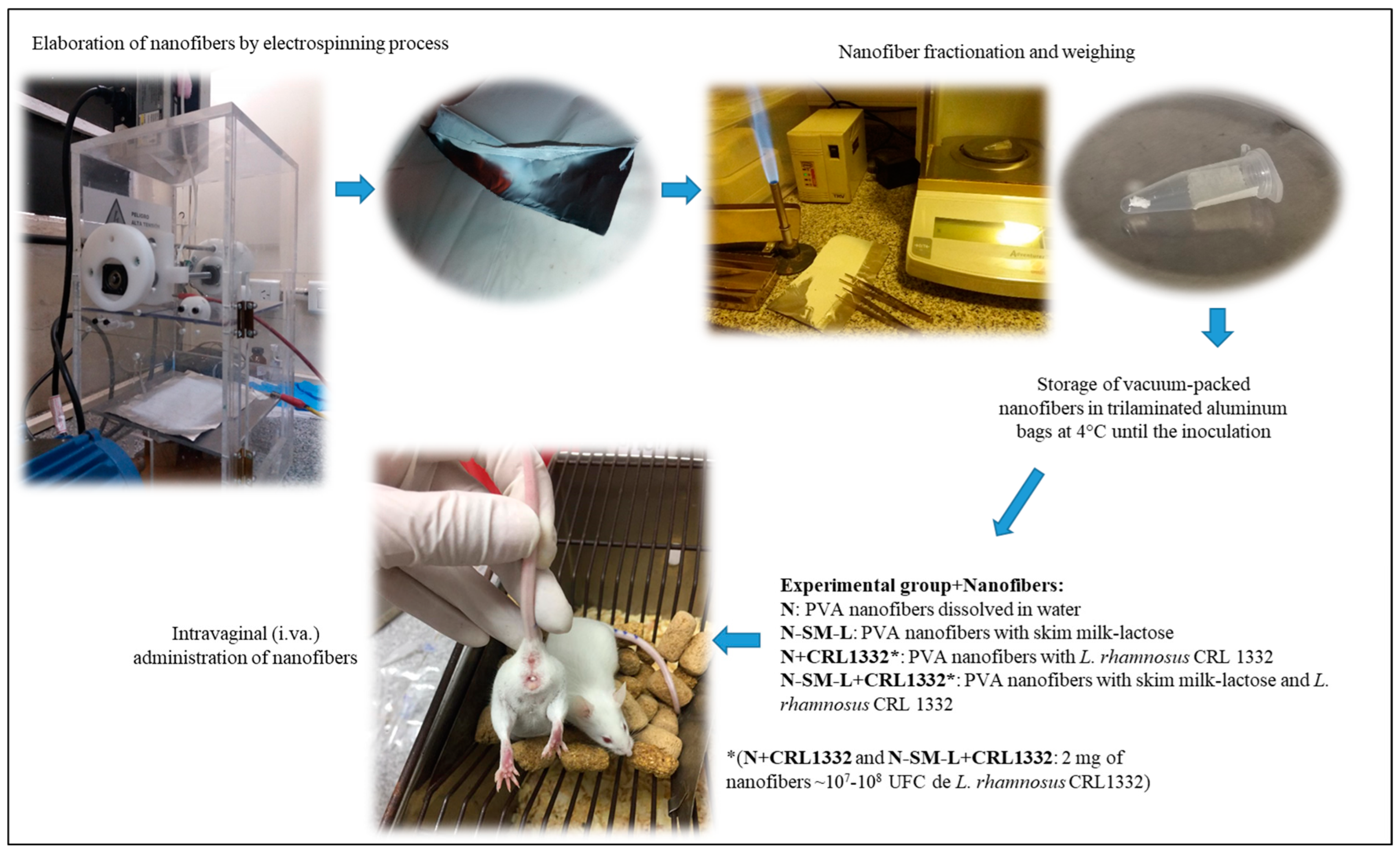
2. Materials and Methods

2.1. Microorganisms and Culture Conditions

The microorganism used in this study was L. rhamnosus CRL1332, isolated from healthy women and included in the Culture Collection of the Reference Center for Lactobacilli (CERELA) (Tucumán, Argentina). This strain was genotypically identified by 16S-RNA–DNA sequencing, and its beneficial probiotic properties were described in previous works [4,19,21,22,23,24,25,26]. The bacteria were stored in frozen aliquots at −80 °C in milk-yeast extract (13% skim milk, 0.5% yeast extract, and 1% glucose; Britania Laboratories, Buenos Aires, Argentina) supplemented with 20% glycerol (Cicarelli Laboratories, Santa Fe, Argentina). The strain was subcultured twice in MRS broth (De Man, Rogosa, and Sharpe) (Biokar Diagnostics, Pantin, France) at 37 °C for 12 h before use.

2.2. Production of Nanofibers with L. rhamnosus CRL1332

To obtain the different types of nanofibers with L. rhamnosus CRL1332 immobilized, the electrospinning technique was applied as described by Silva et al. [4]. The nanofibers obtained were identified as follows: N: PVA nanofibers prepared from a polymeric solution of polyvinyl alcohol (PVA); N+CRL1332: PVA nanofibers with immobilized L. rhamnosus CRL1332; N-SM-L: PVA nanofibers with skim milk lactose; N-SM-L+CRL1332: PVA nanofibers with skim milk lactose and immobilized L. rhamnosus CRL1332. Figure 1 shows the scheme for the elaboration, fractionation, storage, and administration of nanofibers to female mice. The nanofiber blankets obtained by the electrospinning process were handled under sterile conditions and carefully removed from the aluminum foil for their fractionation and distribution after weight in analytical balance. Based on the post-process lactobacilli viability results and considering the maximum volume/weight to be intravaginally administered to mice, the dose was 2 mg, containing approximately 107–108 CFU L. rhamnosus CRL1332. The nanofibers were compacted in such a way as to simulate the shape of a tampon and placed in microtubes stored in vacuum-sealed trilaminar aluminum bags, which were kept refrigerated up to the time of inoculation. The nanofibers were manipulated with tweezers under sterile conditions in order to perform an adequate intravaginal inoculation of the animals.

2.3. Female BALB/c Mice as Experimental Model

The nanofibers were administered in the different experimental groups (EG) in order to assay the safety and effects of the i.va. administration in the murine experimental model. Inbred female BALB/c strain mice, 7 weeks of age, weighing 25 to 30 g, provided by the CERELA biotherium, were used at this stage. The animals were kept in separate cages and fed ad libitum with a conventionally balanced diet under constant environmental conditions. To induce the pseudo-estrus status during the protocol, the hormone 17-β-estradiol-valerate (Sigma-Aldrich, Milan, Italy) was subcutaneously administered to all the mice (0.02 mg in 100 μL of sesame oil, every six days) according to the protocol set up in the laboratory [18]. In this way, the experimental variations of the hormonal changes of the estrous cycle were avoided, promoting the permanence of lactobacilli [17]. Female mice were separated into six EGs, with five mice assigned randomly to each group as follows: N+CRL1332: mice inoculated with 2 mg PVA nanofibers + immobilized L. rhamnosus CRL1332; N-SM-L+CRL1332: mice inoculated with 2 mg PVA nanofibers + skim milk lactose + immobilized L. rhamnosus CRL1332; CRL1332: mice inoculated with 50 µL of peptone agar (% (w/v): 1% peptone meat, 1.5% agar) containing L. rhamnosus CRL1332 (~107–108 CFU); N: mice inoculated with 2 mg of pure PVA nanofibers; N-SM-L: mice inoculated with 2 mg PVA nanofibers + skim milk lactose; and SS: mice inoculated with 50 µL saline solution (control).
The animals that did not reach the pseudo-estrus condition after the hormonal treatment were excluded from the experimental groups. The CERELA Institutional Committee for the Care and Use of Laboratory Animals approved the experimental protocol CRL-BIOT-LMP-2011/2A used in this work. Figure 2 shows the administration scheme that was applied to the different EG for the i.va. mice and the days of sampling. Forty-eight hours after the hormone administration, mice from the different EGs were inoculated by i.va. way once a day for 7 days.

2.4. Sampling of Animals and Analytical Procedures

Prior to the nanofiber administration (day zero) and one day after finishing (day eight), samples of vaginal washes (v.w.) were taken from the different experimental groups under sterile conditions. Vaginal washes were performed with an automatic micropipettor tip loaded with 50 μL of phosphate buffered saline (PBS: 8.1 mM Na2HPO4, 1.5 mM KH2PO4, 140 mM NaCl, pH 7.2) 7 times, which allowed for the pooling of each animal to carry out different determinations (quantification of cultivable microorganisms and cytological analysis) described below. After the last sampling, the mice were sacrificed by cervical dislocation and aseptically dissected to extract vagina, which was transferred to the appropriate solvents for subsequent processes (histological and electronic microscope observation).

2.4.1. Microbiological Analysis

To evaluate the effect of the administration of lactobacilli immobilized in nanofibers on the vaginal microbiota of mice and to demonstrate whether L. rhamnosus CRL1332 was maintained in the murine vagina, the quantification of cultivable microorganisms was performed before and after inoculation. The number of cultivable bacteria in the v.w. was determined by the method of successive dilutions and subsequent plating in selective culture media for different groups of microorganisms. Viable lactobacilli were counted on MRS agar pH 5.5, enterobacteria on Mac Conkey agar, enterococci on Bile Esculin agar, staphylococci on Mannitol Salt Agar, and total aerobic mesophilic bacteria on PCA (Plate Count Agar) [19,27]. The plates were incubated in aerobic conditions at 30 °C and 37 °C for 24–48 h (for vaginal microbiota) or microaerophilic conditions at 37 °C for 48 h (for lactobacilli). The number of microorganisms was expressed as log CFU/mL of v.w.

2.4.2. Cytological and Histological Analysis

To assess whether inoculation of lactobacilli nanofibers administered intravaginally to mice produced any type of adverse effect, cytological and histological studies were performed. Cytology of vaginal lavage was determined in smears of 10 μL of v.w. performed on glass slides, with subsequent staining with May Grünwald-Giemsa (MG-Giemsa) technique [28]. On the other hand, vaginal samples were fixed in 4% (v/v) formaldehyde at 4 °C and embedded in paraffin, applying standard laboratory histological methods [14]. Vaginal washings and vaginal histological sections were observed under an optical microscope (magnification, 40×) to determine the moment of the estrous cycle of mice, the cellular response, and the structural modifications produced by the administration of nanofibers [29]. Photographs were taken on an Axio Scope A1 Carl Zeiss microscope (Göttingen, Germany).

2.4.3. Ultrastructural Analysis

The effect of the i.va. administration of L. rhamnosus CRL1332 in nanofibers on the ultrastructural morphology of vaginal epithelium cells and their permanence was evaluated by scanning electron microscopy. Mouse vaginal samples were placed in Karnovsky’s fixative (2.66% paraformaldehyde, 0.1 M sodium-phosphate buffer, pH 7.4, 1.66% glutaraldehyde) for 1 week. The technique applied was detailed previously by Leccese Terraf [23]. The samples were processed and observed in a scanning electron microscope (Zeiss model SUPRA 55VP: Carl Zeiss microscope, Oberkochen, Germany).

2.5. Statistical Analysis

The mean and standard error were calculated from the obtained experimental data. The analysis of variance was applied using the general linear model (ANOVA-GLM) with the statistical software Minitab version 16.1.0 (Minitab Inc., State College, PA, USA). Significant differences between means were determined using Tukey’s test. Values of p < 0.05 were considered statistically significant.

3. Results

3.1. Effect of Intravaginal Administration of L. rhamnosus CRL1332 Immobilized on Nanofibers on the Cultivable Vaginal Microbiota of BALB/c Mice

The effect of i.va. administration of L. rhamnosus CRL1332 immobilized nanofibers for 7 days on the different cultivable microorganism groups of the murine vaginal microbiota is shown in Figure 3. No modifications were observed in the number of enterobacteria, staphylococci, enterococci, and total aerobic mesophilic bacteria before and after 7 days of inoculation of lactobacilli immobilized on nanofibers. No viable lactobacilli were detected after 7 days of administration in control mice (SS) or in those that received nanofibers without lactobacilli (N and N-SM-L). However, a significant increase in the number of lactobacilli was observed in the v.w. of mice inoculated with free lactobacilli (CRL1332) and immobilized in nanofibers (N+CRL1332 and N-SM-L+CRL1332) after 7 days of administration. The values of CFU/mL of lactobacilli cultivable in the vagina of the different EG inoculated with lactobacilli (CRL1332, N+CRL1332, and N-SM-L+CRL1332) did not show significant differences between them, reaching approximately 104–5 CFU/mL v.w. one day after the last i.va. lactobacilli administration.

3.2. Cytological and Histological Analysis

Cytological evaluation of the vaginal lavage of mice inoculated i.va. with lactobacilli immobilized on nanofibers showed that they did not exhibit an inflammatory response. Figure 4 shows the v.w. cytology of mice inoculated i.va. with: (A) saline solution (SS), (B) L. rhamnosus CRL1332 in agarized peptone (CRL1332), (C) PVA nanofibers (N), (D) PVA nanofibers with skim milk lactose (N-SM-L), (E) PVA nanofibers with immobilized L. rhamnosus CRL1332 nanofibers in PVA (N+CRL1332), and (F) PVA nanofibers with skim milk lactose and L. rhamnosus CRL1332 (N-SM-L+CRL1332). Vaginal smears stained with MG-Giemsa and observed under an optical microscope showed that i.va. of nanofibers with and without immobilized lactobacilli, as well as of L. rhamnosus CRL1332 in agarized peptone, did not produce any type of adverse effect at the cytological level. All the samples showed patterns similar to those of the EG-control mice. The typical state of the pseudo-estrus phase was observed: keratinized epithelial cells (irregular scale shape), absence of nucleated epithelial cells, and leukocytes (indicative of an inflammatory or adverse response). The response to the i.va inoculation of lactobacilli is not comparable with the positive inflammation response observed in previous work by our group in those models of challenge with pathogens [18].
Regarding histological studies, Figure 5 shows the photomicrographs of vaginal sections of i.va. inoculated mice with (A) saline solution, (B) free cells of L. rhamnosus CRL1332, and with the different nanofibers (C–F). The structure of the vagina of mice inoculated i.va. with lactobacilli immobilized on nanofibers, evaluated through histological sections stained with Hematoxylin and Eosin, was comparable between them. Samples studied at the level of histological structure show normal lamina propia characteristics, multilayer epithelium, and keratinized epithelial cells, all of which indicate the pseudo-estrous state induced by the hormonal inoculation. The different EGs showed similar patterns to those of control mice and an absence of an inflammatory response in the murine tissue.

3.3. Ultrastructural Morphological Analysis

The study of the vagina of mice inoculated with lactobacilli in nanofibers through scanning electron microscopy (SEM) showed that there are no adverse effects at the level of the surface of the vaginal epithelium. Examples of scanning microscopy images showing the vaginal cell surface of mice are included in Figure 6. A. shows a microphotograph of the control group mice, in which polygonal squamous cells with very heterogeneous cell borders and microcrusts were observed, typical characteristics of cells from animals in estrus. Only in those animals that received nanofibers with immobilized lactobacilli (E and F) or L. rhamnosus CRL1332 free cells in agarized peptone (B), some bacilli were observed on the surface of the epithelial cells (indicated with black arrows), while in the images from animals that received saline solution (A) or nanofibers without lactobacilli (C and D), the presence of these bacteria was not observed. No modifications were documented on the ultrastructure of the vaginal murine epithelial cells for the different experimental groups, with few modifications related to the normal cultivable vaginal microbiota and at the ultrastructural level.

4. Discussion

Probiotics are applied to women in order to restore the vaginal microbiome and prevent or treat urogenital tract infections [1,2,9,30]. In recent years, the development of nanofibers intended for topical application to the vaginal tract in order to combat urogenital tract infections has expanded [3,4,5,31]. However, at this moment, the evaluation of the safety and effects of intravaginal administration in a murine experimental model of lactobacilli immobilized on nanofibers has not been studied. In this work, the effect of lactobacilli in nanofibers on their application in safe products is demonstrated through protocols applied to evaluate the intravaginal administration of lactobacilli immobilized in nanofibers in a murine experimental model. Although no cytological or histological changes were detected in the murine vaginal tract, complementary assays need to be performed to confirm the absence of adverse effects and the degree of interaction with immune inflammatory cells.
At the moment of selecting the animal experimental model to use, and depending on the type of test that corresponds, the physiological characteristics must be taken into account. The murine vaginal tract is different from those of women, mainly in the vaginal pH, which is neutral, the autochthonous microbiota with a low number or absence of lactobacilli, and the type and duration of the sexual cycle [32]. However, based on the success of using the murine model to study the vaginal tract described by numerous researchers and fine-tuned in our work group, the experimental model in estrogenized mice is useful for predicting the behavior expected in humans [8,15,18,19,27,32,33,34].
Microbiological, cytological, histological, and ultramicroscopic techniques were applied in order to complement the characterization and evaluations carried out in previous works [4,20]. The results showed that lactobacilli nanofibers i.va. administered did not produce adverse effects on murine vaginal tissue and showed normal cytological and histological patterns in the different experimental groups. These results were similar to those previously obtained by our work group, where lactobacilli were administered individually or combined with plant extracts without observing adverse effects on the experimental mouse model [17,19,35]. Moreover, De Gregorio [36] has demonstrated that inoculation of a biosurfactant isolated from L. crispatus BC1, a vaginal strain, was safe and did not disturb vaginal cytology, histology, or cultivable vaginal microbiota in a murine experimental model. No publications showing a complete study of the safety and effects of vaginal lactobacilli in PVA nanofibers i.va. administered were detected in a murine experimental model [37]. Recently, Ilomuanya [38] has shown in a safety assessment at the histological level using female rats that the i.va. administration of an electrospun scaffold with Lactobacillus spp., a tenside, and metronidazole exhibited no irritation or inflammation in the vaginal epithelium compared with the control group.
The use of skim milk and lactose included in the nanofibers was previously demonstrated to be acceptable as excipients for the design of vaginal delivery of probiotic strains [7,38,39]. On the other hand, the PVA polymer was selected, taking into account that it is a mucoadhesive, biocompatible, and hydrophilic polymer and is generally recognized as safe (GRAS), which also supports its safe application as a nanocarrier for vaginal probiotic delivery [40,41,42]. In addition, this polymer was extensively studied for multiple biomedical uses, such as cartilage generators and orthopedic applications, and for vaginal and transdermal drug delivery [4,43,44,45,46,47].
After i.va. administration of L. rhamnosus CRL1332 in nanofibers for 7 days, there were no modifications to the cultivable murine vaginal microbiota. In this study, the microbiologic analysis of viable bacteria was applied to demonstrate the modifications of cultivable microorganisms among the different treatments. However, since this technique does not allow the identification of all the microorganisms of the vaginal microbiota, further study needs to be applied by using molecular techniques in a way to demonstrate the effect of the exogenous administration of lactobacilli on the murine vaginal microbiota. Our results are different from those of De Gregorio [18], who showed the intravaginal administration of L. reuteri CRL1324 to estrogenized mice produced a slight decrease in enterobacteria and staphylococci populations. The results obtained at this work showed that no cultivable lactobacilli were detected in the v.w. of control mice (in MRS pH 5.5 medium), which supports the fact that the quantified cultivable lactobacilli are a consequence of their exogenous i.va. administration, as previously demonstrated [19]. In the same way, in a randomized, double-blind, phase I trial, the i.va. inoculation of healthy women with different strains: L. rhamnosus CRL1332, L. gasseri CRL1256, and CRL1320, evidenced the safety and a significant increase in cultivable lactobacilli after interventions [7]. Although the amount of lactobacilli that remained in the murine vaginal epithelium was lower than those included in the doses that were administered every day, 104–105 CFU/mL viable lactobacilli in v.w. were similar to those previously reported in our research group [16,18,35]. In this study, the viability of lactobacilli on murine vagina could be maintained at similar values by applying a nanosystem delivery based on PVA-nanofiber containing immobilized lactobacilli that could be applied to coat feminine hygiene products instead of the technique with no development of a delivery system. However, more studies are required to evaluate if a longer administration time will show differences between the administration systems (peptone agar and PVA nanofiber) in the lactobacilli viability and colonization in the murine vagina. Similar results were obtained by Daniele [48] with L. fermentum colonization assays in the vagina of BALB/c mice, with similar CFU/mL lactobacilli values and an inhibitory effect on Gardnerella vaginalis. Taking into account the specificity of lactobacilli species and strains previously demonstrated in numerous publications, added to the results obtained of their technological properties and adhesion/colonization capability of vaginal lactobacilli, arises the need to evaluate these systems with other strains of lactobacilli [4,18,24,25,26,49,50,51].
Finally, the absence of nanofibers observed in the SEM images after 1 day of i.va. administration indicates their complete dissolution upon contact with the mucosa, given the hydrophilic nature of the nanofibers as evidenced in previous work. On the other hand, the images of bacilli on the surface of the vaginal cells obtained in scanning electron microscopy in the EG that received lactobacilli i.va. administration show that they produce little change in the normal vaginal epithelial cell ultrastructure. These observations correlate with the results obtained in the microbiological assays, in which the number of viable lactobacilli increased only in those groups to which L. rhamnosus CRL1332 was administered, also suggesting the permanence of lactobacilli in the tract. Then, the results obtained in this work contribute to the next steps in evaluating the efficacy and effectiveness of human clinical trials [2,7,52].

5. Conclusions

A complete evaluation of the safety and effects of i.va. administration of potential probiotic nanofibers on a murine experimental model was reported for the first time. Our results demonstrated that the intravaginal administration of L. rhamnosus CRL1332 immobilized in nanofibers is safe in murine models, given the absence of an inflammatory response at the cytological and histological levels, and little modification of the normal cultivable vaginal microbiota at the ultrastructural level. The inoculation with these lactobacilli-nanofibers produced an increase in viable lactobacilli, promoting their permanence in the murine vaginal tract. Therefore, this bacteria immobilization system is a promising, novel, and safe alternative delivery system to advance in the design of vaginal probiotic products that could reconstitute the vaginal microbiota and prevent urogenital tract infections.

6. Patents

Some of the results were included in a patent presentation published in the Argentine INPI Patent Bulletin No. 1095 on 15 July 2020.

Author Contributions

Methodology, J.A.S., P.R.D.G. and M.E.F.N.-M.; Validation, J.A.S. and M.E.F.N.-M.; Formal analysis, J.A.S., P.R.D.G. and M.E.F.N.-M.; Investigation, M.E.F.N.-M.; Writing—original draft, J.A.S. and M.E.F.N.-M.; Writing—review & editing, M.E.F.N.-M.; Funding acquisition, M.E.F.N.-M. All authors have read and agreed to the published version of the manuscript.

Funding

This research was funded by the Consejo Nacional de Investigaciones Científicas y Técnicas, Argentina (CONICET) (PIP 545) and the Agencia Nacional de Promoción Científica y Tecnológica (ANPCyT) (PICT 2017-4324, PICT 2018-00670, and PICT 2018-02334).

Institutional Review Board Statement

The study was approved by the CERELA Institutional Committee for the Care and Use of Laboratory Animals (experimental protocol CRL-BIOT-LMP-2011/2A).

Informed Consent Statement

Not applicable.

Data Availability Statement

The data are available at the CONICET repository.

Conflicts of Interest

The authors declare no conflict of interest.

References

  1. Puebla-Barragan, S.; Reid, G. Probiotics in Cosmetic and Personal Care Products: Trends and Challenges. Molecules 2021, 26, 1249. [Google Scholar] [CrossRef]
  2. Nader-Macías, M.E.F.; De Gregorio, P.R.; Silva, J.A. Probiotic lactobacilli in formulas and hygiene products for the health of the urogenital tract. Pharmacol. Res. Perspect. 2021, 9, e00787. [Google Scholar] [CrossRef]
  3. Škrlec, K.; Zupančič, Š.; Prpar Mihevc, S.; Kocbek, P.; Kristl, J.; Berlec, A. Development of electrospun nanofibers that enable high loading and long-term viability of probiotics. Eur. J. Pharm. Biopharm. 2019, 136, 108–119. [Google Scholar] [CrossRef]
  4. Silva, J.A.; De Gregorio, P.R.; Rivero, G.; Abraham, G.A.; Nader-Macías, M.E.F. Immobilization of vaginal Lactobacillus in polymeric nanofibers for its incorporation in vaginal probiotic products. Eur. J. Pharm. Sci. 2021, 156, 105563. [Google Scholar] [CrossRef]
  5. Stojanov, S.; Kristl, J.; Zupančič, Š.; Berlec, A. Influence of Excipient Composition on Survival of Vaginal Lactobacilli in Electrospun Nanofibers. Pharmaceutic 2022, 14, 1155. [Google Scholar] [CrossRef]
  6. Domig, K.J.; Kiss, H.; Petricevic, L.; Viernstein, H.; Unger, F.; Kneifel, W. Strategies for the evaluation and selection of potential vaginal probiotics from human sources: An exemplary study. Benef. Microbes 2014, 5, 263–272. [Google Scholar] [CrossRef]
  7. De Gregorio, P.R.; Maldonado, N.C.; Pingitore, E.V.; Terraf, M.C.L.; Tomás, M.S.J.; de Ruiz, C.S.; Santos, V.; Wiese, B.; Bru, E.; Paiz, M.C.; et al. Intravaginal administration of gelatine capsules containing freeze-dried autochthonous lactobacilli: A double-blind, randomised clinical trial of safety. Benef. Microbes 2020, 11, 5–17. [Google Scholar] [CrossRef]
  8. De Gregorio, P.R.; Juárez Tomás, M.S.; Nader-Macías, M.E.F. Immunomodulation of Lactobacillus reuteri CRL1324 on group B Streptococcus vaginal colonization in a murine experimental model. Am. J. Reprod. Immunol. 2016, 75, 23–35. [Google Scholar] [CrossRef]
  9. Ballini, A.; Santacroce, L.; Cantore, S.; Bottalico, L.; Dipalma, G.; Vito, D.; Saini, R.; Inchingolo, F. Probiotics Improve Urogenital Health in Women. Open Access Maced. J. Med. Sci. 2018, 6, 1845–1850. [Google Scholar] [CrossRef]
  10. Gorreja, F.; Walker, W.A. The potential role of adherence factors in probiotic function in the gastrointestinal tract of adults and pediatrics: A narrative review of experimental and human studies. Gut Microbes 2022, 14, 2149214. [Google Scholar] [CrossRef]
  11. European Food Safety Authority. Opinion of the Scientific Committee on a request from EFSA on the introduction of a Qualified Presumption of Safety (QPS) approach for assessment of selected microorganisms referred to EFSA. EFSA J. 2007, 587, 1–16. [Google Scholar] [CrossRef]
  12. Marzal Alfaro, M.B.; Manrique-Rodríguez, S.; Fernández-Llamazares, C.M. Clinical use of probiotics and practical aspects of their use. Hosp. Nutr. 2013, 28, 68–70. Available online: http://scielo.isciii.es/scielo.php?script=sci_arttext&pid=S0212-16112013000700018&lng=es&tlng=en (accessed on 12 May 2023).
  13. Nader-Macías, M.E.F.; Silva de Ruiz, C.; Ocaña, V.S.; Juárez Tomas, M.S. Advances in the Knowledge and Clinical Applications of Lactic Acid Bacteria as Probiotics in the Urogenital Tract. Curr. Womens Health Rev. 2008, 4, 240–257. [Google Scholar] [CrossRef]
  14. Silva de Ruiz, C.; del R. Rey, M.; Nader-Macías, M.E. Structural and ultrastructural studies of the urinary tract of mice inoculated with Lactobacillus fermentum. BJU Int. 2003, 91, 878–882. [Google Scholar] [CrossRef]
  15. Zárate, G.; Santos, V.; Nader-Macias, M.E. Protective effect of vaginal Lactobacillus paracasei CRL 1289 against urogenital infection produced by Staphylococcus aureus in a mouse animal model. Infect. Dis. Obstet. Gynecol. 2009, 48358. [Google Scholar] [CrossRef]
  16. Leccese Terraf, M.C. Biofilm-Forming Urogenital Beneficial Lactic Acid Bacteria (BUGB): Basic Studies and Tests in Experimental Animals. Potential Applications in the Prevention of Urinary Tract Infections. Ph.D. Thesis, Faculty of Biochemistry, Chemistry and Pharmacy, National University of Tucumán, San Miguel de Tucumán, Argentina, 2014. [Google Scholar]
  17. De Gregorio, P.R.; Juárez Tomás, M.S.; Santos, V.; Nader-Macías, M.E. Beneficial lactobacilli: Effects on the vaginal tract in a murine experimental model. Antonie Leeuwenhoek 2012, 102, 569–580. [Google Scholar] [CrossRef]
  18. De Gregorio, P.R.; Juárez Tomás, M.S.; Leccese Terraf, M.C.; Nader-Macías, M.E. Preventive effect of Lactobacillus reuteri CRL1324 on Group B Streptococcus vaginal colonization in an experimental mouse model. J. Appl. Microbiol. 2015, 118, 1034–1047. [Google Scholar] [CrossRef] [PubMed]
  19. De Gregorio, P.R.; Silva, J.A.; Marchesi, A.; Nader-Macias, M.E.F. Anti-Candida activity of beneficial vaginal lactobacilli in in vitro assays and in a murine experimental model. FEMS Yeast Res. 2019, 19, foz008. [Google Scholar] [CrossRef] [PubMed]
  20. Silva, J.A. Beneficial Lactic Acid Bacteria (BLAB) in the Design of Vaginal Hygiene Formulations. Ph.D. Thesis, Faculty of Biochemistry, Chemistry and Pharmacy, National University of Tucumán, San Miguel de Tucumán, Argentina, 2021. [Google Scholar]
  21. Ocana, V.S.; Bru, E.; De Ruiz Holgado, A.A.; Nader-Macias, M.E. Surface characteristics of lactobacilli isolated from human vagina. J. Gen. Appl. Microbiol. 1999, 45, 203–212. [Google Scholar] [CrossRef]
  22. Juárez Tomás, M.S.; Saralegui Duhart, C.I.; De Gregorio, P.R.; Vera Pingitore, E.; Nader-Macías, M.E. Urogenital pathogen inhibition and compatibility between vaginal Lactobacillus strains to be considered as probiotic candidates. Eur. J. Obstet. Gynecol. Reprod. Biol. 2011, 159, 399–406. [Google Scholar] [CrossRef]
  23. Leccese Terraf, M.C.; Juárez Tomás, M.S.; Nader-Macías, M.E.; Silva, C. Screening of biofilm formation by beneficial vaginal lactobacilli and influence of culture media components. J. Appl. Microbiol. 2012, 113, 1517–1529. [Google Scholar] [CrossRef]
  24. Marchesi, A.; Silva, J.A.; Wiese, B.; Nader-Macías, M.E.F. Survival of Beneficial Vaginal Lactobacilli (BVL) to Different Gastrointestinal Tract Conditions. Curr. Pharm. Des. 2020, 26, 3608–3618. [Google Scholar] [CrossRef] [PubMed]
  25. Marchesi, A.; Silva, J.A.; Ficoseco, C.A.; Wiese, B.; Nader-Macías, M.E.F. Effect of phytoderivatives on the growth of homologous beneficial vaginal lactobacilli (BVL) strains and their compatibility for the design of phytobiotics for the vaginal tract health. World J. Pharm. Pharm. Sci. 2020, 9, 236–261. [Google Scholar] [CrossRef]
  26. Silva, J.A.; Marchesi, A.; Aristimuño Ficosecco, M.C.; Nader-Macías, M.E.F. Functional and safety characterization of beneficial vaginal lactic acid bacteria for the design of vaginal hygiene products. J. Appl. Microbiol. 2022, 33, 3041–3058. [Google Scholar] [CrossRef] [PubMed]
  27. De Gregorio, P.R.; Salva, M.S.; Juárez Tomás, M.S.; Nader, M.E.F. Effects of exogenous sex hormones on mouse estrous cycle, vaginal microbiota and immune cells. Scand. J. Lab. Anim. Sci. 2018, 44, 3. [Google Scholar] [CrossRef]
  28. McLean, A.C.; Valenzuela, N.; Fai, S.; Bennett, S.A. Performing vaginal lavage, crystal violet staining, and vaginal cytological evaluation for mouse estrous cycle staging identification. JoVE 2012, 67, e4389. [Google Scholar] [CrossRef]
  29. Silva de Ruiz, C.S.; Rey, M.R.; de Ruiz Holgado, A.P.; Nader-Macías, M.E. Experimental administration of estradiol on the colonization of Lactobacillus fermentum and Escherichia coli in the urogenital tract of mice. Biol. Pharm. Bull. 2001, 24, 127–134. [Google Scholar] [CrossRef]
  30. Dabee, S.; Passmore, J.S.; Heffron, R.; Jaspan, H.B. The Complex Link between the Female Genital Microbiota, Genital Infections, and Inflammation. Infect. Immun. 2021, 89, e00487-20. [Google Scholar] [CrossRef]
  31. Zupančič, Š.; Škrlec, K.; Kocbek, P.; Kristl, J.; Berlec, A. Effects of electrospinning on the viability of ten species of lactic acid bacteria in poly(ethylene oxide) nanofibers. Pharmaceutics 2019, 11, 483. [Google Scholar] [CrossRef]
  32. Patras, K.A.; Wang, N.Y.; Fletcher, E.M.; Cavaco, C.K.; Jimenez, A.; Garg, M.; Fierer, J.; Sheen, T.R.; Rajagopal, L.; Doran, K.S. Group B Streptococcus CovR regulation modulates host immune signalling pathways to promote vaginal colonization. Cell. Microbiol. 2013, 15, 1154–1167. [Google Scholar] [CrossRef]
  33. Joo, H.M.; Kim, K.A.; Myoung, K.S.; Ahn, Y.T.; Lee, J.H.; Huh, C.S.; Han, M.J.; Kim, D.H. Lactobacillus helveticus HY7801 ameliorates vulvovaginal candidiasis in mice by inhibiting fungal growth and NF-κB activation. Int. Immunopharmacol. 2012, 14, 39–46. [Google Scholar] [CrossRef]
  34. De Gregorio, P.R.; Juares Tomás, M.S.; Leccese Terraf, M.C.; Nader-Macías, M.E.F. In vitro and in vivo effects of beneficial vaginal lactobacilli on pathogens responsible for urogenital tract infections. J. Med. Microbiol. 2014, 63, 685–696. [Google Scholar] [CrossRef] [PubMed]
  35. Marchesi, A. Advances in the Design of Novel Phytobiotics with Beneficial Vaginal Lactobacilli (BVL). Ph.D. Thesis, Faculty of Biochemistry, Chemistry and Pharmacy, National University of Tucumán, San Miguel de Tucumán, Argentina, 2020. [Google Scholar]
  36. De Gregorio, P.R.; Parolin, C.; Abruzzo, A.; Luppi, B.; Protti, M.; Mercolini, L.; Silva, J.A.; Giordani, B.; Marangoni, A.; Nader-Macías, M.E.F.; et al. Biosurfactant from vaginal Lactobacillus crispatus BC1 as a promising agent to interfere with Candida adhesion. Microb. Cell Factories 2020, 19, 133. [Google Scholar] [CrossRef] [PubMed]
  37. Diep, E.; Schiffman, J.D. Electrospinning Living Bacteria: A Review of Applications from Agriculture to Health Care. ACS Appl. Bio Mater. 2023, 6, 951–964. [Google Scholar] [CrossRef]
  38. Ilomuanya, M.O.; Bassey, P.O.; Ogundemuren, D.A.; Ubani-Ukoma, U.N.; Tsamis, A.; Fan, Y.; Michalakis, K.; Angsantikul, P.; Usman, A.; Amenaghawon, A.N. Development of Mucoadhesive Electrospun Scaffolds for Intravaginal Delivery of Lactobacilli spp., a Tenside, and Metronidazole for the Management of Bacterial Vaginosis. Pharmaceutics 2023, 15, 1263. [Google Scholar] [CrossRef] [PubMed]
  39. Vitali, B.; Abruzzo, A.; Parolin, C.; Palomino, R.A.; Dalena, F.; Bigucci, F.; Cerchiara, T.; Luppi, B. Association of Lactobacillus crispatus with fructo-oligosaccharides and ascorbic acid in hydroxypropyl methylcellulose vaginal insert. Carbohydr. Polym. 2016, 136, 1161–1169. [Google Scholar] [CrossRef]
  40. Nagy, Z.K.; Wagner, I.; Suhajda, Á.; Tobak, T.; Harasztos, A.H.; Vigh, T.; Sóti, P.L. Nanofibrous solid dosage form of living bacteria prepared by electrospinning. Express Polym. Lett. 2014, 8, 352–361. [Google Scholar] [CrossRef]
  41. Gaaz, T.S.; Sulong, A.B.; Akhtar, M.N.; Kadhum, A.A.; Mohamad, A.B.; Al-Amiery, A.A. Properties and Applications of Polyvinyl Alcohol, Halloysite Nanotubes and Their Nanocomposites. Molecules 2015, 20, 22833–22847. [Google Scholar] [CrossRef]
  42. Nam, S.; Mooney, D. Polymeric Tissue Adhesives. Chem. Rev. 2021, 121, 11336–11384. [Google Scholar] [CrossRef]
  43. Gonzalez, J.S.; Maiolo, A.S.; Hoppe, C.E.; Alvarez, V.A. Composite gels based on poly(vinyl alcohol) for biomedical uses. Procedia Mater. Sci. 2012, 1, 483–490. [Google Scholar] [CrossRef]
  44. Baker, M.I.; Walsh, S.P.; Schwartz, Z.; Boyan, B.D. A review of polyvinyl alcohol and its uses in cartilage and orthopedic applications. J. Biomed. Mater. Res. B Appl. Biomater. 2012, 100, 1451–1457. [Google Scholar] [CrossRef] [PubMed]
  45. Sharma, R.; Garg, T.; Goyal, A.K.; Rath, G. Development, optimization and evaluation of polymeric electrospun nanofiber: A tool for local delivery of fluconazole for management of vaginal candidiasis. Artif. Cells Nanomed. Biotechnol. 2016, 44, 524–531. [Google Scholar] [CrossRef]
  46. Iqbal, Z.; Dilnawaz, F. Nanocarriers For Vaginal Drug Delivery. Recent Pat. Drug Deliv. Formul. 2019, 13, 3–15. [Google Scholar] [CrossRef]
  47. Reddy, V.S.; Tian, Y.; Zhang, C.; Ye, Z.; Roy, K.; Chinnappan, A.; Ramakrishna, S.; Liu, W.; Ghosh, R. A Review on Electrospun Nanofibers Based Advanced Applications: From Health Care to Energy Devices. Polymers 2021, 13, 3746. [Google Scholar] [CrossRef]
  48. Daniele, M.; Pascual, L.; Barberis, L. Curative effect of the probiotic strain Lactobacillus fermentum L23 in a murine model of vaginal infection by Gardnerella vaginalis. Lett. Appl. Microbiol. 2014, 59, 93–98. [Google Scholar] [CrossRef] [PubMed]
  49. Silva, J.A.; Marchesi, A.; Wiese, B.; Nader-Macias, M.E.F. Technological characterization of vaginal probiotic lactobacilli: Resistance to osmotic stress and strains compatibility. J. Appl. Microbiol. 2019, 127, 1835–1847. [Google Scholar] [CrossRef] [PubMed]
  50. Silva, J.A.; Alejandra Marchesi, A.; Wiese, B.; Nader-Macias, M.E.F. Screening of autochthonous vaginal beneficial lactobacilli strains by their growth at high temperatures for technological applications. Antonie Leeuwenhoek 2020, 113, 1393–1409. [Google Scholar] [CrossRef]
  51. Leccese Terraf, M.C.; Mendoza, L.M.; Juárez Tomás, M.S.; Silva, C.; Nader-Macías, M.E. Phenotypic surface properties (aggregation, adhesion and biofilm formation) and presence of related genes in beneficial vaginal lactobacilli. J. Appl. Microbiol. 2014, 117, 1761–1772. [Google Scholar] [CrossRef]
  52. Hill, C.; Guarner, F.; Reid, G.; Gibson, G.R.; Merenstein, D.J.; Pot, B.; Morelli, L.; Canani, R.B.; Flint, H.J.; Salminen, S.; et al. Expert consensus document. The International Scientific Association for Probiotics and Prebiotics consensus statement on the scope and appropriate use of the term probiotic. Nat. Rev. Gastroenterol. Hepatol. 2014, 11, 506–514. [Google Scholar] [CrossRef]
Figure 1. Elaboration of nanofibers by electrospinning, fractionation, storage, and administration to a murine experimental model. N+CRL1332* (Nanofibers dissolved in water) and N-SM-L+CRL1332* (Nanofibers with skim-milk-lactose) groups administrated with 2 mg of nanofiber containing approximately 107-8 UFC/ml of L. rhamnosus CRL1332.
Applmicrobiol 03 00069 g001
Figure 2. Protocol of intravaginal (i.va.) administration of L. rhamnosus CRL1332 immobilized nanofibers and sampling in estrogenized-adult BALB/c female mice.
Applmicrobiol 03 00069 g002
Figure 3. Quantification of cultivable populations of murine vaginal microbiota microorganisms. Data are represented as mean values (log CFU/mL v.w. ± standard error) of viable cells of lactobacilli, enterococci, enterobacteria, staphylococcus, and total aerobic mesophilic bacteria from murine vaginal lavages (v.w.) for the different experimental groups (EG) at days 0 and 7 of the protocol of administration. EG: PVA nanofibers (N); PVA nanofibers with skim milk lactose (N-SM-L); PVA nanofibers with immobilized L. rhamnosus CRL1332 (N+CRL1332); PVA nanofibers with skim milk lactose with L. rhamnosus CRL1332 immobilized (N-SM-L+CRL1332); mice inoculated with L. rhamnosus CRL1332 in agarized peptone (CRL1332); and control mice inoculated with saline (SS). Different letters indicate statistically significant differences (p < 0.05) between EG and over time for each bacterial population.
Applmicrobiol 03 00069 g003
Figure 4. Cytology of vaginal washings (v.w.) from mice inoculated intravaginally with (A) saline solution (SS); (B) L. rhamnosus CRL1332 in agarized peptone (CRL1332); (C) PVA nanofibers (N); (D) PVA nanofibers with skim milk lactose (N-SM-L); (E) PVA nanofibers with L. rhamnosus CRL1332 immobilized (N+CRL1332); and (F) PVA nanofibers with skim milk lactose and immobilized L. rhamnosus CRL1332 (N-SM-L+CRL1332).
Applmicrobiol 03 00069 g004
Figure 5. Vaginal histology of mice inoculated i.va. with (A) saline (SS); (B) L. rhamnosus CRL1332 in agarized peptone (CRL1332); (C) PVA nanofibers (N); (D) PVA nanofibers with skim milk lactose (N-SM-L); (E) PVA nanofibers with L. rhamnosus CRL1332 immobilized (N+CRL1332); and (F) PVA nanofibers with skim milk lactose with immobilized L. rhamnosus CRL1332 (N-SM-L+CRL1332).
Applmicrobiol 03 00069 g005
Figure 6. SEM images (5.00 KX and 10.00 KX) of mouse vaginal epithelial cells i.va. inoculated with: (A) saline solution (SS); (B) L. rhamnosus CRL1332 in agarized peptone (CRL1332); (C) PVA nanofibers (N); (D) PVA nanofibers with skim milk lactose (N-SM-L); (E) PVA nanofibers with L. rhamnosus CRL1332 immobilized (N+CRL1332); and (F) PVA nanofibers with skim milk lactose with immobilized L. rhamnosus CRL1332 (N-SM-L+CRL1332). Bacilli on the surface of the epithelial vaginal cells are indicated with black arrows.
Applmicrobiol 03 00069 g006
Disclaimer/Publisher’s Note: The statements, opinions and data contained in all publications are solely those of the individual author(s) and contributor(s) and not of MDPI and/or the editor(s). MDPI and/or the editor(s) disclaim responsibility for any injury to people or property resulting from any ideas, methods, instructions or products referred to in the content.

