Article

The Role of the Bone Marrow Microenvironment in Physical Function and Quality of Life in Patients with Multiple Myeloma After First-Line Treatment with Novel Agents and Autologous Transplantation

by
Polyxeni Spiliopoulou
1,*,
Pantelis Rousakis
2,
Chrysanthi Panteli
2,
Evangelos Eleutherakis-Papaiakovou
3,
Magdalini Migkou
3,
Nikolaos Kanellias
3,
Ioannis Ntanasis-Stathopoulos
3,
Panagiotis Malandrakis
3,
Foteini Theodorakakou
3,
Despina Fotiou
3,
Evangelos Terpos
3,
Vassilios Myrianthopoulos
4,
Maria Gavriatopoulou
3,
Ourania E. Tsitsilonis
2,
Efstathios Kastritis
3,
Meletios Athanasios Dimopoulos
3 and
Gerasimos Terzis
1
1
Sports Performance Laboratory, School of Physical Education and Sport Science, National and Kapodistrian University of Athens, 17237 Athens, Greece
2
Flow Cytometry Unit, Department of Biology, National and Kapodistrian University of Athens, 15784 Athens, Greece
3
Department of Clinical Therapeutics, School of Medicine, Alexandra General Hospital, National and Kapodistrian University of Athens, 11528 Athens, Greece
4
Laboratory of Pharmaceutical Chemistry, Department of Pharmacy, Panepistimiopolis Zografou, National and Kapodistrian University of Athens, 15771 Athens, Greece
*
Author to whom correspondence should be addressed.
Submission received: 25 March 2025 / Revised: 17 April 2025 / Accepted: 24 April 2025 / Published: 1 May 2025
(This article belongs to the Special Issue Targeting of Tumor Dormancy Pathway)

Simple Summary

Multiple myeloma affects the quality of life and physical functioning of patients. In addition, disease progression is affected by the bone marrow microenvironment. The purpose of this study was to investigate the possible relationship between the bone marrow microenvironment and the quality of life/ physical function of multiple myeloma patients after first-line treatment. It was revealed that the percentage of CD27+ NK/NKT cells was correlated positively with physical function and role functioning, and negatively with fatigue, pain, and dyspnea. This suggests that stronger immune surveillance in the bone marrow from CD27+ NK/NKT cells implies a better quality of life in multiple myeloma patients.

Abstract

Background/Objectives: Multiple myeloma is a malignancy of plasma cells detected in the bone marrow, inducing symptoms like anemia, hypercalcemia, renal problems, and bone fractures in multiple myeloma patients, affecting their quality of life. The bone marrow microenvironment plays a crucial role in the prognosis and progression of the disease. The purpose of this study was to examine the relationship between the percentages of the major cell populations of the bone marrow, including immune cells, and physical function/quality of life in multiple myeloma patients after first-line treatment. Methods: Twenty-one multiple myeloma patients (N = 14 men, N = 7 women) participated in the study after completing first-line treatment. Bone marrow and blood samples were taken one hundred days after transplantation, while physical function (6 min walking test, handgrip test, maximal aerobic power, and isometric strength), health-related quality of life (QLQ-C30), and body composition (DXA) were assessed 2–5 days later. Flow cytometry was used to assess the percentages of plasma cells, mast cells, B cells (total, precursors, naïve, and memory), T cells (total, CD27− and CD27+), NK/NKT cells (total, CD27− and CD27+), eosinophils, monocytes, neutrophils, myeloid progenitors, erythroblasts, and erythroid progenitors, expressed as percentages of total nucleated cells of the bone marrow. Results: The percentage of CD27+ NK/NKT cells was correlated with five parameters of the quality of life questionnaire: physical function (r = 0.78, p = 0.005), role functioning (r = 0.69, p = 0.020), fatigue (r = −0.86, p = 0.000), pain (r = 0.68, p = 0.021), and dyspnea (r = −0.80, p = 0.003). Conclusions: In conclusion, stronger immune surveillance in the bone marrow from CD27+ NK/NKT cells is correlated with better quality of life in multiple myeloma patients.

1. Introduction

Multiple myeloma is a hematological malignancy of bone marrow plasma cells [1]. A percentage of ≥10% clonal plasma cells in the bone marrow is one of the two main multiple myeloma diagnostic criteria [2]. Myeloma cells, along with the paraprotein they overproduce, damage various tissues and lead to hypercalcemia, anemia, renal failure, and/or bone damage [1].
Aside from diagnosis, the level of bone marrow clonal plasma cells is measured after treatment to determine the persistence of residual disease [3]. The residual myeloma cells can become dormant cells, whose possible reactivation will lead to disease relapse [4]. Myeloma cells employ mechanisms to downregulate the anti-tumor immune response [5,6]. Myeloma cells impair the function of, e.g., dendritic cells and macrophages inhibiting T cell activation [7,8] or drive effector NK cells out of the bone marrow [9]. On the other hand, higher levels of cytotoxic T cells and NK cells, combined with lower levels of regulatory T cells in the bone marrow, are believed to control tumor growth in multiple myeloma patients [10].
Physical function and quality of life are secondary outcomes of high importance from the clinical perspective, as they could be exacerbated by multiple myeloma and its symptoms. These two parameters are interconnected, since lower physical function status is associated with lower quality of life in patients with plasma cell disorders [11]. Physical function and quality of life are reduced in patients with multiple myeloma compared to healthy individuals [12], as well as in newly diagnosed multiple myeloma patients compared to patients two or more years after diagnosis [11]. This implies that the tumor burden negatively affects the daily life of multiple myeloma patients. However, whether the bone marrow microenvironment impacts on patients’ daily life is currently unknown.
The purpose of this study was to investigate the relationship between the bone marrow microenvironment and physical function/quality of life in multiple myeloma patients who have undergone first-line treatment. It was hypothesized that higher levels of immune cells with anti-tumor activity, like T cells or NK cells, in the bone marrow will be detected in patients with a better status of physical function and quality of life.

2. Materials and Methods

2.1. Experimental Design

Physical function, body composition, health-related quality of life as well as blood and bone marrow analyses were evaluated in 21 multiple myeloma patients after first-line induction treatment. Physical function was determined by four tests (6 min walking test, handgrip test, isometric strength test, and maximal aerobic power test). Body composition was measured in all patients and a quality of life questionnaire was completed by 19 patients. Blood samples were collected from all patients and bone marrow samples were taken from 16 patients. Blood sampling and bone marrow aspiration were performed one hundred days after autologous stem cell transplantation. Each participant visited the laboratory and performed all the above-mentioned tests one week after bone marrow collection. The study was approved by the bioethics committee of the institutional review board (Protocol Number: 1202/10-06-2020). All procedures followed the Code of Ethics of the World Medical Association (Helsinki Declaration of 1964, as revised in 2013).

2.2. Participants

Twenty-one multiple myeloma patients participated in the study (Table 1) between November 2021 and April 2023. All patients provided written informed consent to participate as volunteers after being informed of the experimental procedures. The inclusion criteria were as follows: (i) participants had completed first-line treatment, undergone autologous hematopoietic stem cell transplantation, completed consolidation therapy, and had just started maintenance treatment; and (ii) their Eastern Cooperative Oncology Group performance status was 0–2. Four patients had bone fractures at diagnosis but performed the tests without any problems or complaints. In addition, the COVID-19 vaccination had been completed in all patients.

2.3. 6 Min Walking Test

The 6 min walking test was performed indoors in a long and spacious corridor. Two cones were placed across one another at a distance of 20 m apart. Each participant started from one cone, walked to the other one, made a counterclockwise rotation, and then repeated the same path for six minutes. Time feedback was given by the researcher every one minute. The instructions were to walk as far as possible, continuously at their own pace. They were encouraged to inform the researcher if they felt unwell or wanted to stop. However, all participants completed the 6 min walk without any problem. The total walking distance was measured and used for the analysis. The ICC for this measurement is 0.95.

2.4. Handgrip Test

Handgrip strength of both the right and left hand was measured with a calibrated hand-held dynamometer (Camry, Model EH101, Zhongshan, China). The patient was in a standing position with the elbow in full extension. Two examinations for each hand were performed with 30 s of rest between each trial. The highest value for each hand was recorded and the sum of right- and left-hand strength was used for the analysis. The ICC in our laboratory is 0.96.

2.5. Isometric Strength

Isometric strength was evaluated with an isometric dynamometer (MicroFET 2 Wireless Manual Muscle Tester, Hoggan Scientific LLC, Salt Lake City, UT, USA, Sampling rate: 100 sample/s) for both the right and left extremities. Specifically, isometric contractions at seven positions for both right and left sides were performed: (1) shoulder adduction, (2) shoulder abduction, (3) hip flexion, (4) hip abduction, (5) hip adduction, (6) knee extension, and (7) knee bend. Two maximal efforts were allowed at each position with a one-minute rest between each effort. The best result was recorded. The sum of shoulder adduction and shoulder abduction on both the right and left sides was calculated as a measure of the upper extremities’ isometric strength. The sum of hip flexion, hip abduction, hip adduction, knee extension, and knee bend on both sides was calculated as a measure of the lower extremities’ isometric strength. Isometric strength of the upper and lower extremities was used for the analysis.

2.6. Maximal Aerobic Power

The aerobic test was performed in a recumbent stationary cycle ergometer (Ergociser, Model Ec-3500, Cateye, Osaka, Japan) according to the adjusted ACSM cycle ergometer protocol [13]. The cycling pace was kept at 50 rpm. Cycling started at 25 Watt, and the intensity was increased by 25 Watt every 3 min until the participant could no longer keep up the cycling pace. Heart rate (Polar A300, 70FF7C15, Polar Electro Oy, Kempele, Finland) and Borg scale were monitored and recorded throughout the aerobic performance test.

2.7. Body Composition

Dual X-ray absorptiometry (DXA) was performed to assess body composition (Lunar Prodigy, General Electrics Medical Systems, Madison, WI, USA). After calibration, the patient was placed in a supine position within the absorptiometry frame. A total body, lumbar, and femur scan was performed. Lunar Prodigy software (Prodigy, encore, version 18, General Electrics, Madison, WI, USA) was used for data analysis. Total mass, total lean body mass, lean body mass of the upper extremities, lean body mass of the lower extremities, lean body mass of the trunk, and total fat mass were determined, as well as bone density for total body, lumbar L2–L4, and femur. Lean body mass index (kg/m2) was calculated according to the following formula: lean body mass (kg) divided by the square of height (m2). The ICC for body composition and bone density is 0.99.

2.8. Health-Related Quality of Life

Health-related quality of life was measured with the QLQ-C30 version 3.0 questionnaire. QLQ-C30 is recommended by the European Organization for Research and Treatment of Cancer for cancer patients. The score range is 0 to 100 and the three main categories were assessed: (a) global health, (b) functional scale (physical/role/emotional/cognitive/social functioning), and (c) symptoms (fatigue/nausea and vomiting/pain/dyspnea/insomnia/appetite loss/constipation/diarrhea/financial difficulties). For categories (a) and (b), a high-value result represents better health level and functioning status, respectively, while for category (c), a high-value result represents worse health-related problems.

2.9. Blood Analysis

Blood samples were acquired through venipuncture during the morning hours, before the start of the maintenance therapy and at the end of the intervention. Tubes containing ethylenediaminetetraacetic acid (EDTA) were used for the collection of blood samples. A hematology analyzer (Beckman Coulter DxH 690T, Brea, CA, USA) was used, and the concentration of white blood cells and percentages of neutrophils, lymphocytes, monocytes, eosinophils, and basophils were calculated. In addition, the concentrations of red blood cells and platelets were also used for the analysis.

2.10. Flow Cytometry

MRD analysis was performed with next-generation flow cytometry [14]. Nucleated cells were isolated from bone marrow aspirates, using the bulk lysis protocol, and their viability was assessed with trypan blue. Cell viability in processed samples was at least 95%. The cell suspension containing 20 × 106 nucleated bone marrow cells was equally divided into two distinct tubes and stained with fluorescent antibodies according to the EuroFlow guidelines for MRD assessment in multiple myeloma [15]. In brief, the panel used for both tubes comprised CD19-PECy7 (J3-119, Beckmann Coulter, Nyon, Switzerland), CD56-PE (C5.9, Cytognos, Salamanca, Spain), CD38-FITC (multi-epitope, Cytognos), CD45-PerCPCy5.5 (HI30, BD Biosciences, San Jose, CA, USA), CD138-BV421 (MI15, BD Biosciences), and CD27-BV510 (O323, BioLegend San Diego, CA, USA), while CD117-APC (104D2, BD Biosciences) and CD81-APCC750 (M38, Cytognos) were used for the surface tube, and CyIgκ-APC (polyclonal, Dako, Glostrup, Denmark) and CyIgλ-APCC750 (polyclonal, Cytognos) for the intracellular tube. Stained samples were acquired in a 3-laser BD FACSCanto II (BD Biosciences) clinical flow cytometer and a minimum of 5 × 10⁶ events were recorded per tube. EuroFlow’s standard operating procedure (SOP) was used for setting the photomultiplier tube (PMT) voltages and spillover compensation set-up, and day-to-day reproducibility was ensured with CS&T beads (BD Biosciences). Analysis of the FCS files was conducted with the Infinicyt software, version 2.0.6 (Cytognos). In MRD positive samples, at least 20 clonal plasma cells were detected.
In the same FCS files, the bone marrow microenvironment was also analyzed. A total of 17 bone marrow cell populations (including plasma cells) were phenotypically characterized for each sample, i.e., B cells and their subsets (B-cell precursors, naive and memory B cells), T cells and NK/NKT cells and their respective CD27+ compartments, erythroblasts, erythroid and myeloid progenitors, mast cells, monocytes, neutrophils, and eosinophils, based on the expression of specific markers as previously described [16].

2.11. Statistical Analysis

Data are presented as mean and standard deviation (mean ± SD). Statistical analysis was performed with SPSS version 21.0 software (SPSS Inc., Chicago, IL, USA). Normal distribution was confirmed with the Kolmogorov–Smirnov test. Correlation analysis was performed, and r Pearson coefficients were calculated. The p values were automatically calculated from SPSS [17]. Significance was accepted at p ≤ 0.05 using a 2-tailed test design.

3. Results

3.1. Physical Function

The 6 min walking distance was 500.7 ± 78.7 m. The handgrip strength for the right arm was 34.4 ± 8.8 kg, for the left arm it was 34.0 ± 7.7 kg, and the sum of right and left handgrip isometric strength was 68.3 ± 16.3 kg. Upper body isometric strength was 44.1 ± 12.4 kg, while lower body isometric strength was 148.0 ± 36.5 kg. Maximal aerobic power was 95.2 ± 29.2 Watt.

3.2. Body Composition

Total body lean mass was 49.7 ± 9.3 kg, and the lean body mass index was 46.7 ± 9.3 kg/m2. Specifically, the lean body mass of the upper extremities was 5.7 ± 1.5 kg, the lean body mass of the lower extremities was 16.3 ± 3.2 kg, and the lean body mass of the trunk was 24.2 ± 4.4 kg. Fat mass was 29.0 ± 9.0 kg. The bone density of the total body was 1.20 ± 0.14 g/cm2, the bone density of the spine was 1.07 ± 0.14 g/cm2, and the bone density of the femur was 0.96 ± 0.13 g/cm2.

3.3. Health-Related Quality of Life

Analysis of the QLQ-C30 questionnaire revealed that global health was 72.5 ± 13.4%, physical function was 73.3 ± 16.7%, role functioning was 69.6 ± 30.2%, emotional functioning was 84.8 ± 18.7%, and cognitive functioning was 85.3 ± 20.3%. In addition, social functioning was 69.6 ± 20.6%, fatigue was 31.4 ± 24.3%, the score for nausea and vomiting was 3.1 ± 9.1%, pain was 20.6 ± 17.2%, and dyspnea was 35.3 ± 27.6%. In addition, insomnia was 15.7 ± 20.8%, appetite loss was 4.2 ± 11.4%, constipation was 10.4 ± 20.1%, diarrhea was 2.1 ± 8.3%, and the score for financial difficulties was 17.7 ± 20.8%.

3.4. Bone Marrow Immune Cells

Immune cell types in the bone marrow were calculated as percentages of the nucleated cells. Twenty immune cell populations are shown in Figure 1. Five patients were MRD positive and eleven were MRD negative.

3.5. Peripheral Blood Cells

The concentration of white blood cells was 4.8 ± 1.5 × 10⁹ cells/L. The percentage of neutrophils was 56.7 ± 10.9%, the percentage of lymphocytes was 27.4 ± 10.2%, the percentage of monocytes was 11.7 ± 3.5%, the percentage of eosinophils was 3.6 ± 2.3%, and the percentage of basophils was 0.6 ± 0.3%. The red blood cell concentration was 4.4 ± 0.6 × 10¹² cells/L, while the platelet concentration was 211.2 ± 54.7 × 10⁹ cells/L.

3.6. Correlations Between Bone Marrow Immune Cells and Physical Function

Performance in the 6 min walking test was not correlated significantly with any of the bone marrow immune cell types (Table 2, p > 0.05). The sum of right and left handgrip strength was not correlated with the percentages of the cell populations in the bone marrow (Table 2, p > 0.05). Upper or lower body isometric strength was not correlated with the bone marrow microenvironment (Table 2). No significant correlation was found between maximal aerobic power and bone marrow immune cells (Table 2).

3.7. Correlations Between Bone Marrow Immune Cells and Health-Related Quality of Life

The percentage of CD27+ NK/NKT in the bone marrow was positively correlated with physical function (r = 0.78, p = 0.005, Figure 2A) and role functioning (r = 0.69, p = 0.020, Figure 2B), while it was negatively correlated with fatigue (r = −0.86, p < 0.001, Figure 2C), pain (r = 0.68, p = 0.021, Figure 2D), and dyspnea (r = −0.80, p = 0.003, Figure 2E). No significant correlations were revealed between the other parameters.

3.8. Correlations Between Bone Marrow Immune Cells and Body Composition

The relationship between bone marrow immune cells and body composition parameters was calculated in 16 multiple myeloma patients. No significant correlations were revealed between the percentages of bone marrow immune cells and body composition parameters.

3.9. Correlations Among Peripheral Blood Cells, Physical Function, Health-Related Quality of Life, and Body Composition

Peripheral blood cells were not correlated with the 6 min walking test, the handgrip isometric strength, the upper or lower isometric strength, and the maximal aerobic capacity (N = 21, p > 0.05). Similarly, peripheral blood cells were not related to the 15 components of the quality of life questionnaire (N = 21, p > 0.05). Finally, peripheral blood cells were not correlated with the lean body mass components, the fat mass, and the bone density of the total body, spine, or femur (N = 21, p > 0.05)
No significant correlations were found between quality of life and physical function components (N = 16, p > 0.05). Fatigue was negatively correlated with lean mass (N = 16, r = −0.51, p = 0.045), lean body mass index (N = 16, r = −0.58, p = 0.018), and the lean mass of the trunk (N = 16, r = −0.50, p = 0.047). There were no other significant correlations between the quality of life and body composition.
As expected, patients’ lean body mass was correlated with physical performance. For example, the sum of right and left handgrip isometric strength was correlated with the total lean mass (N = 2, r = 0.72, p < 0.001), the lean mass index (N = 21, r = 0.72, p < 0.001), the lean mass of the hands (N = 21, r = 0.76, p < 0.001) the lean mass of the legs (N = 21, r = 0.72, p < 0.001), the lean mass of the trunk (N = 21, r = 0.65, p = 0.001), and the total body bone density (N = 21, r = 0.53, p = 0.014). Similarly, maximal aerobic capacity was correlated to the total lean mass (N = 21, r = 0.63, p = 0.002), the lean mass index (N = 21, r = 0.63, p = 0.002), the lean mass of the hands (N = 21, r = 0.68, p < 0.001) the lean mass of the legs (N = 21, r = 0.62, p = 0.003), and the lean mass of the trunk (N = 21, r = 0.66, p = 0.001). Finally, total isometric strength was correlated with lean body mass index (N = 21, r = 0.54, p = 0.012), the lean mass of the hands (N = 21, r = 0.43, p = 0.049), the lean mass of the legs (N = 21, r = 0.64, p = 0.002), and the lean mass of the trunk (N = 21, r = 0.54, p = 0.012). There were no other significant correlations between physical function and body composition.

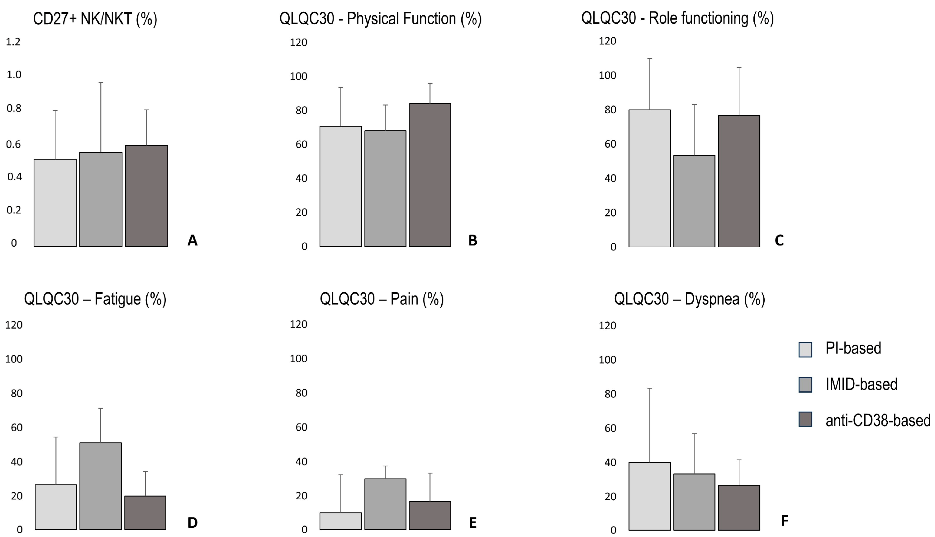
3.10. The Percentage of CD27+ NK/NKT Cells, Quality of Life Parameters, 6 Min Walking Distance, Total Handgrip Strength, Lean Body Mass, and Bone Density According to Treatment-Based Groups

Three treatment-based subgroups were defined according to the main first-line therapy: “proteasome inhibitor-based”, “immunomodulatory drug-based”, and “anti-CD38 antibody-based” (Table 1). The percentage of CD27+ NK/NKT cells and five quality of life parameters are presented in Figure 3. The 6 min walking distance, total of right and left handgrip strength, lean body mass, and bone density are presented in Figure 4.

4. Discussion

The purpose of this study was to investigate if the bone marrow microenvironment correlates with the physical function and the quality of life in multiple myeloma patients. The main finding of the study was that the percentages of bone marrow immune cell populations were not correlated with physical function, but the percentage of CD27+ NK/NKT cells was correlated with health-related quality of life of multiple myeloma patients.
The percentage of NK/NKT cells in the bone marrow was correlated positively with two factors of the quality of life questionnaire, physical function, and role functioning, while they were negatively correlated with three factors, fatigue, pain, and dyspnea. The NK/NKT population includes both NK cells and a subpopulation of T cells expressing both T and NK markers, called NKT cells [18]. NK cells have a strong anti-myeloma activity; however, myeloma cells can develop mechanisms to avoid NK cell responses and proliferate [19]. Changes in the bone marrow microenvironment induced by the disease move NK cells away from the bone marrow and reduce the immune response against myeloma cells in mice with multiple myeloma [9]. Additionally, NKT cells also possess anti-myeloma activity, while their reduced activity is connected with worse disease states in multiple myeloma patients [18]. The CD27 receptor is mostly expressed on CD56bright NK cells, implying an immunomodulatory role and an involvement in cytokine release [20,21]. Also, CD27 is a co-stimulatory receptor expressed on the T cell surface after binding with antigen-presenting cells, indicating that a CD27+ T cell (or CD27+ NKT cell in our case) is in the process of activation [22,23]. This suggests that a higher percentage of CD27+ NK/NKT cells in the bone marrow could indicate an improved anti-myeloma immune surveillance, preventing disease expansion and disease symptoms such as hypercalcemia, renal failure, anemia, and bone disease, which in turn leads to better health-related quality of life. It is important to note that this correlation refers to the NK/NKT population, and not the NK or NKT populations separately. Even though there is no evidence that CD27+ NK/NKT cells improve myeloma outcome, this finding might potentiate the utility of these cells for clinical implications. Specifically, it could be investigated if the presence of CD27+ NK/NKT cells could be used as a prognostic biomarker for multiple myeloma patients or if their increase in the bone marrow microenvironment could be used as a therapeutic target. Previous studies found similar percentages of the immune cell types in the bone marrow of multiple myeloma patients [5], as well as similar quality of life scores [24], although this is the first study investigating the possible connection between bone marrow immunity and quality of life in these patients.
To investigate the possible influence of the heterogeneity in first-line treatments, a subgroup analysis with descriptive statistics was performed according to the main therapy. No obvious trends were observed among the three research groups for CD27+ NK/NKT cells and the five parameters of the questionnaire (physical function, role functioning, fatigue, pain, and dyspnea, Figure 3). However, it should be mentioned that this was only an additional explanatory analysis for the present study. The sample size limits the ability to draw definitive conclusions about treatment effects, although it can be useful for future research.
Physical function was measured through the 6 min walking distance, handgrip, isometric strength, and maximal aerobic power. The hypothesis was that a higher presence of anti-myeloma immune cells would result in better disease status, leading to improved physical function. However, the study found no correlation between any of the bone marrow cell types and the four physical function assessments mentioned above (Table 2). It implies that physical function is mainly dependent on other factors such as bone strength, muscle mass, neural coordination, and psychological parameters. Indeed, handgrip strength was correlated with bone density. This implies that a higher bone density is partly responsible for patients’ better physical function. Poor bone density, as one of the myeloma symptoms, induces bone lesions [25] and, thus, the function of a patient’s daily life is reduced. Conversely, higher bone density indicates less bone weakness and increased daily activity. Previous studies reported similar bone density values in multiple myeloma patients [26]. Finally, the 6 min walking distance, the handgrip isometric strength and bone density did not seem to have a notable trend among the three treatment-based subgroups (Figure 4A,B,D).
Similarly, handgrip strength, maximal aerobic capacity, and isometric strength were correlated with lean body mass. Lean body mass is the sum of water, ligaments, tendons, internal organs, and total muscle mass. Lean body mass is a representative index of muscle mass [27]. So, a patient with a higher lean body mass value seems to have more muscle mass than a patient with a lower one. Muscle mass is correlated with increased movement performance in daily activities [28], leading to better physical functioning. Finally, the subgroup analysis based on the main first-line treatment did not reveal any obvious tendency in lean body mass among the three groups (Figure 4C).
The mean value of lean body mass found in the present study is high compared to a healthy non-exercised population or multiple myeloma patients [26]. However, this group of patients did not exercise regularly, muscle mass could not be increased during their treatment, and their profile was not that of a trained person. The high lean body mass could be explained by a higher amount of body water, another component of the lean body mass. Medical treatment could possibly cause it, as a swelling condition, although there are no data from this or previous studies to support this hypothesis.
The strong correlation found between isometric handgrip strength and lean body mass indicates that handgrip assessment could be a sarcopenia marker in multiple myeloma patients. This has already been proposed by the European Working Group on Sarcopenia in Older People [29]. This is an important finding, since sarcopenia is connected with cardiovascular problems [30], and low muscle mass was associated with higher disease stage, renal failure, anemia, and worse overall survival [31] in multiple myeloma patients. Additionally, handgrip assessment is a low-cost and easily performed measurement for patients.
Previous studies found similar values of physical function in multiple myeloma patients after first-line treatment. More specifically, a study found a similar mean value of the 6 min walking distance in multiple myeloma patients who were assessed at diagnosis [32] or seventeen months after hematopoietic stem cell transplantation [33]. Another study found that handgrip strength values were also similar [34]. However, the maximal aerobic power of the present study was slightly lower than in previous findings [34], possibly due to the recombinant type of bicycle used in the present study.
Finally, lean body mass was negatively correlated with fatigue, indicating that higher muscle mass reduces fatigue in multiple myeloma patients. Exercise training such as resistance training or bicycling increases muscle mass in healthy populations [35,36]. Therefore, it is possible that exercise training programs could be used to enhance lean body mass in multiple myeloma patients, thereby reducing fatigue. A previous study investigated the effect of three months of supervised exercise training [37] in multiple myeloma patients. The program consisted of 30 min of aerobic training at 50–75% of maximum heart rate on a treadmill, bicycle, or stepper, along with total body resistance training with free weights, body weight, or bands. However, the study found that there was no change in muscle mass or fatigue levels either during the exercise program or after unsupervised training three months later [37]. Another study with similar training intervention found no change after three months, but fatigue was improved after six months, although muscle mass was not measured [38]. Despite this, more research is needed to explore the effects of different exercise modalities, intensities, and disease statuses on muscle mass and fatigue levels in multiple myeloma patients.
In the present study, the profile of the bone marrow microenvironment was determined by measuring the percentages of the major bone marrow cell populations with next-generation flow cytometry. The function, anti-tumor activity, proliferation, or cytotoxicity of immune cells were not evaluated here, and there is no conclusive evidence to suggest whether these functional parameters are correlated with the quality of life and physical function of multiple myeloma patients. Also, all measurements were evaluated after the initial medical treatment and hematopoietic stem cell transplantation, which aimed to eliminate myeloma cells and restore the bone marrow microenvironment.
The patients of the present study were at the same stage of the disease, but had received various medical treatments, and the total sample size was small. However, these correlations had not been investigated before. Similar measurements should be conducted in the future with more participants following the same medical treatment. Also, future research should investigate other disease states, like diagnosis or relapse, to determine these relationships. Last but not least, according to the present findings, longitudinal studies evaluating the change in CD27+ NK/NKT cells over time and interventional studies assessing the impact of therapeutic treatments targeting these cells should be instigated.

5. Conclusions

In conclusion, the percentages of major cell populations in the bone marrow microenvironment are not correlated with the physical function of multiple myeloma patients after first-line treatment. Physical function, as measured by physical tests, is correlated with other factors such as lean body mass. On the other hand, the increased percentage of CD27+ NK/NKT cells, which is possibly associated with stronger immune surveillance in the bone marrow, is correlated with better quality of life in multiple myeloma patients.

Author Contributions

Conceptualization, P.S.; methodology, P.S., M.G., E.K., and G.T.; data curation, P.S., P.R., C.P., E.E.-P., M.M., N.K., I.N.-S., P.M., F.T., D.F., E.T., M.G., O.E.T., E.K., M.A.D., and G.T.; writing—original draft preparation, P.S.; writing—review and editing, P.S., P.R., C.P., E.E.-P., M.M., N.K., I.N.-S., P.M., F.T., D.F., E.T., V.M., M.G., O.E.T., E.K., M.A.D., and G.T.; visualization, P.S.; supervision, G.T.; funding acquisition, P.S. All authors have read and agreed to the published version of the manuscript.

Funding

This work was supported by the Stavros Tsakyrakis Scholarships, grant number 16257.

Institutional Review Board Statement

The study was conducted in accordance with the Declaration of Helsinki and approved by the Institutional Review Board (Protocol Number: 1202/10-06-2020; approval date: 10 June 2020).

Informed Consent Statement

Informed consent was obtained from all subjects involved in the study.

Data Availability Statement

Data are unavailable due to privacy and ethical restrictions.

Acknowledgments

The authors would like to thank the patients who participated in the study.

Conflicts of Interest

The authors declare no conflicts of interest.

References

  1. Kumar, A.; Rajkumar, S.V.; Kyle, R.A.; van Duin, M.; Sonneveld, P.; Mateos, M.V.; Gay, F.; Anderson, K.C. Multiple myeloma. Nat. Rev. Dis. Primers 2017, 20, 17046. [Google Scholar] [CrossRef] [PubMed]
  2. Rajkumar, S.V.; Kumar, S. Multiple myeloma: Diagnosis and treatment. Mayo Clin. Proc. 2016, 91, 101–119. [Google Scholar] [CrossRef] [PubMed]
  3. Kumar, S.; Paiva, B.; Anderson, K.C.; Durie, B.; Landgren, O.; Moreau, P.; Munshi, N.; Lonial, S.; Bladé, J.; Mateos, M.V.; et al. International Myeloma Working Group consensus criteria for response and minimal residual disease assessment in multiple myeloma. Lancet Oncol. 2016, 17, e328–e346. [Google Scholar] [CrossRef] [PubMed]
  4. Dadzie, T.G.; Green, A.C. The role of the bone microenvironment in regulating myeloma residual disease and treatment. Front. Oncol. 2022, 12, 999939. [Google Scholar] [CrossRef]
  5. Paiva, B.; Cedena, M.T.; Puig, N.; Arana, P.; Vidriales, M.B.; Cordon, L.; San Miguel, J.F. Minimal residual disease monitoring and immune profiling in multiple myeloma in elderly patients. Blood 2016, 127, 3165–3174. [Google Scholar] [CrossRef]
  6. Desantis, V.; Savino, F.D.; Scaringella, A.; Potenza, M.A.; Nacci, C.; Frassanito, M.A.; Vacca, A.; Montagnani, M. The Leading Role of the Immune Microenvironment in Multiple Myeloma: A New Target with a Great Prognostic and Clinical Value. J. Clin. Med. 2022, 11, 2513. [Google Scholar] [CrossRef]
  7. Raitakari, M.; Brown, R.D.; Gibson, J.; Joshua, D.E. T cells in myeloma. Hematol. Oncol. 2003, 21, 33–42. [Google Scholar] [CrossRef]
  8. Lopes, R.; Caetano, J.; Ferreira, B.; Barahona, F.; Carneiro, E.A.; João, C. The Immune Microenvironment in Multiple Myeloma: Friend or Foe? Cancers 2021, 13, 625. [Google Scholar] [CrossRef]
  9. Ponzetta, A.; Benigni, G.; Antonangeli, F.; Sciume, G.; Sanseviero, E.; Zingoni, A.; Bernardini, G. Multiple myeloma impairs bone marrow localization of effector natural killer cells by altering the chemokine microenvironment. Cancer Res. 2015, 75, 4766–4777. [Google Scholar] [CrossRef]
  10. Pessoa de Magalhães, R.J.; Vidriales, M.B.; Paiva, B.; Fernandez-Gimenez, C.; García-Sanz, R.; Mateos, M.V.; Gutierrez, N.C.; Lecrevisse, Q.; Blanco, J.F.; Hernández, J.; et al. Analysis of the immune system of multiple myeloma patients achieving long-term disease control by multidimensional flow cytometry. Haematologica 2013, 98, 79–86. [Google Scholar] [CrossRef]
  11. Jensen, C.E.; Vohra, S.N.; Nyrop, K.A.; Deal, A.M.; LeBlanc, M.R.; Grant, S.J.; Muss, H.B.; Lichtman, E.I.; Rubinstein, S.M.; Wood, W.A.; et al. Physical Function, Psychosocial Status, and Symptom Burden Among Adults with Plasma Cell Disorders and Associations with Quality of Life. Oncologist 2022, 27, 694–702. [Google Scholar] [CrossRef] [PubMed]
  12. Gulbrandsen, N.; Hjermstad, M.J.; Wisløff, F.; Nordic Myeloma Study Group. Interpretation of quality of life scores in multiple myeloma by comparison with a reference population and assessment of the clinical importance of score differences. Eur. J. Haematol. 2004, 72, 172–180. [Google Scholar] [CrossRef] [PubMed]
  13. American College of Sports Medicine. Laboratory Submaximal Exercise Testing: YMCA Cycle Ergometer Test. In ACSM’s Health-Related Physical Fitness Assessment Manual, 2nd ed.; Wolters Kluwer: Philadelphia, PA, USA, 2008; pp. 112–116. [Google Scholar]
  14. Flores-Montero, J.; Sanoja- Flores, L.; Paiva, B. Next Generation Flow for Highly Sensitive and Standardized Detection of Minimal Residual Disease in Multiple Myeloma. Leukemia 2017, 31, 2094–2103. [Google Scholar] [CrossRef]
  15. Kostopoulos, I.V.; Eleutherakis-Papaiakovou, E.; Rousakis, P.; Ntanasis-Stathopoulos, I.; Panteli, C.; Orologas-Stavrou, N.; Terpos, E. Aberrant plasma cell contamination of peripheral blood stem cell autografts, assessed by next-generation flow cytometry, is a negative predictor for deep response post autologous transplantation in multiple myeloma; a prospective study in 199 patients. Cancers 2021, 13, 4047. [Google Scholar] [CrossRef]
  16. Terpos, E.; Kostopoulos, I.V.; Kastritis, E.; Ntanasis-Stathopoulos, I.; Migkou, M.; Rousakis, P.; Dimopoulos, M.A. Impact of minimal residual disease detection by next-generation flow cytometry in multiple myeloma patients with sustained complete remission after frontline therapy. HemaSphere 2019, 3, 6. [Google Scholar] [CrossRef] [PubMed]
  17. Obilor, E.I.; Amadi, E.C. Test for significance of Pearson’s correlation coefficient. IJIMSEP 2018, 6, 11–23. [Google Scholar]
  18. Dhodapkar, M.V.; Richter, J. Harnessing natural killer T (NKT) cells in human myeloma: Progress and challenges. Clin. Immunol. 2011, 140, 160–166. [Google Scholar] [CrossRef]
  19. Godfrey, J.; Benson, D.M., Jr. The role of natural killer cells in immunity against multiple myeloma. Leuk. Lymphoma 2012, 53, 1666–1676. [Google Scholar] [CrossRef]
  20. Silva, A.; Andrews, D.M.; Brooks, A.G.; Smyth, M.J.; Hayakawa, Y. Application of CD27 as a marker for distinguishing human NK cell subsets. Int. Immunol. 2008, 20, 625–630. [Google Scholar] [CrossRef]
  21. Vossen, M.; Matmati, M.; Hertoghs, K.M.; Baars, P.A.; Gent, M.R.; Leclercq, G.; van Lier, R.A. CD27 defines phenotypically and functionally different human NK cell subsets. J. Immunol. 2008, 180, 3739–3745. [Google Scholar] [CrossRef]
  22. Imanishi, T.; Saito, T. T cell co-stimulation and functional modulation by innate signals. Trends Immunol. 2020, 41, 200–212. [Google Scholar] [CrossRef] [PubMed]
  23. Gaudino, S.J.; Kumar, P. Cross-talk between antigen presenting cells and T cells impacts intestinal homeostasis, bacterial infections, and tumorigenesis. Front. Immunol. 2019, 10, 360. [Google Scholar] [CrossRef]
  24. Proskorovsky, I.; Lewis, P.; Williams, C.D.; Jordan, K.; Kyriakou, C.; Ishak, J.; Davies, F.E. Mapping EORTC QLQ-C30 and QLQ-MY20 to EQ-5D in patients with multiple myeloma. Health Qual. Life Outcomes 2014, 12, 35. [Google Scholar] [CrossRef] [PubMed]
  25. Terpos, E.; Christoulas, D.; Gavriatopoulou, M.; Dimopoulos, M.A. Mechanisms of bone destruction in multiple myeloma. Eur. J. Cancer Care 2017, 26, e12761. [Google Scholar] [CrossRef]
  26. Larsen, R.F.; Jarden, M.; Minet, L.R.; Frølund, U.C.; Hermann, A.P.; Breum, L.; Abildgaard, N. Exercise in newly diagnosed patients with multiple myeloma: A randomized controlled trial of effects on physical function, physical activity, lean body mass, bone mineral density, pain, and quality of life. Eur. J. Haematol. 2024, 113, 298–309. [Google Scholar] [CrossRef]
  27. Pomeroy, E.; Macintosh, A.; Wells, J.C.; Cole, T.J.; Stock, J.T. Relationship between body mass, lean mass, fat mass, and limb bone cross-sectional geometry: Implications for estimating body mass and physique from the skeleton. Am. J. Phys. Anthropol. 2018, 166, 56–69. [Google Scholar] [CrossRef] [PubMed]
  28. Wang, D.X.; Yao, J.; Zirek, Y.; Reijnierse, E.M.; Maier, A.B. Muscle mass, strength, and physical performance predicting activities of daily living: A meta-analysis. JCSM 2020, 11, 3–25. [Google Scholar] [CrossRef]
  29. Cruz-Jentoft, A.J.; Baeyens, J.P.; Bauer, J.M.; Boirie, Y.; Cederholm, T.; Landi, F.; Zamboni, M. Sarcopenia: European consensus on definition and diagnosis: Report of the European Working Group on Sarcopenia in Older People. Age Ageing 2010, 39, 412–423. [Google Scholar] [CrossRef]
  30. Williams, A.; Baruah, D.; Patel, J.; Szabo, A.; Chhabra, S.; Dhakal, B.; D’Souza, A. Prevalence and significance of sarcopenia in multiple myeloma patients undergoing autologous hematopoietic cell transplantation. Bone Marrow Transpl. 2021, 56, 225–231. [Google Scholar] [CrossRef]
  31. Abdallah, N.H.; Nagayama, H.; Takahashi, N.; Gonsalves, W.; Fonder, A.; Dispenzieri, A.; Kumar, S.K. Muscle and fat composition in patients with newly diagnosed multiple myeloma. Blood Cancer J. 2023, 13, 185. [Google Scholar] [CrossRef]
  32. Larsen, R.F.; Jarden, M.; Minet, L.R.; Frølund, U.C.; Möller, S.; Abildgaard, N. Physical function in patients newly diagnosed with multiple myeloma; a Danish cohort study. BMC Cancer 2020, 20, 169. [Google Scholar] [CrossRef] [PubMed]
  33. Tuchman, S.A.; Lane, A.; Hornsby, W.E.; Bishop, C.; Thomas, S.; Herndon II, J.E.; Jones, L.W. Quantitative measures of physical functioning after autologous hematopoietic stem cell transplantation in multiple myeloma: A feasibility study. Clin. Lymphoma Myeloma Leuk 2015, 15, 103–109. [Google Scholar] [CrossRef]
  34. Persoon, S.; Kersten, M.J.; Buffart, L.M.; Vander Slagmolen, G.; Baars, J.W.; Visser, O.; Chinapaw, M.J. Health-related physical fitness in patients with multiple myeloma or lymphoma recently treated with autologous stem cell transplantation. J. Sci. Med. Sport 2017, 20, 116–122. [Google Scholar] [CrossRef] [PubMed]
  35. Häkkinen, K.; Alén, M.; Kraemer, W.J.; Gorostiaga, E.; Izquierdo, M.; Rusko, H.; Paavolainen, L. Neuromuscular adaptations during concurrent strength and endurance training versus strength training. Eur. J. Appl. Physiol. 2003, 89, 42–52. [Google Scholar] [CrossRef] [PubMed]
  36. Spiliopoulou, P.; Zaras, N.; Methenitis, S.; Papadimas, G.; Papadopoulos, C.; Bogdanis, G.C.; Terzis, G. Effect of concurrent power training and high-intensity interval cycling on muscle morphology and performance. JSCR 2021, 35, 2464–2471. [Google Scholar] [CrossRef]
  37. Koutoukidis, D.A.; Land, J.; Hackshaw, A.; Heinrich, M.; McCourt, O.; Beeken, R.J.; Yong, K.L. Fatigue, quality of life and physical fitness following an exercise intervention in multiple myeloma survivors (MASCOT): An exploratory randomised Phase 2 trial utilising a modified Zelen design. Br. J. Cancer 2020, 123, 187–195. [Google Scholar] [CrossRef]
  38. Groeneveldt, L.; Mein, G.; Garrod, R.; Jewell, A.P.; Someren, K.V.; Stephens, R.; Yong, K.L. A mixed exercise training programme is feasible and safe and may improve quality of life and muscle strength in multiple myeloma survivors. BMC Cancer 2013, 13, 31. [Google Scholar] [CrossRef]
Figure 1. The distribution of twenty cell populations in the bone marrow of multiple myeloma patients after first-line treatment (N = 16). Values are presented as mean ± SD. NPCs, normal plasma cells; APCs, abnormal plasma cells; NK, natural killer cells; NKT, natural killer T cells.
Onco 05 00021 g001
Figure 2. Correlation between the percentage of CD27+ NK/NKT cells in the bone marrow with (A) physical function, (B) role functioning, (C) fatigue, (D) pain, and (E) dyspnea (N = 11). NK, natural killer cells; NKT, natural killer T cells.
Onco 05 00021 g002
Figure 3. The percentage of CD27+ NK/NKT cells (A) and five parameters of the QLQC30 questionnaire (BF) according to three first-line treatment-based subgroups, in multiple myeloma patients. NK, natural killer cells; NKT, natural killer T cells; PI, proteasome inhibitors; IMID, immunomodulatory drugs; anti-CD38, anti-CD38 monoclonal antibody.
Onco 05 00021 g003
Figure 4. The 6 min walking distance (A), sum of right and left handgrip strength (B), lean body mass (C), and bone density (D) according to three first-line treatment-based subgroups, in multiple myeloma patients. 6MWT, 6 min walking test; PI, proteasome inhibitors; IMID, immunomodulatory drugs; anti-CD38, anti-CD38 monoclonal antibody.
Onco 05 00021 g004
Table 1. Patient characteristics.
Anthropometrical Characteristics (Mean ± SD)
Total mass (kg)81.4 ± 16.5
Age (years)53.9 ± 7.7
Height (m)1.73 ± 0.1
Gender (N)
Total participants21
Women7
Mem11
Treatment (N)
PI3
IMID8
PI + IMID4
anti-CD38 + IMID1
anti-CD38 + PI2
anti-CD38 + IMID + PI3
Treatment-Based Groups (N)
PI—based7
IMID—based8
Anti-CD38—based6
MRD (N)
Total participants16
MRD positive5
MRD negative11
PI, proteasome inhibitors; IMID, immunomodulatory drugs; anti-CD38, anti-CD38 monoclonal antibody; MRD, minimal residual disease.
Table 2. r Pearson coefficients between twenty immune cell populations in the bone marrow and physical function components of multiple myeloma patients after first-line treatment (N = 16).
% of Bone Marrow Nucleated Cells6MWTHandgrip RH+LHIsometric Strength UBIsometric Strength LBMaximal
Aerobic Power
Plasma cells−0.34−0.200.00−0.230.18
NPCs−0.34−0.190.00−0.230.17
APCs0.38−0.170.030.270.20
Mast cells−0.280.010.180.000.39
Myeloid progenitors0.08−0.34−0.10−0.130.09
B cells−0.12−0.35−0.40−0.56−0.07
B cell precusors−0.17−0.38−0.38−0.63−0.11
Naïve B cells0.08−0.08−0.30−0.120.12
Memory B cells0.32−0.18−0.240.28−0.03
T cells−0.08−0.22−0.090.170.09
CD27− T cells−0.08−0.26−0.150.120.08
CD27+ T cells0.03−0.050.040.190.09
NK/NKT cells−0.230.04−0.080.290.15
CD27− NK/NKT−0.190.04−0.130.270.14
CD27+ NK/NKT−0.260.010.160.200.12
Erythroblasts0.47−0.21−0.010.260.17
Erythroid progenitors0.220.240.240.130.28
Eosinophils−0.06−0.11−0.14−0.48−0.12
Monocytes0.390.270.24−0.090.31
Neutrophils−0.210.330.13−0.11−0.27
6MWT, 6 min walking test; RH, right hand; LH, left hand; UB, upper body; LB, lower body; NPCs, normal plasma cells; APCs, abnormal plasma cells; NK, natural killer cells; NKT, natural killer T cells.
Disclaimer/Publisher’s Note: The statements, opinions and data contained in all publications are solely those of the individual author(s) and contributor(s) and not of MDPI and/or the editor(s). MDPI and/or the editor(s) disclaim responsibility for any injury to people or property resulting from any ideas, methods, instructions or products referred to in the content.

