Review

Targeting Pathways and Mechanisms in Gynecological Cancer with Antioxidant and Anti-Inflammatory Phytochemical Drugs

by
Sandhya Shukla
1,†,
Arvind Kumar Shukla
2,*,†,
Navin Ray
3,
Adarsha Mahendra Upadhyay
4,
Fowzul Islam Fahad
2,
Sayan Deb Dutta
5,6,7,
Arulkumar Nagappan
3,8 and
Raj Kumar Mongre
9,*
1
Department of Pharmacology, Bharti Vidyapeeth Deemed University Medical College, Pune 411043, India
2
School of Biomedical Convergence Engineering, Pusan National University, Yangsan 50612, Republic of Korea
3
Laboratory of Mucosal Exposome and Biomodulation, Department of Integrative Biomedical Sciences, Pusan National University, Yangsan 50612, Republic of Korea
4
Department of Gastrointestinal Surgery, School of Overseas Education, Guizhou Medical University, Guiyang 550025, China
5
Department of Biosystems Engineering, Kangwon National University, Chuncheon 24341, Republic of Korea
6
Institute of Forest Science, Kangwon National University, Chuncheon 24341, Republic of Korea
7
School of Medicine, University of California Davis, Sacramento, CA 95817, USA
8
Department of Obstetrics and Gynecology, College of Medicine, Pusan National University, Yangsan 50612, Republic of Korea
9
Department of Surgery, Boston Children’s Hospital, Harvard Medical School, Harvard University, 300 Longwood Ave, Boston, MA 02115, USA
*
Authors to whom correspondence should be addressed.
These authors contributed equally to this work.
Submission received: 11 April 2025 / Revised: 5 May 2025 / Accepted: 6 May 2025 / Published: 9 May 2025
(This article belongs to the Special Issue Targeting of Tumor Dormancy Pathway)

Simple Summary

Gynecologic cancers, such as ovarian, cervical, and uterine cancers, remain a major health concern for women worldwide. Recent research suggests that inflammation and oxidative stress—an imbalance of harmful molecules in the body—play important roles in how these cancers grow and spread. Certain drugs with antioxidant and anti-inflammatory properties may help slow down or stop tumor development by targeting these harmful processes. In this review, we explore how these drugs work, especially how they influence the environment around the tumor, including the body’s immune response and the formation of new blood vessels that feed cancer cells. By better understanding how these treatments interact with cancer-related pathways, researchers and doctors may find more effective ways to treat gynecologic cancers and improve patient outcomes.

Abstract

Globally, women’s cancer-related morbidity and death are still caused mainly by gynecologic cancer. Antioxidant and anti-inflammatory drugs have shown promise in treating gynecologic cancer because of the complex interactions among oxidative stress, inflammation, and the development of tumors. This review focuses on how these drugs, which include polyphenols, terpenoids, and thiols-related phytochemical-derived compounds target different pathways associated with developing and progressing gynecologic cancer. We investigate what factors affect the tumor microenvironment, specifically how they affect immunological response and vasculogenesis. Through the review of recent studies, we have gained an extensive understanding of the molecular pathways that anti-inflammatory and antioxidant drugs use to achieve their therapeutic benefits. Gynecologic cancer is still a potent cause of cancer-related deaths and fatalities for women globally. Antioxidant and anti-inflammatory drugs have shown promise in treating gynecologic cancer because of the complex interactions among oxidative stress, inflammation, and the development of tumors. This review focuses on how these drugs target different pathways associated with developing and progressing gynecologic cancer. We investigate what factors affect the tumor microenvironment, specifically how they affect immunological response and vasculogenesis. Through the review of recent studies, we have gained an extensive understanding of the molecular pathways that anti-inflammatory and antioxidant drugs use to achieve their therapeutic benefits.

1. Introduction

A major global health burden is posed by gynecologic cancers, which include cancers of the cervix, ovary, endometrium, vulva, and vagina. Together, these cancers cause a significant amount of morbidity and mortality among women globally, and the incidence and survival rates differ according to socioeconomic status, geographic location, and access to healthcare. The most common gynecologic cancers, according to the most recent estimates from the Global Cancer Observatory (GLOBOCAN), are endometrial, ovarian, and cervical cancers. With over 600,000 new cases and about 340,000 deaths reported each year, cervical cancer is still the fourth most common cancer in women [1,2,3,4,5]. It is primarily preventable with vaccination and early screening. Its high burden is ascribed to restricted access to routine screening programs and the human papillomavirus (HPV) vaccine, which disproportionately affects low- and middle-income countries (LMICs). Ovarian cancer is the deadliest gynecologic cancer, accounting for over 310,000 new cases and 200,000 deaths annually [6,7,8]. Because it rarely exhibits early symptoms, it is frequently detected at an advanced stage. With over 420,000 new cases per year, endometrial cancer is becoming more common, especially in high-income nations, and is associated with obesity and hormone imbalances [9,10,11,12]. Despite being less common, vulvar and vaginal cancers are still a concern because of their links to aging populations and HPV infection. Better screening initiatives, easier access to immunizations, and the creation of innovative treatment approaches are all necessary to address the worldwide burden of gynecologic cancer. Reducing disparities in incidence, early detection, and treatment outcomes requires strengthening the healthcare infrastructure, especially in LMICs.
The incidence and mortality rates of gynecological cancers (GCs), which include cervical, ovarian, endometrial, vaginal, and vulvar cancers, vary by region due to differences in healthcare access, socioeconomic factors, and preventive measures. GCs represent a significant global health concern. The age-standardized rates (ASRs) of GC incidence and mortality by country in 2022 are shown in Figure 1A(a,b), which shows notable regional differences, especially in low- and middle-income countries (LMICs), where insufficient access to screening and immunization programs raises the disease burden. In the eight nations with the highest burden, as shown in Figure 1A(c), the proportionate distribution of GC cases and deaths highlights differences in lethality and prevalence among various cancer types. Figure 1A(d) displays the incidence and mortality rates of various GCs in 2022, and Figure 1B(a) shows additional insights into GC epidemiology. This highlights the significant contribution of endometrial, ovarian, and cervical cancers to the overall burden. According to the proportionate distribution of GC cases and deaths across age groups, as shown in Figure 1B(b,c), the incidence of GC rises with age, especially in postmenopausal women. The significant mortality linked to ovarian cancer due to late-stage diagnosis is highlighted in Figure 1B(d,e), which provides specifics on the relative proportions of cervical, corpus uteri, ovarian, vaginal, and vulvar cancers among new cases and deaths. These findings demonstrate how urgently improved prevention measures—such as HPV vaccination, early screening, and fair access to treatment—are needed to reduce inequalities and enhance GC outcomes globally, especially in high-burden areas [11].
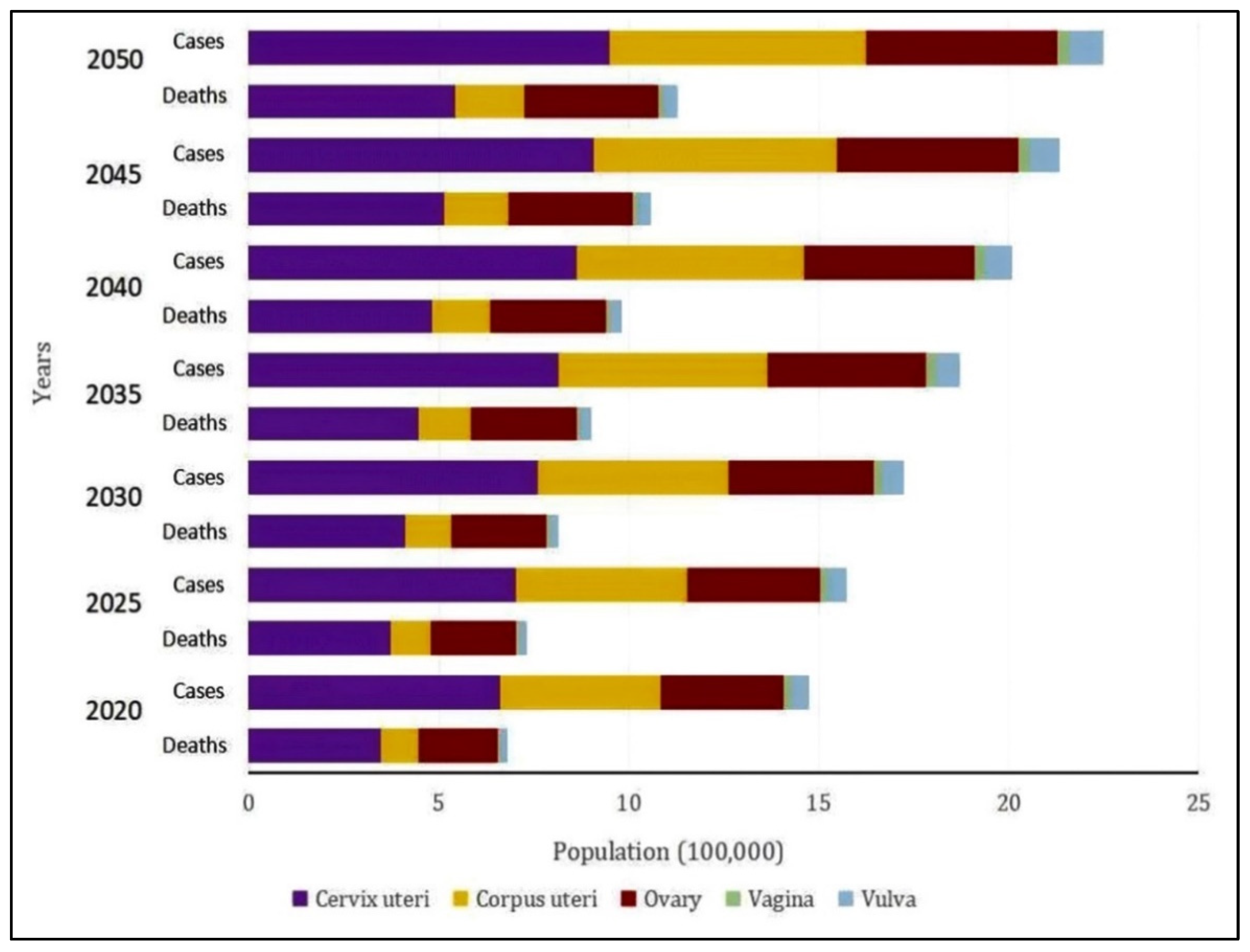
The estimated incidence and mortality rates of gynecological cancers (GCs) by 2050 are shown in Figure 2, which also indicates a significant expected rise in the disease burden over the following three decades. The anticipated increase in both incidence and mortality highlights the pressing need for increased screening programs, improved research initiatives, and creative treatment approaches to lessen the impact of GCs worldwide [11].
The development and spread of gynecologic cancers, such as ovarian, endometrial, and cervical cancers, are significantly influenced by oxidative stress and inflammation [13]. Cellular damage, DNA mutations, and genomic instability result from oxidative stress, which is caused by an imbalance between the generation of reactive oxygen species (ROS) and the antioxidant defense system [5,14]. Oncogenic signaling pathways like PI3K/AKT and MAPK can be activated by chronic oxidative stress, which encourages unchecked cell survival and proliferation. Oxidative stress also plays a role in angiogenesis, resistance to apoptosis, and the epithelial–mesenchymal transition (EMT), all of which promote tumor growth and metastasis. By fostering a pro-tumorigenic microenvironment that is enriched in immune cells, chemokines, and inflammatory cytokines, inflammation worsens tumorigenesis. Tumor necrosis factor-alpha (TNF-α), interleukins (IL-6, IL-8), and nuclear factor-kappa B (NF-κB) are important inflammatory mediators that promote tumor cell proliferation, inhibit antitumor immunity, and cause chronic inflammation. Furthermore, oxidative stress and inflammation are linked because ROS can trigger NF-κB signaling, which results in the continuous production of inflammatory cytokines, and inflammatory reactions intensify the production of ROS [15,16,17,18]. In gynecologic cancers, this feedback loop aids in tumor growth, chemoresistance, and immune evasion. One promising therapeutic approach is to target inflammation and oxidative stress. To interfere with these harmful processes and enhance therapeutic results, researchers are looking into antioxidants, anti-inflammatory medications, and inhibitors of important signaling pathways. Gaining insight into the molecular interactions between inflammation and oxidative stress in gynecologic cancers could result in new treatment strategies that improve patient outcomes and survival. Plant-derived phytochemicals, their botanical sources, and the molecular targets they are examining in pre-clinical cancer treatment trials are listed in Table 1.
The development, metastasis, and resistance to treatment of gynecologic cancers are significantly influenced by the tumor microenvironment (TME). It is made up of a dynamic network of an extracellular matrix (ECM), soluble substances, immune cells, and stromal cells that work together to influence tumor behavior. Cancer-associated fibroblasts (CAFs) facilitate tumor invasion and metastasis by promoting angiogenesis and contributing to ECM remodeling [19]. Tumor-associated macrophages (TAMs) and regulatory T cells (Tregs) are two immune cells that frequently display an immunosuppressive phenotype within the TME, which facilitates immune evasion and resistance to immunotherapy. By promoting stemness, upregulating pro-angiogenic factors like the vascular endothelial growth factor (VEGF), and inducing epithelial-to-mesenchymal transition (EMT), hypoxia within the TME further exacerbates tumor aggressiveness. Furthermore, the TME’s metabolic reprogramming, which includes elevated lactate and glycolysis, promotes tumor cell survival and inhibits anti-tumor immune responses. Developing focused treatment plans requires an understanding of the intricate relationships that exist within the TME. Novel strategies, such as TME-modulating drugs, immune checkpoint inhibitors, and drug delivery-engineered biomaterials, have the potential to increase treatment effectiveness. To create tailored therapeutic interventions and improve clinical outcomes for patients with gynecologic malignancies, future research should concentrate on identifying the major molecular drivers of TME remodeling [20,21,22].
The treatment and management of gynecologic cancers may benefit greatly from the use of antioxidant and anti-inflammatory medications. By encouraging DNA damage, genomic instability, and a pro-tumorigenic microenvironment, oxidative stress and chronic inflammation aid in the development, spread, and resistance to treatment of tumors. By scavenging reactive oxygen species (ROS) and strengthening cellular defenses, antioxidants like polyphenols, flavonoids, and vitamin derivatives reduce oxidative stress. Similarly, anti-inflammatory drugs, such as cytokine modulators, COX-2 inhibitors, and non-steroidal anti-inflammatory drugs (NSAIDs), target inflammatory pathways to lower immune evasion and tumor-promoting cytokines [23,24,25]. According to new research, anti-inflammatory and antioxidant substances may alter important signaling pathways that contribute to the development of cancer, including PI3K/AKT, MAPK, and NF-κB, which would prevent angiogenesis, metastasis, and proliferation [26,27,28]. Furthermore, by enhancing tumor microenvironment conditions and lowering therapy-induced oxidative damage, these agents may increase the effectiveness of traditional therapies like immunotherapy and chemotherapy. However, issues like bioavailability, the best dosage, and possible conflict with conventional therapies call for more research. To confirm these compounds’ therapeutic potential and maximize their incorporation into individualized treatment plans for gynecologic cancers, future research should concentrate on preclinical and clinical studies.
This review explores the molecular pathways that antioxidant and anti-inflammatory medications target in gynecologic cancer, with a particular emphasis on inflammation, oxidative stress, and the control of the tumor microenvironment. Anti-inflammatory agents block pro-inflammatory cytokines, NF-κB signaling, and inflammation-associated genes, while antioxidants control the production of ROS, improve defense mechanisms, and preserve redox balance. Additionally, these medications affect extracellular matrix remodeling, immune cell infiltration, and angiogenesis in the tumor microenvironment. Modification of inflammatory signaling, direct antioxidant activity, and improvement of traditional cancer treatments are some of their therapeutic mechanisms. While preclinical and clinical studies have shown their potential, problems with bioavailability, optimal dosage, and mechanistic complexity remain. The focus of future research should be on overcoming these limitations to optimize their integration into cancer treatment.

2. Methodology: Aim, Review Process, Search Strategy, Data Extraction, and Analysis

The review aims to collect and analyze the molecular targets and mechanisms by which antioxidant and anti-inflammatory phytochemicals exert their anticancer effects in gynecological cancer. The main focus is on identifying plant chemicals that have shown efficacy in preclinical studies (in vitro and in vivo) by targeting specific molecular pathways such as apoptotic induction, proliferation inhibition, metastasis suppression, and PI3K and MAPK modulation. Systematic searches using databases such as PubMed, Google Scholar, and ScienceDirect were carried out for peer-reviewed articles published between 2000 and 2024. The survey included keywords such as plant chemicals, gynecological cancer, preclinical studies, molecular pathways, apoptosis, anti-inflammatory, antioxidant, natural compounds, and specific tumor types such as ovarian, cervical, and endometrial carcinoma. Boolean operators (AND) and (OR) have been used to refine the search predicate. The articles were included if they (1) evaluated plant-derived compounds for the treatment of gynecological cancer; (2) described the molecular mechanisms of action; and (3) were original research papers or peer-reviewed articles. Studies that were focused on clinical trials or did not have sufficient molecular data were excluded. Data collection involved searching the selected articles for details of botanical names, phytochemical class, tumor model, and target molecular pathway. The extracted data were summarized in tables for a summary of the mysterious interactions between the substances and the culprits. This review highlights the therapeutic potential of natural compounds to modulate inflammation and oxidative stress in the case of gynecological cancer and provides a basis for future translational and clinical studies.

3. Clinical Manifestations of Gynecological Cancers

The various clinical manifestations of gynecological cancers, which include cancers of the cervix, uterus, ovaries, vagina, and vulva, frequently differ based on the tumor site, stage, and histological type. Early-stage illnesses can have no symptoms, which makes prompt diagnosis more difficult. Abnormal vaginal bleeding, which can appear as intermenstrual, postcoital, or postmenopausal bleeding, is the most frequent initial symptom, especially in endometrial and cervical cancers [29,30]. Pelvic pressure or pain, which is frequently a sign of advanced disease, can be linked to uterine or ovarian cancers. The ambiguous symptoms of ovarian cancer, often referred to as a “silent killer”, include bloating, early satiety, and urgency in the urine. These symptoms can be mistaken for benign urological or gastrointestinal disorders [31,32,33]. Cancers of the vagina or vulvar region may manifest as visible lesions, bleeding, ulceration, or itching. Anemia, weariness, and inadvertent weight loss are examples of constitutional symptoms that frequently appear later [34]. Both metastasis and local invasion may cause symptoms related to the digestive or urinary systems. There is a significant concern, particularly in women who have undergone menopause or possess known risk factors (e.g., early detection of HPV infection and genetic predispositions is crucial). Early identification of these clinical characteristics, in conjunction with suitable diagnostic tests, is essential for enhancing prognosis and directing treatment plans in gynecological oncology.

4. Clinical Diagnosis of Gynecological Cancer

The clinical diagnosis of gynecological cancers, such as those of the cervical, ovarian, endometrial, vulvar, and vaginal regions, depends on a thorough assessment that includes imaging, histopathological confirmation, physical examination, and patient history. A thorough medical history is the first step in the initial evaluation, with an emphasis on symptoms like irregular vaginal bleeding, pelvic pain, or changes in bowel or urine habits [35,36,37]. It is possible to detect palpable masses, lesions, or anatomical irregularities with a comprehensive pelvic examination. For early detection of cervical neoplasia, cytological screening—specifically the Papanicolaou (Pap) smear and high-risk human papillomavirus (HPV) testing—remains crucial. Serum tumor markers like CA-125 help assess the risk of malignancy, particularly in ovarian cancer, while transvaginal ultrasonography offers vital anatomical information for suspected endometrial or ovarian pathology [38,39]. Computed tomography (CT), magnetic resonance imaging (MRI), and positron emission tomography (PET) are examples of advanced imaging modalities that aid in staging by determining lymph node involvement and tumor spread. A biopsy or surgical sampling for histological analysis is necessary for a conclusive diagnosis [40,41]. Personalized treatment plans are guided by molecular testing and immunohistochemical profiling, which further improve classification. In gynecological oncology, early-stage detection greatly improves prognosis and increases treatment options, so prompt and precise clinical diagnosis is essential for maximizing therapeutic outcomes.

5. Treatment of Gynecological Cancer

Cancer type, stage, and patient-specific factors all influence treatment plans, which combine radiotherapy, chemotherapy, surgery, and more targeted and immunotherapies. The goal of surgical intervention, which is frequently the first line of treatment, is total tumor resection. Radiation therapy and/or adjuvant chemotherapy are used to treat any remaining illness and lower the chance of recurrence [42,43]. By facilitating the use of agents that target particular mutations or signaling pathways, molecular profiling supports personalized medicine approaches. Clinical trial participation and palliative care are thought to enhance the quality of life and outcomes for patients with recurrent or advanced disease [44,45]. To provide holistic care, fertility preservation and psychosocial support are essential. Optimal treatment planning and patient-centered management are guaranteed by interdisciplinary collaboration. In particular, cervical cancer incidence and mortality are greatly decreased by early detection and preventive measures like HPV vaccination and routine screening. In endometriosis, endometrial-like tissue grows outside the uterus and is a chronic, estrogen-dependent condition. It raises the risk of gynecological cancer and is linked to infertility [46].
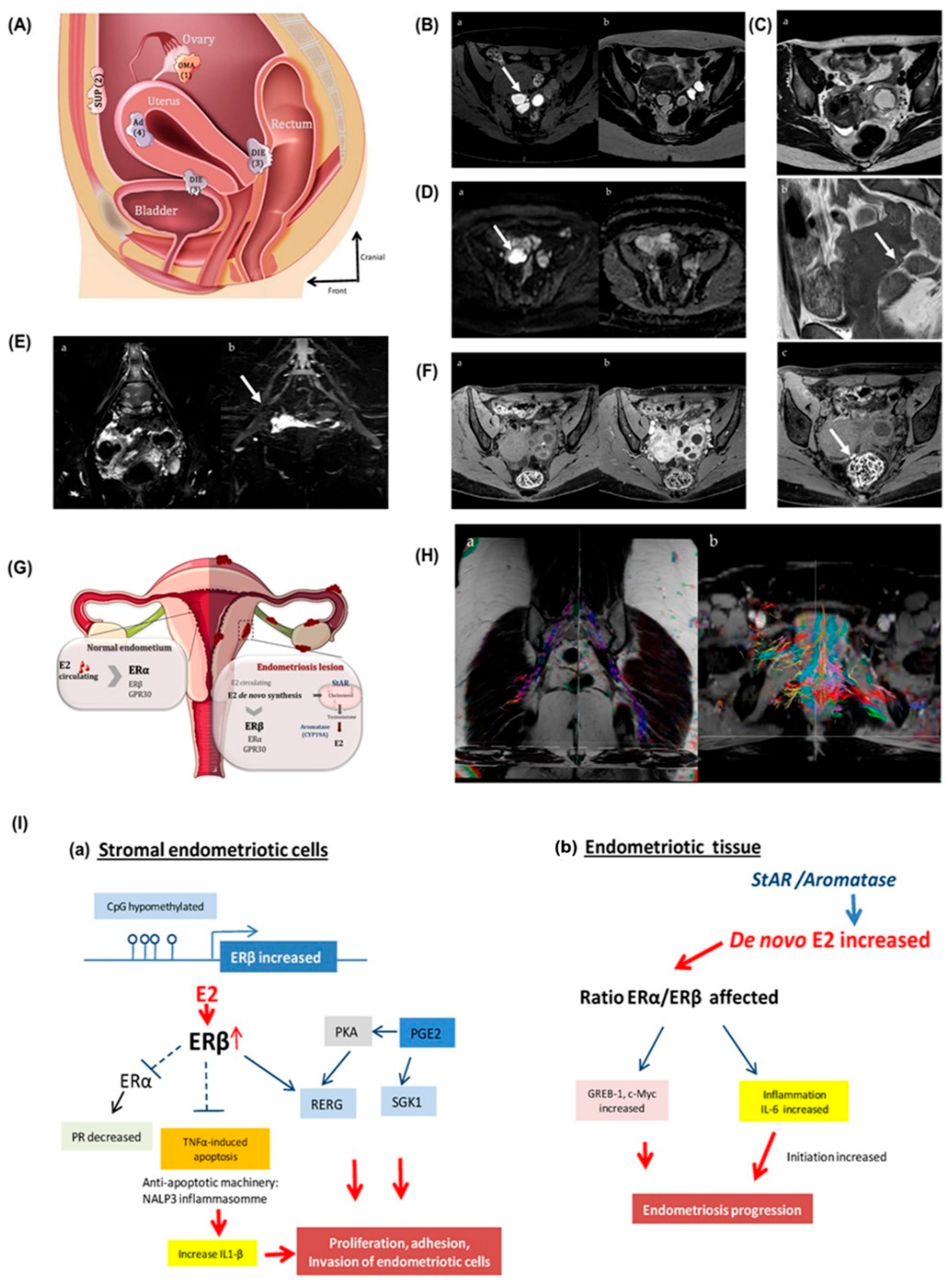
Endometriosis is a prevalent, estrogen-dependent benign gynecological condition characterized by the ectopic growth of endometrial-like tissue outside the uterine cavity, affecting 6–10% of women of reproductive age. Internal adenomyosis, which is characterized by ectopic endometrial glands within the myometrium, deep infiltrating endometriosis (DIE), ovarian endometrioma (OMA), and superficial peritoneal lesions (SUP), are among its various morphological forms (Figure 3A) [47]. Accurate clinical diagnosis is necessary for the best possible treatment planning because endometriosis, despite being benign, shares characteristics with malignancy, including invasive growth, neo-angiogenesis, and the potential for malignant transformation. The most common symptom is ovarian endometriomas, which frequently develop into hemorrhagic cysts as a result of cyclic bleeding. Because MRI has a higher specificity than ultrasound, it has become the gold standard imaging modality. On both T1-weighted and T2-weighted sequences, endometriomas usually show up as multilocular, well-defined cysts with hemosiderin-filled low-intensity walls; on T1-weighted images, the content is hyperintense (Figure 3B) [48]. On T1-weighted fat-suppressed images, “light bulb bright” signals are a telltale sign of chronic bleeding. Deep infiltrating endometriosis (DIE), defined by infiltration of endometrial tissue deeper than 5 mm into the peritoneum, presents with nodular or plaque-like hypointense thickening on T2-weighted sequences, often localized to the retro-cervical region, uterosacral ligaments, posterior vaginal fornix, and rectosigmoid (Figure 3C) [30]. MRI demonstrates a sensitivity and specificity exceeding 90% for DIE detection.
Hemorrhagic foci appear as hyperintense signals on fat-suppressed T1 images, further aiding diagnosis. A functional MRI sequence called diffusion-weighted imaging (DWI) measures tissue diffusivity to improve diagnostic sensitivity. Unlike benign functional cysts, endometriomas have low ADC values and restricted diffusion because of their high cellularity and viscous content (Figure 3D) [48]. By helping to distinguish endometriotic lesions from other adnexal pathologies, DWI improves surgical decision-making. Rectal infiltration and bladder involvement show distinctive MRI signs. Rectal endometriosis can manifest as “mushroom cap” or “fan-shaped” masses, which are indicative of muscular invasion and are pertinent markers for the removal of the segmental bowel. Further evidence of posterior compartment involvement includes fibrotic nodules, a retroflexed uterus, and pelvic structure displacement (Figure 3E) [48]. Endometriomas can be further distinguished from hemorrhagic or infectious adnexal masses using gadolinium-enhanced MRI. Particularly in cysts larger than 9 cm, intense mural enhancement raises the possibility of malignant transformation (Figure 3F) [48]. Neoplastic secretion activity may be correlated with changes in the signal, such as the loss of T2 shading. Two useful techniques for assessing nerve entrapments linked to endometriosis are diffusion tensor imaging (DTI) tractography and magnetic resonance neurography (MR neurography). While MRN detects asymmetry and morphological abnormalities in nerve bundles, tractography shows sacral plexus fiber disarray that is correlated with the intensity of pain (Figure 3G–I). Along with supporting preoperative mapping, these modalities offer insight into the etiology of symptoms. In conclusion, endometriosis mimics malignant processes and has the potential to transform into cancer, making an accurate clinical diagnosis essential. Lesion extent, structural involvement, and possible malignancy can all be comprehensively visualized with multiparametric MRI, especially when combined with DWI, contrast enhancement, and advanced neuroimaging [47,48]. The radiological characteristics necessary to differentiate between endometriosis subtypes are illustrated in Figure 3A–I, which helps physicians customize treatment plans and enhance patient outcomes, especially in the larger framework of gynecological oncology [47,48].

6. Molecular Pathways Targeted by Antioxidant and Anti-Inflammatory Drugs for Gynecological Cancer

Phytochemical-based antioxidant and anti-inflammatory drugs and dietary nutrients both originate from natural sources, but they have quite different purposes, regulations, and applications. Phytochemical-based drugs are defined as standardized, pharmacologically active formulations made from plants; they are usually the subject of rigorous clinical trials and regulatory approval. These compounds are meant to be used therapeutically and have specific dosages, modes of action, and safety profiles. Conversely, dietary components such as vitamins, minerals, and particular bioactive compounds (e.g., flavonoids, carotenoids) primarily support physiological functions and general health. While some nutrients have therapeutic properties, they are typically consumed as dietary supplements or as part of a regular diet and lack the precision and targeted efficacy of pharmaceuticals. Consequently, foodstuffs contain nutrients that support or prevent health, while drugs based on phytochemicals are therapeutic agents.
Antioxidant and anti-inflammatory medications slow the progression of disease by focusing on important molecular pathways related to inflammation, oxidative stress, and the tumor microenvironment. Overproduction of reactive oxygen species (ROS) causes oxidative stress, which upsets cellular homeostasis and exacerbates disease. Enzymatic (superoxide dismutase, catalase, glutathione peroxidase) and non-enzymatic (vitamins C and E, glutathione) antioxidant defense mechanisms control ROS levels to preserve redox balance. Cellular dysfunction, oxidative damage, and disease pathogenesis result from the dysregulation of this equilibrium [49,50,51,52,53]. The inflammatory pathways are important in neurodegenerative diseases and cancer, among other chronic illnesses. Prolonged inflammation results from immune responses triggered by pro-inflammatory cytokines (IL-6, TNF-α, and IL-1β) and chemokines [54,55,56]. The expression of genes linked to inflammation is modulated by the NF-κB signaling pathway, which is a key regulator of inflammation. NF-κB activation is suppressed, cytokine production is inhibited, and gene expression is modulated by anti-inflammatory medications to prevent tissue damage and chronic inflammation [57,58,59].
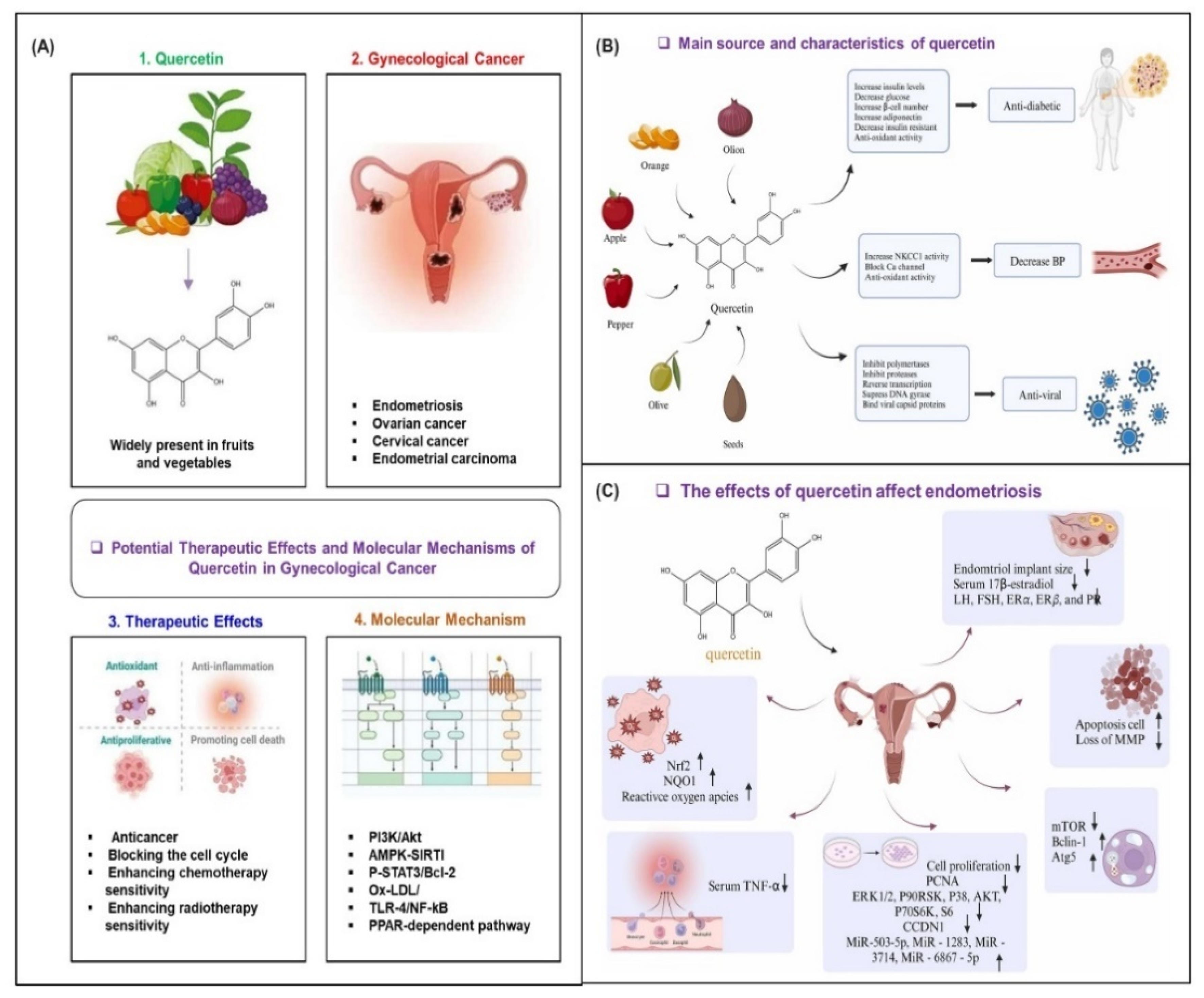
Inflammation and oxidative stress have a significant impact on the tumor microenvironment (TME), which affects how the tumor grows. Angiogenesis, which is controlled by VEGF and hypoxia-inducible factors, provides oxygen and nutrients to support the growth of tumors. ECM remodeling promotes metastasis, while immune cell infiltration, such as that of T cells and macrophages, modifies tumor immune responses. By limiting angiogenesis, controlling immune cell activity, and halting ECM degradation, antioxidant and anti-inflammatory drugs can slow the growth of tumors [49,60,61,62]. One flavonoid that has demonstrated promise in the treatment of gynecological cancers is quercetin, which is known to have anti-inflammatory and antioxidant qualities. Its bioactive properties, especially in lowering inflammation and oxidative stress, are essential in halting the spread of these cancers. Quercetin provides a natural therapeutic approach for gynecological malignancies by targeting multiple molecular pathways linked to tumor growth, as illustrated in Figure 4A. Quercetin’s biological effects are mediated through a variety of mechanisms, and its main sources are fruits, vegetables, and some herbs. One important mechanism is the inhibition of the Na+-K+-2Cl cotransporter 1 (NKCC1), which controls blood pressure and cellular ion balance. Furthermore, as Figure 4B illustrates, quercetin interacts with key enzymes such as NAD(P)H quinone oxidoreductase 1 (NQO1) and activates the nuclear factor (erythroid-derived 2)-like 2 (Nrf2) pathway, which is essential for cellular defense against oxidative damage. In endometriosis, quercetin has been shown to have therapeutic effects via modulation of different factors, including the progesterone receptor (PR), the mitochondrial membrane potential (MMP), and the Nrf2 pathway. This reduces the inflammation and oxidative damage of the tissues concerned, which ultimately reduces the severity of the endometrial symptoms. These results highlight that quercetin has the potential to be an adjunctive treatment option in gynecological conditions and offers a multifaceted approach to treatment, as illustrated in Figure 4C [63].
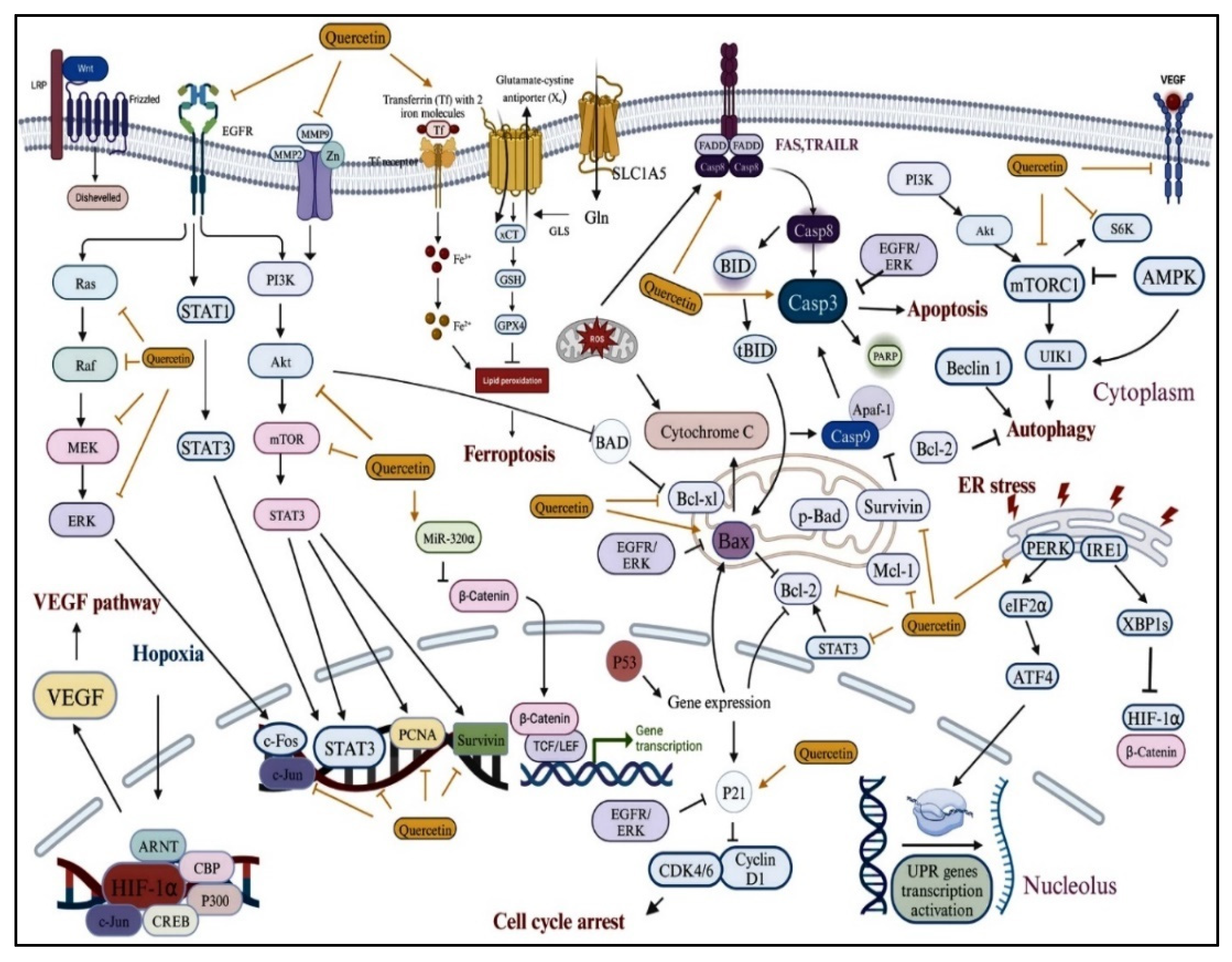
Quercetin has shown promise in the treatment of gynecological cancer. It is a desirable alternative for use as a sensitizing agent or as a preventive measure in the treatment of endometrial cancer (EC), cervical cancer (CC), and ovarian cancer (OC) because it has been demonstrated to inhibit the proliferation of cancer cells. A growing body of research on quercetin’s potential as a treatment has resulted from the natural origin of this compound and its capacity to alter important molecular pathways implicated in the progression of cancer, as illustrated in Figure 5. Several signaling pathways are involved in the intricate molecular processes through which quercetin fights cancer. Several molecular mediators implicated in gynecological cancer, such as vascular endothelial growth factor (VEGF), hypoxia-inducible factors (HIF-1α), and extracellular signal-regulated kinases (ERK), are shown to be affected by quercetin in Figure 4. Furthermore, quercetin activates important proteins like matrix metalloproteinases (MMP), the mammalian target of rapamycin (mTOR), and caspases (Casp3 and Casp8). Together, these elements control angiogenesis, migration, proliferation, and cell survival—all crucial steps in the development of cancer. These interactions show that quercetin can target several facets of tumor biology, ranging from metastasis inhibition to apoptosis induction. In conclusion, because quercetin can alter several molecular pathways that are essential for the growth and spread of tumors, it has potential as a treatment for gynecological cancers. Its potential as a useful supplement to conventional cancer treatments is highlighted by its anti-inflammatory and antioxidant qualities as well as its effects on important signaling molecules like mTOR, VEGF, and HIF-1α. According to ongoing research, quercetin may be crucial in enhancing the results of gynecological cancer treatments, providing a natural and comprehensive strategy to fight these illnesses [63].
Therefore, by focusing on various interconnected molecular pathways that control inflammation, oxidative stress, and the tumor microenvironment (TME), antioxidant and anti-inflammatory medications, like quercetin, provide promising treatment approaches for gynecological cancers. To prevent tumor growth and metastasis, these medications alter important signaling pathways implicated in the advancement of cancer, such as those connected to the production of reactive oxygen species (ROS), cytokine signaling, and cell survival. These substances not only lessen the negative effects of cancer but also improve the effectiveness of traditional treatments by affecting important variables like the Nrf2 pathway, mitochondrial function, and progesterone receptor signaling. Further investigation into these molecular interactions will be essential for creating customized therapies, opening the door for more potent and focused therapeutic approaches in gynecological cancer.

7. Therapeutic Mechanisms of Antioxidant and Anti-Inflammatory Drugs in Gynecological Cancer

Chemicals that are synthetic, natural, or biological are used in chemoprevention to prevent, slow, or even reverse the development of cancer [64,65]. The preventive approach was initially presented by Sporn and colleagues [66,67], and, among other things, makes use of substances called phytochemicals, which are chemically separated into polyphenols, terpenoids, and thiols, as listed in Table 1, with common vegetables, fruit, and whole-grain products [68]. Since lifestyle modifications, including dietary changes, can prevent nearly 33% of cancers, chemotherapy may be a significant factor in lowering the incidence of different cancer types [48]. An oral chemo-preventive compound with a known mechanism of action, high efficiency, and low production cost is considered a chemo-preventive drug. It can be widely used to prevent a variety of diseases, including cancer, because of these features [69]. Numerous phytochemicals have been shown to have anti-carcinogenic effects in both in vitro and in vivo studies. Certain of them reduce inflammation by preventing cells from secreting cytokines, e.g., 3. IL-1, TNF-alfa, IL-6, IL-8, and IL-12 [70,71,72,73]. In turn, they stop cells from undergoing angiogenesis, which controls the growth and progression of cancer, by blocking VEGFA or PI3/Akt [74,75]. Furthermore, the majority of phytochemicals have antioxidant properties that ward off damage and potential carcinogenesis by removing free oxygen radicals from cells. Compounds derived from plants can affect cancer treatment by increasing tumor sensitivity to chemotherapeutics and their proven in vitro cytotoxic effects, in addition to lowering the risk of cancer [76,77]. Because the chosen phytochemicals experimentally demonstrated anticancerogenic properties, studies are now being conducted to evaluate their potential application in preventing cancer, including gynecological cancers. The presentation and discussion of phytochemicals, which we believe are the most important in the chemoprevention of the most prevalent malignant tumors of female genital organs, are covered in this section, including ovarian, endometrial, and cervical cancers (Figure 6) and clinical trials that evaluated phytochemicals in the treatment of breast cancer are listed in Table 2, along with phytochemical classes, dose ranges, clinical indications, and duration [66].
Antioxidant and anti-inflammatory medications are essential for reducing oxidative stress and controlling inflammatory reactions, which are major causes of several pathological conditions, such as cancer, neurological disorders, and cardiovascular diseases. These medicinal substances have the potential to help prevent and treat disease because they work through a variety of mechanisms [78,79,80]. One main way is through direct antioxidant effects, in which these substances counteract reactive oxygen species (ROS) and lessen oxidative damage to DNA, proteins, and lipids in cells. They aid in the prevention of cellular dysfunction and apoptosis by reestablishing redox homeostasis, especially in conditions marked by persistent oxidative stress [81,82]. Additionally, these medications alter inflammatory signaling pathways, which lowers the expression of pro-inflammatory cytokines like interleukin-6 (IL-6), interleukin-8 (IL-8), and tumor necrosis factor-alpha (TNF-α). NF-κB and mitogen-activated protein kinases (MAPKs) are two regulators molecular pathway that suppress chronic inflammation, which is frequently linked to the development of cancer and other inflammatory disorders [83,84,85]. Antioxidant and anti-inflammatory drugs also affect the tumor microenvironment (TME) by lowering tumorigenic signaling brought on by oxidative stress, preventing immune evasion, and boosting anti-tumor immunity. This TME modulation may lessen treatment resistance and increase the efficacy of immune-based therapies [86,87]. Their synergistic interaction with traditional therapies, like chemotherapy and radiation, is another important factor. These agents enhance therapeutic efficacy and decrease side effects by protecting normal tissues and making tumor cells more sensitive to treatment by reducing inflammation and oxidative stress brought on by therapy [88,89,90,91]. In conclusion, the various ways that anti-inflammatory and antioxidant medications work demonstrate their therapeutic potential in the management of chronic illnesses. Their relevance in clinical applications is highlighted by their interplay with current therapies and their capacity to control inflammation, oxidative stress, and the tumor microenvironment. To improve therapeutic outcomes, future research should concentrate on maximizing their use and investigating innovative drug combinations.

8. Clinical Evidence for Treatment of Gynecological Cancer

Gynecological cancers, such as endometrial, cervical, and ovarian cancers, contribute significantly to the worldwide cancer mortality rate. This is frequently because of the limited therapeutic options and late-stage diagnosis. Treating these cancers by focusing on the pathways that cause inflammation and oxidative stress has shown promise. Antioxidant and anti-inflammatory medications are being investigated for their ability to slow the growth of tumors, enhance therapeutic results, and lessen adverse effects associated with treatment [92,93]. According to preclinical research, oxidative stress is a key factor in the development and spread of gynecological cancers. Resveratrol, curcumin, and vitamin C are examples of antioxidants that have demonstrated encouraging results in lowering reactive oxygen species (ROS) levels, which in turn prevent tumor growth, metastasis, and angiogenesis [94,95]. Further anti-inflammatory medications include certain cytokine inhibitors (e.g., nonsteroidal anti-inflammatory drugs, or NSAIDs). It has been demonstrated to reduce inflammation linked to cancer, which limits tumor growth and improves chemotherapeutic sensitivity (IL-6, TNF-α). Early-stage results from clinical trials exploring the effectiveness of these agents in conjunction with traditional therapies are encouraging [96,97,98]. The clinical translation of these treatments is hampered by several issues and restrictions. It is challenging to create medications that precisely target oxidative stress and inflammatory pathways in cancer without interfering with regular cellular processes due to their complexity. Significant challenges are also presented by the bioavailability, stability, and possible toxicity of anti-inflammatory and antioxidant medications. The implementation of these treatments in clinical settings is made more difficult by differences in cancer subtypes, treatment plans, and individual patient responses [99,100,101,102]. Future studies should concentrate on improving drug delivery methods, finding biomarkers for patient selection, and carrying out extensive clinical trials to gain a better understanding of the safety and effectiveness of anti-inflammatory and antioxidant medications in gynecological cancers. Additionally, a thorough comprehension of the molecular processes that underlie inflammation and oxidative stress in cancer will be essential for creating more specialized and individualized treatments. In conclusion, addressing inflammation and oxidative stress in gynecological cancers may improve patient outcomes; however, further study is needed to address present issues and fully utilize the therapeutic potential of anti-inflammatory and antioxidant medications.

9. Future Directions for Treatment of Gynecological Cancer

Gynecological cancers, such as endometrial, cervical, and ovarian cancers, contribute significantly to the worldwide cancer mortality rate. This is frequently because of the limited therapeutic options and late-stage diagnosis. Treating these cancers by focusing on the pathways that cause inflammation and oxidative stress has shown promise. Antioxidant and anti-inflammatory medications are being investigated for their ability to slow the growth of tumors, enhance therapeutic results, and lessen adverse effects associated with treatment [103,104,105].
Table 1. Plant-derived phytochemicals, their botanical sources, and molecular targets investigated in pre-clinical trials for cancer treatment.
Phytochemical
(Compound Class)
Botanical Name
(Family)
Molecular TargetsRef.
6-Shogaol (phenylpropanoid)Zingiber officinale (Roscoe)Akt and STAT
signaling pathway
[106,107]
Allicin (organosulfurs)Allium sativum
(Amaryllidaceae)
STAT3 signaling pathway[108,109]
Alpinumisoflavone
(pyranoisoflavone)
Derris eriocarpa
(Leguminosae)
Nrf2, NQO-1, HO-1,
miR-101, and Akt signaling
[110]
Andrographolide (diterpenoid)Andrographis paniculata
(Acanthaceae)
HIF-1α, VEGF, and PI3K
pathway
[111]
Apigenin (flavonoid)Petroselinum
crispum (Apiaceae)
Intrinsic apoptosis pathway[112]
Baicalein (flavonoid)Scutellaria baicalensis
(Lamiaceae)
MAPK, ERK, and
p38 signaling pathways
[113,114]
Baicalin (flavonoid)Scutellaria baicalensis
(Lamiaceae)
MAPK, ERK, and
p38 signaling pathways
[115]
Curcumin (phytopolyphenol)Curcuma longa
(Zingiberaceae)
Modulates cell signaling
and gene expression
[116]
Decursin and Decursinol (coumarin)Angelica gigas
(Apiaceae)
Not mentioned[117]
DicumarolMelilotus officinalis
(Fabaceae)
Intrinsic apoptosis pathway[118]
Epigallocatechin (flavonoid)Camellia sinensis
(Theaceae)
Inhibit cell proliferation
and apoptosis
[119]
Emodin (resin)Rheum palmatum L.
(Polygonaceae)
PI3K/AKT and
MAPK signaling pathways
[120,121,122]
Genistein (isoflavonoid)Glycine max (Leguminosae)WNT/β-catenin and
Akt signaling pathway
[123,124]
Gingerol (polyphenol)Zingiber officinale (Roscoe)Intrinsic apoptosis pathway[125,126]
Glycyrrhizin (triterpenes)Glycyrrhiza glabra
(Fabaceae)
TxA2 and JAK/STAT
signaling pathway
[127]
Hispidulin (flavone)Salvia involucrata
(Lamiaceae)
Intrinsic apoptosis
pathway
[128,129]
HS-1793 (stilbenoid)Polygonum cuspidatum
(Polygonaceae)
HIF-1α, VEGF, Ki-67,
and CD31
[130]
Licochalcone A (chalcone)Glycyrrhiza glabra
(Fabaceae)
Cyclins and CDKs[131]
Nimbolide (triterpene)Azadirachta indica
(Meliaceae)
PI3K/AKT/mTOR and
ERK signaling
[132]
Physapubescin B (steroid)Physalis pubescens L.
(Solanaceae)
Ki-67, Cdc25C, and PARP[133]
Pterostilbene (polyphenol)Polygonum cuspidatum
(Polygonaceae)
Mitochondrial apoptosis;
ERK and STAT3 signaling
[134,135,136]
Resveratrol (phenol)Polygonum cuspidatum
(Polygonaceae)
Regulating cell cycle and
apoptosis pathways
[137]
Sulforaphane (organosulfur)Brassica oleracea
(Brassicaceae)
Cell cycle arrest and
apoptosis (caspase-8, p21, hsp90)
[138]
Thymol (monoterpenoid)Thymus vulgaris
(Lamiaceae)
Mitochondrial mediated
apoptosis
[139]
Thymoquinone (quinone)Nigella sativa
(Ranunculaceae)
STAT3 and associated proteins[140,141]
Ursolic acid (triterpenoids)Oldenlandia diffusa
(Rubiaceae)
Ki-67, CD31, and miR-29a[142,143]
Withaferin-A (phytosterols)Withania somnifera
(Solanaceae)
AKT/FOXO3a-Par-4
cell death, ERK, p38
[144,145,146]
Table 2. Clinical trials evaluated phytochemicals in breast cancer treatment: phytochemical classes, dose ranges, clinical indication, and duration.
PhytochemicalDose RangeClinical IndicationDurationRef.
Curcumin6000 mg/day (oral)Advanced/metastatic breast cancer (combined with docetaxel)7 days every 3 weeks[147]
Curcumin6 g/day (oral)Radiation dermatitis prevention during radiotherapyDuration of radiotherapy[148]
Curcumin300 mg (IV weekly)Metastatic breast cancer (combined with paclitaxel)12 weeks[148]
Polyphenon E (EGCG)400–800 mg twice dailyChemoprevention in hormone receptor-negative breast cancer6 months[149]
Indole-3-carbinol300–400 mg/day (oral)Breast cancer prevention in high-risk women4–8 weeks[150,151]
Diindolylmethane (BR-DIM)150 mg twice daily (oral)Adjuvant therapy with tamoxifen for breast cancer12 months[152]
Flaxseed lignans (SDG)25 g/day (dietary flaxseed)Reduction in tumor biomarkers (Ki-67, c-erbB2) in postmenopausal breast cancer~32 days[153]
Secoisolariciresinol diglucoside (SDG)50 mg/day (oral)Chemoprevention in premenopausal women at high risk for breast cancer12 months[154,155]
Scutellaria barbata (BZL101)40 g/day (oral extract)Metastatic breast cancer (monotherapy)Until progression[155,156]
Grape seed
proanthocyanidin (GSPE)
100 mg three times daily (oral)Management of radiation-induced breast induration6 months[157]
According to preclinical research, oxidative stress is a key factor in the development and spread of gynecological cancers. Resveratrol, curcumin, and vitamin C are examples of antioxidants that have demonstrated encouraging results in lowering reactive oxygen species (ROS) levels, which in turn prevent tumor growth, metastasis, and angiogenesis [49,94,158]. Further anti-inflammatory medications include certain cytokine inhibitors (e.g., nonsteroidal anti-inflammatory drugs, or NSAIDs). It has been demonstrated to reduce inflammation linked to cancer, which limits tumor growth and improves chemotherapeutic sensitivity (IL-6, TNF-α) [159,160].
In the treatment of gynecological cancers, phytochemicals have demonstrated promise as adjuncts due to their anti-inflammatory, anticancer, and chemopreventive properties, with the potential to enhance clinical outcomes [161,162]. In preclinical research and early-stage clinical trials, curcumin, a polyphenolic compound present in turmeric, has demonstrated effectiveness in modifying signaling pathways implicated in ovarian and endometrial cancer cell proliferation, apoptosis, and metastasis. The compound epigallocatechin gallate (EGCG), which is found in polyphenols E, which is made from green tea, has been studied in clinical trials for its ability to stop tumor growth and improve the efficiency of traditional chemicals. In preclinical research, indole-3-carbinol (I3C) and its derivative diindolylmethane (BR-DIM) from cruciferous vegetables showed anticancer properties [163,164,165]. In clinical trials, they were evaluated for their ability to alter estrogen metabolism and lower the risk of cancers driven by estrogen. Clinical trials suggest that flaxseed lignans, especially secoisolariciresinol diglucoside (SDG), may help manage breast cancer. They have also been associated with decreased tumor growth and the growth of cancer cells. Additionally, clinical trials have shown promise for the antioxidative and anticancer effects of grape seed proanthocyanidin extract (GSPE) and Scutellaria barbata (BZL101). When used alone or in conjunction with traditional therapies, these phytochemicals present encouraging avenues for future research and treatment, helping to lower treatment resistance and increase overall survival rates for patients with gynecological cancer [154,166,167].
Early-stage results from clinical trials exploring the effectiveness of these agents in conjunction with traditional therapies are encouraging. The clinical translation of these treatments is hampered by several issues and restrictions. It is challenging to create medications that precisely target oxidative stress and inflammatory pathways in cancer without interfering with regular cellular processes due to their complexity. Significant challenges are also presented by the bioavailability, stability, and possible toxicity of anti-inflammatory and antioxidant medications. The implementation of these treatments in clinical settings is made more difficult by differences in cancer subtypes, treatment plans, and individual patient responses [168,169,170]. Future studies should concentrate on improving drug delivery methods, finding biomarkers for patient selection, and carrying out extensive clinical trials to gain a better understanding of the safety and effectiveness of anti-inflammatory and antioxidant medications in gynecological cancers. Additionally, a thorough comprehension of the molecular processes that underlie inflammation and oxidative stress in cancer will be essential for creating more specialized and individualized treatments. In conclusion, addressing inflammation and oxidative stress in gynecological cancers may improve patient outcomes; however, further study is needed to address present issues and fully utilize the therapeutic potential of anti-inflammatory and antioxidant medications.

10. Conclusions

In conclusion, research shows that anti-inflammatory and antioxidant substances can be used to treat gynecological cancers by lowering oxidative stress, addressing chronic inflammation, and focusing on important oncogenic pathways. By encouraging tumor suppression, triggering apoptosis, and increasing sensitivity to traditional therapies, these substances—which include curcumin, polyphenon E (EGCG), indole-3-carbinol, diindolylmethane (BR-DIM), flaxseed lignans (SDG), secoisolariciresinol diglucoside (SDG), Scutellaria barbata (BZL101), and grape seed proanthocyanidin (GSPE)—have shown encouraging preclinical results. These phytochemical compounds’ therapeutic efficacy is further supported by recent clinical trials that show promise in enhancing immune responses and lowering treatment-related toxicities. To maximize their integration into clinical practice, more research is required, as the clinical application of these agents is still in its infancy. To maximize therapeutic benefits and reduce side effects, future research should concentrate on enhancing drug formulations, dosage schedules, and combination strategies with traditional therapies. Finding predictive biomarkers will also guarantee the effectiveness of these treatments and allow for individualized treatment plans. In general, developing our knowledge of the molecular pathways that connect oxidative stress, inflammation, and the biology of gynecological cancer will be essential to turning these promising substances into cutting-edge, successful treatment plans that will eventually enhance patient outcomes and quality of life.

Author Contributions

Conceptualization, S.S., A.K.S., N.R. and R.K.M.; methodology, S.S., A.M.U., A.K.S., N.R., F.I.F., A.N., S.D.D. and R.K.M.; writing—original draft preparation, R.K.M.; writing—review and editing, S.S., A.M.U., A.K.S., N.R., F.I.F., A.N., S.D.D. and R.K.M.; visualization, S.S., A.M.U., A.K.S., N.R., F.I.F., A.N., S.D.D. and R.K.M.; management, S.S., A.K.S., N.R. and R.K.M. All authors have read and agreed to the published version of the manuscript.

Funding

This research received no external funding.

Conflicts of Interest

The authors declare no conflicts of interest.

Abbreviations

ASRAge-standardized rate
GCGynecological cancer
AKTProtein kinase B
EMSEpithelial–mesenchymal transition
ILInterleukin
MAMyeloid-derived suppressor cells
MAPKMitogen-activated protein kinase
NF-κBNuclear factor kappa-light-chain-enhancer of activated B cells
ROSReactive oxygen species
TNFTumor necrosis factor
VEGFAVascular endothelial growth factor A

References

  1. Zhang, L.; Bi, Q.; Deng, H.; Xu, J.; Chen, J.; Zhang, M.; Mu, X. Human papillomavirus infections among women with cervical lesions and cervical cancer in Eastern China: Genotype-specific prevalence and attribution. BMC Infect. Dis. 2017, 17, 107. [Google Scholar] [CrossRef]
  2. Polten, R.; Kutle, I.; Hachenberg, J.; Klapdor, R.; Morgan, M.; Schambach, A. Towards novel gene and cell therapy approaches for cervical cancer. Cancers 2022, 15, 263. [Google Scholar] [CrossRef] [PubMed]
  3. Shah, S.; Thanki, R.M.; Diwan, A. Artificial Intelligence for Early Detection and Diagnosis of Cervical Cancer; Springer: Berlin/Heidelberg, Germany, 2024. [Google Scholar]
  4. Dong, H.; Chen, S.; Liang, X.; Cai, Q.; Zhang, X.; Xie, J.; Sun, Z. Vitamin D and its receptors in cervical cancer. J. Cancer 2024, 15, 926. [Google Scholar] [CrossRef] [PubMed]
  5. Renaudin, X. Reactive oxygen species and DNA damage response in cancer. Int. Rev. Cell Mol. Biol. 2021, 364, 139–161. [Google Scholar] [PubMed]
  6. Xiao, Y.; Bi, M.; Guo, H.; Li, M. Multi-omics approaches for biomarker discovery in early ovarian cancer diagnosis. EBioMedicine 2022, 79, 104001. [Google Scholar] [CrossRef]
  7. Reid, F.; Bajwa, A. World Ovarian Cancer Coalition Atlas. 2020. In Global Trends in Incidence, Mortality, and Survival; World Ovarian Cancer Coalition: Toronto, ON, Canada, 2020; pp. 1–42. [Google Scholar]
  8. Zhang, J.; Yu, S.; Li, Q.; Wang, Q.; Zhang, J. Increased co-expression of MEST and BRCA1 is associated with worse prognosis and immune infiltration in ovarian cancer. Gynecol. Oncol. 2022, 164, 566–576. [Google Scholar] [CrossRef]
  9. Colditz, G.; Beers, C. Active cancer prevention. Cancer Control 2010, 45, 23. [Google Scholar]
  10. Gyllensten, U. Novel diagnostics for improved treatment of gynecological cancer. Upsala J. Med. Sci. 2025, 130, 10-48101. [Google Scholar] [CrossRef]
  11. Zhu, B.; Gu, H.; Mao, Z.; Beeraka, N.M.; Zhao, X.; Anand, M.P.; Zheng, Y.; Zhao, R.; Li, S.; Manogaran, P. Global burden of gynaecological cancers in 2022 and projections to 2050. J. Glob. Health 2024, 14, 04155. [Google Scholar] [CrossRef]
  12. Mokdad, A.H.; Bisignano, C.; Hsu, J.M.; Bryazka, D.; Cao, S.; Bhattacharjee, N.V.; Dalton, B.E.; Lindstedt, P.A.; Smith, A.E.; Ababneh, H.S. Burden of disease scenarios by state in the USA, 2022–2050: A forecasting analysis for the Global Burden of Disease Study 2021. Lancet 2024, 404, 2341–2370. [Google Scholar] [CrossRef]
  13. Goswami, B.; Rajappa, M.; Sharma, M.a.; Sharma, A. Inflammation: Its role and interplay in the development of cancer, with special focus on gynecological malignancies. Int. J. Gynecol. Cancer 2008, 18, 591–599. [Google Scholar] [CrossRef] [PubMed]
  14. Dutta, S.; Mitra, M.; Agarwal, P.; Mahapatra, K.; De, S.; Sett, U.; Roy, S. Oxidative and genotoxic damages in plants in response to heavy metal stress and maintenance of genome stability. Plant Signal. Behav. 2018, 13, e1460048. [Google Scholar] [CrossRef]
  15. Iqbal, M.J.; Kabeer, A.; Abbas, Z.; Siddiqui, H.A.; Calina, D.; Sharifi-Rad, J.; Cho, W.C. Interplay of oxidative stress, cellular communication and signaling pathways in cancer. Cell Commun. Signal. 2024, 22, 7. [Google Scholar] [CrossRef]
  16. Milkovic, L.; Siems, W.; Siems, R.; Zarkovic, N. Oxidative stress and antioxidants in carcinogenesis and integrative therapy of cancer. Curr. Pharm. Des. 2014, 20, 6529–6542. [Google Scholar] [CrossRef] [PubMed]
  17. Li, K.; Deng, Z.; Lei, C.; Ding, X.; Li, J.; Wang, C. The role of oxidative stress in tumorigenesis and progression. Cells 2024, 13, 441. [Google Scholar] [CrossRef]
  18. Ju, S.; Singh, M.K.; Han, S.; Ranbhise, J.; Ha, J.; Choe, W.; Yoon, K.-S.; Yeo, S.G.; Kim, S.S.; Kang, I. Oxidative Stress and Cancer Therapy: Controlling Cancer Cells Using Reactive Oxygen Species. Int. J. Mol. Sci. 2024, 25, 12387. [Google Scholar] [CrossRef]
  19. Closset, L.; Gultekin, O.; Salehi, S.; Sarhan, D.; Lehti, K.; Gonzalez-Molina, J. The extracellular matrix–immune microenvironment crosstalk in cancer therapy: Challenges and opportunities. Matrix Biol. 2023, 121, 217–228. [Google Scholar] [CrossRef]
  20. Baghban, R.; Roshangar, L.; Jahanban-Esfahlan, R.; Seidi, K.; Ebrahimi-Kalan, A.; Jaymand, M.; Kolahian, S.; Javaheri, T.; Zare, P. Tumor microenvironment complexity and therapeutic implications at a glance. Cell Commun. Signal. 2020, 18, 59. [Google Scholar] [CrossRef] [PubMed]
  21. Bitsadze, V.; Slukhanchuk, E.; Solopova, A.; Khizroeva, J.K.; Yakubova, F.; Orudzhova, E.; Degtyareva, N.; Egorova, E.; Makatsariya, N.; Samburova, N. The role of the microenvironment in tumor growth and spreading. Obstet. Gynecol. Reprod. 2024, 18, 96–111. [Google Scholar] [CrossRef]
  22. De Nola, R.; Menga, A.; Castegna, A.; Loizzi, V.; Ranieri, G.; Cicinelli, E.; Cormio, G. The crowded crosstalk between cancer cells and stromal microenvironment in gynecological malignancies: Biological pathways and therapeutic implication. Int. J. Mol. Sci. 2019, 20, 2401. [Google Scholar] [CrossRef]
  23. Kan, X.; Zhou, Z.; Liu, L.; Aiskikaer, R.; Zou, Y. Significance of non-steroidal anti-inflammatory drugs in the prevention and treatment of cervical cancer. Heliyon 2025, 11, e42055. [Google Scholar] [CrossRef] [PubMed]
  24. Centini, G.; Schettini, G.; Pieri, E.; Giorgi, M.; Lazzeri, L.; Martire, F.G.; Mancini, V.; Raimondo, D.; Seracchioli, R.; Habib, N. Endometriosis-related ovarian cancer: Where are we now? A narrative review towards a pragmatic approach. J. Clin. Med. 2024, 13, 1933. [Google Scholar] [CrossRef]
  25. Conforti, R.A.; Delsouc, M.B.; Zorychta, E.; Telleria, C.M.; Casais, M. Copper in gynecological diseases. Int. J. Mol. Sci. 2023, 24, 17578. [Google Scholar] [CrossRef]
  26. Khan, H.; Ullah, H.; Castilho, P.C.M.F.; Gomila, A.S.; D’Onofrio, G.; Filosa, R.; Wang, F.; Nabavi, S.M.; Daglia, M.; Silva, A.S. Targeting NF-κB signaling pathway in cancer by dietary polyphenols. Crit. Rev. Food Sci. Nutr. 2020, 60, 2790–2800. [Google Scholar] [CrossRef] [PubMed]
  27. Bahrami, A.; Khalaji, A.; Bahri Najafi, M.; Sadati, S.; Raisi, A.; Abolhassani, A.; Eshraghi, R.; Khaksary Mahabady, M.; Rahimian, N.; Mirzaei, H. NF-κB pathway and angiogenesis: Insights into colorectal cancer development and therapeutic targets. Eur. J. Med. Res. 2024, 29, 610. [Google Scholar] [CrossRef]
  28. Aldhahrani, A.; Alshaye, N.A.; Alshaya, D.S.; Mohamed, D.A.; Fayad, E.; Sophy, M.A.E.; Al-Ashger, N.A.; Salem, M.G.; Farouk, N.A. In Vitro biological evaluation of some hybrid molecules bearing 2-quinoline as anti-inflammatory and anti-cancer agents. J. Mol. Struct. 2025, 1326, 141060. [Google Scholar] [CrossRef]
  29. Choudhari, A.S.; Mandave, P.C.; Deshpande, M.; Ranjekar, P.; Prakash, O. Phytochemicals in Cancer Treatment: From Preclinical Studies to Clinical Practice. Front. Pharmacol. 2019, 10, 1614. [Google Scholar] [CrossRef]
  30. Ali Abdalla, Y.O.; Subramaniam, B.; Nyamathulla, S.; Shamsuddin, N.; Arshad, N.M.; Mun, K.S.; Awang, K.; Nagoor, N.H. Natural Products for Cancer Therapy: A Review of Their Mechanism of Actions and Toxicity in the Past Decade. J. Trop. Med. 2022, 2022, 5794350. [Google Scholar] [CrossRef]
  31. Olson, S.H.; Mignone, L.; Nakraseive, C.; Caputo, T.; Barakat, R.; Harlap, S. Symptoms of ovarian cancer. Obstet. Gynecol. 2001, 98, 212–217. [Google Scholar] [CrossRef]
  32. Baines, P.A.; Allen, G.M. Pelvic pain and menstrual related illnesses. Emerg. Med. Clin. N. Am. 2001, 19, 763–780. [Google Scholar] [CrossRef]
  33. Melnyk, M.; Starczewski, A.; Nawrocka-Rutkowska, J.; Gorzko, A.; Melnyk, B.; Szydłowska, I. Giant Ovarian Tumors in Young Women: Diagnostic and Treatment Challenges—A Report of Two Cases and Narrative Review of the Recent Literature. J. Clin. Med. 2025, 14, 1236. [Google Scholar] [CrossRef]
  34. Naga, O. Last-Minute Review. In Pediatric Board Study Guide: A Last Minute Review; Springer Nature: Cham, Switzerland, 2024; pp. 1017–1126. [Google Scholar]
  35. McCluggage, W.G. Progress in the pathological arena of gynecological cancers. Int. J. Gynaecol. Obstet. 2021, 155 (Suppl. S1), 107–114. [Google Scholar] [CrossRef] [PubMed]
  36. Rizzo, S.; Avesani, G.; Panico, C.; Manganaro, L.; Gui, B.; Lakhman, Y.; Andrieu, P.C.; Bharwani, N.; Rockall, A.; Thomassin-Naggara, I.; et al. Ovarian cancer staging and follow-up: Updated guidelines from the European Society of Urogenital Radiology female pelvic imaging working group. Eur. Radiol. 2025. [Google Scholar] [CrossRef]
  37. Addley, H.; Gregory, D.; Prewett, S.; Smith, J. Gynaecological Cancers. In Radiological Anatomy for Radiation and Particle Therapy; Springer Nature: Cham, Switzerland, 2025; pp. 271–295. [Google Scholar]
  38. Mayeri, D.G.; Kahasha, P.M.; Kibalama, I.B.; Mongane, J.; Louguè, M.; Birindwa, E.K.; Mwimangire, S.C.; Kikuru, C.K.; Materanya, J.M.; Bisimwa, Y.K.; et al. Cervical precancerous and cancerous lesions screening using Pap smear test at Provincial Referral Hospital of Bukavu, Eastern DR Congo: Profile and recommendations to stakeholders. Pan Afr. Med. J. 2024, 47, 57. [Google Scholar] [CrossRef] [PubMed]
  39. Koliopoulos, G.; Nyaga, V.N.; Santesso, N.; Bryant, A.; Martin-Hirsch, P.P.; Mustafa, R.A.; Schünemann, H.; Paraskevaidis, E.; Arbyn, M. Cytology versus HPV testing for cervical cancer screening in the general population. Cochrane Database Syst. Rev. 2017, 8, Cd008587. [Google Scholar] [CrossRef] [PubMed]
  40. Islam, S.; Walker, R.C. Advanced imaging (positron emission tomography and magnetic resonance imaging) and image-guided biopsy in initial staging and monitoring of therapy of lung cancer. Cancer J. 2013, 19, 208–216. [Google Scholar] [CrossRef]
  41. Choi, H.J.; Ju, W.; Myung, S.K.; Kim, Y. Diagnostic performance of computer tomography, magnetic resonance imaging, and positron emission tomography or positron emission tomography/computer tomography for detection of metastatic lymph nodes in patients with cervical cancer: Meta-analysis. Cancer Sci. 2010, 101, 1471–1479. [Google Scholar] [CrossRef]
  42. Anand, U.; Dey, A.; Chandel, A.K.S.; Sanyal, R.; Mishra, A.; Pandey, D.K.; De Falco, V.; Upadhyay, A.; Kandimalla, R.; Chaudhary, A.; et al. Cancer chemotherapy and beyond: Current status, drug candidates, associated risks and progress in targeted therapeutics. Genes Dis. 2023, 10, 1367–1401. [Google Scholar] [CrossRef]
  43. Krzyszczyk, P.; Acevedo, A.; Davidoff, E.J.; Timmins, L.M.; Marrero-Berrios, I.; Patel, M.; White, C.; Lowe, C.; Sherba, J.J.; Hartmanshenn, C.; et al. The growing role of precision and personalized medicine for cancer treatment. Technology 2018, 6, 79–100. [Google Scholar] [CrossRef]
  44. Min, H.Y.; Lee, H.Y. Molecular targeted therapy for anticancer treatment. Exp. Mol. Med. 2022, 54, 1670–1694. [Google Scholar] [CrossRef]
  45. Joshi, D.C.; Sharma, A.; Prasad, S.; Singh, K.; Kumar, M.; Sherawat, K.; Tuli, H.S.; Gupta, M. Novel therapeutic agents in clinical trials: Emerging approaches in cancer therapy. Discov. Oncol. 2024, 15, 342. [Google Scholar] [CrossRef] [PubMed]
  46. Salman, L.; Covens, A. Fertility Preservation in Cervical Cancer-Treatment Strategies and Indications. Curr. Oncol. 2024, 31, 296–306. [Google Scholar] [CrossRef]
  47. Chantalat, E.; Valera, M.-C.; Vaysse, C.; Noirrit, E.; Rusidze, M.; Weyl, A.; Vergriete, K.; Buscail, E.; Lluel, P.; Fontaine, C.; et al. Estrogen Receptors and Endometriosis. Int. J. Mol. Sci. 2020, 21, 2815. [Google Scholar] [CrossRef]
  48. Alonzo, L.; Cannella, R.; Gullo, G.; Piombo, G.; Cicero, G.; Lopez, A.; Billone, V.; Andrisani, A.; Cucinella, G.; Lo Casto, A.; et al. Magnetic Resonance Imaging of Endometriosis: The Role of Advanced Techniques. J. Clin. Med. 2024, 13, 5783. [Google Scholar] [CrossRef] [PubMed]
  49. Reuter, S.; Gupta, S.C.; Chaturvedi, M.M.; Aggarwal, B.B. Oxidative stress, inflammation, and cancer: How are they linked? Free Radic. Biol. Med. 2010, 49, 1603–1616. [Google Scholar] [CrossRef]
  50. Mittal, M.; Siddiqui, M.R.; Tran, K.; Reddy, S.P.; Malik, A.B. Reactive oxygen species in inflammation and tissue injury. Antioxid. Redox Signal 2014, 20, 1126–1167. [Google Scholar] [CrossRef]
  51. Fatima, D.N. Association of Peroxisomes, Reactive Oxygen Species (ROS) and Antioxidants: Insights from Preclinical and Clinical Evaluations. In The Metabolic Role of Peroxisome in Health and Disease; İla, H.B., Ed.; IntechOpen: Rijeka, Croatia, 2022. [Google Scholar] [CrossRef]
  52. Orabi, S.A.; Abou-Hussein, S. Antioxidant defense mechanisms enhance oxidative stress tolerance in plants. A review. Curr. Sci. Int. 2019, 8, 565–576. [Google Scholar]
  53. Martínez, J.P.; Araya, H. Ascorbate–glutathione cycle: Enzymatic and non-enzymatic integrated mechanisms and its biomolecular regulation. In Ascorbate-Glutathione Pathway and Stress Tolerance in Plants; Springer: Dordrecht, The Netherlands, 2010; pp. 303–322. [Google Scholar]
  54. Yeung, Y.T.; Aziz, F.; Guerrero-Castilla, A.; Arguelles, S. Signaling pathways in inflammation and anti-inflammatory therapies. Curr. Pharm. Des. 2018, 24, 1449–1484. [Google Scholar] [CrossRef] [PubMed]
  55. Turner, M.D.; Nedjai, B.; Hurst, T.; Pennington, D.J. Cytokines and chemokines: At the crossroads of cell signalling and inflammatory disease. Biochim. Biophys. Acta (BBA)-Mol. Cell Res. 2014, 1843, 2563–2582. [Google Scholar] [CrossRef]
  56. Wang, R.X.; Zhou, M.; Ma, H.L.; Qiao, Y.B.; Li, Q.S. The role of chronic inflammation in various diseases and anti-inflammatory therapies containing natural products. ChemMedChem 2021, 16, 1576–1592. [Google Scholar] [CrossRef]
  57. Liu, T.; Zhang, L.; Joo, D.; Sun, S.-C. NF-κB signaling in inflammation. Signal Transduct. Target. Ther. 2017, 2, 17023. [Google Scholar] [CrossRef] [PubMed]
  58. Shih, R.-H.; Wang, C.-Y.; Yang, C.-M. NF-kappaB signaling pathways in neurological inflammation: A mini review. Front. Mol. Neurosci. 2015, 8, 77. [Google Scholar] [CrossRef] [PubMed]
  59. Escarcega, R.; Fuentes-Alexandro, S.; Garcia-Carrasco, M.; Gatica, A.; Zamora, A. The transcription factor nuclear factor-kappa B and cancer. Clin. Oncol. 2007, 19, 154–161. [Google Scholar] [CrossRef] [PubMed]
  60. Aboelella, N.S.; Brandle, C.; Kim, T.; Ding, Z.C.; Zhou, G. Oxidative Stress in the Tumor Microenvironment and Its Relevance to Cancer Immunotherapy. Cancers 2021, 13, 986. [Google Scholar] [CrossRef]
  61. Nishida, A.; Andoh, A. The Role of Inflammation in Cancer: Mechanisms of Tumor Initiation, Progression, and Metastasis. Cells 2025, 14, 488. [Google Scholar] [CrossRef]
  62. Fang, J.; Lu, Y.; Zheng, J.; Jiang, X.; Shen, H.; Shang, X.; Lu, Y.; Fu, P. Exploring the crosstalk between endothelial cells, immune cells, and immune checkpoints in the tumor microenvironment: New insights and therapeutic implications. Cell Death Dis. 2023, 14, 586. [Google Scholar] [CrossRef]
  63. Jian, X.; Shi, C.; Luo, W.; Zhou, L.; Jiang, L.; Liu, K. Therapeutic effects and molecular mechanisms of quercetin in gynecological disorders. Biomed. Pharmacother. 2024, 173, 116418. [Google Scholar] [CrossRef]
  64. Mazumder, S.; Chanda, S.; Banerjee, S. Phytochemical Profile and Chemopreventive Properties of Potential Bioactive Ingredients. In Bioactive Ingredients for Healthcare Industry Volume 1: Extraction strategies, Stability and Medicinal Properties; Springer: Singapore, 2025; pp. 15–36. [Google Scholar]
  65. Sichetti, M.; Giuseffi, M.; Giglio, E.; Marino, G.; Mecca, M. Effect of Natural Polyphenols on Breast Cancer Chemoprevention and Treatment. Mol. Nutr. Food Res. 2025, e70055. [Google Scholar] [CrossRef]
  66. Woźniak, M.; Krajewski, R.; Makuch, S.; Agrawal, S. Phytochemicals in Gynecological Cancer Prevention. Int. J. Mol. Sci. 2021, 22, 1219. [Google Scholar] [CrossRef]
  67. Kumar, A.; Pradeep, N.; Kumar, M.; Jose, A.; Tomer, V.; Oz, E.; Proestos, C.; Zeng, M.; Elobeid, T.; K, S.; et al. Major Phytochemicals: Recent Advances in Health Benefits and Extraction Method. Molecules 2023, 28, 887. [Google Scholar] [CrossRef]
  68. Upadhyay, S.; Dixit, M. Role of Polyphenols and Other Phytochemicals on Molecular Signaling. Oxidative Med. Cell Longev. 2015, 2015, 504253. [Google Scholar] [CrossRef]
  69. Xiao, W.; Jiang, W.; Chen, Z.; Huang, Y.; Mao, J.; Zheng, W.; Hu, Y.; Shi, J. Advance in peptide-based drug development: Delivery platforms, therapeutics and vaccines. Signal Transduct. Target. Ther. 2025, 10, 74. [Google Scholar]
  70. Nisar, A.; Jagtap, S.; Vyavahare, S.; Deshpande, M.; Harsulkar, A.; Ranjekar, P.; Prakash, O. Phytochemicals in the treatment of inflammation-associated diseases: The journey from preclinical trials to clinical practice. Front. Pharmacol. 2023, 14, 1177050. [Google Scholar] [CrossRef]
  71. Saleh, H.A.; Yousef, M.H.; Abdelnaser, A. The Anti-Inflammatory Properties of Phytochemicals and Their Effects on Epigenetic Mechanisms Involved in TLR4/NF-κB-Mediated Inflammation. Front. Immunol. 2021, 12, 606069. [Google Scholar] [CrossRef]
  72. Esquivel-Velázquez, M.; Ostoa-Saloma, P.; Palacios-Arreola, M.I.; Nava-Castro, K.E.; Castro, J.I.; Morales-Montor, J. The role of cytokines in breast cancer development and progression. J. Interferon Cytokine Res. 2015, 35, 1–16. [Google Scholar] [CrossRef] [PubMed]
  73. Habanjar, O.; Bingula, R.; Decombat, C.; Diab-Assaf, M.; Caldefie-Chezet, F.; Delort, L. Crosstalk of Inflammatory Cytokines within the Breast Tumor Microenvironment. Int. J. Mol. Sci. 2023, 24, 4002. [Google Scholar] [CrossRef] [PubMed]
  74. Goel, H.L.; Mercurio, A.M. VEGF targets the tumour cell. Nat. Rev. Cancer 2013, 13, 871–882. [Google Scholar] [CrossRef] [PubMed]
  75. Zhang, R.; Yao, Y.; Gao, H.; Hu, X. Mechanisms of angiogenesis in tumour. Front. Oncol. 2024, 14, 1359069. [Google Scholar] [CrossRef]
  76. Dehelean, C.A.; Marcovici, I.; Soica, C.; Mioc, M.; Coricovac, D.; Iurciuc, S.; Cretu, O.M.; Pinzaru, I. Plant-Derived Anticancer Compounds as New Perspectives in Drug Discovery and Alternative Therapy. Molecules 2021, 26, 1109. [Google Scholar] [CrossRef]
  77. Garcia-Oliveira, P.; Otero, P.; Pereira, A.G.; Chamorro, F.; Carpena, M.; Echave, J.; Fraga-Corral, M.; Simal-Gandara, J.; Prieto, M.A. Status and Challenges of Plant-Anticancer Compounds in Cancer Treatment. Pharmaceuticals 2021, 14, 157. [Google Scholar] [CrossRef]
  78. Dash, U.C.; Bhol, N.K.; Swain, S.K.; Samal, R.R.; Nayak, P.K.; Raina, V.; Panda, S.K.; Kerry, R.G.; Duttaroy, A.K.; Jena, A.B. Oxidative stress and inflammation in the pathogenesis of neurological disorders: Mechanisms and implications. Acta Pharm. Sin. B 2025, 15, 15–34. [Google Scholar] [CrossRef] [PubMed]
  79. Blagov, A.V.; Summerhill, V.I.; Sukhorukov, V.N.; Zhigmitova, E.B.; Postnov, A.Y.; Orekhov, A.N. Potential use of antioxidants for the treatment of chronic inflammatory diseases. Front. Pharmacol. 2024, 15, 1378335. [Google Scholar] [CrossRef] [PubMed]
  80. Damasceno, R.O.S.; Pinheiro, J.L.S.; Rodrigues, L.H.M.; Gomes, R.C.; Duarte, A.B.S.; Emídio, J.J.; Diniz, L.R.L.; de Sousa, D.P. Anti-Inflammatory and Antioxidant Activities of Eugenol: An Update. Pharmaceuticals 2024, 17, 1505. [Google Scholar] [CrossRef] [PubMed]
  81. de Almeida, A.; de Oliveira, J.; da Silva Pontes, L.V.; de Souza Júnior, J.F.; Gonçalves, T.A.F.; Dantas, S.H.; de Almeida Feitosa, M.S.; Silva, A.O.; de Medeiros, I.A. ROS: Basic Concepts, Sources, Cellular Signaling, and its Implications in Aging Pathways. Oxidative Med. Cell Longev. 2022, 2022, 1225578. [Google Scholar] [CrossRef]
  82. An, X.; Yu, W.; Liu, J.; Tang, D.; Yang, L.; Chen, X. Oxidative cell death in cancer: Mechanisms and therapeutic opportunities. Cell Death Dis. 2024, 15, 556. [Google Scholar] [CrossRef]
  83. Bastidas-Coral, A.P.; Bakker, A.D.; Zandieh-Doulabi, B.; Kleverlaan, C.J.; Bravenboer, N.; Forouzanfar, T.; Klein-Nulend, J. Cytokines TNF-α, IL-6, IL-17F, and IL-4 Differentially Affect Osteogenic Differentiation of Human Adipose Stem Cells. Stem Cells Int. 2016, 2016, 1318256. [Google Scholar] [CrossRef]
  84. Scheller, J.; Chalaris, A.; Schmidt-Arras, D.; Rose-John, S. The pro- and anti-inflammatory properties of the cytokine interleukin-6. Biochim. Biophys. Acta (BBA)—Mol. Cell Res. 2011, 1813, 878–888. [Google Scholar] [CrossRef]
  85. Vincenzi, A.; Goettert, M.I.; Volken de Souza, C.F. An evaluation of the effects of probiotics on tumoral necrosis factor (TNF-α) signaling and gene expression. Cytokine Growth Factor Rev. 2021, 57, 27–38. [Google Scholar] [CrossRef]
  86. Yang, M.Q.; Zhang, S.L.; Sun, L.; Huang, L.T.; Yu, J.; Zhang, J.H.; Tian, Y.; Han, C.B.; Ma, J.T. Targeting mitochondria: Restoring the antitumor efficacy of exhausted T cells. Mol. Cancer 2024, 23, 260. [Google Scholar] [CrossRef]
  87. Kuo, C.L.; Ponneri Babuharisankar, A.; Lin, Y.C.; Lien, H.W.; Lo, Y.K.; Chou, H.Y.; Tangeda, V.; Cheng, L.C.; Cheng, A.N.; Lee, A.Y. Mitochondrial oxidative stress in the tumor microenvironment and cancer immunoescape: Foe or friend? J. Biomed. Sci. 2022, 29, 74. [Google Scholar] [CrossRef]
  88. Nisar, S.; Masoodi, T.; Prabhu, K.S.; Kuttikrishnan, S.; Zarif, L.; Khatoon, S.; Ali, S.; Uddin, S.; Akil, A.A.-S.; Singh, M.; et al. Natural products as chemo-radiation therapy sensitizers in cancers. Biomed. Pharmacother. 2022, 154, 113610. [Google Scholar] [CrossRef] [PubMed]
  89. Bayat Mokhtari, R.; Homayouni, T.S.; Baluch, N.; Morgatskaya, E.; Kumar, S.; Das, B.; Yeger, H. Combination therapy in combating cancer. Oncotarget 2017, 8, 38022–38043. [Google Scholar] [CrossRef] [PubMed]
  90. Kumar Mongre, R.; Sharma, N.; Singh Sodhi, S.; Ghosh, M.; Kumar Singh, A.; Kim, N.; Park, Y.H.; Shin, Y.G.; Kim, S.J.; Jiao Jiao, Z.; et al. Novel phyto-derivative BRM270 inhibits hepatocellular carcinoma cells proliferation by inducing G2/M phase cell cycle arrest and apoptosis in xenograft mice model. Biomed. Pharmacother. 2017, 87, 741–754. [Google Scholar] [CrossRef]
  91. Mongre, R.K.; Mishra, C.B.; Jung, S.; Lee, B.S.; Quynh, N.T.N.; Anh, N.H.; Myagmarjav, D.; Jo, T.; Lee, M.S. Exploring the Role of TRIP-Brs in Human Breast Cancer: An Investigation of Expression, Clinicopathological Significance, and Prognosis. Mol. Ther. Oncolytics 2020, 19, 105–126. [Google Scholar] [CrossRef]
  92. Pourhanifeh, M.H.; Darvish, M.; Tabatabaeian, J.; Fard, M.R.; Mottaghi, R.; Azadchehr, M.J.; Jahanshahi, M.; Sahebkar, A.; Mirzaei, H. Therapeutic role of curcumin and its novel formulations in gynecological cancers. J. Ovarian Res. 2020, 13, 130. [Google Scholar] [CrossRef] [PubMed]
  93. Yetkin-Arik, B.; Kastelein, A.W.; Klaassen, I.; Jansen, C.H.J.R.; Latul, Y.P.; Vittori, M.; Biri, A.; Kahraman, K.; Griffioen, A.W.; Amant, F.; et al. Angiogenesis in gynecological cancers and the options for anti-angiogenesis therapy. Biochim. Biophys. Acta (BBA)—Rev. Cancer 2021, 1875, 188446. [Google Scholar] [CrossRef]
  94. Di Domenico, F.; Foppoli, C.; Coccia, R.; Perluigi, M. Antioxidants in cervical cancer: Chemopreventive and chemotherapeutic effects of polyphenols. Biochim. Biophys. Acta (BBA)—Mol. Basis Dis. 2012, 1822, 737–747. [Google Scholar] [CrossRef]
  95. Ru, Q.; Li, Y.; Chen, L.; Wu, Y.; Min, J.; Wang, F. Iron homeostasis and ferroptosis in human diseases: Mechanisms and therapeutic prospects. Signal Transduct. Target. Ther. 2024, 9, 271. [Google Scholar] [CrossRef] [PubMed]
  96. Lan, T.; Chen, L.; Wei, X. Inflammatory Cytokines in Cancer: Comprehensive Understanding and Clinical Progress in Gene Therapy. Cells 2021, 10, 100. [Google Scholar] [CrossRef]
  97. Greten, F.R.; Grivennikov, S.I. Inflammation and Cancer: Triggers, Mechanisms, and Consequences. Immunity 2019, 51, 27–41. [Google Scholar] [CrossRef]
  98. Tripathi, S.; Sharma, Y.; Kumar, D. Unveiling the link between chronic inflammation and cancer. Metab. Open 2025, 25, 100347. [Google Scholar] [CrossRef] [PubMed]
  99. Herdiana, Y.; Sriwidodo, S.; Sofian, F.F.; Wilar, G.; Diantini, A. Nanoparticle-Based Antioxidants in Stress Signaling and Programmed Cell Death in Breast Cancer Treatment. Molecules 2023, 28, 5305. [Google Scholar] [CrossRef] [PubMed]
  100. Sidhic, J.; Aswathi, M.K.; Prasad, A.; Tom, A.; Mohan, P.; Sarbadhikary, P.; Narayanankutty, A.; George, S.; Abrahamse, H.; George, B.P. Advancements in metal and metal oxide nanoparticles for targeted cancer therapy and imaging: Mechanisms, applications, and safety concerns. J. Drug Deliv. Sci. Technol. 2025, 105, 106622. [Google Scholar] [CrossRef]
  101. Xia, Y.; Sun, M.; Huang, H.; Jin, W.-L. Drug repurposing for cancer therapy. Signal Transduct. Target. Ther. 2024, 9, 92. [Google Scholar] [CrossRef]
  102. Duranti, E.; Cordani, N.; Villa, C. Edaravone: A Novel Possible Drug for Cancer Treatment? Int. J. Mol. Sci. 2024, 25, 1633. [Google Scholar] [CrossRef]
  103. Brasseur, K.; Gévry, N.; Asselin, E. Chemoresistance and targeted therapies in ovarian and endometrial cancers. Oncotarget 2017, 8, 4008–4042. [Google Scholar] [CrossRef]
  104. Wu, J.; Li, J.; Yan, M.; Xiang, Z. Gut and oral microbiota in gynecological cancers: Interaction, mechanism, and therapeutic value. npj Biofilms Microbiomes 2024, 10, 104. [Google Scholar] [CrossRef]
  105. Agnew, H.J.; Kitson, S.J.; Crosbie, E.J. Gynecological malignancies and obesity. Best Pract. Res. Clin. Obstet. Gynaecol. 2023, 88, 102337. [Google Scholar] [CrossRef]
  106. Islam, M.R.; Rauf, A.; Akash, S.; Trisha, S.I.; Nasim, A.H.; Akter, M.; Dhar, P.S.; Ogaly, H.A.; Hemeg, H.A.; Wilairatana, P.; et al. Targeted therapies of curcumin focus on its therapeutic benefits in cancers and human health: Molecular signaling pathway-based approaches and future perspectives. Biomed. Pharmacother. 2024, 170, 116034. [Google Scholar] [CrossRef]
  107. Zappavigna, S.; Cossu, A.M.; Grimaldi, A.; Bocchetti, M.; Ferraro, G.A.; Nicoletti, G.F.; Filosa, R.; Caraglia, M. Anti-Inflammatory Drugs as Anticancer Agents. Int. J. Mol. Sci. 2020, 21, 2605. [Google Scholar] [CrossRef]
  108. Zhao, H.; Wu, L.; Yan, G.; Chen, Y.; Zhou, M.; Wu, Y.; Li, Y. Inflammation and tumor progression: Signaling pathways and targeted intervention. Signal Transduct. Target. Ther. 2021, 6, 263. [Google Scholar] [CrossRef] [PubMed]
  109. Talib, W.H.; Alsayed, A.R.; Barakat, M.; Abu-Taha, M.I.; Mahmod, A.I. Targeting Drug Chemo-Resistance in Cancer Using Natural Products. Biomedicines 2021, 9, 1353. [Google Scholar] [CrossRef]
  110. Rudzińska, A.; Juchaniuk, P.; Oberda, J.; Wiśniewska, J.; Wojdan, W.; Szklener, K.; Mańdziuk, S. Phytochemicals in Cancer Treatment and Cancer Prevention-Review on Epidemiological Data and Clinical Trials. Nutrients 2023, 15, 1896. [Google Scholar] [CrossRef]
  111. Cháirez-Ramírez, M.H.; de la Cruz-López, K.G.; García-Carrancá, A. Polyphenols as Antitumor Agents Targeting Key Players in Cancer-Driving Signaling Pathways. Front. Pharmacol. 2021, 12, 710304. [Google Scholar] [CrossRef]
  112. Ren, J.; Yan, G.; Yang, L.; Kong, L.; Guan, Y.; Sun, H.; Liu, C.; Liu, L.; Han, Y.; Wang, X. Cancer chemoprevention: Signaling pathways and strategic approaches. Signal Transduct. Target. Ther. 2025, 10, 113. [Google Scholar] [CrossRef] [PubMed]
  113. Hashem, S.; Ali, T.A.; Akhtar, S.; Nisar, S.; Sageena, G.; Ali, S.; Al-Mannai, S.; Therachiyil, L.; Mir, R.; Elfaki, I.; et al. Targeting cancer signaling pathways by natural products: Exploring promising anti-cancer agents. Biomed. Pharmacother. 2022, 150, 113054. [Google Scholar] [CrossRef]
  114. Kim, M.O.; Lee, M.-H.; Oi, N.; Kim, S.-H.; Bae, K.B.; Huang, Z.; Kim, D.J.; Reddy, K.; Lee, S.-Y.; Park, S.J. [6]-Shogaol inhibits growth and induces apoptosis of non-small cell lung cancer cells by directly regulating Akt1/2. Carcinogenesis 2014, 35, 683–691. [Google Scholar] [CrossRef] [PubMed]
  115. Saha, A.; Blando, J.; Silver, E.; Beltran, L.; Sessler, J.; DiGiovanni, J. 6-Shogaol from dried ginger inhibits growth of prostate cancer cells both in vitro and in vivo through inhibition of STAT3 and NF-κB signaling. Cancer Prev. Res. 2014, 7, 627–638. [Google Scholar] [CrossRef]
  116. Huang, L.; Song, Y.; Lian, J.; Wang, Z. Allicin inhibits the invasion of lung adenocarcinoma cells by altering tissue inhibitor of metalloproteinase/matrix metalloproteinase balance via reducing the activity of phosphoinositide 3-kinase/AKT signaling. Oncol. Lett. 2017, 14, 468–474. [Google Scholar] [CrossRef]
  117. Chen, H.; Zhu, B.; Zhao, L.; Liu, Y.; Zhao, F.; Feng, J.; Jin, Y.; Sun, J.; Geng, R.; Wei, Y. Allicin inhibits proliferation and invasion in vitro and in vivo via SHP-1-mediated STAT3 signaling in cholangiocarcinoma. Cell. Physiol. Biochem. 2018, 47, 641–653. [Google Scholar] [CrossRef]
  118. Wang, T.; Jiang, Y.; Chu, L.; Wu, T.; You, J. Alpinumisoflavone suppresses tumour growth and metastasis of clear-cell renal cell carcinoma. Am. J. Cancer Res. 2017, 7, 999. [Google Scholar] [PubMed]
  119. Chang, J.-H.; Cheng, C.-W.; Yang, Y.-C.; Chen, W.-S.; Hung, W.-Y.; Chow, J.-M.; Chen, P.-S.; Hsiao, M.; Lee, W.-J.; Chien, M.-H. Downregulating CD26/DPPIV by apigenin modulates the interplay between Akt and Snail/Slug signaling to restrain metastasis of lung cancer with multiple EGFR statuses. J. Exp. Clin. Cancer Res. 2018, 37, 199. [Google Scholar] [CrossRef]
  120. Kangra, K.; Kakkar, S.; Mittal, V.; Kumar, V.; Aggarwal, N.; Chopra, H.; Malik, T.; Garg, V. Incredible use of plant-derived bioactives as anticancer agents. RSC adv. 2025, 15, 1721–1746. [Google Scholar] [CrossRef]
  121. Zhao, G.; Han, X.; Cheng, W.; Ni, J.; Zhang, Y.; Lin, J.; Song, Z. Apigenin inhibits proliferation and invasion, and induces apoptosis and cell cycle arrest in human melanoma cells. Oncol. Rep. 2017, 37, 2277–2285. [Google Scholar] [CrossRef] [PubMed]
  122. Tao, Y.; Zhan, S.; Wang, Y.; Zhou, G.; Liang, H.; Chen, X.; Shen, H. Baicalin, the major component of traditional Chinese medicine Scutellaria baicalensis induces colon cancer cell apoptosis through inhibition of oncomiRNAs. Sci. Rep. 2018, 8, 14477. [Google Scholar] [CrossRef] [PubMed]
  123. Dou, J.; Wang, Z.; Ma, L.; Peng, B.; Mao, K.; Li, C.; Su, M.; Zhou, C.; Peng, G. Baicalein and baicalin inhibit colon cancer using two distinct fashions of apoptosis and senescence. Oncotarget 2018, 9, 20089–20102. [Google Scholar] [CrossRef]
  124. Kunnumakkara, A.B.; Bordoloi, D.; Harsha, C.; Banik, K.; Gupta, S.C.; Aggarwal, B.B. Curcumin mediates anticancer effects by modulating multiple cell signaling pathways. Clin. Sci. 2017, 131, 1781–1799. [Google Scholar] [CrossRef]
  125. Wu, W.; Tang, S.N.; Zhang, Y.; Puppala, M.; Cooper, T.K.; Xing, C.; Jiang, C.; Lü, J. Prostate Cancer Xenograft Inhibitory Activity and Pharmacokinetics of Decursinol, a Metabolite of Angelica gigas Pyranocoumarins, in Mouse Models. Am. J. Chin. Med. 2017, 45, 1773–1792. [Google Scholar] [CrossRef]
  126. Zhang, W.; Su, J.; Xu, H.; Yu, S.; Liu, Y.; Zhang, Y.; Sun, L.; Yue, Y.; Zhou, X. Dicumarol inhibits PDK1 and targets multiple malignant behaviors of ovarian cancer cells. PLoS ONE 2017, 12, e0179672. [Google Scholar] [CrossRef]
  127. Thangapazham, R.L.; Singh, A.K.; Sharma, A.; Warren, J.; Gaddipati, J.P.; Maheshwari, R.K. Green tea polyphenols and its constituent epigallocatechin gallate inhibits proliferation of human breast cancer cells in vitro and in vivo. Cancer Lett. 2007, 245, 232–241. [Google Scholar] [CrossRef]
  128. Iwanowycz, S.; Wang, J.; Hodge, J.; Wang, Y.; Yu, F.; Fan, D. Emodin Inhibits Breast Cancer Growth by Blocking the Tumor-Promoting Feedforward Loop between Cancer Cells and Macrophages. Mol. Cancer Ther. 2016, 15, 1931–1942. [Google Scholar] [CrossRef]
  129. Lin, W.; Zhong, M.; Yin, H.; Chen, Y.; Cao, Q.; Wang, C.; Ling, C. Emodin induces hepatocellular carcinoma cell apoptosis through MAPK and PI3K/AKT signaling pathways in vitro and in vivo. Oncol. Rep. 2016, 36, 961–967. [Google Scholar] [CrossRef]
  130. Su, J.; Yan, Y.; Qu, J.; Xue, X.; Liu, Z.; Cai, H. Emodin induces apoptosis of lung cancer cells through ER stress and the TRIB3/NF-κB pathway. Oncol. Rep. 2017, 37, 1565–1572. [Google Scholar] [CrossRef] [PubMed]
  131. Zhang, Y.; Li, Q.; Zhou, D.; Chen, H. Genistein, a soya isoflavone, prevents azoxymethane-induced up-regulation of WNT/β-catenin signalling and reduces colon pre-neoplasia in rats. Br. J. Nutr. 2013, 109, 33–42. [Google Scholar] [CrossRef] [PubMed]
  132. Hsiao, Y.C.; Peng, S.F.; Lai, K.C.; Liao, C.L.; Huang, Y.P.; Lin, C.C.; Lin, M.L.; Liu, K.C.; Tsai, C.C.; Ma, Y.S.; et al. Genistein induces apoptosis in vitro and has antitumor activity against human leukemia HL-60 cancer cell xenograft growth in vivo. Environ. Toxicol. 2019, 34, 443–456. [Google Scholar] [CrossRef]
  133. Joo, J.H.; Hong, S.S.; Cho, Y.R.; Seo, D.W. 10-Gingerol inhibits proliferation and invasion of MDA-MB-231 breast cancer cells through suppression of Akt and p38MAPK activity. Oncol. Rep. 2016, 35, 779–784. [Google Scholar] [CrossRef] [PubMed]
  134. Martin, A.; Fuzer, A.M.; Becceneri, A.B.; da Silva, J.A.; Tomasin, R.; Denoyer, D.; Kim, S.H.; McIntyre, K.A.; Pearson, H.B.; Yeo, B.; et al. [10]-gingerol induces apoptosis and inhibits metastatic dissemination of triple negative breast cancer in vivo. Oncotarget 2017, 8, 72260–72271. [Google Scholar] [CrossRef]
  135. Deng, Q.P.; Wang, M.J.; Zeng, X.; Chen, G.G.; Huang, R.Y. Effects of Glycyrrhizin in a Mouse Model of Lung Adenocarcinoma. Cell Physiol. Biochem. 2017, 41, 1383–1392. [Google Scholar] [CrossRef]
  136. Chittasupho, C.; Samee, W.; Mangmool, S.; Karuna, N.; Anuchapreeda, S.; Okonogi, S.; Athikomkulchai, S. Phytochemical Characterization and Anticancer Activity of Clerodendrum chinense Leaf Extract Against Breast and Cervical Cancer Cells. Int. J. Mol. Sci. 2025, 26, 2729. [Google Scholar] [CrossRef]
  137. Chaudhry, G.E.; Zeenia; Sharifi-Rad, J.; Calina, D. Hispidulin: A promising anticancer agent and mechanistic breakthrough for targeted cancer therapy. Naunyn Schmiedebergs Arch. Pharmacol. 2024, 397, 1919–1934. [Google Scholar] [CrossRef]
  138. Kim, D.H.; Sung, B.; Kim, J.A.; Kang, Y.J.; Hwang, S.Y.; Hwang, N.L.; Suh, H.; Choi, Y.H.; Im, E.; Chung, H.Y.; et al. HS-1793, a resveratrol analogue, downregulates the expression of hypoxia-induced HIF-1 and VEGF and inhibits tumor growth of human breast cancer cells in a nude mouse xenograft model. Int. J. Oncol. 2017, 51, 715–723. [Google Scholar] [CrossRef] [PubMed]
  139. Lu, W.J.; Wu, G.J.; Chen, R.J.; Chang, C.C.; Lien, L.M.; Chiu, C.C.; Tseng, M.F.; Huang, L.T.; Lin, K.H. Licochalcone A attenuates glioma cell growth in vitro and in vivo through cell cycle arrest. Food Funct. 2018, 9, 4500–4507. [Google Scholar] [CrossRef] [PubMed]
  140. Subramani, R.; Gonzalez, E.; Arumugam, A.; Nandy, S.; Gonzalez, V.; Medel, J.; Camacho, F.; Ortega, A.; Bonkoungou, S.; Narayan, M.; et al. Nimbolide inhibits pancreatic cancer growth and metastasis through ROS-mediated apoptosis and inhibition of epithelial-to-mesenchymal transition. Sci. Rep. 2016, 6, 19819. [Google Scholar] [CrossRef] [PubMed]
  141. Ding, W.; Hu, Z.; Zhang, Z.; Ma, Q.; Tang, H.; Ma, Z. Physapubescin B Exhibits Potent Activity against Human Prostate Cancer In Vitro and In Vivo. J. Agric. Food Chem. 2015, 63, 9504–9512. [Google Scholar] [CrossRef]
  142. Feng, Y.; Yang, Y.; Fan, C.; Di, S.; Hu, W.; Jiang, S.; Li, T.; Ma, Z.; Chao, D.; Feng, X.; et al. Pterostilbene Inhibits the Growth of Human Esophageal Cancer Cells by Regulating Endoplasmic Reticulum Stress. Cell Physiol. Biochem. 2016, 38, 1226–1244. [Google Scholar] [CrossRef]
  143. Kong, Y.; Chen, G.; Xu, Z.; Yang, G.; Li, B.; Wu, X.; Xiao, W.; Xie, B.; Hu, L.; Sun, X.; et al. Pterostilbene induces apoptosis and cell cycle arrest in diffuse large B-cell lymphoma cells. Sci. Rep. 2016, 6, 37417. [Google Scholar] [CrossRef]
  144. Wen, W.; Lowe, G.; Roberts, C.M.; Finlay, J.; Han, E.S.; Glackin, C.A.; Dellinger, T.H. Pterostilbene, a natural phenolic compound, synergizes the antineoplastic effects of megestrol acetate in endometrial cancer. Sci. Rep. 2017, 7, 12754. [Google Scholar] [CrossRef]
  145. Banerjee, S.; Bueso-Ramos, C.; Aggarwal, B.B. Suppression of 7,12-dimethylbenz(a)anthracene-induced mammary carcinogenesis in rats by resveratrol: Role of nuclear factor-kappaB, cyclooxygenase 2, and matrix metalloprotease 9. Cancer Res. 2002, 62, 4945–4954. [Google Scholar]
  146. Qazi, A.; Pal, J.; Maitah, M.; Fulciniti, M.; Pelluru, D.; Nanjappa, P.; Lee, S.; Batchu, R.B.; Prasad, M.; Bryant, C.S.; et al. Anticancer activity of a broccoli derivative, sulforaphane, in barrett adenocarcinoma: Potential use in chemoprevention and as adjuvant in chemotherapy. Transl. Oncol. 2010, 3, 389–399. [Google Scholar] [CrossRef]
  147. De La Chapa, J.J.; Singha, P.K.; Lee, D.R.; Gonzales, C.B. Thymol inhibits oral squamous cell carcinoma growth via mitochondria-mediated apoptosis. J. Oral Pathol. Med. 2018, 47, 674–682. [Google Scholar] [CrossRef]
  148. Zhu, W.Q.; Wang, J.; Guo, X.F.; Liu, Z.; Dong, W.G. Thymoquinone inhibits proliferation in gastric cancer via the STAT3 pathway in vivo and in vitro. World J. Gastroenterol. 2016, 22, 4149–4159. [Google Scholar] [CrossRef] [PubMed]
  149. Odeh, L.H.; Talib, W.H.; Basheti, I.A. Synergistic effect of thymoquinone and melatonin against breast cancer implanted in mice. J. Cancer Res. Ther. 2018, 14 (Suppl. S2), S324–S330. [Google Scholar] [CrossRef] [PubMed]
  150. Prasad, S.; Yadav, V.R.; Sung, B.; Reuter, S.; Kannappan, R.; Deorukhkar, A.; Diagaradjane, P.; Wei, C.; Baladandayuthapani, V.; Krishnan, S.; et al. Ursolic acid inhibits growth and metastasis of human colorectal cancer in an orthotopic nude mouse model by targeting multiple cell signaling pathways: Chemosensitization with capecitabine. Clin. Cancer Res. 2012, 18, 4942–4953. [Google Scholar] [CrossRef] [PubMed]
  151. Zhang, Y.; Huang, L.; Shi, H.; Chen, H.; Tao, J.; Shen, R.; Wang, T. Ursolic acid enhances the therapeutic effects of oxaliplatin in colorectal cancer by inhibition of drug resistance. Cancer Sci. 2018, 109, 94–102. [Google Scholar] [CrossRef]
  152. Choi, B.Y.; Kim, B.W. Withaferin-A Inhibits Colon Cancer Cell Growth by Blocking STAT3 Transcriptional Activity. J. Cancer Prev. 2015, 20, 185–192. [Google Scholar] [CrossRef]
  153. Suman, S.; Das, T.P.; Sirimulla, S.; Alatassi, H.; Ankem, M.K.; Damodaran, C. Withaferin-A suppress AKT induced tumor growth in colorectal cancer cells. Oncotarget 2016, 7, 13854–13864. [Google Scholar] [CrossRef]
  154. Kuppusamy, P.; Nagalingam, A.; Muniraj, N.; Saxena, N.K.; Sharma, D. Concomitant activation of ETS-like transcription factor-1 and Death Receptor-5 via extracellular signal-regulated kinase in withaferin A-mediated inhibition of hepatocarcinogenesis in mice. Sci. Rep. 2017, 7, 17943. [Google Scholar] [CrossRef]
  155. Ryan, J.L.; Heckler, C.E.; Ling, M.; Katz, A.; Williams, J.P.; Pentland, A.P.; Morrow, G.R. Curcumin for radiation dermatitis: A randomized, double-blind, placebo-controlled clinical trial of thirty breast cancer patients. Radiat. Res. 2013, 180, 34–43. [Google Scholar] [CrossRef]
  156. Saghatelyan, T.; Tananyan, A.; Janoyan, N.; Tadevosyan, A.; Petrosyan, H.; Hovhannisyan, A.; Hayrapetyan, L.; Arustamyan, M.; Arnhold, J.; Rotmann, A.R.; et al. Efficacy and safety of curcumin in combination with paclitaxel in patients with advanced, metastatic breast cancer: A comparative, randomized, double-blind, placebo-controlled clinical trial. Phytomedicine 2020, 70, 153218. [Google Scholar] [CrossRef]
  157. Crew, K.D.; Brown, P.; Greenlee, H.; Bevers, T.B.; Arun, B.; Hudis, C.; McArthur, H.L.; Chang, J.; Rimawi, M.; Vornik, L.; et al. Phase IB randomized, double-blinded, placebo-controlled, dose escalation study of polyphenon E in women with hormone receptor-negative breast cancer. Cancer Prev. Res. 2012, 5, 1144–1154. [Google Scholar] [CrossRef]
  158. Reed, G.A.; Peterson, K.S.; Smith, H.J.; Gray, J.C.; Sullivan, D.K.; Mayo, M.S.; Crowell, J.A.; Hurwitz, A. A phase I study of indole-3-carbinol in women: Tolerability and effects. Cancer Epidemiol. Biomark. Prev. 2005, 14, 1953–1960. [Google Scholar] [CrossRef] [PubMed]
  159. Wong, G.Y.; Bradlow, L.; Sepkovic, D.; Mehl, S.; Mailman, J.; Osborne, M.P. Dose-ranging study of indole-3-carbinol for breast cancer prevention. J. Cell Biochem. Suppl. 1997, 28–29, 111–116. [Google Scholar] [CrossRef]
  160. Thomson, C.A.; Chow, H.H.S.; Wertheim, B.C.; Roe, D.J.; Stopeck, A.; Maskarinec, G.; Altbach, M.; Chalasani, P.; Huang, C.; Strom, M.B.; et al. A randomized, placebo-controlled trial of diindolylmethane for breast cancer biomarker modulation in patients taking tamoxifen. Breast Cancer Res. Treat. 2017, 165, 97–107. [Google Scholar] [CrossRef]
  161. Thompson, L.U.; Chen, J.M.; Li, T.; Strasser-Weippl, K.; Goss, P.E. Dietary flaxseed alters tumor biological markers in postmenopausal breast cancer. Clin. Cancer Res. 2005, 11, 3828–3835. [Google Scholar] [CrossRef]
  162. Fabian, C.J.; Khan, S.A.; Garber, J.E.; Dooley, W.C.; Yee, L.D.; Klemp, J.R.; Nydegger, J.L.; Powers, K.R.; Kreutzjans, A.L.; Zalles, C.M.; et al. Randomized Phase IIB Trial of the Lignan Secoisolariciresinol Diglucoside in Premenopausal Women at Increased Risk for Development of Breast Cancer. Cancer Prev. Res. 2020, 13, 623–634. [Google Scholar] [CrossRef] [PubMed]
  163. Rugo, H.; Shtivelman, E.; Perez, A.; Vogel, C.; Franco, S.; Tan Chiu, E.; Melisko, M.; Tagliaferri, M.; Cohen, I.; Shoemaker, M.; et al. Phase I trial and antitumor effects of BZL101 for patients with advanced breast cancer. Breast Cancer Res. Treat. 2007, 105, 17–28. [Google Scholar] [CrossRef]
  164. Perez, A.T.; Arun, B.; Tripathy, D.; Tagliaferri, M.A.; Shaw, H.S.; Kimmick, G.G.; Cohen, I.; Shtivelman, E.; Caygill, K.A.; Grady, D.; et al. A phase 1B dose escalation trial of Scutellaria barbata (BZL101) for patients with metastatic breast cancer. Breast Cancer Res. Treat. 2010, 120, 111–118. [Google Scholar] [CrossRef]
  165. Brooker, S.; Martin, S.; Pearson, A.; Bagchi, D.; Earl, J.; Gothard, L.; Hall, E.; Porter, L.; Yarnold, J. Double-blind, placebo-controlled, randomised phase II trial of IH636 grape seed proanthocyanidin extract (GSPE) in patients with radiation-induced breast induration. Radiother. Oncol. 2006, 79, 45–51. [Google Scholar] [CrossRef]
  166. Tannous, S.; Haykal, T.; Dhaini, J.; Hodroj, M.H.; Rizk, S. The anti-cancer effect of flaxseed lignan derivatives on different acute myeloid leukemia cancer cells. Biomed. Pharmacother. 2020, 132, 110884. [Google Scholar] [CrossRef]
  167. Delman, D.M.; Fabian, C.J.; Kimler, B.F.; Yeh, H.; Petroff, B.K. Effects of Flaxseed Lignan Secoisolariciresinol Diglucosideon Preneoplastic Biomarkers of Cancer Progression in a Model of Simultaneous Breast and Ovarian Cancer Development. Nutr. Cancer 2015, 67, 857–864. [Google Scholar] [CrossRef]
  168. Weth, F.R.; Hoggarth, G.B.; Weth, A.F.; Paterson, E.; White, M.P.J.; Tan, S.T.; Peng, L.; Gray, C. Unlocking hidden potential: Advancements, approaches, and obstacles in repurposing drugs for cancer therapy. Br. J. Cancer 2024, 130, 703–715. [Google Scholar] [CrossRef] [PubMed]
  169. Xiao, Y.; Yu, D. Tumor microenvironment as a therapeutic target in cancer. Pharmacol. Ther. 2021, 221, 107753. [Google Scholar] [CrossRef] [PubMed]
  170. Liu, M.; Wang, Y.; Zhang, Y.; Hu, D.; Tang, L.; Zhou, B.; Yang, L. Landscape of small nucleic acid therapeutics: Moving from the bench to the clinic as next-generation medicines. Signal Transduct. Target. Ther. 2025, 10, 73. [Google Scholar] [CrossRef] [PubMed]
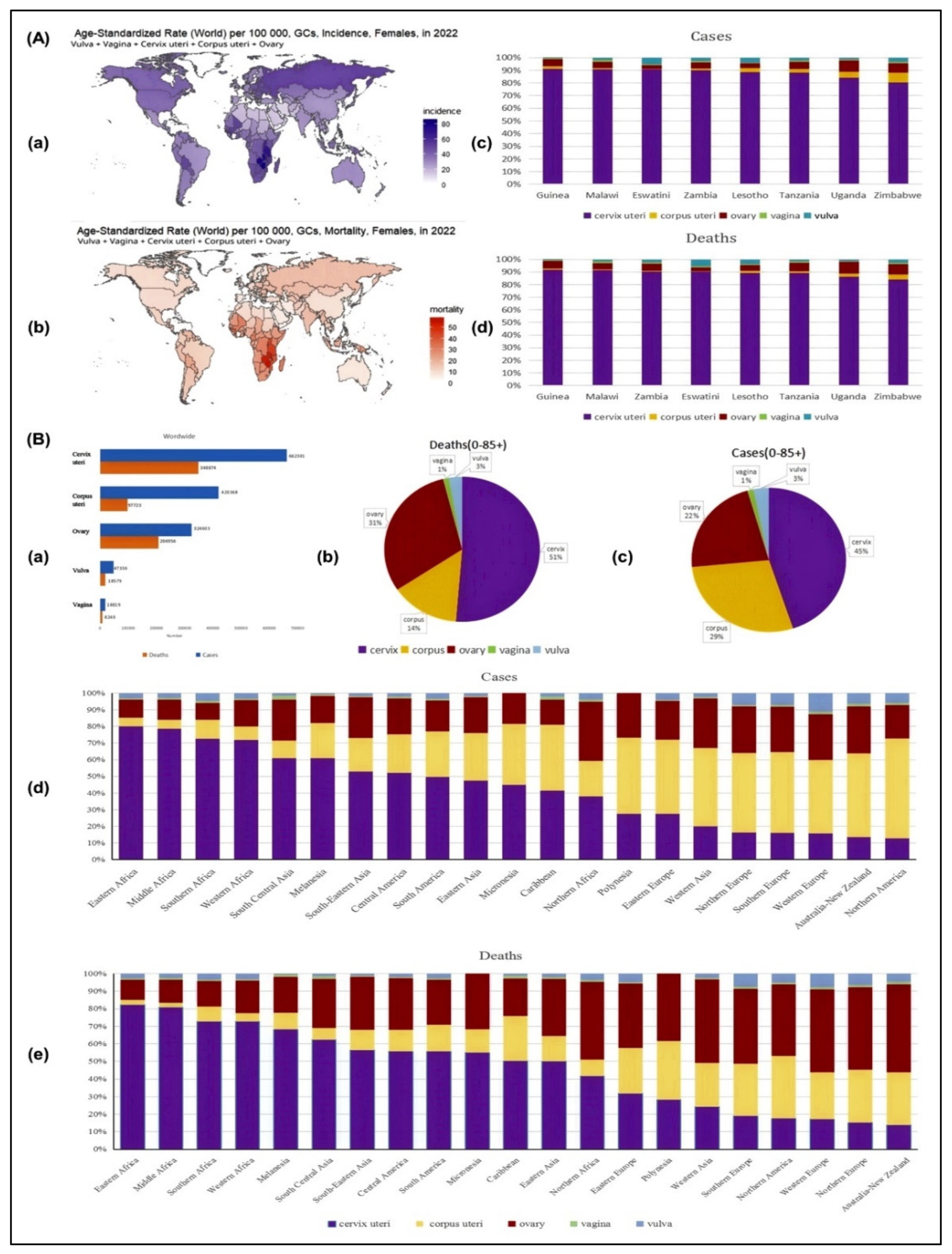
Figure 1. (A) Country-wise age-standardized rates (ASRs) of gynecological cancer (GC) incidence and mortality in 2022. Panels (a,b) illustrate the incidence and mortality rates per 100,000 individuals across various countries, revealing significant geographical disparities. Proportions of different GC types among new cases (c) and deaths (d) in the eight countries with the highest burden highlight the distribution of cervical, corpus uteri, ovarian, vaginal, and vulvar cancers. These data provide insights into regional differences in GC prevalence and lethality, informing targeted public health interventions. (B) (a) Incidence and mortality rates of various GCs in 2022, demonstrating the overall disease burden. (b,c) Age-stratified proportions of different GC types among total cases and deaths, spanning populations from infancy to over 85 years. These charts depict the distribution of GCs across age groups, emphasizing trends in disease occurrence and fatality. (d,e) Proportions of major GC types in total cases and deaths, highlighting the relative prevalence and lethality of cervical, corpus uteri, ovarian, vaginal, and vulvar cancers. These visualizations facilitate a better understanding of the impact of individual cancer types, supporting resource allocation and policy development to mitigate the global burden of GCs. This section is adapted from under Creative Commons Attribution 4.0 (CC BY 4.0) license [11].
Onco 05 00024 g001
Figure 2. The projected incidence and mortality rates of gynecological cancers (GCs) by 2050 indicate a substantial increase in disease burden over the next three decades. This anticipated rise underscores the urgent need for enhanced research, improved screening programs, and innovative therapeutic strategies to reduce the global impact of GCs. Strengthening preventive measures, expanding access to early detection, and advancing treatment modalities will be critical in mitigating the growing burden of these malignancies. This section is adapted from under Creative Commons Attribution 4.0 (CC BY 4.0) license [11].
Onco 05 00024 g002
Figure 3. (A) Schematic lateral view of the pelvis illustrating four endometriosis subtypes: ovarian endometrioma (OMA), superficial peritoneal endometriosis (SUP), deep infiltrating endometriosis (DIE), and adenomyoma (Ad). Reproduced/adapted from [47]. (B) Bilateral ovarian cysts with high signal on axial fat-saturated T1-weighted (a) and low signal on T2-weighted (b) MRIs, indicative of hemorrhagic content and “kissing ovaries” in the rectouterine pouch. (C) Axial T2-weighted image showing hypointense nodules in the pouch of Douglas (a) with high-signal foci on coronal (b) and axial fat-saturated T1-weighted images (c), suggestive of deep pelvic endometriosis with hemorrhage. (D) Diffusion-weighted imaging (b = 800) reveals hyperintensity (a) with corresponding hypo-intensity on ADC map (b), consistent with endometrioma and restricted diffusion. (E) Three-dimensional MR neurography of the lumbosacral plexus showing intact nerve fibers in a healthy subject (a) and discontinuity in fiber pathways in a patient with endometriosis (b). (F) Pre- (a) and post-contrast (b) axial images showing a left pyosalpinx and multi-cystic ovary with wall enhancement, consistent with tubo-ovarian abscess. Reproduced/adapted from [48]. (G) Comparative schematic of ERα and ERβ activity in normal endometrium vs. endometriosis: normal tissue predominantly expresses ERα, while lesions exhibit upregulated ERβ, local estradiol synthesis via increased StAR and CYP19A, and reduced ERα expression. This section is adapted from under Creative Commons Attribution 4.0 (CC BY 4.0) license [29]. (H) The comparative analysis of fiber tractography in patients with posterior compartment endometriosis in two women (a,b) shows the short, branched, and disrupted neural fibers. (I) Molecular pathways in ER signaling: (a) ERβ overexpression in stromal cells suppresses apoptosis and ERα, promotes inflammatory and growth signals (e.g., IL-1, RERG, SGK1, PGE2); (b) local E2 elevation disrupts ERα/ERβ balance, enhancing expression of genes (e.g., Greb-1, c-Myc) involved in disease progression. This section is adapted from under Creative Commons Attribution 4.0 (CC BY 4.0) license [48].
Onco 05 00024 g003
Figure 4. (A) In gynecological cancer, quercetin flavonoids have anti-inflammatory and antioxidant properties. (B) An illustration shows the primary source and properties of quercetin. Na+-K+-2Cl−cotransporter 1 (NKCC1); blood pressure (BP). (C) A representation of how quercetin impacts endometriosis. (Nrf2, nuclear factor (erythroid-derived 2)-like 2; NQO1, NAD(P)H quinone oxidoreductase 1; PR, progesterone receptor; MMP, mitochondrial membrane potential). This section is adapted from under Creative Commons Attribution 4.0 (CC BY 4.0) license [63].
Onco 05 00024 g004
Figure 5. A schematic representation of the signaling pathway in gynecological cancer through which quercetin influences ovarian cancer (OC), cervical cancer (CC), and endometrial carcinoma (EC), regarding key molecular mediators. This illustration was created using Biorender (https://www.biorender.com/; accessed on 13 March 2025). The pathway involves various factors, including hypoxia-inducible factors 1-alpha (HIF-1α), vascular endothelial growth factor (VEGF), extracellular regulated protein kinases (ERK), rapidly accelerated fibrosarcoma (Raf), rat sarcoma (Ras), caspase 3 (Casp3), caspase 8 (Casp8), proliferating cell nuclear antigen (PCNA), activating transcription factor 4 (ATF4), matrix metalloproteinases (MMP), mammalian target of rapamycin (mTOR), MAPK extracellular kinase (MEK), and signal transducer and activator of transcription 3 (STAT3). This section is adapted from under Creative Commons Attribution 4.0 (CC BY 4.0) license [63].
Onco 05 00024 g005
Figure 6. The role of polyphenols, terpenoids, and thiols in preventing gynecological cancer. This section is adapted from under Creative Commons Attribution 4.0 (CC BY 4.0) license [66].
Onco 05 00024 g006
Disclaimer/Publisher’s Note: The statements, opinions and data contained in all publications are solely those of the individual author(s) and contributor(s) and not of MDPI and/or the editor(s). MDPI and/or the editor(s) disclaim responsibility for any injury to people or property resulting from any ideas, methods, instructions or products referred to in the content.