Article

Stabilizing the Shield: C-Terminal Tail Mutation of HMPV F Protein for Enhanced Vaccine Design

1
Department of Biotechnology & Bioengineering, School of Biosciences and Technology, Galgotias University, Gautam Buddha Nagar, Greater Noida 201310, Uttar Pradesh, India
2
Department of Biotechnology, Invertis University, Bareilly 243123, Uttar Pradesh, India
3
Department of Biotechnology, GLA University, 17km Stone, NH−2 Mathura-Delhi Road Mathura, Chaumuhan, Mathura 281406, Uttar Pradesh, India
4
Department of Radiology, University of Michigan, Ann Arbor, MI 48109, USA
5
Department of Life Sciences, School of Biosciences and Technology, Galgotias University, Gautam Buddha Nagar, Greater Noida 201310, Uttar Pradesh, India
6
Division of Cell Biology and Immunology, Biomedical Parasitology and Translational-Immunology Lab, CSIR-Institute of Microbial Technology (IMTECH), Sec−39A, Chandigarh 160036, Chandigarh, India
7
Structural Bioinformatics Laboratory (GRIB-IMIM), Universitat Pompeu Fabra (UPF), 08003 Barcelona, Catalonia, Spain
*
Authors to whom correspondence should be addressed.
BioMedInformatics 2025, 5(3), 47; https://doi.org/10.3390/biomedinformatics5030047
Submission received: 19 July 2025 / Revised: 13 August 2025 / Accepted: 20 August 2025 / Published: 28 August 2025
(This article belongs to the Section Computational Biology and Medicine)

Abstract

Background: Human Metapneumovirus (HMPV) is a respiratory virus in the Pneumoviridae family. HMPV is an enveloped, negative-sense RNA virus encoding three surface proteins: SH, G, and F. The highly immunogenic fusion (F) protein is essential for viral entry and a key target for vaccine development. The F protein exists in two conformations: prefusion and postfusion. The prefusion form is highly immunogenic and considered a potent vaccine antigen. However, this conformation needs to be stabilized to improve its immunogenicity for effective vaccine development. Specific mutations are necessary to maintain the prefusion state and prevent it from changing to the postfusion form. Methods: In silico mutagenesis was performed on the C-terminal domain of the pre-F protein, focusing on five amino acids at positions 469 to 473 (LVDQS), using the established pre-F structure (PDB: 8W3Q) as the reference. The amino acid sequence was sequentially mutated based on hydrophobicity, resulting in mutants M1 (IIFLL), M2 (LLIVL), M3 (WWVLL), and M4 (YMWLL). Increasing hydrophobicity was found to enhance protein stability and structural rigidity. Results: Epitope mapping revealed that all mutants displayed significant B and T cell epitopes similar to the reference protein. The structure and stability of all mutants were analyzed using molecular dynamics simulations, free energy calculations, and secondary structure analysis. Based on the lowest RMSD, clash score, MolProbity value, stable radius of gyration, and low RMSF, the M1 mutant demonstrated superior structural stability. Conclusions: Our findings indicate that the M1 mutant of the pre-F protein could be the most stable and structurally accurate candidate for vaccine development against HMPV.

1. Introduction

Human Metapneumovirus (HMPV) is a respiratory virus belonging to the family Pneumoviridae [1,2,3], and cases of HMPV were reported in 2016 in India. Despite its worldwide prevalence, HMPV is under-reported and less explored than other respiratory viruses like influenza and Respiratory Syncytial Virus (RSV) [4]. Recent reports of increasing cases, particularly among infants and the elderly, indicate a public health threat [4,5]. In the absence of an effective vaccine and antivirals, there is an urgent need to develop an effective vaccine [6]. HMPV is an enveloped negative-sense RNA virus that encodes three surface-expressed membrane proteins, namely Small Hydrophobic (SH), Glycoprotein (G), and Fusion (F) [7,8]. The fusion protein, which mediates viral entry into the host cell, is a potent vaccine target [8]. RSV-F and HMPV-F are class I viral fusion proteins with approximately 30% sequence similarity, and both exist in two conformational states: prefusion (pre-F) and postfusion (post-F) forms [9]. The pre-F conformation is critical for eliciting neutralizing antibodies and could therefore be an effective vaccine candidate for HMPV [8,9,10,11,12].
Post-F protein-based vaccine formulation failed to generate robust neutralizing antibody responses, making them less effective as stand-alone immunogens [13,14]. Various mutations have been introduced to arrest the F protein in the prefusion stage, namely proline substitution, disulfide interaction, hydrophobic to hydrophilic substitution, cavity filling substitution, etc. [10,15,16,17,18]. Given the immunological advantages of the prefusion F protein, most modern HMPV vaccine efforts focus on stabilizing and presenting this form to the immune system [16,19,20,21]. These structural and immunogenic differences between pre- and postfusion are crucial for designing an effective HMPV vaccine. The mature pre-F protein is a cleavage product of single-chain precursor F0, which is cleaved by host cell serine proteases into disulfide-linked F1 and F2 subunits [13,22]. The mature F1 subunit interacts with the host membrane using N N-terminal fusion peptide sequence and forms the functional trimer structure [8,15,23,24]. The pre-F protein interacts with the host cell membrane and undergoes a significant conformational shift. Hydrophobic fusion peptides are ejected from the central cavity of the F trimer and inserted into the host cell membranes [25,26]. This intermediate structure then collapses into a stable six-helix bundle, composed of the N-terminal (HRA) and C-terminal (HRB) heptad repeats, adopting its more stable post-F conformation [27]. The pre-F is a metastable, high-energy form and contains critical neutralizing epitopes, particularly in the antigenic sites Ø and I, which are recognized by highly potent neutralizing antibodies [8,28]. The fusion proteins of both RSV and HMPV are class I viral fusion proteins with ~30% sequence identity and share many structural and functional similarities in their pre- and postfusion states [15]. Earlier RSV vaccine research and clinical studies demonstrate that post-F elicits weak neutralizing antibodies or nonfunctional antibody responses with adverse effects, making it unsuitable for use as a vaccine antigen [29,30,31]. In contrast, pre-F induces superior neutralizing antibody responses accounting for most of the RSV-neutralizing activity in human immune sera. Recently, an antibody targeting the trimer interface of HMPV F was identified, as evidenced by the similar structural transitions seen in the in vivo investigation [32,33]. Prefusion, being a viable vaccine antigen, is inherently unstable and tends to transition into the more stable postfusion form. This transition poses a challenge for in vitro expression since the wild-type protein naturally adopts the postfusion conformation due to its greater thermodynamic stability [34,35]. Specific mutations are indeed required to arrest the protein in its prefusion state and prevent its conformation change to achieve the postfusion form [36,37,38]. Earlier, different mutations were incorporated, such as introducing disulfide bonds and proline substitutions to lock the HMPV F protein [8,39,40,41]. The earlier identified pre-F (PDB: 8W3Q) represents the prefusion-arrested trimeric protein. This structure serves as a valuable template for designing stabilized prefusion proteins, supporting the development of more effective vaccine candidates [13,15]. Vaccines based on the stabilized pre-F protein elicit robust protective immunity in animal models, reducing viral replication and disease severity [16,42]. The success of RSV pre-F-based vaccines, such as those recently approved for human use, has further reinforced the potential of this approach for HMPV [43].
This study aimed to introduce a mutation in the already mutated protein (8W3Q) by inserting the hydrophobic residue on the C-terminal end of the protein. These hydrophobic mutations in the C-terminal end are the first to develop new mutant variants with a more stable structure. These modifications are strategically designed to enhance hydrophobic interactions, thereby reducing excessive motion and stabilizing the overall structure. These mutations help maintain the more stable prefusion state for a longer duration, making the modified 8W3Q variant a more suitable candidate for vaccine development. This stabilization strategy ensures that the protein remains in its optimal immunogenic form, improving its potential effectiveness in eliciting a strong immune response. The modifications specifically targeted amino acid positions 469 to 473, a region selected based on prior structural analysis and experimental insights. Four new mutations have been introduced, optimizing the protein conformational integrity following our approach. These mutations are carefully designed to avoid disrupting critical epitope regions, ensuring that the protein retains its ability to trigger an effective immune response. Instead of altering antigenicity, the introduced mutations primarily function to increase the structural stability of the prefusion form, reducing its tendency to transition into a more stable postfusion state. The new mutant variant of pre-F protein may enhance the efficacy of the vaccine candidate, which is needed for the HMPV virus, as well as for other highly mutation-prone RNA viruses. Overall, the present work aims to retain the most stable and structurally accurate prefusion stage model suitable for downstream applications such as docking, vaccine design, or mechanistic studies.

2. Materials and Methods

2.1. Mutation of the Target Protein

To investigate the structural impact of C-terminal mutations in the prefusion-stabilized F protein of Human Metapneumovirus (HMPV), we utilized the cryo-EM-resolved structure (PDB ID: 8W3Q) as the reference model. This structure represents the biologically relevant prefusion conformation, preserving key neutralizing epitopes critical for vaccine development, as previously reported by Hsieh et al. [8]. Targeted mutations were introduced at residues 469–473 (native sequence: LVDQS) to generate four variants—M1 (IIFLL), M2 (LLIVL), M3 (WWVLL), and M4 (YMWLL) to enhance hydrophobic interactions and stabilize the prefusion fold. Mutation design was guided by residue hydrophobicity and executed using SwissSidechain mutagenesis. Homology models of the variants were generated using the ModWeb server [44], employing 8W3Q as the sole template to preserve conformational integrity. The Protein Structure Analysis (ProSA) web server was used to verify proper folding in the three-dimensional mutated proteins [45]. Following energy minimization and structural refinement with GalaxyRefine [46], each model underwent comprehensive pre-MD validation to assess structural quality [47]. This included the calculation of GDT-HA and RMSD scores to measure backbone fidelity; MolProbity scores, clash scores, and rotamer outliers for stereochemical assessment; and Ramachandran plot analysis via PROCHECK [48,49] to evaluate dihedral angle distributions. Global folding accuracy was assessed using ProSA Z-scores [45] and QMEAN/GMQE scores from SwissModel, while intrinsic flexibility and collective motions were examined through Normal Mode Analysis (NMA) using DynaMut2 [47]. Structural validation [50,51] and Structural alignments with the wild-type reference were performed using TM-align, and immunological relevance was validated by epitope mapping of B-cell and T-cell regions through the IEDB resource [52] to ensure the introduced mutations did not disrupt antigenic sites. Among all variants, M1 displayed the most favourable structural metrics, including the lowest RMSD (0.232 nm), the best MolProbity score (1.59), and minimal steric clashes (clash score: 11.9), designating it as the optimal candidate for downstream molecular dynamics simulations. All mutant structures were further inspected and visualized in PyMOL [53]. Although the HMPV F protein exists in both prefusion and postfusion conformations, this study exclusively utilized the prefusion state (8W3Q), given its immunogenic superiority and relevance in vaccine design. In contrast, the most stable postfusion form (PDB: 5L1X), while structurally useful, is less suitable as a vaccine antigen due to the loss of critical neutralizing epitopes.

2.2. MD (Molecular Dynamics) Simulation

The optimized mutated models, along with the wild-type protein, were further subjected to MD simulation using GROMACS v.2021.4 [54] with CHARMM36 force field [55]. The TIP3P water model was used to solvate the systems, which were in a 1.2 nm cubic box and were neutralized with 150 mM NaCl. The systems were minimized using the steepest-descent method comprising 5000 steps. The Particle Mesh Ewald (PME) method [56] was used to account for electrostatic forces. The LINCS algorithm [57] was used to constrain the hydrogen bonds. Each system was subjected to equilibration through 500 ps constant temperature, constant volume (NVT) followed by 1 ns under constant temperature and constant pressure (NPT) ensembles at 300K, maintaining a constant pressure of 1.0 bar. The V-rescale [58] thermostat was used for temperature coupling, while pressure was controlled using the Berendsen pressure coupling [59]. Final production runs of each system were carried out for 100 ns each. The post-MD analyses, including the calculation of root mean square deviation (RMSD), radius of gyration (Rg), and hydrogen bonds (H-bonds), were carried out using the in-built GROMACS utilities.

3. Results and Discussion

The HMPV F glycoprotein exists in two conformational states—prefusion (PDB: 8W3Q) and postfusion (PDB: 5L1X)—as shown in Figure 1. The F1 and F2 subunits of the F protein are made up of both the prefusion and postfusion conformations. The F1 subunit experiences substantial conformational changes following the cleavage at the activation site, whereas the F2 subunit maintains structural conservation between the two states. These alterations are highlighted by structural superimposition of the prefusion and postfusion forms, which mainly show the rearrangement within the F1 subunit. Despite the structural characterization of both prefusion and postfusion conformations of the HMPV F protein, the postfusion form (e.g., PDB: 5L1X) was not utilized in this investigation for various reasons. The postfusion state is devoid of essential neutralizing epitopes (specifically antigenic sites Ø and I) that are retained solely in the metastable prefusion conformation. These epitopes are essential for generating robust neutralizing antibodies and are a principal reason why prefusion-based immunogens regularly surpass postfusion-based designs in RSV and HMPV vaccine research. Secondly, the postfusion conformation signifies the most thermodynamically stable state of the protein, indicating that it has already experienced the extensive rearrangements linked to membrane fusion. Consequently, all structural characteristics pertinent to initiating or sustaining the prefusion state are lacking, rendering it inappropriate for the development of variations designed to halt the protein before fusion. Third, previous vaccine experiments utilizing postfusion antigens have produced inadequate immune responses and, in several instances, elicited non-protective or disease-enhancing antibodies. Ultimately, from a modelling perspective, employing a postfusion template would obscure the analysis of stabilizing mutations intended to diminish C-terminal flexibility in the prefusion state, as the postfusion backbone configuration is inherently distinct and fails to represent the authentic prefusion epitope landscape. Due to these integrated structural, immunological, and design considerations, solely the prefusion template (PDB: 8W3Q) was chosen, guaranteeing that all computational evaluations and mutational impacts were directly related to the biologically pertinent and immunogenically optimal conformation. In addition, the glycosylation represents a significant post-translational modification found in the HMPV F glycoprotein. Recognized N-linked glycosylation sites within this protein are indeed crucial for proper protein folding [60], stability, and surface expression. Furthermore, glycan moieties may help in immune evasion by concealing essential antigenic sites from neutralizing antibodies. Although the dynamic and heterogeneous nature of glycosylation often prevents complete resolution in existing PDB structures, it remains a vital component of the glycoprotein structural and functional characteristics. Consequently, even in cases where glycosylation cannot be directly modelled or visualized, it is important to recognize its probable impact and the constraints it presents on structural interpretation. The significance of glycosylation in HMPV F glycoproteins is difficult to understand, but it contributes to structural and functional analyses. Though the prefusion form is highly immunogenic and is considered an ideal antigen for vaccine development, it is highly unstable in nature. Therefore, considering the glycosylation site, its conformation must be stabilized to ensure its effective use as an immunogen in vaccine formulations.

3.1. Design of Prefusion-Stabilized HMPV F Protein

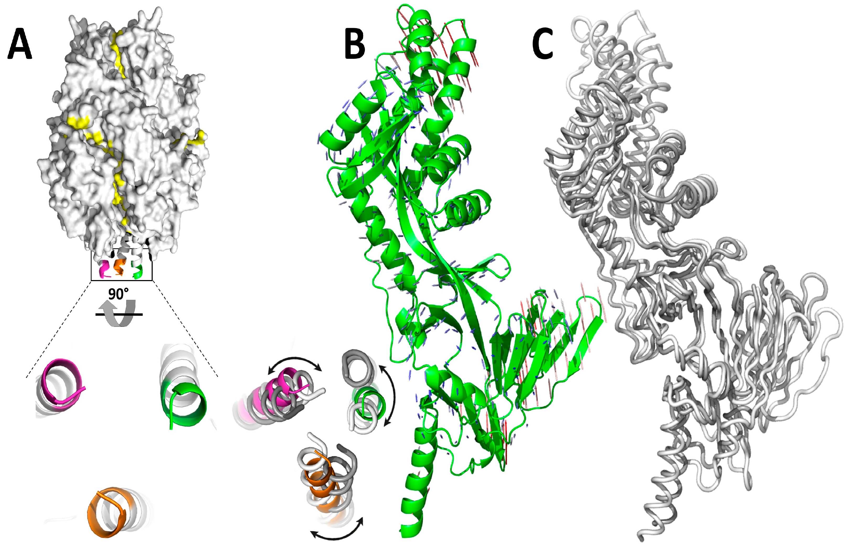
The reference protein (PDB: 8W3Q) is depicted in Figure 2, both under control and simulation conditions. In Figure 2A, the trimeric structure is shown as a surface, whereas the C-terminal end is highlighted in magenta, green, and orange. Based on the NMA data, the superimposition of the first and last frames from the 469th to the 473rd amino acid (LVDQS) is shown at the bottom right (Figure 2A), revealing the movement of these amino acids and indicating that strong interactions among them contribute to the enhanced stability of the trimeric structure. Therefore, the C-terminal domain demonstrated a high degree of flexibility, suggesting that it is inherently unstable and prone to structural shifts. This excessive flexibility in the C-terminal region could contribute to protein instability, potentially affecting its ability to maintain the prefusion conformation required for effective immunogenicity. To address this issue, targeted mutations were introduced within this region under hydrophobic conditions. These modifications were strategically designed to enhance hydrophobic interactions, thereby reducing excessive motion and stabilizing the overall structure. The monomeric structure of 8W3Q was subjected to NMA, illustrated under porcupine cartoon presentation (Figure 2B). The initial frame (grey) and the final frame (white) of the MD simulation are presented in Figure 2C. To better justify our approach, the first and last frames of the MD simulation were primarily selected to visualize the net conformational change and local stabilization at the mutation site (residues 469–473). This approach illustrates the cumulative structural impact of the simulation; however, it does not capture the complete conformational sampling. Therefore, we additionally performed clustering analysis of the full MD trajectory using the GROMACS gmx cluster tool (RMSD-based, GROMOS method). The wild-type structure exhibited higher structural variability, forming multiple low-population clusters, whereas the M1 mutant maintained over 85% of frames within a single dominant cluster, indicating reduced conformational heterogeneity. No large-scale transitions from the prefusion to the postfusion state were observed in any variant, consistent with the stability of the input template (PDB ID: 8W3Q). All conformational differences observed thus represent fluctuations within the prefusion ensemble rather than state transitions. The amino acid segment spanning residues 469 to 473 (LVDQS), highlighted within the black box in Figure 2A, is shown in an enlarged 90° rotated view in the lower panel of Figure 2A (bottom left). The same region, when superimposed between the initial and final frames of the MD simulation (right panel), reveals notable positional shifts, indicating a high degree of local flexibility in this segment. To evaluate the quality and reliability of the designed mutant structures before MD simulation, a multi-tiered validation pipeline was implemented using several computational tools. Each variant was assessed based on structural deviation (RMSD, GDT-HA), stereochemical quality (MolProbity and clash scores), and backbone geometry (Ramachandran analysis via PROCHECK). Global model quality was verified through ProSA Z-scores and QMEAN/GMQE, while intrinsic flexibility was evaluated using Normal Mode Analysis (NMA) in DynaMut2. The reference protein 8W3Q (WT) exhibited a QMEAN score of 0.78 ± 0.05 and a GMQE of 0.82, indicating excellent structural quality and high expected reliability. Mutants M1 and M2 showed slightly reduced QMEAN scores (0.77 ± 0.05) with GMQE values of 0.81, suggesting minimal deviation from the reference in predicted accuracy. M3 and M4 retained identical QMEAN scores to the reference (0.78 ± 0.05) but shared the slightly lower GMQE (0.81) observed in all mutants. Overall, all variants remained within the excellent range for both metrics, confirming that the introduced mutations did not compromise model reliability. These results support the structural robustness of all designed mutants relative to the reference model. TM-align was used to ensure structural conservation with the wild-type template, and B-/T-cell epitope mapping (via IEDB) confirmed that antigenic regions were preserved. These results, summarized in Supplementary Table S1, demonstrated that variant M1 displayed the most favourable metrics across all categories and was thus selected for downstream molecular dynamics simulations. The reference structure (PDB: 8W3Q) used for all modelling was confirmed to represent the biologically relevant prefusion conformation, which is essential for eliciting effective neutralizing antibody responses and is thus preferred for rational vaccine design.
Therefore, the high degree of flexibility suggests that it is inherently unstable and prone to structural shifts. This excessive flexibility in the C-terminal region could contribute to protein instability, potentially affecting its ability to maintain the prefusion conformation, conferring higher immunogenicity. Therefore, to address this issue, targeted mutations were introduced within this region under hydrophobic conditions. These modifications were strategically designed to enhance hydrophobic interactions, thereby reducing excessive motion and stabilizing the overall structure. By the structural integrity of the protein, these mutations help to maintain the prefusion state for a longer duration, making the modified 8W3Q variant a more suitable candidate for vaccine development [10].

3.2. Immunogenic Sites of the Prefusion F-Protein

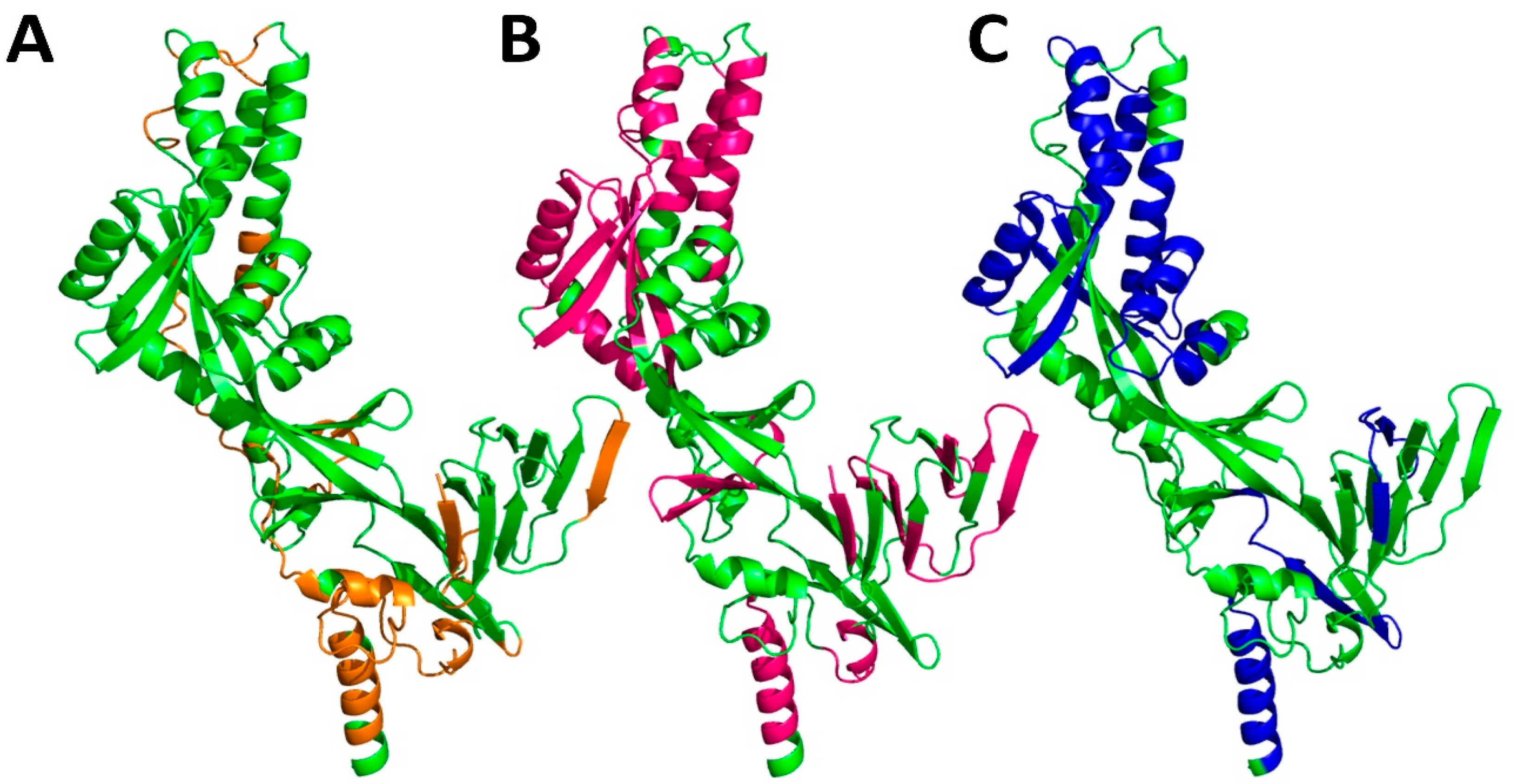
This stabilization strategy ensures that the protein remains in its optimal immunogenic form, improving its potential effectiveness to elicit the long-lasting and potent immune response [61]. The monomeric structure of the 8W3Q encompasses well-defined immunogenic regions instrumental for eliciting sizeable immune response (Figure 3). T- and B-cell epitopes of the target protein were predicted de novo in this study for all the mutants. T-cell epitopes were identified by NetCTL v1.2 and NetMHCpan 4.0server based on the different scoring parameters [62]. The affinity of the epitopes with the complete reference set of human alleles of MHC class I and MHC class II was analyzed by using the IEDB MHC-I, and most potential epitopes were screened, showing a percentile rank threshold of 0.8 and IC50 < 50. B-cell epitopes were predicted using the BepiPred 2.0 algorithm, which evaluates surface accessibility, hydrophilicity, and sequence conservation to identify potential antigenic regions. The predicted epitope sequences were then mapped onto the 3D protein structure using PyMOL to confirm their surface exposure and to ensure that the mutated region did not overlap with any predicted immunogenic sites. This combined sequence- and structure-based approach ensured accurate epitope identification, and all relevant tool references are provided herein. These regions include the B cell epitope (highlighted in orange), which is responsible for direct antibody recognition and binding, thereby facilitating the humoral immunity (Figure 3A). Additionally, the structure features the T cell MHC-I epitope (marked in magenta), which is crucial for antigen presentation to cytotoxic T lymphocytes (CTLs), thereby activating a cell-mediated immune response essential for targeting infected or abnormal cells (Figure 3B). Furthermore, the T cell MHC-II epitope (depicted in blue) plays a significant role in stimulating helper T cells, which, in turn, enhance both humoral and cellular immunity by supporting B cell activation and cytokine production (Figure 3C). The precise mapping of these epitopes within the monomeric 8W3Q structure provides valuable insights into its immunogenic potential. Understanding these epitope distributions is essential for vaccine design, as it aids in optimizing antigen presentation and enhancing the immune system’s ability to recognize and neutralize the target pathogen effectively. The analysis of the selected epitope regions, encompassing both B cell and T cell epitopes (MHC-I and MHC-II), indicates that the proposed mutation is not located within these regions, showing the conserved epitopes in wild type and mutants as well. Consequently, these mutations do not compromise the efficacy of the proposed vaccine candidate. The proposed design emphasizes a well-balanced incorporation of hydrophobic residues at the C-terminal end, considering protein stability and structural integrity. The selection of hydrophobic amino acids is intended to mitigate degradation and aggregation, while ensuring a balance with flexible and stable residues to facilitate proper folding.

3.3. Substitution of Hydrophobic Mutations in the C-Terminal Trimeric Structure of HMPV F Protein

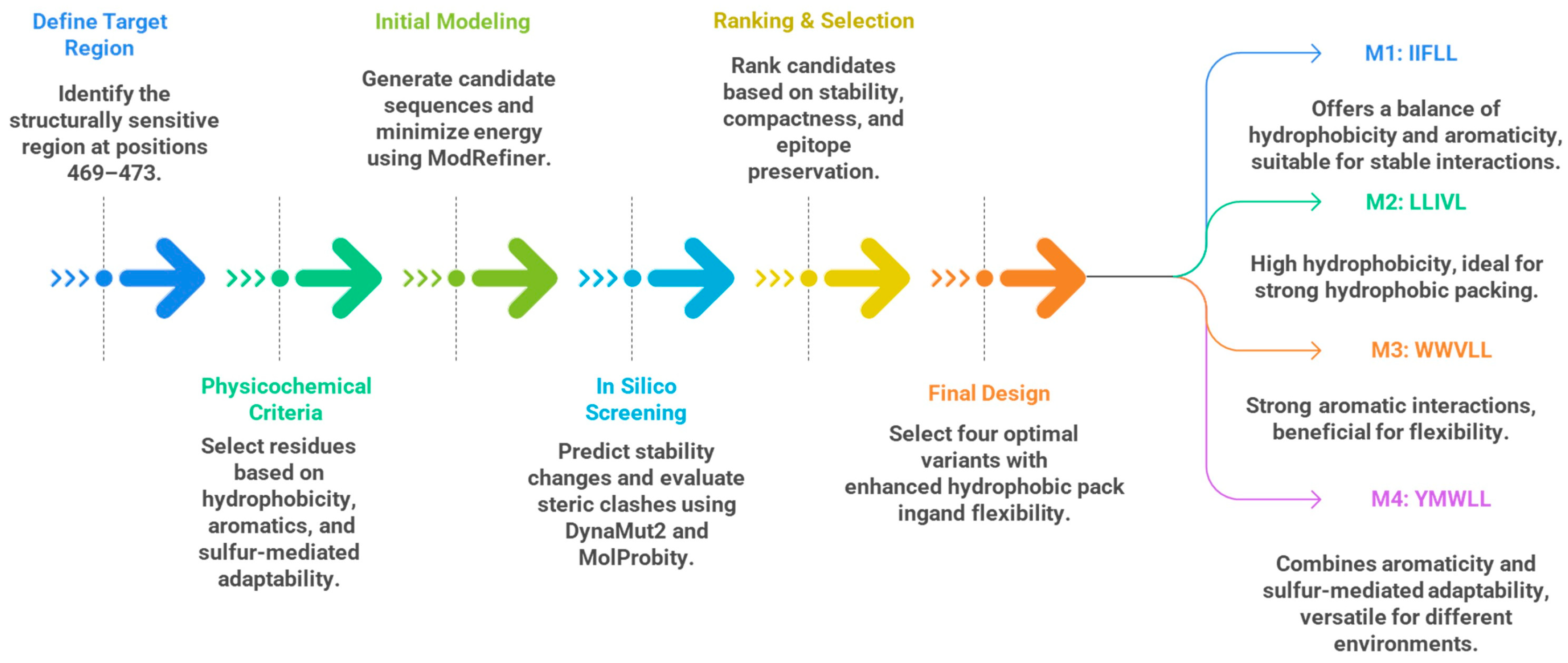
The C-terminal trimer interface is a structurally sensitive area where minor changes in side-chain arrangement and local flexibility can significantly affect overall protein stability and functional integrity. Hydrophobic replacements were implemented at locations 469–473 to enhance this interface, based on physicochemical and geographical analyses. Residues exhibiting elevated hydrophobicity indices, such as leucine and isoleucine, were chosen to augment the van der Waals interactions and fortify interprotomer packing, while aromatic residues (phenylalanine, tryptophan, tyrosine) were included to leverage π–π stacking and π–hydrophobic interactions that enhance conformational stability. Methionine was deliberately incorporated in one variation to introduce a sulphur-containing side chain that facilitates methionine–aromatic interactions, thereby providing restricted conformational flexibility while maintaining hydrophobic properties. Sterically bulky or β-branched residues were excluded from congested places to avert conflicts and maintain backbone geometry, as verified by structural validation methods. Candidate combinations were subjected to in silico screening that included the predictions of stability-free energy (ΔΔG), the assessment of steric clashes, and epitope mapping to achieve an ideal equilibrium among stability, structural compactness, and functional compatibility. This systematic, computationally driven selection workflow, as summarized in Figure 4, facilitated the rational design of mutations that improve interfacial stability while preserving crucial flexibility and immunological significance.
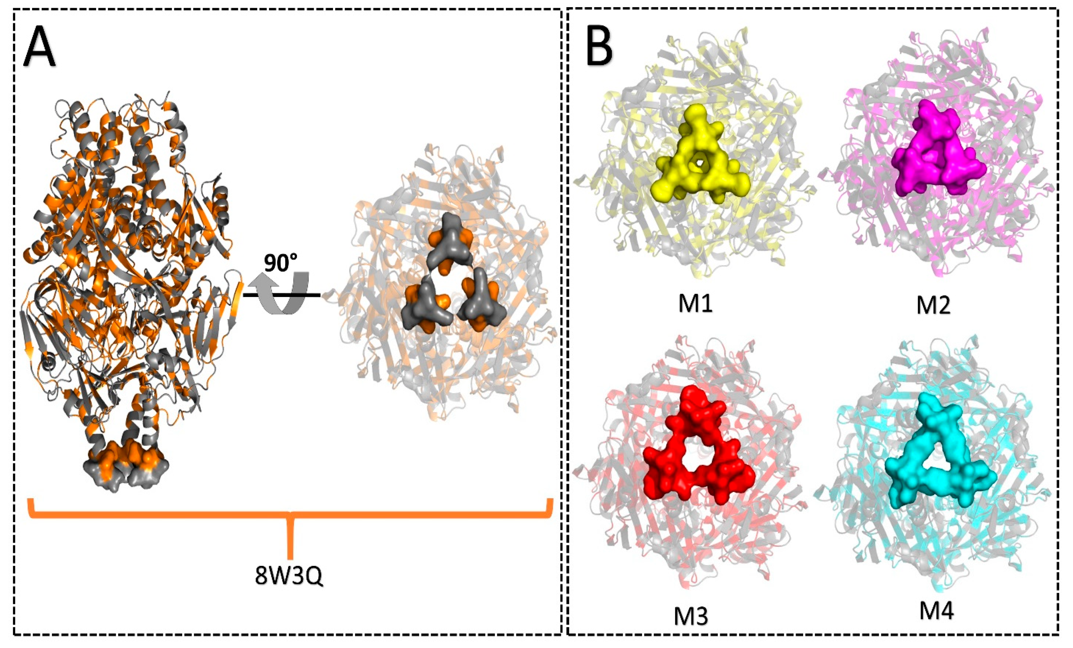
The 468th amino acid in reference protein 8W3Q is alanine, a small and hydrophobic residue that contributes to the protein’s flexibility. Consequently, the subsequent five amino acids at positions 469 to 473 (LVDQS) were selected as the mutation site, as shown in Figure 5. Figure 5A illustrates the three-dimensional structure of 8W3Q, which is considered as reference protein. The orange colour depicts the hydrophobic region, whereas the grey regions are hydrophilic. The right-angle orientation of the reference protein illustrates the lower bottom region, as shown on the right side in Figure 5A. The grey region, which is hydrophilic, was further modified, and corresponding mutants are shown in Figure 5B. This selected region (469 to 473) was subsequently subjected to mutation through homology modelling, resulting in the creation of mutants: M1 (IIFLL), M2 (LLIVL), M3 (WWVLL), and M4 (YMWLL). The M1 mutant, the sequence from positions 469 to 473 (LVDQS) of reference 8W3Q, is replaced with IIFLL. These five residues are characterized by their high hydrophobicity, which enhances protein stability. The centrally located phenylalanine residue plays a crucial role in maintaining flexibility and proper folding due to its larger electron cloud [63,64], while the leucine residues at positions 472 and 473 of adjacent monomers form strong hydrophobic core interactions, thereby altogether enhancing the protein stability. In another mutant, M2, the corresponding substitution is LLIVL, which maintains the sequence structural stability and resistance to aggregation. The Leucine amino acids play an important role in structural stability and reinforce hydrophobic interactions that contribute to core stability and structural rigidity [65]. The amino acid isoleucine plays a crucial role in facilitating protein folding, while valine significantly contributes to the stability of hydrophobic amino acid regions, thereby supporting compact folding [66]. Leucine and valine are specific amino acids commonly found in stable α-helices and β-sheets [67]. For M3, the corresponding substitution is WWVLL. Tryptophan, known for its high hydrophobicity, stabilizes peptide interactions and enhances hydrophobicity through π-stacking effects by aromatic stacking effects [68]. The presence of strong hydrophobic residues at the C-terminal inhibits the degradation and aggregation of the trimeric assembly, ensuring proper protein folding. Therefore, the strategic placement of residues prevents uncontrolled aggregation and is considered an aggregation-resistant candidate [69]. These hydrophobic interactions facilitate strong monomer–monomer attachment and prevent steric clashes, thereby maintaining structural integrity and promoting trimeric assembly through strategic hydrophobic packing [70]. In the final mutation M4, the corresponding amino acid is replaced by YMWLL. The YMWLL mutation was selected due to the stabilization of tyrosine and tryptophan, both aromatic, through π-stacking, which enhances intermonomer interactions. Methionine was chosen for its relatively lower hydrophobicity compared to aromatic residues, yet it facilitates disulfide-like interactions [71]. Collectively, tryptophan, methionine, and leucine form strong hydrophobic interactions, creating a hydrophobic pocket at the trimeric interface. These selected residues align effectively in a trimeric assembly without excessive bulk, thereby minimizing steric hindrance [72]. Consequently, each mutation contributes to a stable trimeric assembly by enhancing hydrophobic interactions at the C-terminal.
The prefusion conformation of the F protein is critical for its efficacy as a vaccine candidate. To enhance the stability of this prefusion state, various mutations have been introduced, and among these, the structure represented by PDB ID: 8W3Q has been utilized as the reference protein in this study. The 8W3Q variant contains specific mutations that stabilize the F protein in its trimeric prefusion conformation. Additionally, to further improve protein stability, the C-terminal end region was modified through the substitution of hydrophobic amino acids. The 468th amino acid in reference protein 8W3Q is alanine, a small and hydrophobic residue that contributes to the protein’s flexibility. Consequently, the subsequent five amino acids at positions 469 to 473 (LVDQS) were selected as the mutation site. This selected region was subsequently subjected to mutation through homology modelling, resulting in the development of mutants M1 (IIFLL), M2 (LLIVL), M3 (WWVLL), and M4 (YMWLL) (Figure 5).

3.4. Characterization of Protein Structure of Mutants

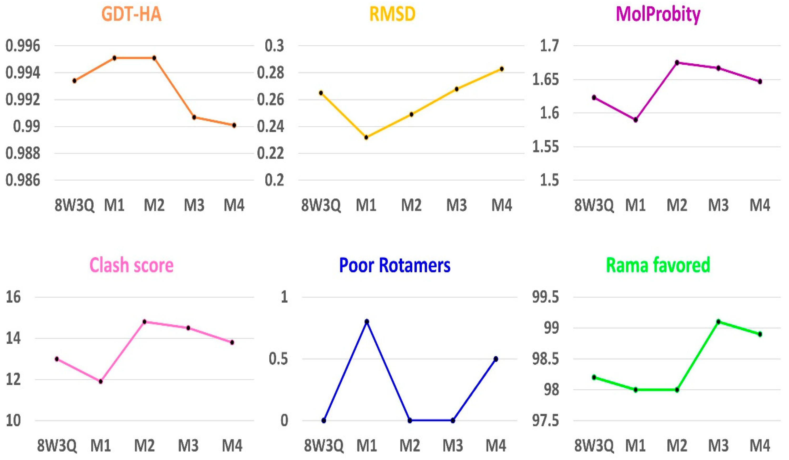
The developed mutants underwent energy minimization and were evaluated using the Galaxy Refine web server, as detailed in Table 1 and Figure 6. The assessment focused on standard quality parameters, including GDT-HA, RMSD, MolProbity score, clash score, rotamer outliers, and Ramachandran plot statistics. The Global Distance Test (GDT)–High Accuracy (HA) analysis measures how closely the mutated model matches the reference structure. Based on the principle that higher is better, the GDT-HA predicts that M1 and M2 (0.9951) are very close to 8W3Q (0.9934), which indicates excellent backbone alignment. The RMSD data (root mean square deviation) indicates how closely the atomic positions of a model match the reference structure; a lower value means the model is closer in atomic position to the reference 8W3Q, representing a better fit. M1 showed the lowest RMSD value (0.232), indicating it fits more comfortably and is the most stable structure among the mutated models. MolProbity evaluates all-atom contacts and geometry, and like RMSD, lower scores reflect better quality. M1 (1.59) had the lowest MolProbity score, identifying it as the top model in this metric as well. The clash score represents the quality of a model. Thus, M1 (1.59) is considered the model with the best fit amongst them, as shown in Table 1. The clash score illustrates the number of steric clashes per 1000 atoms; thus, the lower the clash score, the less steric hindrance there is. M1 had the lowest clash score (11.9), ranking again as the model with the best fit. Poor rotamers inform that rotamers represent the percentage of side chains in unfavourable conformation; as a result, models with a lower value are considered better. In such a side chain geometry, M2 and M3 (each had 0%) are considered as poor rotamers, indicating ideal side-chain conformations. The Ramachandran plot “favoured region” score indicates the percentage of residues in optimal backbone conformations; higher values are better. M3 achieved the highest score (99.1%), reflecting exceptional geometry. Overall, based on these structural validation metrics, M1 and M3 emerged as the top candidates when compared to the reference structure. The Rama represents the percentage of residues in the favoured region of the Ramachandran plot, which indicates that a higher value indicates a better model. Thus, M3 (99.1%) possesses the highest structural geometric consistency and fits perfectly among other mutated models. When comparing every mutated model against the reference structure 8W3Q, models M1 and M3 exhibited top scores based on structural validation metrics as revealed by Galaxyweb server. The 8W3Q. M1 consistently outperformed other models, showing the highest GDT-HA score, lowest RMSD, best MolProbity score, and the lowest clash score. While M1 presents a minor drawback with a slightly higher percentage of poor rotamers (0.8%), it is still within the acceptable limits for high-resolution modelling. The Ramachandran favoured region score (98%) of M1 was also higher, only slightly lower than that of the reference 8W3Q (98.2%). Although these minor imperfections are offset by M1 and M3, which are ranked second in overall quality, they stand out as strong candidates among the models. Next to M1, M3 is another strong contender, excelling with a maximum Ramachandran favoured score of 99.1%. Additionally, M3 has zero poor rotamers, highlighting its excellent geometrical precision and clean structure, which indicates optimal side-chain packing. Nevertheless, M3 remains. However, it falls slightly behind M1 because of its slightly lower GDT-HA (0.9907), as well as marginally higher RMSD and MolProbity scores, positioning itself as the second most compatible candidate (Figure 6, Table 1). The superior performance of M1 observed in molecular dynamics simulations, reflected by its lower RMSD, reduced C-terminal flexibility, and more compact radius of gyration, was consistent with its favourable pre-MD validation metrics. These included minimal steric clashes, an optimal MolProbity score, and the preservation of secondary structure and epitope integrity. This strong agreement and correlation between pre-MD structural assessments and post-MD behaviour validates and supports the robustness of the modelling workflow and supports the selection of highlight M1 as a structurally stable and immunogenically intact prefusion-stabilized variant.
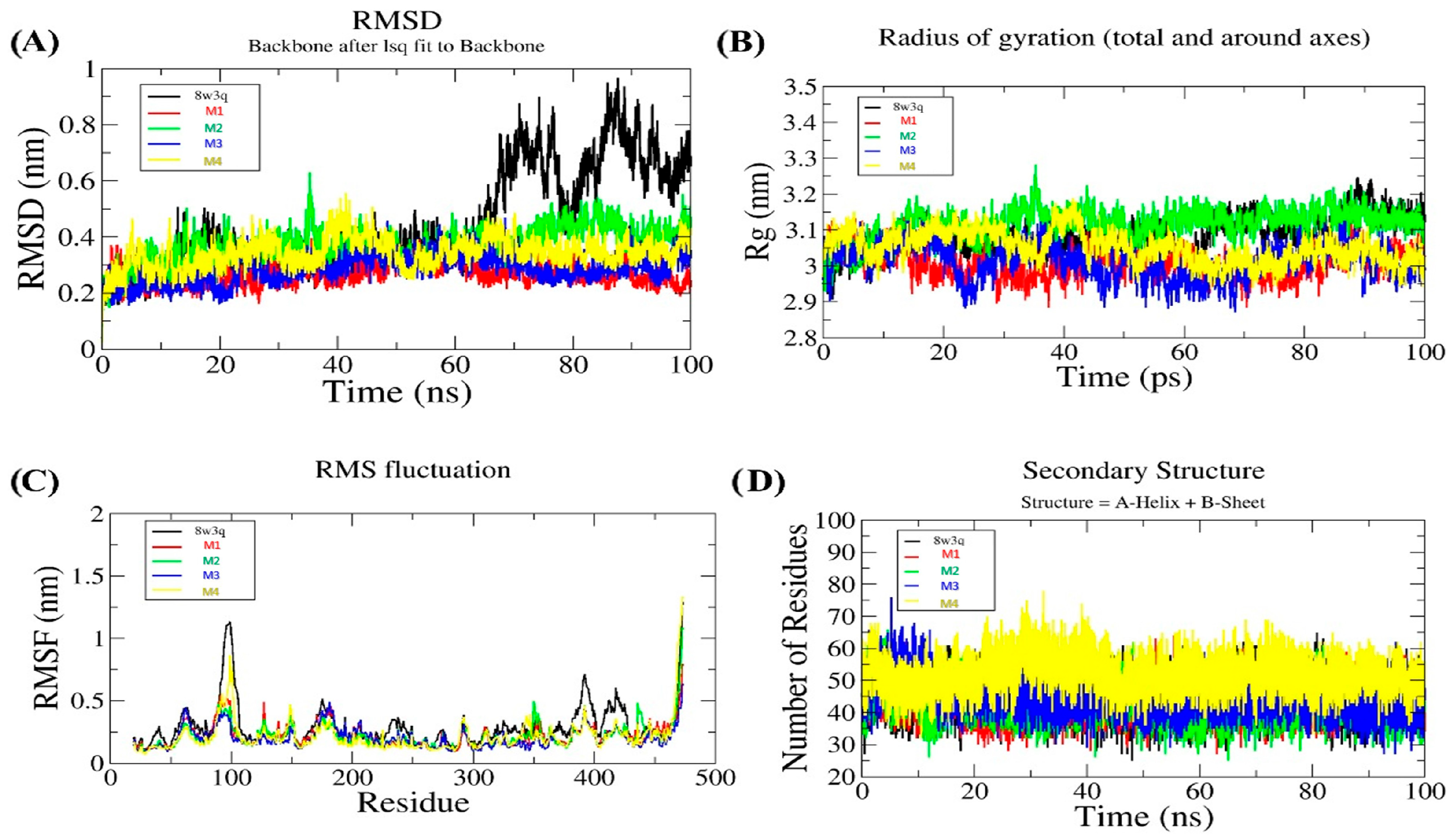
After the MD simulation analysis, these models were further analyzed to observe the structural stability upon inserting mutations (Figure 7). The RMSD profile (Figure 7A) suggested that the mutated versions of the protein maintain a, more stable RMSD pattern compared to the wild type. The red line (M1) shows the lowest and most stable RMSD, whereas the black line, which denotes 8W3Q, shows the highest fluctuations. Further, the radius of gyration (Rg) plot (Figure 7B) suggested that M1 and M3 show very compact and stable structures (~3.01 nm), whereas 8W3Q shows a slightly higher and comparatively less consistent Rg (~3.08 nm). Altogether, the mutated versions M1 (IIFLL), M3 (WWVLL), and M4 (YMWLL) are more compact than M2 (LLVLA) and reference 8W3Q. Moreover, we have also calculated the average RMSF of the mutants. The RMSF plot (Figure 7C) indicates that M3 has the lowest average RMSF, while 8W3Q has the highest average RMSF. Other mutants M1, M2, and M4 have the same average RMSF (Table 2), but M1 and M3 show the lowest residue level fluctuations, especially at the C-terminal region. M4 shows some structural improvements, although it fails to deliver the intended stability at the mutation site (C-terminal). The increase in local flexibility and potential disorder outweighs the global gains, making M4 a less favourable candidate compared to M1–M3. Although the WT displays larger peaks that indicate more flexibility or instability at the C-terminal region, the analysis of the secondary structure profile (Figure 7D) shows that WT and M2 maintained almost identical overall levels of secondary structure elements throughout the simulation. M1 and M3 exhibited slightly reduced secondary structure content compared to the reference, whereas M4 showed the highest retention of secondary structure. In Figure 7D, the y-axis denotes the number of residues adopting α-helical or β-sheet conformations at each time point, providing a direct measure of structural element retention during the simulation.
To gain a deeper insight into the local dynamics of the C-terminal mutation site (residues 469–473), we have calculated RMSD, RMSF, and solvent-accessible surface area (SASA) for both WT and the four mutant variants (M1–M4) (Figure 8A). The M1 mutant has the lowest average RMSD for the 469–473 region, which indicates lower positional deviation and greater local conformational stability compared to WT and other mutants. M1 mutant is also characterized by low RMSF, indicating restricted flexibility and, therefore, decreased dynamic fluctuations at the mutated region, which corroborates the hypothesis of interface stabilization. Although all the mutants present higher SASA than the WT, M1 has a level of exposure balanced at a moderate level, which corroborates the hypothesis that its augmented hydrophobic interactions effectively participate in interface packing without residue overexposure to the solvent. M3 and M4 have high SASA, which may reflect less buried side chains or poor packing. To better estimate the energetic implications of these mutations, we compared the average potential energy from full-length production MD simulations for each system (Figure 8B). The results indicate that M1 and M3 had a lower average potential energy than WT, confirming the hypothesis of a more energetically favourable conformation.
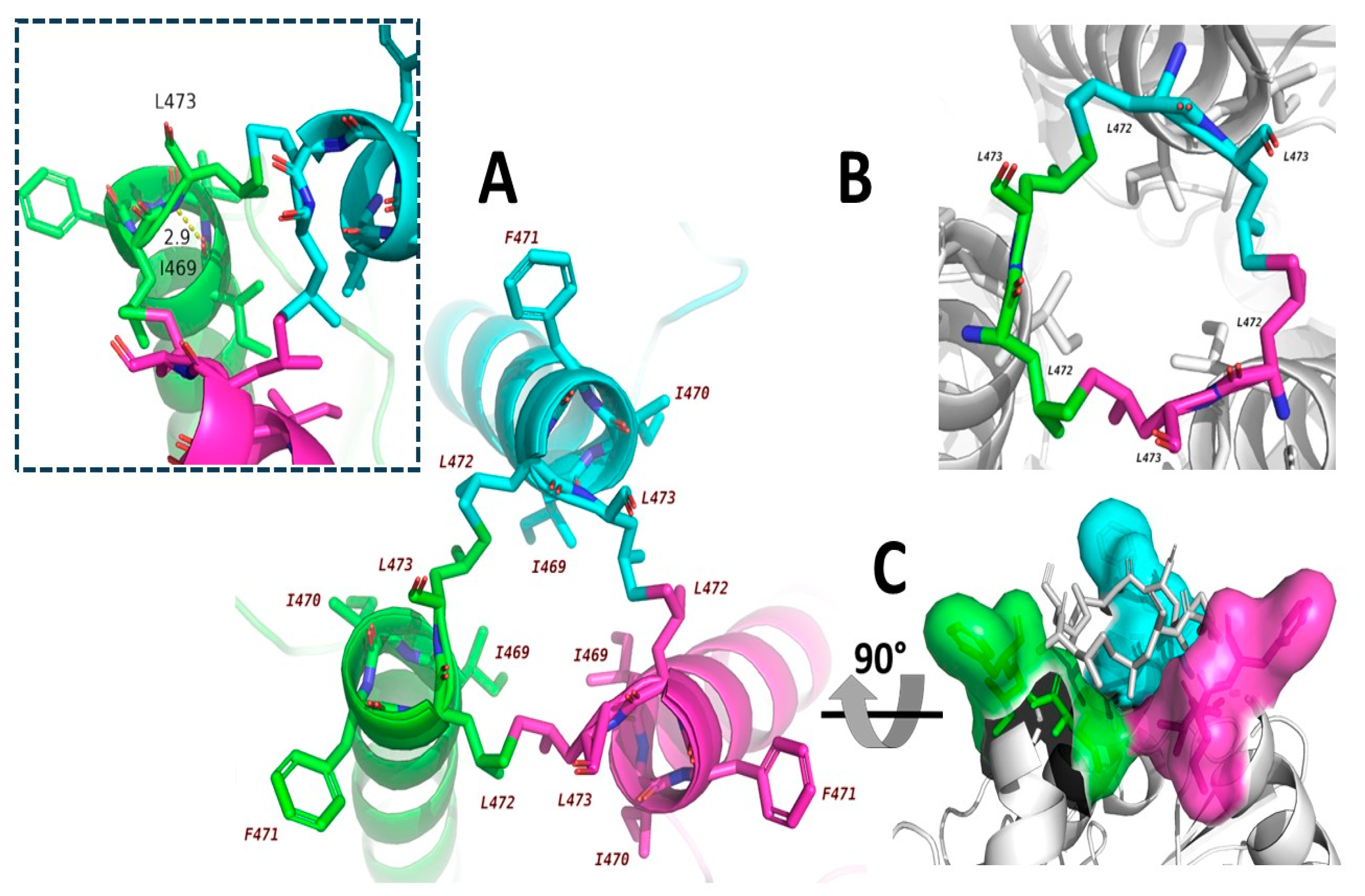
Thus, based on the structural quality score by GalexyWEB server and molecular dynamics simulation score by Gromacs, the best mutation was M1, showing the lowest RMSD, lowest clash score, lowest MolProbity value, stable Rg, and a low RMSF value. Therefore, M1 provides the best overall accuracy and stability. Next to M1, M3 has the best Ramachandran favoured score, zero poor rotamer, low RMSD, low RMSF, and a low Rg value. Thus, M3 is considered the best per-residue stable mutant. M4 is ranked third, with zero poor rotamers and a decent MolProbity value, high secondary structure retention, but higher RMSD. M4 is considered a strong α/β content mutant. M2 has an identical GDT-HA value to M1 but retains a slight rotamer issue, higher RMSD, and a fluctuating radius of gyration. M2 is considered the most unstable amongst them, but it still has a good backbone fit compared to the reference 8W3Q. The final mutant, M1, was selected as the final model and considered as one of the best vaccine candidates. M1 is illustrated in Figure 9, with all possible orientations. Figure 9A explains the overall stoichiometry of the C-terminal residue from I469 to L473. The figure illustrates a trimeric helical bundle composed of three α-helices, each shown in a distinct colour—cyan, green, and magenta—which contribute to the formation of a hydrophobic core, a hallmark of coiled coil motifs. The hydrophobic residues I469, I470, L472, L473, and the aromatic F471 occupy the “a” and “d” positions of the heptad repeat [73] positions that align along the inner face of the coiled coil. The side chains of these residues projected inward positions that buried themselves from the solvent and established a tightly packed water-excluded core region [74]. The hydrophobic clustering introduces a thermodynamic driving force that minimizes the system’s free energy and consequently promotes structural integrity [75]. In addition, the hydrophobic core and the van der Waals interactions further stabilize the interface. The close packing of the side chains of the mutated residues allows for dispersion forces between adjacent helices that eventually enhance the affinity among these subunits [76]. Figure 9B illustrates the trimeric complex (the interaction between L472 and L473 of the subsequent monomer) which exhibits three-fold rotational symmetry. The symmetry promotes cooperative stabilization; therefore, the conformational changes in one of the helices are counterbalanced by similar interactions in the other two helices. It reduces the entropy cost of oligomerization, subsequently supports robust folding, and achieves high structural fidelity [77]. Furthermore, Figure 9C particularly demonstrates the shape complementarity between the helices that cause precise interdigitation, though side chains from one monomer fit into cavities formed by neighbouring helices. The bulky aromatic ring of F471 participates in π-stacking or CH-π interactions, which also adds a layer of non-covalent stabilization [78]. In addition, with F471, the L473 may contribute to C-terminal capping of the helices, which reduces terminal fraying and helix unwinding, further stabilizing the coiled coil structure. Furthermore, these residues also participate in terminal hydrophobic interaction, adding extra rigidity to the end of the C-terminal. Thus, altogether, the trimeric helical assembly is stabilized through hydrophobic core formation, van der Waals interactions, symmetry-driven cooperativity, and terminal capping. All suggested characteristics are essential for maintaining the structure and function of prefusion protein. Hence, the suggested mutation would be considered a milestone in vaccine design strategies for future researchers.

4. Conclusions

We have performed a detailed structural and dynamic characterization of four mutant protein models (M1, M2, M3, and M4) in this research and compared them to the reference structure PDB ID: 8W3Q. The overall aims were to achieve a more stable prefusion HMPV F protein to be added to the antigen armoury. The structural parameters of key interest were employed for assessing model quality using the GalaxyRefine server and molecular dynamics (MD) simulations. Among all mutants, M1 consistently showed better structural stability and integrity. With the best backbone fit, atomic accuracy, and fewer steric clashes, it possessed the highest GDT-HA score (0.9951), lowest RMSD (0.232), best MolProbity score (1.59), and lowest clash score (11.9). Despite only 0.8% of the rotamer outliers being detected, M1 was still within decent limits for models at high resolutions. With 98% of residues in preferred regions, about as many as the reference, the Ramachandran plot further confirms its quality. These results, confirmed by MD simulations, showed that M1 had the most stable and lowest RMSD profile, the most compact structure (Rg 3.01 nm), and the lowest residue fluctuations, particularly at the C-terminal. Most critical to the preservation of the coiled coil motifs of vaccine significance, its helical trimeric structure adopted a compact hydrophobic core stabilized by van der Waals contacts, shape complementarity, and terminal capping. With the best Ramachandran favoured score (99.1%) and without rotamer outliers, M3 was the second most promising mutant, reflecting outstanding side-chain geometry. While M3 had relatively lower GDT-HA and relatively higher RMSD and MolProbity scores compared to M1, it still reflected excellent structural and dynamic behaviour, especially low RMSF and compact Rg, thereby showing stability on a per-residue basis. M4, with nearly flawless secondary structure conservation and no rotamer outliers, was constrained by high RMSD and poor stability at the mutation site, ranking third in the group of candidates. M2, although equal in the GDT-HA score of M1, had greater RMSD, changed Rg, and rotamer issues, and ranked as the least stable mutant. Overall, the results are strongly in favour of M1 as the most structurally and dynamically stable mutant and, thus, the best vaccine candidate for second-generation vaccine development. Structural aspects of M1 trimeric symmetry, hydrophobic core formation, and interhelical complementarity exhibit the major features of effective antigen design. These findings are a good model to follow in the rational design of stable prefusion protein structures for next-generation vaccine development. The overall assessment framework can be employed to guide future research to choose and design protein candidates with enhanced structural stability and immunogenicity. The methods and findings provide a valuable tool for scientists who aim to design next-generation vaccines, particularly against structurally difficult viral proteins. This research, therefore, provides the basis for rational, stability-driven vaccine design in future research.

Supplementary Materials

The following supporting information can be downloaded at https://www.mdpi.com/article/10.3390/biomedinformatics5030047/s1. Supplementary Table S1: Comparative structural validation parameters for the reference model (8W9Q) and four designed mutants (M1–M4). Parameters include GDT-HA (Global Distance Test–High Accuracy), RMSD (root mean square deviation), MolProbity score (structure quality assessment), clash score, percentage of poor rotamers, Ramachandran favoured residues, RMSD (base-MD) after molecular dynamics simulation, radius of gyration (Rg), average RMSF (root mean square fluctuation), C-terminal RMSF, α-helix and β-sheet content, disruption of known epitopes, and TM-aligned RMSD to the wild-type. The best overall model is indicated based on a combination of structural stability, quality scores, and minimal epitope disruption.

Author Contributions

Conceptualization, R.K.; methodology, R.K., and B.O.; software, R.K., S.B., and R.G.; validation, R.K., S.B., and J.G.; formal analysis, R.K., S.M., and S.T.; investigation, R.K., S.T., and R.K.T.; resources, R.K., S.B., and J.G.; data curation, R.K.; writing—original draft preparation, R.K.; writing—review and editing, R.K., R.K.T., and S.M.; visualization, R.K.; supervision, R.K. All authors have read and agreed to the published version of the manuscript.

Funding

This research received no external funding.

Institutional Review Board Statement

Not applicable.

Informed Consent Statement

Not applicable.

Data Availability Statement

The data presented in this study are available on request from the corresponding authors.

Conflicts of Interest

The authors declare no conflicts of interest.

References

  1. Akingbola, A.; Adegbesan, A.; TundeAlao, S.; Adewole, O.; Ayikoru, C.; Benson, A.E.; Shekoni, M.; Chuku, J. Human Metapneumovirus: An Emerging Respiratory Pathogen and the Urgent Need for Improved Diagnostics, Surveillance, and Vaccine Development. Infect. Dis. 2025, 57, 304–310. [Google Scholar] [CrossRef]
  2. van den Hoogen, B.G.; de Jong, J.C.; Groen, J.; Kuiken, T.; de Groot, R.; Fouchier, R.A.; Osterhaus, A.D. A Newly Discovered Human Pneumovirus Isolated from Young Children with Respiratory Tract Disease. Nat. Med. 2001, 7, 719–724. [Google Scholar] [CrossRef]
  3. Shafagati, N.; Williams, J. Human Metapneumovirus—What We Know Now. F1000Research 2018, 7, 135. [Google Scholar] [CrossRef]
  4. Deval, H.; Kumar, N.; Srivastava, M.; Potdar, V.; Mehta, A.; Verma, A.; Singh, R.; Kavathekar, A.; Kant, R.; Murhekar, M. Human Metapneumovirus (hMPV): An Associated Etiology of Severe Acute Respiratory Infection in Children of Eastern Uttar Pradesh, India. Access Microbiol. 2024, 6, 000829.v4. [Google Scholar] [CrossRef]
  5. Mishra, B.; Mohapatra, D.; Tripathy, M.; Mamidi, P.; Mohapatra, P.R. A Re-Emerging Respiratory Virus: Human Metapneumovirus (hMPV). Cureus 2025, 17, e78354. [Google Scholar] [CrossRef]
  6. Tannock, G.A.; Kim, H.; Xue, L. Why Are Vaccines against Many Human Viral Diseases Still Unavailable; an Historic Perspective? J. Med. Virol. 2020, 92, 129–138. [Google Scholar] [CrossRef]
  7. van den Hoogen, B.G.; Bestebroer, T.M.; Osterhaus, A.D.M.E.; Fouchier, R.A.M. Analysis of the Genomic Sequence of a Human Metapneumovirus. Virology 2002, 295, 119–132. [Google Scholar] [CrossRef]
  8. Hsieh, C.-L.; Rush, S.A.; Palomo, C.; Chou, C.-W.; Pickens, W.; Más, V.; McLellan, J.S. Structure-Based Design of Prefusion-Stabilized Human Metapneumovirus Fusion Proteins. Nat. Commun. 2022, 13, 1299. [Google Scholar] [CrossRef]
  9. Battles, M.B.; McLellan, J.S. Respiratory Syncytial Virus Entry and How to Block It. Nat. Rev. Microbiol. 2019, 17, 233–245. [Google Scholar] [CrossRef]
  10. Liang, Y.; Shao, S.; Li, X.Y.; Zhao, Z.X.; Liu, N.; Liu, Z.M.; Shen, F.J.; Zhang, H.; Hou, J.W.; Zhang, X.F.; et al. Mutating a Flexible Region of the RSV F Protein Can Stabilize the Prefusion Conformation. Science 2024, 385, 1484–1491. [Google Scholar] [CrossRef]
  11. Mas, V.; Nair, H.; Campbell, H.; Melero, J.A.; Williams, T.C. Antigenic and Sequence Variability of the Human Respiratory Syncytial Virus F Glycoprotein Compared to Related Viruses in a Comprehensive Dataset. Vaccine 2018, 36, 6660–6673. [Google Scholar] [CrossRef]
  12. Trinité, B.; Durr, E.; Pons-Grífols, A.; O’Donnell, G.; Aguilar-Gurrieri, C.; Rodriguez, S.; Urrea, V.; Tarrés, F.; Mane, J.; Ortiz, R.; et al. VLPs Generated by the Fusion of RSV-F or hMPV-F Glycoprotein to HIV-Gag Show Improved Immunogenicity and Neutralizing Response in Mice. Vaccine 2024, 42, 3474–3485. [Google Scholar] [CrossRef]
  13. Lee, Y.-Z.; Han, J.; Zhang, Y.-N.; Ward, G.; Braz Gomes, K.; Auclair, S.; Stanfield, R.L.; He, L.; Wilson, I.A.; Zhu, J. Rational Design of Uncleaved Prefusion-Closed Trimer Vaccines for Human Respiratory Syncytial Virus and Metapneumovirus. Nat. Commun. 2024, 15, 9939. [Google Scholar] [CrossRef]
  14. Schneider-Ohrum, K.; Cayatte, C.; Bennett, A.S.; Rajani, G.M.; McTamney, P.; Nacel, K.; Hostetler, L.; Cheng, L.; Ren, K.; O’Day, T.; et al. Immunization with Low Doses of Recombinant Postfusion or Prefusion Respiratory Syncytial Virus F Primes for Vaccine-Enhanced Disease in the Cotton Rat Model Independently of the Presence of a Th1-Biasing (GLA-SE) or Th2-Biasing (Alum) Adjuvant. J. Virol. 2017, 91, e02180-16. [Google Scholar] [CrossRef]
  15. Battles, M.B.; Más, V.; Olmedillas, E.; Cano, O.; Vázquez, M.; Rodríguez, L.; Melero, J.A.; McLellan, J.S. Structure and Immunogenicity of Pre-Fusion-Stabilized Human Metapneumovirus F Glycoprotein. Nat. Commun. 2017, 8, 1528. [Google Scholar] [CrossRef]
  16. Krarup, A.; Truan, D.; Furmanova-Hollenstein, P.; Bogaert, L.; Bouchier, P.; Bisschop, I.J.M.; Widjojoatmodjo, M.N.; Zahn, R.; Schuitemaker, H.; McLellan, J.S.; et al. A Highly Stable Prefusion RSV F Vaccine Derived from Structural Analysis of the Fusion Mechanism. Nat. Commun. 2015, 6, 8143. [Google Scholar] [CrossRef]
  17. Stewart-Jones, G.B.E.; Thomas, P.V.; Chen, M.; Druz, A.; Joyce, M.G.; Kong, W.-P.; Sastry, M.; Soto, C.; Yang, Y.; Zhang, B.; et al. A Cysteine Zipper Stabilizes a Pre-Fusion F Glycoprotein Vaccine for Respiratory Syncytial Virus. PLoS ONE 2015, 10, e0128779. [Google Scholar] [CrossRef]
  18. Swanson, K.A.; Settembre, E.C.; Shaw, C.A.; Dey, A.K.; Rappuoli, R.; Mandl, C.W.; Dormitzer, P.R.; Carfi, A. Structural Basis for Immunization with Postfusion Respiratory Syncytial Virus Fusion F Glycoprotein (RSV F) to Elicit High Neutralizing Antibody Titers. Proc. Natl. Acad. Sci. USA 2011, 108, 9619–9624. [Google Scholar] [CrossRef]
  19. Lin, M.; Yin, Y.; Zhao, X.; Wang, C.; Zhu, X.; Zhan, L.; Chen, L.; Wang, S.; Lin, X.; Zhang, J.; et al. A Truncated Pre-F Protein mRNA Vaccine Elicits an Enhanced Immune Response and Protection against Respiratory Syncytial Virus. Nat. Commun. 2025, 16, 1386. [Google Scholar] [CrossRef]
  20. Su, C.; Zhong, Y.; Zhao, G.; Hou, J.; Zhang, S.; Wang, B. RSV Pre-Fusion F Protein Enhances the G Protein Antibody and Anti-Infectious Responses. npj Vaccines 2022, 7, 168. [Google Scholar] [CrossRef]
  21. Wang, S.; Liang, B.; Wang, W.; Li, L.; Feng, N.; Zhao, Y.; Wang, T.; Yan, F.; Yang, S.; Xia, X. Viral Vectored Vaccines: Design, Development, Preventive and Therapeutic Applications in Human Diseases. Signal Transduct. Target. Ther. 2023, 8, 149. [Google Scholar] [CrossRef]
  22. Bakkers, M.J.G.; Ritschel, T.; Tiemessen, M.; Dijkman, J.; Zuffianò, A.A.; Yu, X.; van Overveld, D.; Le, L.; Voorzaat, R.; van Haaren, M.M.; et al. Efficacious Human Metapneumovirus Vaccine Based on AI-Guided Engineering of a Closed Prefusion Trimer. Nat. Commun. 2024, 15, 6270. [Google Scholar] [CrossRef]
  23. Gilman, M.S.A.; Furmanova-Hollenstein, P.; Pascual, G.; B van ’t Wout, A.; Langedijk, J.P.M.; McLellan, J.S. Transient Opening of Trimeric Prefusion RSV F Proteins. Nat. Commun. 2019, 10, 2105. [Google Scholar] [CrossRef]
  24. Más, V.; Rodriguez, L.; Olmedillas, E.; Cano, O.; Palomo, C.; Terrón, M.C.; Luque, D.; Melero, J.A.; McLellan, J.S. Engineering, Structure and Immunogenicity of the Human Metapneumovirus F Protein in the Postfusion Conformation. PLoS Pathog. 2016, 12, e1005859. [Google Scholar] [CrossRef]
  25. Huang, J.; Chopra, P.; Liu, L.; Nagy, T.; Murray, J.; Tripp, R.A.; Boons, G.-J.; Mousa, J.J. Structure, Immunogenicity, and Conformation-Dependent Receptor Binding of the Postfusion Human Metapneumovirus F Protein. J. Virol. 2021, 95, e0059321. [Google Scholar] [CrossRef]
  26. White, J.M.; Delos, S.E.; Brecher, M.; Schornberg, K. Structures and Mechanisms of Viral Membrane Fusion Proteins: Multiple Variations on a Common Theme. Crit. Rev. Biochem. Mol. Biol. 2008, 43, 189–219. [Google Scholar] [CrossRef]
  27. Yang, K.; Wang, C.; White, K.I.; Pfuetzner, R.A.; Esquivies, L.; Brunger, A.T. Structural Conservation among Variants of the SARS-CoV-2 Spike Postfusion Bundle. Proc. Natl. Acad. Sci. USA 2022, 119, e2119467119. [Google Scholar] [CrossRef]
  28. Rush, S.A.; Brar, G.; Hsieh, C.-L.; Chautard, E.; Rainho-Tomko, J.N.; Slade, C.D.; Bricault, C.A.; Kume, A.; Kearns, J.; Groppo, R.; et al. Characterization of Prefusion-F-Specific Antibodies Elicited by Natural Infection with Human Metapneumovirus. Cell Rep. 2022, 40, 111399. [Google Scholar] [CrossRef]
  29. Kelleher, K.; Subramaniam, N.; Drysdale, S.B. The Recent Landscape of RSV Vaccine Research. Ther. Adv. Vaccines Immunother. 2025, 13, 25151355241310601. [Google Scholar] [CrossRef]
  30. McLellan, J.S.; Ray, W.C.; Peeples, M.E. Structure and Function of Respiratory Syncytial Virus Surface Glycoproteins. Curr. Top. Microbiol. Immunol. 2013, 372, 83–104. [Google Scholar] [CrossRef]
  31. Nuttens, C.; Moyersoen, J.; Curcio, D.; Aponte-Torres, Z.; Baay, M.; Vroling, H.; Gessner, B.D.; Begier, E. Differences Between RSV A and RSV B Subgroups and Implications for Pharmaceutical Preventive Measures. Infect. Dis. Ther. 2024, 13, 1725–1742. [Google Scholar] [CrossRef] [PubMed]
  32. Guo, L.; Li, L.; Liu, L.; Zhang, T.; Sun, M. Neutralising Antibodies against Human Metapneumovirus. Lancet Microbe 2023, 4, e732–e744. [Google Scholar] [CrossRef] [PubMed]
  33. Huang, J.; Diaz, D.; Mousa, J.J. Antibody Recognition of the Pneumovirus Fusion Protein Trimer Interface. PLoS Pathog. 2020, 16, e1008942. [Google Scholar] [CrossRef]
  34. Belot, L.; Ouldali, M.; Roche, S.; Legrand, P.; Gaudin, Y.; Albertini, A.A. Crystal Structure of Mokola Virus Glycoprotein in Its Post-Fusion Conformation. PLoS Pathog. 2020, 16, e1008383. [Google Scholar] [CrossRef]
  35. Olmedillas, E.; Rajamanickam, R.R.; Avalos, R.D.; Sosa, F.A.; Zandonatti, M.A.; Harkins, S.S.; Shresta, S.; Hastie, K.M.; Saphire, E.O. Structure of a SARS-CoV-2 Spike S2 Subunit in a Pre-Fusion, Open Conformation. bioRxiv 2023. [Google Scholar] [CrossRef]
  36. He, L.; Kumar, S.; Allen, J.D.; Huang, D.; Lin, X.; Mann, C.J.; Saye-Francisco, K.L.; Copps, J.; Sarkar, A.; Blizard, G.S.; et al. HIV-1 Vaccine Design through Minimizing Envelope Metastability. Sci. Adv. 2018, 4, eaau6769. [Google Scholar] [CrossRef]
  37. He, L.; Lin, X.; Wang, Y.; Abraham, C.; Sou, C.; Ngo, T.; Zhang, Y.; Wilson, I.A.; Zhu, J. Single-Component, Self-Assembling, Protein Nanoparticles Presenting the Receptor Binding Domain and Stabilized Spike as SARS-CoV-2 Vaccine Candidates. Sci. Adv. 2021, 7, eabf1591. [Google Scholar] [CrossRef]
  38. Roche, S.; Gaudin, Y. Characterization of the Equilibrium between the Native and Fusion-Inactive Conformation of Rabies Virus Glycoprotein Indicates That the Fusion Complex Is Made of Several Trimers. Virology 2002, 297, 128–135. [Google Scholar] [CrossRef]
  39. Gonzalez, K.J.; Huang, J.; Criado, M.F.; Banerjee, A.; Tompkins, S.M.; Mousa, J.J.; Strauch, E.-M. A General Computational Design Strategy for Stabilizing Viral Class I Fusion Proteins. Nat. Commun. 2024, 15, 1335. [Google Scholar] [CrossRef]
  40. Kinder, J.T.; Klimyte, E.M.; Chang, A.; Williams, J.V.; Dutch, R.E. Human Metapneumovirus Fusion Protein Triggering: Increasing Complexities by Analysis of New HMPV Fusion Proteins. Virology 2019, 531, 248–254. [Google Scholar] [CrossRef]
  41. Ou, L.; Chen, S.J.; Teng, I.-T.; Yang, L.; Zhang, B.; Zhou, T.; Biju, A.; Cheng, C.; Kong, W.-P.; Morano, N.C.; et al. Structure-Based Design of a Single-Chain Triple-Disulfide-Stabilized Fusion-Glycoprotein Trimer That Elicits High-Titer Neutralizing Responses against Human Metapneumovirus. PLoS Pathog. 2023, 19, e1011584. [Google Scholar] [CrossRef] [PubMed]
  42. McLellan, J.S.; Chen, M.; Joyce, M.G.; Sastry, M.; Stewart-Jones, G.B.E.; Yang, Y.; Zhang, B.; Chen, L.; Srivatsan, S.; Zheng, A.; et al. Structure-Based Design of a Fusion Glycoprotein Vaccine for Respiratory Syncytial Virus. Science 2013, 342, 592–598. [Google Scholar] [CrossRef] [PubMed]
  43. Deng, L.; Cao, H.; Li, G.; Zhou, K.; Fu, Z.; Zhong, J.; Wang, Z.; Yang, X. Progress on Respiratory Syncytial Virus Vaccine Development and Evaluation Methods. Vaccines 2025, 13, 304. [Google Scholar] [CrossRef]
  44. Pieper, U.; Webb, B.M.; Dong, G.Q.; Schneidman-Duhovny, D.; Fan, H.; Kim, S.J.; Khuri, N.; Spill, Y.G.; Weinkam, P.; Hammel, M.; et al. ModBase, a Database of Annotated Comparative Protein Structure Models and Associated Resources. Nucleic Acids Res. 2014, 42, D336–D346. [Google Scholar] [CrossRef] [PubMed]
  45. Wiederstein, M.; Sippl, M.J. ProSA-Web: Interactive Web Service for the Recognition of Errors in Three-Dimensional Structures of Proteins. Nucleic Acids Res. 2007, 35, W407–W410. [Google Scholar] [CrossRef]
  46. Ko, J.; Park, H.; Heo, L.; Seok, C. GalaxyWEB Server for Protein Structure Prediction and Refinement. Nucleic Acids Res. 2012, 40, W294–W297. [Google Scholar] [CrossRef]
  47. Rodrigues, C.H.M.; Pires, D.E.V.; Ascher, D.B. DynaMut2: Assessing Changes in Stability and Flexibility upon Single and Multiple Point Missense Mutations. Protein Sci. 2021, 30, 60–69. [Google Scholar] [CrossRef]
  48. Laskowski, R.A.; MacArthur, M.W.; Moss, D.S.; Thornton, J.M. PROCHECK: A Program to Check the Stereochemical Quality of Protein Structures. J. Appl. Crystallogr. 1993, 26, 283–291. [Google Scholar] [CrossRef]
  49. Saih, A.; Bouqdayr, M.; Baba, H.; Hamdi, S.; Moussamih, S.; Bennani, H.; Saile, R.; Wakrim, L.; Kettani, A. Computational Analysis of Missense Variants in the Human Transmembrane Protease Serine 2 (TMPRSS2) and SARS-CoV-2. Biomed Res. Int. 2021, 2021, 9982729. [Google Scholar] [CrossRef]
  50. Bagaria, A.; Jaravine, V.; Huang, Y.J.; Montelione, G.T.; Güntert, P. Protein Structure Validation by Generalized Linear Model Root-Mean-Square Deviation Prediction. Protein Sci. 2012, 21, 229–238. [Google Scholar] [CrossRef]
  51. Kufareva, I.; Abagyan, R. Methods of Protein Structure Comparison. Methods Mol. Biol. 2012, 857, 231–257. [Google Scholar] [CrossRef] [PubMed]
  52. Sanchez-Trincado, J.L.; Gomez-Perosanz, M.; Reche, P.A. Fundamentals and Methods for T- and B-Cell Epitope Prediction. J. Immunol. Res. 2017, 2017, 2680160. [Google Scholar] [CrossRef]
  53. Schrödinger, L.; DeLano, W. PyMOL. 2020. Available online: http://www.pymol.org/pymol (accessed on 8 July 2025).
  54. Van Der Spoel, D.; Lindahl, E.; Hess, B.; Groenhof, G.; Mark, A.E.; Berendsen, H.J.C. GROMACS: Fast, Flexible, and Free. J. Comput. Chem. 2005, 26, 1701–1718. [Google Scholar] [CrossRef]
  55. Huang, J.; MacKerell, A.D. CHARMM36 All-Atom Additive Protein Force Field: Validation Based on Comparison to NMR Data. J. Comput. Chem. 2013, 34, 2135–2145. [Google Scholar] [CrossRef] [PubMed]
  56. Darden, T.; York, D.; Pedersen, L. Particle Mesh Ewald: An N⋅log(N) Method for Ewald Sums in Large Systems. J. Chem. Phys. 1993, 98, 10089–10092. [Google Scholar] [CrossRef]
  57. Hess, B.; Bekker, H.; Berendsen, H.J.C.; Fraaije, J.G.E.M. LINCS: A Linear Constraint Solver for Molecular Simulations. J. Comput. Chem. 1997, 18, 1463–1472. [Google Scholar] [CrossRef]
  58. Bussi, G.; Donadio, D.; Parrinello, M. Canonical Sampling through Velocity Rescaling. J. Chem. Phys. 2007, 126, 014101. [Google Scholar] [CrossRef]
  59. Berendsen, H.J.C.; Postma, J.P.M.; van Gunsteren, W.F.; DiNola, A.; Haak, J.R. Molecular Dynamics with Coupling to an External Bath. J. Chem. Phys. 1984, 81, 3684–3690. [Google Scholar] [CrossRef]
  60. Roth, J.; Zuber, C.; Park, S.; Jang, I.; Lee, Y.; Kysela, K.G.; Fourn, V.L.; Santimaria, R.; Guhl, B.; Cho, J.W. Protein N-Glycosylation, Protein Folding, and Protein Quality Control. Mol. Cells 2010, 30, 497–506. [Google Scholar] [CrossRef]
  61. Scheiblhofer, S.; Laimer, J.; Machado, Y.; Weiss, R.; Thalhamer, J. Influence of Protein Fold Stability on Immunogenicity and Its Implications for Vaccine Design. Expert Rev. Vaccines 2017, 16, 479–489. [Google Scholar] [CrossRef]
  62. Crooke, S.N.; Ovsyannikova, I.G.; Kennedy, R.B.; Poland, G.A. Immunoinformatic Identification of B Cell and T Cell Epitopes in the SARS-CoV-2 Proteome. Sci. Rep. 2020, 10, 14179. [Google Scholar] [CrossRef]
  63. Ramirez-Ramirez, J.; Martin-Diaz, J.; Pastor, N.; Alcalde, M.; Ayala, M. Exploring the Role of Phenylalanine Residues in Modulating the Flexibility and Topography of the Active Site in the Peroxygenase Variant PaDa-I. Int. J. Mol. Sci. 2020, 21, 5734. [Google Scholar] [CrossRef]
  64. Senthil, R.; Usha, S.; Saravanan, K.M. Importance of Fluctuating Amino Acid Residues in Folding and Binding of Proteins. Avicenna J. Med. Biotechnol. 2019, 11, 339–343. [Google Scholar]
  65. Kathuria, S.V.; Chan, Y.H.; Nobrega, R.P.; Özen, A.; Matthews, C.R. Clusters of Isoleucine, Leucine, and Valine Side Chains Define Cores of Stability in High-Energy States of Globular Proteins: Sequence Determinants of Structure and Stability. Protein Sci. 2016, 25, 662–675. [Google Scholar] [CrossRef] [PubMed]
  66. Zhu, B.Y.; Zhou, N.E.; Kay, C.M.; Hodges, R.S. Packing and Hydrophobicity Effects on Protein Folding and Stability: Effects of Beta-Branched Amino Acids, Valine and Isoleucine, on the Formation and Stability of Two-Stranded Alpha-Helical Coiled Coils/Leucine Zippers. Protein Sci. 1993, 2, 383–394. [Google Scholar] [CrossRef]
  67. Brosnan, J.T.; Brosnan, M.E. Branched-Chain Amino Acids: Enzyme and Substrate Regulation. J. Nutr. 2006, 136, 207S–211S. [Google Scholar] [CrossRef]
  68. Shao, J.; Kuiper, B.P.; Thunnissen, A.-M.W.H.; Cool, R.H.; Zhou, L.; Huang, C.; Dijkstra, B.W.; Broos, J. The Role of Tryptophan in π Interactions in Proteins: An Experimental Approach. J. Am. Chem. Soc. 2022, 144, 13815–13822. [Google Scholar] [CrossRef]
  69. Zhou, H.-X.; Pang, X. Electrostatic Interactions in Protein Structure, Folding, Binding, and Condensation. Chem. Rev. 2018, 118, 1691–1741. [Google Scholar] [CrossRef] [PubMed]
  70. Xiao, F.; Chen, Z.; Wei, Z.; Tian, L. Hydrophobic Interaction: A Promising Driving Force for the Biomedical Applications of Nucleic Acids. Adv. Sci. 2020, 7, 2001048. [Google Scholar] [CrossRef] [PubMed]
  71. Valley, C.C.; Cembran, A.; Perlmutter, J.D.; Lewis, A.K.; Labello, N.P.; Gao, J.; Sachs, J.N. The Methionine-Aromatic Motif Plays a Unique Role in Stabilizing Protein Structure. J. Biol. Chem. 2012, 287, 34979–34991. [Google Scholar] [CrossRef]
  72. Qing, R.; Hao, S.; Smorodina, E.; Jin, D.; Zalevsky, A.; Zhang, S. Protein Design: From the Aspect of Water Solubility and Stability. Chem. Rev. 2022, 122, 14085–14179. [Google Scholar] [CrossRef] [PubMed]
  73. Fan, R.; Yuan, Y.; Zhang, Q.; Zhou, X.-R.; Jia, L.; Liu, Z.; Yu, C.; Luo, S.-Z.; Chen, L. Isoleucine/Leucine Residues at “a” and “d” Positions of a Heptad Repeat Sequence Are Crucial for the Cytolytic Activity of a Short Anticancer Lytic Peptide. Amino Acids 2017, 49, 193–202. [Google Scholar] [CrossRef]
  74. Ulmschneider, M.B.; Sansom, M.S. Amino Acid Distributions in Integral Membrane Protein Structures. Biochim. Biophys. Acta 2001, 1512, 1–14. [Google Scholar] [CrossRef]
  75. Sun, Q. The Hydrophobic Effects: Our Current Understanding. Molecules 2022, 27, 7009. [Google Scholar] [CrossRef]
  76. Li, Y.; Huang, Y.; Swaminathan, C.P.; Smith-Gill, S.J.; Mariuzza, R.A. Magnitude of the Hydrophobic Effect at Central versus Peripheral Sites in Protein-Protein Interfaces. Structure 2005, 13, 297–307. [Google Scholar] [CrossRef] [PubMed]
  77. Best, R.B.; Mittal, J. Protein Simulations with an Optimized Water Model: Cooperative Helix Formation and Temperature-Induced Unfolded State Collapse. J. Phys. Chem. B 2010, 114, 14916–14923. [Google Scholar] [CrossRef] [PubMed]
  78. Aurora, R.; Rose, G.D. Helix Capping. Protein Sci 1998, 7, 21–38. [Google Scholar] [CrossRef]
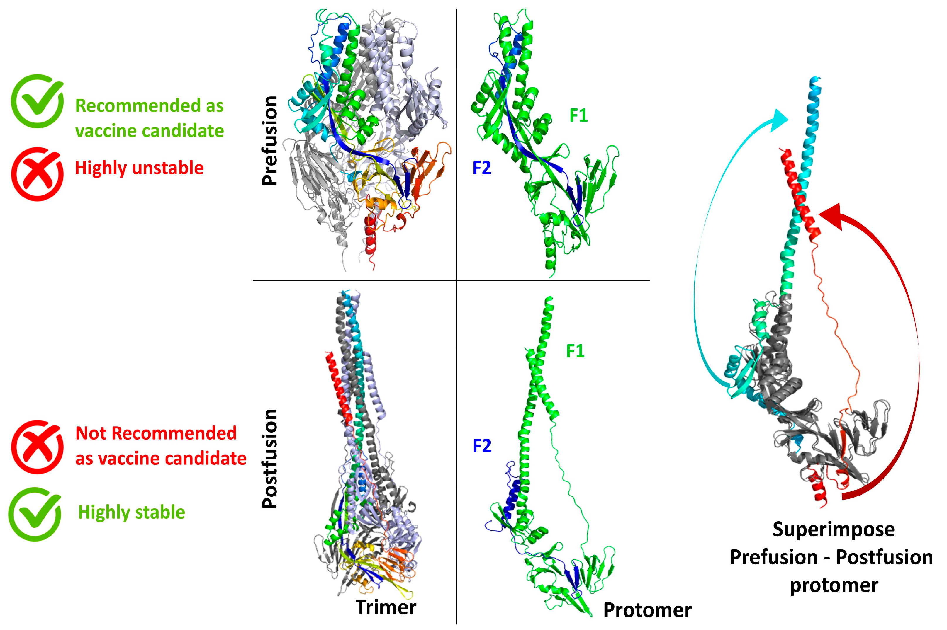
Figure 1. Overview of prefusion and postfusion states of F glycoprotein of the HMPV virus. The prefusion state is shown at the top, while the postfusion state is displayed at the bottom. Both the F1 (green) and F2 (blue) subunits are shown for the prefusion and postfusion conformations, highlighting structural features (middle). On the far right, the prefusion and postfusion states are superimposed to illustrate conformational changes. Grey regions indicate structurally conserved domains, whereas the cyan and red arrows highlight domain movements that occur during the transition from the prefusion to the postfusion state.
Biomedinformatics 05 00047 g001
Figure 2. Strategy for prefusion F protein stabilization. (A) Crystal structure of the trimeric prefusion F protein (PDB ID: 8W3Q), displayed as a surface representation (white). Monomers (C-terminal regions) are shown in magenta, green, and orange, with the interprotomer interface residues highlighted in yellow. A black box indicates the C-terminal region (residues 469–473: LVDQS), which is further magnified and rotated 90° for clarity in the lower panel. (B) Molecular dynamics (MD) simulation analysis of the 8W3Q structure, visualized using porcupine plots to depict the direction and magnitude of amino acid movements. Colour coding matches panel A (magenta, green, orange), with arrows indicating regions of structural flexibility. (C) Structural superposition of the final stabilized model (white ribbon) with the original HMPV F protein structure (grey ribbon; PDB ID: 8W3Q), showing overall structural alignment post-MD simulation.
Biomedinformatics 05 00047 g002
Figure 3. Antigenic sites of prefusion F-proteins (A). B cell epitopes present in the F protein, shown in orange, are responsible for direct antibody recognition and binding (B). T cell MHC-I and T cell MHC-II epitope present within the monomeric 8W3Q structure are marked in magenta (B) and blue (C), respectively. The precise mapping of these epitopes within the monomeric 8W3Q structure provides valuable insights into its immunogenic potential.
Biomedinformatics 05 00047 g003
Figure 4. C-terminal trimer interface mutation selection pipeline. Schematic representation of the computational workflow used to design hydrophobic amino acid substitutions at positions 469–473. The pipeline begins with identification of the structurally sensitive C-terminal trimer interface, followed by selection of residues based on hydrophobicity, aromaticity, and methionine-mediated adaptability. Candidate sequences are generated and energy-minimized, then evaluated through in silico screening for predicted stability changes (ΔΔG), steric compatibility, geometric quality, and epitope preservation. Ranked variants are assessed for an optimal balance of hydrophobic packing and flexibility, resulting in four final designs: M1 (IIFLL), M2 (LLIVL), M3 (WWVLL), and M4 (YMWLL), each with distinct physicochemical advantages for enhancing interfacial stability while maintaining functional compatibility.
Biomedinformatics 05 00047 g004
Figure 5. The three-dimensional structure of reference protein PDB: 8W3Q and the corresponding mutants is illustrated. (A) The 3D structure of the reference protein is shown in the left panel, while the right panel shows the trimeric structure of the lower bottom under 900 orientations. The corresponding mutants M1, M2, M3, and M4 are shown in (B), and the orange, yellow, magenta, red, and cyan regions are hydrophobic and present in the corresponding mutants.
Biomedinformatics 05 00047 g005
Figure 6. The GalaxyRefine web server data interpretation. The structure similarity of mutants relative to the 8W3Q structures in terms of GDT-HA, RMSD, and MolProbity score is presented in the plot. Three components of the MolProbity score, namely, the number of atomic clashes per 1000 atoms, the percentages of rotamer outliers, and Ramachandran favoured backbone torsion angles, are represented in the plot.
Biomedinformatics 05 00047 g006
Figure 7. (A) The root mean square deviation (RMSD) pattern of the reference protein and mutants is shown in the Apo state. The RMSD in nanometers is shown on the y-axis, while the simulation time is shown on the x-axis. M1, M2, M3, and M4 are represented by red, green, blue, and yellow lines, respectively, while the black line denotes the WT. (B) The radius of gyration (Rg) of the 8W3Q and mutants M1, M2, M3, and M4. The x-axis shows the time in nanoseconds, while the y-axis shows the Rg. (C) Residual flexibility of WT and mutants M1, M2, M3, and M4. The x-axis represents the total number of residues, while the y-axis represents the RMSF in nm. (D) The number of residues vs. secondary structure is illustrated for the different mutants.
Biomedinformatics 05 00047 g007
Figure 8. (A) The dynamics of C-terminal mutated residues compared to WT. (B) The potential energy comparison of the complete proteins from the MD simulation trajectories.
Biomedinformatics 05 00047 g008
Figure 9. The mutant M1 three-dimensional analysis. (A) The α-helical structure of the trimeric C-terminal of the M1 mutant is shown in cyan, green, and magenta, contributing to the formation of the hydrophobic core. The C-terminal hydrophobic residues, from I469 to L473, are highlighted. The zoomed-in view in the top right illustrates the binding interaction in one of the monomers of the M1 mutant (B). Hydrophobic residues 469 Isoleucine, 470 Isoleucine, 472 Leucine, 473 Leucine are showing the hydrophobic interaction, forming a trimeric complex in the pocket and exhibiting threefold rotational symmetry. (C) The complex structure formed by the neighbouring helices, using side chains from one monomer, fits into the cavities. The bulky aromatic ring of F471 participates in π-stacking or CH-π interactions, which also adds a layer of non-covalent stabilization.
Biomedinformatics 05 00047 g009
Table 1. Values of different parameters GDT-HA, RMSD, MolProbity, clash score, poor rotamers, and Rama favoured score of different mutants M1, M2, M3, and M4 and the reference protein.
VariantGDT-HARMSDMolProbityClash ScorePoor RotamersRama Favoured
8W3Q0.99340.2651.62313098.2
M10.99510.2321.5911.90.898
M20.99510.2491.67514.8098
M30.99070.2681.66714.5099.1
M40.99010.2831.64713.80.598.9
Table 2. The values of parameters of secondary structure predictions of mutants M1, M2, M3, and M4 and the reference protein.
VariantRMSD
(nm)
Rg
(nm)
RMSF
(nm)
C-Term RMSF
(nm)
α-Helix
(%)
β-Sheet
(%)
WT0.443.080.290.4516.168.25
M10.273.010.220.3615.268.17
M20.373.120.220.3916.268.08
M30.293.010.20.2815.518.45
M40.343.050.220.516.139.62
Disclaimer/Publisher’s Note: The statements, opinions and data contained in all publications are solely those of the individual author(s) and contributor(s) and not of MDPI and/or the editor(s). MDPI and/or the editor(s) disclaim responsibility for any injury to people or property resulting from any ideas, methods, instructions or products referred to in the content.