
Article

Electronically Controlled Conformational Equilibria in Symmetrically Substituted 3,7,10-Triarylphenothiazines: Tuning Redox and Emission Properties †

Institut für Organische Chemie und Makromolekulare Chemie, Heinrich-Heine-Universität Düsseldorf, Universitätsstrasse 1, D-40225 Düsseldorf, Germany
*
Author to whom correspondence should be addressed.
Dedicated to Prof. Dr. Peter Bäuerle on the occasion of his 70th birthday.
Photochem 2026, 6(3), 30; https://doi.org/10.3390/photochem6030030
Submission received: 15 July 2026 / Revised: 9 August 2026 / Accepted: 12 August 2026 / Published: 17 August 2026

Abstract

The combination of Suzuki arylation and Buchwald–Hartwig amination provides a sequentially Pd-catalyzed pseudo-four-component strategy for the synthesis of symmetrically substituted 3,7,10-triarylphenothiazines in moderate to good yields. Using p-anisyl-derived donor units and p-benzonitrile-derived acceptor units, the electronic and photophysical properties of four representative derivatives were investigated by cyclic voltammetry, absorption and emission spectroscopy, and (TD-)DFT calculations. The calculated electronic transitions are in good agreement with the experimental absorption spectra and enable assignment of the underlying optical transitions, while the observed photophysical behavior is interpreted in the context of previous studies on related 3,10-diarylphenothiazines. A p-anisyl donor substituent at the phenothiazine nitrogen atom favors the intra-oriented ground-state conformation, resulting in intense low-energy absorption bands and high fluorescence quantum yields. In contrast, a p-benzonitrile substituent at this position shifts the conformational equilibrium toward the extra-oriented conformation, leading to altered electronic transitions and reduced fluorescence efficiency. Combined with p-anisyl donor units at the 3,7-positions, the extra-oriented conformation becomes predominant, resulting in pronounced emission quenching. These findings demonstrate that 3,7,10-triarylphenothiazines represent a class of redox-active luminophores in which electronic substitution and conformational preferences provide complementary handles for tuning ground- and excited-state properties.

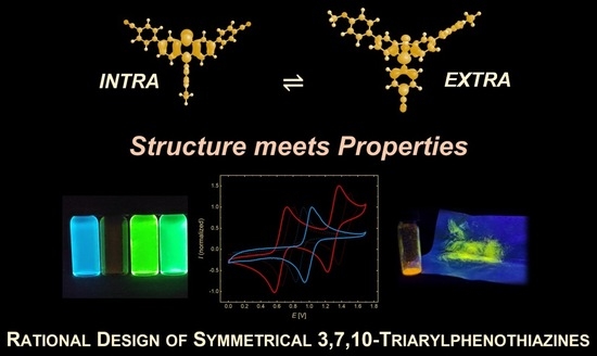
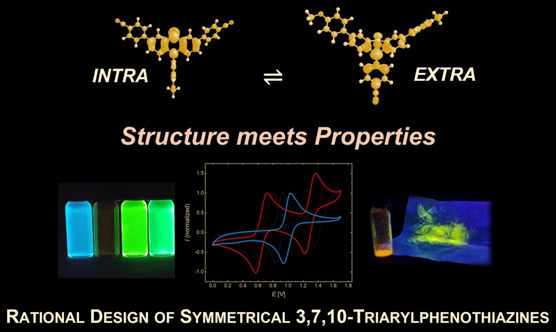
Graphical Abstract

1. Introduction

Phenothiazines are tricyclic electron-rich heterocycles that form very stable radical cations at low oxidation potentials and therefore play an important role as donors in organic field effect transistors [1,2], organic photovoltaics [3,4,5,6,7,8,9], and in lithium batteries [10,11] as well as in photoredox catalysis [12,13,14,15]. As a consequence of its electronic structure, where the bisbenzoanellated 1,4-thiazine core would be antiaromatic due to 16 electrons in conjugation, 10H-phenothiazine and its derivatives are not planar but bent into a “butterfly” conformation by folding on the S,N-axis [16,17].
From a systematic meta-study on the electronic effect of electron-donating and electron-withdrawing N-substituents on the distribution of extra- and intra-conformers, based on DFT calculations on the ground state structures of the corresponding extra- and intra-conformers, a Boltzmann distribution can be established favoring the extra-conformer for strongly electron-withdrawing substituents caused by donor–acceptor resonance stabilization [18]. In contrast, donor substituents on the phenothiazine nitrogen atom clearly favor the intra-conformer. In turn, the intra-conformer causes preferential overlap with the adjacent benzo rings, causing those phenothiazines to become stronger donors. The electronic influence of the N-aryl substituents is further confirmed by linear correlation of Hammett σp parameters for the N-aryl substituents with first oxidation potentials determined by cyclic voltammetry.
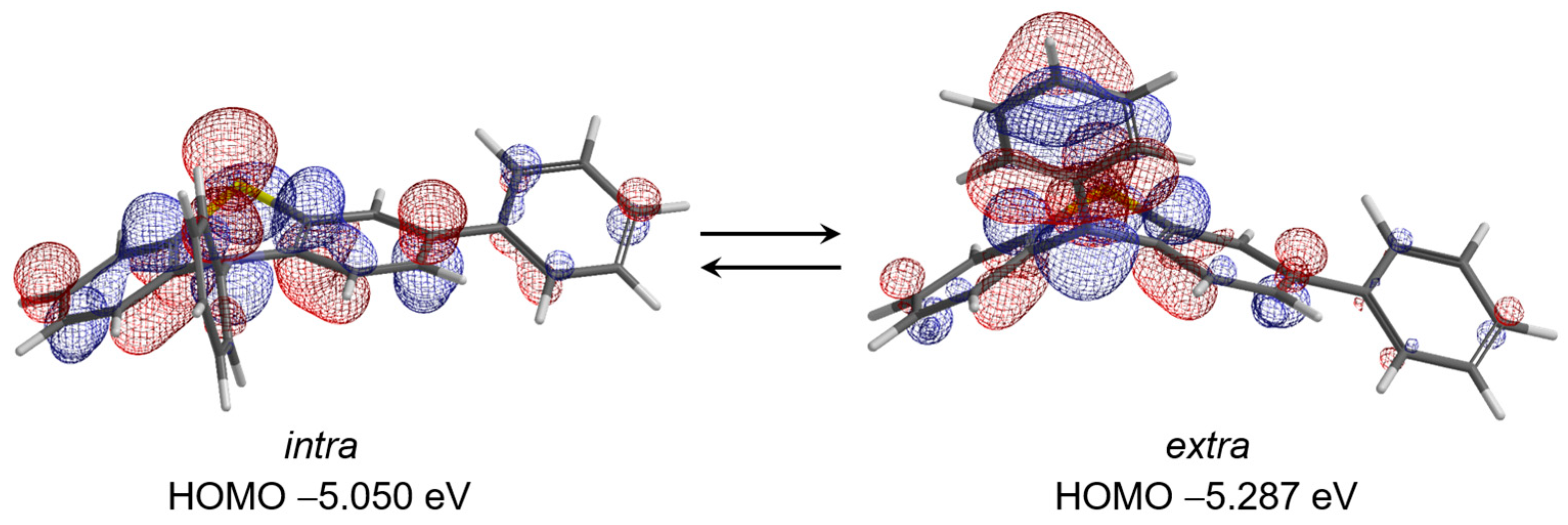
Recently, we have disclosed a general approach to 3,10-diaryl substituted phenothiazines in a one-pot fashion by employing a sequentially Pd-catalyzed Suzuki arylation–Buchwald–Hartwig amination consecutive three-component synthesis [19,20]. The library of electronically diverse 3,10-diaryl phenothiazines was therefore employed in comprehensive investigations of electronic properties by absorption and emission spectroscopy, cyclic voltammetry, and quantum chemical calculations to elucidate and rationalize their electronic structure. A quick inspection of the electronic ground state structure of intra- and extra-conformers of the parent 3,10-diphenyl-10H-phenothiazine by DFT calculations (B3LYP/6-31G*/C-PCM for dichloromethane as a dielectric model) [21] shows that the HOMO of the intra-conformer (−5.050 eV) not only is 0.24 eV above the HOMO of the extra-conformer (−5.287 eV) but also that the coefficient density of the former extends over the tricyclic scaffold into the 3-phenyl substituent (Scheme 1). The origin of the significant differences between intra- and extra-conformers lies in the improved overlap of the nitrogen atom with both benzo anellants in the former conformer. An intra-conformation, therefore, can more efficiently exert phenothiazine’s electron donor capacity onto conjugated substituents in the 7-position.
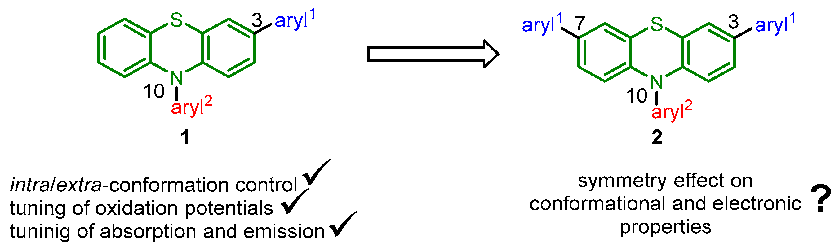
Moreover, the physical-organic treatment of the electronic data of a consanguineous series established semiquantitative linear free energy relationships (LFERs), eventually as a 3D plot for the first oxidation potentials, thereby paving the way to a rational design of tailored 3,10-diaryl phenothiazines with predictable oxidation potentials and photophysical properties. With respect to the photophysical properties, aryl substituents at the 3-position obey LFER, whereas the effect of 10-aryl substituents affects the photophysical properties predominantly indirectly by modulating the extra/intra-conformational equilibrium in the ground and excited state. Introducing two identical substituents at the 3- and 7-positions of the phenothiazine core additionally will create a symmetric substitution pattern with altered electronic structure and thereby different electronic and photonic properties (Scheme 2).
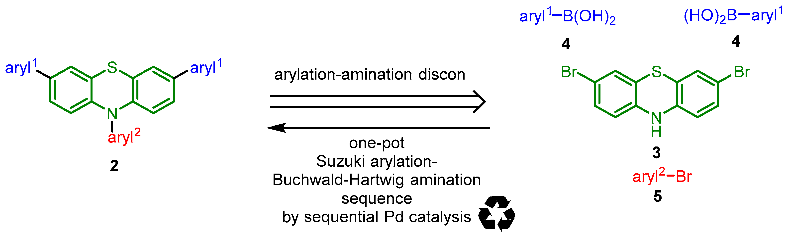
Therefore, the retrosynthetic analysis of 3,7,10-triarylsubstituted phenothiazines 2 suggests employing a sequentially Pd-catalyzed process [22] as a one-pot synthetic route (Scheme 3).
Herein, we report the concise modular synthesis of a small library of 3,7,10-triarylsubstituted phenothiazines via a consecutive pseudo-four-component reaction and investigate the electronic properties by cyclic voltammetry and optical spectroscopy, and elucidate the electronic structure by (TD)DFT calculations.

2. Materials and Methods

All reactions were carried out in oven-dried Schlenk flasks using septa and syringes under nitrogen atmosphere. 3,7-Dibromo-10H-phenothiazine (3) was synthesized by halogenating 10H-phenothiazine with elemental bromine under standard conditions on a multi-gram scale according to literature procedure [23]. All electronic data of the series 1 used for discussion in this work were taken from our previous publication (where details on the preparation and characterization were documented) [20]. Reagents and catalysts were purchased reagent-grade and used without purification. Dry solvents were dried by a solvent purification system.
The purification of the phenothiazines was performed on silica gel 60 M (0.04–0.063 mm) from MACHEREY-NAGEL GmbH & Co. KG, Düren, Germany, using flash technique under a pressure of 2 bar. The crude mixtures were absorbed on Celite® 545 from Carl Roth GmbH & Co. KG, Karlsruhe, Germany, before chromatographic purification.
The reaction progress was monitored qualitatively using TLC Silica gel 60 F254 aluminum sheets obtained from MACHEREY-NAGEL GmbH & Co. KG, Düren, Germany. The spots were detected with UV light at 254 and 366 nm.
1H, 13C and 135-DEPT 13C NMR spectra were recorded on Bruker AVIII-300 and AVIII-600, Karlsruhe, Germany. Acetone-d6 and CD2Cl2 were used as deuterated solvents. The resonances of the solvents were locked as internal standard (acetone-d6: 1H δ 2.05 (water in acetone-d6 1H δ 2.84) (due to the water content in acetone-d6, the water peaks in the NMR spectra are also labeled with “actone-d6”), 13C δ 29.84, 206.26; CD2Cl2: 1H δ 5.32, 13C δ 54.00). The multiplicities of the signals were abbreviated as follows: s: singlet; d: doublet; t: triplet; q: quartet; dd: doublet of doublet; td: triplet of doublets; ddd: doublet of doublet of doublets; m: multiplet. The type of carbon atoms was determined based on 135-DEPT 13C NMR spectra. For the description of the 13C NMR spectra primary carbon nuclei are abbreviated as CH3, secondary carbon nuclei as CH2, tertiary carbon nuclei as CH and quaternary carbon nuclei as Cquat.
EI mass spectra were measured on Finnigan MAT TSQ 7000, Thermo Fisher Scientific, Darmstadt, Germany. IR spectra were obtained on Shimadzu IR Affinity-1, which works with the attenuated total reflection (ATR) method. The intensity of signals is abbreviated as follows: s (strong), m (medium), w (weak). The melting points (uncorrected) were measured on Büchi Melting Point B-540, Essen, Germany. Absorption spectra were recorded in CH2Cl2 high performance liquid chromatography (HPLC) grade at 298 K on a Perkin–Elmer UV/Vis/NIR Lambda 19 spectrometer, Hohenfels-Liggersdorf, Germany. For the determination of the molar extinction coefficients ε absorption measurements at five different concentrations were carried out. Emission spectra were recorded in CH2Cl2 HPLC grade at 298 K on a Hitachi F-7000 spectrometer, Krefeld, Germany.
Cyclic voltammetry experiments were performed with 263A E&G Princeton Applied Research, AMETEK Scientific Instruments, Krefeld, Germany, as potentiostatic instrumentation under argon in dry and degassed dichloromethane at 298 K and at scan rates of 100, 250, 500 and 1000 mVs−1. The working electrode was a 1 mm platinum disk, the counter electrode was a platinum wire, and the reference electrode was a silver/silver chloride electrode filled with saturated sodium chloride solution. The electrolyte was tetrabutylammonium hexafluorophosphate at a concentration of c = 0.1 M. The potentials were calibrated using [FeCp2]/[FeCp2]+ and [FeCp*2]/[FeCp*2]+ as an internal potential standard. The absolute potential of this standard was determined against [FeCp2]/[FeCp2]+ (E00/+1 = 450 mV) [24]. This procedure provided a value of E00/+1 = −103 mV for [FeCp*2]/[FeCp*2]+. The determined potentials are the arithmetic average of the potential by the different scan rates. Combustion analyses were carried out on Perkin Elmer Series II Analyser 2400, Hohenfels-Liggersdorf, Germany, in the micro analytical laboratory of the Institute for Pharmaceutical and Medicinal Chemistry at Heinrich Heine University Düsseldorf. Quantum chemical calculations were carried out utilizing the HPC-Cluster Ivybridge of the Zentrum für Informations- und Medientechnologie (ZIM) at the Heinrich Heine University Düsseldorf. During the preparation of this manuscript, the author(s) used ChatGPT (GPT-5.6, OpenAI, San Francisco, CA, USA, 2026) for language polishing and consistency checking. The authors have reviewed and edited the output and take full responsibility for the content of this publication.

3. Results and Discussion

3.1. Synthesis and Structure

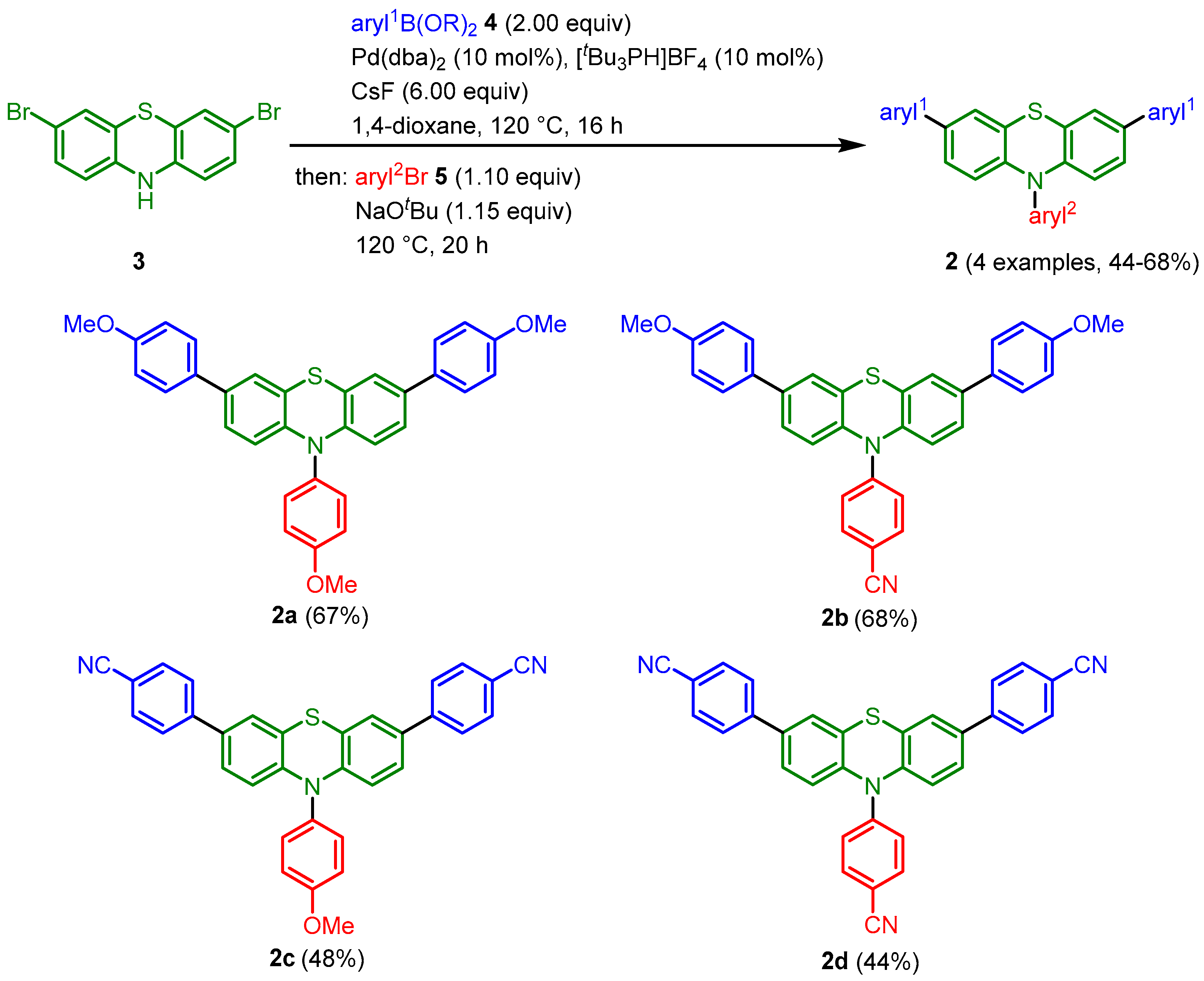
The optimized conditions for the coupling-amination sequence developed for the synthesis of 3,10-diarylphenothiazines [20] can be employed for the reaction of 3,7-dibromo-10H-phenothiazine (3) [23] with arylboronic acid (esters) 4 and aryl bromides 5 in the presence of catalytic amounts of Pd(dba)2 (dba = dibenzylidene acetone) and tris(tert-butyl)phosphonium tetrafluoroborate and cesium fluoride (for the Suzuki step) and sodium tert-butoxide (for the Buchwald–Hartwig step) as bases. After purification by chromatography on silica gel, four novel symmetrical 3,7,10-triarylphenothiazines 2 were obtained in a consecutive pseudo-four-component reaction in moderate to good yields (Scheme 4). For this study, the p-anisyl moiety was chosen as a donor and the p-cyanophenyl substituent as an acceptor. Just recently, structure 2d has been reported to be synthesized by a stepwise approach, and the compound has been investigated with respect to solid-state photophysics of crystal isomorphs [25].
The structure of 3,7,10-triarylphenothiazines 2 was unambiguously assigned by 1H and 13C NMR spectroscopy, mass spectrometry, and IR spectroscopy, and the molecular compositions and purities were verified by combustion analysis. Due to the CS symmetry of the molecules 2 (plane of symmetry along the S/N axis of the thiazine), the 1H and 13C NMR spectra of 3,7,10-triarylphenothiazines exhibited reduced sets of signals.
In analogy to the previously studied 3,10-diarylphenothiazines, the preferred conformations for the 3,7,10-triarylphenothiazines 2 were determined by DFT calculations employing the Gaussian 09 program [26] with the PBEh1PBE functional [27] and 6-311++G** basis set [28] applying IEFPCM (Integral Equation Formalism Polarizable Continuum Model) for dichloromethane as a dielectric model (Table 1) [29]. The relative Gibbs free energy difference between the two conformers was defined as Δ Δ G extra-intra =   Δ G i n t r a   Δ G e x t r a . Accordingly, positive values indicate a preference for the intra-conformer, whereas negative values indicate a preference for the extra-conformer. According to quantum chemical conformational analysis in dichloromethane, the N-anisyl substituents in compounds 2a and 2c adopt almost exclusively the intra-conformation. In contrast, the 3,7,10-triarylphenothiazines 2b and 2d bearing p-cyanophenyl substituents at the nitrogen atom, the ratio shifts significantly toward the extra-conformation due to the increasing acceptor strength of the aryl2 substituent.

3.2. Electronic Properties

To determine the electrochemical properties, cyclovoltammetric experiments were performed for all 3,7,10-triarylphenothiazines 2. Furthermore, the photophysical properties of the series of 3,7,10-triarylphenothiazines 2 were thoroughly and systematically investigated by UV/Vis and static fluorescence spectroscopy.

3.2.1. Electrochemical Properties

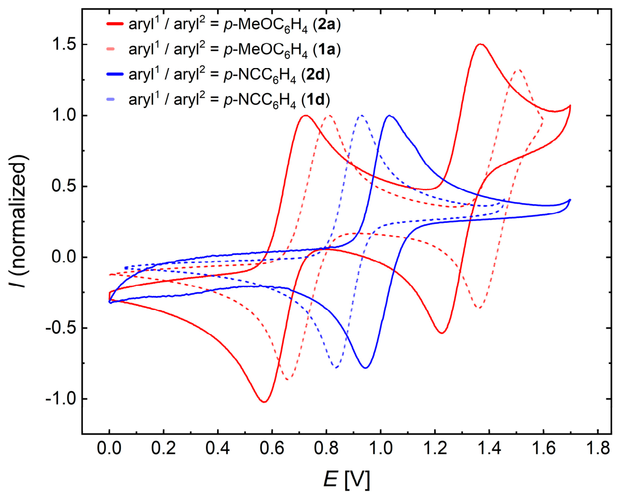
All compounds 2 exhibit two clearly separated, electrochemically and Nernstian reversible one-electron oxidation processes (except for 2d, for which the second oxidation lies outside the measurement range) (Table 2). The first oxidation potentials E 0 0 / + 1 appear in a range between 0.61 and 0.91 V, with a distinct dependence on the electronic nature of the aryl substituents at the 3-, 7-, and 10-positions. Thus, in comparison to 3,10-diarylphenothiazines [20], placing an additional p-anisyl donor at the 7-position causes a slight cathodic shift in the first oxidation potential by 0.03 V, whereas an additional p-benzonitrile substituent in the 7-position leads to a slight anodic shift of 0.04 V. Both redox potentials can be adjusted over a broad potential range ( E 0 0 / + 1  = 0.30 V; E 0 + 1 / + 2  = 0.24 V) depending on the substitution pattern (Figure 1). Consequently, the range of the oxidation potential of 3,7,10-triarylphenothiazines 2 is broader than of the corresponding 3,10-diarylphenothiazines 1 ( E 0 0 / + 1  = 0.23 V; E 0 + 1 / + 2  = 0.15 V) [20]. Thus, extending the aryl substitution expands the accessible potential window and allows a more precise tuning of the electronic properties.
The semiquinone formation constants K S E M [30], calculated from the difference in first and second oxidation potentials, are quite large and underline a considerable stability of the formed radical cations against disproportionation.
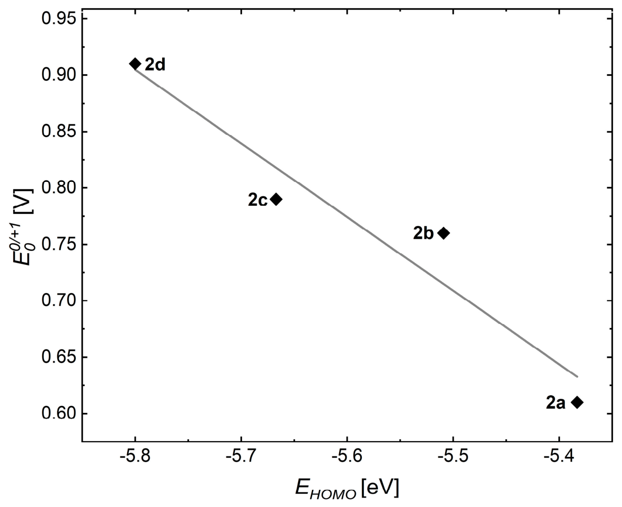
Plotting E 0 0 / + 1 of 3,7,10-triarylphenothiazines 2 against the calculated HOMO energies of the corresponding intra-conformations yields a good correlation (Figure 2).
Indeed, the pronounced correlation of the first oxidation potentials E 0 0 / + 1 with the calculated HOMO energies of the intra-conformers indicates that this conformation represents the electronically relevant structure for the oxidation process. This correlation is also observed for compounds in which the calculated conformational equilibria favor the extra-conformation in the electronic ground state, suggesting that the oxidation process is governed by the electronic properties of the intra-conformation rather than by its ground-state population. This behavior can be rationalized by the structural similarity of the intra-conformation to the fully planarized phenothiazine radical cation, as both exhibit an almost orthogonal arrangement of the aryl substituents relative to the nitrogen lone pair. Accordingly, as already established for 3,10-diarylphenothiazines [20], HOMO energies of intra-conformers can be semiquantitatively estimated from the first oxidation potentials E 0 0 / + 1 of 3,7,10-triarylphenothiazines 2 ( E 0 0 / + 1 = 0.652   E H O M O 2.88   V   R 2 = 0.926 ).

3.2.2. Photophysical Properties

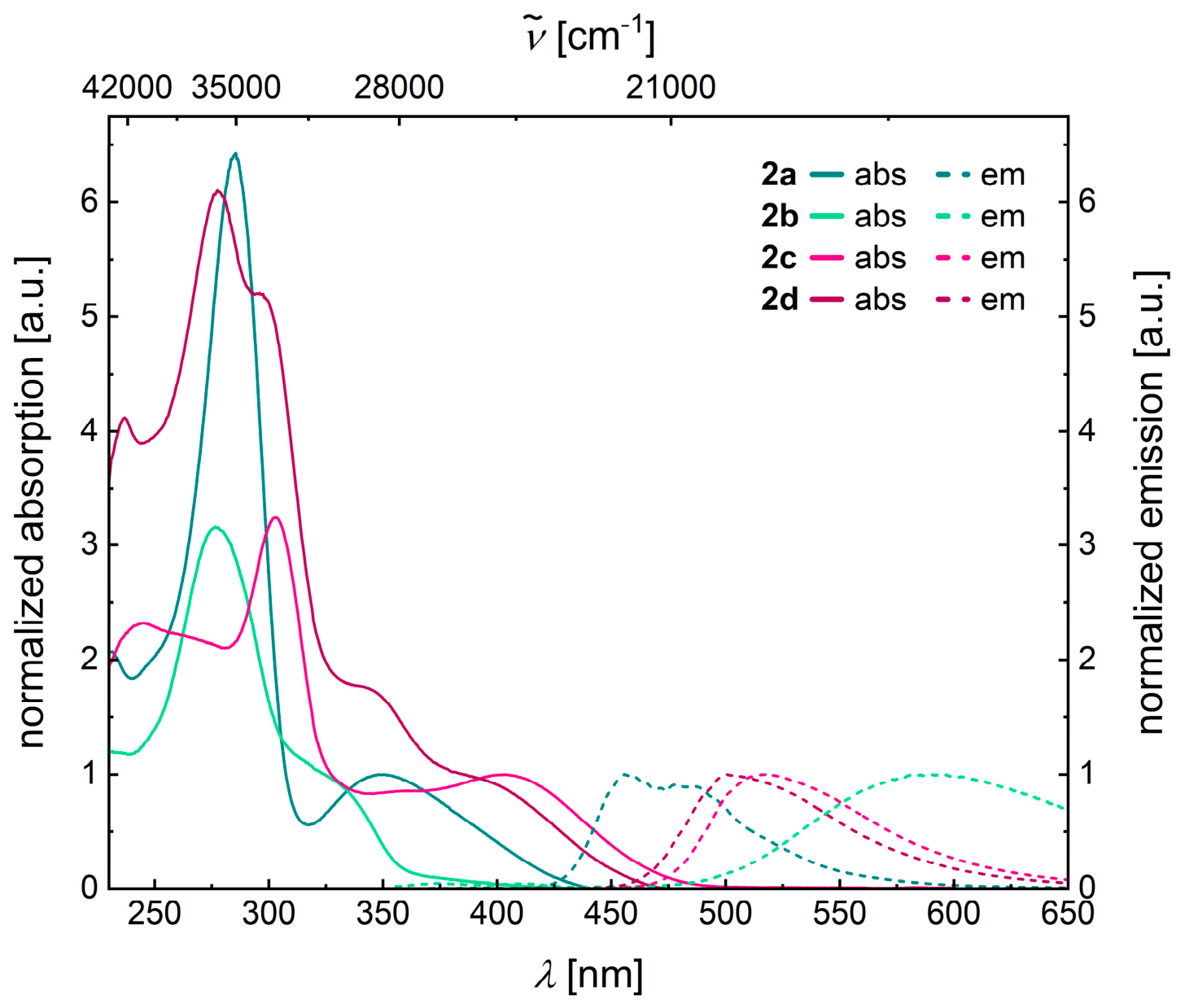
The dyes 2 are colorless to orange solids that exhibit two to five broad, featureless absorption maxima in dichloromethane solution. The photophysical properties of the 3,7,10-triarylphenothiazines 2 were investigated by UV/Vis absorption and fluorescence spectroscopy in dichloromethane (Table 3, Figure 3). All 3,7,10-triarylphenothiazines 2 possess characteristic longest wavelength absorption maxima between 325 (2b) and 403 nm (2c) with molar absorption coefficients ε in the range of 9100 to 15,300 L mol−1 cm−1. Furthermore, the fluorescence quantum yields of all compounds were determined using the relative method with the standard coumarin 1 ( Φ F = 73% in ethanol) [31] according to the procedure known from the literature [32].
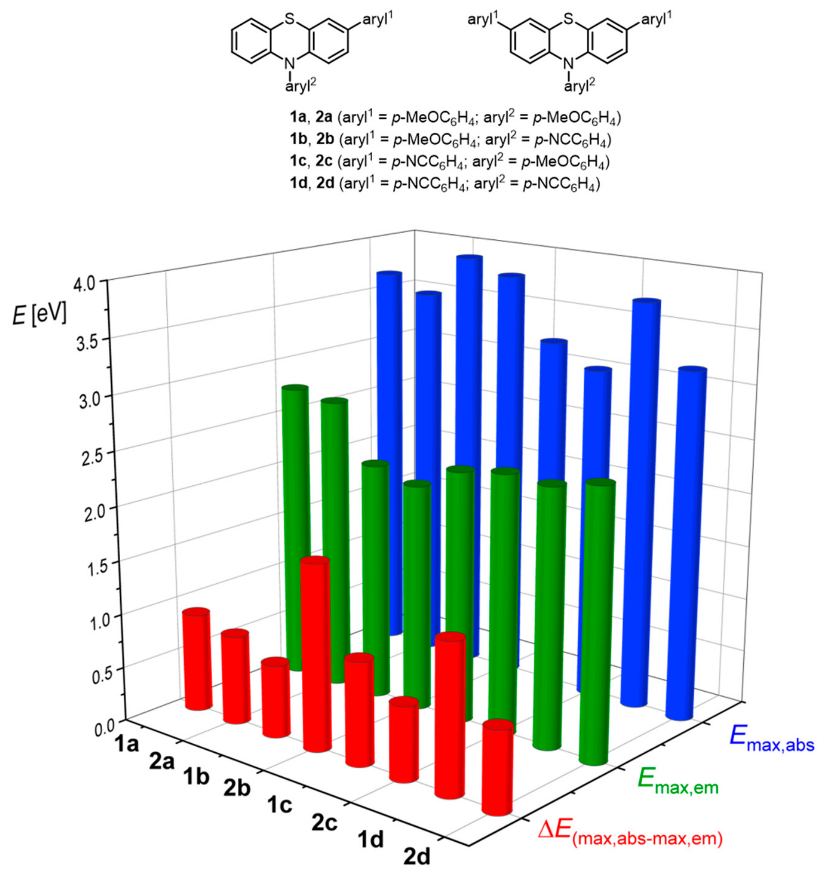
The direct comparison between the two series of 3,10-diaryl phenothiazines 1 and 3,7,10-triaryl phenothiazines 2 (see Supplementary Materials, Table S2) reveals similarities in the lowest absorption and highest emission energies as well as their differences (representing Stokes shifts); however, it also reveals differences, such as a slight red shift in the absorption energies of the title compounds 2 relative to their corresponding disubstituted analogs 1 (Figure 4). This can also be seen from the adiabatic transition energies E0-0, which are readily determined as the arithmetic mean of absorption and emission energies (see Supplementary Materials, Table S3).
In the combination of both series 1 and 2, for the dyes 1a, 2a, 1b, and 2b, where p-anisyl donor(s) are placed in 3- (and 7-) position, a linear correlation for plotting the lowest energy absorption bands Eabs,max against the first oxidation potential E 0 0 / + 1 can be established (Eabs,max = 1.834 · E 0 0 / + 1 + 2.457 [eV]; r2 = 0.946) (see Supplementary Materials, Figure S19). This reflects an enhanced electron density on the central phenothiazine by unperturbed conjugation of the p-anisyl substituents. The increase in the oxidation potential is caused by the p-benzonitrile acceptors at position 10 (dyes 1b and 2b). Interestingly, the higher electron density on the phenothiazine core causes a bathochromic shift in the absorption band. For the inverse placement of p-benzonitrile acceptors at positions 3- (and 7-) position (dyes 1c, 2c, 1d, and 2d), the absorption bands are considerably redshifted (Table 3); however, no correlation with the oxidation potentials can be established. In addition, the extra-intra equilibrium in the electronic ground state becomes the dominant factor for the observed absorption characteristics, which even overcompensates the push-pull effect of donor–acceptor substitution.
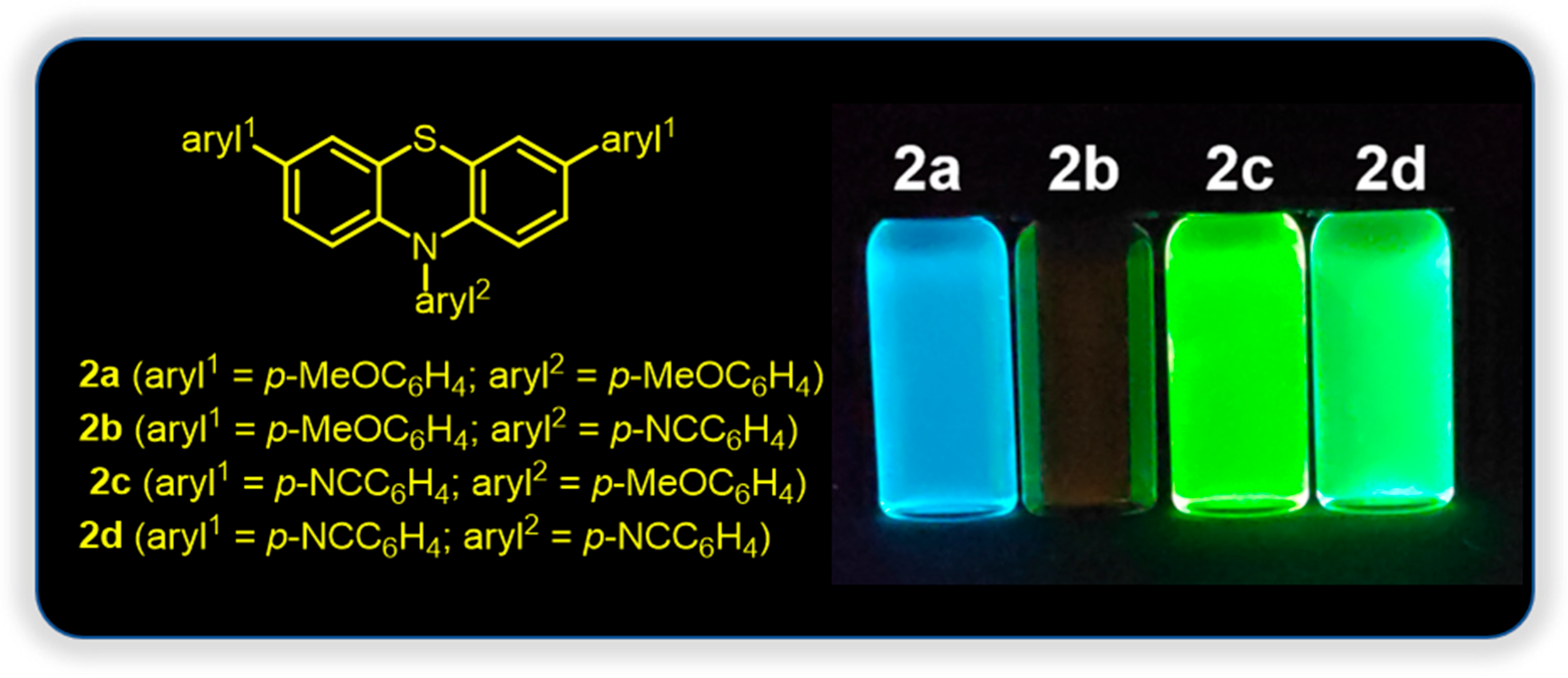
The emission data of series 1 and 2 are definitely worth discussing, in particular, since the vibrationally relaxed S1 state of phenothiazines is usually fully planarized in contrast to the S0 state [18,33,34,35]. However, for N-acceptor substituted 3,10-diaryl phenothiazines the observed occurrence of dual emission was assigned to the presence of two different conformations in the excited state, i.e., a fully planarized intra-type and a folded extra-type conformer, which are energetically different [20]. Although the energy levels of extra- and intra-conformers in the excited state are quite different, an equilibration in the sense of a conformational change cannot be ruled out. As a consequence, besides electronic effects significant structural changes from the bent, butterfly-shaped electronic ground state structure to the planarized relaxed excited state cause quite large Stokes shifts as a typical signature of phenothiazines’ photophysics [33,34,35]. While p-anisyl substituents at the phenothiazine nitrogen cause Stokes shifts between 5500 and 7600 cm−1 (dyes 1a/2a, 1c/2c), p-benzonitrile moieties in the 10-position even lead to Stokes shifts between 11,000 and 13,900 cm−1. Markedly, this effect is pronounced for 3-mono- and 3,7-di(p-anisyl)-substituted dyes bearing an N-p-benzonitrile substituent with Stokes shifts of 13,900 (dye 1b) and 13,800 cm−1 (dye 2b). The weak emission maxima are found at 560 (1b) and 589 nm (2b) with fluorescence quantum yields of 1% (dye 1b) [20] and below 1% (dye 2b) (Table 3, Figure 5).
In contrast, the remaining pairs of the series 1 and 2 display quite intense emission bands around 450 nm with fluorescence quantum yields of 12 (dye 1a) and 16% (dye 2a), between 500 and 520 nm with fluorescence quantum yields of 11 (dye 1d) and 41% (dye 2d), and with the highest fluorescence quantum yields of 46 (dye 1c) and 49% (dye 2c) and emission maxima at 533 and 517 nm. For the latter pair, the intense emission band can be rationalized again due to the favorable extended π-conjugation imposed by the intra-conformation of the 10-p-anisyl substituent and the terminal electron-withdrawing p-benzonitrile unit. For the intra-conformers of series 1, with several electronically diverse 3-aryl substituents, a significant linear correlation between the fluorescence quantum yields and the discretely negative charge stabilizing σp Hammett parameter for the remote substituent has been established [20].

3.2.3. Calculated Electronic Structure

The electronic structure of the absorptions of 3,7,10-triarylphenothiazines 2 can be further assessed by calculations on the TD-DFT level of theory. Therefore, using Gaussian 09 [26], TD-DFT calculations (PBEh1PBE [26]/6–311++G** [28], polarizable continuum model (PCM) [36] with dichloromethane as a dielectric model) were also performed (Table 4). In addition, the natural transition orbitals (NTOs) were calculated to investigate the nature of the electronic transitions in detail. As a consequence of the inherent CS-symmetry of 3,7,10-triarylphenothiazines 2 a comparison to the previously studied systems 1 [20] does not appear to be necessary.
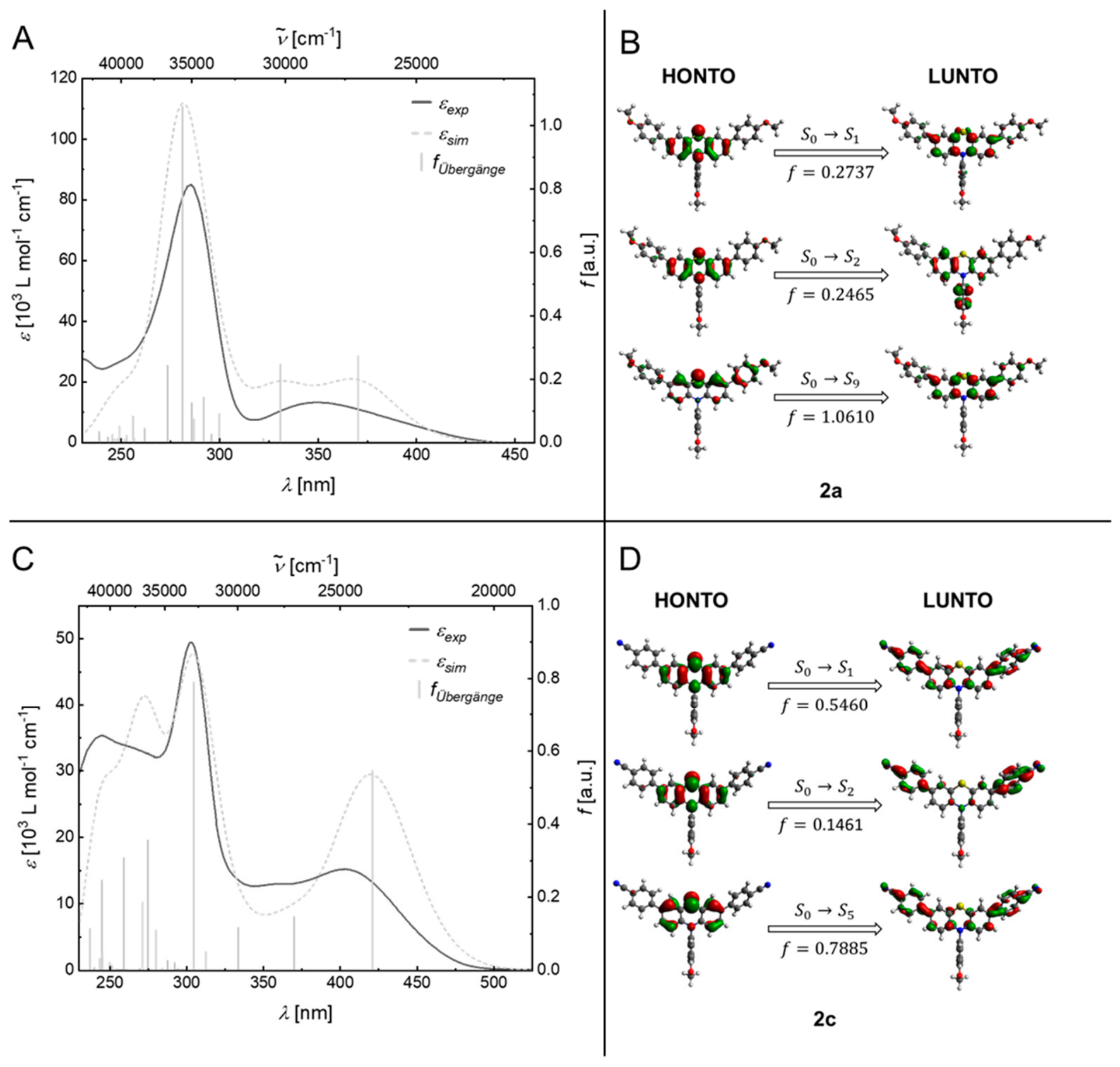
The dyes 2a and 2c, bearing a p-anisyl donor at position 10 of the phenothiazine core, exhibit a similar absorption pattern characterized by a locally excited state and differ only in minor details. The simulated UV/Vis spectra, calculated from the transitions of the intra-conformers, are in good agreement with the experimental data (Figure 6A,C). Both absorption spectra exhibit a pronounced maximum in the long-wavelength absorption region, which is bathochromically shifted in dye 2c due to its increased CT character (vide infra).
The TD-DFT calculated, simulated UV/Vis spectra of dyes 2a and 2c clearly show that the longest wavelength absorption bands in each case consist of a superposition of the S0→S1 transitions and the S0→S2 transitions (Figure 6B,D). Due to its dominant contribution (≈95%), the S0→S1 transition can also be simply described as a HOMO→LUMO transition. The HONTOs of the S0→S1 transition in 3,7,10-triarylphenothiazines 2a and 2c are distributed across the entire phenothiazine core, while the LUNTOs of the S0→S1 transition become increasingly localized on the adjacent aryl substituents at the 3- and 7-positions with increasing acceptor strength.
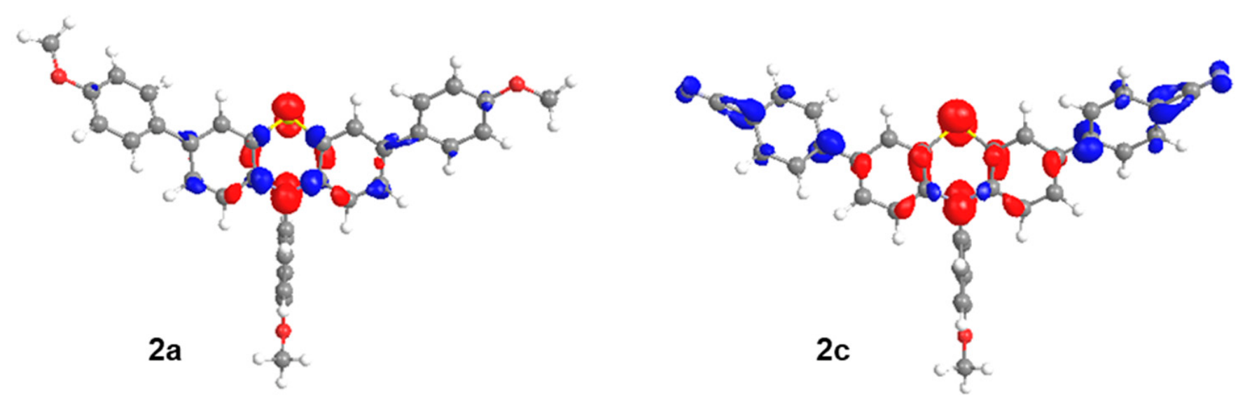
The substantial overlap between HONTO and LUNTO reflects the predominantly locally excited (LE) character of the transition. However, the overlap between the hole and electron distributions decreases with increasing acceptor strength of the substituent aryl1, thereby enhancing the charge-transfer (CT) character of the longest wavelength absorption band (Figure 7).
As discussed for 3,10-diarylphenothiazines 1, electron-withdrawing N-aryl substituents lead to significant structural and, consequently, electronic changes [20]. Consequently, the UV/Vis spectra of dye 2b and 2d, bearing a p-benzonitrile acceptor at position 10 of the phenothiazine core, differ significantly from those of dyes 2a and 2c. In contrast to the previously discussed 3,7,10-triarylphenothiazine derivatives 2a and 2c, the longest wavelength absorption bands of compounds 2b and 2d can only be detected as weak shoulders (see Supplementary Materials, Figure S17). At first glance, the longest wavelength absorption band of compound 2b appears to be located at 325 nm. However, closer examination of the longest wavelength region of the UV/Vis spectra of dyes 2b reveals a very weak absorption onset at approximately 425 nm, which gradually increases in intensity to about 360 nm. This indicative of electronic transitions with low oscillator strengths.
To gain a deeper understanding of how the experimentally determined absorption bands relate to the corresponding electronic transitions, TD-DFT calculations were also performed for the 3,7,10-triarylphenothiazine 2b.
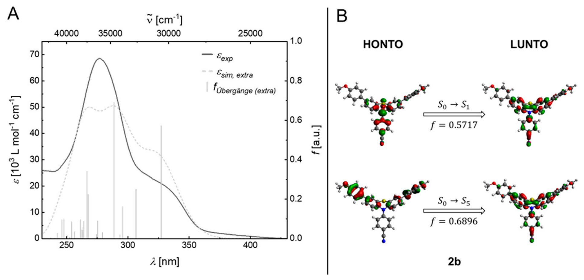
The simulated UV/Vis spectrum of the extra-conformation shows good agreement with the experimental data. However, it cannot explain the weak absorption band in the 360–425 nm range (Figure 8A). In contrast, the S0→S1 transition of the intra-conformer of compound 2b is bathochromically shifted and exhibits an almost negligible oscillator strength (f ≈ 0), which can be attributed to the nearly orthogonal D-A geometry. This low intensity of the first electronic transition is due to its charge-transfer character and is consistent with the weak absorption band in the 360–425 nm range. The natural transition orbitals (NTOs) of the extra-conformer show that the HONTO of the S0→S1 transition is completely localized on the phenothiazine core and the adjacent N-benzonitrile substituent, while the LUNTO of the S0→S1 transition is weakly extended to the aryl substituents at positions 3 and 7 (Figure 8B). For the extra-conformer, the substantial overlap between HONTO and LUNTO results in a high oscillator strength for the S0→S1 transition and suggests an LE transition with a weak charge transfer character.
The experimental absorption spectrum of compound 2b can thus best be reproduced by the simulated absorption spectrum assuming an intra/extra conformer equilibrium ratio of 12:88, obtained by superposition of the calculated absorption spectra of both conformers, weighted by the theoretical molar fractions (see Supplementary Materials, Figure S20). The influence of conformational heterogeneity on the absorption properties is even more pronounced in dye 2d. Compared to the other members of the 3,7,10-triarylphenothiazine series, DFT calculations indicate that dye 2d does not adopt a clearly preferred conformation in dichloromethane in the electronic ground state (54% intra-conformer at T = 298 K). Similarly, as for dye 2b, the influence of conformation on the absorption properties can be illustrated from an overlay of the simulated UV/Vis spectra for both conformations of dye 2d (see Supplementary Materials, Figure S21). A comparison of the experimental spectra and the calculated transitions of the simulated spectra shows that the longest wavelength absorption band consists of a superposition of the S0→S1 and S0→S2 transitions of the intra-conformer, whereas the absorption band at 340 nm is attributed to the S0→S1 and S0→S2 transitions of the extra-conformer. Due to the different molecular orbital profiles of both conformers and the calculated contribution of the equilibrium constant, the influence of both conformers on the absorption properties is significant.

4. Conclusions

In summary, the consecutive Suzuki arylation–Buchwald–Hartwig amination sequence provides an efficient Pd-catalyzed route to symmetrically substituted 3,7,10-triarylphenothiazines through a concise pseudo-four-component strategy, affording the target compounds in moderate to good yields. Four electronically distinct dyes bearing p-anisyl donor and/or p-benzonitrile acceptor substituents were comprehensively investigated by cyclic voltammetry, absorption and emission spectroscopy and interpreted in the context of our previously reported TD-DFT. Together, these investigations establish clear structure–property relationships linking electronic substitution pattern, conformational equilibria, electrochemical behavior, and photophysical response.
Consistent with our previous studies on 3,10-diarylphenothiazines, the rapidly equilibrating intra-/extra-conformational distribution in the ground state is governed by the interplay of the electronic properties of the three aryl substituents, with the 10-aryl substituent exerting the dominant influence, thereby affecting the electronic structure underlying absorption and oxidation. The pronounced differences in fluorescence observed across the series indicate that excited-state conformational effects significantly contribute to the emission behavior of 3,7,10-triarylphenothiazines.
These findings provide a molecular design concept for reversibly oxidizable luminophores with tunable redox and emission properties. Further studies are aimed at gaining deeper insight into excited-state structural dynamics and conformational effects by investigating the influence of environmental parameters and by combining advanced spectroscopic techniques with quantum chemical calculations. In addition, unsymmetrically substituted 3,7,10-triarylphenothiazines are being explored to investigate how deliberate conformational restriction can influence intersystem crossing and singlet–triplet population dynamics in the excited state.

Supplementary Materials

The following supporting information can be downloaded at https://www.mdpi.com/article/10.3390/photochem6030030/s1, general procedure (GP) for the pseudo-four-component synthesis of 3,7,10-triarylphenothiazines 2; Analytic characterization of compounds 2ad; Figure S1: 1H NMR spectrum of compound 2a (acetone-d6, 300 MHz, 298 K); Figure S2: 13C NMR spectrum of compound 2a (acetone-d6, 75 MHz, 298 K); Figure S3: 1H NMR of compound 2b (acetone-d6, 300 MHz, 298 K); Figure S4: 13C NMR spectrum of compound 2b (acetone-d6, 75 MHz, 298 K); Figure S5: 1H NMR of compound 2c (acetone-d6, 300 MHz, 298 K); Figure S6: 13C NMR spectrum of compound 2c (acetone-d6, 75 MHz, 298 K); Figure S7: 1H NMR of compound 2d (acetone-d6, 300 MHz, 298 K); Figure S8: 13C NMR spectrum of compound 2d (acetone-d6, 75 MHz, 298 K); Figure S9: Cyclic voltammogram in the anodic region of compound 2a (recorded in dichloromethane, T = 298 K, electrolyte: [Bu4N][PF6], Pt working electrode, Pt counter electrode, Ag/AgCl reference electrode, v = 100 mV/s, standard: [FeCp*2]/[FeCp*2]+); Figure S10: Cyclic voltammogram in the anodic region of compound 2b (recorded in dichloromethane, T = 298 K, electrolyte: [Bu4N][PF6], Pt working electrode, Pt counter electrode, Ag/AgCl reference electrode, v = 100 mV/s, standard: [Cp2Fe]/[Cp2Fe]+); Figure S11: Cyclic voltammogram in the anodic region of compound 2c (recorded in dichloromethane, T = 298 K, electrolyte: [Bu4N][PF6], Pt working electrode, Pt counter electrode, Ag/AgCl reference electrode, v = 100 mV/s, standard: [FeCp2]/[FeCp2]+); Figure S12: Cyclic voltammogram in the anodic region of compound 2d (recorded in dichloromethane, T = 298 K, electrolyte: [Bu4N][PF6], Pt working electrode, Pt counter electrode, Ag/AgCl reference electrode, v = 100 mV/s, standard: [FeCp2]/[FeCp2]+); Figure S13: Normalized absorption (solid) and emission (dashed) spectra of compound 2a (recorded in dichloromethane, T = 298 K, cabs(2a) = 10−5 m, cem(2a) = 10−7 m, λexc = λmax,abs); Figure S14: Normalized absorption (solid) and emission (dashed) spectra of compound 2b (recorded in dichloromethane, T = 298 K, cabs(2b) = 10−5 m, cem(2b) = 10−5 m, λexc = λmax,abs); Figure S15: Normalized absorption (solid) and emission (dashed) spectra of compound 2c (recorded in dichloromethane, T = 298 K, cabs(2c) = 10−5 m, cem(2c) = 10−7 m, λexc = λmax,abs); Figure S16: Normalized absorption (solid) and emission (dashed) spectra of compound 2d (recorded in dichloromethane, T = 298 K, cabs(2d) = 10−5 m, cem(2d) = 10−7 m, λexc = λmax,abs); Figure S17: UV/Vis absorption spectra of 3,7,10-triarylphenothiazines 2 (recorded in CH2Cl2, T = 298 K, c(2) = 10−5 m); Figure S18: Comparison of normalized UV/Vis absorption (recorded in CH2Cl2, T = 298 K, c(2) = 10−5 m, solid lines) and emission spectra of 3,7,10-triarylphenothiazines 2 (recorded in CH2Cl2, T = 298 K, c(2) = 10−7 m and c(2b) = 10−5 m, λ e x c = 360 nm, dashed line); Figure S19: Linear correlation of the lowest energy absorption bands Eabs,max of compounds 1a/2a and 1b/2b and the first oxidation potential E 0 0 / + 1 can be established (Eabs,max = 1.8338 · E 0 0 / + 1 + 2.019 [eV]; r2 = 0.9455); Figure S20: Experimental UV/Vis absorption spectrum of dye 2b (recorded in CH2Cl2, T = 298 K, c(2b) = 10−5 m) and overlay with simulated spectra of intra-and extra-conformers (PBEh1PBE/6-311++G**, PCM (CH2Cl2)) and weighted overlay of 12% intra- and 88% extra-conformers; Figure S21: Experimental UV/Vis absorption spectrum of dye 2d (recorded in CH2Cl2, T = 298 K, c(2d) = 10−5 m) and overlay with simulated spectra of intra- and extra-conformers (PBEh1PBE/6-311++G**, PCM (CH2Cl2)) and weighted overlay of 54% intra- and 46% extra-conformers; Table S1: Experimental details of the synthesis of 3,7,10-triarylphenothiazines 2; Table S2: Comparison of selected photophysical data of 3,10-diarylphenothiazines 1 [20] and 3,7,10-triarylphenothiazines 2 (UV/Vis absorption maximum λmax,abs, emission maximum λmax,em, fluorescence quantum yield and ΦF, and Stokes shift Δ ν ~ ); Table S3: Selected photophysical data of 3,10-diarylphenothiazines 1 [20] and 3,7,10-triarylphenothiazines 2 in eV.

Author Contributions

Conceptualization, L.M. and T.J.J.M.; methodology, L.M.; validation, L.M.; formal analysis, L.M.; investigation, L.M.; data curation, L.M.; writing—original draft preparation, L.M. and T.J.J.M.; writing—review and editing, L.M. and T.J.J.M.; supervision, T.J.J.M.; project administration, T.J.J.M.; funding acquisition, T.J.J.M. All authors have read and agreed to the published version of the manuscript.

Funding

The authors cordially thank the Fonds der Chemischen Industrie and the Deutsche Forschungsgemeinschaft (Mu 1088/9-1) for the financial support. Computational support and infrastructure were provided by the “Centre for Information and Media Technology” (ZIM) at the University of Düsseldorf (Germany).

Institutional Review Board Statement

Not applicable.

Informed Consent Statement

Not applicable.

Data Availability Statement

The original contributions presented in this study are included in the article/Supplementary Materials. Further inquiries can be directed to the corresponding author.

Conflicts of Interest

The authors declare no conflicts of interest.

Abbreviations

The following abbreviations are used in this manuscript:
C-PCMconductor-like polarizable continuum model
135-DEPTDistortionless Enhancement by Polarization Transfer at a pulse angle of 135°
HOMOhighest occupied molecular orbital
HONTOhighest occupied natural transition orbital
KSEMsemiquinone formation constant
IEFPCMintegral equation formalism polarizable continuum model
LFERlinear free enthalpy relationship
LUMOlowest unoccupied molecular orbital
LUNTOlowest unoccupied natural transition orbital
PBEh1PBEhybrid density functional theory combining 25% exact (Hartree–Fock) exchange, 75% Perdew–Burke–Ernzerhof (PBE) exchange and 100% PBE correlation
(TD-)DFT(time-dependent) density functional theory

References

  1. Zou, Y.; Wu, W.; Sang, G.; Yang, Y.; Liu, Y.; Li, Y. Polythiophene Derivative with Phenothiazine−Vinylene Conjugated Side Chain:  Synthesis and Its Application in Field-Effect Transistors. Macromolecules 2007, 40, 7231–7237. [Google Scholar] [CrossRef] [Scilit]
  2. Hwang, D.-H.; Kim, S.-K.; Park, M.-J.; Lee, J.-H.; Koo, B.-W.; Kang, I.-N.; Kim, S.-H.; Zyung, T. Conjugated Polymers Based on Phenothiazine and Fluorene in Light-Emitting Diodes and Field Effect Transistors. Chem. Mater. 2004, 16, 1298–1303. [Google Scholar] [CrossRef] [Scilit]
  3. Thokala, S.; Singh, S.P. Phenothiazine-Based Hole Transport Materials for Perovskite Solar Cells. ACS Omega 2020, 5, 5608–5619. [Google Scholar] [CrossRef] [Scilit] [PubMed]
  4. Mao, L.; Wu, Y.; Jiang, J.; Guo, X.; Heng, P.; Wang, L.; Zhang, J. Rational Design of Phenothiazine-Based Organic Dyes for Dye-Sensitized Solar Cells: The Influence of π-Spacers and Intermolecular Aggregation on Their Photovoltaic Performances. J. Phys. Chem. C 2020, 124, 9233–9242. [Google Scholar] [CrossRef] [Scilit]
  5. Venkatraman, V.; Foscato, M.; Jensen, V.R.; Alsberg, B.K. Evolutionary de novo design of phenothiazine derivatives for dye-sensitized solar cells. J. Mater. Chem. A 2015, 3, 9851–9860. [Google Scholar] [CrossRef] [Scilit]
  6. Huang, Z.-S.; Meier, H.; Cao, D. Phenothiazine-based dyes for efficient dye-sensitized solar cells. J. Mater. Chem. C 2016, 4, 2404–2426. [Google Scholar] [CrossRef] [Scilit]
  7. Sailer, M.; Franz, A.W.; Müller, T.J.J. Synthesis and Electronic Properties of Monodisperse Oligophenothiazines. Chem. Eur. J. 2008, 14, 2602–2614. [Google Scholar] [CrossRef] [Scilit] [PubMed]
  8. Luo, J.-S.; Wan, Z.-Q.; Jia, C.-Y. Recent advances in phenothiazine-based dyes for dye-sensitized solar cells. Chin. Chem. Lett. 2016, 27, 1304–1318. [Google Scholar] [CrossRef] [Scilit]
  9. Meyer, T.; Ogermann, D.; Pankrath, A.; Kleinermanns, K.; Müller, T.J.J. Phenothiazinyl Rhodanylidene Merocyanines for Dye-Sensitized Solar Cells. J. Org. Chem. 2012, 77, 3704–3715. [Google Scholar] [CrossRef] [Scilit] [PubMed]
  10. Narayana, K.A.; Casselman, M.D.; Elliott, C.F.; Ergun, S.; Parkin, S.R.; Risko, C.; Odom, S.A. N-Substituted Phenothiazine Derivatives: How the Stability of the Neutral and Radical Cation Forms Affects Overcharge Performance in Lithium-Ion Batteries. ChemPhysChem 2015, 16, 1179–1189. [Google Scholar] [CrossRef] [Scilit] [PubMed]
  11. Casselman, M.D.; Kaur, A.P.; Narayana, K.A.; Elliott, C.F.; Risko, C.; Odom, S.A. The fate of phenothiazine-based redox shuttles in lithium-ion batteries. Phys. Chem. Chem. Phys. 2015, 17, 6905–6912. [Google Scholar] [CrossRef] [Scilit] [PubMed]
  12. Sartor, S.M.; Chrisman, C.H.; Pearson, R.M.; Miyake, G.M.; Damrauer, N.H. Designing High-Triplet-Yield Phenothiazine Donor–Acceptor Complexes for Photoredox Catalysis. J. Phys. Chem. A 2020, 124, 817–823. [Google Scholar] [CrossRef] [Scilit] [PubMed]
  13. Speck, F.; Rombach, D.; Wagenknecht, H.-A. N-Arylphenothiazines as strong donors for photoredox catalysis–pushing the frontiers of nucleophilic addition of alcohols to alkenes. Beilstein J. Org. Chem. 2019, 15, 52–59. [Google Scholar] [CrossRef] [Scilit] [PubMed]
  14. Treat, N.J.; Sprafke, H.; Kramer, J.W.; Clark, P.G.; Barton, B.E.; Read de Alaniz, J.; Fors, B.P.; Hawker, C.J. Metal-Free Atom Transfer Radical Polymerization. J. Am. Chem. Soc. 2014, 136, 16096–16101. [Google Scholar] [CrossRef] [Scilit] [PubMed]
  15. Pan, X.; Lamson, M.; Yan, J.; Matyjaszewski, K. Photoinduced Metal-Free Atom Transfer Radical Polymerization of Acrylonitrile. ACS Macro Lett. 2015, 4, 192–196. [Google Scholar] [CrossRef] [Scilit] [PubMed]
  16. Bell, J.D.; Blount, J.F.; Briscoe, O.V.; Freeman, H.C. The crystal structure of phenothiazine. Chem. Commun. 1968, 4, 1656–1657. [Google Scholar] [CrossRef] [Scilit]
  17. Klein, C.L.; Conrad, J.M., III; Morris, S.A. Structure of N-phenylphenothiazine, C18H13NS. Cryst. Struct. Commun. 1985, 41, 1202–1204. [Google Scholar] [CrossRef] [Scilit]
  18. Mayer, L.; May, L.; Müller, T.J.J. The interplay of conformations and electronic properties in N-aryl phenothiazines. Org. Chem. Front. 2020, 7, 1206–1217. [Google Scholar] [CrossRef] [Scilit]
  19. Mayer, L.; Kohlbecher, R.; Müller, T.J.J. Concatenating Suzuki Arylation and Buchwald-Hartwig Amination by A Sequentially Pd-catalyzed One-Pot Process–Consecutive Three-Component Synthesis of C,N-Diarylated Heterocycles. Chem. Eur. J. 2020, 26, 15130–15134. [Google Scholar] [CrossRef] [Scilit] [PubMed]
  20. Mayer, L.; Müller, T.J.J. 3,10-Diaryl Phenothiazines–One-pot Synthesis and Conformational Tuning of Ground and Excited State Electronics. Eur. J. Org. Chem. 2021, 2021, 3516–3527. [Google Scholar] [CrossRef] [Scilit]
  21. Spartan’24; Wavefunction Inc.: Irvine, CA, USA, 2025.
  22. Kornet, M.M.; Müller, T.J.J. Recent Advances in sequentially Pd-catalyzed one-pot syntheses of heterocycles. Molecules 2024, 29, 5265. [Google Scholar] [CrossRef] [Scilit] [PubMed]
  23. Bodea, C.; Raileanu, M. Über Phenthiazone, III Brom-phenthiazine und Brom-phenthiazone. Liebigs Ann. Chem. 1960, 631, 194–198. [Google Scholar] [CrossRef] [Scilit]
  24. Zanello, P. Electrochemical and X-ray Structural Aspects of Transition Metal Complexes Containing Redox-Active Ferrocene Ligands. In Ferrocenes: Homogeneous Catalysis, Organic Synthesis, Materials Science; Wiley-VCH Verlag GmbH: Weinheim, Germany, 2007; pp. 317–430. [Google Scholar] [CrossRef] [Scilit]
  25. Fan, B.; Zhao, H.; Wei, Q.; Liu, G.; Xue, P. Three color fluorescent switching and stimulus-response of a phenothiazine derivative for anti-counterfeiting. J. Mol. Struct. 2026, 1356, 145118. [Google Scholar] [CrossRef] [Scilit]
  26. Frisch, M.J.; Trucks, G.W.; Schlegel, H.B.; Scuseria, G.E.; Robb, M.A.; Cheeseman, J.R.; Scalmani, G.; Barone, V.; Mennucci, B.; Petersson, G.A.; et al. Gaussian 09; Gaussian, Inc.: Wallingford, CT, USA, 2009. [Google Scholar]
  27. Adamo, C.; Barone, V. Toward reliable density functional methods without adjustable parameters: The PBE0 model. J. Chem. Phys. 1999, 110, 6158–6170. [Google Scholar] [CrossRef] [Scilit]
  28. Krishnan, R.; Binkley, J.S.; Seeger, R.; Pople, J.A. Self-consistent molecular orbital methods—XX—A basis set for correlated wave functions. J. Chem. Phys. 1980, 72, 650–654. [Google Scholar] [CrossRef] [Scilit]
  29. Marenich, A.V.; Cramer, C.J.; Truhlar, D.G. Universal Solvation Model Based on Solute Electron Density and on a Continuum Model of the Solvent Defined by the Bulk Dielectric Constant and Atomic Surface Tensions. J. Phys. Chem. B. 2009, 113, 6378–6396. [Google Scholar] [CrossRef] [Scilit] [PubMed]
  30. Michaelis, L. Semiquinones, the Intermediate Steps of Reversible Organic Oxidation-Reduction. Chem. Rev. 1935, 16, 243–286. [Google Scholar] [CrossRef] [Scilit]
  31. Dixon, J.M.; Taniguchi, M.; Lindsey, J.S. PhotochemCAD 2: A Refined Program with Accompanying Spectral Databases for Photochemical Calculations. Photochem. Photobiol. 2005, 81, 212–213. [Google Scholar] [CrossRef] [Scilit]
  32. Fery-Forgues, S.; Lavabre, D. Are Fluorescence Quantum Yields So Tricky to Measure? A Demonstration Using Familiar Stationery Products. J. Chem. Educ. 1999, 76, 1260–1264. [Google Scholar] [CrossRef] [Scilit]
  33. Yang, L.; Feng, J.K.; Ren, A.M. Theoretical study on electronic structure and optical properties of phenothiazine-containing conjugated oligomers and polymers. J. Org. Chem. 2005, 70, 5987–5996. [Google Scholar] [CrossRef] [Scilit] [PubMed]
  34. May, L.; Müller, T.J.J. Widely Electronically Tunable 2,6-Disubstituted Dithieno [1,4] thiazines—Electron-Rich Fluorophores Up to Intense NIR Emission. Chem. Eur. J. 2020, 26, 12978–12986. [Google Scholar] [CrossRef] [Scilit] [PubMed]
  35. Hauck, M.; Stolte, M.; Schönhaber, J.; Kuball, H.G.; Müller, T.J.J. Synthesis, electronic, and electro-optical properties of emissive solvatochromic phenothiazinyl merocyanine dyes. Chem. Eur. J. 2011, 17, 9984–9998. [Google Scholar] [CrossRef] [Scilit] [PubMed]
  36. Scalmani, G.; Frisch, M.J. Continuous surface charge polarizable continuum models of solvation. I. General formalism. J. Chem. Phys. 2010, 132, 114110. [Google Scholar] [CrossRef] [Scilit] [PubMed]
Scheme 1. DFT-calculated (B3LYP/6-31G*/C-PCM for dichloromethane as a dielectric) intra- and extra-conformers of 3,10-diphenyl-10H-phenothiazine and HOMO energies.
Photochem 06 00030 sch001
Scheme 2. Conformational and electronic properties by transitioning from 3,10-diaryl- (1) to 3,7,10-triarylphenothiazines (2).
Photochem 06 00030 sch002
Scheme 3. Retrosynthetic analysis and consecutive pseudo-four-component synthesis of 3,7,10-triarylphenothiazines 2 by sequential palladium catalysis in a one-pot fashion.
Photochem 06 00030 sch003
Scheme 4. Pseudo-four-component Suzuki arylation–Buchwald–Hartwig amination syntheses of symmetrical 3,7,10-triarylphenothiazines 2.
Photochem 06 00030 sch004
Figure 1. Comparison of the normalized cyclic voltammograms of the 3,7,10-triarylphenothiazines 2a and 2d (solid line) and the 3,10-diarylphenothiazines 1a and 1d (dashed line) (recorded in CH2Cl2, T = 298 K, 0.1 m [Bu4N][PF6], v = 100 mV/s, Pt working electrode, Ag/AgCl reference electrode, and Pt counter electrode, referenced against [Cp*2Fe]/[Cp*2Fe]+ and [Cp2Fe]/[Cp2Fe]+, respectively).
Photochem 06 00030 g001
Figure 2. Plotting of the first oxidation potentials E 0 0 / + 1   (measured in CH2Cl2, T = 298 K, 0.1 m [Bu4N][PF6], v = 100 mV/s, Pt working electrode, Ag/AgCl reference electrode, and Pt counter electrode, referenced against [[Cp*2Fe]/[Cp*2Fe]+ or [Cp2Fe]/[Cp2Fe]+) versus the HOMO energies (PBEh1PBE/6-311++G**, PCM CH2Cl2) E H O M O of the 3,7,10-triarylphenothiazines 2 ( E 0 0 / + 1 = 0.652   E H O M O 2.88   V   R 2 = 0.926   o r   E H O M O =   E 0 0 / + 1 0.652   4.417   [ e V ] ).
Photochem 06 00030 g002
Figure 3. Comparison of normalized UV/Vis absorption (recorded in CH2Cl2, T = 298 K, c(2) = 10−5 m, solid lines) and emission spectra of 3,7,10-triarylphenothiazines 2 (recorded in CH2Cl2, T = 298 K, c(2) = 10−7 m and c(2b) = 10−5 m, λ e x c = 360 nm, dashed line).
Photochem 06 00030 g003
Figure 4. Comparison of selected photophysical data (lowest absorption Emax,abs and highest emission energies Emax,em as well as their differences ΔE(max,abs–max,em), i.e., Stokes shifts) of 3,10-diarylphenothiazines 1 and 3,7,10-triarylphenothiazines 2 (energies are given in eV).
Photochem 06 00030 g004
Figure 5. Emission of 3,7,10-triarylphenothiazines 2 in solution (recorded in CH2Cl2, T = 298 K, c(2) = 10−5 m, λ e x c = 365 nm).
Photochem 06 00030 g005
Figure 6. (A,C): Comparison of experimental (solid) (recorded in CH2Cl2, T = 298 K, c(2) = 10−5 m) and calculated (dashed) (PBEh1PBE/6-311++G**, PCM CH2Cl2) spectra of dyes 2a (A) and 2c (C) (calculated transitions are depicted as bars). (B,D): Natural transition orbitals (NTOs) for dominant transitions (for each transition, the oscillator strength is indicated as determined by TD-DFT calculations (PBEh1PBE/6-311++G**, PCM CH2Cl2; isosurface value at 0.04 a.u.) of dyes 2a (B) and 2c (D).
Photochem 06 00030 g006
Figure 7. Charge-transfer character of S0→S1 transitions of dyes 2a und 2c illustrated by difference in coefficient density of HOMO and LUMO (red = decrease in coefficient density; blue = increase in coefficient density (PBEh1PBE/6-311++G**, PCM CH2Cl2, isosurface at 0.002 a.u.).
Photochem 06 00030 g007
Figure 8. (A): Comparison of experimental (solid) (recorded in CH2Cl2, T = 298 K, c(2b) = 10−5 m) and calculated (dashed, extra-conformer) (PBEh1PBE/6-311++G**, PCM CH2Cl2) spectra of dye 2b (calculated transitions are depicted as bars). (B): Natural transition orbitals (NTOs) for dominant transitions (for each transition, the oscillator strength is indicated as determined by TD-DFT calculations (PBEh1PBE/6-311++G**, PCM CH2Cl2; isosurface value at 0.04 a.u.) of the extra-conformer of dye 2b.
Photochem 06 00030 g008
Table 1. DFT-calculated Boltzmann conformer distribution and preferred conformation of 3,7,10-triarylphenothiazines 2 (PBEh1PBE/6-311++G**, PCM CH2Cl2).
Compoundaryl1aryl2 Δ Δ G extra-intra  [1] [kcal/mol]Kextra-intra (extra:intra Ratio)Preferred Conformation
2ap-MeOC6H4p-MeOC6H4−3.543439.6 (0.23:99.77)intra
2bp-MeOC6H4p-NCC6H41.1770.1324 (88.31:11.69)extra
2cp-NCC6H4p-MeOC6H4−5.2277934 (0.01:99.99)intra
2dp-NCC6H4p-NCC6H4−0.0921.172 (46.05:53.95)intra
[1]  Δ Δ G extra-intra =   Δ G i n t r a   Δ G e x t r a .
Table 2. Selected electrochemical data of 3,7,10-triarylphenothiazines 2ad.
Compoundaryl1aryl2 E 0 0 / + 1 [V] [a] E 0 + 1 / + 2 [V] [a] K S E M  [b]
2ap-MeOC6H4p-MeOC6H40.611.261.19·1011
2bp-MeOC6H4p-NCC6H40.761.301.09·109
2cp-NCC6H4p-MeOC6H40.791.501.18·1012
2dp-NCC6H4p-NCC6H40.91[c]
[a] Recorded in CH2Cl2, T = 298 K, 0.1 m [Bu4N][PF6], v = 100 mV/s, Pt working, Ag/AgCl reference, and Pt counter electrode, referenced against [Cp*2Fe]/[Cp*2Fe]+ or [Cp2Fe]/[Cp2Fe]+. [b] Semiquinone formation constant K S E M = 10 E 0 + 1 / + 2 E 0 0 / + 1 0.059   V . [c] Outside of the measurement range.
Table 3. Selected photophysical data of 3,7,10-triarylphenothiazines 2a–d (bold absorptions are used for calculating the Stokes shifts).
Compoundaryl1aryl2 λ m a x , a b s  [a] [nm] ( ε [L mol−1 cm−1]) λ m a x , e m  [b] [nm] ( Φ F ) Δ ν ~  [c] [cm−1]
2ap-MeOC6H4p-MeOC6H4285 (86,400), 350 (13,300)456 (16%)6600
2bp-MeOC6H4p-NCC6H4277 (69,000), 325 (sh, 15,000)589 (<1%)13,800
2cp-NCC6H4p-MeOC6H4245 (35,000), 303 (49,700),
403 (15,300)
517 (49%)5500
2dp-NCC6H4p-NCC6H4237 (37,400), 278 (55,600), 297 (47,400), 340 (16,000), 385 (sh, 9100)500 (41%)6000
[a] Recorded in CH2Cl2, T = 298 K, c(2) = 10−5 m. [b] Recorded in CH2Cl2, T = 298 K, c(2) = 10−5–10−7 m, Relative fluorescence quantum yields Φ F were determined with coumarin 1 as a standard in ethanol ( Φ F = 0.73 ) [30], λ e x c (2) = λ m a x , a b s [c]  ν ~ =   1 λ m a x ,   a b s   1 λ m a x ,   e m (determined from the bold absorption maxima).
Table 4. TD-DFT calculations of absorption maxima of 3,7,10-triarylphenothiazine 2 (PBEh1PBE/6-311++G**, PCM CH2Cl2).
Compoundsaryl1aryl2λmax,abs(exp) [a] [nm] (ε [M−1 cm−1])λmax,abs(calcd) [nm]Oscillator StrengthDominant Contributions
2ap-MeOC6H4p-MeOC6H4350 (13,300)3700.2737HOMO→LUMO (95%)
3310.2465HOMO→LUMO + 1 (93%)
285 (86,400)2811.0610HOMO − 2→LUMO (70%)
2bp-MeOC6H4p-NCC6H4~380 (1800)437 [b]0.0028HOMO→LUMO (99%)
365 [b]0.2057HOMO→LUMO + 1 (94%)
325 (15,000)327 [c]0.5715HOMO→LUMO (94%)
277 (69,000)289 [c]0.6896HOMO − 1→LUMO (85%)
2cp-NCC6H4p-MeOC6H4403 (15,300)4210.5460HOMO→LUMO (96%)
3700.1461HOMO→LUMO + 1 (98%)
303 (49,700)3050.7885HOMO − 1→LUMO (77%)
245 (35,000)2450.2463HOMO − 1→LUMO + 2 (68%)
2dp-NCC6H4p-NCC6H4385 (9100)416 [b]0.2332HOMO→LUMO (79%) HOMO→LUMO + 1 (18%)
403 [b]0.2741HOMO→LUMO + 1 (80%)
HOMO→LUMO (17%)
340 (16,000)356 [c]0.3431HOMO→LUMO (95%)
347 [c]0.1951HOMO→LUMO + 1 (96%)
297 (47,400)302 [b]0.8935HOMO − 1→LUMO (71%)
HOMO→LUMO + 7 (12%)
278 (55,600)281 [c]1.1191HOMO − 2→LUMO (81%)
237 (37,400)236 [b]0.1441HOMO − 4→LUMO (24%)
HOMO − 3→LUMO (23%)
HOMO − 1→LUMO + 7 (16%)
[a] Recorded in CH2Cl2, T = 298 K, c(2) = 10−5 m. [b] The intra-conformer is weighted with the theoretical mass fraction (11.69% (2b) and 53.95% (2d)). [c] The extra-conformer is weighted with the theoretical mass fraction (88.31% (2b) and 46.05% (2d)).
Disclaimer/Publisher’s Note: The statements, opinions and data contained in all publications are solely those of the individual author(s) and contributor(s) and not of MDPI and/or the editor(s). MDPI and/or the editor(s) disclaim responsibility for any injury to people or property resulting from any ideas, methods, instructions or products referred to in the content.